powerpoint presentation - union high...
TRANSCRIPT
3/17/2014
1
The Lymphatic System
• Lymphatic System Overview
• Lymphatic Vessels and Flow of Lymph
• Lymphoid Cells, Tissues, and Organs
(7th edition)
Overview of the Lymphatic System – Slide 2
• Major Components of the Lymphatic System (fig. 20.1)
LYMPH - an extracellular fluid (ECF) similar to plasma; ECF is found in
several places in the body: body tissues (ECF = interstitial fluid), blood
(ECF = plasma), and lymphatic vessels (ECF = lymph)
LYMPHATIC VESSELS - a network of vessels that carry lymph
throughout the body, and eventually to the venous blood system
LYMPHATIC TISSUE - protective tissue scattered throughout the body;
lymphatic tissue is a specialized connective tissue containing large
numbers of lymphocytes
LYMPHATIC ORGANS - e.g. the spleen and thymus
LYMPHOCYTES - white blood cells involved with immunity
• The Lymphatic System and Cardiovascular System are collectively called the
Circulatory System; the lymphatic system circulates lymph and the
cardiovascular system circulates blood
(7th edition)

3/17/2014
2
Overview of the Lymphatic System – Slide 4
Major Functions of the Lymphatic System:
• fluid balance - excess interstitial fluid from tissue spaces (interstitial spaces) is returned to the blood by the
lymphatic capillaries; once interstitial fluid enters a lymphatic capillary it is called “lymph”
every day, about 30 liters of fluid leave your blood capillaries and enter the interstitial fluid of the body
tissues
approximately 27 of those liters are reabsorbed back into the blood capillaries
the remaining 3 liters move into the lymphatic capillaries, so that they are removed from the tissue
• distribution - of hormones, nutrients, and waste products from the site of origin to general circulation
the lymphatic system absorbs fats from the small intestines, and lymphatic vessels carry the fats to the
bloodstream
transport of lipid-soluble vitamins (A, D, E, and K) from the gastrointestinal tract to the blood
• defense - microorganisms and antigens are destroyed by WBCs
(7th edition)

3/17/2014
3
Lymphatic Vessels and Lymph Flow – SLIDE 6
• LYMPHATIC CAPILLARIES (fig. 20.1) - are found in most tissues of the body;
they differ from blood capillaries in several ways:
they originate as blind pockets; each of the lymphatic capillaries has one
closed end; extracellular fluid enters the lymphatic capillary through a
flap-like minivalve
they are larger in diameter than blood capillaries
they have thinner walls than blood capillaries
there are gaps between the endothelial cells that comprise the wall of the
lymphatic capillary making them more porous; fluids can enter the
lymphatic capillary but cannot leave
• Lymphatic vessels - resemble veins in structure; from the capillaries, lymph
moves into lymphatic vessels; they contain valves which keep the lymph
moving in one direction through these low pressure vessels
(7th edition)

3/17/2014
4
Lymphatic Vessels and Lymph Flow = Slide 7
• In general a tissue will contain many more lymphatic vessels than veins but
they will be much smaller
• Lymph moves in response to:
contraction of surrounding skeletal muscles (remember that veins return
blood to the heart using a similar mechanism); like veins, lymph vessels
have valves to keep the fluid moving in one direction
contraction of smooth muscle in wall of the lymphatic vessel
pressure changes in the thoracic cavity during respiration
• Major lymph-collecting vessels: (fig. 20.2)
THORACIC DUCT - largest lymph vessel; drains into the left subclavian
vein; drains the entire body except the upper right quarter
CYSTERNA CHYLI - expanded abdominal portion of the thoracic duct;
this is where fat-filled vessels from the digestive tract enter the lymphatic
system
RIGHT LYMPHATIC DUCT - drains lymph from the upper right quarter of
the body; drains into the right subclavian vein
(7th edition)
Drainage of different regions of the
body by lymph nodes

3/17/2014
5
Lymphoid Cells, Tissues, and Organs – Slide 9
• Types of Lymphocytes:
T CELLS (T lymphocytes) - attack foreign cells or body cells infected by viruses; T cells mature and divide
in the thymus; T cells are responsible for cell-mediated immunity (meaning that the protection is directly
from living cells)
B CELLS (B lymphocytes) - responsible for antibody-mediated immunity (=humoral immunity); a
percentage of circulating B lymphocytes mature into PLASMA CELLS; plasma cells produce and secrete
antibodies which destroy antigens
NK CELLS (natural killer cells) - attack foreign cells and cells infected with viruses and cancer cells
• LYMPHOID NODULES - lymphocytes densely packed into an area of areolar tissue
occur in the connective tissue deep to the epithelia that line the respiratory, digestive, and urinary tracts
have a central zone called a germinal center which contains dividing lymphocytes
the boundaries of the nodule are not distinct because they have no fibrous capsule surrounding them
(7th edition)

3/17/2014
6
Lymphoid Cells, Tissues, and Organs – Slide 11
• LYMPH NODES (fig. 20.4)
small oval lymphoid organs distributed along the lymphatic vessels; each
lymph node is covered by a capsule of dense fibrous connective tissue;
connective tissue strands call trabeculae extend inward to divide the node
into compartments
they filter the lymph, removing bacteria and antigens
lymphocytes congregate, function, and proliferate in the lymph nodes
lymph nodes are found throughout the body: superficial aggregations of lymph
nodes are found in the inguinal, axillary, and cervical regions
afferent lymphatic vessel - lymph vessel that carries lymph to the lymph
node; as lymph flows through the lymph node it is exposed to B and T cells
and macrophages; at least 99% of the pathogens in the lymph are removed
efferent lymphatic vessel - lymph vessel that carries lymph away from the
lymph node
(7th edition)
3/17/2014
7
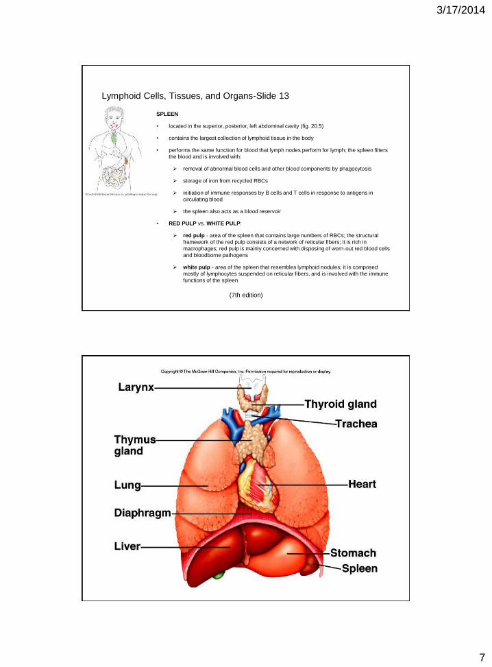
Lymphoid Cells, Tissues, and Organs-Slide 13
SPLEEN
• located in the superior, posterior, left abdominal cavity (fig. 20.5)
• contains the largest collection of lymphoid tissue in the body
• performs the same function for blood that lymph nodes perform for lymph; the spleen filters
the blood and is involved with:
removal of abnormal blood cells and other blood components by phagocytosis
storage of iron from recycled RBCs
initiation of immune responses by B cells and T cells in response to antigens in
circulating blood
the spleen also acts as a blood reservoir
• RED PULP vs. WHITE PULP:
red pulp - area of the spleen that contains large numbers of RBCs; the structural
framework of the red pulp consists of a network of reticular fibers; it is rich in
macrophages; red pulp is mainly concerned with disposing of worn-out red blood cells
and bloodborne pathogens
white pulp - area of the spleen that resembles lymphoid nodules; it is composed
mostly of lymphocytes suspended on reticular fibers, and is involved with the immune
functions of the spleen
(7th edition)
3/17/2014
8
Lymphoid Cells, Tissues, and Organs – Slide 15
• TONSILS
Large lymphoid nodules in the walls of the pharynx and oral cavity (fig. 20.5)
pharyngeal tonsil - also called the “adenoids” is located in the posterior superior wall
of the nasopharynx
palatine tonsils - a pair of tonsils located at the posterior margin of the oral cavity
along the border along its boundary with the oropharynx
lingual tonsil - located under the attached base of the tongue
• PEYER’S PATCHES
Peyer's patches are clusters of lymphoid nodules deep to the epithelial lining of the
small intestine (fig. 20.5)
contain lymphocytes and macrophages which remove microorganisms, debris, and
antigens from the digestive tract
• APPENDIX - large concentrations of lymphoid tissue are located in the wall of the appendix;
thus, the appendix has some lymphatic function (fig. 20.5)
(7th edition)
Innate Host Defenses; innate defenses are present at birth and are
genetically determined = Slide 16
• Innate External Defense System - first line of defense
surface barriers include the skin and mucous membranes of the respiratory, digestive, urinary, and
reproductive tracts
characteristics of skin that help it to resist invasion:
water-resistant and tough keratin outer layer
intercellular junctions hold skin cells tightly together
skin secrections are acidic and have chemicals that make the skin inhospitable to pathogens; e.g.
lysozyme destroys cell walls of certain bacteria
mucous membranes not only provide a barrier, but also produce a variety of protective chemicals (e.g.
lysozyme) and acidic secretions
the stomach secretes digestive enzymes and has a very low pH
the digestive and respiratory pathways are lined with sticky mucous that traps pathogens
(7th edition)
3/17/2014
9
Innate Host Defenses = Slide 17
• Innate Internal Defense System - second line of defense; attempts to limit the spread of pathogens; this system
is fast-acting and nonspecific
the internal defense system has 5 components:
phagocytic cells (e.g. neutrophils and monocytes/macrophages)
NK cells (natural killer cells)
antimicrobial proteins (complement and interferon)
inflammation
fever
(7th edition)
Innate Host Defenses = Slide 18
• Innate Internal Defense System - second line of defense; attempts to limit the
spread of pathogens; this system is fast-acting and nonspecific
phagocytes (fig. 21.2)
neutrophils are the first cells to leave the blood and enter tissues at
the sites of infection or trauma; these cells are short-lived
monocytes follow the influx of neutrophils into the affected tissue;
once in the tissue, they transform into macrophages; they
phagocytize many more pathogens than neutrophils
phagocytes use special membrane receptors to recognize and bind
molecules that are found on pathogens, but not on normal body cells
when a phagocyte recognizes a pathogen it:
- ingests the pathogen
- releases chemical alarm signals that mobilize other cells of
innate and adaptive immunity
OPSONIZATION - some bacteria have capsules that make it difficult
for phagocytes to grab them; the immune system makes molecules
that “coat” the bacteria and enhance phagocytosis; this is called
opsonization; both complement and antibodies can act as
opsonins
(7th edition)

3/17/2014
10
Innate Host Defenses – Slide = 19
• Innate Internal Defense System - second line of defense; attempts to limit the
spread of pathogens; this system is fast-acting and nonspecific
NK cells (natural killer cells)
type of lymphocyte involved in innate immunity
attack body cells that have been invaded by pathogens (e.g. viruses)
or cancer; they will also attack the cells of transplanted tissues
NK cells are larger than B and T cells, and unlike B and T cells, do
not have antigen receptors
both NK cells and T cells are involved in IMMUNE SURVEILLANCE
(they continually scan our cells for abnormalities)
(7th edition)
Innate Host Defenses = Slide 20
• Innate Internal Defense System - second line of defense; attempts to limit
the spread of pathogens; this system is fast-acting and nonspecific
antimicrobial proteins
interferons (fig. 21.5)- interfere with viral replication and activate
immune cells; cells that have been attacked by a virus release
interferon to help protect neighboring cells that have not yet been
affected
complement (complement system) (fig. 21.6) - it
“complements” or enhances other components of both innate
and adaptive defenses; it can mark cells for phagocytosis,
promote inflammation, and kill some bacteria
(7th edition)

3/17/2014
11
Innate Host Defenses = Slide 21
• Innate Internal Defense System - second line of defense; attempts to limit the spread of pathogens; this system
is fast-acting and nonspecific
inflammation
when the body is injured (e.g. a cut, abrasion, or bruise) a sequence of events called inflammation is
initiated
tonsillitis, tendonitis, and laryngitis are examples of short-lived, or acute, inflammation; arthritis is an
example of long-term, or chronic, inflammation
there are 4 cardinal signs of inflammation: pain, swelling, redness, and heat
the purpose of inflammation is to bring white blood cells and plasma proteins into an injured area;
inflammatory mediators (e.g. histamine from basophils and mast cells) cause vasodilation
(increasing blood flow to the area) and an increase in vascular permeability (allowing phagocytes
and plasma proteins to enter the tissue)
plasma proteins and more fluid than usual leak into the injured area causing EDEMA (increased
interstitial fluid); edema causes swelling, which can contribute to the sensation of pain
(7th edition)
Innate Host Defenses = Slide 22
• Innate Internal Defense System - second line of defense; attempts to limit the spread of pathogens; this system
is fast-acting and nonspecific
fever
generalized increase in body temperature
PYROGENS - chemicals secreted by leukocytes and macrophages that have been exposed to foreign
substances in the body; they cause the body’s thermostat (located in the hypothalamus) to set its
temperature higher
higher body temperatures enhance phagocytosis and cause the liver and spleen to sequester iron and
zinc (making these essential elements less available to bacteria); pathogens also do not grow very
well at higher temperatures
(7th edition)

3/17/2014
12
Adaptive Defenses - the body’s third line of defense = Slide 23
• Adaptive Defenses are Specific, Systemic, and have Memory; they include
Humoral Immunity (antibody-mediated) and Cellular Immunity (cell-meditated)
• B and T Lymphocytes are key players in adaptive immunity
• Antigens (fig. 21.7)
have multiple antigenic determinants (based on shapes)
self-antigens are the shapes that lymphocytes expect to find in the body
(thus lymphocytes do not normally attack them)
antigen receptors are specific and diverse
(7th edition)
Adaptive Defenses = Slide 24
• “Education” of Lymphocytes
immunocompetence - the lymphocyte is able to recognize its one specific
antigen by binding to it
self-tolerance - the lymphocyte is unresponsive to self-antigens, so that it
does not attack the body’s own cells
T cells become immunocompetent and self-tolerant in the thymus, whereas
for B cells this occurs in the bone marrow
• Autoimmune Diseases - lymphocytes attack the body’s own cells; e.g. Type 1
Diabetes mellitus, Grave’s disease, and Multiple sclerosis
• Memory Cells - are created in large numbers during a primary immune response
(exposed to antigen for first time); memory cells create a larger number of
effector cells during a secondary immune response (exposed to antigen again);
thus, the response to the second attack will be much greater
(7th edition)