powerpoint presentationamos3.aapm.org/abstracts/pdf/127-35574-418554-126273.pdf · indicator of...
TRANSCRIPT

8/2/2017
1
Imaging in Clinical Trials: An Overview
Keyvan Farahani, PhD
Cancer Imaging Program, NCI
AAPM Beyond Clinical Imaging Aug 2, 2017
2
Outline Introduction to Oncology Clinical Trials
Role of Imaging in Oncology Clinical Trials (Onc, Rad Onc,
Interventional)
Quantitative measures and biomarkers
NCI Clinical Trials Network
Examples of imaging in clinical trials
Introduction: Clinical Trials
Definition: A clinical Trial is a prospective study comparing the effect and
value of interventions(s) against a control in human beings.

8/2/2017
2
Introduction: Clinical Trials
Definition: A clinical Trial is a prospective study comparing the effect and
value of interventions(s) against a control in human beings.*
“Stretchy glue inspired by slugs could be the future of sutures”
Washington Post, July 27, 2017
Introduction: Clinical Trials
Definition: A clinical Trial is a prospective study comparing the effect and
value of interventions(s) against a control in human beings.*
Key Components
Primary and secondary questions
Study design
Study population and sample size
Data collection and quality control
Data analysis
Study monitoring
Close out and reporting
* Friedman LM, Furberg CD, DeMets DL. Fundamentals of Clinical Trials. Springer-Verlag, New York Inc., 1998 (3rd Ed.)
Phases of Clinical Trials
Phase I
Safety
Tx administration
n ~ 15-30
Phase II
Efficacy
n ~ < 100
Randomized (some)
Phase III
Comparison of new application with standard
n ~ 100 – thousands
All randomized
Treated
Control

8/2/2017
3
Oncology Clinical Trials
Overall goal in an oncology trial is typically one of the following:
Treat cancer
Diagnose cancer
Prevent cancer
Manage symptoms and side effects of treatment
Oncology Therapy Trials
Designed to address questions on safety, efficacy, and advantage
compared to current standard therapies
Drugs
Vaccines
Interventional
Combinations
Oncology Clinical Trial Team Members
Principal Investigator
Research nurse
Staff physician or nurse
Data manager
Statistician
Physicist – imaging and/or RT

8/2/2017
4
Imaging in Clinical Trials
Imaging applications in clinical trials
Screen imaging Early Detection & Prevention
Diagnostic imaging Diagnosis & Management
Imaging to guide therapy & Therapy
Monitor response
Imaging Capabilities
Lambin P, et. al., EJC 2012
Quantitative Imaging
Definition – Quantitative Imaging: Extraction of quantifiable features
from medical images for the assessment of normal or the severity,
degree of change, or status of a disease, injury, or chronic condition
relative to normal.
Quantitative Imaging Biomarker Alliance (QIBA)
ww.rsna.org/QIBA/

8/2/2017
5
Biomarkers
Definition: Biomarker - defined
characteristic that is measured as an
indicator of normal biological processes,
pathogenic processes or responses to an
exposure or intervention, including
therapeutic interventions.
FDA–NIH Biomarker Working Group:
Molecular, histologic, radiographic or
physiologic characteristics are examples
of biomarkers.
• O’Connor JBP, et. al., Nature Reviews Clinical Oncology 2016
• FDA and NIH: BEST (Biomarkers, Endpoints, and other tools) resource. NCBI 2016
DNA Repair
PARPi (FTT)
Molecular Imaging Biomarkers Measure Factors Affecting Tumor Behavior Variable Levels in Tumor
Surface
Receptors
SSR, HER2
Proliferative Rate
Thymidine &
Analogs, Sigma-2
Cancer Metabolism
FDG, Glutamine
Hypoxia
FMISO, EF-5
Drug Transport
MIBI, Verapamil
Nuclear
Receptors
FES, FDHT
Angiogenesis
Water
RGD Peptides
Courtesy of Dr. D. Mankoff, U Penn
Imaging Requirement for Biomarker Imaging Simultaneously Localize and Characterize Disease Sites
FDG
PET
PET/CT
Fusion
FES FDG
Glucose
Metabolism Estradiol
Binding
Functional/Anatomic
Imaging
Functional Imaging
Combinations
Courtesy of Dr. D. Mankoff, U Penn

8/2/2017
6
Imaging and Cancer Therapy
Novel Approaches to Biomarker Imaging
Choosing the right patients
Is the therapeutic target present?
Choosing the right drug
Does the drug reach the target?
Getting the right result
Is there a pharmacodynamic response?
Predicting the outcome
Will response lead to better patient survival?
Courtesy of Dr. D. Mankoff, U Penn
Clinical Indications for Imaging in a Clinical Trial Setting
Role Definition
Diagnosis and staging To determine whether a lesion is positive or negative for malignancy
Prognostic marker To determine the expected outcome under standard therapy for the
patient's disease stage
Predictive biomarker assay To differentiate between patients expected to benefit clinically on one
treatment relative to another from those not expected to experience such
a benefit
Pharmacokinetics marker To confirm that the drug has reached the intended target
Pharmacodynamics marker To measure the effects of the drug on the body
Early response indicator To determine the expected ultimate outcome on a particular therapy
regimen from changes in a tumor characteristic following a few cycles of
treatment
Basis of a Phase II trial end point A pre- to posttreatment change measurement used to determine whether
to proceed to the subsequent Phase III investigation
Basis of a Phase III trial end point A pre- to posttreatment change that serves as a surrogate for a definitive
clinical end point
Lin F, et. al., Acad Radiol 24 (8), 2017
Statistical Tests in the Context of Clinical Trials
Test Purpose
Log-rank test To test whether the distributions of a time-to-event outcome between two
groups are equal
Logistic regression To model the probability of an event (e.g., pathologic complete response)
occurring as a function of one or more explanatory variables
Cox regression To model the rate at which an event (e.g., progression or death) occurs as
a function of one or more explanatory variables
Fisher exact test To test the association between two categorical variables
Mann-Whitney U test To test whether the distributions of a quantitative variable between two
groups are equal
Test for qualitative
interaction
To test whether one treatment is more efficacious than another in one
subset of patients but not in another subset
Kendall tau rank
Correlation coeff.
A measure of the association between two quantitative or ordered
categorical variables
Wilcoxon
signed-rank test
To test whether repeat measurements on
a particular patient differ
Hu
ang EP, et. al., A
cad R
adio
l 24
(8), 2
01
7
8/2/2017
7
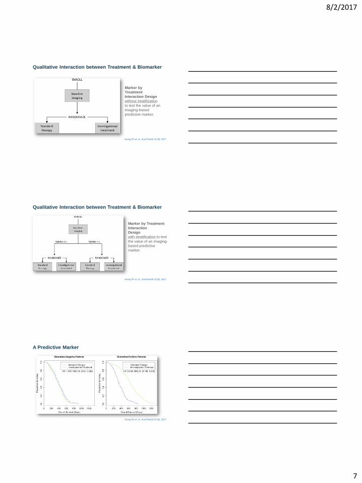
Qualitative Interaction between Treatment & Biomarker
Marker by
Treatment
Interaction Design
without stratification
to test the value of an
imaging-based
predictive marker.
Huang EP, et. al., Acad Radiol 24 (8), 2017
Marker by Treatment
Interaction
Design
with stratification to test
the value of an imaging-
based predictive
marker.
Huang EP, et. al., Acad Radiol 24 (8), 2017
Qualitative Interaction between Treatment & Biomarker
A Predictive Marker
Huang EP, et. al., Acad Radiol 24 (8), 2017
8/2/2017
8
22
1) Selection of the appropriate imaging endpoint and modality
2) Qualification of the QI capabilities of participating sites
3) Data collection and image analysis for imaging endpoint
determination
4) Auditing and quality control for quantitative imaging data
Challenges and Approaches for Quantitative Imaging in Cancer Clinical Trials
Yankeelov T., et. al., Clin Cancer Res. 2016 Jan 15; 22(2): 284–290.
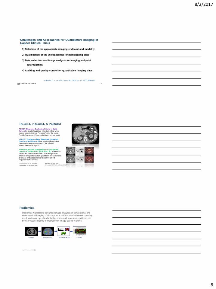
RECIST, irRECIST, & PERCIST
Eisenhauer EA, et. al., EJC 2009 Bohnsack O, et., al., ESMO 2014
irRECIST (Immune-related Response Evaluation
Criteria In Solid Tumors) is a set of published rules
that provide better assessment of the effect of
immunotherapeutic agents.
RECIST (Response Evaluation Criteria In Solid
Tumors) is a set of published rules that define when
cancer patients improve ("respond"), stay the same
("stable") or worsen ("progression") during treatments.
Positron Emission Tomography (PET) Response
Criteria in Solid Tumors (PERCIST 1.0) - methods to
ensure the comparability of PET FDG images from
different time points to allow quantitative measurements
of change and assessment of overall treatment response in PET studies.
Wahl R, et. al., JNM 2009 O JH, Lodge M, Whal R, Radiology 2016
Radiomics
Lambin P, et. al., EJC 2012
Radiomics hypothesis: advanced image analysis on conventional and
novel medical imaging could capture additional information not currently
used, and more specifically, that genomic and proteomics patterns can
be expressed in terms of macroscopic image-based features.

8/2/2017
9
NCI Quantitative Imaging Excellence (CQIE)
initiated in 2010 in collaboration with ACRIN to establish a resource of
clinical trial-ready sites within the National Cancer Institute (NCI)-
designated Cancer Centers (NCI-CCs).
The intent was to enable imaging centers in the NCI-CCs network
capable of conducting treatment trials with advanced quantitative
imaging end points.
CQIE provided PET/CT and MRI phantoms and protocols for site
qualification.
Rosen M, et. al., Acad Radiol 2017; 24:232–245
NCI Network Trials
National Clinical Trials Network (NCTN)
NCI Community Oncology Research Program (NCORP)
Experimental Therapeutics Clinical Trials Network (ETCTN)
…and
NCI-designated cancer centers
NIH Clinical Center in Bethesda, Maryland
NCI National Clinical Trials Network Structure
Phase II and III trials:
help establish new standards of care
prepare for FDA approval
new approaches to interventions
validate new biomarkers
https://www.cancer.gov/research/areas/clinical-trials/nctn/nctn-clinical-trials-network

8/2/2017
10
NCI NTCN Imaging and Radiation Oncology Core
• FitzGerald TJ, et. al., IJROBP 2016 • ASTRONews 2014
ClinicalTrials.gov https://clinicaltrials.gov/
Some Examples of
Imaging in Clinical Trials in Cancer

8/2/2017
11
Imaging: DSC-MRI
Primary Outcome Measures:
Change in rCBV within enhancing tumor [Baseline to 2 weeks]
OS [Up to 5 years]
Secondary Outcome Measures:
CBF [Baseline]
Change in CBF [Baseline to 2 weeks]
PFS [Up to 5 years]
rCBV [Baseline]
Estimated Enrollment: 165
https://clinicaltrials.gov/ct2/show/record/NCT03115333
Example 1 - Change in Relative Cerebral Blood Volume as a
Biomarker for Early Response to Bevacizumab in Recurrent
Glioblastoma [ECOG-ACRIN: EAF151]
Example 2 - FDG-PET/CT in Tumor Assessment and Surgical in
Patients With Newly Diagnosed H&N Cancer (ACRIN 6685)
Primary Outcome Measures:
Negative predictive value of PET/CT imaging for staging the N0 neck based upon pathologic
sampling of the neck lymph nodes [Within 2 Weeks Before Surgery]
Potential of PET/CT imaging to change treatment of the N0 neck [Within 2 Weeks Before Surgery]
Secondary Outcome Measures: (Partial list)
Sensitivity and diagnostic yield of PET/CT imaging for detecting occult metastasis in the clinically N0 neck (both by neck
and lymph node regions) or other local sites [Within 2 Weeks Before Surgery ]
Effect of other factors (e.g., tumor size, location, second primary tumors, or intensity of FDG uptake) that can lead to
identification of subsets of patients that could potentially forego neck dissection or …[Within 2 Weeks Before Surgery ]
Cost-effectiveness and cost-benefit of using PET/CT imaging for staging of head and neck cancer vs current good clinical
practices [ Time Frame: Within 2 Weeks Before Surgery ]
Correlation of PET/CT imaging findings with CT/MRI findings and biomarker results [Within 2 Weeks Before Surgery ]
Target Enrollment: A total of 292 participants will be enrolled.
https://clinicaltrials.gov/ct2/show/NCT00983697?term=NCT00983697&rank=1
Example 3 - Randomized Phase II Trial of Individualized Adaptive RT
Using During-Treatment FDG-PET/CT in Locally Advanced NSCLC
[RTOG 1106/ACRIN 6697]
• Primary Outcome Measures: • Local-regional, progression-free (LRPF) rate (NRG) [2 years]
• Relative change in SUV peak from the baseline to the during-treatment FDG-PET/CT to LRPF
(ECOG-ACRIN) [Baseline to 2 years]
• Secondary Outcome Measures: (partial list) • Baseline FMISO uptake (tumor-to-blood pool ratio) association with LRPF (i.e. the assessment of using
baseline FMISO-PET uptake as a prognostic marker) (ECOG-ACRIN) [Baseline]
• Change in metabolic tumor volume (ECOG-ACRIN) [Baseline to 5 years]
• Change of peak SUVs for FDG from pre- to during-treatment (ECOG-ACRIN) [Baseline to 5 years ]
• FMISO total hypoxic volume (ECOG-ACRIN) [Up to 5 years]
• FMISO tumor-to-blood pool ratio (ECOG-ACRIN) [ Time Frame: Up to 5 years ]
https://clinicaltrials.gov/ct2/show/record/NCT01507428?term=NCT01507428&rank=1
8/2/2017
12
Example 4 - Abbreviated Breast MRI and Digital Tomosynthesis
Mammography in Screening Women With Dense Breasts
A Randomized Phase II trial
PRIMARY OBJECTIVES:
To compare the rates of detection of invasive cancers with abbreviated breast (AB)-MRI and
digital tomosynthesis mammography (DBT).
SECONDARY OBJECTIVES: (partial list)
To compare the positive predictive value (PPV) of biopsies, call back rates, and short-term follow up
rates after AB-MR and DBT on both the initial and 1 year follow up studies.
To estimate and compare the sensitivity and specificity of AB-MR and DBT, using the 1 year follow up
to define a reference standard.
To compare patient-reported short-term quality of life related to diagnostic testing with AB-MR and DBT using the Testing Morbidities Index.
To compare willingness to return for testing with AB-MRI versus (vs) DBT within the recommended
screening interval and explore factors associated with willingness to return for screening.
https://clinicaltrials.gov/ct2/show/NCT02933489?term=ea+1141&recrs=a&rank=1
A Funding Opportunity for
Early Phase Imaging Trials
8/2/2017
Early Phase Clinical Trials in Imaging & IGI (R01) [PAR-17-167]
3 year clinical trials in novel imaging or IGI
Intended to accelerate the development of imaging and IGI modalities, methodologies, and agents through the early stages of clinical development -such as trials evaluating safety and preliminary efficacy
Phase I & II studies to establish treatment parameters and early therapeutic efficacy
SEP Review (CSR)
https://grants.nih.gov/grants/guide/pa-files/PAR-17-167.html
36

8/2/2017
13
Acknowledgment
Lalitha Shankar, MD, PhD
Cancer Imaging Program, NCI
8/2/2017
www.cancer.gov www.cancer.gov/espanol