powering through pigmentary disorders
TRANSCRIPT
Powering Through Pigmentary Disorders
Sandy Tsao, MD
Co-Director, Multi-Ethnic Skin Clinic
Assistant Professor, Harvard Medical School
Dermatology Laser and Cosmetic Center
Massachusetts General Hospital

Disclosures
Neither I nor my spouse/partner has a relevant financial relationship with a commercial interest
to disclose

Objectives
• Review some of the common pigmentary disorders
• Review diagnostic tools available in the diagnosis and management of these disorders
• Discuss patient and treatment selection to maximize benefits and limit potential side effects
Skin Color
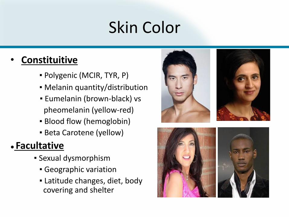
• Constituitive
▪ Polygenic (MCIR, TYR, P)
▪ Melanin quantity/distribution
▪ Eumelanin (brown-black) vs
pheomelanin (yellow-red)
▪ Blood flow (hemoglobin)
▪ Beta Carotene (yellow)
● Facultative▪ Sexual dysmorphism
▪ Geographic variation
▪ Latitude changes, diet, body covering and shelter
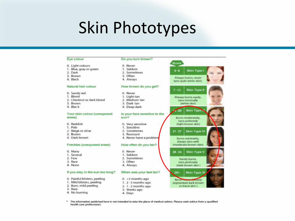
Skin Phototypes
Melanosomes and Melanin Synthesis
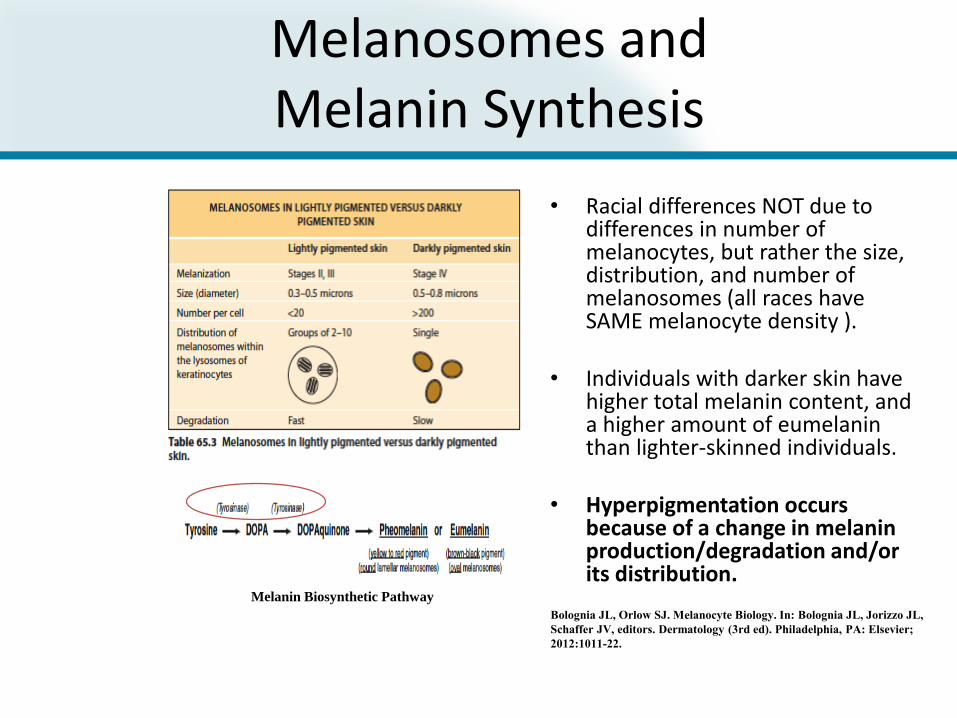
• Racial differences NOT due to differences in number of melanocytes, but rather the size, distribution, and number of melanosomes (all races have SAME melanocyte density ).
• Individuals with darker skin have higher total melanin content, and a higher amount of eumelanin than lighter-skinned individuals.
• Hyperpigmentation occurs because of a change in melanin production/degradation and/or its distribution.
Melanin Biosynthetic Pathway
Bolognia JL, Orlow SJ. Melanocyte Biology. In: Bolognia JL, Jorizzo JL,
Schaffer JV, editors. Dermatology (3rd ed). Philadelphia, PA: Elsevier;
2012:1011-22.
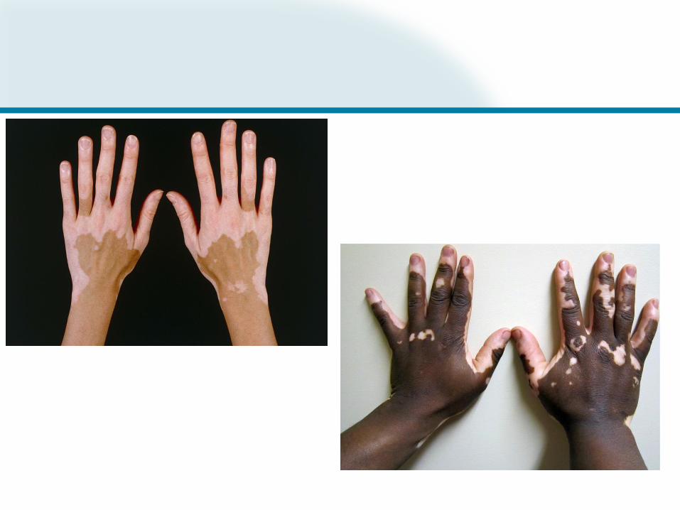
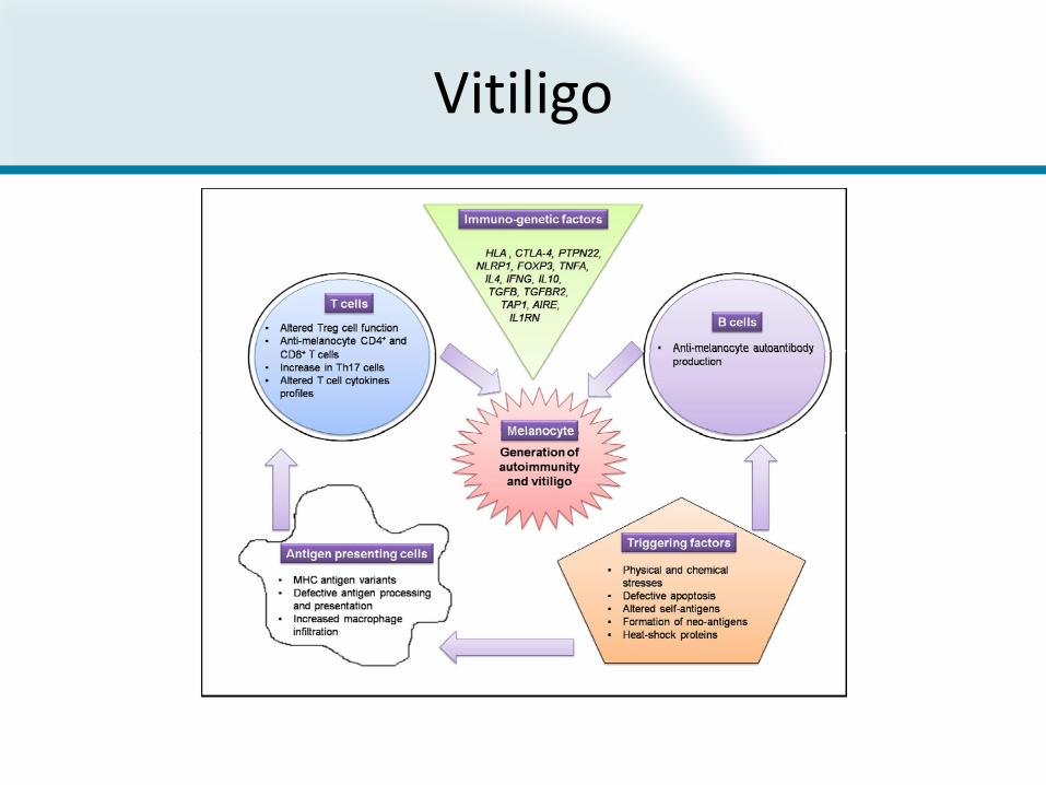
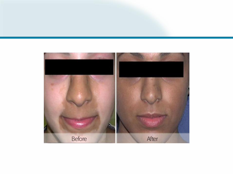
Vitiligo

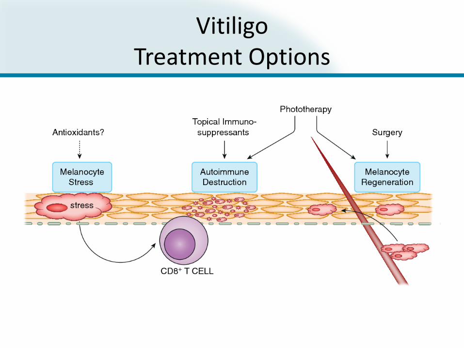
VitiligoTreatment Options


Diagnosis
• Physical Examination
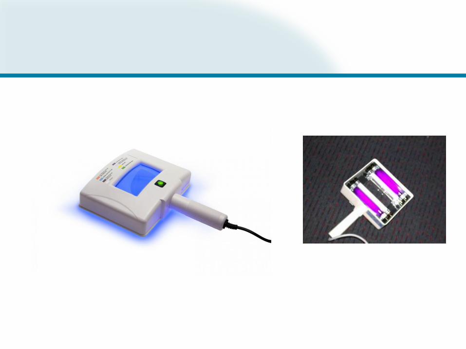
• Wood’s Lamp
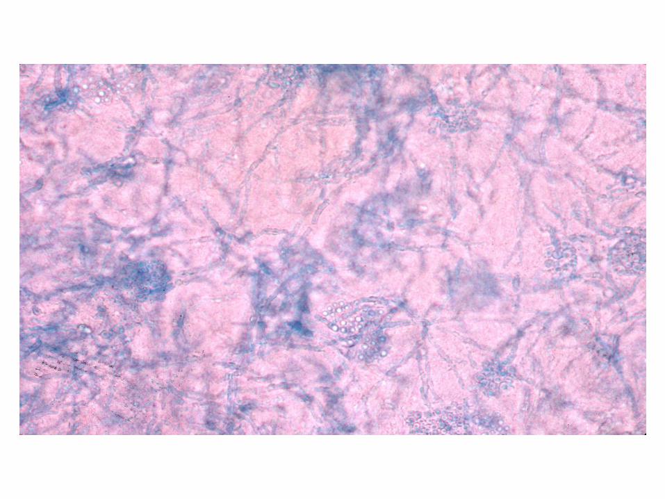
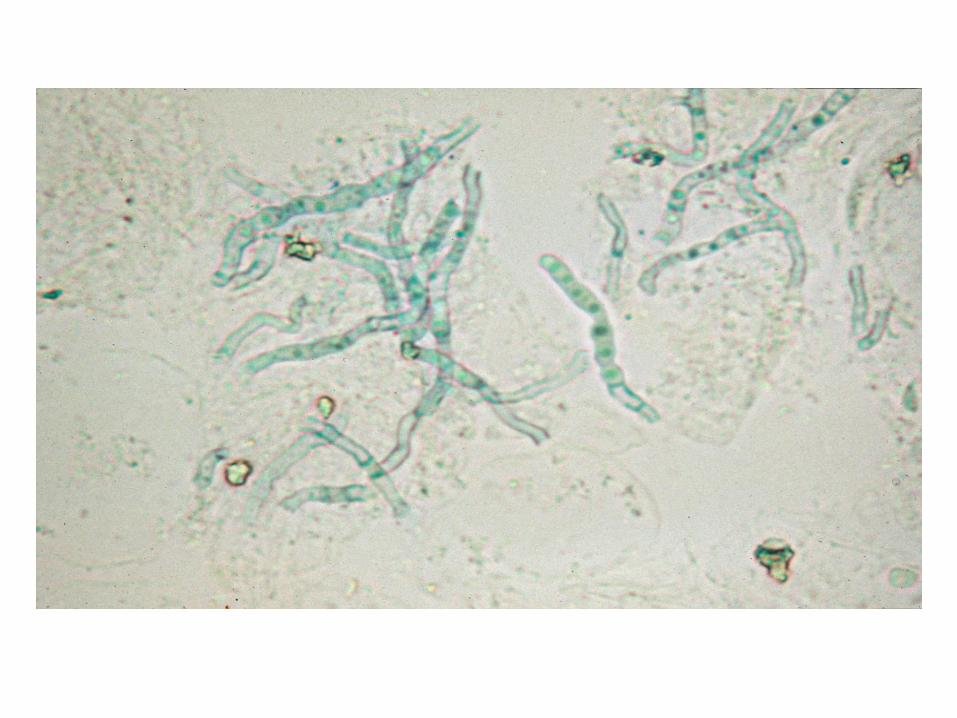
• Potassium hydroxide (KOH) microscopic evaluation
• Fungal culture
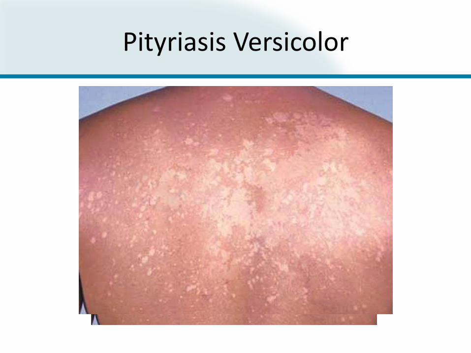
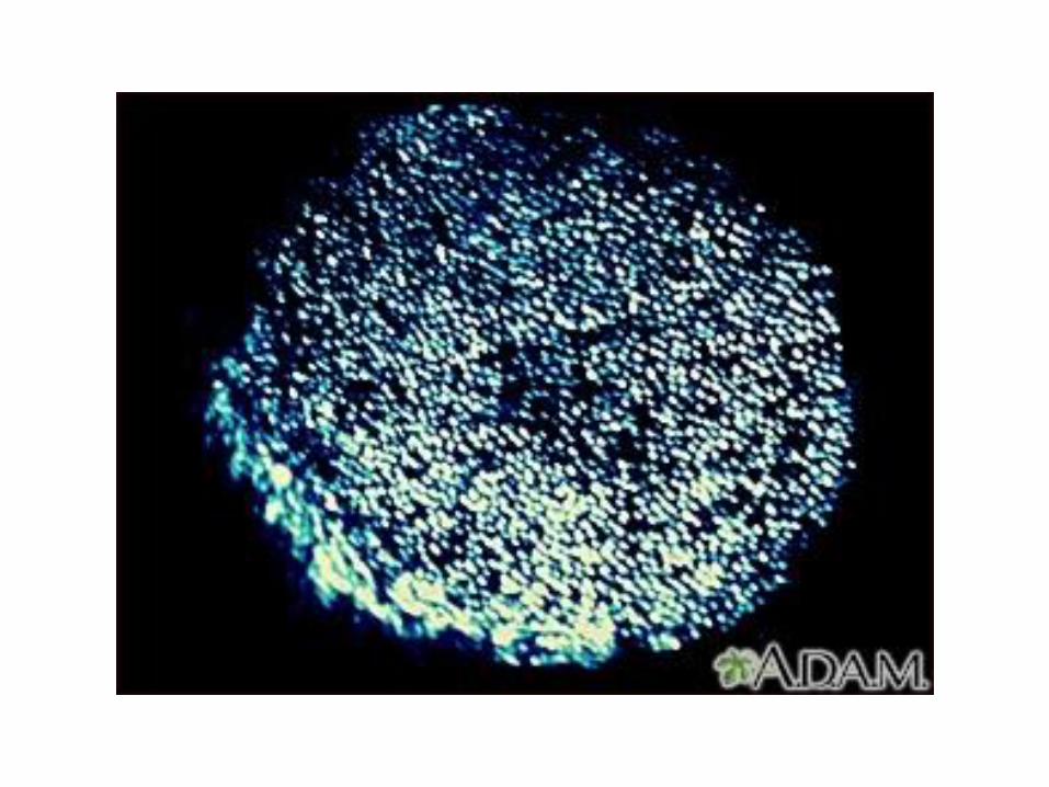
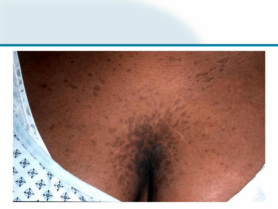
Pityriasis Versicolor

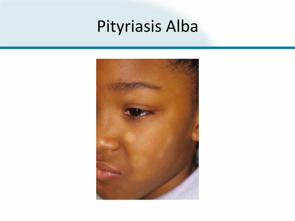
Pityriasis Alba
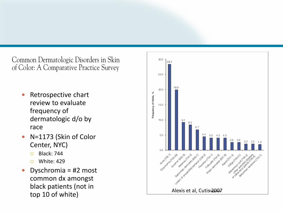
Retrospective chart review to evaluate frequency of dermatologic d/o by race
N=1173 (Skin of Color Center, NYC) Black: 744
White: 429
Dyschromia = #2 most common dx amongst black patients (not in top 10 of white)
Alexis et al, Cutis 2007
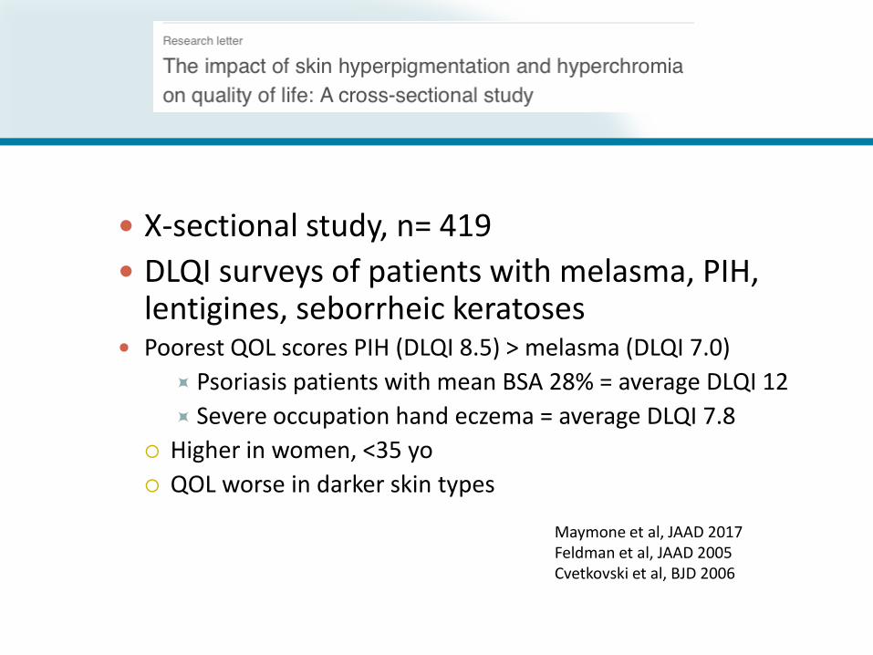
X-sectional study, n= 419
DLQI surveys of patients with melasma, PIH, lentigines, seborrheic keratoses
Poorest QOL scores PIH (DLQI 8.5) > melasma (DLQI 7.0)
Psoriasis patients with mean BSA 28% = average DLQI 12
Severe occupation hand eczema = average DLQI 7.8
Higher in women, <35 yo
QOL worse in darker skin types
Maymone et al, JAAD 2017Feldman et al, JAAD 2005Cvetkovski et al, BJD 2006
Where Is the Pigment?
• Epidermal vs Dermal vs Mixed– Epidermal pigmentation may be easier to target– Treatment options will vary depending on
pigment location
• Facial vs Non-Facial– Treatment response and healing times will vary
depending of body site involved

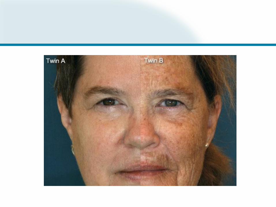
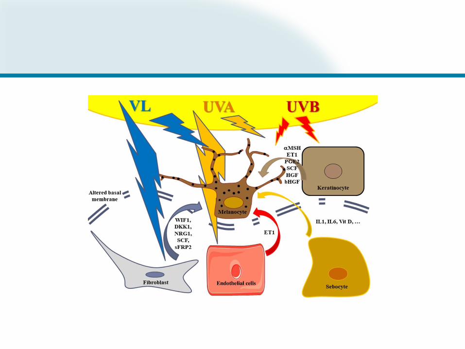
Photoaging-Related Dyschromia• Solar Energy
– UV light, ranging from 280 to 400 nm,
– Visible light (VL) ranging from 400 to 700 nm
– Infrared light (IR) ranging from 700 to 2500 nm.
• Cutaneous Changes– Rhytides, lentigines, dyspigmentation, loss of elasticity and
telangiectases
– Involve facial and non-facial regions
• Based on recent literature, UV radiation, VL, IR all induce pigmentation.
Schalka S. New data on hyperpigmentation disorders. J Eur Acad Dermatol Venereol. 2017 Sep;31 Suppl 5:18-21.
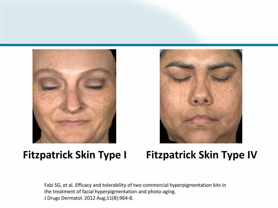
Fabi SG, et al. Efficacy and tolerability of two commercial hyperpigmentation kits in the treatment of facial hyperpigmentation and photo-aging.J Drugs Dermatol. 2012 Aug;11(8):964-8.
Fitzpatrick Skin Type I Fitzpatrick Skin Type IV
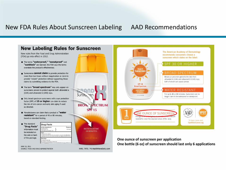
New FDA Rules About Sunscreen Labeling AAD Recommendations
One ounce of sunscreen per applicationOne bottle (6 oz) of sunscreen should last only 6 applications

Vascular Changes in Melasma
• 50 Korean women with melasma
• Biopsy of lesional and perilesional skin
• Lesional skin:
– ⬆️ vessel size and density
– ⬆️ in vascular endothelial growth factor (VEGF) expression by keratinocytes
– Number of vessels correlated with intensity of pigmentation
Kim EH, Kim YC, et al. The vascular characteristics of melasma. J Dermatol Sci. 2007 May;46(2):111-6.
Camouflage Makeup
• Many patients find camouflage makeup to be an important component in the treatment of their hyperpigmentation
• Several widely available brands come in a broad range of shades and offer heavy coverage to help even out skin tone:
– Dermablend (Vichy Laboratories, Paris, France),
– Covermark/CM Beauty (CM Beauty, Northvale, NJ)
– Cover FX (Cover FX Skin Care; Toronto, Ontario, Canada)

Skin Lightening Agents
• Hydroquinone
– Reduces conversion of dihydroxyphenylalanine to melanin by tyrosinase inhibition
– Resultant distorted melanosome formation, increased melanosome destruction and inhibition of DNA and RNA synthesis
– Available in 2% OTC and prescription formulations
– Highly reactive oxidative nature, with efficacy diminishing as discoloration progresses

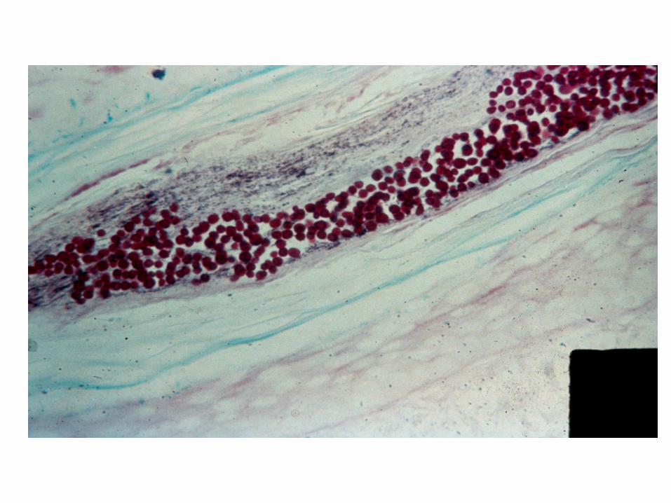
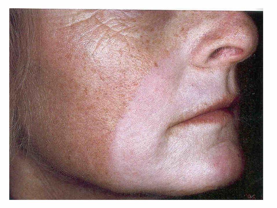
Exogenous Ochronosis
• Associated with the prolonged use of skin-lightening products, most commonly those containing hydroquinone.
• Most commonly seen among the indigenous Black population in African countries and is thought to be due to localized homogentisic acid collecting within the dermis
• Bluish, brown, and/or black mottled macules in areas where topical lightening agents have been applied
• Histology: Yellow–brown banana-shaped deposits in the dermis
• Treatment:
– Stop the offending medication.
– Can try medium depth peels or QS lasers or ablative lasers.
Chang MW. Disorders of Hyperpigmentation. In: Bolognia JL, Jorizzo JL, Schaffer JV, editors. Dermatology (3rd ed). Philadelphia, PA: Elsevier; 2012:1049-74.
Silpa-Archa N, Kohli I, et al. Post inflammatory hyperpigmentation: A comprehensive overview: Epidemiology, pathogenesis, clinical presentation, and noninvasive assessment technique. J Am Acad Dermatol. 2017 Oct;77(4):591-605.
Active Products Against Melanin
• Prevent melanin synthesis
• Lower melanin synthesis
• Accelerate epidermal turnover to limit melanin transfer through melanosomes
• Tretinoin, tranexamic acid, other antiflammatory meds
• Azelaic acid, arbutine, licorice, HQ, kojic acid, vitamin C, phytic acid, resveratrol
• Arbutrin, glabridin, niacinamide, retinoids, linoleic acids
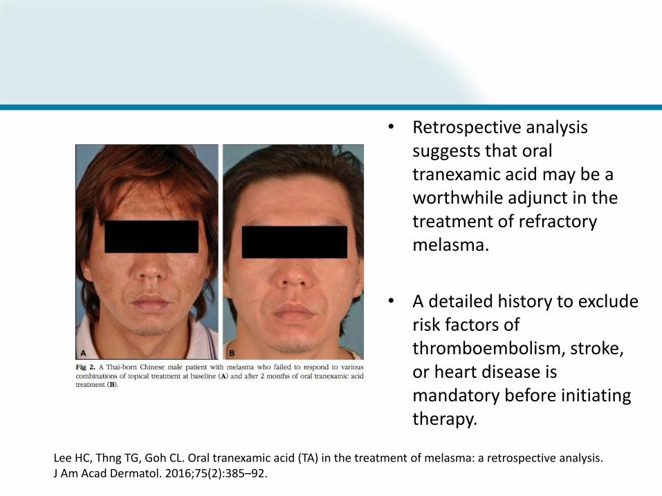
• Retrospective analysis suggests that oral tranexamic acid may be a worthwhile adjunct in the treatment of refractory melasma.
• A detailed history to exclude risk factors of thromboembolism, stroke, or heart disease is mandatory before initiating therapy.
Lee HC, Thng TG, Goh CL. Oral tranexamic acid (TA) in the treatment of melasma: a retrospective analysis. J Am Acad Dermatol. 2016;75(2):385–92.
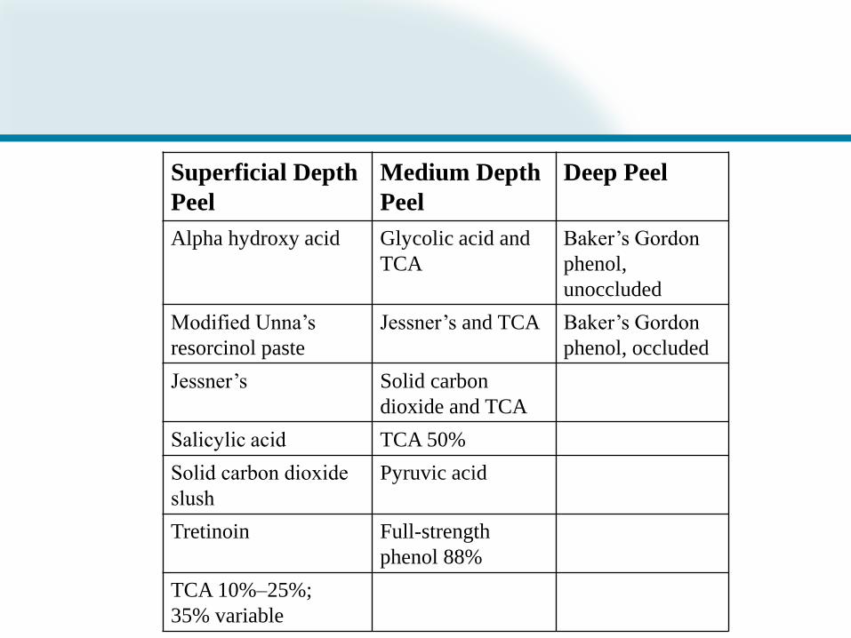
Superficial Depth
Peel
Medium Depth
Peel
Deep Peel
Alpha hydroxy acid Glycolic acid and
TCA
Baker’s Gordon
phenol,
unoccluded
Modified Unna’s
resorcinol paste
Jessner’s and TCA Baker’s Gordon
phenol, occluded
Jessner’s Solid carbon
dioxide and TCA
Salicylic acid TCA 50%
Solid carbon dioxide
slush
Pyruvic acid
Tretinoin Full-strength
phenol 88%
TCA 10%–25%;
35% variable

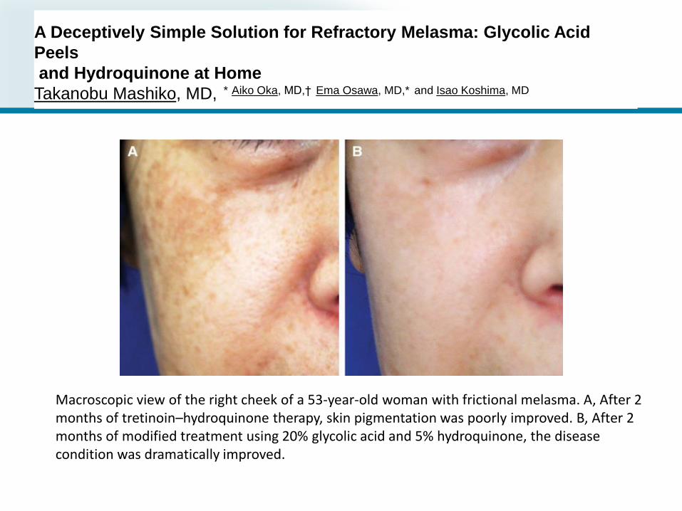
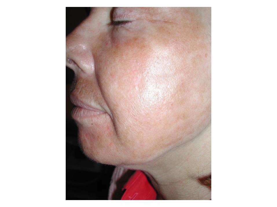
A Deceptively Simple Solution for Refractory Melasma: Glycolic Acid
Peels
and Hydroquinone at Home
Takanobu Mashiko, MD, * Aiko Oka, MD,† Ema Osawa, MD,* and Isao Koshima, MD
Macroscopic view of the right cheek of a 53-year-old woman with frictional melasma. A, After 2 months of tretinoin–hydroquinone therapy, skin pigmentation was poorly improved. B, After 2 months of modified treatment using 20% glycolic acid and 5% hydroquinone, the disease condition was dramatically improved.


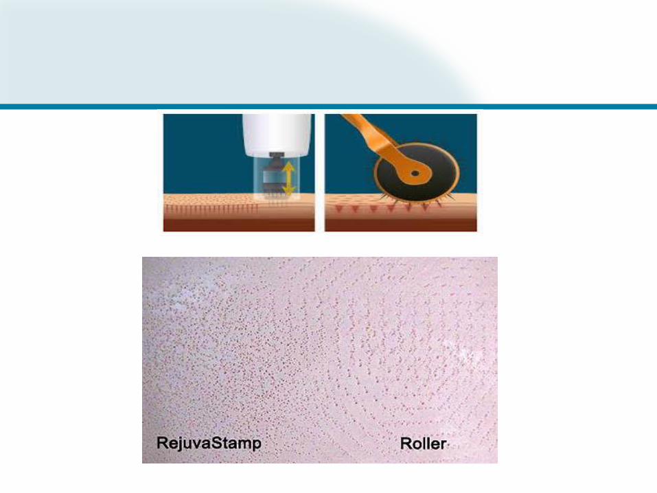
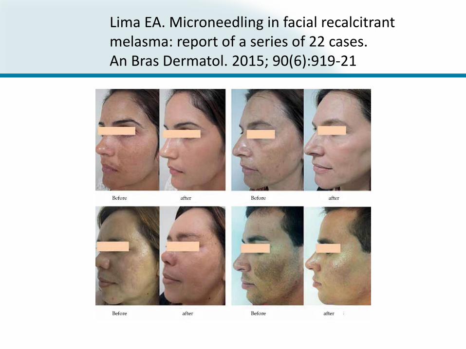
Lima EA. Microneedling in facial recalcitrant melasma: report of a series of 22 cases. An Bras Dermatol. 2015; 90(6):919-21
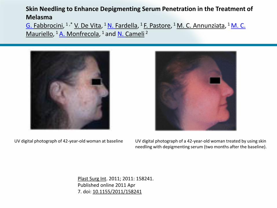
UV digital photograph of 42-year-old woman at baseline UV digital photograph of a 42-year-old woman treated by using skin needling with depigmenting serum (two months after the baseline).
Plast Surg Int. 2011; 2011: 158241.Published online 2011 Apr 7. doi: 10.1155/2011/158241
Skin Needling to Enhance Depigmenting Serum Penetration in the Treatment of MelasmaG. Fabbrocini, 1 ,* V. De Vita, 1 N. Fardella, 1 F. Pastore, 1 M. C. Annunziata, 1 M. C. Mauriello, 1 A. Monfrecola, 1 and N. Cameli 2
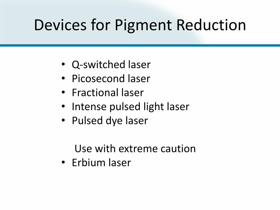
Devices for Pigment Reduction
• Q-switched laser• Picosecond laser• Fractional laser• Intense pulsed light laser• Pulsed dye laser
Use with extreme caution• Erbium laser• Carbon dioxide laser
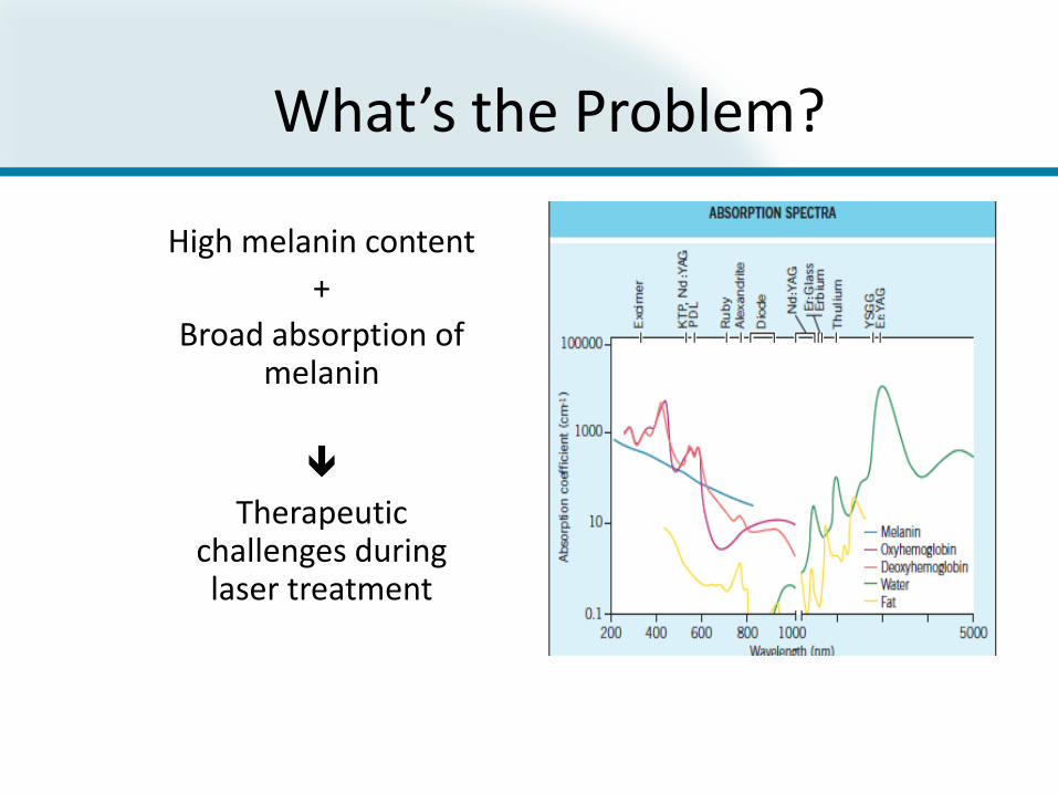
What’s the Problem?
High melanin content
+
Broad absorption of melanin
Therapeutic challenges during laser treatment

Safely Treating Patients with Darker Skin Phototypes
• Goal:– Minimize Epidermal Pigment Absorption– Minimize Epidermal Irritation & Erythema ➔ PIH– Minimize Epidermal Heating ➔ blistering and scarring
• How to achieve that goal:– Longer wavelengths– Lower fluence– Longer pulse duration– Maximize cooling
For Darker Skin Phototypes…
• Less is more (Fluence, density, passes)• Consider laser test site• Stress need for strict photoprotection• Discuss increased risk of PIH• Consider pre-treatment use of retin-a or
hydroquinone• Use caution with ablative fractional
devices

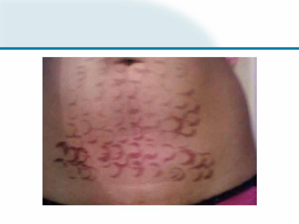
Microscopic Epidermal Necrotic Debris (MEND)
Fractionated Laser TreatmentHuman Tissue – Cross Section
Results:
▪ Complete re-epithelialization
in 24 hrs.
▪ Clear collagen denaturation
from papillary dermis into
mid reticular dermis
▪ Healing occurs from viable
tissue. Zones of spared
tissue contain clusters of
epidermal stem cells and
Transit Amplifying (TA) cells
In Vivo Histology
(16 days post exposure)
Controlled Zones of
Denatured Collagen in the Dermis
Courtesy of Chris Zachary, MD
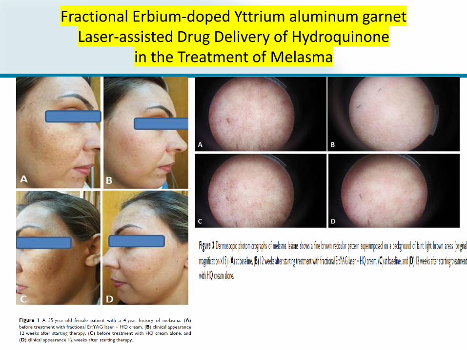
Fractional Erbium-doped Yttrium aluminum garnetLaser-assisted Drug Delivery of Hydroquinone
in the Treatment of Melasma
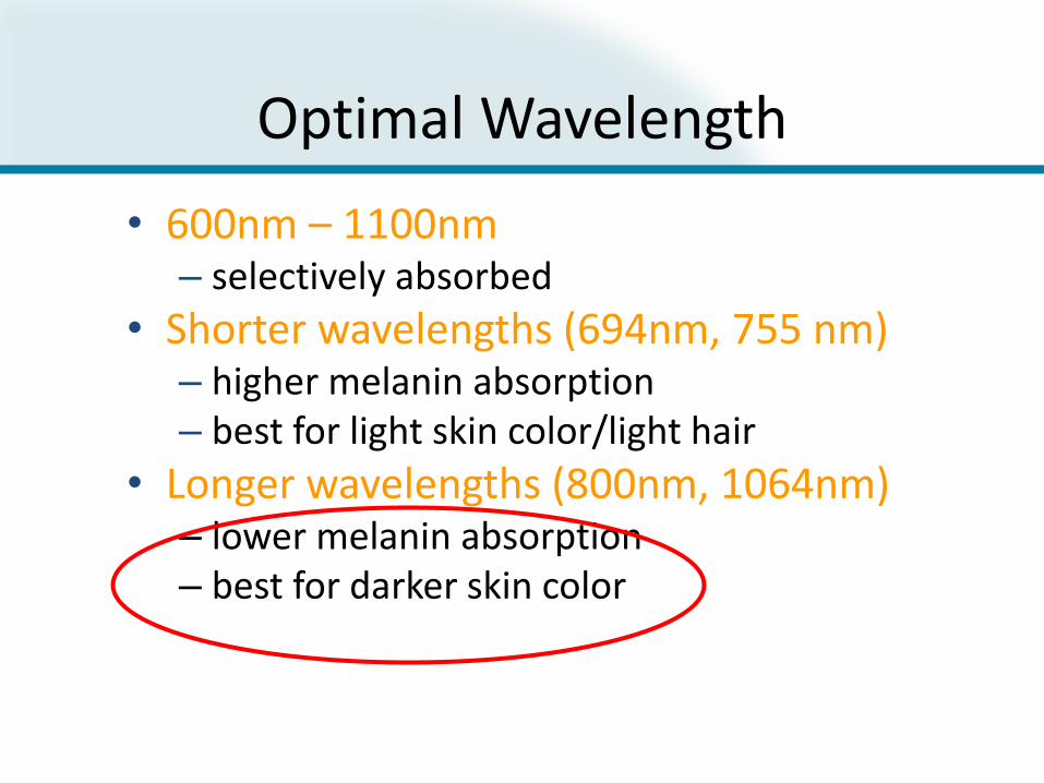
Optimal Wavelength
• 600nm – 1100nm– selectively absorbed by melanin
• Shorter wavelengths (694nm, 755 nm)– higher melanin absorption– best for light skin color/light hair
• Longer wavelengths (800nm, 1064nm)– lower melanin absorption– best for darker skin color


Advancing Laser Treatments
Q-switched Nd:YAG 1064nm laser:
• Low fluence
• Combination therapy with pulsed dye laser
Picosecond Nd:YAG 1064nm or Alex 755nm laser:
• Low fluence

Lee YJ, Shin HJ, Noh T-K, Choi K-H, Chang S-E. Treatment of
Melasma and Post-Inflammatory Hyperpigmentation by a
Picosecond 755-nm Alexandrite Laser in Asian Patients. Annals of
Dermatology. 2017;29(6):779-781. doi:10.5021/ad.2017.29.6.779.

Post-inflammatory hyperpigmentation in a 20-year-old female. (A) Findings at baseline. (B) Findings after seven treatments of 5.25 J/cm2 with a 2-mm spot size using a Picosecond 755-nm Alexandrite laser.
Ann Dermatol. 2017 Dec; 29(6): 779–781.Published online 2017 Oct 30. doi: 10.5021/ad.2017.29.6.779
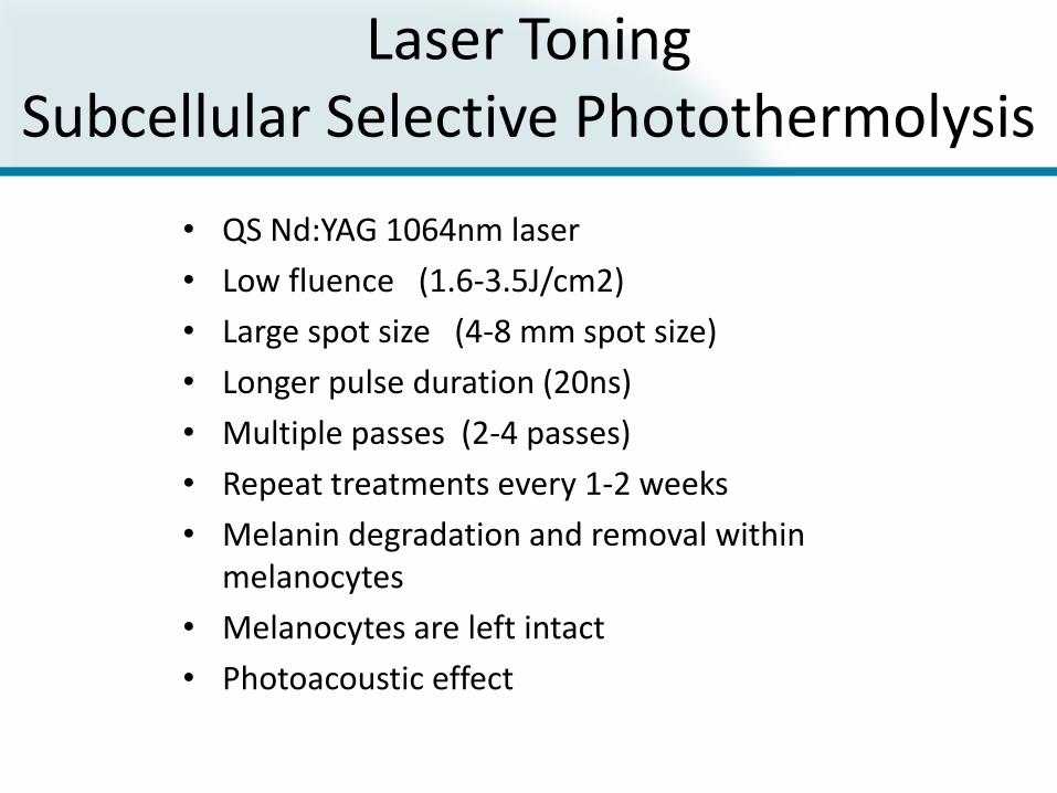
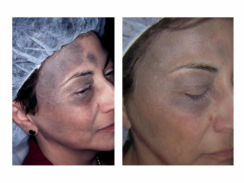
Laser ToningSubcellular Selective Photothermolysis
• QS Nd:YAG 1064nm laser
• Low fluence (1.6-3.5J/cm2)
• Large spot size (4-8 mm spot size)
• Longer pulse duration (20ns)
• Multiple passes (2-4 passes)
• Repeat treatments every 1-2 weeks
• Melanin degradation and removal within melanocytes
• Melanocytes are left intact
• Photoacoustic effect
• for several weeks

Laser Ther. 2011; 20(3): 189–194.doi: 10.5978/islsm.20.189
Clinical findings in a 43 year-old female patient (Patient No 19). a: Baseline condition. b: Findings 6 weeks after dual toning with the 1064 nm Nd:YAG laser, showing very good clearance of the melasma and improved general skin condition including reduced pore size and disappearance of the fine periocular lines.
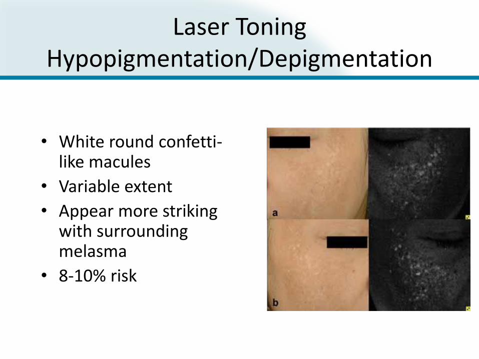
Laser ToningHypopigmentation/Depigmentation
• White round confetti-like macules
• Variable extent
• Appear more striking with surrounding melasma
• 8-10% risk
• Frequently permanent

Pulsed Dye Laser Treatment
• Melasma – targeting vascular component– Longer pulse duration (10-20msec)– Larger spot size (10mm)– Fluence 7.5-8.5 J/cm; DCD 30/20– Consider combination treatment with fractionated
laser
• Lentigines– Requires compression lens to purge tissue blood– Longer pulse duration– Conservative fluence; DCD 30/20
Geddes ER, Stout AB, Friedman PM Lasers Surg Med. 2017 Jan;49(1):20-26.

Pulsed Dye Laser Treatmentof Post-Procedure Ecchymoses
Treatment Protocol
Energy density of 6J/cm2,, pulse duration 6ms, 10mm spot,
DCD 30ms spurt, 20ms delay
3 passes
Arch Dermatol. 2010;146(1):94-95.

Hydrogen Peroxide 40% Solution
• Applied in office
• Pen applicator
• Generally requires 1-2 treatments spaced 3 weeks apart
• Side effects include stinging, irritation, redness, blister, scar
• FDA approved
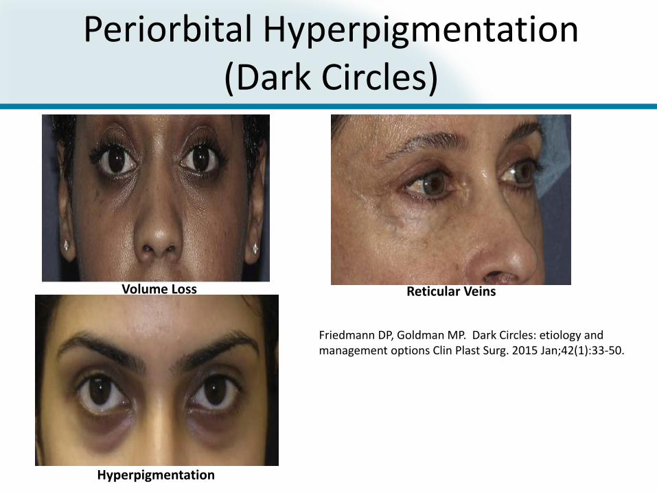
Volume Loss Reticular Veins
Hyperpigmentation
Friedmann DP, Goldman MP. Dark Circles: etiology and management options Clin Plast Surg. 2015 Jan;42(1):33-50.
Periorbital Hyperpigmentation (Dark Circles)
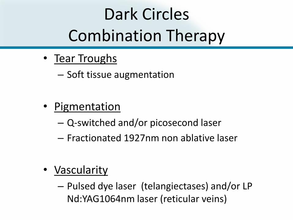
Dark CirclesCombination Therapy
• Tear Troughs
– Soft tissue augmentation
• Pigmentation
– Q-switched and/or picosecond laser
– Fractionated 1927nm non ablative laser
• Vascularity
– Pulsed dye laser (telangiectases) and/or LP Nd:YAG1064nm laser (reticular veins)veins)
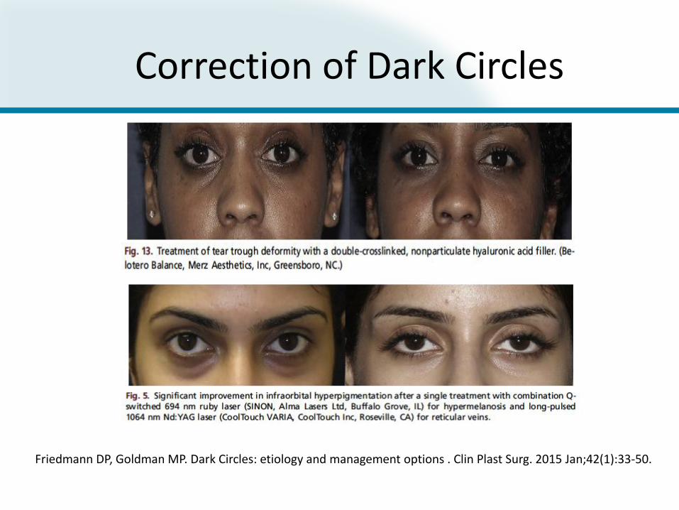
Friedmann DP, Goldman MP. Dark Circles: etiology and management options . Clin Plast Surg. 2015 Jan;42(1):33-50.
Correction of Dark Circles

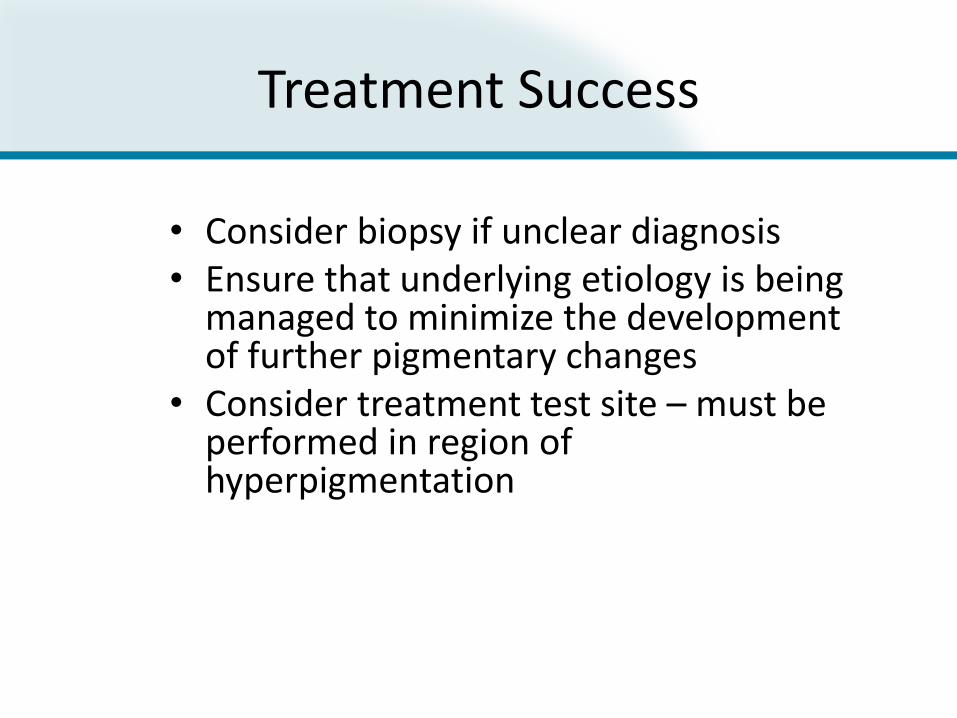
Treatment Success
• Consider biopsy if unclear diagnosis• Ensure that underlying etiology is being
managed to minimize the development of further pigmentary changes
• Consider treatment test site – must be performed in region of hyperpigmentation
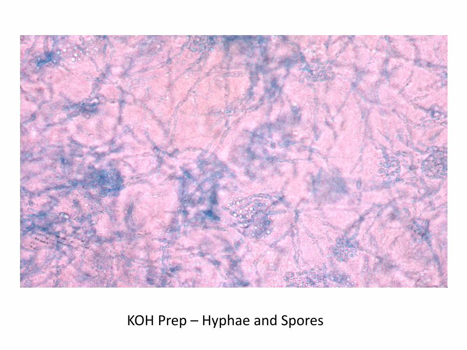
KOH Prep – Hyphae and Spores
What Does the Future Hold?
• Laser assisted drug delivery
• Combination therapies
• Gene modification therapies