potassium fluxes in desheathed frog sciatic nerve
TRANSCRIPT

Potassium Fluxes in
Desheathed Frog Sciatic Nerve
W I L L I A M P. H U R L B U T
From The Rockefeller Institute, New York
ABSTRACT Desheathed frog (R. pipiens) sciatic nerves were soaked in Na- deficient solutions, and measurements were made of their Na and K contents and of the movements of K 4z. When a nerve is in Ringer's solution, the Na fluxes are equal to the K fluxes, and about 75 per cent of the K influx is due to active transport. The Na content and the Na efflux are linearly related to the Na concentration of the bathing solution, while the K content and the K fluxes are not so related. When a nerve is in a solution in which 75 per cent of the NaG1 has been replaced by choline chloride or sucrose, the active K influx exceeds the active Na effiux, and the K content is maintained. When a nerve is soaked in a solution that contains Li, the K 42 uptake is inhibited, and the nerve loses K and gains Li. When a Li-loaded nerve recovers in a Li-free solution, K is taken up in exchange for Li. This uptake of K requires Na in the external solution. It is concluded that the active transports of K and of Na may be due to different processes, that an accumulation of K occurs only in exchange for an intracellular cation, which need not be Na, and that Na plays a specific, but unknown, role in K transport.
I N T R O D U C T I O N
T h e effiux of N a f rom frog sciatic nerves is near ly i ndependen t of the K con- cen t ra t ion of the ba th ing solution (7). This suggests tha t in a nerve in s t anda rd Ringer ' s solution no coupl ing exists be tween the act ive N a efflux and the act ive K influx. T o prove the independence of the active t ranspor t processes for N a and K, one must demons t ra te also tha t the active K influx is no t d i rec t ly d e p e n d e n t on the active Na efflux. This r epor t presents results in- d ica t ing tha t the active K influx is not s toichiometr ical ly re la ted to the act ive N a efflux. I t is conc luded tha t the active t ransports of Na and K m a y be b rough t abou t by i ndependen t mechanisms.
M E T H O D AND M A T E R I A L S
The method of preparing the nerves and the analytical techniques used in this in- vestigation were the same as those reported in a previous paper (7). The choline con- tent of a nerve was determined with choline labeled with C 14 in the methyl position.
x223
The Journal of General Physiology

1224 T H E J O U R N A L O F G E N E R A L P H Y S I O L O G Y • V O L U M E 4 6 • i963
Preparation of I~otope Solution 2 mc of K 42 in HC1 were diluted to 5.0 ml with water and adjusted to pH 6-8 with NaOH. Appropriate aliquots of this stock solu- tion were added to the physiological fluids to give convenient levels of radioactivity. The stock solution contained K at concentrations of 30 to 60 raM, and this source of K was allowed for when making up the experimental solutions. Except in some solu- tions used for loading prior to effiux measurements, the concentrations of K in the experimental radioactive solutions fell within -4-0.1 mw of the standard concentra-
tion of 2.0 mM.
Uptake o] K 42 Usually, each nerve was soaked in 5 to 10 ml of a solution that contained K 42 at an initial concentration of about 0.2 #c/mL To determine the influx of K 42, the nerves were exposed to radioactive solutions for 0.5 to 2 hours, usually for 1 hour.
When the Na contents of the nerves were not determined, the extracellular K 42 was calculated from the equation:
* D - W , ] / D (1)
where * K 42 q, c8 -- the content of the extraceUular space in cl.M/gm (dry) a = ml of solution per gram water, assumed to be equal to I • the concentration of K 42 in the bathing solution in cP~/ml C O
Wr = the wet weight of the nerve in grams D = the dry weight of the nerve in grams W¢ = the intracellular water in grams.
The intracellular K 4~ content, Q*, expressed in units of #mols/gm, was calculated from the formula:
* "~C /C* O * = (q* - q.o. o/ o (2)
where: q* = the total K 42 in the nerves in Cl'M/gm (dry) co ---- the K concentration of the bathing solution in/zrnols/ml.
This is the K 4~ content that would have been attained if all the K in the external solution had been replaced with K 4~.
Previous results (6) indicated that the ratio, W~/D, was relatively constant and equal to 1.1, approximately, and they showed that most of the variations in the water contents of desheathed nerves were due to variations in the sizes of the extracellular spaces. Therefore, when equation (1) was used, W J D was assumed to be 1. After a nerve has been soaked for 1 hour in a solution that contains K 42, the extracellular space contains about 25 per cent of the total K 42 in the nerve. Consequently, the value adopted for W J D in equation (1) is not critical.
When the Na contents of the nerves were measured, the nerves were washed for 1 hour at 2°C in a solution that contained 118 ~ choline chloride and 1.8 rnM CaC12 (solution W) before they were extracted for analysis. When this was done, it was assumed that no K 42 was present in the extracellular space. The estimates of intracellular K 42 obtained using equations (1) and (2) agreed with the estimates ob- tained from washed nerves.

W. P. HURLBUT K Fluxes in Frog Nerve i225
Ejflux Experiments Nerves were soaked for 3 to 4 hours at 20°C in Ringer 's solution that contained K 42 at an initial concentration of approximately 2 #e/mi. In some cases, the K concentration of the loading solution was as high as 6 mu, because of the extra K that was added with the radioisotope. The high concentra- tion of K had no obvious effects. In the early efflux experiments, the radioactive nerves were placed in a capillary arranged so that the eluting solution flowed past the central section of the nerve only, thereby minimizing the effects of the cut ends. In later experiments, the entire preparation was passed through planchets, as was done in the Na 2~ efflux studies (7). These techniques gave similar results.
Extraction Methods Because of the short half-life of K e, it was necessary to extract the nerves rapidly. In the early effiux and uptake experiments, this was done by leaching the nerves for 15 minutes at 100°C in 1.0 ml of 2 M HNO~. The extract was diluted to 6.0 ml with water, and an aliquot was dried and counted. I f the extract was not diluted, a scum formed in the planchet and interfered with the counting. In later effiux experiments, the nerves were extracted for 15 minutes in water at 100°C, as described previously for Na 22 (7). In the uptake experiments in which K, Na, and K 4~ were determined, the nerves were extracted overnight in water in an oven at 80°C. This procedure removed 98 per cent of the isotope from the nerves (this was determined for Rb 86 and Cs134), and gave values for the K contents similar to those obtained for nerves that had been leached in water for several days at room temperature.
Unless indicated otherwise, all experiments were conducted at 20°C. The nu- merical values given in the text are mean values -4- 1 SD. The results of all analyses have been referred to the dry weights of the nerves.
R E S U L T S
Solutions Containing Na at Concentrations near 116 m~. Fig. 1 shows the t ime course of the u p t a k e of K 42 b y nerves soaked in rad ioac t ive R inge r ' s
solut ion a t 2 or 20°C. These nerves were not washed in solution W, a n d no correct ions were m a d e for the K 4~ con ta ined in the ext race l lu lar spaces. T h e
values of the K contents of these nerves a re listed in T a b l e I. N o significant
differences in K contents were no t ed be tween the nerves t ha t had been soaked a t 20°C a n d those t ha t h a d been soaked a t 2°C. These nerves were ex t r ac t ed in HNO3, a n d the m e a n K con ten t of re la t ive ly fresh nerves was 196 ~ m o l s / gin, a b o u t 4 0 / ~ m o l s / g m grea te r t h a n the K contents of nerves ex t r ac t ed in
w a t e r (6, 7). W h e n the K content , as g iven b y the acid ext rac t ion, was used to c o m p u t e the specific ac t iv i ty of the K in the nerve, the results i nd ica t ed t ha t not all of the K was exchangeable . Consequent ly , the d a t a shown in Fig. 1 were c o m p u t e d b y assuming tha t 20 pe r cent of the K in these nerves could not exchange wi th K 42 in the b a t h i n g solution. T h e points in Fig. 1 are fi t ted
wi th exponen t ia l curves. At 20°C, the t ime cons tan t of the exponen t ia l is 10 hours, and a t 2°C the t ime cons tan t is 56 hours. T h e t e m p e r a t u r e coefficient
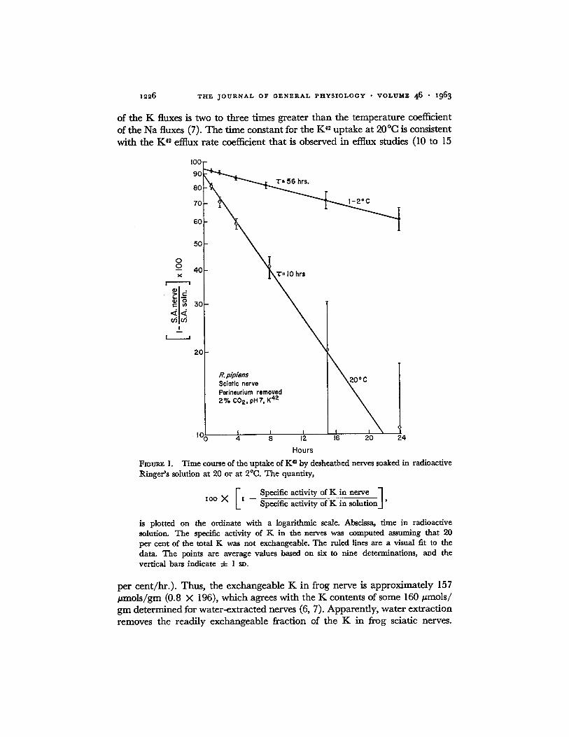
1226 THE JOURNAL OF GENERAL PHYSIOLOGY • VOLUME 46 • 1963
of the K fluxes is two to three times greater than the temperature coefficient of the Na fluxes (7). The time constant for the K 42 uptake at 20°C is consistent with the K 4~ effiux rate coefficient that is observed in efflux studies (I0 to 15
I00
90
80
70
60
50
0 0 - - 40
! I ]o u~u~
I
I
20
T= 56 hrs.
T= I0 hrs
R. pip/ens S c i a t i c n e r v e
Perineurium r e m o v e d 2% C02, pHT, K 42
I I I I / 2O 104 4 8 12 16 24
Hours
FIOUt~ 1. Time course of the uptake of K a by desheathed nerves soaked in radioactive Ringer's solution at 20 or at 2°C. The quantity,
I Specific activity of K in nerve ] lOG X x -- Specific activity of K in solution.]'
is plotted on the ordinate with a logarithmic scale. Abscissa, time in radioactive solution. The specific activity of K in the nerves was computed assuming that 90 per cent of the total K was not exchangeable. The ruled lines are a visual fit to the data. The points are average values based on six to nine determinations, and the vertical bars indicate 4- l sv.
per cent/hr.) . Thus, the exchangeable K in frog nerve is approximately 157 #mois /gm (0.8 X 196), which agrees with the K contents of some 160 #mols/ gin determined for water-extracted nerves (6, 7). Apparently, water extraction removes the readily exchangeable fraction of the K in frog sciatic nerves.
W. P. HURLBUT K Fluxes in Frog Nerve I227
About 5 per cent of the exchangeable K (8 ~mols/gm) exchanges rapidly in the cold and presumably represents extracellular K. A previous estimate of extracellular K, based on equations (1) and (2), was 6 #mols /gm (6).
Fig. 2 illustrates the effects on the K ~2 efflux rate coefficient that were pro- duced by the solutions used in a previous study of the Na fluxes (7). A K-free solution had little effect on the effiux rate coefficient, but a solution that con- tained 5 m u NaN3, a Ca-free solution, or a solution that contained 8.5 mM K caused a prompt and reversible increase in the effiux rate coefficient. It is not known whether the increase in the K 4~ effiux rate coefficient that was produced by NaN3 or by the Ca-free solution, was due to an increase in the K permeabili ty of the fibers, to a depolarization of the fibers, or to a combination of these factors. It also is possible that some of the increase in the K efflux that occurred in the Ca-free solution was caused by spontaneous activity in
T A B L E I
K C O N T E N T S OF D E S H E A T H E D F R O G S C I A T I C N E R V E S SOAKED F O R V A R I O U S P E R I O D S OF T I M E IN R I N G E R ' S S O L U T I O N
AT 20°C O R AT 2°C Nerves ext rac ted in HNOa.
Time in radioact ive solution, hrs. K conten t (/janols/gm dry) -4- s v
No. of nerves
I -8 15 24 1964-16 1774-14 1654-11
58 15 17
the nerve fibers. The effects of the solution with 8.5 mM K were probably due to depolarization of the membrane (14).
Fig. 3 shows the dependence of the initial rate of K 42 uptake on the con- centration of K in the bathing solution. These points were obtained with paired nerves; one member of each pair was soaked in standard Ringer 's solution (2 mM K), and its mate was soaked in a solution that contained K at the concentration indicated on the abscissa. The nerves were soaked in their respective solutions for 2 hours before they were transferred to radio- active solutions for 2 hours. The nerves were extracted in HNO~, and the intraceUular K 4. was estimated from equations (t) and (2). The data in Fig. 3 indicate that there are two components to the curve relating the rate of K 42 uptake to the external K concentration, one being proportional to the ex- ternal K concentration, the other showing saturation kinetics at low levels of extracellular K. If the linear portion of the curve is extrapolated to zero K concentration, the extrapolation passes close to the point at 2 m_~ K and intersects the ordinate at a level of 75 per cent, approximately. This indicates that when the external K concentration is 2 raM, the saturable component of the K influx accounts for about 75 per cent of the total K influx. The K influx into a nerve in Ringer's solution is about 20 #mols / (gm X hr.), which is equal to the Na effiux, approximately (7).

1228 T H E J O U R N A L O F G E N E R A L P H Y S I O L O G Y • V O L U M E 46 • 1963
Fig. 4 summarizes the changes in the initial rates of K 4~ uptake that oc- curred when nerves were soaked in various solutions and then were allowed to recover in standard Ringer's solution. Paired nerves were used exclusively; the control nerve was soaked in oxygenated Ringer's solution, while the ex- perimental nerve was soaked in one of the modified solutions. The symbols used in this figure refer to the conditions that prevailed during the treatment
20 20
r - I O
o
( 3 .
0 t--
o
(1) o 2c o
~ I0 N
A
2 mMK
I0
I I I I I I 5 ,5 7
C
NaN 3 off
5 mM NoN 3
20
I I | I I } 3 5 7
I0
B 2mMK
8.5 mM K
I I I I I 3 5 7
D I I .8 mM Ca
R. p/pleas Sci0tic nerve Perineurium removed pH 7, P04, 20°C
I I I I [ I 3 5 7
Hours
FIGUreS 2. Effects of various solutions on the rate coefficient for the effiux of K 4~. Ordi- nate, rate coefficient. Abscissa, time after beginning washout of K ~. All nerves were loaded with isotope by soaking about 4 hours at 20°C in Ringer's solution containing K ~. In each case, the initial washing solution was standard Ringer's fluid, and modi- fied Ringer's fluid was substituted at the time indicated by the first arrow and removed at the second. A, Effect of K-free Ringer's solution. B, Effect of a solution containing 8.5 m~ K. C, Effect of a solution containing 5 mM NaNv D, effect of a Ca-free solution.
period (0 to 5 hours). To obtain the recovery data (times greater than 5 hours), the nerves were first soaked for 5 hours in the appropriate experimental solu- tion and then allowed to recover in standard Ringer's solution. The time courses of the changes in the initial rates of K 4~ uptake were followed in the manner described for Na 2~ (7), except that, in most cases, the nerves were exposed to K 42 for 1 hour. The nerves were extracted in HNO3, and the intra- cellular K 42 was computed from equations (1) and (2). Anoxia and NaN~ reversibly depressed the rate of K 42 uptake, while Ca lack increased it about

W. P. HURLBUT If. Fluxes in Frog Nerve x2~9
20 per cent. After a nerve had soaked for a few hours in 5 mM NaNs, the K
influx was 4 to 5 #mols/(gm × hr.). Ouabain or dinitrophenol inhibited the rate of K 42 uptake about as strongly
as did 5 rnM NAN3. Soaking nerves for 5 hours in a solution that contained
20o Sciatic nerve 2 Perineurium removed / o
• B -s
150 8
==
IOC
9 '6
50
0 | I l I I I I I I I I 5 I0
K in bathing solution (rnM)
FzouR~ 3. Initial rate of K ~ uptake as a function of the K concentration of the bath- ing solution. Ordinate, initial rates of K a uptake of nerves in the experimental solution expressed as a percentage of the initial rates of K e uptake of control nerves in standard Ringer 's solution (2 mM K). Abscissa, K concentration of the bathing solution. Each point represents one pair of nerves. The ruled line is a visual fit to the data. The average value of the initial rate of K 42 uptake of the control nerves was 21.6 4- 3.6 #mols/(gr~ X hr.) (N = 19).
0.01 nee ouabain reduced their rates of K .2 uptake from 15.8 4- 3.5 #mols/ (gm × hr.) to 3.9 -4- 0.4 #mols/(gm × hr.) (N = 6); soaking them for 5 hours in 0.2 m u a-dinitrophenol reduced their rates of K 4. uptake from 20.1 -4- 0.4 ~mols/(gm X hr.) to 3.1 4- 0.4 #mols/(gm × hr.) (N = 4). During the 1st hour that nerves were exposed to 0.01 m_u ouabain their rates of K 4~ up- take were reduced by about 50 per cent, and 0.2 rnu a-dinitrophenol reduced the K influxes by about one-third during the first hour of exposure.
Approximately 75 per cent of the K influx, or 15 #mols/(gm × hr.), is due to active transport, since this fraction of the influx can be abolished by ouabain or by metabolic inhibition. This estimate of the size of the active

z 2 3 o T H E J O U R N A L O F G E N E R A L P H Y S I O L O G Y • V O L U M E 46 I963
c o m p o n e n t of the K influx is consistent with the da ta in Fig. 3 which show
tha t the saturable c o m p o n e n t of the K influx represents abou t 75 per cent of
the total influx in Ringer ' s solution. T h e passive c o m p o n e n t of the K influx
represents only 25 per cent of the total influx, or approx imate ly 5 #mol s /
(gin X hr.).
o Anoxia • 0 mM Ca
140 E] 0 mM K, OmM Co Z~ 5 mM NaN 3 x OmMK
o 120 • E o
_ I O 0 r- iD
~ 8o
- ~ 6 0
= 4 0
~ 2o n,-
R. pipiens Sc io t ic nerve Perineurium removed 2% CO 2,pH 7, 20°C
T r e a t m e n t , . , Recovery • I I O( I I I I I I I I I 5 I0
Hours
Fioum~ 4. Time course of the changes in the initial rates of K a uptake that occur when nerves are soaked in modified Ringer's solutions and then are allowed to recover in standard Ringer's solution. Ordinate, rates of K ~ uptake of experimental nerves ex- pressed as a percentage of the rates of K ~ uptake of control nerves in oxygenated Ringer's fluid. Abscissa, time since beginning exposure to the experimental solution. The sym- bols indicate the solution used during the treatment period. The points are averages of from three to six determinations. The average value of the deviations of the individual data from the means was -4- I0 per cent. In most cases, the nerves were exposed to the radioactive solution for 1 hour. However, to obtain the data in 0 mM Ca, to obtain the point, ×, plotted at 9 hours, and to obtain the point, ix, plotted at 9.5 hours, the nerves were soaked for 2 hours in radioactive solutions. In each instance, the points are plotted at the midpoint of the period during which the nerves were soaked in the radioactive solution. The average value of the rate of K ~ uptake by the control nerves was 21.8 ± 5,0 #mols/(gm × hr.) (N = 60).
W h e n nerves are re turned to Ringer ' s solution after they have been soaked
for several hours in one of the exper imental solutions shown in Fig. 4, their rates of K 42 uptake are increased slightly above the control rates. T h e K 42 efflux experiments indicate tha t the K 42 eftlux rate coefficient is no rma l dur ing
the recovery periods (Fig. 2). Since the K contents of the nerves are less t han no rma l at these times, the K efflux also is less t han normal . Therefore, the
recover o f the K contents of the nerves is due to a reduced K efflux as well

W. P. HURI.,.B'UT K Fluxes in Frog Nerve I23I
as to an increased K influx. It is useful to estimate the relative contribution of these two factors to the recovery of the K contents.
Consider first the recovery, in Ringer's solution, of a nerve that has been soaked for 5 hours in a solution containing 5 mM NaNs. During the 1st hour of recovery the K influx is less than normal by 2 #mols/(gm X hr.); during the 2nd hour of recovery, the K influx is greater than normal by 4/zmols/ (gm × hr.); during the 4th and 5th hours of recovery, the K influx is greater than normal by 0.6 #mols/(gm X hr.). If the K influx during the 3rd hour of recovery is assumed to be increased above normal by 3 #mols/gm × hr.), then in 5 hours the net K uptake due to the increased K influx is about 6 #mols/gm ( - 2 + 4 + 3 + 2 X 0.6). The K contents of nerves that have been soaked for 5 hours in 5 mM NaN3 change from 83 -4- 12 #mols/(gm(N = 36) to 134 -4- 18 #mols/gm (N = 15) during 4 to 6 hours of recovery in Ringer's solution. Thus, the net K uptake that can be attributed to the increased K influx is only one-eighth of the total net K uptake.
Consider next the recovery of nerves that have been soaked for 2.5 hours in a K- and Ca-free solution. During the Ist hour of recovery the K influx is increased above normal by about 6 #mols/(gm X hr.); during the 5th hour of recovery, the K influx is increased above normal by 4 #mols/(grn X hr.). If the average increase in the K influx is assumed to be 5 #mols/(gm X hr.), then in 5 hours the net K uptake that is due to the increased K influx is about 25 #mols/gm. The K contents of nerves that have been soaked for 9.5 hours in a K- and Ca-free solution change from 67 4- 16 #mols/gm (N = 99) to 138 4- 7 #mols/gm (iV = 7) during 5 hours of recovery in Ringer's solution. In this case, the net K uptake that is due to the increased K influx is about one-third of the total net K uptake.
When nerves are recovering from a soaking in a K-free solution, most of the net K uptake is due to the increase in the K influx. During the Ist hour of recovery the K influx is increased above normal by I0 #mols/(gm X hr.); during the 2nd hour the increase is 4 #mols/(grn X hr.); and during the 4th and 5th hours the increase is 2 #mols/(gm X hr.). If one assumes that during the 3rd hour of recovery the K influx is greater than normal by 3 #mols/ (gm × hr.), then in 5 hours the net K uptake produced by the increased K influx is about 21 #mols/gm [10 + 4 + 3 + 2 × 2]. This is 60 per cent of the measured net K uptake of 35 #mols/gm.
These results indicate that for small displacements of the ionic distribution, the reaccumulation of K may be due primarily to the enhanced K influx. For large displacements of the ionic distribution, however, the low K efflux is primarily responsible for the net uptake of K. It is also clear that the mag- nitudes of the increases in the K influxes are not proportional to the extents of the displacements of the ionic contents from the resting values, in contrast to the case with the Na effiuxes (7).
1232 THE JOURNAL OF GENERAL PHYSIOLOGY • VOLUME 4 6 • 1963
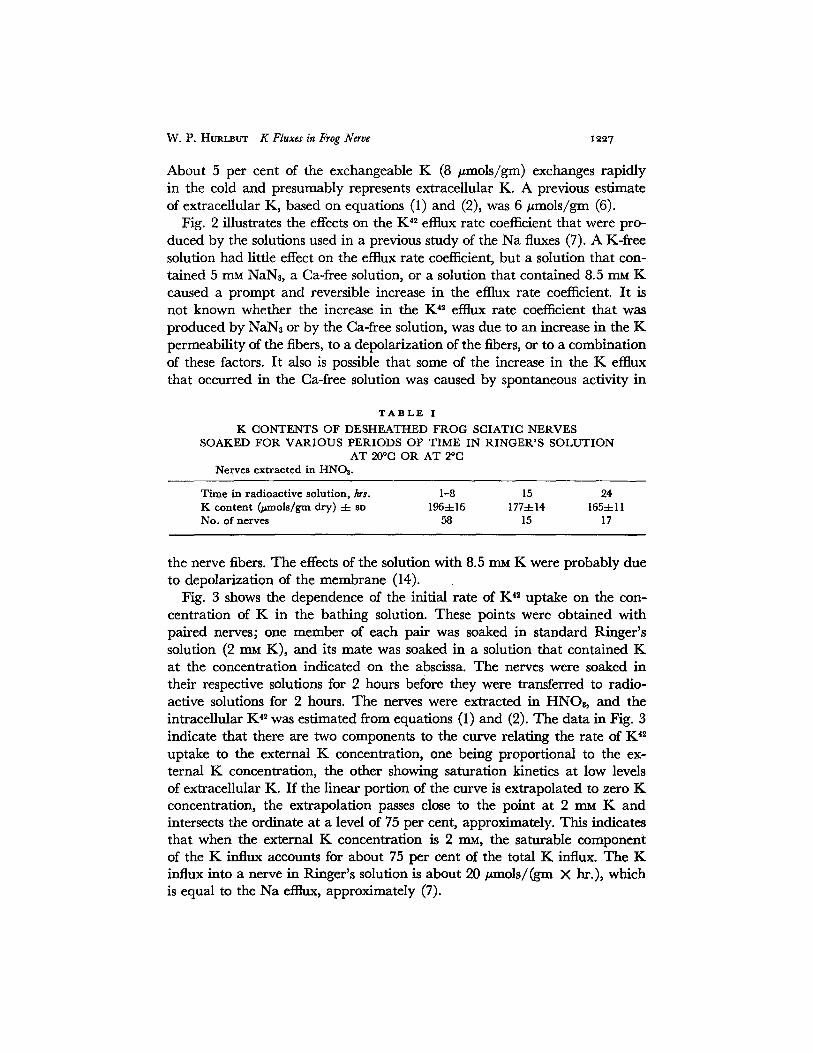
It is important to determine whether, in recovering nerves, the active K influxes are directly related to the active Na effiuxes. The active K influx in resting nerve was estimated to be 15 #mols/(gm X hr.). If it is assumed that the increases in the K influxes that occur during the recovery periods are due to increased rates of active transport, then the maximum active K influxes, which occur during the first few hours of the recovery period, can be estimated readily. When nerves are recovering after they have been soaked for 5 hours in a K-free solution or after they have been soaked for 5 hours in a solution that contains 5 mu NaNs, the maximum active K influxes are about 25 #mols/(gm X hr.) and 19 #mols/(gm X hr.), respectively. When nerves are recovering after they have been soaked for 2.5 hours or for 5 hours in a K- and Ca-free solution, the maximum active K influxes are about 21 #mols/ (gm X hr.) and 18/zmols/(gm X hr.), respectively. The Na effluxes under these conditions have been estimated to be 40, 60, 77, and 96/zmols/(gm X hr.), respectively (7). Approximately half of the Na efflux may be inhibited by 5 m_u NaN3 (7), so the active Na effluxes under these conditions are roughly 20, 30, 39, and 48/zmols/(gm X hr.), respectively.
There is no apparent correlation between these estimates of the active K influxes and of the active Na effluxes, and the results suggest that the active fluxes of these ions are not coupled in a stoichiometric manner. This indication of a lack of coupling between these fluxes is not particularly significant, since the active K influx could be accounted for by assuming that only part of the active Na efttux is coupled to it. If conditions could be found under which the active K influx exceeded the active Na efflux, the case for the absence of coupling between these active fluxes would be put on a firmer basis. To do this, experiments were carried out with Na-deficient solutions.
Previous results (7) have shown that in recovering nerves the time constant for the Na ~2 effiux is about 2 hours, which is equivalent to a half-time of 1.4 hours. If the K influx were tightly coupled to all of the Na effiux, then the K content of a nerve should recover with the half-time for Na 2~ exchange. If the K influx were not in- creased at all during the recovery time, then the K content of a nerve should recover with the half-time of 5 to 7 hours that is characteristic of K 4~ exchange. Following a 2.5 hour soaking in a K- and Ca-free solution, or a 5 hour soaking in a solution with 5 mM NAN3, the K content of a nerve recovers with a half-time of 3 to 4 hours (6, 7). When nerves recover after having been soaked for 5 hours in a K-free solution, or after having been stimulated for I hour at 50 volleys/see., the half-time for the recovery of the K contents is about 2 hours (2, 7), which is comparable to the half-time for Na 2~ exchange. This shows that when large changes in the ionic distribution of nerve have occurred, all of the Na efflux cannot be linked to inward K movement, and it shows that the recovery of the K contents is due to an increased influx and to a reduced effiux.

W. P. HURI_mUT K Fluxes in Frog Nerve xe33
Na-Deficient Solutions N e r v e s w e r e s o a k e d for p e r i o d s u p to 10 h o u r s
in s o l u t i o n s in w h i c h p a r t o f t h e n o r m a l c o m p l e m e n t o f N a C I h a d b e e n r e -
p l a c e d b y i s o s m o t i c q u a n t i t i e s o f c h o l i n e c h l o r i d e , sucrose , o r IAC1. A t t h e
40
E e E § -.~2o
~t3
: : k i t
25
e. p/piens T Sciatic nerve ,I Perineurium removed ~ J" 20 °c., p H T , ~ -
,
A
30 dO 910 120
B 20 ~ Choline 'I
" ~ 15
g: 5
o 6'0 ' 120
Na concentration of bathing solution (mM)
FIOURE 5. Steady-state values of the Na contents and initial rates of K ~ uptake of nerves soaked in Na-deficient solutions. Ordinate, A, Na content, B, K ~ taken up in 1 hour. Abscissa, concentration of Na in the bathing solution. Mean values -4- sD. In A and the uppermost curve in B, the points at Na concentrations of 116 mu and 30 mM are based on twenty-five or more determinations; other points are averages of three to six measurements. The points at zero external Na were obtained using solutions buffered with trls. For the upper curve in B, choline chloride or sucrose was used as a substitute for NaC1; and for the middle curve, LiCI was used. Choline was used as the Na sub- stitute to obtain the point in 5 m.M NaN8 at an external Na concentration of 30 raM. The curves are a visual fit to the data.
c o n c l u s i o n o f e a c h e x p e r i m e n t , t h e n e r v e s w e r e w a s h e d for 1 h o u r a t 2 ° C
in s o l u t i o n W , a n d t h e n w e r e e x t r a c t e d for ana lys i s . T h e N a c o n t e n t s o f t h e
n e r v e s a n d t h e i n i t i a l r a t e s o f K ** u p t a k e fel l to n e w s t e a d y - s t a t e v a l u e s in a
few hou r s . T h e s e s t e a d y - s t a t e v a l u e s a r e p r e s e n t e d in F ig . 5. W h e n c h o l i n e
c h l o r i d e o r suc rose w a s s u b s t i t u t e d fo r 75 p e r c e n t o f t h e NaC1 in t h e b a t h i n g
s o l u t i o n , t h e K c o n t e n t s of t h e n e r v e s fel l s l i g h t l y : 10 4- 8 # m o l s / g m in 9

x234 T H E J O U R N A L O F G E N E R A L P H Y S I O L O G Y • V O L U M E 4 6 • i963
hours, (N = 13). When Li was substituted for Na in the bathing solution, the K contents of the nerves fell markedly (Table III) . It is clear from Fig. 5 that the rate of K 4~ uptake is related to the Na concentration of the bathing solution. Shanes has observed a similar effect with toad nerves (12). The question to be decided is: Is the depression of the rate of K 4~ uptake due to a reduction of the Na effiux or is it due to another action of Na?
The Na content of a nerve varies linearly with the concentration of Na in the external solution (Fig. 5). The small amount of indiffusible Na in- dicated in Fig. 5 may be due to contamination of the nerve extracts with extraneous Na. The Na ~2 effiux rate coefficient is not affected by Na-deficient solutions. Therefore, in the steady state, the Na efflux, which is given by the product of the Na '~ efflux rate coefficient and the Na content of a nerve, should be linearly related to the Na concentration of the bathing solution. The Na fluxes in a nerve in Ringer's solution are about 20 pmols/(gm X hr.) (7), and the normal K fluxes are also about 20 #mols / (gm × hr.) (Figs. 3 to 5). When a nerve is soaked in a solution in which 75 per cent of the NaC1 has been replaced by sucrose or choline chloride, the Na fluxes should fall to about 5 pmols / (gm X hr.). Under these conditions, the K influx is 15 /~mols/(gm × hr.), which is considerably higher than the Na effiux.
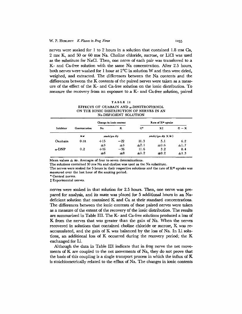
The active component of the K influx is also higher than the NaNt-sensitive component of the Na efflux. Five millimolar NaN8 inhibits about 50 per cent of the Na efflux from a nerve that is soaking in a solution containing 30 m_M Na. The NaN3-sensitive component of the Na efflux is, therefore, 2 to 3 pmols/ .(gin X hr.). Under these conditions, 5 rnM Na.Na reduces the K influx by about 9/~mols/(gm X hr.) (Fig. 5); 0.2 mM dinitrophenol reduces the K influx by about 8 pmols / (gm X hr.) (Table I I ) ; and 0.01 rnu ouabain re- duces the K influx by about 6 pmols / (gm × hr.) (Table II). These three estimates of the size of the active K influx are two to three times greater than the estimate of the size of the NaNt-sensitive component of the Na efflux. Therefore, it is unlikely that the effects of Na-deficient solutions on the active K influx are directly related to the decline in the Na efflux that occurs in such solutions.
Thus, under some conditions, the active Na effluxes in frog nerve exceed the active K influxes, and under other conditions the active K influxes exceed the active Na effiuxes. In the steady state, the relation between the active Na efflux and the external Na concentration is linear, but the relation between the active K influx and the external Na concentration is hyperbolic. These results show that the active K influx is not stoichiometrically related to the active Na efflux.
The evidence for a lack of coupling between the Na efflux and the K influx would be strengthened if one could demonstrate an accumulation of K in exchange for an ion other than Na. Experiments were done as follows. Paired
W . P. HURLBUT 1~ Fluxes in Frog Nerve I,:,35
nerves were soaked for 1 to 2 hours in a solution that contained 1.8 rnM Ca, 2 mM K, and 30 or 60 mM Na. Choline chloride, sucrose, or LiC1 was used as the substitute for NAG1. Then, one nerve of each pair was transferred to a K- and Ca-free solution with the same Na concentration. After 2.5 hours, both nerves were washed for 1 hour at 2 °C in solution W and then were dried, weighed, and extracted. The differences between the Na contents and the differences between the K contents of the paired nerves were taken as a meas- ure of the effect of the K- and Ca-free solution on the ionic distribution. To measure the recovery from an exposure to a K- and Ca-free solution, paired
T A B L E I I
E F F E C T S O F O U A B A I N A N D ~ x - D I N I T R O F H E N O L O N T H E I O N I C D I S T R I B U T I O N O F N E R V E S I N A N
N a - D E F I C I E N T S O L U T I O N
Inhibitor
Change in ionic content Rate of Ka uptake
Concentration Na K C* X~ G -- X
rajt ~mds/gm dry ~mols/(gm d,~ × hr.)
O u a b a i n 0.01 -4-15 --22 11.3 5.1 6 . 2 ± 3 ± 3 ± 2 . 1 4 -0 .6 ± 1.7
a - D N P 0 .2 -{-16 - -36 11.6 3 .2 8 .4 ± 6 ± 8 4 -1 .2 -4-0.2 ± 1 . 3
M e a n v a l u e s ± 8D. A v e r a g e s of f ou r to s e v e n d e t e r m i n a t i o n s . T h e so lu t ions c o n t a i n e d 30 mM N a a n d cho l i ne was u s e d as t h e N a s u b s t i t u t e . T h e ne rve s were soaked for 5 h o u r s in t he i r r e spec t ive so lu t ions a n d t he r a t c o f K 4s u p t a k e was m e a s u r e d over t h e las t h o u r o f t h e soak ing pe r iod . * C o n t r o l ne rves .
E x p e r i m e n t a l ne rves .
nerves were soaked in that solution for 2.5 hours. Then, one nerve was pre- pared for analysis, and its mate was placed for 5 additional hours in an Na- deficient solution that contained K and Ca at their standard concentrations. The differences between the ionic contents of these paired nerves were taken as a measure of the extent of the recovery of the ionic distribution. The results are summarized in Table III . The K- and Ca-free solutions produced a loss of K from the nerves that was greater than the gain of Na. When the nerves recovered in solutions that contained choline chloride or sucrose, K was re- accumulated, and the gain of K was balanced by the loss of Na. In Li solu- tions, an additional loss of K occurred during the recovery period; the K exchanged for Li.
Although the data in Table I I I indicate that in frog nerve the net move- ments of K are coupled to the net movements of Na, they do not prove that the basis of this coupling is a single transport process in which the influx of K is stoichiometrically related to the efflux of Na. The changes in ionic contents
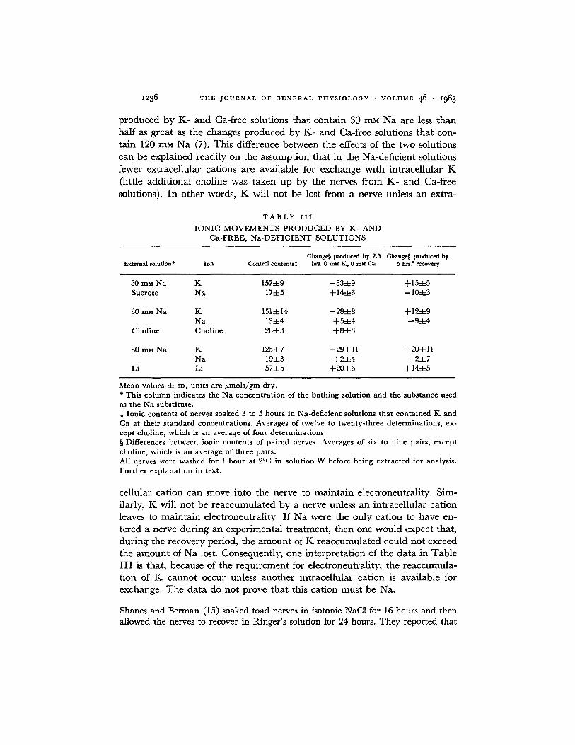
I236 T H E J O U R N A L O F G E N E R A L P H Y S I O L O G Y • V O L U M E 4 6 • i963
p roduced by K - and Ca-free solutions tha t conta in 30 mM N a are less t han half as great as the changes p roduced by K - and Ca-free solutions tha t con-
tain 120 mM N a (7). This difference between the effects of the two solutions
can be explained readi ly on the assumption tha t in the Na-deficient solutions
fewer extracellular cations are available for exchange wi th intracel lular K
(little addi t ional choline was taken up by the nerves f rom K - and Ca-free
solutions). I n other words, K will not be lost f rom a nerve unless an extra-
T A B L E I I I
IONIC MOVEMENTS PRODUCED BY K- AND Ca-FREE, Na-DEFICIENT SOLUTIONS
Change§ produced by 2.5 Change§ produced by External solution* Ion Control contents~ hrs. 0 mu K, 0 mM Ca 5 hrs.' recovery
30 mM Na K 157-4-9 --334-9 +154-5 Sucrose Na 174-5 +144-3 -- 104-3
30 mM Na K 1514-14 --284-8 +124-9 Na 134-4 +54-4 --9-4-4
Choline Choline 284-3 +84-3
60 mM Na K 1254-7 --294-11 --204-11 Na 194-3 +24-4 --24-7
Li Li 574-5 +204-6 + 144-5
Mean values -4- SD; units are #mols/gm dry. * This column indicates the Na concentration of the bathing solution and the substance used as the Na substitute.
Ionic contents of nerves soaked 3 to 5 hours in Na-deficient solutions that contained K and Ca at their standard concentrations. Averages of twelve to twenty-three determinations, ex- cept choline, which is an average of four determinations. § Differences between ionic contents of paired nerves. Averages of six to nine pairs, except choline, which is an average of three pairs. All nerves were washed for 1 hour at 2°C in solution W before being extracted for analysis. Further explanation in text.
cellular cat ion can move into the nerve to ma in ta in electroneutral i ty. Sim-
ilarly, K will not be r eaccumula ted by a nerve unless an intracel lular cat ion leaves to ma in ta in electroneutral i ty. I f N a were the only cat ion to have en-
tered a nerve dur ing an exper imental t reatment , then one would expect that,
dur ing the recovery period, the a m o u n t of K r eaccumula t ed could not exceed the a m o u n t of N a lost. Consequent ly , one in terpre ta t ion of the da ta in Tab le
I I I is that, because of the requ i rement for electroneutral i ty, the r eaccumula - t ion of K canno t occur unless ano ther intracel lular cat ion is available for
exchange. The da ta do not prove tha t this cat ion must be Na.
Shanes and Berman (15) soaked toad nerves in isotonic NaC1 for 16 hours and then allowed the nerves to recover in Ringer's solution for 24 hours. They reported that
W. P. HURLBUT If Fluxes in Frog Nerve i237
K was taken up by the nerves during the recovery period but that no Na was lost. The experiments of Shanes and Berman were much longer in duration than those reported here. It has been my experience that for experiments lasting 10 hours or less, the total cation content of frog nerve remains nearly constant.
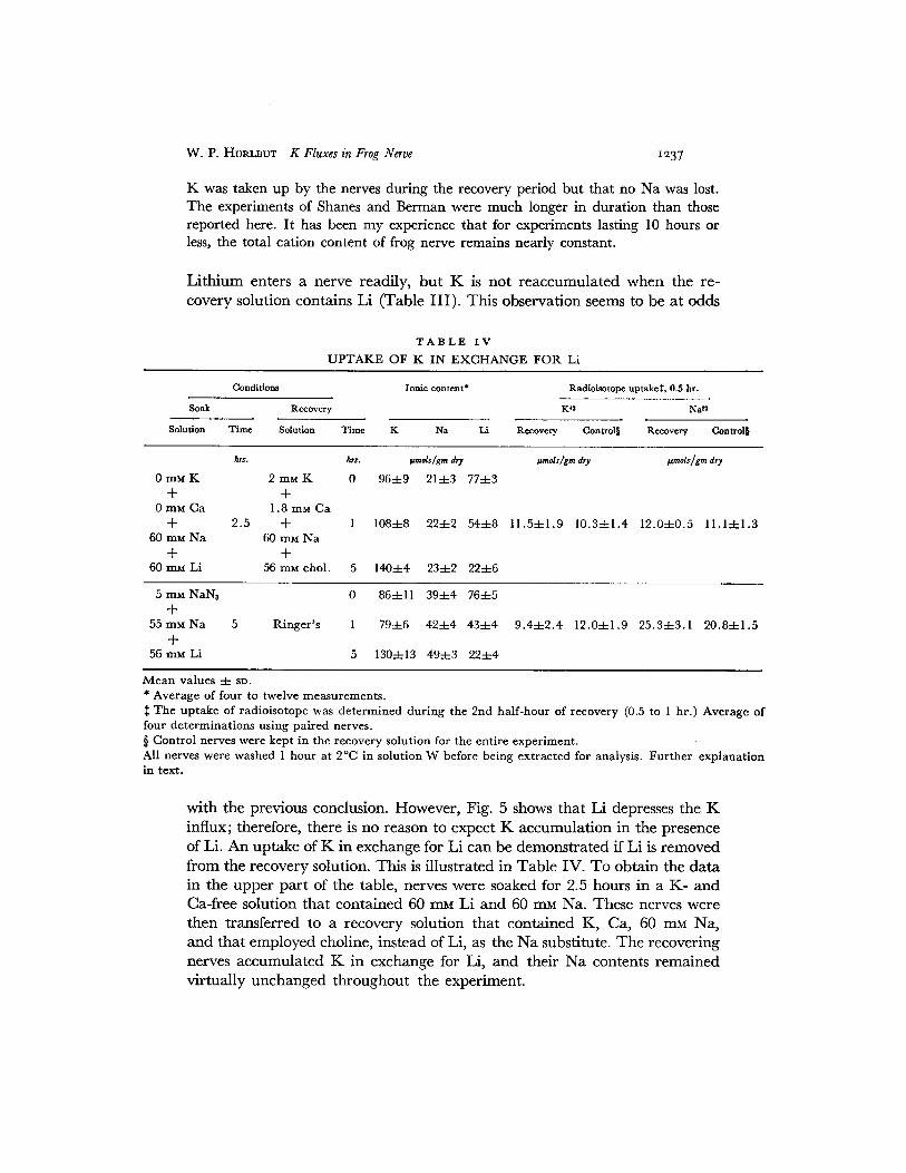
Li thium enters a nerve readily, but K is not reaccumulated when the re- covery solution contains Li (Table III) . This observation seems to be at odds
T A B L E I V
U P T A K E O F K I N E X C H A N G E FOR. Li
Conditions Ionic content* Radioisotope uptakeS, 0.5 hr.
Soak Recovery Ke Na ~
Solution Time Solution Time K Na Li Recovery Control§ Recovery Control§
hrs. hrs. ~mols/gm dry
0 mM K 2 mM K 0 964-9 214-3 774-3 + +
0 mM Ca 1.8 mM C a -b 2 .5 -t- 1 1084-8 224-2 544-8
60 mM N a 60 mM N a + +
60 mM Li 56 mM chol . 5 1404-4 234-2 224-6
~unols/gm dry I.,mols/gm dry
11.54-1 .9 10 .34-1 .4 12 .04-0 .5 11 .14-1 .3
5 mM NaN3 0 864-11 394-4 764-5 +
55 mM N a 5 R i n g e r ' s 1 794-6 424-4 434-4 +
56 mM Li 5 1304-13 494-3 224-4
9.4::t:2.4 12 .04-1 .9 25 .34-3 .1 20 .84 -1 .5
M e a n va l ue s 4- SD. * A v e r a g e of fou r to twelve m e a s u r e m e n t s .
T h e u p t a k e of r ad io i so tope was d e t e r m i n e d d u r i n g the 2nd h a l f - h o u r of r ecovery (0.5 to 1 hr . ) A v e r a g e of four d e t e r m i n a t i o n s u s i n g pa i r ed nerves . § Con t ro l ne rves were kep t in t he r ecove ry so lu t ion for t he en t i re e x p e r i m e n t . All nerves were w a s h e d 1 h o u r at 2°C in so lu t ion W before be ing e x t r a c t e d for analys is . F u r t h e r e x p l a n a t i o n in text .
with the previous conclusion. However, Fig. 5 shows that Li depresses the K influx; therefore, there is no reason to expect K accumulation in the presence of Li. An uptake of K in exchange for Li can be demonstrated if Li is removed from the recovery solution. This is illustrated in Table IV. To obtain the data in the upper part of the table, nerves were soaked for 2.5 hours in a K- and Ca-free solution that contained 60 mM Li and 60 mM Na. These nerves were then transferred to a recovery solution that contained K, Ca, 60 mM Na, and that employed choline, instead of Li, as the Na substitute. The recovering nerves accumulated K in exchange for Li, and their Na contents remained virtually unchanged throughout the experiment.

1238 T H E J O U R N A L O F G E N E R A L P H Y S I O L O G Y • V O L U M E 46 • I963
Keynes and Swan observed a similar phenomenon with frog muscle (10). These workers suggested that extracellular Na exchanged passively for intra- cellular Li, and that the Na then was extruded in exchange for extracellular K. A similar explanation of the data in Table IV cannot be ruled out entirely. However, the data in Table IV indicate that in the recovering nerves the active K influxes exceeded the active Na effluxes, and it seems unlikely that all of the K uptake was coupled to Na extrusion. The data on the right side of Table IV show the amounts of Na 22 and K 42 that were taken up by the nerves during the second half-hour interval of the recovery period. This interval was chosen, rather than the first half-hour, to enable Li, which in- hibits K 4~ uptake, to be washed from the extracellular spaces. The uptakes of Na 2~ and K 42 were about 12 #mols/gm. These nerves had been washed for 1 hour at 2°C in solution W. In nerves equilibrated with standard Ringer's solution and then washed for 1 hour in solution W, the intracellular Na 2~ contents are overestimated by 10/zmols/gm (7). In the present instance, the nerves were soaked in solutions that contained half the normal complement of Na, and the intracellular Na 22 contents should have been overestimated by 5/zmols/gm. Therefore, the amount of Na 22 that entered the fibers during the test period was 7 /zmols/gm, which corresponds to an Na influx of 14 #mols/(gm × hr.). Since the Na contents of the nerves were in a steady state (Table IV, top), the Na influx should have been equal to the Na effiux. The Na 22 effiux rate coefficient is about 60 per cent/hr. (7), and the Na effiux was, therefore, 10 #mols / (gm X hr.) [ ( 2 2 - 5 ) X 0.6], which agrees with the estimate for the Na influx.
The K influx in these nerves was about 23 ~mols/(gm X hr.). Approxi- mately 75 per cent of this influx, or 18 #mols / (gm X hr.), should have been active K influx. If the active Na effiux was 50 per cent of the total Na effiux, or about 5 to 7/zmols / (gm X hr.), then the active K influx was clearly the larger of the two. On the other hand, if all the Na efflux were due to active transport, then the discrepancy between the sizes of the active K influx and the active Na efflux may not have been significant.
Nerves recovering from metabolic inhibition also could accumulate K in exchange for Li (Table IV, bottom). When nerves had been soaked for several hours in a solution in which half of the NaC1 had been replaced by LiC1, the Na contents of the nerves were about 20 #mols /gm (Fig. 5). When the soaking solution also contained 5 mM NAN3, the Na contents of the nerves rose to 39 #mols /gm in 5 hours (Table IV, bottom), a value close to the value of the Na contents of nerves in Ringer's solution. Therefore, to demonstrate that such nerves could accumulate K in exchange for Li, although net Na movements were small, the nerves were allowed to recover in standard Ringer's solution. The data in Table IV indicate clearly that the nerves ac- cumulated K in exchange for Li. The Na contents of the recovering nerves

W. P. HUR~Z~UT K Fluxes in Frog Nerve I~39
also increased by about 10/zmols/gm. Only half of this increase represented a change in intracellular Na, for the Na contents of the extracellular spaces should have increased by 5/zmols/gm when the Na concentration of the bath- ing solution was changed from 60 m_~ to 116 m~ (7). The explanation sug- gested by Keynes and Swan for the reaccumulation of K by these nerves can- not be excluded because the active Na effiuxes and the active K influxes were equal, approximately.
One explanation of the data in Table IV is that Li is substituting for Na in the ionic transport system, but the data in Table V argue against such an interpretation. Nerves were soaked for 4 hours in a solution in which all of
T A B L E V
E F F E C T O F N a O N K ~2 U P T A K E BY L i - L O A D E D N E R V E S
Concentration of recovery solution
Na Li Choline Rate of K 4~ uptake Li content
r a m r a m r a m uraols/gm dry X hr #mols]gm dry
0 0 116 2.9:1:0.5 57q-4 0 30 86 3.3-4-0.7 90-4-4
30 30 56 I0.1 "4-3.5 74:k::8 30 0 86 15.4:t::4.6 46:t:6 90 30 0 11.9-4-0.9 70-1-3
Tr i s buffer . E a c h so lu t ion c o n t a i n e d 1.8 m ~ CaCI2 a n d 2 m ~ KC1. E a c h n u m b e r is a n a v e r a g e (q-SD) of the resu l t s o f t h r ee m e a s u r e m e n t s u s i n g ne rves f r om differ- e n t frogs. Al l ne rves were w a s h e d 1 h o u r a t 2°C in so lu t ion W before b e i n g e x t r a c t e d for ana lys i s . F u r t h e r e x p l a n a t i o n in t ex t .
the Na had been replaced by Li. They were then transt~rred for I hour to one of the solutions indicated in the first column of Table V, and then the initial rate of K ~2 uptake was measured in this solution. It is clear that the uptake of K 42 required Na in the external solution, and that Li could not substitute for Na in this regard. If the data in the fourth row of Table V are compared with the data in Fig. 5, one sees that Li in the nerve does not reduce the K 4~ uptake. The inhibitory action of Li appears to be due to its presence in the bathing solution.
When a nerve is soaking in a solution in which 75 per cent of the NaC1 has been replaced b y sucrose or choline chloride, the K influx exceeds the Na effiux. Since the K content of a nerve changes little under these conditions, it seems likely that K in the bathing solution is exchanging directly for K within the fibers. Support for this interpretation is furnished by the experi- ments shown in Fig. 6. Fig. 6A shows that the K 4~ effiux rate coefficient is not changed greatly when 75 per cent of the NaC1 in Ringer's solution is replaced by an isosmotic quanti ty of sucrose. Similar results are obtained if the Na is
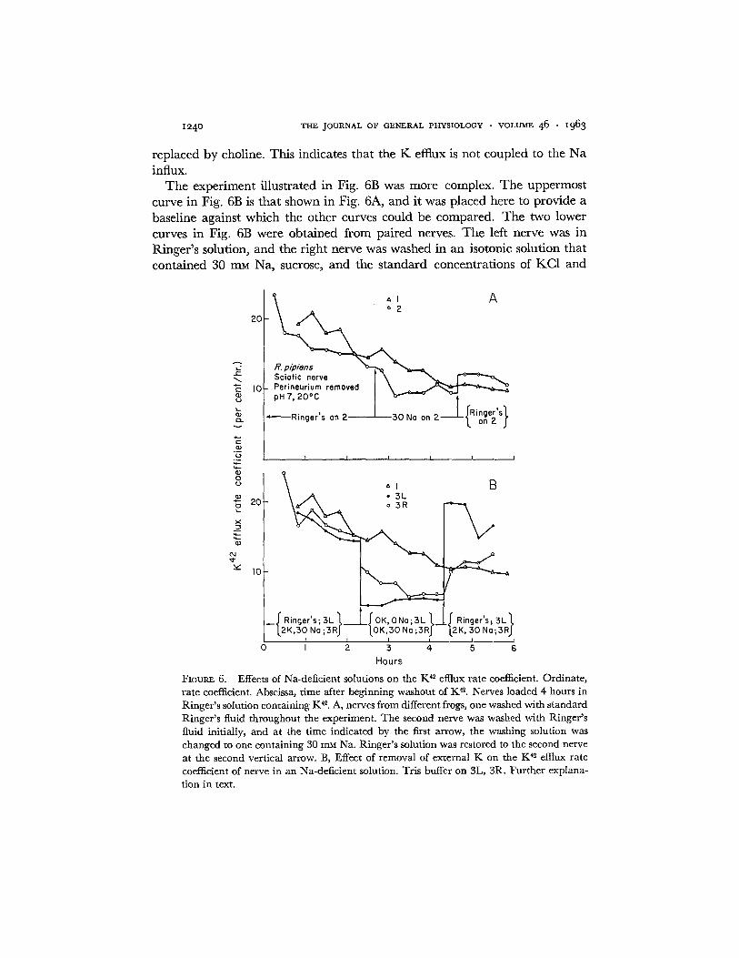
z~4o THE JOURNAL OF GENERAL PHYSIOLOGY • VOLUME 46 • z963
replaced by choline. This indicates that the K efflux is not coupled to the Na influx.
The experiment illustrated in Fig. 6B was more complex. The uppermost curve in Fig. 6B is that shown in Fig. 6A, and it was placed here to provide a baseline against which the other curves could be compared. The two lower curves in Fig. 6B were obtained from paired nerves. The left nerve was in Ringer's solution, and the right nerve was washed in an isotonic solution that contained 30 m ~ Na, sucrose, and the standard concentrations of KC1 and
• o 2
20
. . Solo , i t nerve l \ - "E I0 P e r i n e u r i u m r e m o v e d I \ q) ,.a pH 7, 20°C | b " " - ~ '~
, , Ringer s on 2 ' 30 Na on 2 on 2
*E o,} "~ I I I i i I
q~ o c) '~1 B
• 3L
X: 10
12K,30 No ;3RJ IOK,30 No ;3RJ "L2K, 30 No;3RJ I I I i I I
0 I 2 3 4 5 6
Hours
FIOUR~ 6. Effects of Na-deficient solutions on the K e efflux rate coefficient. Ordinate , ra te coefficient. Abscissa, t ime after beginning washout of K ~. Nerves loaded 4 hours in Ringer ' s solution conta ining Ke. A, nerves from different frogs, one washed with s tandard Ringer ' s fluid th roughout the experiment. T h e second nerve was washed with Ringer ' s fluid initially, and at the t ime indicated by the first arrow, the washing solution was changed to one conta ining 30 mM Na. Ringer ' s solution was restored to the second nerve at the second vert ical arrow. B, Effect of removal of external K on the K 42 efttux rate coefficient of nerve in an Na-deficlent solution. Tris buffer on 3L, 3R. Fur the r explana- t ion in text.

W. P. HURLBUT K Fluxes in Frog Nerve i241
CaC12. At a time indicated by the first vertical arrow, the KC1 was removed from the Na-deficient solution that bathed the right nerve, and an isotonic solution that contained only sucrose and CaCI~ was placed on the left nerve. The original solutions were replaced at the time indicated by the second vertical arrow. When the KC1 was removed from the Na-deficient solution that bathed the right nerve, the K 4~ efflux rate coefficient fell about 40 per cent in an hour. Measurements of the membrane potential were not made, but this decline in the K 42 etttux rate coefficient may have been accompanied by an increase in the membrane potential.
When a nerve is soaking in standard Ringer's solution, removal of the K from the solution has little effect on the K 4~ effiux rate coefficient (Fig. 2), and the membrane potential increases only a few millivolts (9). Presumably, the small effect of a K-free solution on the K 4~ efflux rate coefficient, and on the membrane potential, is due to the fact that when a nerve is put into a K-free Ringer's solution the Na influx increases (7). When K is removed from an Na-deficient solution in which a nerve is bathing, the Na influx increases also, but the increase in the Na influx is much less than that observed when the K is removed from Ringer's solution.
To measure this, four pairs of nerves were used; the experimental member of each pair was soaked for 4 hours in a K-free solution that contained 30 mM Na and choline chloride, while the control nerve was soaked in a similar solu- tion that contained 2 mM K. Then the rates of Na ~ uptake were measured. The amount of Na ~2 taken up in 0.5 hour by the experimental nerves exceeded the amount taken up by the control nerves by 6.4 4- 0.4 #mols/gm.
When this experiment is carried out with standard Ringer's solution and K-free Ringer's solution, the increase in the amount of Na 22 taken up in 0.5 hour by the experimental nerves is 16.9 4- 4.9 #mols/gm (N = 6) (7). This is two to three times larger than the increase that occurs in the Na-deficient solution. This suggests that the reason the K 42 efflux rate coefficient declines when K is removed from an Na-deficient solution is that the Na influx does not increase sufficiently to enable the nerve to gain Na at a rate equal to the original K efflux. Therefore, the K efflux must decline to maintain the elec- troneutrality of the axoplasm (assuming that anions do not move). The nerve fibers should hyperpolarize if, as seems to be the case (14), the membrane potential is a major part of the driving force for K ions in the membrane.
Eventually, the rate coefficient of the right nerve (Fig. 6B), which was bathed in a K-free solution that contained 30 mM Na, attained a value only slightly higher than the rate coefficient of the left nerve, which was in a K- and Na-free solution. This indicates that the residual K efflux from the right nerve was not exchanging for extracellular Na and may have been accom- panied by an intracellular anion.

THE JOURNAL OF ClgNIgRAL PHYSlOLOOY • VOLUME 46 • ~963
D I S C U S S I O N
Ussing (19) has derived a relation between the influx and the efflux of a monovalent cation that crosses a ceil membrane solely under the influence of its own concentration gradient and the electric field. According to this rela- tion,
M,e/MoF = (ao/a~) exp (FV/RT) (a)
where: M ~ = the passive influx of the ion Mo~ = the passive efflux of the ion ao = the activity of the ion in the external solution a~ = the activity of the ion in the axoplasm V -- the membrane potential in volts and R, T, and F have their
conventional meanings. It is useful to apply this relation to the Na and the K fluxes in resting frog
nerve at 20°C to estimate the magnitude of the passive component of the K influx and the passive component of the Na efflux. The intracellular Na and K contents of frog nerves are about 40/~mols/gm and 160/zmols/gm, respec- tively. The intracellular water content of frog and toad nerves has been es- t imated to be 1.1 to 1.5 gm H~O/gm (6, 15). The mean value of 1.3 gin/ gm (dry) will be used for calculation. The intracellular Na and K concentra- tions, therefore, are 31 mM and 123 mM, respectively. When the external K concentration is 2 mM, the resting membrane potential of frog fibers is ap- proximately 71 mv (8). If the activities of the ions are replaced by the con- centrations, and if the Na influx and the K efftux of 20 #mols/(gm × hr.) are assumed to be passive, then the values calculated for the passive Na efltux and for the passive K influx are 0.3/~mol/(gm X hr.) and 5.3/zmols/(gm X hr.), respectively. This computed value for the passive K influx agrees with the K influx measured in nerves soaked for several hours in a solution that contained 5 r r~ NAN,, 0.2 mM a-dinitrophenol, or 0.01 mM ouabain, and with the size of the linear component of the K influxes in Fig. 3. Thus, four independent estimates indicate that for a nerve in Ringer's solution about 75 per cent of the K influx, or 15 ~mols/(gm X hr.), is due to active transport. When a nerve is soaking in a solution that contains 30 m_u Na, the K influx is 11 to 15/~mols/(gm X hr.). The passive K influx still is about 5 /zmols/ (gm X hr.), and the active component is 6 to 10 #mols/(gm X hr.). Shanes (13), working with toad nerves, has also obtained good agreement between the computed value of the passive K influx and the K influx observed in asphyxiated nerves poisoned with iodoacetate.
In contrast to these rather consistent estimates of the size of the active K influx, the various ways of estimating the active Na efflux yield different re- sults. The Ussing criterion indicates that practically all of the Na efflux is due

W. P. HURImUT K Fluxes in Frog Nerve i~43
to active transport, but only about 50 per cent of the Na effiux is abolished by NaNs, or dinitrophenol, or low temperatures, and only about 25 per cent is abolished by ouabain (7). For a nerve in Ringer's solution, the sizes of the active Na effiux defined by Ussing's relation, sensitivity to metabolic inhibi- tion, or sensitivity to ouabain are 20, 10, and 5 #mols / (gm X hr.), respec- tively. For a nerve in a solution with 30 m~ Na, the three estimates of the active Na efflux are 5, 3, and 1 #mols / (gm × hr.), respectively. It seems that more than one mechanism must contribute to the Na effiux. In nerves in Na-deficient solutions, the NaNs-sensitive component of the K influx is two or three times greater than the NaN3-sensitive component of the Na efflux. Therefore, it seems unlikely that this component of these fluxes is mediated by a single mechanism that transports Na and K in stoichiometrically related amounts. One conclusion that can be drawn from these results is that the active transports of Na and K are brought about by independent mechanisms.
The conclusion that the active K influx exceeds the active Na effiux in a nerve soaking in an Na-deficient solution is only as reliable as the estimates of the ionic fluxes. The validation of the estimates of the Na fluxes was pre- sented in a previous paper (7). The estimates of the K influx, effiux, and net flux also form a self-consistent set of data. The rate coefficient for K 42 effiux is 10 to 15 per cent/hr. , the exchangeable K is about 160 #mols/gm, and the efflux is, therefore, 16 to 24 #mols / (gm X hr.), which agrees with the influx estimate of some 20 /~mols/(gm × hr.). Since K-free solutions have little effect on the K effiux from a nerve and abolish the K influx, one would expect nerves exposed to K-free solutions to lose K at an initial rate of ap- proximately 20/~mols/ (gm × hr.). The observed loss in an hour is 11 4- 5 /~mols/gm (N = 7). Calcium-free solutions double the K ~2 efflux rate co- efficient and have little effect on the K influx. Therefore, a nerve should lose about 20 #mols K/grn during the 1st hour of soaking in a Ca-free solution. The observed loss is 18 4- 4 #mols /gm (N = 2). Since Ca lack affects only the K efflux, while K lack affects only the K influx, one would expect their combined effects to be additive. Therefore, during the Ist hour of soaking in a K- and Ca-free solution, a nerve should lose 40 #mols K / g m ; the observed loss is 26 -4- 10/~mols/gm in 0.5 hour (N = 6). During the 1st hour that a nerve is exposed to 5 mM NAN3, the K influx is reduced about 40 per cent and the K 42 effiux rate coefficient is increased about 50 per cent. The initial rate of loss of K produced by this agent would be 20 × (1.5 - 0.6) = 18 #mols / (gm × hr.). The observed loss is 22 -4- 5/zmols/gm (N = 8) in 1 hour. Since the estimates of the K influx and the K efflux and the observed changes in the K contents of a nerve comprise a self-consistent set of data, it seems unlikely that the estimates of the K fluxes are in serious error.
For nerves undergoing recovery, it is useful to compare the uptake of K that occurs in exchange for Li, with the uptake of K that occurs in exchange

~244 THE JOURNAL OF GENERAL PHYSIOLOGY • VOLUME 4 6 • i963
for Na. After nerves have been soaked for 2.5 hours in a K- and Ca-free solu- tion that contains 120 mM Na, their K and Na contents are 67 ± 16 t~mols/ gm and 149 ± 12 /zmols/gm (N = 29), respectively (7). If such nerves are transferred to standard Ringer's solution for 5 additional hours, their K and Na contents change to become 138 ± 7/zmols /gm and 86 ± 12 #mols /gm (N = 7), respectively. During the recovery, the K contents of the nerves in- creased about 71 /zmols/gm; if the normal K content is taken to be 160 /~mols/gm, then recovery is 76 per cent complete (71/[160 - 67]). When the K is accumulated in exchange for Li, the increase in the K content is about 44/zmols /gm and is about 69 per cent complete (Table IV, top).
After nerves have been soaked for 5 hours in Ringer's solution with 5 mM NAN,, their K and Na contents are 83 ± 12 t~mols/gm and 114 ± 12 #tools/ gm (N = 36), respectively. If such nerves are allowed to recover for 4 to 6 hours in standard Ringer's solution without NAN3, their K and Na contents change to become 134 ± 18 #tools/gin and 76 ± 14/zmols/gm (N = 15), respectively. These figures for the K contents are similar to those shown in Table IV (bottom). These comparisons show that K can be taken up in exchange for Li about as readily as in exchange for Na.
The Li effiux rate coefficient, k, was calculated from the data in Table IV with the formula:
k = (~,'~) I n [ (L i0 - - 5)~Lib]
where: t~0 = the Li contents of nerves that had not recovered in an Li-free solution
L~5 = the Li contents of nerves that had recovered for 5 hours in an Li-free solution.
Because of the Li in the extracellular spaces, the data in Table IV over- estimate by 5 #tools/gin the intracellular Li contents of those nerves that had not recovered in an Li-free solution. Therefore, when k was calculated, this number was deducted from the figure for the Li contents of those nerves. The value of k is 22 per cent/hr. , which is about one-third of the value of the 1Na ~2 effiux rate coefficient (60 per cent/hr.) . Therefore, for a given cation content the Li effiux should be about one-third of the Na efflux. In spite of this quantitative difference between the effiuxes of Li and Na, the intracellular Li is nearly as effective as intracellular Na in producing an accumulation of K. This is because the extra Na effiux is largely balanced by an increased Na influx (7), and does not contribute to net Na movements or produce a large increase in the K influx.
One interpretation of these considerations and of the data in Table V is that Na plays two roles in K transport: one role, in which Li is an effective substitute for Na, is for intracellular Na to exchange for extracellular K; the second role that Na plays is highly specific but appears not to involve

W. P. Htm.lmlrr K Fluxes in Frog Nerve I245
exchange with K. In a strictly operational sense, this interpretation is true. However, the ability of intracellular Li or intracellular Na to exchange for extracellular K does not arise because the active K influx is tightly coupled to the active effiux of Na or Li. This is indicated by the fact that when nerves with a nearly normal K content are soaking in an Li-free and Na-deficient solution, the active K influxes are larger than the active Na emuxes and K appears to exchange for itself. It would be incongruous to speak of intracellu- lar K substituting for intracellular Na in the extrusion process.
In nerves that are accumulating K in exchange for Na or Li, the K influx is increased only slightly above normal (Fig. 4, Table IV). The principal reason that Na- or Li-loaded nerves can accumulate K is not that these ions enhance appreciably the active inward transport of K, but simply that these ions, having displaced the K that is normally present in the axoplasm, thereby lower the K effiux to levels less than normal. It is the lowering of the K effiux that is mainly responsible for the accumulation of K by a nerve that contains large quantities of Na or Li in its axoplasm. Perhaps any cation that could cross the nerve membrane, and that had no deleterious effects on the metabo- lism or structure of the nerve, could exchange for extracellular K and produce K accumulation. This is not to say that the inward transport of K is responsi- ble for the outward transport of Na, for it was shown previously (7) that the effiux of Na is not affected by a K-free solution. Rather, it appears that the active transport of K into the fibers and the active transport of Na out of the fibers may be mediated by different processes.
Although the active K influx is not stoichiometrically related to the active Na effiux when these fluxes are examined over a wide range of intracellular Na contents, the small increases in the K influxes of recovering nerves could be explained by assuming that these increases were due to the activation of a coupled transport process that could pump ions at a maximum rate of 5 to 10 #mols / (gm X hr.) and that was saturated when the Na content of a nerve increased about 50 per cent. Such a pump would be the major determinant of the net ionic movements in frog nerve only when the ionic contents had been displaced a small extent from their resting values. Therefore, the present results do not exclude the possibility that small changes in the ionic distribu- tion, such as those produced by a bout of activity (2), activate a saturable transport process of limited capacity in which the influx of K is stoichiomet- rically related to the effiux of Na. However, if there be such a transport proc- ess in frog fibers, it still appears that the Na effiux is not dependent on external K, since the Na 22 effiux from nerves recovering from stimulation is not greatly reduced by a K-free solution (7).
If Na and K are transported by independent mechanisms, then it is pos- sible that the mechanisms are not electroneutral, as they seem to be in cephalopod axons (5), and the active transport of Na, or K, or both ions may

THE JOURNAL OF GENERAL PHYSIOLOGY • VOLUME 4 6 • i963
play a role in determining the membrane potential of frog nerve fibers, when they are not conducting action potentials. In this regard, Connelly (3) has suggested that the prolonged hyperpolarization of the membrane that follows the tetanization of frog nerve fibers may be due to an electrogenic Na pump; Lorente de Nd has maintained for many years that metabolic events play a direct role in determining the membrane potential of frog nerve (l 1); and St~mpfli has suggested that the resting potential of single frog nerve fibers may not be a K diffusion potential (17).
The suggestion was made that when a nerve is soaking in a solution that contains 30 mM Na, the K fluxes represent a self-exchange. The membrane potentials (17, 18) and the K fluxes of nerves in Ringer's solution are similar to the membrane potentials and the K fluxes of nerves in Na-deficient solu- tions. Therefore, neither the driving force for the K ions nor the K perme- ability of the nerve membrane appears to be greatly affected by an Na- deficient solution. If the state of the membrane is the same in an Na-deficient solution as in Ringer's solution, it is not unreasonable to suppose that under normal conditions the K effiux {which appears to be due to the passive move- ment of K ions since it is dependent on the membrane potential (14)) and the K influx {which is mostly due to active transport) represent an ionic ex- change.
The notion that the K fluxes represent an ionic exchange that is independ- ent of Na exchange is supported by the effects of temperature on the ionic fluxes and on the ionic distribution. At 2~C the Na fluxes are one-half to one-third as large as at 20°C (7) and the K fluxes are one-fifth to one-sixth as large as at 20°C {Fig. l). The K content of frog nerve (and presumably the Na content also) is well maintained at 2°C {Table I). This indicates that the K influx and the K effiux have the same temperature coefficient and that this temperature coefficient is different from that of the Na fluxes. This sug- gests that the Na and K movements are due to different processes and sup- ports the interpretation that K is exchanging for itself across the nerve mem- brane.
If it is true that K exchange across the membranes of frog nerve fibers oc- curs independently of Na exchange, then one possible explanation for the increases in the Na influx that occur when nerves are soaked in K-free solu- tions or are exposed to metabolic inhibitors is as follows. One could assume that K movements through the membrane occurred along charged sites whose specificity for K was dependent on the cellular metabolism. In the absence of K, or during metabolic inhibition, any cation of appropriate size present in the external solution could move along the sites. This proposal is similar to Shanes's Na exclusion pump, except that there is no need to assume a hidden influx of Na and a rapid turnover of the pump. However, such a scheme does not explain why Na is required for K transport. This would re-

W. P. HURLBUT K Fluxes in Frog Nerve 1247
quire the additional assumption that Na is important to the membrane structure, or that Na is a kind of cofactor in K transport. The participation of an Na-dependent ATPase in K transport is one possibility (16). The trans- port of several non-electrolytes by the frog intestine (4) and the transport of amino acids by calf thymus nuclei (1) are Na-dependent, and in these cases it seems unlikely that the non-electrolytes are exchanging for the Na ion.
The conclusions drawn from these studies are: both Na and K are actively transported in frog nerve, bu t probably by different mechanisms; because of the low anion permeabili ty of the nerve membrane and because of the re- quirement for electroneutrality, K accumulation cannot occur unless another intracellular cation, not necessarily Na, is available for exchange; and Na plays a specific, but unknown, role in K transport.
I wish to thank Miss Virginia Haws, Miss Florence Ling, and Mrs. Shelley Tobia~ for their patient help and capable technical assistance, and to thank Dr. Frank Brink, Jr., and Dr. C. M. ConneUy for reading the first draft of the manuscript. Received for publication, October 5, 1962.
L I T E R A T U R E C I T E D
1. ALLFREY, V. G., MEUDT, R., HOPKINS, J. W., and MIRSKY, A. E., 1961, Sodium dependent "transport" reactions in the cell nucleus and their role in protein and nucleic acid synthesis, Pro¢. Nat. Acad. Sc., 47 ,907.
2. ASANO, T., and HURLBIrr, W. P., 1958, Effects of potassium, sodium, and azide on the ionic movements that accompany activity in frog nerves, J. Gen. Physiol., 41, 1187.
3. CONNELLY, C. M., 1959, Recovery processes and metabolism of nerve, Rev. Mod. Physics, 31, 475.
4. C~AXY, T. Z., 1961, Significance of sodium ions in active intestinal transport of nonelectrolytes, Am. J. Physiol., 201, 999.
5. HODOKIN, A. L., 1959, Ionic movements and electrical activity in nerve and muscle, Proc. Roy. Soc. London, Series B, 148, I.
6. HURLEUT, W. P., 1958, Effects of azide and chloretone on the sodium and po- tassinm contents and the respiration of frog sciatic nerves, J. Gen. Physiol., 41,959.
7. HURLEUT, W. P., 1963, Sodium fluxes in desheathed frog sciatic nerves, or. Gen. Physiol., 1963, 46, 1191.
8. HUXLEY, A. L., and STXUPFLI, R., 1951 a, Direct determination of membrane resting potential and action potential in single myelinated nerve fibers, 3.. Physiol., 112,476
9. HuxLEY, A. L., and ST;~MPFLI, R., 1951 b, Effect of potassium and sodium on resting and action potentials of single myelinated nerve fibers, J. Physiol., 112,, 496.
10. KXYNES, R. D., and SWAn, R. C., 1959, The permeability of frog muscle fibers to lithium ions, J. Physiol., 147,626.

I248 THE JOURNAL OF GENERAL PHYSIOLOGY • VOLUME 46 • i963
11. LORENTE DE N6, R., 1947, A study of nerve physiology Part I, Studies from the Rockefeller Institute for Medical Research, 131.
12. SHANES, A. M., 1956, Distinction between effects on metabolic transport and passive transport of ions, Science, 124,727.
13. SHANES, A. M., 1957, Ionic transport in a vertebrate nerve, in Metabolic Aspects of Transfer Across Cell Membranes, (Q. R. Murphy, editor), Madison, University of Wisconsin Press, 127.
14. SHANES, A. M., 1958, Electrochemical aspects of physiological and pharma- cological action in excitable cells. I. The resting cell and its alteration by extrinsic factors, Pharmacol. Rev., 10, 59.
15. SHANES, A. M., and BERMAN, M. D., 1955, Penetration of the desheathed toad sciatic nerve by ions and molecules. I. Steady state and equilibrium distribu- tions, J. Cell. and Comp. Physiol., 45, 177.
16. SKOU, J. C., 1961, The relationship of a (Mg 2+ -[- Na+)-activated K+-stimulated enzyme or enzyme system to the active, linked transport of Na + and K + across the cell membrane, in Membrane Transport and Metabolism, (A. Kleinzeller and A. Kotyk editors), New York, Academic Press Inc., 228.
17. STAMPFLI, R., 1959, Is the resting potential of Ranvier nodes a potassium po- tential? Ann. New York Acad. Sc., 81, 265.
18. STRAUB, R., 1956, Die Wirkung yon Veratridin and Ionen auf des Ruhepotential markhaltiger Nervenfasern des Frosches, Helv. Physiol. and Pharm. Acta, 14, I.
19. Ussmo, H. H., 1949, The distinction by means of tracers between active transport and diffusion, Acta. Physiol. Scan&, 19, 43.