postpartum optic neuritis: etiologic and pathophysiologic
TRANSCRIPT

Journal of Neuro-Ophlhalmology 20(2): 85-88, 2000. 2000 Lippincott Williams & Wilkins, Inc., Philadelphia
Postpartum Optic Neuritis: Etiologic and Pathophysiologic Considerations
Hana Leiba, MD, Joel S. Glaser, MD, Norman J. Schatz, MD, and R. Michael Siatkowski, MD
The clinical course of four patients with visual loss in the postpartum period due to acute optic neuritis is described. Factors that disclosed the underlying etiology and expression of disease are discussed. The clinical records of four women examined and managed for visual loss after uncomplicated pregnancies and term deliveries were reviewed. Neurodiagnostic examination, treatment modalities, and outcomes were assessed. These four women with varied and confounding medical histories, all with optic neuropathy, eventually were demonstrated to harbor demyelinating disease. Although visual loss in the postpartum period evokes differential diagnostic considerations, the authors' experience suggests that puerperal immune-mediated changes are responsible for activation of optic neuritis associated with relapsing multiple sclerosis. Key Words: Multiple sclerosis—Optic neuritis—Postpartum—Pregnancy.
The differential diagnosis of visual disturbance in the postpartum period is potentially extensive (1), including changes in corneal curvature, nonorganic factitious visual dysfunction, retinopathy of eclampsia and anemia, amniotic fluid embolization, disseminated intravascular coagulopathy, enlargement of pituitary adenoma, stimulation of meningioma growth, and cortical ischemic events. However, when considerations are limited to optic nerve and chiasm disease, the diagnostic possibilities are considerably narrowed. Although lactation optic neuritis is an entity mentioned in early medical literature, this condition does not survive close scrutiny, or at least there is no useful modern documentation that supports such an isolated event.
We present four patients with optic neuropathies sustained during the first 3 postpartum months; each case was a dramatic and difficult diagnostic dilemma. One
Manuscript received October 6, 1998; accepted February 1, 2000. From the Bascom Palmer Eye Institute, Department of Ophthalmol
ogy, University of Miami School of Medicine; and the Cleveland Clinic Florida, Fort Lauderdale, Florida.
H. Leiba is currently at the Eye Department, Kaplan Hospital, Re-hovot, Israel. Supported in part by a grant from the American Israeli Ophthalmological Society and the American Physician Fellowship for Medicine in Israel. R. M. Siatkowski is currently at the Dean A. McGee Eye Institute, Oklahoma City, Oklahoma.
Address correspondence and reprint requests to J. S. Glaser, MD, 325 Alhambra Circle, Coral Gables, FL 33134.
patient was initially diagnosed with probable demyelina-tive disease, one with chiasmal ischemia, one with inflammatory adenohypophysitis, and the condition of the fourth patient was a diagnostic quandary, despite preexisting multiple sclerosis (MS).
CASE REPORTS
Case 1 A 31-year-old previously healthy woman sought treat
ment in February 1994 for bilateral ocular pain and progressive visual loss that she experienced over the preceding 5 days. Two months earlier, she gave birth to healthy twins. On examination, visual acuity was 20/400 OD and hand motion OS. Pupils were 7 mm and sluggish, but without an asymmetric afferent pupillary defect. Visual fields showed extreme constriction on the right and bilateral dense central scotomas. Results of slit-lamp biomicroscopy were unremarkable. Both optic discs were edematous; the retinas were normal.
Results of gadolinium-enhanced magnetic resonance imaging (MRI) were unremarkable. Results of a lumbar puncture revealed an opening pressure of 160 mm water and clear colorless fluid, with 6 white blood cells, 1 red blood cell, no malignant cells, and negative bacterial studies. The protein level was 68 mg/dL, and glucose was 115 mg/dL; the immunoglobulin G level was 5.7 mg/dL (normal, 2-A mg/dL). Venereal disease research laboratory (VDRL) and anticryptococcal antigens were negative. The erythrocyte sedimentation rate was 19, and blood glucose levels, protein levels, and a complete blood count were all within normal limits. An antinuclear antibody titer was less than 1:140, and an angiotensin converting enzyme titer was 30 u/L, both of which were within normal limits. Results of chest films were normal.
Despite unremarkable MRI results, bilateral optic neuritis, likely of a demyelinating nature, was diagnosed. The patient underwent a 5-day course of intravenous methylprednisolone, 250 mg four times daily, followed by oral treatment of 60 mg/d prednisone, which was slowly tapered over several weeks. Visual acuity gradually recovered to 20/200 OD and 20/300 OS, and the disc swelling resolved. The patient remained stable for 5 months and then developed right hearing loss, diplopia,
86 H. LEIBA ETAL.
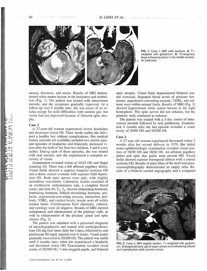
FIG. 1. Case 1; MRI axial sections. A: T1-weighted with gadolinium. B: T2-weighted; large enhancing lesion in the middle cerebel-
| lar peduncle.
nausea, dizziness, and ataxia. Results of MRI demonstrated white matter lesions in the brainstem and cerebellum (Fig. 1). The patient was treated with intravenous steroids, and the symptoms gradually improved. At a follow-up visit 9 months later, she was aware of no residua except for mild difficulties with tandem gait, but vision had not improved because of bilateral optic atrophy.
Case 2 A 27-year-old woman experienced severe headaches
and decreased vision OS. Three weeks earlier she delivered a healthy boy without complications. Her medical history (records not available) included two similar separate episodes of headaches and bilaterally decreased vision after the birth of her first two children, 5 and 6 years earlier. During each of these episodes, she was treated with oral steroids, and she experienced a complete recovery of vision.
Examination revealed vision of 20/25 OD and finger counting OS. There was a left afferent pupillary defect. Visual fields showed a superior temporal scotoma OD and a dense central scotoma with superior field depression OS. Both optic nerves were pale, with slightly anomalous vasculature. Laboratory studies consisted of an erythrocyte sedimentation rate, a complete blood count, and tests for T3, T4, thyroid-stimulating hormone, leutinizing hormone, follicle-stimulating hormone, prolactin, angiotensin-converting enzyme, antinuclear antibody, VDRL, and Cortisol levels; results were all within normal limits. Cerebrospinal fluid chemistry, cultures, and cytology were all negative. Results of MRI showed enlargement and enhancement of the pituitary stalk, as well as enhancement of the pituitary gland and optic chiasm (Fig. 2).
The patient was admitted with a presumed diagnosis of adenohypophysitis and treated with methylpredniso-lone 250 mg four times daily for 5 days, followed by oral prednisone 80 mg/d, tapered over 8 weeks. Visual acuity gradually recovered to 20/200 OS. The patient was stable until 4 months later, when she experienced a headache and decreased vision OD. Examination revealed visual acuity of 20/200 OU, 7-mm sluggish pupils, and bilateral
optic atrophy. Visual fields demonstrated bilateral central scotomas. Repeated blood levels of pituitary hormones, angiotensin-converting enzyme, VDRL, and cultures were within normal limits. Results of MRI (Fig. 3) showed hyperintense white matter lesions in the right hemisphere. The optic nerves did not enhance, but the pituitary stalk continued to enhance.
The patient was treated with a 5-day course of intravenous steroids followed by oral prednisone. Examination 4 months after the last episode revealed a visual acuity of 20/60 OD and 20/200 OS.
Case 3 A 27-year-old woman experienced decreased vision 3
months after her second delivery in 1979. Her initial neuro-ophthalmologic examination revealed visual acuities of 20/50 OD and 20/20 OS. An afferent pupillary defect and optic disc pallor were present OD. Visual fields showed superior bitemporal defects with a central scotoma OD. Results of plain films of the skull and pneumoencephalography demonstrated an empty sella. Results of a bilateral carotid angiography and a computed
FIG. 2. Case 2; MRI sagittal section. T1-weighted with gadolinium. Enlarged pituitary gland (open arrow) and enhancing chiasm and hypophyseal stalk (curved arrow).
J Neuro-Ophthalmol. Vol. 20, No. 2, 2000

POSTPARTUM OPTIC NEURITIS 87
•
FIG. 3. Case 2; MRI axial section, T2-weighted. Punctate white matter lesions (arrows).
tomograph (CT) scan of the brain were normal. Results of laboratory work-up, including endocrine functions, were all within normal limits. Her symptoms gradually resolved, and visual acuity recovered to 20/20. Six years later, she experienced double vision that was exacerbated by tennis playing. Examination revealed a right lateral rectus paresis, which resolved spontaneously.
In 1986, the patient returned with decreased visual acuity OS accompanied by left-sided headaches. Visual acuity was 20/15 OD, and 20/30 OS. Color vision was 9/10 plates OD and 3/10 OS. A left afferent pupillary defect was present. Motility testing was consistent with mild right internuclear ophthalmoplegia, which resolved spontaneously.
In 1994, the patient developed spastic paraparesis, and MRI revealed multiple white matter lesions consistent with MS.
Case 4 A 32-year-old woman experienced a full-term preg
nancy and uncomplicated delivery. Five weeks later, she developed left visual loss associated with orbital pain. She recalled that 4 years earlier, her left arm and leg were "clumsy and numb," but recovery was spontaneous in 1 month. Examination revealed acuity of 20/15 OD and 20/100 OS, a left afferent pupil defect and central scotoma, left color sense was only one of 11 plates, and an edematous left disc. Neurologic examination revealed a mild ataxia. Results of gadolinium-enhanced MRI dis
closed hyperintensity of the left optic nerve, multiple periventricular punctate white matter signal abnormalities, and a left frontal cystic lesion. The patient declined lumbar puncture and received an intravenous course of 500 mg methylprednisolone twice daily for 5 days, followed by an oral taper of prednisone over 10 days. Vision recovered to 20/30, and a course of.IFN-beta 1A 1 mL/wk began.
DISCUSSION
During and after pregnancy, the possible causes of visual disturbances, including hormonal, mechanical, and functional etiologies, are surprisingly broad. Sunness and Santos (1) provide a comprehensive overview that includes numerous nonneuro-ophthalmologic problems, with which we are not concerned in the current study. However, visual disturbances in the form of field defects during pregnancy require comment. While it is recognized that the pituitary gland undergoes a small degree of enlargement during pregnancy, principally because of hypertrophy and hyperplasia of prolactin cells, it must be recalled that the chiasm generally lies a full centimeter above the level of the diaphragma sellae. Therefore, in the absence of a preexisting adenoma, no visual change may causally be related to pregnancy alone. Pregnant women with pituitary m/croadenomas are not at risk for chiasmal compression, although pregnant women with macroadenomas greater than 1.1 cm may develop field loss (2). Supra-sellar meningiomas may be sensitive to levels of estrogen and progesterone and can undergo a growth spurt, especially during the second half of pregnancy (3,4). In general, however, we conclude that intracranial neoplasms do not appear to present more often during pregnancy.
Distinction must be made between true temporal hemianopic visual field defects and spuriously contracted fields or vague temporal depressions of a functional nature. Pregnancy and the postpartum interval are times of physiologic and psychological stress; the possibility of nonorganic visual problems or corneal and fundus alterations should be kept in mind.
Four patients who developed optic neuritis in the postpartum period have been described. Patient 1, with severe bilateral papillitis and normal initial MRI, developed a brainstem lesion 5 months later, which was confirmed by MRI and was consistent with MS. Patient 2 initially was diagnosed with adenohypophysitis. However, the history of episodes of optic neuritis after two previous term deliveries and a fourth episode of optic neuritis 4 months later, along with demonstrable white matter lesions on MRI, confirmed MS. Patient 3 was thought to suffer from ischemic chiasmal syndrome; the first episode of optic neuritis 3 months after delivery was thought to represent Sheehan syndrome and originally was reported as such (5). Six years later, a transient sixth nerve paresis with Uhthoff phenomenon, and 9 years thereafter, a contralateral optic neuritis with brainstem signs, clearly established the clinical diagnosis of MS. Thirteen years after the initial episode, the patient devel-
J Neuro-Ophthalmol, Vol. 20, No. 2, 2000

88 H. LEi
oped spastic paraparesis, and MRI revealed multiple white matter lesions. Patient 4 had a previous episode of left arm and leg symptoms with no specific diagnosis, but with MRI findings at the subsequent acute episode of papillitis. Therefore, postpartum optic neuritis in these patients occurred from 3 weeks to 3 months after the delivery, and for patient two, it occurred after three consecutive pregnancies.
Studies of patients with MS suggest that remissions tend to occur during gestation, and relapses tend to occur during the postpartum period (6,7,8). The overall incidence of MS is not correlated with pregnancy, and the relative risk for developing MS is the same for nullipa-rous women as for those who have had one or two children (9,10). Abramski (6) and Korn-Lubetzki et al. (7) have suggested that pregnancy simply delays exacerbations, allowing these exacerbations to occur after the im-munesupressive changes of pregnancy are no longer present.
Multiple sclerosis is at least twice as common among women as among men, as is true with other autoimmune-mediated diseases. In a European study of 254 women by Confavreux et al. (11), the following mean (±SD) rates of MS relapse were observed: in the prepregnancy year, 0.7 ± 0.9/woman/y; during the first trimester, 0.5 ± 1.3; during the second trimester, 0.6 ± 1.6; during the third trimester, 0.2 ± 1.0; and during the first 3 postpartum months, 1.2 ± 2.0. That is, compared with the year before conception, there was a decrease of about 70% in the relapse rate during the third trimester of pregnancy and about a 70% increase in relapse rate in the 3 initial postpartum months. Pregnancy seems to be associated with a shift from cell-mediated immunity toward increased humoral immunity (12). The fetal-placental unit secretes cytokines that down-regulate the production of maternal cytokines, which mediate cellular immunity. Indeed, this depression of maternal cell-mediated immunity could explain the tolerance of the fetus by the mother.
The effect of pregnancy on MS is similar to the effect of pregnancy on other autoimmune diseases. A tendency for remission during pregnancy and for exacerbations in the postpartum period also has been described in myasthenia gravis, in thyroiditis, and in systemic lupus ere-thematosus (1,13,14). A similar protective effect of pregnancy on autoimmunity has been demonstrated in laboratory animals (7,11), including the prolongation of graft tolerance in pregnant animals.
The federal Food and Drug Administration has not yet tested interferons (3-la and (3-lb or glatiramir acetate for use during pregnancy (15), but implications for MS man-
ETAL.
agement suggest the possibility of beginning such therapies in patients at risk as soon as possible in the postpartum period.
In summary, we present four patients with six episodes of postpartum optic neuropathies who had current or past evidence of demyelinating disease, or who subsequently developed clinical and radiologic evidences of MS. Pregnancy appears to be an immuneprotective period, but the postpartum period is immunologically prime for exacerbation of demyelinating or autoimmune diseases. Although the differential diagnosis of optic nerve disease is varied, optic neuritis in the postpartum period usually reflects MS, as it does in any other young woman.
REFERENCES
1. Sunness JS, Santos A. Pregnancy and the mother's eye. In: Tasman W, Jaeger EA, eds. Duane's clinical ophthalmology. Vol. 5. Philadelphia: Lippincott-Raven, 1997.
2. Kupersmith MJ, Rosenberg C, Kleinberg D. Visual loss in pregnant women with pituitary adenomas. Ann Int Med 1994; 121: 473-9.
3. Hsu DW, Efird JT, Hedley-White ET. Progesterone and estrogen receptors in meningiomas: prognostic considerations. J Neurosurg 1997;86:113-20.
4. Roelvink NCA, Kamphorst W, van Alphen HAM, et al. Pregnancy-related primary brain and spinal tumors. Arch Neurol 1987; 44:209-14.
5. Schatz NJ, Schlezinger NS. Noncompressive causes of chiasmal disease: Proceedings from the New Orleans Academy of Ophthalmology Symposium on Neuro-ophthalmology, St. Louis: CV Mosby Co., 1976:90-7.
6. Abramsky O. Pregnancy and multiple sclerosis. Ann Neurol 1994; 36:38^41.
7. Korn-Lubetzki I, Kahana E, Cooper G, Abramsky O. Activity of multiple sclerosis during pregnancy and puerperium. Ann Neurol 1984;16:229-31.
8. Birk K, Rudick R. Pregnancy and multiple sclerosis. Arch Neurol 1986;43:719-26.
9. Villard-Mackintosh L, Vessey MP. Oral contraceptives and reproductive factors in multiple sclerosis incidence. Contraception 1993;47:161-8.
10. Hunt JS. Immunobiology of pregnancy. Curr Opin Immunol 1992; 4:591-6.
11. Confavreux C, Hutchinson M, Hours MM, et al. Rate of pregnancy-related relapse in multiple sclerosis. N Engl J Med 1998; 339:285-91.
12. Wegmann TG, Lin H, Guilbert L, et al. Bidirectional cytokine interactions in the maternal-fetal relationship: is successful pregnancy a TH2 phenomenon? Immunol Today 1993;14:343-6.
13. Amino N, Tanizawa O, Mori H, et al. Aggravation of thyrotoxicosis in early pregnancy and after delivery in Graves' disease. J Clin Endocrinol Metab 1982;55:108-12.
14. Garsenstein M, Pollak VE, Kark RM. Systemic lupus erythematosus and pregnancy. New Engl J Med 1962;267:165-9.
15. Bashir K, Whitaker JN. Current immunotherapy in multiple sclerosis. Immunol Cell Biol 1998;76:55-64.
J Neuro-Ophthalmol, Vol. 20, No. 2. 2000