postpartum myocardial infarction: association with primary
TRANSCRIPT

C L I N I C A L C A S E ' / \ |_CMECREPIT 1
Postpartum myocardial infarction: association with primary coronary
artery dissection
LISA I. MANSUR, MD; G A R O L D O. MINNS, MD; RICHARD A. STECKLEY, MD
A 36-year-old woman presented with an acute myocardial infarction 2 weeks after the birth of her first child. The patient had smoked two packs of cigarettes a day for 12 years and had been taking bromocriptine to suppress lactation. While in the emergency room, the patient went into ventricu-lar fibrillation, but defibrillation successfully restored sinus rhythm. Coronary angiography revealed several atherosclerotic lesions. The patient refused to undergo coronary artery bypass grafting, and she was discharged receiving medical therapy.
^ B S i Forty-eight cases of postpartum myocardial in-farction have previously been reported; the present case makes 49. In 41 cases a cardiac examination was performed, either on necroscopy or by angiography. Twenty-two (54%) of the 41 pa-tients had occlusion related to primary coronary artery dissec-tion, 11 (27%) had normal coronary arteries and presumed spasm, and only 6 (15%) had atherosclerosis-related coronary occlusion. The mortality rate was 39%, and surviving patients had significant cardiac limitation.
m— - w Postpartum myocardial infarction and coronary oc-clusion, when they do occur, are frequently caused by primary coronary artery dissection. Immediate recognition of this asso-ciation may significantly reduce morbidity and mortality for these patients.
INDEX TERMS: PUERPERAL DISORDERS; MYOCARDIAL INFARCTION; ANEU-RYSM, DISSECTING; CORONARY VASOSPASM CLEVE CLIN ] MED 1995; 62:261-268
From the University of Kansas School of Medicine, Wichita. Address reprint requests to L.I.M., 1993 W. Champagne Avenue, Tay-
lorsville, UT 84118.
MYOCARDIAL infarc-tion during the post-partum period is un-common but devas-
tating. This paper documents a case in a previously healthy 36-year-old cigarette smoker who was taking bromocriptine to suppress lactation; it also reviews 48 pre-viously reported cases. Surpris-ingly, in 22 of the 41 cases in which a coronary examination was performed, the occlusion was caused by primary coronary artery dissection, an otherwise rare cause of myocardial infarction. Eleven other cases were presumed to result from coronary artery spasm, possi-bly related to peripartum medica-tion use in some. Unlike in the general population, only 15% of the postpartum cases were related to atherosclerotic coronary artery disease.
C A S E S U M M A R Y
A 36-year-old white woman pre-sented to the emergency depart-ment of a local hospital with chest discomfort. She described this as a sensation of pressure that radiated
JULY • AUGUST 1995 CLEVELAND CLINIC JOURNAL OF MEDICINE 2 6 1
on December 14, 2021. For personal use only. All other uses require permission.www.ccjm.orgDownloaded from
P O S T P A R T U M M Y O C A R D I A L I N F A R C T I O N • M A N S U R
to the right arm and neck; it had begun 2 hours previously while she was carrying packages. Her in-itial electrocardiogram demonstrated ST elevation in the anterior and lateral leads. Soon after her arri-val she went into ventricular fibrillation, but defibril-lation successfully restored normal sinus rhythm. The patient was transferred to a tertiary-care hospi-tal for cardiac catheterization.
Two weeks previously, the patient had given birth to her first child in an uneventful, normal vaginal delivery. Her pregnancy had been entirely uncom-plicated, and she denied having any chest discom-fort during the pregnancy or delivery. After delivery, she was given propoxyphene for pain and bro-mocriptine 2.5 mg twice a day to suppress lactation. She had smoked two packs of cigarettes a day for 12 years. She had no family history of coronary artery disease, and she denied using cocaine, ampheta-mines, or excessive amounts of alcohol.
The patient's initial creatine phosphokinase (CPK) concentration, drawn before her cardiac ar-rest, was 1.95 |J.kat/L, and her lactate dehydro-genase (LDH) concentration was 3.7 (ikat/L. Her subsequent CPK concentrations, drawn at 8-hour intervals, were 8.57 |J.kat/L (19% MB fraction), 30.91 (xkat/L (23% MB fraction), 52.07 |akat/L (15% MB fraction), and 37.87 |lkat/L (15% MB fraction).
Left ventriculography demonstrated anterolateral hypokinesis and apical dyskinesis. The calculated left ventricular ejection fraction was 34%. Coronary arteriography revealed several atherosclerotic le-sions, including a 60% left main artery stenosis, a 90% intermediate diagonal stenosis, and a total left anterior descending artery occlusion distal to the intermediate diagonal branch. The circumflex ar-tery had a 90% elongated stenosis. The right coro-nary artery was free of obstruction.
Coronary artery bypass grafting was recom-mended, but the patient declined this procedure and was discharged receiving medical therapy.
D I S C U S S I O N
The incidence of myocardial ischemia and in-farction in women of usual reproductive age is so rare it is difficult to determine realistically. A recent study estimated the incidence of myocardial in-farction in women age 25 to 34 years as 0.03 per 1000 woman-years, with an estimated annual risk of 1 in 30 000.1
The independent risk factor most associated with myocardial infarction in young women is cigarette smoking (especially more than 15 cigarettes per day).1'2 Other risk factors include a family history of coronary artery disease, as well as diabetes, hyper-tension, and hyperlipidemia.2 The effects of oral contraceptives are controversial.
Hemodynamic changes during pregnancy During pregnancy, the maternal cardiovascular
system undergoes remarkable changes.3"18 The in-travascular volume (plasma and red blood cell mass) increases 40% to 50%,4-8 the heart rate increases by 10 to 15 beats per minute, and the stroke volume increases 30%.9 Because of these changes, the car-diac output increases by 1.5 L/minute above base-line, reaching a peak at the beginning of the second trimester.9 Owing to increased filling volume secon-dary to increased intravascular volume, the heart undergoes concentric hypertrophy.10 However, sys-temic vascular resistance decreases, and arterial blood pressure therefore remains stable or decreases during a normal pregnancy.9,11,12
The cardiac output increases an additional 25% during the first stage of labor, 50% during the second stage, and 80% at delivery (less if conduction anes-thesia is used).13 In addition, every uterine contrac-tion adds up to 500 mL of blood into the central circulation, compounding cardiovascular stress with increased arterial pressure and stroke volume.14,15
During a vaginal delivery, blood loss of 300 to 500 mL is not uncommon; during a cesarean section, 1000 mL.16 Further, the use of oxytocin to induce labor likely decreases coronary blood flow.17 The cardiovascular system usually returns to a baseline state over the 7 to 10 days after delivery.18
Myocardial infarction during or after pregnancy
Despite these remarkable cardiovascular changes (which could alter coronary blood flow), myocardial ischemia and infarction during pregnancy, parturi-tion, and the puerperium are rare, with a frequently cited incidence of 1 in 10 000 pregnancies.3 Over 100 cases of myocardial infarction and coronary oc-clusion during pregnancy, delivery, and the puer-perium have been reported since 1922; 48 of these cases occurred during the puerperium.3,19"59
Reports of myocardial infarction during the puer-perium are scant because the problem is uncommon and because all cases are not reported. By definition,
262 CLEVELAND CLINIC JOURNAL OF MEDICINE VOLUME 62 • NUMBER 4
on December 14, 2021. For personal use only. All other uses require permission.www.ccjm.orgDownloaded from
P O S T P A R T U M M Y O C A R D I A L I N F A R C T I O N • M A N S U R
the puerperium is the period of "confinement" dur-ing and just after birth.60 However, this period is traditionally recognized as the time after delivery in which the reproductive system resumes a nonpreg-nant state (approximately 6 weeks).
A coronary artery examination was performed either by coronary angiography or at necropsy in 41 of the 49 postpartum cases (including the present one). Coronary atherosclerosis was documented as the primary cause of coronary occlusion in 40% to 50% of the reported cases occurring during preg-nancy and delivery,61,62 but only 15% of the postpar-tum cases (Table). In contrast, primary coronary ar-tery dissection was documented in 22 of the 41 postpartum cases in which coronary examination was performed. Coronary artery spasm was assumed to be the cause of occlusion in 11 cases in which the coronary arteries were normal on angiography. Ad-ditional possible causes include the same conditions that cause myocardial infarction in patients of simi-lar age (congenital coronary atresia or malforma-tion, hypercoagulable states, vasculitis, and coro-nary artery aneurysms).
The average age of the patients with postpartum myocardial infarction was 31.5 years (range 17 to 42 years). Over 75% were multigravid or multiparous or both, and virtually all had been previously healthy. Excluding the six cases that were diagnosed after 6 weeks, an average of 12 days elapsed between parturition and the cardiac event (range 3.5 hours to 42 days).
The mortality rate in myocardial infarction dur-ing pregnancy and parturition is as high as 45%.63
Nineteen (39%) of the 49 patients with postpartum coronary occlusion died as an immediate result of the cardiac event. Three additional patients were successfully resuscitated after cardiopulmonary ar-rest. Many of the surviving patients suffered se-verely limiting cardiac damage. The cardiac debili-tation was likely related to involvement of the anterior or anterolateral wall in 85% of the postpar-tum cases.
Coronary artery dissection Primary coronary artery dissection is ordinarily a
rare cause of coronary occlusion. Recent reviews of the nearly 100 reported cases, dating back to 1933, revealed that almost 75% occurred in women.49'54,64,65
Of the 49 cases of postpartum coronary artery occlu-sion and infarction, 22 were related to primary coro-nary artery dissection. The dissection involved the
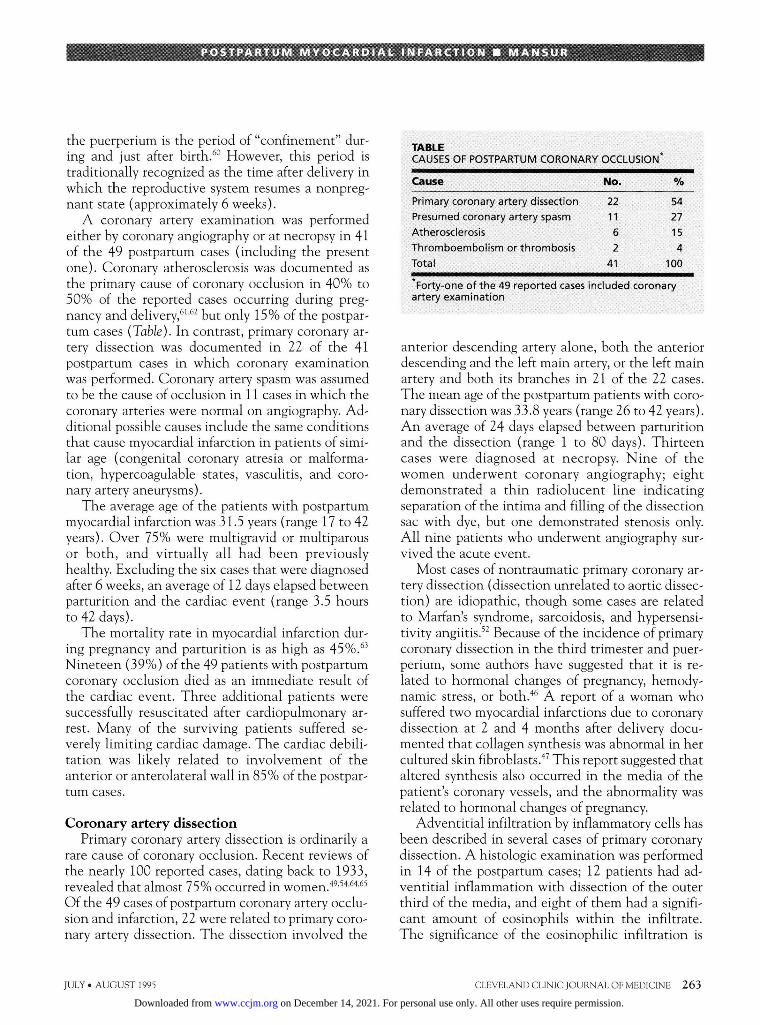
TABLE CAUSES OF POSTPARTUM CORONARY OCCLUSION*
Cause No. %
Primary coronary artery dissection 22 54 Presumed coronary artery spasm 11 27 Atherosclerosis 6 15 Thromboembolism or thrombosis 2 4 Total 41 100
* Forty-one of the 49 reported cases included coronary artery examination
anterior descending artery alone, both the anterior descending and the left main artery, or the left main artery and both its branches in 21 of the 22 cases. The mean age of the postpartum patients with coro-nary dissection was 33.8 years (range 26 to 42 years). An average of 24 days elapsed between parturition and the dissection (range 1 to 80 days). Thirteen cases were diagnosed at necropsy. Nine of the women underwent coronary angiography; eight demonstrated a thin radiolucent line indicating separation of the intima and filling of the dissection sac with dye, but one demonstrated stenosis only. All nine patients who underwent angiography sur-vived the acute event.
Most cases of nontraumatic primary coronary ar-tery dissection (dissection unrelated to aortic dissec-tion) are idiopathic, though some cases are related to Marian's syndrome, sarcoidosis, and hypersensi-tivity angiitis.52 Because of the incidence of primary coronary dissection in the third trimester and puer-perium, some authors have suggested that it is re-lated to hormonal changes of pregnancy, hemody-namic stress, or both.46 A report of a woman who suffered two myocardial infarctions due to coronary dissection at 2 and 4 months after delivery docu-mented that collagen synthesis was abnormal in her cultured skin fibroblasts.47 This report suggested that altered synthesis also occurred in the media of the patient's coronary vessels, and the abnormality was related to hormonal changes of pregnancy.
Adventitial infiltration by inflammatory cells has been described in several cases of primary coronary dissection. A histologic examination was performed in 14 of the postpartum cases; 12 patients had ad-ventitial inflammation with dissection of the outer third of the media, and eight of them had a signifi-cant amount of eosinophils within the infiltrate. The significance of the eosinophilic infiltration is
JULY • AUGUST 1995 CLEVELAND CLINIC JOURNAL OF MEDICINE 2 6 3
on December 14, 2021. For personal use only. All other uses require permission.www.ccjm.orgDownloaded from
P O S T P A R T U M M Y O C A R D I A L I N F A R C T I O N • M A N S U R
not known. Some speculate that the eosinophils contain several enzymes and cytotoxic substances, including histaminase, arylsulfatase, phospholipase, fibrolysin, and major basic protein, that could con-tribute to lysis and damage of the arterial wall.66,67
Other vascular-wall abnormalities have been documented in postpartum coronary artery dissec-tion. Cystic medial necrosis without inflammation was described histologically in two patients who had no history of hypertension or features of Marian's syndrome. Significant atherosclerosis is rarely found in association with coronary dissection,51 and atherosclerosis in addition to adventitial inflamma-tion was reported in only two of the cases of postpar-tum dissection. Some investigators believe the in-tramural hemorrhages of dissection arise from the thin-walled vasculature within the atheromatous plaques.68 Others, citing the prevalence of variant angina and primary coronary artery dissection in Japan,50 believe coronary artery spasm to be the pri-mary event in dissection. Hypertension does not appear to be necessary in the pathogenesis of post-partum coronary dissection, as none of the patients had essential or pregnancy-induced hypertension.
Aortic dissection has also been associated with pregnancy. Studies have demonstrated aortic mi-crostructural changes during pregnancy, including disorganization and fragmentation of elastic and reticulum fibers in the tunica media, hypertrophy and hyperplasia of smooth muscle fibers, decreased amounts of interstitial acidic mucopolysaccharides, and changes in the ground substance of the connec-tive tissue.69,70 Some investigators believe the altera-tions of the aortic and coronary walls are associated with the "softening" of tissues that occurs before delivery. However, other investigators have found no consistent alterations in aortic structure or ten-sile strength when they compared women of all tri-mesters of pregnancy with controls.71,72
Hypertension and eclampsia Nine women with postpartum myocardial in-
farction had pregnancy-induced hypertension or preeclampsia-eclampsia that developed during or af-ter delivery. Five of these nine patients underwent coronary examination; only one had normal coro-nary arteries and no other possible contributing fac-tors; atherosclerosis was documented in three.
The causes of pregnancy-induced hypertension and preeclampsia-eclampsia are not completely un-derstood, but generalized vasospasm is important.73
Whether pregnancy-induced hypertension or pre-eclampsia-eclampsia directly result in coronary oc-clusion is not known, but evidence supports a pathogenic role.73
Vasospasm Postpartum myocardial infarction in patients
with normal coronary arteries poses an etiologic puzzle. Eleven patients with postpartum myocardial infarction who underwent coronary angiography had normal coronary arteries; vasospasm was the presumed mechanism of coronary occlusion. None of the patients had a history consistent with variant angina. Use of vasoconstrictive medications or nicotine was documented in seven of these patients. As in all young patients with myocardial infarction, the surreptitious use of cocaine and amphetamines should be suspected if results of coronary angiogra-phy are normal and vasospasm is suspected.
Bromocriptine and postpartum vasospasm Bromocriptine, a semisynthetic hydrogenated er-
got alkaloid derivative of alpha-ergocryptine, is a D2-dopamine receptor agonist.59,74 It was approved in 1980 by the Food and Drug Administration for postpartum lactation suppression at doses of 2.5 mg twice or three times a day. In low doses (less than 10 mg/day), it typically acts as a potent vasodilator, is well known for causing severe postural hypotension, and does not usually activate alpha-adrenergic re-ceptors.59,75 However, bromocriptine in low doses has been implicated in cases of hypertension, cere-brovascular accident, Raynaud's phenomenon, vas-cular headaches, seizures, vasospasm, and myocar-dial infarction.76,77
According to one theory, bromocriptine-induced vasospasm is a paradoxical response70; according to another, the constrictive response is mediated by receptors that accept both nonhydrogenated and hydrogenated ergot alkaloids,48 or by abnormally po-sitioned D2-dopamine receptors.78 Alternatively, a decreased sensitivity of peripheral dopaminergic re-ceptors may unmask the vasoconstrictor effects of bromocriptine.78 Finally, up-regulation of dopamine receptors (similar to the up-regulation demon-strated with cocaine use) may occur, causing a dopamine-agonist supersensitivity response to bro-mocriptine.79
Six case reports have suggested an association be-tween postpartum use of bromocriptine and myocar-dial ischemia and infarction.48,53,56,58,59 Two of the pa-
264 CLEVELAND CLINIC JOURNAL OF MEDICINE VOLUME 62 • NUMBER 4
on December 14, 2021. For personal use only. All other uses require permission.www.ccjm.orgDownloaded from
P O S T P A R T U M M Y O C A R D I A L I N F A R C T I O N • M A N S U R
tients had normal angiographic findings, one had a single stenosis (possibly caused by spasm) and no evidence of atherosclerosis, one had pregnancy-in-duced hypertension and used ergonovine, and two used other vasoconstrictive medications (isomethep-tene, benzquinamide, oxytocin) in addition to bro-mocriptine. Because bromocriptine has no known effects on coagulation, the presumed mechanism of occlusion is coronary vasospasm.
Some of these cases are confounded by the con-comitant use of other potentially vasoconstricting or cardioactive agents (eg, ergonovine maleate for uterine atony and postpartum hemorrhage, isometheptene for migraine headache, benzqui-namide for nausea and vomiting, and nicotine). Bromocriptine may have potentiated the vasocon-strictive activity of these agents by interfering with drug metabolism.48
Patients prone to vasospasm and the possible va-soconstrictive effects of bromocriptine include those who develop hypertension during pregnancy or after delivery and those with a history of vascular head-aches, atherosclerotic disease, cocaine abuse, and Raynaud's phenomenon.80"82 In 1991, the Food and Drug Administration suggested that lactation sup-pression is safer without the use of drugs.83 The asso-ciation of bromocriptine use with postpartum myo-cardial infarction is speculative but should be recognized as a possibility.
Atherosclerotic coronary artery disease Myocardial infarction related to atherosclerotic
coronary artery disease is rare in women younger than 35 years.1 In reported cases, atherosclerosis seems a less common cause of myocardial infarction during the puerperium than during pregnancy and delivery. Only six of the 41 postpartum patients who underwent coronary artery examinations had atherosclerosis-related occlusions. Two of these pa-tients smoked cigarettes, three received bro-mocriptine or ergonovine after delivery, and four had essential or pregnancy-induced hypertension.
Thrombosis, thromboembolism Finally, in two reported cases, postpartum myo-
cardial infarction was caused by thrombosis or thromboembolism. Although embolization usually occurs in association with other conditions such as mitral stenosis, cardiomyopathy, atrial septal defect, endocarditis, or atrial fibrillation, these patients had no such history.
JULY • AUGUST 1995
D I A G N O S I N G P O S T P A R T U M M Y O C A R D I A L I N F A R C T I O N
The diagnosis of myocardial ischemia and in-farction in the postpartum population requires the same criteria as in any other patients, with a few caveats. Myocardial ischemia may be mistakenly identified as gastroesophageal reflux, a common cause of chest discomfort during and after preg-nancy. Peripheral edema, distended neck veins, an S3 gallop, a systolic ejection murmur at the left sternal border, and increased splitting of the first and second heart sounds are not uncommon during pregnancy but usually resolve during the first weeks after delivery. The electrocardiogram may show nonspecific ST- and T-wave changes as well as left axis deviation. The serum LDH level during preg-nancy is 18% to 100% higher than in nonpregnant women, and serum LDH and CPK levels increase during the muscular work of childbirth. However, the MB fraction of CPK should not increase during pregnancy and childbirth, and serial MB fractions should be followed for diagnosis.
Because of the apparent frequency of primary coronary artery dissection in postpartum coronary artery occlusion, early catheterization is necessary to determine the cause of occlusion. Typical angio-graphic findings for dissection confirm the diagno-sis, but single stenotic lesions may be caused by dissection as well.
T H E R A P Y
Reports in the medical literature on the treat-ment of postpartum myocardial infarction are lim-ited. Treatment depends on the cause of coronary occlusion. A thorough drug and nicotine history should be taken, and a screen for drugs of abuse should be ordered. Medications should be evaluated and discontinued if a potential for vasoconstriction exists. Nitroglycerin (given intravenously) and an-algesics (morphine) have been used without ad-verse consequences. Depending on the cause of oc-clusion, the length of time since delivery, and the risk of hemorrhage, aspirin may be used for its anti-platelet action and heparin for its anticoagulant effect. Beta blockers, if not contraindicated, may be used in an attempt to limit infarction size and pre-vent ventricular arrhythmias. However, beta-blocker use could potentiate spasm in patients with normal coronary arteries.84 Calcium antagonists
CLEVELAND CLINIC JOURNAL OF MEDICINE 265
on December 14, 2021. For personal use only. All other uses require permission.www.ccjm.orgDownloaded from

P O S T P A R T U M M Y O C A R D I A L I N F A R C T I O N • M A N S U R
may be of benefit in coronary vasospasm and in dissection.
Thrombolytics are relatively contraindicated within 10 days of obstetric delivery because of the risk of bleeding.85,86 However, thrombolytics have been used successfully during the postpartum period to treat isolated thrombosis of a prosthetic mitral valve and superior sagittal sinus thrombosis, with few complications.87'88 Given the possibility of coro-nary artery dissection in this population, using thrombolytics without knowing the cause of coro-nary occlusion is not advised.
Percutaneous transluminal balloon coronary angioplasty was successful in two postpartum pa-tients, including one with atherosclerotic disease and one with presumed spasm. Coronary artery by-pass grafting may be indicated, especially given the frequent involvement of the left main and proximal anterior descending arteries.
Of the nine women who survived coronary artery dissection, one underwent thoracotomy and evacu-ation of a coronary artery hematoma, and three un-derwent bypass grafting (one with aneurysmec-tomy). The remaining five patients received medical therapy, including calcium antagonists, ni-trates, and warfarin. One of these patients sub-sequently received a cardiac transplant.
Whether to perform surgery for dissection de-pends on the degree of stenosis, hemodynamic
status, and myocardial viability.52 Past surgical expe-rience indicates that internal mammary grafts are best,89,90 and that proximal ligation of the dissected artery is probably not necessary.
S U M M A R Y
Postpartum myocardial infarction, though rare, often results in severe morbidity or death. In re-ported cases, the most common cause of postpartum coronary occlusion was primary coronary artery dis-section. Immediate recognition of this association is essential for appropriate decisions concerning diag-nostic procedures and treatment.
Only 15% of the reported cases were attributed to atherosclerotic disease, though all cases of atherosclerosis-related occlusion were probably not reported because of the rarity of the disease. The 11 cases in which the coronary arteries were demon-strated normal on angiography pose an etiologic and therapeutic puzzle, though vasospasm is a likely mechanism of occlusion. Possible pathogenic roles of vasospasm caused by medications (bromocriptine and ergonovine) and of pregnancy-induced hyper-tension have been postulated.
Thrombolytics are relatively contraindicated within 10 days after delivery because of the risk of hemorrhage and should not be used until coronary dissection has been excluded.
1. Mant D, Villard-Mackintosh L, Vessey MP, Yeates D. Myocar-dial infarction and angina pectoris in young women. J Epidemiol Community Health 1987; 41:215-219.
2. Rosenberg L, Shapiro S, Kaufman DW, Slone D, Miettinen OS, Stolley PD. Cigarette smoking in relation to the risk of myocar-dial infarction in young women. Modifying influence of age and predisposing factors. IntJ Epidemiol 1980; 9:57-63.
3. Ginz B. Myocardial infarction in pregnancy. J Obstet Gynaecol Br Commonw 1970; 77:610-615.
4. Scott DE. Anemia in pregnancy. Obstet Gynecol Annu 1972; 1:219-244.
5. Lees MM, Taylor SH, Scott DB. A study of cardiac output at rest throughout pregnancy. J Obstet Gynaecol Br Commonw 1967; 74:319-328.
6. Walters WAW, MacGregor WG, Hills M. Cardiac output at rest during pregnancy and the puerperium. Clin Sei 1966; 3 0 : 1 -11.
7. Brehm H, Kindling E. Der kreislauf wahrend Schwangerschaft und Wochenbett. Arch Gynakol 1955; 185:696.
8. Pyorala T. Cardiovascular response to the upright position dur-ing pregnancy. Acta Obstet Gynecol Scand 1966; 45:8.
9. de Swiet M, The cardiovascular system. In: Hytten FE, Cham-berlain G, editors. Clinical physiology in obstetrics. Oxford:
Blackwell Scientific Publications, 1991:3-38. 10. Sala, DJ. Myocardial infarction. NAACOG Clin Issu Perinat
Womens Health Nurs 1992; 3:443—453. 11. Kerr MG. The mechanical effects of the gravid uterus in late
pregnancy. J Obstet Gynaecol Br Commonw 1965; 72:513. 12. Lees MM, Scott DB, Kerr MG, Taylor SH. The circulatory
effects of recumbent postural change in late pregnancy. Clin Sei 1967;32:453-465.
13. Ueland K, Hansen JM. Maternal cardiovascular dynamics. III. Labor and delivery under local and caudal analgesia. Am J Obstet Gynecol 1969; 103:8-18.
14- Hendricks CH. The hemodynamics of uterine contraction. Am J Obstet Gynecol 1958; 76:969.
15. Adams JQ, Alexander AM Jr. Alterations in cardiovascular physiology during labor. Obstet Gynecol 1958; 12:542.
16. Lamb MA. Myocardial infarction during pregnancy: a team challenge. Heart Lung 1987; 16:658-661.
17. Pauerstein CJ. Use and abuse of oxytocic agents. Clin Obstet Gynecol 1976; 16:262-267.
18. Jacobi AG. Obstetric anesthesia and the cardiac patient. J Cardiovasc Pulmonary Technique 1982; 10:35.
19. Kolisko O. Dittrichs Handbuch der arztlichen. Sachverstandi-gen. 1916; 2:1916.
20. Lovitt WV Jr, Corzine WJ Jr. Case reports: dissecting intramu-ral hemorrhage of anterior descending branch of left coronary artery. Arch Pathol Lab Med 1952; 43:458-462.
266 CLEVELAND CLINIC JOURNAL OF MEDICINE VOLUME 62 • NUMBER 4
on December 14, 2021. For personal use only. All other uses require permission.www.ccjm.orgDownloaded from
P O S T P A R T U M M Y O C A R D I A L I N F A R C T I O N • M A N S U R
21. Freedman JR, Gilbert JT. Coronary occlusion with myocardial infarction in a puerperal patient. Am J Obstet Gynecol 1956; 71 :1106-1110.
22. Maternal Health Committee. Maternal health in Ohio: mater-nal deaths involving cardiac disease. Ohio State Med J 1958; 54 :187-188 .
23. Urdan BE, Madden WJ . Pregnancy complicated by myocardial infarction: report of a fatal case. Obstet Gynecol 1959; 1 4 : 3 7 8 -380.
24. Vasicka AL, Lin TJ. Fatal coronary artery disease during the early postpartum period: report of one case. Am J Obstet Gynecol 1959; 77 :899-904 .
25. Wells AL. Dissecting aneurysm of coronary artery in the puer-perium. J Pathol 1960; 79:404^106.
26. Brown A. Myocardial infarction associated with pregnancy. N Engl J Med 1960; 262 :1163-1166 .
27. Watson H, Emslie-Smith D, Herring J, Hill IGW. Myocardial infarction during pregnancy and puerperium. Lancet 1960; 2 : 5 2 3 -525.
28. Burton JF, Zawadzki ES. The coronary aneurysm. J Forensic Sci 1962; 7 :486-492 .
29. Brody GL, Burton JF, Zawadski ES, French AJ. Dissecting aneurysm of the coronary artery. N Engl J Med 1965; 2 7 3 : 1 - 6 .
30. Palamino SJ. Dissecting intramural hematoma of the left coro-nary artery in the puerperium: a case report and survey of the literature. Am J Clin Pathol 1969; 51 :119-125 .
31. Glancy DL, Marcus ML, Epstein SE. Myocardial infarction in young women with normal coronary arteriograms. Circulation 1971; 44 :495-501 .
32. DiMaio VJM, DiMaio DJ. Postpartum dissecting coronary aneu-rysm. N Y State J Med 1971; 71 :767-769 .
33. Asuncion CM, Hyun J. Dissecting intramural hematoma of the coronary artery in pregnancy and the puerperium. Obstet Gynecol 1972; 40 :202-210 .
34. Claudon DG, Claudon DB, Edwards JE. Primary dissecting aneurysm of coronary artery. A cause of acute myocardial is-chemia. Circulation 1972; 45 :259-266 .
35. Razavi M. Unusual forms of coronary artery disease. Cardiovasc Clin 1975; 7 :25-46 .
36. Shaver PJ, Carrig TF, Baker WP. Postpartum coronary artery dissection. Br Heart J 1978; 40 :83 -86 .
37. Jewett JF. Committee on maternal welfare. Two dissecting coro-nary artery aneurysms postpartum. N Engl J Med 1978; 2 9 8 : 1 2 5 5 -1256.
38. Beary JF, Summer WR, Bulkley BH. Postpartum acute myocar-dial infarction: a rare occurrence of uncertain etiology. Am J Cardiol 1979 ;43 :158-161 .
39. Ciraulo DA, Markovitz A. Myocardial infarction in pregnancy associated with a coronary artery thrombus. Arch Intern Med 1979; 139:1046-1047.
40. Chant GN. Coronary anatomy in postpartum acute myocardial infarction [letter]. Am J Cardiol 1980; 45 :912 .
41. Henion WA, Hilal A, Matthew PK, Lazarus AR, Cohen J . Postpartum myocardial infarction. N Y State J Med 1982; 1 :57 -62.
42. Robinowitz M, Virmani R, McAllister HA. Spontaneous coro-nary artery dissection and eosinophilic inflammation: a cause and effect relationship? Am J Med 1982; 72 :923-928.
43. Salem DN, Isner JM, Hopkins P, Konstam MA. Ergonovine provocation in postpartum myocardial infarction. Angiology 1984 ;35 :110-114 .
44. Bornstein A, Dalai P, Tischler J, Novack S, Michaelson S. Acute myocardial infarction in a thirty-six year old postpartum female. Angiology 1984; 53 :591-594 .
45. Taylor GJ, Cohen B. Ergonovine-induced artery spasm and myocardial infarction after normal delivery. Obstet Gynecol 1985; 66:821-822.
46. lung B, Squara P, Fruchaud J, et al. Postpartum myocardial infarction with normal coronary arteries. Apropos of a case. Arch Mai Coeur Vaiss 1986; 79 :1951-1955.
47. Bonnett J, Aumailley M, Thomas D, Grosgogeat Y, Broustet JP, Bricaud H . Spontaneous coronary artery dissection: case report and evidence for a defect in collagen metabolism. Eur Heart J 1986; 7 :904-909.
48. Iffy L, TenHove W, Frisoli G. Acute myocardial infarction in the puerperium in patients receiving bromocriptine. Am J Obstet Gynecol 1986; 155 :371-372 .
49. Vicari R, Eybel C, Monson D. Survival following spontaneous coronary artery dissection: surgical repair by extrusion of intramu-ral hematoma. Am Heart J 1986; 111 :593-594 .
50. Nishikiwa H, Nakanishi S, Nishiyama S, Nishimura S, Seki A, Yamaguchi H. Primary coronary artery dissection observed at coronary angiography. Am J Cardiol 1988; 61 :645-648 .
51. Movesian MA, Wray RB. Postpartum myocardial infarction. Br Heart J 1989; 62 :154-156 .
52. Lette J, Gagnon A, Cerino M, Prenovault J. Apical hypertro-phic cardiomyopathy with spontaneous postpartum coronary ar-tery dissection. C a n ) Cardiol 1989; 5 :311-314 .
53. Ruch A, Duhring JL. Postpartum myocardial infarction in a patient receiving bromocriptine. Obstet Gynecol 1989; 7 4 : 4 4 8 -451.
54. DeMaio SJ, Kinsella SH, Silverman ME. Clinical course and long-term prognosis of spontaneous coronary artery dissection. Am J Cardiol 1989; 64 :471-474 .
55. Pardo J, Gonzalez B, Novoa O, Pumarino R, Opplinger E, Godoy D. Acute postpartum infarct of the myocardium secon-dary to a spontaneous dissection of the coronary artery. Rev Med Chil 1990; 118:300-305.
56. Kulig K, Moore LL, Kirk M, et al. Bromocriptine-associated headache: possible life threatening sympathomimetic interaction. Obstet Gynecol 1991; 78 :941-943.
57. Saxena R, Nolan TE, von Dohlen T, Houghton JL. Postpar-tum myocardial infarction treated by balloon coronary angioplasty. Obstet Gynecol 1992; 79 :810-812 .
58. Eikman FM. Recurrent myocardial infarction in a postpartum patient receiving bromocriptine. Clin Cardiol 1992; 15 :781-783.
59. Larrazet F, Spaulding C, Lobreau HJ, Weber S, Guerin F. Pos-sible bromocriptine-induced myocardial infarction. Ann Intern Med 1993; 118:199-200.
60. The puerperium. In: Cunningham FG, MacDonald PC, Gant NF, editors. Williams obstetrics. Norwalk, Conn, and San Mateo, Calif: Appleton and Lange, 1989:245-256.
61. Douglas PS. Heart disease in women. FA Davis Company: Philidelphia, 1989:122-123.
62. Hands ME, Johnson MD, Saltzman DH, Rutherford JD. The cardiac, obstetric and anaesthetic management of pregnancy com-plicated by acute myocardial infarction. J Clin Anesth 1990; 2 :258-268.
63. Clark SL. Cardiac disease in pregnancy. Obstet Gynecol 1987; 18 :237-253.
64- Ramamurti S, Mahrer PR, Magnusson P, Bowyer JV, Sasse L, Shaperman M. Idiopathic coronary artery dissection: a rare in vivo diagnosis. Clin Cardiol 1985; 8 :57-60 .
65. Mathieu D, Larde D, Vasile N. Primary dissecting aneurysms of the coronary arteries: case report and literature review. Cardiovasc Intervent Radiol 1984; 7 :71-74 .
66. Beeson PB, Bass DA. The eosinophil. Major Probl Intern Med 1977; 14 :1-269.
67. Gleich GJ, Frigas E, Loegering DA, Wasson DL, Steinmuller D. Cytotoxic properties of the eosinophil major basic protein. J Immunol 1979; 123:2925-2927.
68. Horn H, Finkelstein LE. Arteriosclerosis of the coronary arter-ies and mechanism of their occlusion. Am Heart J 1940; 19:655.
69. Manalo-Estrello P, Barker A. Histopathologic findings in hu-man aortic media associated with pregnancy: a study of 16 cases. Arch Pathol 1967; 83 :336-341 .
70. Perl E, Catchpole HR. Changes induced in the connective tissue of the pubic symphysis of the guinea pig with estrogen and relaxin. Arch Pathol 1950; 50 :233-239 .
JULY • AUGUST 1995 CLEVELAND CLINIC JOURNAL OF MEDICINE 267
on December 14, 2021. For personal use only. All other uses require permission.www.ccjm.orgDownloaded from

M A N S U R
CLEVELAND CLINIC
J O U R N A L o f MEDICINE
INTERNAL MEDICINE
BOARD REVIEW
IM BOARD REVIEW presents clinical vignettes along with questions on the differential diagnosis and treatment of medical conditions likely to be encountered on the Qualifying Examination in Medicine -as well as in practice. Take the challenge.
IN THIS ISSUE Page 206
71. Cavanzo FJ, Taylor HB. Effect of pregnancy on the human aorta and its relationship to dissecting aneurysm. Am J Obstet Gynecol 1969; 105 :567-568.
72. Johnson WL, Conrad JT, Whitney D, Graybeal N. The effect of pregnancy and norethynodrel with mestranol on length tension relationship in the rabbit aorta. Am J Obstet Gynecol 1965; 93 :179-180 .
73. Bauer TW, Moore GW, Hutchins GM. Morphologic evidence for coronary artery spasm in eclampsia. Circulation 1982; 6 5 : 2 5 5 -259.
74- Cedarbaum JM, Schleifer LS. Drugs for Parkinson's disease, spasticity and acute muscle spasm. In; Goodman LS, Gilman A, editors. The pharmacological basis of therapeutics. Tarrytown, NY: Pergamon Press, 1990:473-475.
75. Feldman H N . Myocardial infarction in the puerperium [letter]. Am J Obstet Gynecol 1987; 157:515-516.
76. Division of Drug Experience. Postpartum hypertension, seizures, strokes reported with bromocriptine. FDA Drug Bull 1984; 1 4 : 3 -4.
77. Package Insert. Sandoz Pharmaceutical Corp. Route 10. East Hanover, NJ 07936, July 1, 1987.
78. Crea F, Chierchia S, Kaski JC, et al. Provocation of coronary spasm by dopamine in patients with active variant angina pecto-ris. Circulation 1986; 74 :262-269.
79. Gawin FH. Chronic neuropharmacology of cocaine: progress in pharmacotherapy. J Clin Psychiatry 1988; 4 9 (Suppl 2) : 11-16.
80. Miller D, Waters DD, Warnica W. Is variant angina the coro-nary manifestation of a generalized vasospastic disorder? N Engl J Med 1981; 304 :763-766.
81. Miller LG, Bakht FR, Baker T, Kirshon B. Possible cocaine predisposition to adverse cerebrovascular and cardiovascular se-quelae of bromocriptine administered postpartum. J Clin Pharma-col 1989; 29 :781-785.
82. Katz M, Kroll D, Pak I, Osimoni A, Hirsch M. Puerperal hypertension, stroke and seizures after suppression of lactation with bromocriptine. Obstet Gynecol 1985; 66 :822-824 .
83. Stehlin D. Lactation suppression safer without drugs. FDA Con-sumer 1990; (April): 25-27.
84. Tilmant PY, Lablanche JM, Thieuleux FA, Depuis BA, Ber-trand ME. Detrimental effect of propranolol in patients with coronary arterial spasm countered by combination with diltiazem. Am J Cardiol 1983; 52 :230-233 .
85. 1993 Physicians' Desk Reference. 47th edition. Montvale, NJ: Medical Economics Data, 1993:657.
86. McEvoy GK, Litvak K, Welsh OH. AHFS Drug Information 93. Bethesda, Md: American Society of Hospital Pharmacists Inc, 1993:880.
87. Jost CM, Yancy CW Jr, Ring WS. Combined thrombolytic therapy for prosthetic mitral valve thrombosis. Ann Thorac Surg 1993 ;55 :159-161 .
88. Manthous CA, Chen H. Case report: treatment of superior sagittal sinus thrombosis with urokinase. Conn Med 1992; 56 :529-530 .
89. Thayer JO, Healy RW, Maggs PR. Spontaneous coronary artery dissection. Ann Thorac Surg 1987; 44 :97-102 .
90. Forker AD, Rosenlof RC, Weaver WF. Primary dissecting aneurysm of the right coronary artery with survival. Chest 1973; 64 :656-658 .
2 6 8 CLEVELAND CLINIC JOURNAL OF MEDICINE VOLUME 62 • NUMBER 4
on December 14, 2021. For personal use only. All other uses require permission.www.ccjm.orgDownloaded from