postoperative care - geistlich.de · you will normally spend a few days in the hospital. you will...
TRANSCRIPT
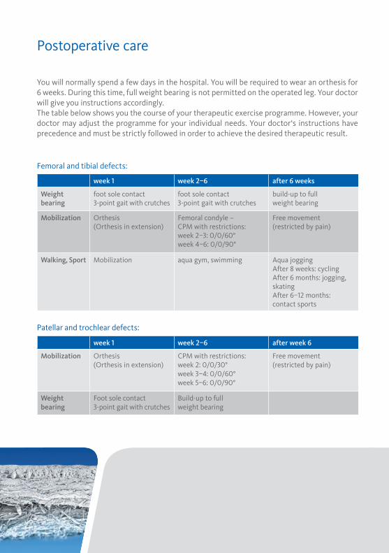
You will normally spend a few days in the hospital. You will be required to wear an orthesis for 6 weeks. During this time, full weight bearing is not permitted on the operated leg. Your doctor will give you instructions accordingly.The table below shows you the course of your therapeutic exercise programme. However, your doctor may adjust the programme for your individual needs. Your doctor‘s instructions have precedence and must be strictly followed in order to achieve the desired therapeutic result.
Femoral and tibial defects:
Patellar and trochlear defects:
week � week ��� after 6 weeks
Weight bearing
foot sole contact�-point gait with crutches
foot sole contact�-point gait with crutches
build-up to full weight bearing
Mobilization Orthesis(Orthesis in extension)
Femoral condyle – CPM with restrictions:week ���: ������°week ���: ������°
Free movement (restricted by pain)
Walking, Sport Mobilization aqua gym, swimming Aqua joggingAfter 8 weeks: cyclingAfter 6 months: jogging, skatingAfter ���� months: contact sports
week � week ��� after week 6
Mobilization Orthesis(Orthesis in extension)
CPM with restrictions:week �: ������°week ���: ������°week ���: ������°
Free movement(restricted by pain)
Weight bearing
Foot sole contact�-point gait with crutches
Build-up to full weight bearing
AMIC® Patient InformationAutologous Matrix Induced Chondrogenesis
����
�.��
���
��e
Manufacturer and Distribution in SwitzerlandGeistlich Pharma AGBusiness Unit SurgeryBahnhofstrasse 40CH-���� Wolhusenwww.geistlich.com
Postoperative care
France Geistlich Pharma France SAParc des Nations��� rue de la Belle EtoileBP ���������� Roissy CDG Cedexwww.geistlich.fr
GermanyGeistlich BiomaterialsVertriebsgesellschaft mbHSchneidweg �D-����� Baden-Badenwww.geistlich.de
ItalyGeistlich Biomaterials Italia S.r.lVia A. Fogazzaro ��I-����� Thiene VIwww.geistlich.it
United KingdomGeistlich Sons Ltd.Long LaneChester CH� � PFwww.geistlich.co.uk
Your Surgeon
Your doctor has identified damage to your articular cartilage. To repair the damage, he has re-commended surgical treatment using the AMIC® method. AMIC® (Autologous Matrix Induced Chondrogenesis) is a biological method for cartilage repair. This innovative technique uses the body’s own healing potential and the regenerative capacity of mesenchymal stem cells found in the bone marrow to grow into chondrocytes or cartilage tissue. The cartilage defect is accessed through the opening to the joint (arthrotomy). Special instruments are used to remove the degenerative cartilage tissue.
Several perforations (microfractures) into the subchondral bone plate are made with a sharp instrument (awl or pick). The defect is then covered with the Chondro-Gide® matrix. Through the perforations, bone marrow elements including stem cells and growth factors are released into the defect. Chondro-Gide® stabilizes and protects the migrating cells and thereby provides an ideal environment for the generation of new cartilage tissue.
If cartilage defects are not treated, there is the risk that they can continue to spread and ultimately lead to arthrosis.
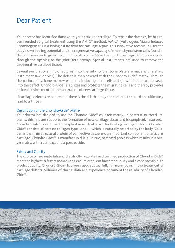
Description of the Chondro-Gide® MatrixYour doctor has decided to use the Chondro-Gide® collagen matrix. In contrast to metal im-plants, this implant supports the formation of new cartilage tissue and is completely resorbed.Chondro-Gide® is a CE-marked implant or medical device for treating cartilage defects. Chondro-Gide® consists of porcine collagen type I and III which is naturally resorbed by the body. Colla-gen is the main structural protein of connec tive tissue and an important component of articular cartilage. Chondro-Gide® is manufactured in a unique, patented process which results in a bila-yer matrix with a compact and a porous side.
Safety and QualityThe choice of raw materials and the strictly regulated and certified production of Chondro-Gide® meet the highest safety standards and ensure excellent biocompatibility and a con sistently high product quality. Chondro-Gide® has been used successfully for many years in the treatment of cartilage defects. Volumes of clinical data and experience docu ment the reliability of Chondro-Gide®.
MicrofracturingA sharp awl or pick is used and, if necessary, a mallet to perforate the subchondral bone plate every ��� mm to allow bone marrow ma-terial to flow into the defect.
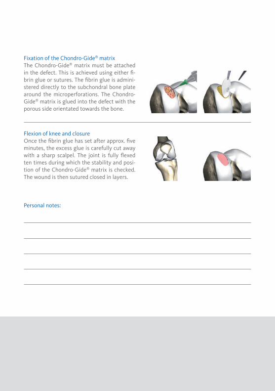
Fixation of the Chondro-Gide® matrixThe Chondro-Gide® matrix must be attached in the defect. This is achieved using either fi-brin glue or sutures. The fibrin glue is admini-stered directly to the subchondral bone plate around the microperforations. The Chondro-Gide® matrix is glued into the defect with the porous side orientated towards the bone.
Flexion of knee and closureOnce the fibrin glue has set after approx. five minutes, the excess glue is carefully cut away with a sharp scalpel. The joint is fully flexed ten times during which the stability and posi-tion of the Chondro-Gide® matrix is checked. The wound is then sutured closed in layers.
Personal notes:
Surgical TechniqueDear Patient
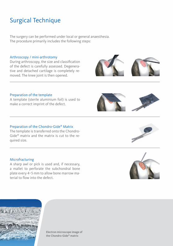
The surgery can be performed under local or general anaesthesia.The procedure primarily includes the following steps:
Arthroscopy / mini-arthrotomyDuring arthroscopy, the size and classification of the defect is carefully assessed. Degenera-tive and detached cartilage is completely re-moved. The knee joint is then opened.
Preparation of the templateA template (sterile aluminium foil) is used to make a correct imprint of the defect.
Preparation of the Chondro-Gide® MatrixThe template is transferred onto the Chondro-Gide® matrix and the matrix is cut to the re-quired size.
Electron microscope image of the Chondro-Gide® matrix
Your doctor has identified damage to your articular cartilage. To repair the damage, he has re-commended surgical treatment using the AMIC® method. AMIC® (Autologous Matrix Induced Chondrogenesis) is a biological method for cartilage repair. This innovative technique uses the body’s own healing potential and the regenerative capacity of mesenchymal stem cells found in the bone marrow to grow into chondrocytes or cartilage tissue. The cartilage defect is accessed through the opening to the joint (arthrotomy). Special instruments are used to remove the degenerative cartilage tissue.
Several perforations (microfractures) into the subchondral bone plate are made with a sharp instrument (awl or pick). The defect is then covered with the Chondro-Gide® matrix. Through the perforations, bone marrow elements including stem cells and growth factors are released into the defect. Chondro-Gide® stabilizes and protects the migrating cells and thereby provides an ideal environment for the generation of new cartilage tissue.
If cartilage defects are not treated, there is the risk that they can continue to spread and ultimately lead to arthrosis.
Description of the Chondro-Gide® MatrixYour doctor has decided to use the Chondro-Gide® collagen matrix. In contrast to metal im-plants, this implant supports the formation of new cartilage tissue and is completely resorbed.Chondro-Gide® is a CE-marked implant or medical device for treating cartilage defects. Chondro-Gide® consists of porcine collagen type I and III which is naturally resorbed by the body. Colla-gen is the main structural protein of connec tive tissue and an important component of articular cartilage. Chondro-Gide® is manufactured in a unique, patented process which results in a bila-yer matrix with a compact and a porous side.
Safety and QualityThe choice of raw materials and the strictly regulated and certified production of Chondro-Gide® meet the highest safety standards and ensure excellent biocompatibility and a con sistently high product quality. Chondro-Gide® has been used successfully for many years in the treatment of cartilage defects. Volumes of clinical data and experience docu ment the reliability of Chondro-Gide®.
MicrofracturingA sharp awl or pick is used and, if necessary, a mallet to perforate the subchondral bone plate every ��� mm to allow bone marrow ma-terial to flow into the defect.
Fixation of the Chondro-Gide® matrixThe Chondro-Gide® matrix must be attached in the defect. This is achieved using either fi-brin glue or sutures. The fibrin glue is admini-stered directly to the subchondral bone plate around the microperforations. The Chondro-Gide® matrix is glued into the defect with the porous side orientated towards the bone.
Flexion of knee and closureOnce the fibrin glue has set after approx. five minutes, the excess glue is carefully cut away with a sharp scalpel. The joint is fully flexed ten times during which the stability and posi-tion of the Chondro-Gide® matrix is checked. The wound is then sutured closed in layers.
Personal notes:
Surgical TechniqueDear Patient
The surgery can be performed under local or general anaesthesia.The procedure primarily includes the following steps:
Arthroscopy / mini-arthrotomyDuring arthroscopy, the size and classification of the defect is carefully assessed. Degenera-tive and detached cartilage is completely re-moved. The knee joint is then opened.
Preparation of the templateA template (sterile aluminium foil) is used to make a correct imprint of the defect.
Preparation of the Chondro-Gide® MatrixThe template is transferred onto the Chondro-Gide® matrix and the matrix is cut to the re-quired size.
Electron microscope image of the Chondro-Gide® matrix
You will normally spend a few days in the hospital. You will be required to wear an orthesis for 6 weeks. During this time, full weight bearing is not permitted on the operated leg. Your doctor will give you instructions accordingly.The table below shows you the course of your therapeutic exercise programme. However, your doctor may adjust the programme for your individual needs. Your doctor‘s instructions have precedence and must be strictly followed in order to achieve the desired therapeutic result.
Femoral and tibial defects:
Patellar and trochlear defects:
week � week ��� after 6 weeks
Weight bearing
foot sole contact�-point gait with crutches
foot sole contact�-point gait with crutches
build-up to full weight bearing
Mobilization Orthesis(Orthesis in extension)
Femoral condyle – CPM with restrictions:week ���: ������°week ���: ������°
Free movement (restricted by pain)
Walking, Sport Mobilization aqua gym, swimming Aqua joggingAfter 8 weeks: cyclingAfter 6 months: jogging, skatingAfter ���� months: contact sports
week � week ��� after week 6
Mobilization Orthesis(Orthesis in extension)
CPM with restrictions:week �: ������°week ���: ������°week ���: ������°
Free movement(restricted by pain)
Weight bearing
Foot sole contact�-point gait with crutches
Build-up to full weight bearing
AMIC® Patient InformationAutologous Matrix Induced Chondrogenesis
����
�.��
���
��e
Manufacturer and Distribution in SwitzerlandGeistlich Pharma AGBusiness Unit SurgeryBahnhofstrasse 40CH-���� Wolhusenwww.geistlich.com
Postoperative care
France Geistlich Pharma France SAParc des Nations��� rue de la Belle EtoileBP ���������� Roissy CDG Cedexwww.geistlich.fr
GermanyGeistlich BiomaterialsVertriebsgesellschaft mbHSchneidweg �D-����� Baden-Badenwww.geistlich.de
ItalyGeistlich Biomaterials Italia S.r.lVia A. Fogazzaro ��I-����� Thiene VIwww.geistlich.it
United KingdomGeistlich Sons Ltd.Long LaneChester CH� � PFwww.geistlich.co.uk
Your Surgeon