post-transplant lymphoproliferative disorder - case presentation - alison jazwinski, md flinders...
TRANSCRIPT

Post-transplant Lymphoproliferative Disorder
- Case Presentation -
Post-transplant Lymphoproliferative Disorder
- Case Presentation -
Alison Jazwinski, MD
Flinders Medical Center
Adelaide, South Australia

PatientPatient
• 43 yo male • HPI:
• Sent to FMC with persistently elevated liver enzymes on routine lab check (ALT 971, AST 521)
• On admission reported taking 2 Paracetamol tablets every 2 hours for a headache
• Denied abdominal pain, melena/hematemesis, vomiting, diarrhea
• Also denied numbness, weakness, difficultly swallowing or speaking
PatientPatient
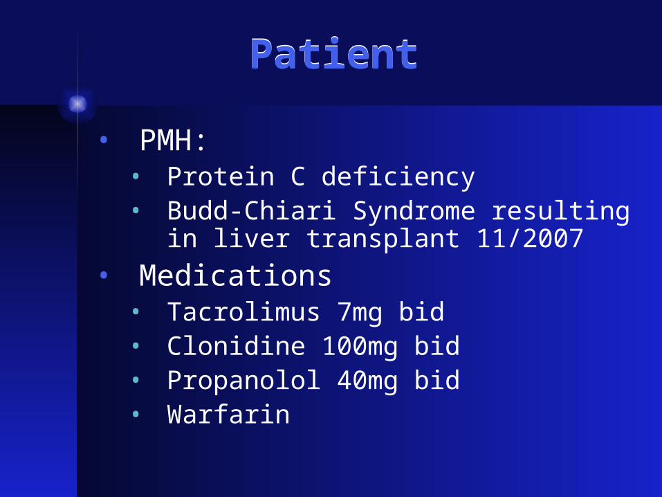
• PMH:• Protein C deficiency• Budd-Chiari Syndrome resulting in liver
transplant 11/2007
• Medications• Tacrolimus 7mg bid• Clonidine 100mg bid• Propanolol 40mg bid• Warfarin

Physical ExamPhysical Exam
• Vitals: BP 130/78, HR 80, RR 16, temp 36.8• Gen: well appearing male in NAD• HEENT: no scleral icterus, MMM• Neck: no lymphadenopathy• CV: RRR no M/R/G• Lungs; CTAB no W/R/R• Abdomen: soft, mildly TTP RUQ, no rebound/guarding,
NABS• Extrem: no edema• Neuro: CN II-XII intact, strength 5/5 all muscle groups,
reflexes 2+ throughout, gait normal, sensation intact to light touch, pinprick, vibration
Admission LabsAdmission Labs
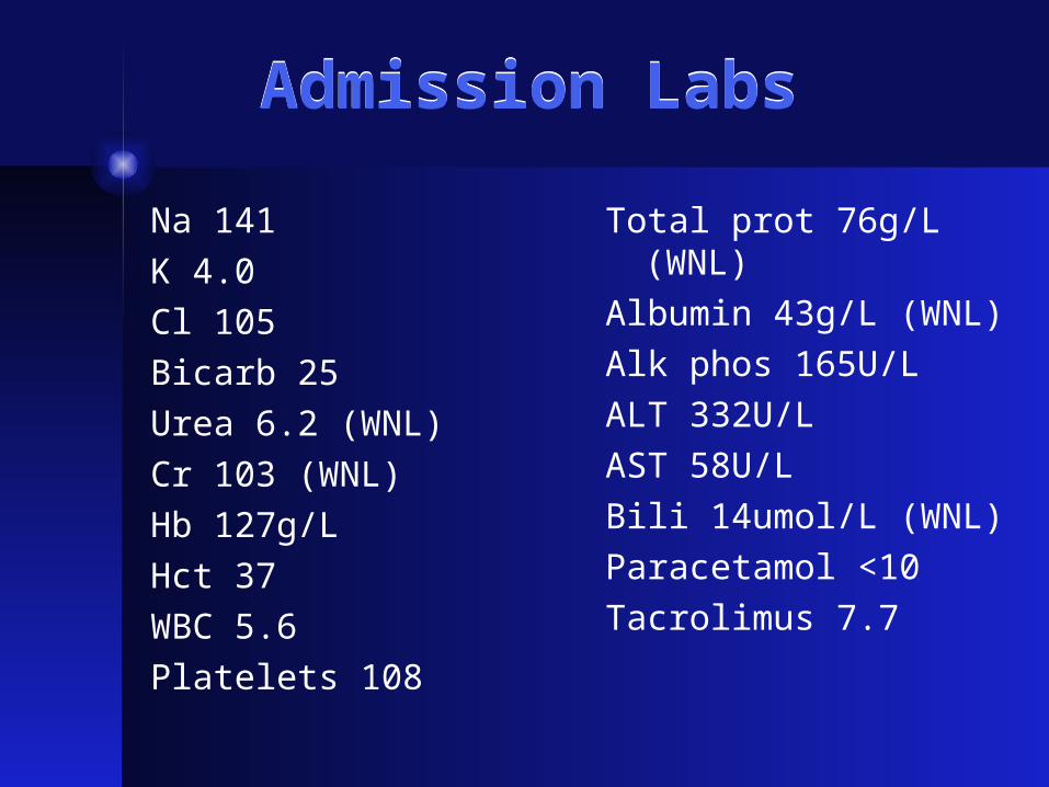
Na 141
K 4.0
Cl 105
Bicarb 25
Urea 6.2 (WNL)
Cr 103 (WNL)
Hb 127g/L
Hct 37
WBC 5.6
Platelets 108
Total prot 76g/L (WNL)
Albumin 43g/L (WNL)
Alk phos 165U/L
ALT 332U/L
AST 58U/L
Bili 14umol/L (WNL)
Paracetamol <10
Tacrolimus 7.7

PatientPatient
• LFT abnormalities thought to be related to Paracetamol over-use.
• He was using it for a headache… why did he have a headache?
• Further evaluation revealed…
Head CTHead CT

Head CT Head CT
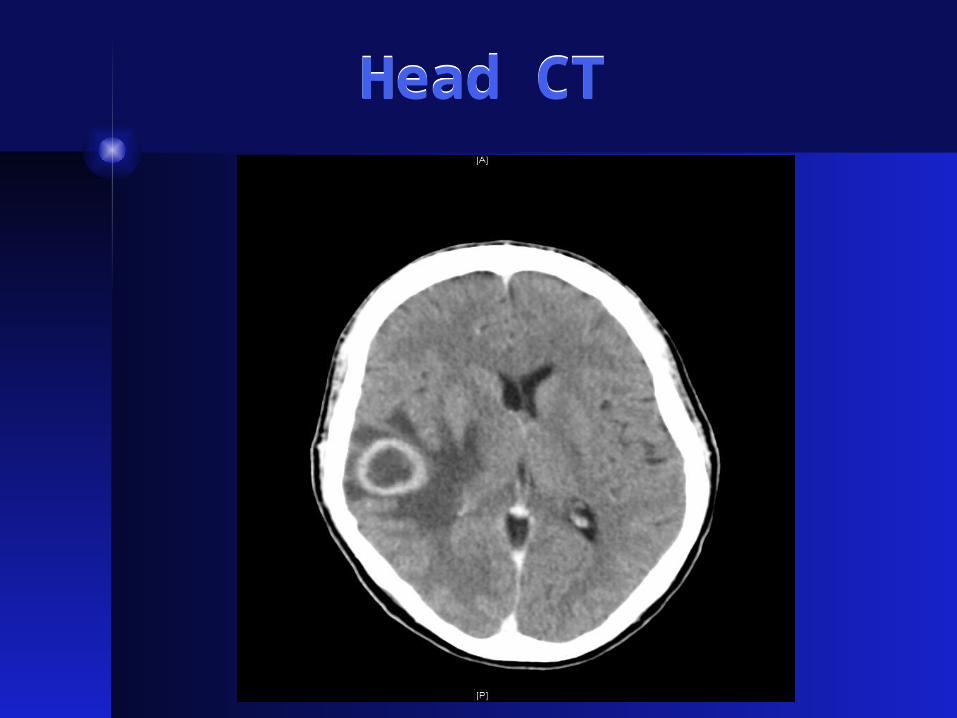
28mm ring enhancing mass in right temporal lobe with moderate surrounding vasogenic edema. There is 6mm midline shift and effacement of overlying cerebral sulci.

Brain MRIBrain MRI

Brain MRIBrain MRI
Solitary, thick walled ring enhancing lesion in right temporal lobe measuring 2.8cm x 2.3 cm x 1.8cm associated with extensive vasogenic edema and adjacent mass effect. Appearances are indeterminate, could represent a cerebral abscess however a high-grade glioma or solitary metastasis may also give this appearance.
When spectroscopy was added, the findings were keeping with a high grade primary cerebral neoplasm such as a GBM.

Differential DiagnosisDifferential Diagnosis
• Infection• Bacterial abscess• Cryptococcus• Toxoplasma
• Malignancy• Lymphoma• Primary CNS tumor• Metastatic disease

Further stepsFurther steps
• Patient was initiated on dexamethasone and loaded with phenytoin for seizure proph
• CT chest/abdomen/pelvis negative for source of primary malignancy
• On to surgery with resection
• Cultures sent for AFB, cryptococcus, toxoplasma, and bacterial culture, all returned negative

HistologyHistology
• Features most in keeping with an EBV driven post-transplant lymphoproliferative disorder with no convincing monoclonality identified on immunoperoxidase stains and associated with considerable tissue necrosis

PTLDPTLD
• Mostly large cell lymphomas• Most B cell type
• Extranodal involvement in 30-70%
• Appears to be related to EBV inducing B cell proliferation in setting of chronic immunosuppression
• PTLD cells are of host origin in the majority of cases
Transplantation 2006;81:888Transplantation 1990;49:1080

Putative Checkpoints in the EBV Life Cycle That Might Give Rise to Lymphoma
Putative Checkpoints in the EBV Life Cycle That Might Give Rise to Lymphoma
N Engl J Med 350:1328, March 25, 2004

Forms of DiseaseForms of Disease
• Benign polyclonal lymphoproliferation (55%)• Infectious mono-type illness
• Develops 2-8 weeks after immunosuppression initiated
• Polyclonal B cell proliferation with normal cytogenetics
• Polyclonal lymphoproliferation with early malignant transformation (30%)
• Localized solid tumors (15%)• Monoclonal B cell proliferation with malignant
cytogenetic abnormalities
Am J Pathol 1988; 133:173

Areas of InvolvementAreas of Involvement
• Gastrointestinal tract
• Lungs
• Skin
• Liver
• CNS (20-25%)
• Allograft lesions (20-25%)
Transplantation 1995; 59:240

Treatment ApproachesTreatment Approaches
• Reduction in immunosuppression
• Antiviral agents
• Chemotherapy
• Immune globulin
• Surgical resection
• Radiation
• Interferon-alpha
Pediat Transplant 2001; 5:198

Reduction of ImmunosuppressionReduction of Immunosuppression
• Most will resolve with this• Best response among those with early disease
where immunosuppression is a major contributing factor
• Depends on severity of disease• Could reduce Prednisone to maintenance doses (7.5-
10mg) and stop other agents
• Could reduce Cyclosporine or Tacrolimus by 50% and discontinue Azathioprine or MMF
• Risk is allograft rejection
Transplantation 1999; 68:1517

Other methods of treatmentOther methods of treatment
• Only case reports at this time
• Largely dependent on severity of disease and treatment center

Thanks!