post screen ryan f. donnelly and eneko...
TRANSCRIPT

Reviews�P
OST
SCREEN
REVIEWS Drug Discovery Today �Volume 23, Number 5 �May 2018
Microarray patches: potentially usefuldelivery systems for long-actingnanosuspensionsRyan F. Donnelly and Eneko Larrañeta
School of Pharmacy, Queen’s University Belfast, Medical Biology Centre, 97 Lisburn Road, Belfast BT9 7BL, UK
Long-acting drug nanosuspension formulations are coming to the fore as controlled release strategies for
several medical conditions and as a preventative measure against HIV infection. However, such delivery
systems must, by necessity, be given by hypodermic injection, typically into muscle. This poses problems
for patients who are needle-phobic, given that injections have to be administered on a weekly or
monthly basis. Needle-stick injuries, inappropriate reuse of needles, and poor disposal practices are
major challenges in developing countries. Dissolving microneedles (MNs) are capable of delivering high
drug doses, if suitably designed and formulated, and are also capable of delivering nanoparticles (NPs)
into viable skin. Given that such microneedles are minimally invasive and self-disabling, the potential
for major enhancement in patient care and compliance exists. In this review, we explore the key
considerations in the development of these combination drug delivery systems.
IntroductionMNs are minimally invasive devices that by-pass the stratum
corneum (SC) barrier of the skin, thus granting access to the dermal
microcirculation and antigen-presenting cells located in the infe-
rior layers of the skin. MNs comprise multiple micron-scale pro-
jections positioned on a baseplate in various geometries. When
applied to the skin, they painlessly puncture the SC, creating
microscopic aqueous channels through which drugs can diffuse.
MNs are long enough to penetrate the SC (50–900 Mm in height,
up to 2000 MN cm�2), but short enough to avoid stimulation of
dermal nerves. They are manufactured from various materials (e.
g., silicon, metal, or polymer using microfabrication techniques
[1–3]. Originally described in a 1970s patent and finally realized in
practical terms during the late 1990s [4], MNs (Fig. 1) are currently
of great interest because of several advantages that they have over
traditional methods of drug delivery. Some of these advantages
include the ability to painlessly administer the active pharmaceu-
tical ingredient (API), bypass the hepatic first-pass metabolism,
and the extension of the variety of drug types that can be delivered
both intra- and transdermally.
Corresponding author: Donnelly, R.F. ([email protected])
1026 www.drugdiscoverytoday.com
The first substance delivered using MNs was the low-molecular-
weight compound, calcein [4]. Multiple investigations rapidly
followed, leading to the current ever-growing body of evidence
for the significant drug delivery capabilities of MNs. Although a
variety of strategies has been used (Fig. 2), MNs fabricated from
silicon and metal continue to be extensively investigated for drug
delivery. Their use typically involves the pretreatment of skin,
followed by application of a topical solution, gel, or patch contain-
ing the drug to be delivered [5–7]. Although this conventional
‘poke and patch’ methodology has progressed somewhat from the
original studies, it has been recognized that such a cumbersome
two-step application process is a major drawback [8].
To create a one-step application process, solid MNs have been
coated with the material to be delivered. Coated MNs have been
used for the delivery of several different compounds, including
fluorescein sodium [9], salmon calcitonin [10], desmopressin [11],
parathyroid hormone (PTH) [12], and DNA/RNA [13,14], among
others. Aside from this, because of the limited drug-loading ca-
pacity of this method, coated MNs are more frequently used for the
delivery of highly potent molecules and vaccines.
Research into hollow MNs has focused mainly on array design
and characterization, with several sophisticated engineering strat-
egies presented [15–18]. However, a major limitation to their use is
1359-6446/ã 2017 Elsevier Ltd. All rights reserved.https://doi.org/10.1016/j.drudis.2017.10.013

Drug Discovery Today �Volume 23, Number 5 �May 2018 REVIEWS
(b)(a) (c)
(d) (f)(e)
(g) (i)(h) (j)
Drug Discovery Today
FIGURE 1
Microneedle (MN) designs. Wet-etched silicon MNs approximately 280 Mm in height suitable for coating with capture proteins or antibodies (a,b). MNsapproximately 600 Mm in height produced from micromolding of aqueous gels of poly(methylvinylether-co-maleic acid) and poly(ethylene glycol) (PEG) 10 000that swell in skin to capture skin interstitial fluid (c). Poly(carbonate) MNs approximately 1000 Mm in height with a 100 Mm off-center through-hole suitable forblood extraction (d), Orion Helium-ion microscope images of 7-MN arrays of this design (e) and 3D optical coherence tomographic representation of these MNsin situ, following insertion into excised neonatal porcine skin in vitro (f). Swollen hydrogel-forming MNs approximately 600 Mm in height produced frommicromolding of aqueous gels of poly(methylvinylether-co-maleic acid) and PEG 10 000 completely intact following removal from skin (g) and MNsapproximately 280 Mm in height produced from micromolding of aqueous gels of poly(methylvinylether-co-maleic acid) and glycerol following removal fromskin (h). The latter type of hydrogel-forming MNs following uptake of meso-tetra (N-methyl-4-pyridyl) porphine tetra tosylate in vitro (I). Hydrogel-forming MNsapproximately 600 Mm in height produced from micromolding of aqueous gels of poly(methylvinylether-co-maleic acid) and PEG 10 000 swelling in human skinin vivo (j).
Review
s� P
OST
SCREE
N
the potential blockage of the MN bore by compressed dermal tissueupon insertion, reducing drug release [18].
Dissolving MNs have been used to deliver several small-mole-
cule drugs, including caffeine, lidocaine, theophylline, and met-
ronidazole [19]. Additionally, they have been used to specifically
target various clinical needs by the delivery of several biopharma-
ceutical molecules, including low-molecular-weight heparins [20],
insulin [21], leuprolide acetate [22], erythropoietin [23], and hu-
man growth hormone [24]. A central criticism of the dissolving
platform was the perceived inability to deliver therapeutically
relevant doses of low-potency drug substances [25]. However,
McCrudden et al. have taken steps to address these concerns,
having successfully delivered therapeutically relevant doses of
ibuprofen sodium in a rat model [26].
Hydrogel-forming MNs have been demonstrated to proficiently
deliver both small molecules, such as metronidazole and theoph-
ylline, and larger molecules, such as insulin and proteins [8]. The
benefit of the hydrogel system is that the MN swelling rate can be
controlled by altering the polymer cross-linked density, thus
conferring the ability to govern drug release rate, which can be
tailored for specific drugs. The delivered dose is not limited by
what can be loaded into the needles themselves, given that the
drug is contained within an attached solid drug reservoir. Accord-
ingly, sustained delivery of high drug doses is readily achievable.
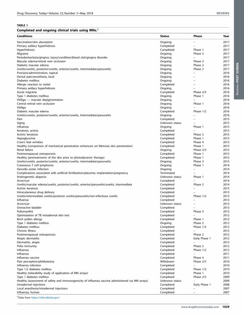
As can be seen from Table 1, there has been a range of completed
and ongoing clinical trials involving the use of MNs. Although
several investigations involving humans have considered the per-
ception, safety, and practical applications of MN technology
www.drugdiscoverytoday.com 1027

REVIEWS Drug Discovery Today �Volume 23, Number 5 �May 2018
(a)
(b)
(c)
(d)
(e)
Stratum corneumEpidermis
Drug Discovery Today
FIGURE 2
Microneedle (MN) delivery strategies. A schematic representation of five different MN types used to facilitate drug delivery transdermally. (a) Solid MNs forincreasing the permeability of a drug formulation by creating microholes across the skin. (b) Coated MNs for rapid dissolution of the coated drug into the skin. (c)Dissolvable MNs for rapid or controlled release of the drug incorporated within the MNs. (d) Hollow MNs used to puncture the skin and enable release of a liquiddrug following active infusion or diffusion of the formulation through the needle bores. (e) Hydrogel-forming MNs take up interstitial fluids from the tissue,inducing diffusion of the drug located in a patch through the swollen microprojections.
Reviews�P
OST
SCREEN
[27,28] and a few human volunteer trials have studied MN-medi-
ated transdermal drug delivery [12,29], the predominant focus in
the field to date has been on vaccines [30–33]. This is hardly
surprising, given the potential for a stable, dry-state formulation,
the avoidance of needle-stick injuries common with hypodermic
syringes, dose-sparing through direct targeting of the abundance
of professional antigen-presenting cells in viable skin, and the self-
disabling nature of dissolving MNs. Consequently, several clinical
trials covering the use of MNs for vaccine delivery are detailed in
Table 1. Influenza intradermal vaccination has been extensively
studied because there is a constant demand for a seasonal vaccine.
These trials were conducted around the world in thousands of
volunteers, including a randomized, open-label Phase 2 clinical
trial (978 healthy adults) [34], a Phase 3 randomized, double-blind
trial (2255 healthy adults) [35], and a Phase 2/3 trial in older
individuals (aged 60 years and older) [36]. The obtained results
suggested that the MN-based vaccine provided an equivalent (and,
in some cases, superior) immune response compared with the
conventional intramuscular vaccine.
MN vaccines have the potential to revolutionize vaccination,
especially in the developing world. However, in studies where the
delivery of therapeutic drug substances using MNs has been ex-
emplified, the focus has tended to be on illustration of the capa-
bility of MNs to deliver a substance with particular
1028 www.drugdiscoverytoday.com
physicochemical characteristics and little mention is typically
made of the actual amount delivered or its relevance to therapeutic
human doses.
Vaccines tend to be potent and, thus, delivery of even micro-
gram quantities of antigen, antigen/adjuvant combination, virus-
like particle, or even DNA is often sufficient to elicit an immune
response, especially when targeted to the viable epidermis and/or
dermis. This means that small, postage stamp-sized MN patches
that can be inserted into the skin by fairly gentle thumb pressure
are sufficient to achieve successful vaccination.
In addition to MN-mediated vaccination, the delivery of insulin
using MNs has been extensively studied in clinical trials (Table 1).
The use of MNs for this purpose provided better patient compli-
ance compared with traditional subcutaneous injections. In addi-
tion, clinical trials showed that the MN-mediated administration
of insulin enabled faster uptake of this molecule and equivalent
bioavailability/blood-glucose effects compared with subcutaneous
administrations [37,38].
The use of MN devices for clinically relevant does in human
volunteers has not been extensively studied, as shown in Table 1.
However, recent work in the field has focused strongly on the
transdermal delivery of therapeutically relevant doses of drugs
using MN patches. Plasma levels in animal models have been
measured and extrapolated to estimate suitable patch sizes for
Drug Discovery Today �Volume 23, Number 5 �May 2018 REVIEWS
TABLE 1
Completed and ongoing clinical trials using MNs.a
Conditions Status Phase Year
Vaccination/skin absorption Ongoing – 2017Primary axillary hyperhidrosis Completed – 2017Hyperhidrosis Completed Phase 1 2017Migraine Ongoing Phase 3 2017Periodontoclasia/gingiva; injury/condition/blood clot/gingiva disorder Ongoing – 2017Macular edema/retinal vein occlusion Ongoing Phase 3 2017Diabetic macular edema Ongoing Phase 2 2017Uveitis/uveitis, posterior/uveitis, anterior/uveitis, intermediate/panuveitis Ongoing Phase 3 2017Psoriasis/administration, topical Ongoing – 2016Dental pain/anesthesia, local Ongoing – 2016Diabetes mellitus Ongoing – 2016Allergic reaction to nickel Completed – 2016Primary axillary hyperhidrosis Ongoing – 2016Acute migraine Completed Phase 2/3 2016Type 1 diabetes mellitus Ongoing Phase 1 2016Vitiligo — macular depigmentation Ongoing – 2016Central retinal vein occlusion Ongoing Phase 1 2016Vitiligo Ongoing – 2016Diabetic macular edema Completed Phase 1/2 2016Uveitis/uveitis, posterior/uveitis, anterior/uveitis, Intermediate/panuveitis Ongoing – 2016Pain Completed – 2015Aging Unknown status – 2015Influenza Ongoing Phase 1 2015Keratosis, actinic Completed – 2015Actinic keratosis Completed Phase 2 2015Hypoglycemia Completed Phase 1 2015Crow’s feet wrinkles Completed Phase 4 2015Healthy (comparison of mechanical penetration enhancers on Metvixia skin penetration) Completed Phase 1 2015Renal failure Ongoing Phase 2/3 2015Postmenopausal osteoporosis Completed Phase 1 2015Healthy (pretreatments of the skin prior to photodynamic therapy) Completed Phase 1 2015Uveitis/uveitis, posterior/uveitis, anterior/uveitis, intermediate/panuveitis Ongoing Phase 3 2015Cutaneous T cell lymphoma Ongoing Phase 1 2014Varicella zoster infection Ongoing – 2014Complications associated with artificial fertilization/placenta; implantation/pregnancy Terminated – 2014Androgenetic alopecia Unknown status Phase 1 2014Acne/scar Completed – 2014Uveitis/macular edema/uveitis, posterior/uveitis, anterior/panuveitis/uveitis, intermediate Completed Phase 2 2014Actinic keratosis Completed – 2013Intracutaneous drug delivery Completed – 2013Uveitis/intermediate uveitis/posterior uveitis/panuveitis/non-infectious uveitis Completed Phase 1/2 2013Influenza Completed – 2013Acne/scar Unknown status – 2013Overactive bladder Completed – 2013Poliomyelitis Completed Phase 3 2013Optimization of TB intradermal skin test Completed – 2012Birch pollen allergy Completed Phase 1 2012Type 1 diabetes mellitus Ongoing Phase 2 2012Diabetes mellitus Completed Phase 1/2 2012Chronic Illness Completed – 2012Postmenopausal osteoporosis Completed Phase 2 2012Atopic dermatitis Completed Early Phase 1 2012Dermatitis, atopic Completed – 2012Polio immunity Completed Phase 2 2012Influenza Completed Phase 1/2 2012Influenza Completed – 2011Influenza vaccine Completed Phase 4 2011Pain perception/phlebotomy Withdrawn Phase 2/3 2010Influenza infection Completed – 2010Type 1/2 diabetes mellitus Completed Phase 1/2 2010Healthy (tolerability study of application of MN arrays) Completed Phase 1 2010Type 1 diabetes mellitus Completed Phase 2/3 2009Healthy (assessment of safety and immunogenicity of influenza vaccine administered via MN arrays) Unknown status – 2009Intradermal Injections Completed Early Phase 1 2008Local anesthesia/intradermal Injections Completed – 2007Influenza, human Completed – 2007aData from https://clinicaltrials.gov/.
www.drugdiscoverytoday.com 1029
Review
s� P
OST
SCREE
N

REVIEWS Drug Discovery Today �Volume 23, Number 5 �May 2018
Reviews�P
OST
SCREEN
the achievement of therapeutic plasma levels in humans
[18,20,22]. Given that most commonly used small-molecule drugs
tend to require oral doses in the range of tens to hundreds of
milligrams daily, the patch sizes estimated have ranged from
10 cm2 to 30 cm2. Such patch sizes are well within the range of
marketed transdermal patches. Indeed, Novartis market
Nicotinell1 (nicotine) patches of 30 cm2 (www.nicotinell.co.uk),
whereas Janssen market Duragesic1 CII (fentanyl) patches of 32
and 42 cm2 (www.duragesic.com). It was recently shown that
human volunteers can insert the MNs of large patches as efficiently
and reliably as those on smaller patches, thus making such a
delivery system viable [39].
The ability to deliver high drug doses has raised interest in the
possibility of delivering long-acting drug nanosuspensions intra-
dermally without the need for a conventional hypodermic needle
injection. Nanosuspensions are, in simple terms, aqueous suspen-
sions of NPs made from solid-drug crystals stabilized with a coating
of surfactant and/or polymer. They are typically up to 90% w/w
pure drug and can be prepared by ‘top-down’ (e.g., wet milling or
high-pressure homogenization) or ‘bottom-up’ (solution-based
nanoprecipitation) methods [40–42]. Preparation of solid-drug
NPs can be used to improve aqueous solubility for applications
in enhanced oral or pulmonary bioavailability. However, if one
selects a potent drug with relatively poor aqueous solubility and
can tailor particle size appropriately, an injectable preparation
capable of sustained delivery of clinically relevant drug doses
for up to 3 months from a single injection can be produced.
The particles are deposited as a depot and slowly release drug
for absorption into the systemic circulation as they dissolve in
interstitial fluid. If enough particles can be deposited, then thera-
peutic plasma levels can be maintained for prolonged periods [40–
42].
Originally used for the hormonal treatment of endometriosis,
long-acting drug nanosuspensions then found use in the manage-
ment of schizophrenia. Most interestingly, however, this formu-
lation type is now undergoing clinical trials for the prevention and
treatment of HIV [42]. It is because of this exciting new indication
that the mode of nanosuspension delivery has come sharply into
focus. Oral administration of nanosuspensions does not allow
sustained drug absorption over weeks or months and, thus, such
products have typically been administered subcutaneously or, if
higher volumes are required (up to 2.7 ml from a single injection)
to deliver therapeutic doses, intramuscularly. Hypodermic needle
injections cause problems in developing countries, in particular,
because of the lack of skilled healthcare personnel, frequency of
needle-stick injuries, inappropriate reuse of needles, and poor
disposal practices. An alternative, minimally invasive, self-dis-
abling, delivery system that avoids such problems would not only
be particularly useful in low-resource settings, but could also
improve compliance in developed countries.
Solid MN arrays have been used to enhance the delivery of
microparticle and NP suspensions. However, in these cases, the
application process requires the application of a solid MN array
before administration of a formulation containing the suspension
into the treated area [17,43,44]. A two-step process is not ideal
from a patient point of view.
Several examples of hollow MNs for the delivery of nanosus-
pensions can be found in the literature [45–48]. These devices have
1030 www.drugdiscoverytoday.com
been extensively studied for the intraocular administration of NPs
and microparticles [46–48]. However, they require complex
pumping systems, which are expensive and difficult to manufac-
ture and could present issues for correct use in the absence of
skilled healthcare workers in developing countries [2]. The safe
and hygienic disposal of such systems in low-resource settings
might also prove problematic. Coated MNs have limited dosing
capacity and are also removed from the skin intact, therefore
presenting challenges for disposal [2]. Diffusion of hydrophobic
NPs through swollen hydrogel matrices is likely to be poor, thus
ruling that system out [8]. However, dissolving MNs have con-
siderable promise in the intradermal delivery of long-acting
nanosuspensions.
The literature contains a variety of examples of the use of
dissolving MNs for nanosuspension administration [49–51]. How-
ever, most of these papers focused on the delivery of encapsulated
vaccines [49,52,53] or were designed as a proof of concept to show
the capabilities of MNs to enhance the delivery of particulate
formulations through the SC [49]. Consequently, basic technical
and scientific challenges are being addressed currently, such as: the
possibility of manufacturing two-layered MN arrays concentrating
suspensions in the needle tips or the study [54,55] of the fate of NPs
administered intradermally [55]. Nevertheless, the delivery of
therapeutically relevant doses of nanomedicines using dissolving
MNs remains relatively unexplored.
Advantages of a dissolving MN system over conventional nee-
dle-and-syringe-based methods for the administration of long-
acting drug nanosuspensions would include: (i) no requirement
for skilled medical personnel to administer the dose; (ii) potential
for at-home use by patients; (iii) possibility for improved compli-
ance; (iv) avoidance of needle-stick injuries; (v) specialized dispos-
al not required; and (vi) possibility for enhanced storage stability
because of the dried nature of the formulations.
However, to be a realistic proposition, such a dissolving MN
system would need to be able to incorporate a high loading of
hydrophobic NPs in the needles themselves. Given the viscous
nature of the gel formed in skin upon needle dissolution, it is
unlikely that any NPs in the baseplate upon which the needles
were formed would be able to diffuse into the viable skin. Doses of
several hundred milligrams would, in most cases (apart from
hormones), have to be delivered into skin upon needle dissolu-
tion. If the patch size required to deliver such a dose would, by
necessity, be greater than that of conventional transdermal
patches, then the delivery of a particular drug would not be
feasible. However, if one considers that the MNs themselves can
weigh 10 mg for every cm2 of patch upon which they are formed
and the drug loading is a minimum of 80% w/w, then 8 mg/cm2 of
drug could be delivered upon total needle dissolution in skin. If the
drug dose for a month of treatment were 300 mg, then the patch
size would be 37.5 cm2. If the drug dose were 600 mg, then two
patches could be applied, one on each arm. This mimics the
current rilpivirine regimen for HIV prevention, where two injec-
tions are given at different sites. Given that large MN patches can
be reliably applied by human volunteers, the approach appears
feasible. However, what would be useful would be an indicator
confirming when the patch can be removed. A low-cost system
would not be able to tell when the needles had dissolved, but a
moisture sensor or time-dependent color change indicator based

Drug Discovery Today �Volume 23, Number 5 �May 2018 REVIEWS
Review
s� P
OST
SCREE
N
on average in-skin dissolution time would not add prohibitive
expense.
The needles should dissolve quickly in skin (in <1 h ideally), so
as to be convenient for patients. This means that the polymer
matrix must be carefully chosen so as to provide sufficient me-
chanical strength to allow ready puncture of the SC upon applica-
tion of relatively gentle pressure, while not being brittle, but must
not be slowly soluble in the relatively small volume of fluid
available in viable skin. Given that possibly >80% by weight of
the MN might comprise hydrophobic NPs, repulsion of interstitial
fluid could be a consideration requiring addition of disintegrants
to boost MN breakdown. Excipients chosen, including the matrix
polymer and water, must not significantly alter the characteristics
of the particles of the nanosuspension (e.g., size, charge or aggre-
gation status) or cause dissolution during manufacture or storage.
The excipients should also be biodegradable or of sufficiently low
molecular weight to allow ready clearance from the body, given
that the patch will need to be applied by the patient weekly or
monthly. Accumulation of excipients in skin or elsewhere in the
body would be undesirable and would be likely to raise regulatory
Drugnanosuspe nsion
Drugnanosuspe nsi
(a)
(c)
FIGURE 3
Microarray patches loaded with long-acting nanosuspensions. Preparation of minanosuspensions where the preformed baseplate is added after initial casting of
where the particles are loaded into the entire shaft length of the microneedles (MNloaded into the MN tips only (c).
concerns. Indeed, from a regulatory viewpoint, clinical studies
aimed at supporting market authorization would need to show
that therapeutic blood levels comparative to existing dosage forms
are achievable, even if the pharmacokinetic patterns differ.
Whether regulators would require a sterile or low bioburden
product, considering that MNs enter viable skin, is not clear as
yet. It is likely that a suitable applicator, or an in-built method of
confirming correct use (MN insertion) would also be required,
especially if the product is to be used by patients themselves in
low-resource settings. In-built feedback mechanisms would be
more desirable than applicator devices, especially if the cost could
be kept low. Such systems have recently been described [39,56,57].
Dissolving MN systems loaded with hydrophobic NPs are likely
to be manufactured in two stages to avoid waste of drug (Fig. 3a).
The baseplate could be separately produced as a polymeric film
using well-established knife-casting techniques. Mixing a freeze-
dried NP powder with a polymeric gel or adding polymer powder
directly to a concentrated nanosuspension might be required to
maximize drug loading in the needles of the array. A suitable mold
could be filled by the particle-loaded gel by a range of methods,
Drugnanosuspe nsion
on
(b)
Drug Discovery Today
croarray patches using aqueous polymeric gels containing solid-drugthe gel into the molds (a). Delivery of solid-drug nanosuspensions into skin) (b). Delivery of solid-drug nanosuspensions into skin where the particles are
www.drugdiscoverytoday.com 1031

REVIEWS Drug Discovery Today �Volume 23, Number 5 �May 2018
Reviews�P
OST
SCREEN
including applying a vacuum or utilising compressed air. Needles
and baseplate can then be merged before or after drying, using a
thin adhesive layer, if necessary. For most drugs, the NPs are likely
to have to be distributed throughout the entire length of the
needles (Fig. 3b), which could be increased to further enhance
the drug-loading capacity, while taking care not to exceed 1 mm,
in which case pain and pinprick bleeding can occur. However, for
potent hormones, the MN could be formed by two gel castings so
as to localize the drug in the needle tips alone (Fig. 3c).
Some of the manufacturing approaches previously described
have been evaluated by different research groups for the laborato-
ry-scale preparation of MNs. However, one of the important
challenges in MN technology is the scale-up the manufacturing
processes. Several studies described new processes to improve the
manufacturing of MN arrays [58,59]. Lutton et al. described a
scalable method for MN production using a micromolding proce-
dure [58]. The described method can be easily applied to the
manufacturing of nanosuspension-containing MN arrays. Never-
theless, there is another key issue that needs to be addressed to
design a suitable manufacturing process: product sterility/low
bioburden requirements [60]. As described previously, the regula-
tory bodies have not yet defined the requirements for MN products
in terms of sterility. Consequently, the manufacturing procedures
should be designed with this challenge in mind.
Concluding remarksDissolving MN delivery systems have been shown to deliver NPs
into the viable skin layers in vitro and in vivo [55] and have the
1032 www.drugdiscoverytoday.com
capability to incorporate and release high doses of undissolved
solids [26]. Accordingly, their potential as a next-generation
delivery system for emerging long-acting nanosuspensions is
becoming clear. Indeed, the first report of the development
and successful in vivo evaluation of such a combination system
was recently presented [61]. To date, MNs, including dissolving
MNs, have been rather narrowly viewed as vaccine delivery
systems for the developing world. However, the demonstrated
ability of properly formulated systems to incorporate and release
high drug doses should soon begin to change this mindset. One
could view the dissolving MN simply as a tool to deposit the ‘real’
delivery system, the drug nanosuspension, in the viable skin
layers in sufficient amounts to allow prolonged drug administra-
tion. It is likely that we will soon see an increasing number of
hydrophobic solid drug nanoparticulate systems delivered using
dissolving MNs, with a variety of therapeutic indications. For
translation to clinic and, ultimately market, methods of manu-
facture will need to be refined and scaled up [62] and the
‘microneedle’ aspect of the name of the final patch systems
removed. The term currently being used by the World Health
Organization for all microneedle-based systems is ‘microarray
patches’, or MAPs. Such an adjustment might appear minor,
but could be important for patient acceptance. Dissolving MAPs
could well be the dosage form of the near future, with needle-free
administration of long-acting drug nanosuspensions an exciting
application with a range of potential benefits for patients and
healthcare providers, especially in the poorest countries of the
world. Watch this space!
References
1 Arora, A. et al. (2008) Micro-scale devices for transdermal drug delivery. Int. J. Pharm.
364, 227–236
2 Donnelly, R.F. et al. (2012) Microneedle-Mediated Transdermal and Intradermal Drug
Delivery. Wiley
3 Tuan-Mahmood, T. et al. (2013) Microneedles for intradermal and transdermal drug
delivery. Eur. J. Pharm. Sci. 50, 623–637
4 Henry, S. et al. (1998) Microfabricated microneedles: a novel approach to
transdermal drug delivery. J. Pharm. Sci. 87, 922–925
5 Zhou, C.P. et al. (2010) Transdermal delivery of insulin using microneedle rollers in
vivo. Int. J. Pharm. 392127–392133
6 Mikolajewska, P. et al. (2010) Microneedle pre-treatment of human skin improves 5-
aminolevulininc acid (ALA)- and 5-aminolevulinic acid methyl ester (MAL)-
induced PpIX production for topical photodynamic therapy without increase in
pain or erythema. Pharm. Res. 27, 2213–2220
7 Stahl, J. et al. (2012) Microneedle pre-treatment enhances the percutaneous
permeation of hydrophilic compounds with high melting points. BMC Pharmacol.
Toxicol. 13, 5
8 Donnelly, R.F. et al. (2012) Hydrogel-forming microneedle arrays for enhanced
transdermal drug delivery. Adv. Funct. Mater. 22, 4879–4890
9 Chen, J. et al. (2013) Controllable coating of microneedles for transdermal drug
delivery. Drug Dev. Ind. Pharm. 41, 415–422
10 Tas, C. et al. (2012) Delivery of salmon calcitonin using a microneedle patch. Int. J.
Pharm. 423, 257–263
11 Cormier, M. et al. (2004) Transdermal delivery of desmopressin using a coated
microneedle array patch system. J. Control. Release 97, 503–511
12 Daddona, P.E. et al. (2011) Parathyroid hormone (1-34)-coated microneedle patch
system: clinical pharmacokinetics and pharmacodynamics for treatment of
osteoporosis. Pharm. Res. 28, 159–165
13 Pearton, M. et al. (2012) Microneedle delivery of plasmid DNA to living human skin:
formulation coating, skin insertion and gene expression. J. Control. Release 160,
561–569
14 Chong, R.H.E. et al. (2013) Gene silencing following siRNA delivery to skin via
coated steel microneedles: in vitro and in vivo proof-of-concept. J. Control. Release
166, 211–219
15 Wang, P.C. et al. (2014) Hypodermic-needle-like hollow polymer microneedle array:
fabrication and characterization. J. Micromech. Syst. 23, 991–998
16 Martanto, W. et al. (2006) Microinfusion using hollow microneedles. Pharm. Res. 23,
104–113
17 McAllister, D.V. et al. (2003) Microfabricated needles for transdermal delivery of
macromolecules and nanoparticles: fabrication methods and transport studies.
Proc. Natl. Acad. Sci. U. S. A. 100, 13755–13760
18 Gardeniers, H.J.G.E. et al. (2003) Silicon micromachined hollow microneedles for
transdermal liquid transport. J. Micromech. Syst. 12, 855–862
19 Garland, M.J. et al. (2012) Influence of skin model on in vitro performance of drug-
loaded soluble microneedle arrays. Int. J. Pharm. 434, 80–89
20 Gomaa, Y.A. et al. (2012) Laser engineered dissolving microneedles for active
transdermal delivery of nadroparin calcium. Int. J. Pharm. 82, 299–307
21 Migalska, K. et al. (2011) Laser-engineered dissolving microneedle arrays for
transdermal macromolecular drug delivery. Pharm. Res. 28, 1919–1930
22 Ito, Y. et al. (2011) Incidence of low bioavailability of leuprolide acetate after
percutaneous administration to rats by dissolving microneedles. Int. J. Pharm. 407,
126–131
23 Ito, Y. et al. (2010) Self-dissolving micropile array chip as percutaneous delivery
system of protein drug. Biol. Pharm. Bull. 33, 683–690
24 Lee, J.W. et al. (2011) Dissolving microneedle patch for transdermal delivery of
human growth hormone. Small 7, 531–539
25 Tuan-Mahmood, T.M. et al. (2013) Microneedles for intradermal and transdermal
drug delivery. Eur. J. Pharm. Sci. 44, 623–637
26 McCrudden, M.T.C. et al. (2014) Design and physicochemical characterisation of
novel dissolving polymeric microneedle arrays for transdermal delivery of high
dose, low molecular weight drugs. J. Control. Release 180C, 71–80
27 Bal, S.M. et al. (2008) In vivo assessment of safety of microneedle arrays in human
skin. Eur. J. Pharm. Sci. 35, 193–202
28 Donnelly, R.F. et al. (2014) Hydrogel-forming microneedle arrays can be effectively
inserted in skin by self-application: a pilot study centred on pharmacist
intervention and a patient information leaflet. Pharmacol. Res. 31, 1989–1999
29 Wermeling, D.P. et al. (2008) Microneedles permit transdermal delivery of a skin-
impermeant medication to humans. Proc. Natl. Acad. Sci. U. S. A. 105, 2058–2063
Drug Discovery Today �Volume 23, Number 5 �May 2018 REVIEWS
Review
s� P
OST
SCREE
N
30 McGrath, M.G. et al. (2011) Determination of parameters for successful spray
coating of silicon microneedle arrays. Int. J. Pharm. 415, 140–149
31 Vrdoljak, A. et al. (2012) Coated microneedle arrays for transcutaneous delivery of
live virus vaccines. J. Control. Release 159, 34–42
32 Pearton, M. et al. (2013) Host responses in human skin after conventional
intradermal injection or microneedle administration of virus-like-particle influenza
vaccine. Adv. Healthc. Mater. 2, 1401–1410
33 Fernando, G.J. et al. (2016) Influenza nucleoprotein DNA vaccination by a skin
targeted, dry coated, densely packed microprojection array (Nanopatch) induces
potent antibody and CD8(+) T cell responses. J. Control. Release 237, 35–41
34 Leroux-Roels, I. et al. (2008) Seasonal influenza vaccine delivered by intradermal
microinjection: a randomised controlled safety and immunogenicity trial in adults.
Vaccine 26, 6614–6619
35 Arnou, R. et al. (2010) Immunogenicity, large scale safety and lot consistency of an
intradermal influenza vaccine in adults aged 18–60 years: randomized, controlled,
Phase III trial. Hum. Vaccines 6, 346–354
36 Holland, D. et al. (2008) Intradermal influenza vaccine administered using a new
microinjection system produces superior immunogenicity in elderly adults: a
randomized controlled trial. J. Infect. Dis. 198, 650–658
37 Gupta, J. et al. (2009) Minimally invasive insulin delivery in subjects with type 1
diabetes using hollow microneedles. Diabetes Technol. Ther. 11, 329–337
38 Gupta, J. et al. (2011) Rapid pharmacokinetics of intradermal insulin administered
using microneedles in type 1 diabetes subjects. Diabetes Technol. Ther. 13, 451–456
39 Ripolin, A. et al. (2017) Successful application of large microneedle patches by
human volunteers. Int. J. Pharm. 521, 92–101
40 Sun, B. et al. (2012) Nanocrystals for the parenteral delivery of poorly water-soluble
drugs. Curr. Opin. Solid State Mater. Sci. 16, 295–301
41 Chin, W.W. et al. (2014) A brief literature and patent review of nanosuspensions to a
final drug product. J. Pharm. Sci. 103, 2980–2999
42 Own, A. and Ranard, S. (2016) Strengths, weaknesses, opportunities and challenges
for long acting injectable therapies: insights for applications in HIV therapy. Adv.
Drug Deliv. Rev. 103, 144–156
43 Gomaa, Y.A. (2014) Microneedle/nanoencapsulation-mediated transdermal
delivery: mechanistic insights. Eur. J. Pharm. Biopharm. 86, 145–155
44 Wing, S.C. (2015) Drug delivery into microneedle-porated nails from nanoparticle
reservoirs. J. Control. Release 220, 98–106
45 Hafeli, U.O. et al. (2009) In vivo evaluation of a microneedle-based miniature
syringe for intradermal drug delivery. Biomed. Microdevices 11, 943–950
46 Kim, Y.C. et al. (2014) Particle-stabilized emulsion droplets for gravity-mediated
targeting in the posterior segment of the eye. Adv. Healthc. Mater. 3, 1272–1282
47 Patel, S.R. et al. (2011) Suprachoroidal drug delivery to the back of the eye using
hollow microneedles. Pharm. Res. 28, 166–176
48 Jiang, J. et al. (2009) Intrascleral drug delivery to the eye using hollow microneedles.
Pharm. Res. 26, 395–403
49 Larraneta, E. et al. (2016) Microneedles: a new frontier in nanomedicine delivery.
Pharm. Res. 33, 1055–1073
50 Ke, C.J. et al. (2012) Multidrug release based on microneedle arrays filled with pH-
responsive PLGA hollow microspheres. Biomaterials 33, 5156–5165
51 Lee, S.G. et al. (2014) Nanostructured lipid carrier-loaded hyaluronic acid
microneedles for controlled dermal delivery of a lipophilic molecule. Int. J.
Nanomed. 9, 289–299
52 Cole, G. et al. (2017) Dissolving microneedles for DNA vaccination: improving
functionality via polymer characterization and RALA complexation. Hum. Vaccines
Immunother. 13, 50–62
53 Yang, H.W. et al. (2017) Ebola vaccination using a DNA vaccine coated on PLGA-
PLL/gamma PGA nanoparticles administered using a microneedle patch. Adv.
Healthc. Mater. 6, 1600750
54 Lalit, V. and Donnelly, R.F. (2017) Novel bilayer dissolving microneedle arrays with
concentrated PLGA nano-microparticles for targeted intradermal delivery: proof of
concept. J. Control. Release http://dx.doi.org/10.1016/j.jconrel.2017.10.005
Published online October 14, 2017
55 Kennedy, J. et al. (2017) In vivo studies investigating biodistribution of
nanoparticle-encapsulated rhodamine B delivered via dissolving microneedles. J.
Control. Release http://dx.doi.org/10.1016/j.jconrel.2017.04.022 Published online
April 14, 2017
56 Vicente-Perez, E.M. et al. (2016) The use of a pressure-indicating sensor film to
provide feedback upon hydrogel-forming microneedle array self-application in
vivo. Pharm. Res. 33, 3072–3080
57 Norman, J.J. et al. (2014) Microneedle patches: usability and acceptability for self-
vaccination against influenza. Vaccine 32, 1856–1862
58 Lutton, R.E.M. et al. (2015) A novel scalable manufacturing process for the
production of hydrogel-forming microneedle arrays. Int. J. Pharm. 494, 417–429
59 Larraneta, E. et al. (2015) Microwave-assisted preparation of hydrogel-forming
microneedle arrays for transdermal drug delivery applications. Macromol. Mater.
Eng. 300, 586–595
60 Lutton, R.E.M. et al. (2015) Microneedle characterisation: the need for universal
acceptance criteria and GMP specifications when moving towards
commercialisation. Drug Deliv. Transl. Res. 5, 313–331
61 McCrudden, M.T.C. et al. (2016) Two-step casting of dissolving MN arrays for use in
the sustained release of rilpivirine for HIV pre-exposure prophylaxis. Proc. 43rd
Annu. Meet. Controlled Release Soc. 174
62 Larraneta, E. et al. (2016) Microneedle arrays as transdermal and intradermal drug
delivery systems: materials science, manufacture and commercial development.
Mater. Sci. Eng. R 104, 1–32
www.drugdiscoverytoday.com 1033