possibility of drug delivery due to hydrogen bonds...
TRANSCRIPT
NANOSYSTEMS: PHYSICS, CHEMISTRY, MATHEMATICS, 2018, 9 (3), P. 370–377
Possibility of drug delivery due to hydrogen bonds formation in nanodiamondsand doxorubicin: molecular modeling
A. N. Bokarev, I. L. Plastun
Saratov State Technical University named after Yury Gagarin, Saratov, Russia
[email protected], inna [email protected]
PACS 36.40.Mr DOI 10.17586/2220-8054-2018-9-3-370-377
The possibility of drug delivery and retention in cells due to hydrogen bond formation between enriched nanodiamonds and highly toxic drugs
(for example doxorubicin), is investigated by numerical simulation. Using molecular modeling by the density functional theory method with the
B3LYP functional and the 6-31G(d) basic set, we analyze hydrogen bond formation and their influence on IR-spectra and structure of molecular
complex which is formed due to interaction between doxorubicin and nanodiamonds enriched by carboxylic groups. Numerical modeling of
carboxylated nanodiamonds and doxorubicin interaction is based on nanodiamond representation by a diamond-like nanoparticle with simpler
structure. Enriched adamantane (1,3,5,7-adamantanetetracarboxylic acid) is used as an example of carboxylated diamond-like nanoparticle.
Combined IR spectrum as imposing of IR spectra for doxorubicin and 1,3,5,7-adamantanetetracarboxylic acid various interaction positions is
obtained. The combined IR spectrum demonstrates good agreement with experimental data. The obtained results demonstrate that there can be
strong hydrogen bonds between doxorubicin and nanodiamond as one of basic mechanism for drug delivery and retention in cells.
Keywords: doxorubicin, nanodiamond, drug delivery, 1,3,5,7-adamantanetetracarboxylic acid, molecular complex, hydrogen bond, molecular
modeling, density functional theory, IR spectrum.
Received: 23 December 2017
Revised: 15 March 2018
Final revision: 20 March 2018
1. Introduction
Detonation nanodiamond (ND) is one of most promising materials for targeted drug delivery [1–3] – one ofrapidly developing areas of modern chemistry, pharmacology and medicine. Wide possibilities for surface modifica-tion and advantageous dimensions make nanodiamonds very attractive objects for use in the drug delivery process.Many experimental studies are devoted to increasing the efficiency of tumor treatment by using drugs in combina-tion with nanodiamonds [4–12]. It is nontoxic, biocompatible and able to overcome biological barriers, penetratinginto the cells [10]. For the immobilization of drugs on ND surface are commonly used an adsorption [11] andcovalent grafting [8, 12] methods.
A number of studies have shown that therapeutic efficacy of drugs is enhanced and their toxicities may beattenuated with immobilization on ND. Such studies are: investigation of amikacin-immobilized ND antibacterialactivity [8], enhance of drug retention in cancer cells due to ND-mitoxantrone molecular complexes [13], flavonoidstransport due to diamond-like nanoparticles [14], enhance therapeutic efficacy of doxorubicin due to ND [9, 15]and many other studies (see, for example, reviews [4] and [6]).
As noted above, the most popular methods of drug immobilization on ND surface are adsorption and covalentgrafting, but also there is one more simple immobilization method – creation of molecular complex due to hydrogenbond formation, that is due to electrostatic interaction. One of example for drug and ND electrostatic interactionanalysis is doxorubicin (DOX) (C27H29NO11). It is one of the most well-known and widely used drugs forthe treatment of cancer. Unfortunately, DOX, like most anticancer drugs, is toxic. Doctors must limit the doseadministered since doxorubicin can cause major side effects such as hair loss, nausea [16], and weakening of theheart muscle over time (cardiomyopathy) [17]. Significant reduction in side effects can be achieved by increasingthe efficiency of the mechanism for drug delivery to tumor cells.
Theoretical investigation of DOX and ND electrostatic interaction and analysis of DOX–ND molecular dynamicdepending on a pH-factor was conducted in [15], where in modeling process used a bare ND. However, in realnature experiments usually used an enriched ND, containing various functional groups as –COOH, –CONH, –OH,–CO, –NH2, –SH, –CH2, –H, etc. (see, for example, [6]). The enriched ND surface promotes strengthening of theintermolecular interactions that, as a result, causes increase in the drug delivery efficacy.
Experimental studies of DOX and enriched ND molecular complexes, formed as a result of electrostaticinteraction, demonstrate a significant enhancement of DOX therapeutic efficacy [9]. In this article conclusion, it is
Possibility of drug delivery due to hydrogen bonds formation in nanodiamonds and doxorubicin 371
said that the effect of DOX enhancement is found, but the mechanism of its emergence is not explained. Theoreticalstudies of DOX and ND intermolecular interaction will be able to offer an explanation for this mechanism, butnow investigation of DOX and ND electrostatic interaction and analysis of hydrogen bonds formation in DOX–NDmolecular complex have not been carried out. For this reason, such study is quite relevant as attempt of anexplanation of DOX efficacy enhancement mechanisms.
Thus, the aim of present work is investigation of hydrogen bond formation between DOX and COOH-enrichedND by analysis of calculated IR spectra and optimized structures of formed molecular complexes. ND are a meansof drug retention in cancer cells. Energy of formed hydrogen bonds will define a use possibility for ND as tool ofdrug delivery and retention in cells due to electrostatic interaction mechanism.
2. Molecular modeling
Molecular modeling was executed by numerical simulation based on the density functional theory [18], inparticular, on the B3LYP functional method [19]. In B3LYP method there is representation of the molecularorbital as a linear combination of linearly independent functions, which are called basis functions. For numericalsimulations, we used split-valence basis sets. These sets are characterized by high accuracy, which is achievedby the valence orbital representation by two sets of functions. The notation for the split-valence basis sets istypically M -NPG, where M denotes the number of primitive Gaussian functions which are included in the basisfunction of atomic orbital (M takes values from 3 to 6), the N and P show that the valence orbital are composedof two basis functions each, the first one composed of a linear combination of N primitive Gaussian functions(N is typically equal to 2 or 3), the other composed of a linear combination of P primitive Gaussian functions.In present work we use the split-valence 6-31G(d) basis set. Obviously, the atomic orbital of the inner electronshell is approximated by six Gaussian functions. Orbital of the valence shell is described, respectively, threeand one Gaussian functions. For numerical modeling of intermolecular interaction in molecular compound weused the Gaussian 09 program complex [20], which is traditionally applied for investigations in various areas ofcomputational physics and chemistry and approved by the authors in [14] and other investigations.
Procedures of molecular modeling and spectra calculations require a large amount of computational resourcesand time depending on the structure complexity of molecules, which form the compound, and on the total numberof atoms in the complex. The typical ND has diameter of ∼ 4 to 40 nm [4,6] and includes more than 1000 atoms.Therefore, the modeling of real ND and DOX interaction at the level of studying the possibility of intermolecularbond formation is very difficult for personal computers. For this reason, the question arises concerning thepossibility of using small size diamond-like nanoparticles, which would be similar to large size nanodiamonds intheir characteristics and intermolecular interaction capabilities.
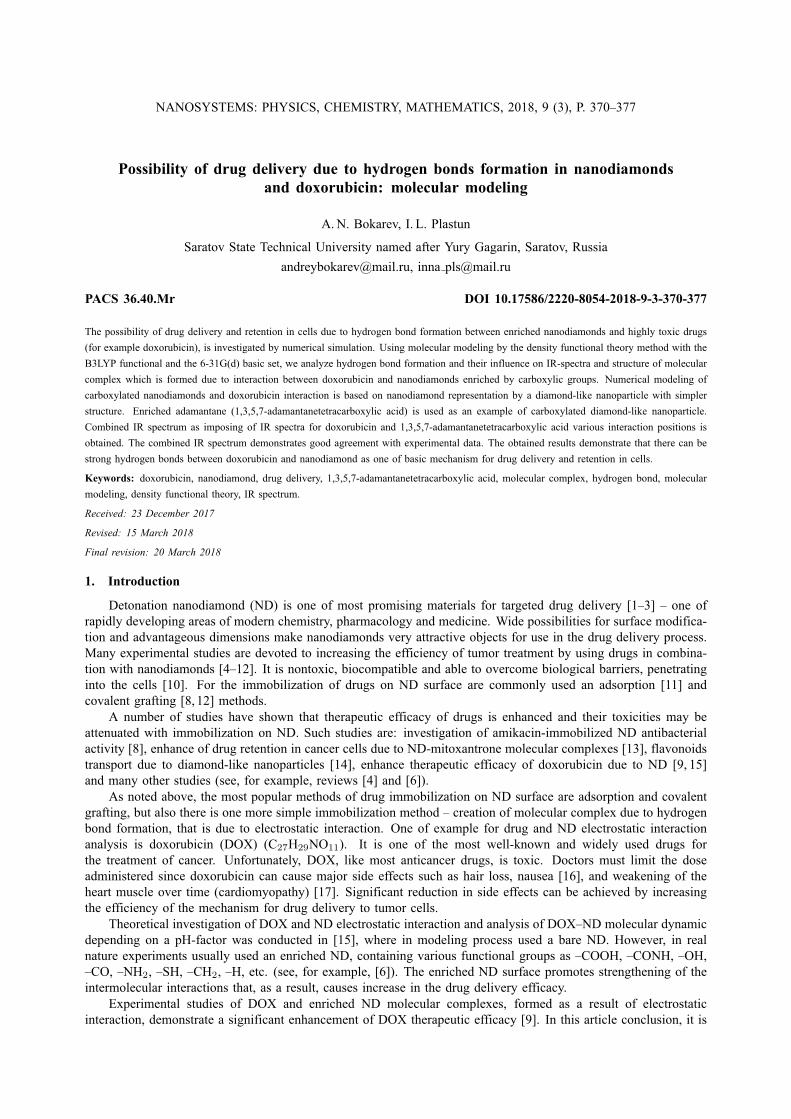
In the process of molecular modeling by numerical simulations, we used a model based on the simplestdiamondoid, adamantane. Adamantane (C10H16) is the smallest diamond-like hydrocarbon particle. The spatialarrangement of carbon atoms in adamantane molecule is the same as in the diamond crystal. This structure isthe basic “building” block for larger diamond-like structures [21]. In present work, we consider the modifiedadamantane, named 1,3,5,7-adamantantetracarboxylic acid (AdTCA) (C14H16O8), which contains 4 carboxylic(–COOH) groups [22]. The modified structure is resulted from the substitution of 4 hydrogen atoms for carboxylicgroups in original adamantane. To assess the possibility of using AdTCA as an object that approximates a large-size carboxylated ND, we also calculated IR spectra for 1 nm diameter ND (C88H64O8) enriched by 4 carboxylicgroups. The chemical structures of adamantane, AdTCA and 1 nm diameter ND are presented on Fig. 1.
In order to account for anharmonicity in intermolecular interactions, we used the following scaling factors forresulted vibrational modes: 0.9713 (range 0 – 1000 cm−1); 0.9744 (range 1000 – 2000 cm−1); 0.956 (range above2000 cm−1). These factors are used to decrease the divergence between calculated and experimental data, whichis caused by using the harmonic approximation in numerical simulations.
3. Results and discussion
In our investigations, we based our work on experimental results obtained in [9]. In this work, the ND–DOXmolecular complex was obtained using a novel adsorption technique developed with a pH 8.5 coupling buffer.Follow the experimental results [9], we consider a ND–DOX molecular complex in crystal phase. As is shown inour preliminary calculation results, all hydrogen bonds are present in an aqueous phase, but their energy becomesa little less than in a crystal phase, and their investigation is not a subject for research in this article.
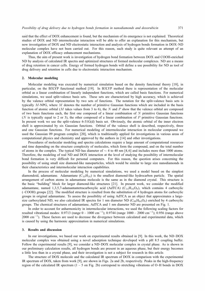
The structure of DOX molecule and the calculated IR spectrum of DOX in comparison with the experimentalIR spectrum of DOX, taken from work [9], are shown in Figs. 2a and 2b, respectively. Peaks in the high-frequencyregion of the calculated IR spectrum (1 – 5 on Fig. 2b) correspond to stretching vibrations of O–H bonds in DOX
372 A. N. Bokarev, I. L. Plastun
(a) (b) (c)
FIG. 1. Structure of adamantane (a), 1,3,5,7-adamantanetetracarboxylic acid C14H16O8 (b) and1 nm diameter nanodiamond C88H64O8, enriched by 4 carboxylic (–COOH) groups (c)
(1 – 5 on Fig. 2a). After carrying out the frequencies scaling, the calculated spectrum is in good agreement withthe experimental data.
(a) (b)
FIG. 2. Structure (a) and experimental (upper) and calculated (lower) IR spectra (b) of doxoru-bicin with designated 1–5 O–H bonds
The calculated frequencies of stretching vibrations for O–H bonds in DOX, which can participate in theformation of hydrogen bonds, are: 1 – 3472 cm−1, 2 – 3591 cm−1, 3 – 3495 cm−1, 4 – 3517 cm−1, 5 –3610 cm−1. The lengths of all O–H bonds are on the order of 0.97 A.
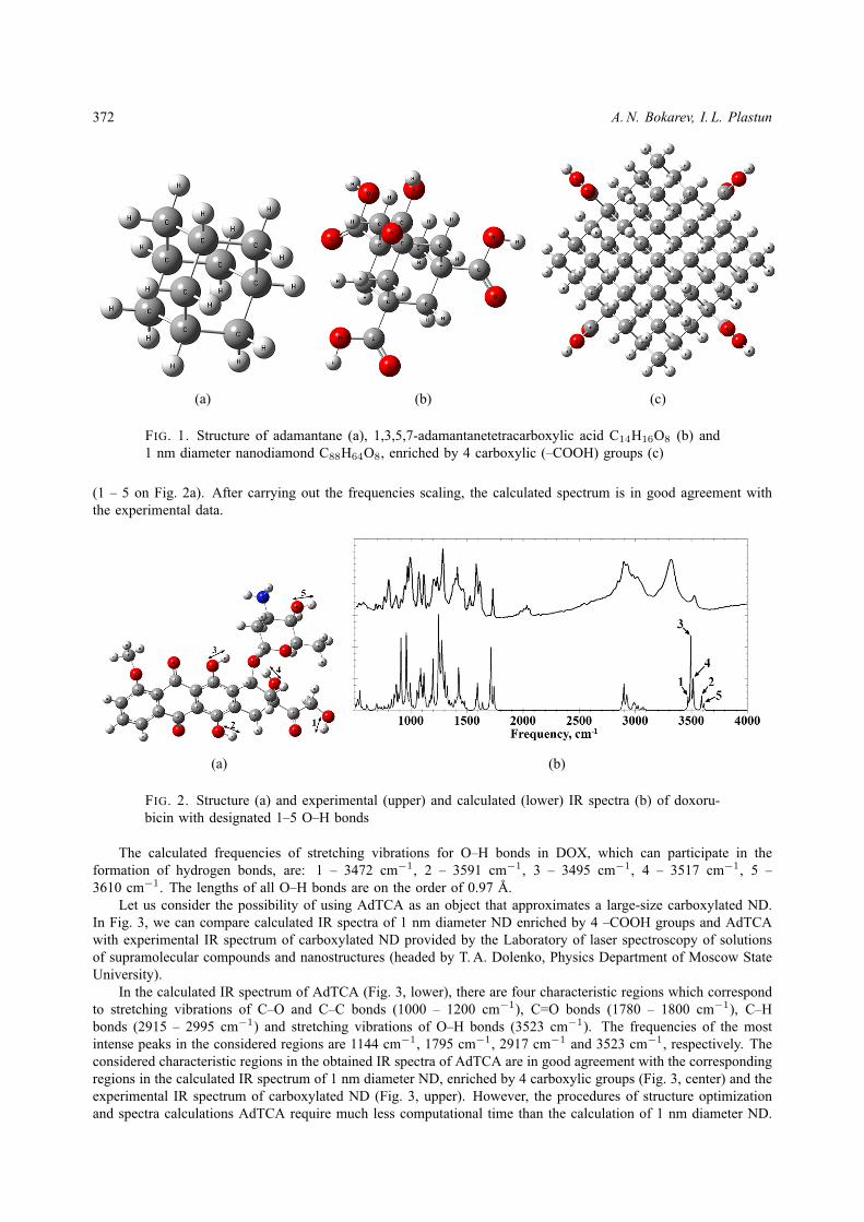
Let us consider the possibility of using AdTCA as an object that approximates a large-size carboxylated ND.In Fig. 3, we can compare calculated IR spectra of 1 nm diameter ND enriched by 4 –COOH groups and AdTCAwith experimental IR spectrum of carboxylated ND provided by the Laboratory of laser spectroscopy of solutionsof supramolecular compounds and nanostructures (headed by T. A. Dolenko, Physics Department of Moscow StateUniversity).
In the calculated IR spectrum of AdTCA (Fig. 3, lower), there are four characteristic regions which correspondto stretching vibrations of C–O and C–C bonds (1000 – 1200 cm−1), C=O bonds (1780 – 1800 cm−1), C–Hbonds (2915 – 2995 cm−1) and stretching vibrations of O–H bonds (3523 cm−1). The frequencies of the mostintense peaks in the considered regions are 1144 cm−1, 1795 cm−1, 2917 cm−1 and 3523 cm−1, respectively. Theconsidered characteristic regions in the obtained IR spectra of AdTCA are in good agreement with the correspondingregions in the calculated IR spectrum of 1 nm diameter ND, enriched by 4 carboxylic groups (Fig. 3, center) and theexperimental IR spectrum of carboxylated ND (Fig. 3, upper). However, the procedures of structure optimizationand spectra calculations AdTCA require much less computational time than the calculation of 1 nm diameter ND.
Possibility of drug delivery due to hydrogen bonds formation in nanodiamonds and doxorubicin 373
Thus, the high level of consistency of the characteristic regions in considered spectra and a smaller amount of therequired computational time allows the use of AdTCA as an object that approximates a large-size carboxylated NDat the level that allows qualitative estimation of compound formation during molecular modeling.
FIG. 3. Experimental IR spectrum of carboxylated nanodiamond (upper), calculated IR spectraof 1 nm diameter nanodiamond (central) and 1,3,5,7-adamantanetetracarboxylic acid (lower)
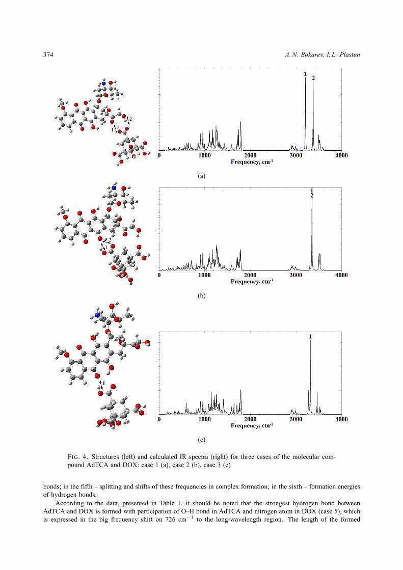
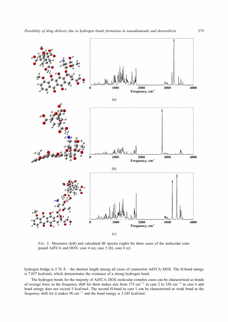
As we spoke above, the sizes of NDs, which are used in scientific experiments with DOX, are from 4 nm andhigher [9], and the width of the DOX molecule is about 1.5 nm, therefore we can assume that the DOX moleculecan be attached to the ND at several points simultaneously. The doxorubicin molecule includes five O–H bonds(1 – 5 on Fig. 2a) and two N–H bonds, whose hydrogen atoms can theoretically participate in the formation ofhydrogen bonds as a donor, as well as eleven oxygen atoms and one nitrogen atom that can participate in theformation of hydrogen bonds as an acceptor. In our studies, we considered possible variants of interaction with allthese atoms, but due to the limitations of molecular modeling caused by the structural features of AdTCA and NDand computation mechanism of energy optimization, only six variants of the complexes were obtained. Thus, inour calculations we investigate the parameters of hydrogen bonds, which are formed between AdTCA and DOX atall possible interaction positions. As a result of numerical simulation, we obtained optimized structures and the IRspectra of the six formed molecular complexes (Figs. 4 and 5) and analyzed the energy of intermolecular hydrogenbonds (H-bonds).
The energy of H-bonds is investigated by analysis of such parameters as length of a hydrogen bridgeand the frequency shift. The energy of hydrogen bonds was estimated using empirical Iogansen formula [23]:−∆H = 0.3(∆ν − 40)1/2, where ∆ν – frequency shift of the stretching vibration of O–H bond (∆H in kcal/mol,∆ν – cm−1).
According to the optimized structures and calculated IR spectra for six cases of molecular compound (Fig. 4and Fig. 5), it can be noted that in all interaction positions the one H-bond or two H-bonds are formed betweenAdTCA and DOX.
Two peaks (1 and 2 in Fig. 4a and in Fig. 5c) at 3207 cm−1 and at 3376 cm−1 for case 1 (Fig. 4a) and for case6 – at 3187 cm−1 and at 3362 cm−1 (Fig. 5c) correspond to stretching vibrations of O–H bond in AdTCA, whichparticipates in the H-bond formation with DOX, and in DOX, which participates in the H-bond formation withAdTCA, respectively. In case 2, there are two formed intermolecular H-bonds between AdTCA and DOX whichparticipate in intermolecular H-bond formation, occur at the same frequency of 3350 cm−1 (1 and 2 in Fig. 4b).
In interaction positions named as cases 3, 4 and 5 (Fig. 4c and Fig. 5a,b), only one intermolecular H-bond isformed between AdTCA and DOX. Peaks at 3314 cm−1 for case 3 (1 in Fig. 4c) and at 3251 cm−1 for case 4(1 in Fig. 5a) corresponds to stretching vibrations of O–H bond in AdTCA, which participates in hydrogen bondformation with DOX. Let’s note that peak at 2797 cm−1 for case 5 (1 in Fig. 5b) which corresponds to H-bondformation with nitrogen atom in DOX.
We analyze the H-bonds parameters in six cases of AdTCA–DOX molecular complex to estimate the strengthof the formed H-bonds. The calculated parameters, whose analysis provides the estimation of a hydrogen bond’s in-fluence on the structure and the IR spectrum, are presented in Table 1. In the first column, the case numbers ofmolecular complex and H-bonds numbers are given; in the second – lengths of O–H bond in them; in the third –lengths of O–H. . . O and O–H. . .N hydrogen bridges; in the fourth – frequencies of stretching vibrations of O–H
374 A. N. Bokarev, I. L. Plastun
(a)
(b)
(c)
FIG. 4. Structures (left) and calculated IR spectra (right) for three cases of the molecular com-pound AdTCA and DOX: case 1 (a), case 2 (b), case 3 (c)
bonds; in the fifth – splitting and shifts of these frequencies in complex formation; in the sixth – formation energiesof hydrogen bonds.
According to the data, presented in Table 1, it should be noted that the strongest hydrogen bond betweenAdTCA and DOX is formed with participation of O–H bond in AdTCA and nitrogen atom in DOX (case 5), whichis expressed in the big frequency shift on 726 cm−1 to the long-wavelength region. The length of the formed
Possibility of drug delivery due to hydrogen bonds formation in nanodiamonds and doxorubicin 375
(a)
(b)
(c)
FIG. 5. Structures (left) and calculated IR spectra (right) for three cases of the molecular com-pound AdTCA and DOX: case 4 (a), case 5 (b), case 6 (c)
hydrogen bridge is 2.76 A – the shortest length among all cases of connection AdTCA–DOX. The H-bond energyis 7.857 kcal/mol, which demonstrates the existence of a strong hydrogen bond.
The hydrogen bonds for the majority of AdTCA–DOX molecular complex cases can be characterized as bondsof average force as the frequency shift for them makes size from 173 cm−1 in case 2 to 336 cm−1 in case 6 andbond energy does not exceed 5 kcal/mol. The second H-bond in case 1 can be characterized as weak bond as thefrequency shift for it makes 96 cm−1 and the bond energy is 2.245 kcal/mol.

376 A. N. Bokarev, I. L. Plastun
TABLE 1. Calculated H-bonds parameters in six cases of AdTCA–DOX molecular complexes
Case number/bond number
OH lengthROH , A
Hydrogen bridge lengthROH−O, A
Frequencyν, cm−1
Frequency shift∆ν, cm−1
Bond energy−∆H , kcal/mol
1/1 0.99 2.78 3207 316 4.984
1/2 0.98 2.78 3376 96 2.245
2/1 0.99 2.95 3350 173 3.46
2/2 0.98 2.8 3350 241 4.253
3/1 0.99 2.76 3314 209 3.9
4/1 0.99 2.8 3251 272 4.57
5/1 1.02 2.76 (OH–N) 2797 726 7.857
6/1 0.99 2.82 3187 336 5.161
6/2 0.98 2.86 3362 248 4.327
Thus, it is possible to note that in AdTCA–DOX molecular complex there can be seven H-bonds of averageforce, one strong and one weak hydrogen bond what proves existence of the rather strong electrostatic interactionbetween nanodiamond and DOX.
We compare our results with the experimental IR spectrum of the powder of the nanodiamond-doxorubicincomplex taken from [9]. For comparison, we created the combined IR spectrum as imposing of calculated IRspectra for six cases of molecular complexes, corresponded to six interaction positions between AdTCA and DOX(Fig. 6).
FIG. 6. Experimental IR spectrum of ND–DOX molecular complex (upper) and combined IRspectrum of six variants of AdTCA–DOX molecular complexes (lower)
As can be seen on Fig. 6, there is the good agreement of the characteristic regions in the experimental andcombined IR spectra, in particular, the wide area of stretching vibrations of O–H bonds, which participate in thehydrogen bonds formation (3150 to 3600 cm−1) and the peak on 2797 cm−1, corresponded to stretching vibrationsof O–H bond in AdTCA, which participates in hydrogen bond formation with nitrogen atom in DOX. Wideboundaries of the region, which corresponds to stretching vibrations of O–H bonds, indicate a large number offormed hydrogen bonds of various configurations in the experimental nanodiamond-doxorubicin complex, includingthe considered interaction variants.
4. Conclusions
During molecular modeling, it was established that AdTCA–DOX molecular complex can occur due to ninehydrogen bonds formed in six various cases for the molecular compound. One of H-bond is strong and seven
Possibility of drug delivery due to hydrogen bonds formation in nanodiamonds and doxorubicin 377
H-bonds are bonds of average force. Since the sizes of NDs, which are used in scientific experiments withDOX, are from 5 nm and higher, and the width of the DOX molecule is about 1.5 nm, it can be assumed thatthe DOX molecule can be attached to the ND on several points simultaneously. The combined IR spectrum,with imposing of calculated IR spectra for six cases of molecular complexes, corresponded to six interactionpositions between AdTCA and DOX and was created for comparison with experimental IR spectra of ND–DOXmolecular complex [9]. We can observe a good agreement between the calculated and the experimental IR spectra,especially in the high-frequency area corresponding to stretching vibrations of O–H bonds, which participate inthe intermolecular hydrogen bond formation. This indicates a large number of formed hydrogen bonds in theexperimental ND–DOX complex and the stability of compound as a result of electrostatic interaction.
The obtained results demonstrate that there can be strong electrostatic interaction between doxorubicin andcarboxylated detonation nanodiamonds. The formed hydrogen bonds can be considered as one of main mechanismsfor targeted drug delivery and for drug retention in cells and, thus, for enhancement of doxorubicin therapeuticefficacy, as observed in experiment [9].
References
[1] Nanotherapeutics: Drug Delivery Concepts in Nanoscience. Ed. by Lamprecht A. CRC Press, Taylor and Francis Group, Boca Raton,2008, 292 p.
[2] Gupta R.B., Kompella U.B. Nanoparticles Technology for Drug Delivery. N.-Y. Taylor & Francis Group, 2006, 403 p.[3] Popova N.R., Popov A.L., Shcherbakov A.B., Ivanov V.K. Layer-by-layer capsules as smart delivery systems of CeO2 nanoparticle based
theranostic agents. Nanosystems: physics, chemistry, mathematics, 2017, 8 (2), P. 282–289.[4] Yakovlev R.Y., Solomatin A.S., et al. Detonation diamond a perspective carrier for drug delivery systems. Rus. J. Gen. Chem., 2014,
84 (2), P. 379–390.[5] Ho D., Wang C-H.K., Chow E.K-H. Nanodiamonds: The intersection of nanotechnology, drug development, and personalized medicine.
Science Advances, 2015, 1 (7), P. 1500439.[6] Shenderova O.A., McGuire G.E. Science and engineering of nanodiamond particle surfaces for biological applications (Review). Biointer-
phases, 2015, 10 (3), P. 030802.[7] Schrand A.M., Ciftan H.S.A., Shenderova O.A. Nanodiamond particles: properties and perspectives for bioapplications. Crit. Rev. Solid
State Mater. Sci., 2009, 34, P. 18–74.[8] Solomatin A.S., Yakovlev R.Yu., et al. Antibacterial activity of Amikacin-immobilized detonation nanodiamond. Nanosystems: physics,
chemistry, mathematics, 2017, 8 (4), P. 531–534.[9] Salaam A.D., Hwang P.T.J., et al. Nanodiamonds enhance therapeutic efficacy of doxorubicin in treating metastatic hormone-refractory
prostate cancer. Nanotechnology, 2014, 25 (42), P. 425103.[10] Zhang X., Hu W., et al. A comparative study of cellular uptake and cytotoxicity of multiwalled carbon nanotubes, graphene oxide, and
nanodiamond. Toxicol. Res., 2012, 1, P. 62–68.[11] Shugalei I.V., Voznyakovskii A.P., et al. Biological activity of detonation nanodiamond and prospects in its medical and biological
applications. Russ. J. Gen. Chem., 2013, 83 (5), P. 851–883.[12] Liu K.K., Zheng W.W., et al. Covalent linkage of nanodiamondpaclitaxel for drug delivery and cancer therapy. Nanotechnology, 2010, 21,
P. 1–14.[13] Toh T.-B., Lee D.-K., et al. Nanodiamondmitoxantrone complexes enhance drug retention in chemoresistant breast cancer cells. Mol.
Pharmaceutics, 2014, 11, P. 2683–2691.[14] Plastun I.L., Agandeeva K.E., Bokarev A.N., Zenkin N.S. Diamond-like nanoparticles influence on flavonoids transport: molecular
modeling. Proc. SPIE, 2017, 103360K.[15] Adnan A., Lam R., et al. Atomistic simulation and measurement of pH dependent cancer therapeutic interactions with nanodiamond
carrier. Mol. Pharmaceutics, 2011, 8, P. 368–374.[16] Petrioli R., Fiaschi A.I., et al. The role of doxorubicin and epirubicin in the treatment of patients with metastatic hormone refractory
prostate cancer. Cancer Treat. Rev., 2008, 34, P. 710-8.[17] Saltiel E., McGuire W. Doxorubicin (adriamycin) cardiomyopathy – a critical review. West J. Med., 1983, 139, P. 332–341.[18] Kohn W. Nobel Lecture: Electronic structure of matterwave functions and density functionals. Reviews of Modern Physics, 1999, 71 (5),
P. 1253–1266.[19] Pople J. Nobel Lecture: Quantum chemical models. Reviews of Modern Physics, 1999, 71 (5), P. 1267–1274.[20] Frisch M.J., Trucks G.W., et al. Gaussian 09, Revision A.02. Wallingford CT, Gaussian Inc., 2009.[21] Fort R.C.Jr., Schleyers P. von R. Adamantane: Consequences of diamondoid structure. Chem. Rev., 1964, 64 (3) P. 277–300.[22] Ermer . Five-fold diamond structure of adamantane-1,3,5,7-tetracarboxylic acid. Journal of American Chemical Society, 1988, 110 (12),
P. 3747–3754.[23] Iogansen A.V. IR Spectroscopy and Hydrogen Bond Energy Determination. Hydrogen Bond, Nauka, Moscow, 1981, P. 112–155 (in
Russian).