positron emission tomography (pet) oncology … · conventional imaging studies, when it is...
TRANSCRIPT
Positron Emission Tomography (PET) Oncology Applications
Policy # 00105 Original Effective Date: 01/28/2002 Current Effective Date: 01/02/2018
©2017 Blue Cross and Blue Shield of Louisiana
Blue Cross and Blue Shield of Louisiana is an independent licensee of the Blue Cross and Blue Shield Association and
incorporated as Louisiana Health Service & Indemnity Company.
No part of this publication may be reproduced, stored in a retrieval system, or transmitted, in any form or by any means, electronic, mechanical, photocopying, or otherwise, without permission from Blue Cross and Blue Shield of Louisiana.
Page 1 of 65
Applies to all products administered or underwritten by Blue Cross and Blue Shield of Louisiana and its subsidiary, HMO Louisiana, Inc.(collectively referred to as the “Company”), unless otherwise provided in the applicable contract. Medical technology is constantly evolving, and we reserve the right to review and update Medical Policy periodically.
Note: Cardiac applications of positron emission tomography (PET) scanning are considered in medical policy 00103. Note: Miscellaneous applications of positron emission tomography (PET) scanning are considered in medical policy 00104. Note: This policy only addresses the use of radiotracers detected with the use of dedicated PET scanners. Radiotracers such as fluorodeoxyglucose (FDG) may be detected using single photon emission computed tomography (SPECT) cameras, a hybrid PET/SPECT procedure that may be referred to as FDG-SPECT or molecular coincidence. For this policy, PET scanning is discussed for the following four applications in oncology: Diagnosis. Diagnosis refers to use of positron emission tomography (PET) as part of the testing used in establishing whether or not a patient has cancer. Staging. This refers to use of positron emission tomography (PET) to determine the stage (extent) of the cancer at the time of diagnosis, before any treatment is given. Imaging at this time is generally to determine whether or not the cancer is localized. This may also be referred to as initial staging. Restaging. This refers to imaging following treatment in two situations. Restaging is part of the evaluation of a patient in whom a disease recurrence is suspected based on signs and/or symptoms. Restaging also includes determining the extent of malignancy following completion of a full course of treatment. Surveillance. This refers to use of imaging in asymptomatic patients (patients without objective signs or symptoms of recurrent disease). This imaging is completed 6 months or more (12 months or more for lymphoma) following completion of treatment. Coverage Eligibility The following apply to the listed oncologic applications of positron emission tomography (PET) scanning:
Eligible for Coverage Coverage for eligible medical treatments or procedures, drugs, devices or biological products may be provided only if:
Benefits are available in the member’s contract/certificate, and
Positron Emission Tomography (PET) Oncology Applications
Policy # 00105 Original Effective Date: 01/28/2002 Current Effective Date: 01/02/2018
©2017 Blue Cross and Blue Shield of Louisiana
Blue Cross and Blue Shield of Louisiana is an independent licensee of the Blue Cross and Blue Shield Association and
incorporated as Louisiana Health Service & Indemnity Company.
No part of this publication may be reproduced, stored in a retrieval system, or transmitted, in any form or by any means, electronic, mechanical, photocopying, or otherwise, without permission from Blue Cross and Blue Shield of Louisiana.
Page 2 of 65
Medical necessity criteria and guidelines are met. Investigational Coverage is not available for investigational medical treatments or procedures, drugs, devices or biological products.
All policy statements apply to both positron emission tomography (PET) scans and positron emission tomography (PET) plus computed tomography (CT) scans, i.e., positron emission tomography (PET) scans with or without positon emission tomography/computed tomography (PET/CT) fusion. For the clinical situations indicated that may be considered medically necessary, this assumes that the results of the positron emission tomography (PET) scan will influence treatment decisions. If the results will not influence treatment decisions, these situations would be considered investigational. Based on review of available data, the Company considers the use of positron emission tomography (PET) scans for oncology applications to be either eligible for coverage or investigational* as indicated below:
Bone Cancer Eligible for Coverage Staging When used in the staging of Ewing sarcoma and osteosarcoma may be eligible for
coverage. Restaging When used in the restaging of Ewing sarcoma and osteosarcoma may be eligible for
coverage. Investigational Staging When used in the staging of chondrosarcoma is considered investigational.* Brain Cancer Eligible for Coverage Diagnosis When utilized to differentiate scar tissue or tumor necrosis from active disease following
radiation or chemotherapy the use of positron emission tomography (PET) scanning may be eligible for coverage.
Staging When used for staging of brain cancer positron emission tomography (PET) scanning may be eligible for coverage.
Restaging When used for restaging of brain cancer positron emission tomography (PET) scanning may be eligible for coverage.
Positron Emission Tomography (PET) Oncology Applications
Policy # 00105 Original Effective Date: 01/28/2002 Current Effective Date: 01/02/2018
©2017 Blue Cross and Blue Shield of Louisiana
Blue Cross and Blue Shield of Louisiana is an independent licensee of the Blue Cross and Blue Shield Association and
incorporated as Louisiana Health Service & Indemnity Company.
No part of this publication may be reproduced, stored in a retrieval system, or transmitted, in any form or by any means, electronic, mechanical, photocopying, or otherwise, without permission from Blue Cross and Blue Shield of Louisiana.
Page 3 of 65
Investigational Diagnosis When used for diagnosis (other than described above) positron emission tomography
(PET) scanning is considered investigational.* Breast Cancer Eligible for coverage Staging When used in the staging of breast cancer may be eligible for coverage for the following
application:
Detecting locoregional or distant recurrence or metastasis (except axillary lymph nodes).
Restaging When used in restaging breast cancer may be eligible for coverage for the following application:
Detecting locoregional or distant recurrence or metastasis (except axillary lymph nodes).
Investigational All other applications of positron emission tomography (PET) scans in the evaluation of breast cancer are considered investigational* including, but not limited to, the following:
Differential diagnosis in patients with suspicious lesions or an indeterminate/low suspicion finding on mammography;
Staging axillary lymph nodes.
Predicting pathologic response to neoadjuvant therapy for locally advanced disease. Cervical Cancer Eligible for Coverage Staging When used in the staging of cervical cancer positron emission tomography (PET) scans
may be eligible for coverage. Restaging When used in the restaging of cervical cancer positron emission tomography (PET)
scans may be eligible for coverage.
Positron emission tomography (PET) scanning in the evaluation of known or suspected recurrence of cervical
cancer may be eligible for coverage.
Positron Emission Tomography (PET) Oncology Applications
Policy # 00105 Original Effective Date: 01/28/2002 Current Effective Date: 01/02/2018
©2017 Blue Cross and Blue Shield of Louisiana
Blue Cross and Blue Shield of Louisiana is an independent licensee of the Blue Cross and Blue Shield Association and
incorporated as Louisiana Health Service & Indemnity Company.
No part of this publication may be reproduced, stored in a retrieval system, or transmitted, in any form or by any means, electronic, mechanical, photocopying, or otherwise, without permission from Blue Cross and Blue Shield of Louisiana.
Page 4 of 65
Colorectal Cancer Eligible for Coverage Diagnosis When PET results may assist in the following situations positron emission tomography
(PET) scanning may be eligible for coverage: o Avoiding an invasive diagnostic procedure, or o Determining the optimal anatomical location to perform an invasive diagnostic
procedure. Staging When positron emission tomography (PET) results may assist in the following situations
positron emission tomography (PET) scanning may be eligible for coverage: o To detect and assess resectability of hepatic or extrahepatic metastases of
colorectal cancer (CRC); or o Cancer stage remains in doubt after completion of a standard diagnostic workup;
or o positron emission tomography (PET) could potentially replace one or more
conventional imaging studies, when it is expected that conventional study information is insufficient for the clinical management of the patient, or
o Clinical management would differ depending on the cancer stage. Restaging When positron emission tomography (PET) results may assist in the following situations
positron emission tomography (PET) scanning may be eligible for coverage: o To detect and assess resectability of hepatic or extrahepatic metastases of
colorectal cancer (CRC); or o Detecting residual disease (after completion of treatment), or o Detecting suspected recurrence (example: rising carcinoembryonic antigen
[CEA] levels; clinical signs/symptoms suspicious for recurrence); or o Determination of the extent of known recurrence.
Investigational Differentiation Positron emission tomography (PET) scanning is considered investigational* as:
A technique to assess the presence of scarring versus local bowel recurrence in patients with previously resected colorectal cancer (CRC).
A technique contributing to radiotherapy treatment planning. Endometrial Cancer Eligible for Coverage Staging Positron emission tomography (PET) scanning may be eligible for coverage in the:
o Detection of lymph node metastases.
Positron Emission Tomography (PET) Oncology Applications
Policy # 00105 Original Effective Date: 01/28/2002 Current Effective Date: 01/02/2018
©2017 Blue Cross and Blue Shield of Louisiana
Blue Cross and Blue Shield of Louisiana is an independent licensee of the Blue Cross and Blue Shield Association and
incorporated as Louisiana Health Service & Indemnity Company.
No part of this publication may be reproduced, stored in a retrieval system, or transmitted, in any form or by any means, electronic, mechanical, photocopying, or otherwise, without permission from Blue Cross and Blue Shield of Louisiana.
Page 5 of 65
Restaging When positron emission tomography (PET) results may assist in the following situations
positron emission tomography (PET) scanning may be eligible for coverage: o Assessment of endometrial cancer recurrence.
Esophageal Cancer Eligible for Coverage Diagnosis When positron emission tomography (PET) results may assist in the following situations
positron emission tomography (PET) scanning may be eligible for coverage: o To avoid an invasive diagnostic procedure; or o To determine the optimal anatomical location to perform an invasive diagnostic
procedure. Staging When positron emission tomography (PET) results may assist in the following situations
positron emission tomography (PET) scanning may be eligible for coverage: o Staging of esophageal cancer; or o Staging of esophageal cancer when the stage of the cancer remains in doubt
after completion of a standard diagnostic workup. Restaging When PET results may assist in the following situations positron emission tomography
(PET) scanning may be eligible for coverage: o Restaging after the completion of treatment; or o Detection of residual disease, suspected recurrence, or to determine the extent
of a known recurrence; or o Determining response to preoperative induction therapy.
Investigational Diagnosis When used in the evaluation and detection of primary esophageal cancer positron
emission tomography (PET) scanning is considered investigational.* Gastric Cancer Eligible for Coverage Diagnosis When positron emission tomography (PET) results may assist in the following situation
positron emission tomography (PET) scanning may be eligible for coverage: o Initial diagnosis of gastric cancer, and
Staging When positron emission tomography (PET) results may assist in the following situation positron emission tomography (PET) scanning may be eligible for coverage:
o Initial staging of gastric cancer, and Restaging Evaluation for recurrent gastric cancer after surgical resection, when other imaging
modalities are inconclusive.
Positron Emission Tomography (PET) Oncology Applications
Policy # 00105 Original Effective Date: 01/28/2002 Current Effective Date: 01/02/2018
©2017 Blue Cross and Blue Shield of Louisiana
Blue Cross and Blue Shield of Louisiana is an independent licensee of the Blue Cross and Blue Shield Association and
incorporated as Louisiana Health Service & Indemnity Company.
No part of this publication may be reproduced, stored in a retrieval system, or transmitted, in any form or by any means, electronic, mechanical, photocopying, or otherwise, without permission from Blue Cross and Blue Shield of Louisiana.
Page 6 of 65
Head and Neck Cancers (excluding CNS and Thyroid) Eligible for Coverage Diagnosis When positron emission tomography (PET) results may assist in the following situation
positron emission tomography (PET) scanning may be eligible for coverage: o In the evaluation of head and neck cancer in the initial diagnosis of suspected
cancer. Staging When positron emission tomography (PET) results may assist in the following situation
positron emission tomography (PET) scanning may be eligible for coverage: o In the evaluation of head and neck cancer in the initial staging of disease.
Restaging When positron emission tomography (PET) results may assist in the following situation positron emission tomography (PET) scanning may be eligible for coverage:
o In the evaluation of head and neck cancer in the restaging of residual or recurrent disease during follow-up; and
o In the evaluation of response to treatment. Investigational When used for applications not discussed above, positron emission tomography (PET) scanning for the evaluation of head and neck cancer is considered investigational*. Lung Cancer/Solitary Pulmonary Nodule Eligible for Coverage Diagnosis When positron emission tomography (PET) results may assist in the following situations
positron emission tomography (PET) scanning may be eligible for coverage: o Solitary Pulmonary Nodule - In patients with a solitary pulmonary nodule to
distinguish between benign and malignant disease when prior CT and chest x-ray findings are inconclusive or discordant; or
o Lung Cancer - To determine resectability for patients with a presumed solitary metastatic lesion from lung cancer; or
o Lung Cancer -To distinguish between benign and malignant disease when prior computed tomography (CT) scan and chest x-ray findings are inconclusive or discordant.
Staging When positron emission tomography (PET) results may assist in the following situations positron emission tomography (PET) scanning may be eligible for coverage:
o In clinical situations in which the stage of cancer remains in doubt after completion of a standard diagnostic workup; or
o As staging technique in those with known non-small cell lung cancer (NSCLC). Restaging When positron emission tomography (PET) results may assist in the following situations
positron emission tomography (PET) scanning may be eligible for coverage:
Positron Emission Tomography (PET) Oncology Applications
Policy # 00105 Original Effective Date: 01/28/2002 Current Effective Date: 01/02/2018
©2017 Blue Cross and Blue Shield of Louisiana
Blue Cross and Blue Shield of Louisiana is an independent licensee of the Blue Cross and Blue Shield Association and
incorporated as Louisiana Health Service & Indemnity Company.
No part of this publication may be reproduced, stored in a retrieval system, or transmitted, in any form or by any means, electronic, mechanical, photocopying, or otherwise, without permission from Blue Cross and Blue Shield of Louisiana.
Page 7 of 65
o As a restating technique in those with known non-small cell lung cancer (NSCLC); or
o For restaging after the completion of treatment; or o For the purpose of detecting residual disease, detecting suspected recurrence, or
to determine the extent of a known recurrence. Investigational Staging When used as a technique in the staging of small cell lung cancer positron emission
tomography (PET) scanning is investigational.*
Lymphoma, including Hodgkin’s Disease Eligible for Coverage Diagnosis Only in clinical situations in which the positron emission tomography (PET) results may
assist in avoiding an invasive diagnostic procedure, or in which the positron emission tomography (PET) results may assist in determining the optimal anatomical location to perform an invasive diagnostic procedure. In general, for most solid tumors, a tissue diagnosis is made prior to the performance of positron emission tomography (PET) scanning. Positron emission tomography (PET) scans following a tissue diagnosis are performed for the purpose of staging, not diagnosis. Therefore, the use of positron emission tomography (PET) in the diagnosis of lymphoma should be rare.
Staging When positron emission tomography (PET) results may assist in the following situations positron emission tomography (PET) scanning may be eligible for coverage:
o For staging lymphoma during initial staging; or o In clinical situations in which the stage of the cancer remains in doubt after
completion of a standard diagnostic workup. Restaging When positron emission tomography (PET) results may assist in the following situations
positron emission tomography (PET) scanning may be eligible for coverage: o For restaging at follow-up; or o For the purpose of detecting residual disease, detecting suspected recurrence, or
to determine the extent of a known recurrence; or o For restaging after the completion of treatment.
Investigational When used for applications not discussed above, positron emission tomography (PET) scanning for the evaluation of lymphoma is considered investigational*.
Positron Emission Tomography (PET) Oncology Applications
Policy # 00105 Original Effective Date: 01/28/2002 Current Effective Date: 01/02/2018
©2017 Blue Cross and Blue Shield of Louisiana
Blue Cross and Blue Shield of Louisiana is an independent licensee of the Blue Cross and Blue Shield Association and
incorporated as Louisiana Health Service & Indemnity Company.
No part of this publication may be reproduced, stored in a retrieval system, or transmitted, in any form or by any means, electronic, mechanical, photocopying, or otherwise, without permission from Blue Cross and Blue Shield of Louisiana.
Page 8 of 65
Melanoma Eligible for Coverage Diagnosis When positron emission tomography (PET) results may assist in the following situations
PET scanning may be eligible for coverage: o Only in clinical situations in which the positron emission tomography (PET)
results may assist in avoiding an invasive diagnostic procedure, or in which the positron emission tomography (PET) results may assist in determining the optimal anatomical location to perform an invasive diagnostic procedure. In general, for most solid tumors, a tissue diagnosis is made prior to the performance of positron emission tomography (PET) scanning. Positron emission tomography (PET) scans following a tissue diagnosis are performed for the purpose of staging, not diagnosis. Therefore, the use of positron emission tomography (PET) in the diagnosis of melanoma should be rare; or
o As a technique for assessing extranodal spread of malignant melanoma at initial staging.
Restaging When positron emission tomography (PET) results may assist in the following situations positron emission tomography (PET) scanning may be eligible for coverage:
o For assessing extranodal spread of malignant melanoma at initial staging or at restaging during follow-up treatment; or
o For the purpose of detecting residual disease, detecting suspected recurrence, or to determine the extent of a known recurrence.
Investigational When used for applications not discussed above positron emission tomography (PET) scanning for the evaluation of melanoma is considered investigational.* Positron emission tomography (PET) scanning is considered investigational* as a technique to detect regional lymph node metastases in patients with clinically localized melanoma who are candidates to undergo sentinel node biopsy. When used as a technique for restaging in managing stage 0, I, or II melanoma, positron emission tomography (PET) scanning is considered investigational*.
Multiple Myeloma Eligible for Coverage Staging
o To assess extent of disease at time of diagnosis
Positron Emission Tomography (PET) Oncology Applications
Policy # 00105 Original Effective Date: 01/28/2002 Current Effective Date: 01/02/2018
©2017 Blue Cross and Blue Shield of Louisiana
Blue Cross and Blue Shield of Louisiana is an independent licensee of the Blue Cross and Blue Shield Association and
incorporated as Louisiana Health Service & Indemnity Company.
No part of this publication may be reproduced, stored in a retrieval system, or transmitted, in any form or by any means, electronic, mechanical, photocopying, or otherwise, without permission from Blue Cross and Blue Shield of Louisiana.
Page 9 of 65
Restaging o Restaging after completion of treatment o Detection of residual disease, suspected recurrence, or to determine the extent
of a known recurrence. Ovarian Cancer Eligible for Coverage Diagnosis When PET results may assist in the following situations positron emission tomography
(PET) scanning may be eligible for coverage: o Avoiding an invasive diagnostic procedure, or o Determining the optimal anatomical location to perform an invasive diagnostic
procedure. Staging When positron emission tomography (PET) results may assist in the following situations
positron emission tomography (PET) scanning may be eligible for coverage: o For staging ovarian cancer during initial staging; or o In clinical situations in which the stage of the cancer remains in doubt after
completion of a standard diagnostic workup. Restaging When PET results may assist in the following situations positron emission tomography
(PET) scanning may be eligible for coverage: o For restaging at follow-up; or o For the purpose of detecting residual disease; or o For detecting suspected recurrence; or o To determine the extent of a known recurrence; or o For restaging after the completion of treatment.
Pancreatic Cancer Eligible for Coverage Diagnosis When used as a technique in the initial diagnosis of pancreatic cancer when other
imaging and biopsy are inconclusive positron emission tomography (PET) scanning may be eligible for coverage.
Staging When used as a technique for staging of pancreatic cancer when other imaging and biopsy are inconclusive positron emission tomography (PET) scanning may be eligible for coverage.
Investigational When used as a technique to evaluate other aspects of pancreatic cancer positron emission tomography (PET) scanning is considered investigational.*
Positron Emission Tomography (PET) Oncology Applications
Policy # 00105 Original Effective Date: 01/28/2002 Current Effective Date: 01/02/2018
©2017 Blue Cross and Blue Shield of Louisiana
Blue Cross and Blue Shield of Louisiana is an independent licensee of the Blue Cross and Blue Shield Association and
incorporated as Louisiana Health Service & Indemnity Company.
No part of this publication may be reproduced, stored in a retrieval system, or transmitted, in any form or by any means, electronic, mechanical, photocopying, or otherwise, without permission from Blue Cross and Blue Shield of Louisiana.
Page 10 of 65
Prostate Cancer Eligible for Coverage
Restaging When used with 11
C-choline for evaluating response to primary treatment in prostate cancer positron emission tomography (PET) scanning may be eligible for coverage.
Investigational When used with
68Gallium in all aspects of managing prostate cancer positron emission tomography
(PET) scanning is considered investigational*. When used for all other indications in known or suspected prostate cancer positron emission tomography (PET) scanning is considered investigational*. Soft Tissue Sarcoma Investigational Positron emission tomography (PET) scanning is considered investigational* in evaluation of soft tissue sarcoma, including but not limited to the following applications:
Distinguishing between benign lesions and malignant soft tissue sarcoma; or
Distinguishing between low grade and high grade soft tissue sarcoma; or
Detecting locoregional recurrence; or
Detecting distant metastasis.
Evaluating response to imatinib and other treatments for gastrointestinal stromal tumors. Testicular Cancer Eligible for Coverage Restaging When used as a technique in evaluation of residual mass following chemotherapy of
stage IIB and III seminomas positron emission tomography (PET) scanning may be eligible for coverage.
Note: The positron emission tomography (PET) scan should be completed not sooner than 6 weeks following chemotherapy. Investigational Except as noted above for seminoma, positron emission tomography (PET) scanning is investigational* in evaluation of testicular cancer, including but not limited to the following applications:
Initial staging of testicular cancer; or
Distinguishing between viable tumor and necrosis/fibrosis after treatment of testicular cancer; or
Detection of recurrent disease after treatment of testicular cancer.
Positron Emission Tomography (PET) Oncology Applications
Policy # 00105 Original Effective Date: 01/28/2002 Current Effective Date: 01/02/2018
©2017 Blue Cross and Blue Shield of Louisiana
Blue Cross and Blue Shield of Louisiana is an independent licensee of the Blue Cross and Blue Shield Association and
incorporated as Louisiana Health Service & Indemnity Company.
No part of this publication may be reproduced, stored in a retrieval system, or transmitted, in any form or by any means, electronic, mechanical, photocopying, or otherwise, without permission from Blue Cross and Blue Shield of Louisiana.
Page 11 of 65
Thyroid Cancer, Differentiated Eligible for Coverage Diagnosis When used as a technique in the diagnosis of patients with differentiated thyroid cancer
when thyroglobulin (Tg) levels are elevated and whole-body I-131 imaging is negative positron emission tomography (PET) scanning may be eligible for coverage
Restaging When used as a technique for restaging patients with differentiated thyroid cancer when
thyroglobulin (Tg) levels are elevated and whole-body I-131 imaging is negative positron emission tomography (PET) scanning may be eligible for coverage
Investigational When used as a technique in the evaluation of known or suspected differentiated thyroid cancer in all other situations positron emission tomography (PET) scanning is considered investigational.* Unknown Primary Eligible for Coverage Diagnosis When used in patients with an unknown primary who meet all of the following criteria
positron emission tomography (PET) scanning may be eligible for coverage: o Single site of disease outside the cervical lymph nodes; and o Patient is considering local or regional treatment for a single site of metastatic
disease; and o Negative workup for an occult primary tumor; and o Positron emission tomography (PET) scan will be used to rule out or detect
additional sites of disease that would eliminate the rationale for local or regional treatment.
Investigational Diagnosis Positron emission tomography (PET) scanning is considered investigational* in
evaluation of unknown primary, including but not limited to the following applications: o As part of the initial workup of an unknown primary; or o As part of the workup of patients with multiple sites of disease.
Cancer Surveillance Positron emission tomography (PET) scanning is considered investigational* when used as a surveillance tool for patients with cancer or with a history of cancer. A scan is considered surveillance if performed more than 6 months after completion of cancer therapy (12 months for lymphoma) in patients without objective signs or symptoms suggestive of cancer recurrence.
Positron Emission Tomography (PET) Oncology Applications
Policy # 00105 Original Effective Date: 01/28/2002 Current Effective Date: 01/02/2018
©2017 Blue Cross and Blue Shield of Louisiana
Blue Cross and Blue Shield of Louisiana is an independent licensee of the Blue Cross and Blue Shield Association and
incorporated as Louisiana Health Service & Indemnity Company.
No part of this publication may be reproduced, stored in a retrieval system, or transmitted, in any form or by any means, electronic, mechanical, photocopying, or otherwise, without permission from Blue Cross and Blue Shield of Louisiana.
Page 12 of 65
Other Oncologic Applications Positron emission tomography (PET) scanning for other oncologic applications is considered investigational* for the following:
In all aspects of managing neuroendocrine tumors; and
In all aspects of managing penile cancer; and
In all aspects of managing renal cancer.
POLICY GUIDELINES PATIENT SELECTION As with any imaging technique, the medical necessity of positron emission tomography (PET) scanning depends in part on what imaging techniques are used before or after the positron emission tomography (PET) scanning. Due to its expense, positron emission tomography (PET) scanning is typically considered after other techniques, such as computed tomography (CT), magnetic resonance imaging (MRI), or ultrasonography, provide inconclusive or discordant results. In patients with melanoma or lymphoma, positron emission tomography (PET) scanning may be considered an initial imaging technique. If so, the medical necessity of subsequent imaging during the same diagnostic evaluation is unclear. Thus, positron emission tomography (PET) should be considered for the medically necessary indications above only when standard imaging (e.g., computed tomography [CT], magnetic resonance imaging [MRI]) is inconclusive or not indicated. Patient selection criteria for positron emission tomography (PET) scanning also may be complex. For example, it may be difficult to determine from claims data whether a positron emission tomography (PET) scan in a patient with malignant melanoma is being done primarily to evaluate extranodal disease or regional lymph nodes. Similarly, it may be difficult to determine whether a positron emission tomography (PET) scan in a patient with colorectal cancer (CRC) is being performed to detect hepatic disease or evaluate local recurrence. Due to the complicated hierarchy of imaging options in patients with malignancy and complex patient selection criteria, a possible implementation strategy for this policy is its use for retrospective review, possibly focusing on cases with multiple imaging tests, including positron emission tomography (PET) scans. Use of positron emission tomography (PET) scanning for surveillance as described in the policy statement and policy rationale refers to the use of positron emission tomography (PET) to detect disease in asymptomatic patients at various intervals. This is not the same as the use of positron emission tomography (PET) for detecting recurrent disease in symptomatic patients; these applications of positron emission tomography (PET) are considered within tumor-specific categories in the policy statements.
Background/Overview A variety of tracers are used for PET scanning, including oxygen-15, nitrogen-13, carbon-
11choline, and
fluorine-18
. In 2016, 2 additional tracers, gallium-68
, and fluciclovine-18
, were approved by the Food and Drug
Positron Emission Tomography (PET) Oncology Applications
Policy # 00105 Original Effective Date: 01/28/2002 Current Effective Date: 01/02/2018
©2017 Blue Cross and Blue Shield of Louisiana
Blue Cross and Blue Shield of Louisiana is an independent licensee of the Blue Cross and Blue Shield Association and
incorporated as Louisiana Health Service & Indemnity Company.
No part of this publication may be reproduced, stored in a retrieval system, or transmitted, in any form or by any means, electronic, mechanical, photocopying, or otherwise, without permission from Blue Cross and Blue Shield of Louisiana.
Page 13 of 65
Administration (FDA). Because of their short half-life, some tracers must be made locally using an onsite cyclotron. The radiotracer most commonly used in oncology imaging has been fluorine-
18 coupled with
fluorodeoxyglucose (FDG), which correlates with glucose metabolism. FDG has been considered useful in cancer imaging because tumor cells show increased metabolism of glucose. The most common malignancies studied have been melanoma, lymphoma, lung, colorectal, and pancreatic cancer. For this evidence review, PET scanning is discussed for the following 4 applications in oncology: diagnosis, staging, restaging, and surveillance. Diagnosis refers to use of PET as part of the testing used in establishing whether a patient has cancer. Staging refers to use of PET to determine the stage (extent) of the cancer at the time of diagnosis, before any treatment is given. Imaging at this time is generally to determine whether the cancer is localized. This also may be referred to as initial staging. Restaging refers to imaging after treatment in 2 situations. Restaging is part of the evaluation of a patient in whom disease recurrence is suspected based on signs and/or symptoms. Restaging also includes determining theextent of malignancy after completion of a full course of treatment. Surveillance refers to use of imaging in asymptomatic patients (patients without objective signs or symptoms of recurrent disease). This imaging is completed 6 months or more (≥12 months for lymphoma) after completion of treatment. This evidence review focuses on the use of radiotracers detected with dedicated PET scanners. Radiotracers such as FDG may be detected using single-photon emission computerized tomography (SPECT) cameras, a technique that may be referred to as FDG-SPECT imaging. The use of SPECT cameras for PET radiotracers presents unique issues of diagnostic performance and is not considered herein.
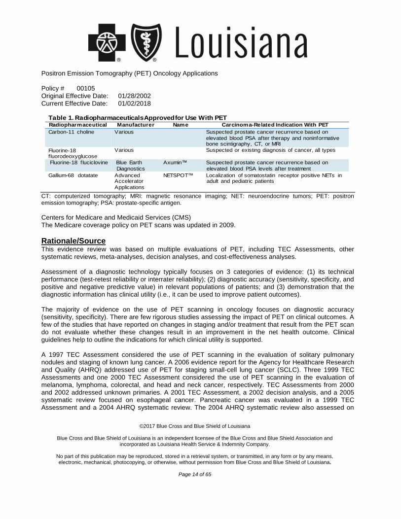
FDA or Other Governmental Regulatory Approval U.S. Food and Drug Administration The FDA website includes various PET-related documents. As of June 2016, the following radiopharmaceuticals have been granted FDA-approval, to be used with PET for carcinoma-related indications (see Table 1).
Positron Emission Tomography (PET) Oncology Applications
Policy # 00105 Original Effective Date: 01/28/2002 Current Effective Date: 01/02/2018
©2017 Blue Cross and Blue Shield of Louisiana
Blue Cross and Blue Shield of Louisiana is an independent licensee of the Blue Cross and Blue Shield Association and
incorporated as Louisiana Health Service & Indemnity Company.
No part of this publication may be reproduced, stored in a retrieval system, or transmitted, in any form or by any means, electronic, mechanical, photocopying, or otherwise, without permission from Blue Cross and Blue Shield of Louisiana.
Page 14 of 65
CT: computerized tomography; MRI: magnetic resonance imaging; NET: neuroendocrine tumors; PET: positron emission tomography; PSA: prostate-specific antigen.
Centers for Medicare and Medicaid Services (CMS) The Medicare coverage policy on PET scans was updated in 2009.
Rationale/Source This evidence review was based on multiple evaluations of PET, including TEC Assessments, other systematic reviews, meta-analyses, decision analyses, and cost-effectiveness analyses. Assessment of a diagnostic technology typically focuses on 3 categories of evidence: (1) its technical performance (test-retest reliability or interrater reliability); (2) diagnostic accuracy (sensitivity, specificity, and positive and negative predictive value) in relevant populations of patients; and (3) demonstration that the diagnostic information has clinical utility (i.e., it can be used to improve patient outcomes). The majority of evidence on the use of PET scanning in oncology focuses on diagnostic accuracy (sensitivity, specificity). There are few rigorous studies assessing the impact of PET on clinical outcomes. A few of the studies that have reported on changes in staging and/or treatment that result from the PET scan do not evaluate whether these changes result in an improvement in the net health outcome. Clinical guidelines help to outline the indications for which clinical utility is supported. A 1997 TEC Assessment considered the use of PET scanning in the evaluation of solitary pulmonary nodules and staging of known lung cancer. A 2006 evidence report for the Agency for Healthcare Research and Quality (AHRQ) addressed use of PET for staging small-cell lung cancer (SCLC). Three 1999 TEC Assessments and one 2000 TEC Assessment considered the use of PET scanning in the evaluation of melanoma, lymphoma, colorectal, and head and neck cancer, respectively. TEC Assessments from 2000 and 2002 addressed unknown primaries. A 2001 TEC Assessment, a 2002 decision analysis, and a 2005 systematic review focused on esophageal cancer. Pancreatic cancer was evaluated in a 1999 TEC Assessment and a 2004 AHRQ systematic review. The 2004 AHRQ systematic review also assessed on
Table 1. Radiopharmaceuticals Approved for Use With PET Radiopharmaceutical Manufacturer Name Carcinoma-Related Indication With PET
Carbon-11 choline Various Suspected prostate cancer recurrence based on
elevated blood PSA after therapy and noninformative bone scintigraphy, CT, or MRI
Fluorine-18 fluorodeoxyglucose
Various Suspected or existing diagnosis of cancer, all types
Fluorine-18 fluciclovine Blue Earth
Diagnostics
Gallium-68 dotatate Advanced Accelerator
Applications
Axumin™ Suspected prostate cancer recurrence based on
elevated blood PSA levels after treatment
NETSPOT™ Localization of somatostatin receptor positive NETs in adult and pediatric patients
Positron Emission Tomography (PET) Oncology Applications
Policy # 00105 Original Effective Date: 01/28/2002 Current Effective Date: 01/02/2018
©2017 Blue Cross and Blue Shield of Louisiana
Blue Cross and Blue Shield of Louisiana is an independent licensee of the Blue Cross and Blue Shield Association and
incorporated as Louisiana Health Service & Indemnity Company.
No part of this publication may be reproduced, stored in a retrieval system, or transmitted, in any form or by any means, electronic, mechanical, photocopying, or otherwise, without permission from Blue Cross and Blue Shield of Louisiana.
Page 15 of 65
ovarian cancer and testicular cancer. Soft tissue sarcoma was the subject of a 2002 AHRQ systematic review. Breast cancer was evaluated in 2 TEC Assessments (2001, 2003), a 2005 systematic review, a 2007 systematic review, and a 2005 cost-effectiveness analysis. Several uses of PET were reviewed in National Comprehensive Cancer Network (NCCN) documents released in 2007 and 2009. Another AHRQ systematic review evaluating use of PET for 9 cancers was published in 2008. Systematic reviews and meta-analyses published in addressed 10 indications for 9 malignancies. In the Assessments, PET scanning was considered an adjunct to other imaging methods (i.e., CT, MRI, ultrasonography), often used when the results of previous imaging studies were inconclusive or discordant. In this setting, the clinical value of PET scans is the rate of discordance among imaging techniques and the percentage of time that PET scanning results in the correct diagnosis, as confirmed by tissue biopsy. These Assessments, systematic reviews, and randomized controlled trials (RCTs) offered the following observations and conclusions. BONE CANCER AND
18F-FDG-PET AND
18F-FDG-PET/CT
A systematic review and meta-analysis (35 studies, total N=2171 patients) by Liu et al (2015) evaluated fluorine
18fludeoxyglucose PET (
18F-FDG-PET) and
18F-PET with/CT (
18F-PET/CT) in the diagnosis, staging,
and recurrence assessment of bone sarcoma. Most selected studies used PET/CT (n=29). Meta-analyses showed high sensitivity (96%; 95% confidence interval [CI], 93% to 98%) and specificity (79%; 95% CI, 63% to 90%) of
18F-FDG-PET and –PET/CT to differentiate primary bone sarcomas from benign lesions. For
pooled results for detecting recurrence, sensitivity was 92% (95% CI, 85% to 97%) and specificity was 93% (95% CI, 88% to 96%). For pooled results for detecting distant metastases, sensitivity was 90% (95% CI, 86% to 93%) and specificity was 85% (95% CI, 81% to 87%). Subgroup analysis by specific metastatic site revealed that PET alone was less effective in detecting lung metastases than other metastatic sites (sensitivity, 71%; 95% CI, 52% to 86%; specificity, 92%; 95% CI, 87% to 96%). A systematic review (13 studies, total N=342 patients) and meta-analysis (5 studies, n=279 patients) by Treglia et al (2012) examined the diagnostic accuracy of
18F-FDG-PET and -PET/CT in Ewing sarcoma.
The meta-analysis showed high estimates of sensitivity and specificity for 18
F-FDG-PET and -PET/CT (pooled sensitivity, 96%; pooled specificity, 92%). In a prospective study by Völker et al (2007) of 46 pediatric patients with sarcoma (Ewing sarcoma, osteosarcoma, rhabdomyosarcoma), PET was compared with conventional imaging techniques (ultrasound, CT, MRI, bone scintigraphy). The study showed that PET was as effective as conventional imaging in detecting primary tumors, and PET was superior to conventional imaging in detecting lymph node and bone involvement. However, CT was more reliable in detecting lung metastases. The most thorough assessment of cancer involvement used both PET and conventional tests and resulted in important changes in therapy decisions.
Positron Emission Tomography (PET) Oncology Applications
Policy # 00105 Original Effective Date: 01/28/2002 Current Effective Date: 01/02/2018
©2017 Blue Cross and Blue Shield of Louisiana
Blue Cross and Blue Shield of Louisiana is an independent licensee of the Blue Cross and Blue Shield Association and
incorporated as Louisiana Health Service & Indemnity Company.
No part of this publication may be reproduced, stored in a retrieval system, or transmitted, in any form or by any means, electronic, mechanical, photocopying, or otherwise, without permission from Blue Cross and Blue Shield of Louisiana.
Page 16 of 65
Current NCCN guidelines for bone cancer state that PET and CT may be considered for:
Workup of patients with chordoma, Ewing sarcoma, or osteosarcoma,
Restaging in patients with Ewing sarcoma or osteosarcoma, and
Surveillance of patients with Ewing sarcoma or osteosarcoma (category 2B). Section Summary: Bone Cancer Evidence for the use of
18F-FDG-PET and
18F-PET/CT for the diagnosis and for the staging and restaging of
bone cancer consists of systematic reviews and meta-analyses of many studies. Pooled analyses have shown that PET is effective in staging of bone cancer. PET has also shown high sensitivities and specificities in detecting metastases in bone and lymph nodes, but low sensitivity in detecting lung metastases. The evidence supports the use of
18F-FDG-PET and
18F-PET/CT for the diagnosis and staging
and restaging of bone cancer but does not support their use for surveillance. BRAIN TUMORS AND
18F-FDG-PET,
18F-FET-PET, AND
11C METHIONINE PET
18
F-FET PET A systematic review and meta-analysis by Dunet et al (2016) included studies published through January 2015 in which patients with suspected primary or recurrent brain tumors underwent both fluorine
18fluoro-
ethyl-tyrosine PET (18
F-FET-PET) and 18
F-FDG-PET. Four studies (total N=109 patients) met inclusion criteria. All 4 studies included in the meta-analysis had scores greater than 10 in the 15-point Quality Assessment of Diagnostic Accuracy Studies (QUADAS) tool.
18F-FET PET (pooled sensitivity, 94%; 95%
CI, 79% to 98%; pooled specificity, 88%; 95% CI, 37% to 99%) performed better than 18
F- FDG-PET (pooled sensitivity, 38%; 95% CI, 27% to 50%; pooled specificity, 86%; 95% CI, 31% to 99%) in the diagnosis of brain tumors. Target to background ratios of both FDG and FET were similar in detecting low- and high-grade gliomas. A systematic review and meta-analysis including studies published through January 2011 addressed the use of FET in detecting primary brain tumors (Dunet et al, 2012). Thirteen studies (total N=462 patients) were included in the systematic review and 5 (n=224 patients) were included in the meta-analysis. All 5 studies in the meta-analysis had scores above 10 on the 14-point QUADAS scale. The pooled sensitivity for 18
F-FET PET in detecting primary brain tumors was 82% (95% CI, 74% to 88%) and pooled specificity was 76% (95% CI, 44% to 92%). Other imaging modalities for diagnosing brain tumors were not included in this analysis, so no conclusions can be made about comparative effectiveness. 11
C Methionine PET A 2014 meta-analysis compared the diagnostic performance of
18F-FDG-PET with
11C methionine PET in
the detection of suspected primary brain tumors and suspected recurrence of brain tumors following treatment. The literature search included studies published through February 2013; 24 studies provided
Positron Emission Tomography (PET) Oncology Applications
Policy # 00105 Original Effective Date: 01/28/2002 Current Effective Date: 01/02/2018
©2017 Blue Cross and Blue Shield of Louisiana
Blue Cross and Blue Shield of Louisiana is an independent licensee of the Blue Cross and Blue Shield Association and
incorporated as Louisiana Health Service & Indemnity Company.
No part of this publication may be reproduced, stored in a retrieval system, or transmitted, in any form or by any means, electronic, mechanical, photocopying, or otherwise, without permission from Blue Cross and Blue Shield of Louisiana.
Page 17 of 65
data on the use of 18
F-FDG-PET and 11 studies on the use of 11
C-methionine PET. The pooled sensitivity and specificity of
18F-FDG-PET in detecting primary or recurrent brain tumors were 71% (95% CI, 63% to
78%) and 77% (95% CI, 67% to 85%), respectively. Diagnostic performance was better with 11
C-methionine PET, with a pooled sensitivity and specificity of 91% (95% CI, 85% to 94%) and 86% (95% CI, 78% to 92%), respectively. Another meta-analysis (Deng et al, 2013) assessed the ability of
11C methionine PET and MRI to detect
glioma recurrence. The literature search included articles through March 2012. All selected studies were retrospective cohorts, 11 using
11C-methionine PET (n=244 patients) and 7 using MRI (n=214 patients).
Meta-analyses found dynamic susceptibility contrast-enhanced MRI (pooled sensitivity, 88%; 95% CI, 82% to 93%; pooled specificity, 85%; 95% CI, 75% to 92%) performed similarly to
11C methionine PET (pooled
sensitivity, 87%; 95% CI, 81% to 92%; pooled specificity, 81%; 95% CI, 72% to 89%) in glioma recurrence detection, with
11C methionine slightly less specific.
Current NCCN guidelines for brain cancer state that PET can assess metabolism within tumor and normal tissue by using radio-labeled tracers, which may be useful in differentiating tumor from radiation necrosis, may correlate with tumor grade, or provide optimal area for biopsy. The guidelines warn that limitations include accuracy of interpretations and availability of equipment and isotopes. Section Summary: Brain Tumors Evidence for the use of PET to diagnose and stage brain cancer consists of several systematic reviews and meta-analyses. The diagnostic capabilities of PET vary depending on the radiotracer used. There was 1 direct comparison of radiotracers, with
18F-FET-PET showing better diagnostic accuracy than
18F- FDG-
PET. An indirect comparison between 18
F-FDG-PET and 11
C methionine PET showed that 11
C methionine PET performed better, and another indirect comparison of
11C methionine PET and MRI showed a
comparable diagnostic capability between the 2 methods. The evidence supports the use of 18
F-FDG-PET, 18
F-FET-PET, and 11
C methionine PET for the diagnosis and staging and restaging of brain tumors cancer but does not support their use for surveillance. BREAST CANCER AND
18F-FDG-PET AND
18F-FDG-PET/CT
The 2001 TEC Assessment focused on multiple applications of PET scanning in breast cancer, including characterizing breast lesions, staging axillary lymph nodes, detecting recurrence, and evaluating response to treatment. The 2003 TEC Assessment reexamined all indications except for characterizing breast lesions. The bulk of the data on
18F-FDG-PET for breast cancer focuses on its ability to characterize breast lesions
further such that patients could avoid biopsy of a mammographically indeterminate or suspicious lesion. The key statistic in this analysis is the false-negative rate, because patients with a false-negative result on a PET scan may inappropriately forgo a biopsy and subsequent treatment. The false-negative rate will vary
Positron Emission Tomography (PET) Oncology Applications
Policy # 00105 Original Effective Date: 01/28/2002 Current Effective Date: 01/02/2018
©2017 Blue Cross and Blue Shield of Louisiana
Blue Cross and Blue Shield of Louisiana is an independent licensee of the Blue Cross and Blue Shield Association and
incorporated as Louisiana Health Service & Indemnity Company.
No part of this publication may be reproduced, stored in a retrieval system, or transmitted, in any form or by any means, electronic, mechanical, photocopying, or otherwise, without permission from Blue Cross and Blue Shield of Louisiana.
Page 18 of 65
with the underlying prevalence of the disease, but may range from 5.5% to 8.5%. Given the relative ease of breast biopsy, this false-negative rate may be considered unacceptable, and thus patients may undergo biopsy regardless of the results of a PET scan. A 2005 meta-analysis focused on PET for detecting recurrence and metastases. The analysis concluded that PET is a valuable tool; however, they did not compare PET performance with that of other diagnostic modalities, so it is unclear whether use of PET resulted in different management decisions and health outcomes. A systematic review published in 2007 on PET for staging axillary lymph nodes identified 20 studies. Three of these 20 studies were rated highest quality, indicating broad generalizability to a variety of patients and no significant flaws in research methods. The remaining studies were less generalizable due to flaws in the methodology. Reviewers observed that there was great variability in estimates of sensitivity and specificity from the selected studies and that it was difficult to draw conclusions from the evidence. Breast Cancer Diagnosis In a meta-analysis of 8 studies (total N=873 patients) of
18F-FDG-PET performed in women with newly
discovered suspicious breast lesions, Caldarella et al (2014) reported pooled sensitivity and specificity of 85% (95% CI, 83% to 88%) and 79% (95% CI, 74% to 83%), respectively, on a per-lesion basis. As previously noted, a false-negative rate of 15% (1 ‒ sensitivity) may be considered unacceptable given the relative ease of breast biopsy. Breast Cancer Staging A 2013 meta-analysis by Hong et al reported a sensitivity and a specificity of
18F-FDG-PET/CT in
diagnosing distant metastases in breast cancer patients of 96% (95% CI, 90% to 98%) and 95% (95% CI, 92% to 97%), respectively, based on 8 studies (n=748). In a meta-analysis of 6 comparative studies (n=664 patients), the sensitivity and specificity were 97% (95% CI, 84% to 99%) and 95% (95% CI, 93% to 97%) compared with 56% (95% CI, 38% to 74%) and 91% (95% CI, 78% to 97%) with conventional imaging, all respectively. Rong et al (2013) conducted a meta-analysis of 7 studies (total N=668 patients) and reported that the sensitivity and specificity of
18F-FDG-PET/CT were greater than bone scintigraphy for detecting bone
metastasis in breast cancer patients. The sensitivity and specificity of 18
F-FDG-PET/CT were 93% (95% CI, 82% to 98%) and 99% (95% CI, 95% to 100%) compared with 81% (95% CI, 58% to 93%) and 96% (95% CI, 76% to 100%) for bone scintigraphy, all respectively. Breast Cancer Restaging A 2016 systematic review by Xiao et al evaluated the diagnostic efficacy of
18F-FDG-PET and
18F-FDG-
PET/CT in detecting breast cancer recurrence. The literature search, conducted through January 2016,
Positron Emission Tomography (PET) Oncology Applications
Policy # 00105 Original Effective Date: 01/28/2002 Current Effective Date: 01/02/2018
©2017 Blue Cross and Blue Shield of Louisiana
Blue Cross and Blue Shield of Louisiana is an independent licensee of the Blue Cross and Blue Shield Association and
incorporated as Louisiana Health Service & Indemnity Company.
No part of this publication may be reproduced, stored in a retrieval system, or transmitted, in any form or by any means, electronic, mechanical, photocopying, or otherwise, without permission from Blue Cross and Blue Shield of Louisiana.
Page 19 of 65
identified 26 studies (total N=1752 patients) for inclusion in the analysis; 12 studies used PET and 14 studies used PET/CT. Fourteen studies had QUADAS scores greater than 10. Reasons for suspected recurrence in the 1752 patients were: elevated tumor markers (57%), suspicion from conventional imaging modalities (34%), and suggestive clinical symptoms or physical examination results (9%). Pooled sensitivity and specificity for PET and PET/CT were 90% (95% CI, 88% to 90%) and 81% (95% CI, 78% to 84%), respectively. Subgroup analyses showed that PET/CT was more specific than PET alone in diagnosing recurrent breast cancer (p=0.035). A 2016 systematic review by Liu et al compared
18F-FDG-PET or PET/CT with MRI in assessing pathologic
complete response to neoadjuvant chemotherapy (NAC) in patients with breast cancer. The literature search, conducted through August 2015, identified 6 studies (total N=382 patients) for inclusion. Quality assessment of the studies was satisfactory using the QUADAS-2 scale. Meta-analysis showed that
18F-
FDG-PET or -PET/CT was more sensitive than MRI and MRI was more specific than 18
F-FDG- PET or -PET/CT in assessing complete response to NAC. The pooled sensitivities and specificities for
18F-FDG-PET
or -PET/CT were 86% (95% CI, 76% to 93%) and 72% (95% CI, 49% to 87%) and 65% (95% CI, 45% to 80%) and 88% (95% CI, 75% to 95%), for MRI, all respectively. In another 2016 meta-analysis comparing
18F-FDG-PET with MRI and evaluating pathologic complete
response to NAC in patients with breast cancer, Sheikhbahaei et al (2016) selected 10 studies for analysis. The inclusion criteria differed slightly from Liu (2016). Liu et al required that both
18F-FDG-PET and MRI be
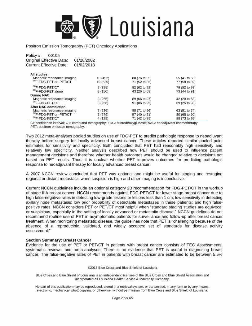
performed before and during (or after) NAC, while Sheikhbahaei did not require the scanning before NAC. Pooled sensitivities and specificities are listed in Table 2. Subgroup analysis was performed, by time of scanning (during NAC and after NAC was completed).
Table 2. Pooled Diagnostic Performance of 18
F-FDG-PET and Magnetic Resonance Imaging in
Detection of Residual Disease After NAC for Breast Cancer50
Type of Imaging No. of Studies
(No. of Patients) Sensitivity (95% CI), % Specificity (95% CI), %
Positron Emission Tomography (PET) Oncology Applications
Policy # 00105 Original Effective Date: 01/28/2002 Current Effective Date: 01/02/2018
©2017 Blue Cross and Blue Shield of Louisiana
Blue Cross and Blue Shield of Louisiana is an independent licensee of the Blue Cross and Blue Shield Association and
incorporated as Louisiana Health Service & Indemnity Company.
No part of this publication may be reproduced, stored in a retrieval system, or transmitted, in any form or by any means, electronic, mechanical, photocopying, or otherwise, without permission from Blue Cross and Blue Shield of Louisiana.
Page 20 of 65
Two 2012 meta-analyses pooled studies on use of FDG-PET to predict pathologic response to neoadjuvant therapy before surgery for locally advanced breast cancer. These articles reported similar pooled point estimates for sensitivity and specificity. Both concluded that PET had reasonably high sensitivity and relatively low specificity. Neither analysis described how PET should be used to influence patient management decisions and therefore whether health outcomes would be changed relative to decisions not based on PET results. Thus, it is unclear whether PET improves outcomes for predicting pathologic response to neoadjuvant therapy for locally advanced breast cancer. A 2007 NCCN review concluded that PET was optional and might be useful for staging and restaging regional or distant metastasis when suspicion is high and other imaging is inconclusive. Current NCCN guidelines include an optional category 2B recommendation for FDG-PET/CT in the workup of stage IIIA breast cancer. NCCN recommends against FDG-PET/CT for lower stage breast cancer due to high false-negative rates in detecting low-grade lesions or lesions less than 1 cm; low sensitivity in detecting axillary node metastasis; low prior probability of detectable metastases in these patients; and high false-positive rates. NCCN considers PET or PET/CT most helpful when “standard staging studies are equivocal or suspicious, especially in the setting of locally advanced or metastatic disease.” NCCN guidelines do not recommend routine use of PET in asymptomatic patients for surveillance and follow-up after breast cancer treatment. When monitoring metastatic disease, the guidelines note that PET is “challenging because of the absence of a reproducible, validated, and widely accepted set of standards for disease activity assessment.” Section Summary: Breast Cancer Evidence for the use of PET or PET/CT in patients with breast cancer consists of TEC Assessments, systematic reviews, and meta-analyses. There is no evidence that PET is useful in diagnosing breast cancer. The false-negative rates of PET in patients with breast cancer are estimated to be between 5.5%
All studies Magnetic resonance imaging 10 (492) 88 (76 to 95) 55 (41 to 68) 18
F-FDG-PET or -PET/CT 10 (535) 71 (52 to 85) 77 (58 to 89)
18F-FDG-PET/CT 7 (385) 82 (62 to 92) 79 (52 to 93)
18F-FDG-PET alone 3 (150) 43 (26 to 63) 73 (44 to 91)
During NAC Magnetic resonance imaging 3 (256) 89 (66 to 97) 42 (20 to 68) 18
F-FDG-PET/CT 3 (256) 91 (86 to 95) 69 (25 to 93) After NAC completion
Magnetic resonance imaging 7 (236) 88 (71 to 96) 63 (51 to 74) 18
F-FDG-PET or -PET/CT 7 (279) 57 (40 to 71) 80 (65 to 90) 18
F-FDG-PET/CT 4 (129) 71 (42 to 89) 88 (73 to 95)
CI: confidence interval; CT: computed tomography; FDG: fluorodeoxyglucose; NAC: neoadjuvant chemotherapy; PET: positron emission tomography.
Positron Emission Tomography (PET) Oncology Applications
Policy # 00105 Original Effective Date: 01/28/2002 Current Effective Date: 01/02/2018
©2017 Blue Cross and Blue Shield of Louisiana
Blue Cross and Blue Shield of Louisiana is an independent licensee of the Blue Cross and Blue Shield Association and
incorporated as Louisiana Health Service & Indemnity Company.
No part of this publication may be reproduced, stored in a retrieval system, or transmitted, in any form or by any means, electronic, mechanical, photocopying, or otherwise, without permission from Blue Cross and Blue Shield of Louisiana.
Page 21 of 65
and 8.5%, which can be considered unacceptable, given that breast biopsy can provide more definitive results. PET/CT might be useful in detecting metastases when results from other imaging techniques are inconclusive. The evidence supports the use of
18F-FDG-PET and
18F-PET/CT for staging and restaging
only if standard staging methods are inconclusive, but does not support their use for diagnosis, staging/restaging when standard staging methods are conclusive, and or surveillance. CERVICAL CANCER AND
18F-FDG-PET AND
18F-FDG-PET/CT
An AHRQ review published in 2008 identified several studies using 18
F-FDG-PET or -PET/CT to stage advanced cervical cancer and to detect and stage recurrent disease. The report concluded that most studies supported enhanced diagnostic accuracy, which would improve the selection of appropriate treatment for patients. For recurrent disease, PET identified additional sites of metastasis, which would alter treatment decisions in some cases. For example, in a 2004 study by Yen et al of 55 patients whose recurrences were initially considered curable with radical surgical treatment, 27 instead underwent palliative therapy based on PET results. An NCCN report on PET also identified several studies supporting the use of PET for initial staging and identifying and staging recurrent disease. In a 2013 meta-analysis of 9 cervical cancer recurrence studies, Rong et al (2013) reported a sensitivity and a specificity for PET/CT of 94.8% (95% CI, 91.2% to 96.9%) and 86.9% (95% CI, 82.2% to 90.5%), respectively. Reviewers found the quality of studies on recurrence was average with some limitations. For example, studies included mostly symptomatic women and did not differentiate between PET for diagnosis or surveillance. In a systematic review of 20 studies, Chu et al (2014) reported pooled sensitivity and specificity for FDG-PET or FDG-PET/CT of 87% (95% CI, 80% to 92%) and 97% (95% CI, 96% to 98%), respectively, for distant metastasis in recurrent cervical cancer. For local regional recurrence, pooled sensitivity and specificity were 82% (95% CI, 72% to 90%) and 98% (95% CI, 96% to 99%), respectively. Current NCCN guidelines state that PET/CT may be considered part of the initial nonfertility sparing workup for patients with stage I cervical cancer. The guidelines also state that PET/CT may be considered as part of the initial workup for detection of stage II, III, or IV metastatic disease. A single PET/CT scan at 3 to 6 months after therapy for locally advanced cervical cancer is recommended to detect persistent or recurrent disease. PET/CT is not recommended for surveillance. Section Summary: Cervical Cancer Evidence for the use of PET in patients with cervical cancer consists of systematic reviews and meta- analyses. Pooled results have shown that PET can be used for staging or restaging and detecting recurrent disease. The evidence supports the use of
18F-FDG-PET and
18F-PET/CT for the diagnosis and staging and
restaging of cervical cancer but does not support their use for surveillance.
Positron Emission Tomography (PET) Oncology Applications
Policy # 00105 Original Effective Date: 01/28/2002 Current Effective Date: 01/02/2018
©2017 Blue Cross and Blue Shield of Louisiana
Blue Cross and Blue Shield of Louisiana is an independent licensee of the Blue Cross and Blue Shield Association and incorporated as Louisiana Health Service & Indemnity Company.
No part of this publication may be reproduced, stored in a retrieval system, or transmitted, in any form or by any means, electronic, mechanical, photocopying, or otherwise, without permission from Blue Cross and Blue Shield of Louisiana.
Page 22 of 65
COLORECTAL CANCER AND 18F-FDG-PET AND 18
F-FDG-PET/CTTwo clinical applications of PET scanning were considered in the 1999 TEC Assessment: (1) To detect hepatic or extrahepatic metastases and to assess their resectability in patients with CRC, either as part of initial staging or after primary resection, and (2) to evaluate the presence of postoperative scar versus recurrent disease as a technique to determine the necessity of tissue biopsy.
The body of evidence indicated that PET scanning added useful information to conventional imaging in detecting hepatic and extrahepatic metastases. In particular, PET detected additional metastases leading to more identification of nonresectable disease, allowing patients to avoid surgery. The strongest evidence came from a study that directly assessed the additional value of PET. In a group of 37 patients thought to have a solitary liver metastasis by conventional imaging, PET correctly upstaged 4 patients and falsely overstaged 1 patient. This and another study found that when PET results were discordant with conventional imaging results, PET was correct in 88% and 97% of patients, respectively. When PET affected management decisions, it was more often used to recommend against surgery.
When used to distinguish between local recurrence and scar, the comparison is between performing histologic sampling in all patients with a suspected local recurrence and avoiding sampling in patients whose PET scans suggest the presence of postoperative scar. The key concern is whether the negative predictive value (NPV) for PET is sufficiently high to influence decision making, specifically to avoid tissue biopsy when the PET scan is negative. The TEC Assessment found that studies then available suggested an 8% probability of false-negative results making it unlikely that patients and physicians would forgo histologic sampling and delay potentially curative repeat resection.
A 2012 systematic review of different imaging techniques for radiotherapy treatment planning of rectal cancer concluded that additional studies would be needed to validate use of PET in this setting. Three systematic reviews published in 2014 included overlapping studies that assessed the predictive value of FDG-PET/CT in patients with locally advanced rectal cancer who received neoadjuvant chemoradiotherapy. Various PET parameters were investigated (standardized uptake value, response index [percentage of the standardized uptake value decrease from baseline to post neoadjuvant treatment]), and cutoff values varied. Pooled sensitivities ranged from 74% to 82%, and pooled specificities ranged from 64% to 85%. The value of FDG-PET/CT in this setting has yet to be clarified.
CRC Diagnosis A 2015 systematic review by Jones et al evaluated the role of
18F-FDG-PET and
18F-FDG-PET/CT
compared with conventional imaging in the detection of primary nodal disease. Twelve studies met inclusion criteria (total N=494 patients), Meta-analysis for detecting primary disease in situ showed that PET and PET/CT had a higher sensitivity (99%; 95% CI, 96% to 100%) than CT alone (60%; 95% CI, 46% to 75%).
Positron Emission Tomography (PET) Oncology Applications
Policy # 00105 Original Effective Date: 01/28/2002 Current Effective Date: 01/02/2018
©2017 Blue Cross and Blue Shield of Louisiana
Blue Cross and Blue Shield of Louisiana is an independent licensee of the Blue Cross and Blue Shield Association and
incorporated as Louisiana Health Service & Indemnity Company.
No part of this publication may be reproduced, stored in a retrieval system, or transmitted, in any form or by any means, electronic, mechanical, photocopying, or otherwise, without permission from Blue Cross and Blue Shield of Louisiana.
Page 23 of 65
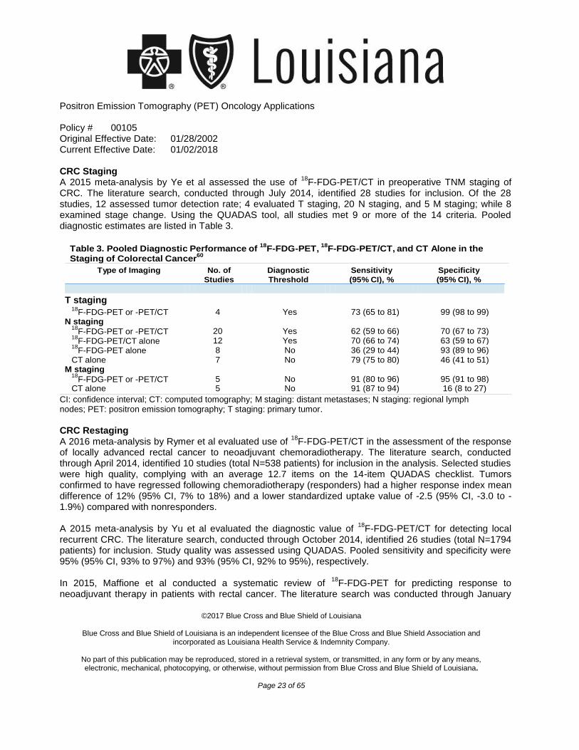
CRC Staging A 2015 meta-analysis by Ye et al assessed the use of
18F-FDG-PET/CT in preoperative TNM staging of
CRC. The literature search, conducted through July 2014, identified 28 studies for inclusion. Of the 28 studies, 12 assessed tumor detection rate; 4 evaluated T staging, 20 N staging, and 5 M staging; while 8 examined stage change. Using the QUADAS tool, all studies met 9 or more of the 14 criteria. Pooled diagnostic estimates are listed in Table 3.
T staging
CI: confidence interval; CT: computed tomography; M staging: distant metastases; N staging: regional lymph nodes; PET: positron emission tomography; T staging: primary tumor.
CRC Restaging A 2016 meta-analysis by Rymer et al evaluated use of
18F-FDG-PET/CT in the assessment of the response
of locally advanced rectal cancer to neoadjuvant chemoradiotherapy. The literature search, conducted through April 2014, identified 10 studies (total N=538 patients) for inclusion in the analysis. Selected studies were high quality, complying with an average 12.7 items on the 14-item QUADAS checklist. Tumors confirmed to have regressed following chemoradiotherapy (responders) had a higher response index mean difference of 12% (95% CI, 7% to 18%) and a lower standardized uptake value of -2.5 (95% CI, -3.0 to -1.9%) compared with nonresponders. A 2015 meta-analysis by Yu et al evaluated the diagnostic value of
18F-FDG-PET/CT for detecting local
recurrent CRC. The literature search, conducted through October 2014, identified 26 studies (total N=1794 patients) for inclusion. Study quality was assessed using QUADAS. Pooled sensitivity and specificity were 95% (95% CI, 93% to 97%) and 93% (95% CI, 92% to 95%), respectively. In 2015, Maffione et al conducted a systematic review of
18F-FDG-PET for predicting response to
neoadjuvant therapy in patients with rectal cancer. The literature search was conducted through January
Table 3. Pooled Diagnostic Performance of 18
F-FDG-PET, 18
F-FDG-PET/CT, and CT Alone in the
Staging of Colorectal Cancer60
Type of Imaging No. of
Studies Diagnostic
Threshold Sensitivity
(95% CI), % Specificity
(95% CI), %
18F-FDG-PET or -PET/CT 4 Yes 73 (65 to 81) 99 (98 to 99)
N staging 18
F-FDG-PET or -PET/CT
20
Yes
62 (59 to 66)
70 (67 to 73) 18
F-FDG-PET/CT alone 12 Yes 70 (66 to 74) 63 (59 to 67) 18
F-FDG-PET alone 8 No 36 (29 to 44) 93 (89 to 96) CT alone 7 No 79 (75 to 80) 46 (41 to 51)
M staging 18
F-FDG-PET or -PET/CT
5
No
91 (80 to 96)
95 (91 to 98) CT alone 5 No 91 (87 to 94) 16 (8 to 27)
Positron Emission Tomography (PET) Oncology Applications
Policy # 00105 Original Effective Date: 01/28/2002 Current Effective Date: 01/02/2018
©2017 Blue Cross and Blue Shield of Louisiana
Blue Cross and Blue Shield of Louisiana is an independent licensee of the Blue Cross and Blue Shield Association and
incorporated as Louisiana Health Service & Indemnity Company.
No part of this publication may be reproduced, stored in a retrieval system, or transmitted, in any form or by any means, electronic, mechanical, photocopying, or otherwise, without permission from Blue Cross and Blue Shield of Louisiana.
Page 24 of 65
2014, with 29 studies meeting inclusion criteria for the meta-analysis. The studies had QUADAS scores ranging from 8 to 14 (median, 12). The pooled sensitivity and specificity for
18F-FDG-PET assessment of
response to chemoradiotherapy in locally advanced rectal cancer were 73% (95% CI, 71% to 76%) and 77% (95% CI, 75% to 79%), respectively. In a 2013 systematic review, Lu et al evaluated 510 patients from 11 studies on
18F-FDG-PET for CRC
tumor recurrence detection in patients with elevated carcinoembryonic antigen. The literature search ran through April 2012. FDG-PET and PET/CT pooled sensitivity estimates were 90.3% (95% CI, 85.5% to 94.0%) and 94.1% (95% CI, 89.4% to 97.1%), respectively, and specificities were 80.0% (95% CI, 67.0% to 89.6%) and 77.2% (95% CI, 66.4% to 85.9%), respectively. Current NCCN guidelines for colon cancer “strongly discourage the routine use of PET/CT scanning for staging, baseline imaging, or routine follow-up and recommend consideration of a preoperative PET/CT scan at baseline only if prior anatomic imaging indicates the presence of potentially surgically curable M1 disease.” For initial workup of nonmetastatic patients, the guidelines state “PET/CT does not supplant a contrast-enhanced diagnostic CT scan. PET/CT should only be used to evaluate an equivocal finding on a contrast-enhanced CT scan or in patients with strong contraindications to IV [intravenous] contrast.” For workup of proven metastatic synchronous adenocarcinoma, the guidelines state that PET/CT may be considered. PET/CT is not recommended for surveillance. NCCN has noted that PET/CT should not be used to assess response to chemotherapy. NCCN was divided on appropriateness of PET/CT when CEA level is rising; PET/CT might be considered when imaging study results (e.g., a good quality CT scan) are normal. Current NCCN guidelines for rectal cancer state that PET/CT is “not routinely indicated” and “should only be used to evaluate an equivocal finding on a contrast-enhanced CT scan.” PET/CT is not recommended for surveillance. PET/CT can be considered if serial CEA elevation occurs or if there is documented metachronous metastases. Section Summary: Colorectal Cancer Evidence for the detection of primary nodal disease, staging, restaging, and detecting recurrence of CRC consists of a TEC Assessment and several meta-analyses published after the assessment. A meta-analysis evaluating the diagnostic accuracy of PET or PET/CT found a high sensitivity but a low specificity. Several pooled analyses evaluating staging or restaging using PET or PET/CT resulted in sensitivities and specificities ranging low 60s to high 90s. The evidence for the use of PET or PET/CT did not show a benefit over the use of contrast CT in patients with CRC. The evidence does not support the use of
18F-FDG-PET
and 18
F-PET/CT for the diagnosis, staging and restaging, or surveillance of CRC. ENDOMETRIAL CANCER AND
18F-FDG-PET AND
18F-FDG-PET/CT
Positron Emission Tomography (PET) Oncology Applications
Policy # 00105 Original Effective Date: 01/28/2002 Current Effective Date: 01/02/2018
©2017 Blue Cross and Blue Shield of Louisiana
Blue Cross and Blue Shield of Louisiana is an independent licensee of the Blue Cross and Blue Shield Association and
incorporated as Louisiana Health Service & Indemnity Company.
No part of this publication may be reproduced, stored in a retrieval system, or transmitted, in any form or by any means, electronic, mechanical, photocopying, or otherwise, without permission from Blue Cross and Blue Shield of Louisiana.
Page 25 of 65
In 2016, Bollineni et al published a systematic review and meta-analysis on the diagnostic value of 18
F- FDG-PET for endometrial cancer. The literature search, conducted through August 2015, identified 21 studies for inclusion in the meta-analysis: 13 on detection of lymph node metastases (n=861) and 8 on detection of endometrial cancer recurrence (n=378). Pooled sensitivity and specificity for
18F-FDG-PET for
detecting lymph node metastases were 72% (95% CI, 63% to 80%) and 94% (95% CI, 93% to 96%), respectively. Pooled sensitivity and specificity for
18F-FDG-PET for detecting endometrial cancer recurrence
following primary surgical treatment were 95% (95% CI, 91% to 98%) and 91% (95% CI, 86% to 94%), respectively. Current NCCN guidelines for endometrial cancer state that whole body PET/CT can be considered if metastases are suspected in select patients (based on clinical symptoms, physical findings, or abnormal laboratory findings). Following treatment, PET/CT can be considered in select patients for surveillance. Section Summary: Endometrial Cancer and
18F-FDG-PET and
18F-FDG-PET/CT
The evidence supports the use of 18
F-FDG-PET and 18
F-PET/CT for the diagnosis, staging and restaging, or surveillance of endometrial cancer. ESOPHAGEAL CANCER AND
18F-FDG-PET AND
18F-FDG-PET/CT
For initial diagnosis, PET is generally not considered for detecting primary esophageal tumors, and evidence is lacking on its ability to differentiate between esophageal cancer and benign conditions. In 2016, Cong et al published a meta-analysis evaluating the predictive value of
18F-FDG-PET and -
PET/CT for tumor response during or after neoadjuvant chemoradiotherapy in patients with esophageal cancer. The literature search, conducted through January 2016, identified 4 studies (n=192 patients) in which PET or PET/CT was performed during neoadjuvant chemoradiotherapy and 11 studies (n=490 patients) in which PET or PET/CT was performed after neoadjuvant chemoradiotherapy. All studies scored between 9 and 12 using the QUADAS tool. Pooled sensitivity and specificity for PET and PET/CT performed during NRCT is 85% (95% CI, 76% to 91%) and 59% (95% CI, 48% to 69%), respectively. Pooled sensitivity and specificity for PET and PET/CT performed after neoadjuvant chemoradiotherapy were 67% (95% CI, 60% to 73%) and 69% (95% CI, 63% to 74%), respectively. In 2016, Goense et al published a systematic review evaluating
18F-FDG-PET and -PET/CT for the
detection of recurrent esophageal cancer after treatment with curative intent. The literature search, conducted through December 2014, identified 8 studies (total N=486 patients) for inclusion. The quality of the studies was considered reasonable using the QUADAS tool, with low risk of bias for a majority of the studies, and high risk of bias in a few studies for patient selection. Pooled estimates of sensitivity and specificity of
18F-FDG-PET and -PET/CT combined were 96% (95% CI, 93% to 97%) and 78% (95% CI,
66% to 86%), respectively. Subgroup analysis by technique (PET alone and PET/CT) was not possible for
Positron Emission Tomography (PET) Oncology Applications
Policy # 00105 Original Effective Date: 01/28/2002 Current Effective Date: 01/02/2018
©2017 Blue Cross and Blue Shield of Louisiana
Blue Cross and Blue Shield of Louisiana is an independent licensee of the Blue Cross and Blue Shield Association and
incorporated as Louisiana Health Service & Indemnity Company.
No part of this publication may be reproduced, stored in a retrieval system, or transmitted, in any form or by any means, electronic, mechanical, photocopying, or otherwise, without permission from Blue Cross and Blue Shield of Louisiana.
Page 26 of 65
sensitivity due to heterogeneity. Specificity subgroup analysis showed no statistical difference between PET alone and PET/CT in detecting recurrent esophageal cancer. A 2009 NCCN report found studies showing that PET is more sensitive than other diagnostic imaging in detecting stage IV disease with distant lymph node involvement. A meta-analysis described in the report found a 67% pooled sensitivity, 97% specificity, and small added value after conventional staging in detecting distant metastasis. In a 2013 meta-analysis of 245 patients with esophageal cancer from 6 studies, Shi et al reported that, for detection of regional nodal metastases, FDG-PET/CT had a sensitivity of 55% (95% CI, 34% to 74%) and specificity of 76% (95% CI, 66% to 83%), respectively. Another use of PET in esophageal cancer is in determining whether to continue chemotherapy for potential curative resection. The 2009 NCCN report described several studies in which response to chemotherapy, defined as a decline in standardized uptake values, correlated with long-term survival. Patients who do not respond to chemotherapy might benefit from this test by being spared futile and toxic chemotherapy. However, the treatment strategy of PET-directed chemotherapy does not appear to have been validated with RCTs showing improved net health outcome. Current NCCN guidelines for esophageal cancer recommend that PET/CT be considered to assess treatment response 5 to 6 weeks after preoperative therapy. Current NCCN guidelines for esophageal cancer indicate that PET/CT be considered in the initial workup of esophageal cancer if there is no evidence of M1 disease and to assess response to preoperative or definitive chemoradiation. The guidelines note that PET/CT for these indications is preferable to PET alone. Section Summary: Esophageal Cancer Evidence for PET or PET/CT to detect metastases, predict tumor response to treatment, or to detect recurrence in patients with esophageal cancer consists of meta-analyses. The meta-analyses have shown high sensitivity and specificity estimates for these indications. The evidence supports the use of
18F-FDG-
PET and 18
F-PET/CT for the diagnosis and staging and restaging of esophageal cancer, but does not support their use for surveillance. GASTRIC CANCER AND
18F-FDG-PET AND
18F-FDG-PET/CT
A 2016 systematic review by Li et al evaluated 18
F-FDG-PET and 18
F-FDG-PET/CT for detecting recurrent gastric cancer. The literature search, conducted through February 2015, identified 14 studies (total N=828 patients) to be included in the analysis. The analysis combined both imaging techniques; 3 studies used PET alone and 11 studies used PET/CT. Pooled sensitivity and specificity were 85% (95% CI, 75% to 92%) and 78% (95% CI, 72% to 84%), respectively. In a 2013 meta-analysis, Zou and Zhou evaluated studies published through May 2013 and calculated the sensitivity and specificity of
18F-FDG-PET/CT for detecting recurrence of gastric cancer after surgical
Positron Emission Tomography (PET) Oncology Applications
Policy # 00105 Original Effective Date: 01/28/2002 Current Effective Date: 01/02/2018
©2017 Blue Cross and Blue Shield of Louisiana
Blue Cross and Blue Shield of Louisiana is an independent licensee of the Blue Cross and Blue Shield Association and
incorporated as Louisiana Health Service & Indemnity Company.
No part of this publication may be reproduced, stored in a retrieval system, or transmitted, in any form or by any means, electronic, mechanical, photocopying, or otherwise, without permission from Blue Cross and Blue Shield of Louisiana.
Page 27 of 65
resection. Eight studies (total N=500 patients) were eligible for the meta-analysis. The studies fulfilled 12 of the 14 QUADAS criteria for methodologic quality. Pooled sensitivity was 86% (95% CI, 71% to 94%) and pooled specificity was 88% (95% CI, 75% to 94%), respectively. A systematic review by Wu (2012) pooled 9 studies (total N=562 patient) published through July 2011 that used
18F-FDG-PET alone for evaluating recurrent gastric cancer. Each selected study fulfilled at least 9 of
the 14 criteria in the QUADAS tool for methodologic quality. Pooled sensitivity and specificity were 78% (95% CI, 68% to 86%) and 82% (05% CI, 76% to 87%), respectively. Reviewers concluded that PET/CT might be more effective than either PET alone or CT alone, but it was unclear what sources reviewers used for their estimates for PET/CT and CT alone. Current NCCN guidelines for gastric cancer indicate that PET/CT (but not PET alone) can be used as part of an initial workup if there is no evidence of metastatic disease. The guidelines note that the sensitivity of PET/CT is lower than that for CT alone due to low tracer accumulation in diffuse and mucinous tumor types, but specificity is higher. PET/CT adds value to the diagnostic workup with higher accuracy in staging. NCCN guidelines also indicate that PET/CT can be used to evaluate response to treatment, in cases of renal insufficiency or allergy to CT contrast. Section Summary: Gastric Cancer Evidence for the use of PET to diagnose recurrent gastric cancer consists of meta-analyses. One meta- analysis evaluated
18F-FDG-PET alone, one evaluated
18F-FDG-PET/CT, and another combined the 2
techniques into a single estimate. Sensitivity estimates ranged from 78% to 85% and specificity estimates ranged from 78% to 88%. The evidence supports the use of
18F-FDG-PET and
18F-PET/CT for the
diagnosis and staging and restaging of esophageal cancer, but does not support their use for surveillance. HEAD AND NECK CANCER AND
18F-FDG-PET AND
18F-FDG-PET/CT
Among the 3 studies identified in the TEC Assessment (2000) that used other diagnostic modalities to identify a primary tumor in patients with positive cervical lymph nodes, PET found more primary tumors than the other modalities in 2 studies and identified similar proportions in the third. When data from these 3 studies were pooled, PET was found to identify tumor in 38% of cases and other modalities in 21% of cases. When PET was used to stage cervical lymph nodes initially, the addition of PET to other imaging modalities increased the proportion of patients correctly staged, as confirmed histologically. When compared head to head with other imaging modalities, pooled data from a variety of studies suggested that PET had a better diagnostic performance than CT and MRI. Of 8 studies focusing on the use of PET to detect residual or recurrent disease, 5 found PET to be more specific and sensitive, 2 reported mixed or equivalent results, and 1 reported worse results compared with CT.
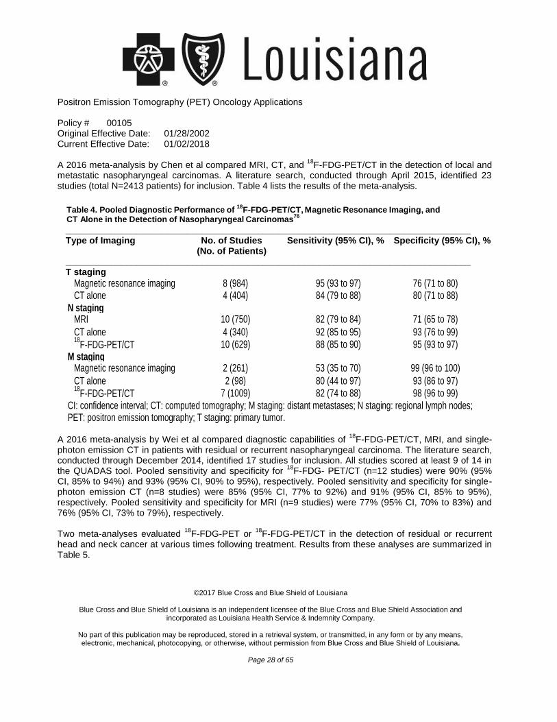
Positron Emission Tomography (PET) Oncology Applications
Policy # 00105 Original Effective Date: 01/28/2002 Current Effective Date: 01/02/2018
©2017 Blue Cross and Blue Shield of Louisiana
Blue Cross and Blue Shield of Louisiana is an independent licensee of the Blue Cross and Blue Shield Association and
incorporated as Louisiana Health Service & Indemnity Company.
No part of this publication may be reproduced, stored in a retrieval system, or transmitted, in any form or by any means, electronic, mechanical, photocopying, or otherwise, without permission from Blue Cross and Blue Shield of Louisiana.
Page 28 of 65
A 2016 meta-analysis by Chen et al compared MRI, CT, and 18
F-FDG-PET/CT in the detection of local and metastatic nasopharyngeal carcinomas. A literature search, conducted through April 2015, identified 23 studies (total N=2413 patients) for inclusion. Table 4 lists the results of the meta-analysis.
________________________________________________________________________________ Type of Imaging No. of Studies Sensitivity (95% CI), % Specificity (95% CI), % (No. of Patients) ________________________________________________________________________________ T staging
A 2016 meta-analysis by Wei et al compared diagnostic capabilities of
18F-FDG-PET/CT, MRI, and single-
photon emission CT in patients with residual or recurrent nasopharyngeal carcinoma. The literature search, conducted through December 2014, identified 17 studies for inclusion. All studies scored at least 9 of 14 in the QUADAS tool. Pooled sensitivity and specificity for
18F-FDG- PET/CT (n=12 studies) were 90% (95%
CI, 85% to 94%) and 93% (95% CI, 90% to 95%), respectively. Pooled sensitivity and specificity for single-photon emission CT (n=8 studies) were 85% (95% CI, 77% to 92%) and 91% (95% CI, 85% to 95%), respectively. Pooled sensitivity and specificity for MRI (n=9 studies) were 77% (95% CI, 70% to 83%) and 76% (95% CI, 73% to 79%), respectively. Two meta-analyses evaluated
18F-FDG-PET or
18F-FDG-PET/CT in the detection of residual or recurrent
head and neck cancer at various times following treatment. Results from these analyses are summarized in Table 5.
Table 4. Pooled Diagnostic Performance of 18
F-FDG-PET/CT, Magnetic Resonance Imaging, and
CT Alone in the Detection of Nasopharyngeal Carcinomas76
Magnetic resonance imaging 8 (984) 95 (93 to 97) 76 (71 to 80) CT alone 4 (404) 84 (79 to 88) 80 (71 to 88)
N staging MRI 10 (750) 82 (79 to 84) 71 (65 to 78)
CT alone 4 (340) 92 (85 to 95) 93 (76 to 99) 18
F-FDG-PET/CT 10 (629) 88 (85 to 90) 95 (93 to 97) M staging
Magnetic resonance imaging 2 (261) 53 (35 to 70) 99 (96 to 100)
CT alone 2 (98) 80 (44 to 97) 93 (86 to 97) 18
F-FDG-PET/CT 7 (1009) 82 (74 to 88) 98 (96 to 99) CI: confidence interval; CT: computed tomography; M staging: distant metastases; N staging: regional lymph nodes; PET: positron emission tomography; T staging: primary tumor.
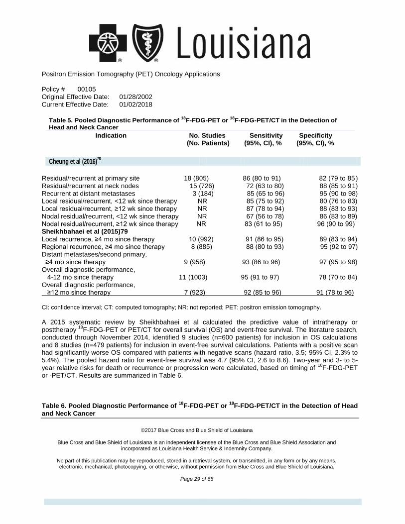
Positron Emission Tomography (PET) Oncology Applications
Policy # 00105 Original Effective Date: 01/28/2002 Current Effective Date: 01/02/2018
©2017 Blue Cross and Blue Shield of Louisiana
Blue Cross and Blue Shield of Louisiana is an independent licensee of the Blue Cross and Blue Shield Association and
incorporated as Louisiana Health Service & Indemnity Company.
No part of this publication may be reproduced, stored in a retrieval system, or transmitted, in any form or by any means, electronic, mechanical, photocopying, or otherwise, without permission from Blue Cross and Blue Shield of Louisiana.
Page 29 of 65
Indication No. Studies Sensitivity Specificity
(No. Patients) (95%, CI), % (95%, CI), %
Residual/recurrent at primary site 18 (805) 86 (80 to 91) 82 (79 to 85) Residual/recurrent at neck nodes 15 (726) 72 (63 to 80) 88 (85 to 91) Recurrent at distant metastases 3 (184) 85 (65 to 96) 95 (90 to 98) Local residual/recurrent, <12 wk since therapy NR 85 (75 to 92) 80 (76 to 83) Local residual/recurrent, ≥12 wk since therapy NR 87 (78 to 94) 88 (83 to 93) Nodal residual/recurrent, <12 wk since therapy NR 67 (56 to 78) 86 (83 to 89) Nodal residual/recurrent, ≥12 wk since therapy NR 83 (61 to 95) 96 (90 to 99) Sheikhbahaei et al (2015)79 Local recurrence, ≥4 mo since therapy 10 (992) 91 (86 to 95) 89 (83 to 94) Regional recurrence, ≥4 mo since therapy 8 (885) 88 (80 to 93) 95 (92 to 97) Distant metastases/second primary, ≥4 mo since therapy 9 (958) 93 (86 to 96) 97 (95 to 98) Overall diagnostic performance, 4-12 mo since therapy 11 (1003) 95 (91 to 97) 78 (70 to 84) Overall diagnostic performance, ≥12 mo since therapy 7 (923) 92 (85 to 96) 91 (78 to 96) CI: confidence interval; CT: computed tomography; NR: not reported; PET: positron emission tomography.
A 2015 systematic review by Sheikhbahaei et al calculated the predictive value of intratherapy or posttherapy
18F-FDG-PET or PET/CT for overall survival (OS) and event-free survival. The literature search,
conducted through November 2014, identified 9 studies (n=600 patients) for inclusion in OS calculations and 8 studies (n=479 patients) for inclusion in event-free survival calculations. Patients with a positive scan had significantly worse OS compared with patients with negative scans (hazard ratio, 3.5; 95% CI, 2.3% to 5.4%). The pooled hazard ratio for event-free survival was 4.7 (95% CI, 2.6 to 8.6). Two-year and 3- to 5-year relative risks for death or recurrence or progression were calculated, based on timing of
18F-FDG-PET
or -PET/CT. Results are summarized in Table 6. Table 6. Pooled Diagnostic Performance of
18F-FDG-PET or
18F-FDG-PET/CT in the Detection of Head
and Neck Cancer
Table 5. Pooled Diagnostic Performance of 18
F-FDG-PET or 18
F-FDG-PET/CT in the Detection of
Head and Neck Cancer
Cheung et al (2016)
78
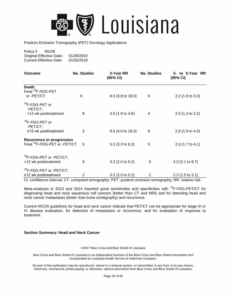
Positron Emission Tomography (PET) Oncology Applications
Policy # 00105 Original Effective Date: 01/28/2002 Current Effective Date: 01/02/2018
©2017 Blue Cross and Blue Shield of Louisiana
Blue Cross and Blue Shield of Louisiana is an independent licensee of the Blue Cross and Blue Shield Association and
incorporated as Louisiana Health Service & Indemnity Company.
No part of this publication may be reproduced, stored in a retrieval system, or transmitted, in any form or by any means, electronic, mechanical, photocopying, or otherwise, without permission from Blue Cross and Blue Shield of Louisiana.
Page 30 of 65
Outcome No. Studies 2-Year RR No. Studies 3- to 5-Year RR
(95% CI) (95% CI)
Death 18
Final F-FDG-PET or -PET/CT
6 8.3 (3.8 to 18.0) 6 2.2 (1.6 to 3.2) 18
F-FDG-PET or -PET/CT, <12 wk posttreatment 8 3.0 (1.9 to 4.6) 4 2.0 (1.3 to 3.2) 18
F-FDG-PET or -PET/CT, ≥12 wk posttreatment 3 8.5 (4.0 to 18.3) 6 2.8 (1.9 to 4.0) Recurrence or progression
18Final F-FDG-PET or -PET/CT
6 5.2 (3.3 to 8.3) 5 2.6 (1.7 to 4.1)
18
F-FDG-PET or -PET/CT, <12 wk posttreatment 9 3.2 (2.0 to 5.2) 6 4.3 (2.1 to 8.7) 18
F-FDG-PET or -PET/CT, ≥12 wk posttreatment 2 3.2 (2.0 to 5.2) 2 2.2 (1.5 to 3.1)
CI: confidence interval; CT: computed tomography; PET: positron emission tomography; RR: relative risk. Meta-analyses in 2013 and 2014 reported good sensitivities and specificities with
18F-FDG-PET/CT for
diagnosing head and neck squamous cell cancers (better than CT and MRI) and for detecting head and neck cancer metastases (better than bone scintigraphy) and recurrence. Current NCCN guidelines for head and neck cancer indicate that PET/CT can be appropriate for stage III or IV disease evaluation, for detection of metastases or recurrence, and for evaluation of response to treatment. Section Summary: Head and Neck Cancer
Positron Emission Tomography (PET) Oncology Applications
Policy # 00105 Original Effective Date: 01/28/2002 Current Effective Date: 01/02/2018
©2017 Blue Cross and Blue Shield of Louisiana
Blue Cross and Blue Shield of Louisiana is an independent licensee of the Blue Cross and Blue Shield Association and
incorporated as Louisiana Health Service & Indemnity Company.
No part of this publication may be reproduced, stored in a retrieval system, or transmitted, in any form or by any means, electronic, mechanical, photocopying, or otherwise, without permission from Blue Cross and Blue Shield of Louisiana.
Page 31 of 65
Evidence for the use of 18
F-FDG-PET/CT in the management of patients with head and neck cancer consists of systematic reviews and meta-analyses. In patients with head and neck cancers, PET or PET/CT is better able to detect local and metastatic disease than other imaging techniques. Evidence has also shown that
18F-FDG-PET/CT may be useful in predicting response to therapy. The evidence supports the
use of 18
F-FDG-PET and 18
F-PET/CT for the diagnosis and staging and restaging of esophageal cancer, but does not support their use for surveillance. LUNG CANCER AND
18F-FDG-PET AND
18F-FDG-PET/CT
PET scanning may have a clinical role in patients with solitary pulmonary nodules for whom a diagnosis is uncertain after CT scan or chest radiograph. Younger patients who have no smoking history have a relatively low risk for lung cancer and, in this setting, the NPV of a PET scan is relatively high. If presented with a negative PET scan and information about the very low probability of undetected malignancy, it is quite likely that some patients would choose to avoid the harms of an invasive sampling procedure (i.e., biopsy). A 2012 meta-analysis on evaluating pulmonary nodules using dual-time PET (a second scan added after a delay) found that its additive value relative to a single PET scan is questionable. Non-Small-Cell Lung Cancer In patients with known NSCLC, the clinical value of PET scanning relates to improved staging information regarding the involvement of mediastinal lymph nodes, which generally excludes patients from surgical excision. The 1997 TEC Assessment discussed a decision analysis that suggested use of CT plus PET scanning in staging mediastinal lymph nodes resulted in fewer surgeries and an average gain in life expectancy of 2.96 days.3 This suggests that the reduction in surgeries was not harmful to patients. A 2017 systematic review by Ruilong et al evaluated the diagnostic value of
18F-FDG PET/CT for detecting
solitary pulmonary nodules. The literature search, conducted to May 2015, identified 12 studies (1297 patients) for inclusion in the analysis. The pooled sensitivity and specificity of
18F-FDG-PET/CT to detect
malignant pulmonary nodules were 82% (95% CI, 76% to 87%) and 81% (95% CI, 66% to 90%), respectively. Other meta-analyses have reported good sensitivities and specificities in detection of lung cancer metastases and recurrence with PET/CT. A meta-analysis by Li et al (2013) calculated the following sensitivity and specificity for
18F-FDG-PET/CT in detecting distant metastases in patients with lung cancer:
93% (95% CI, 88% to 96%) and 96% (95% CI, 95% to 96%), respectively. He et al (2014) compared PET, PET/CT, and conventional imaging techniques in detecting recurrent lung cancer. Table 7 summarizes the diagnostic performances of the different imaging techniques: Table 7. Pooled Diagnostic Performance of PET, PET/CT, and CIT in the Detection of Lung Cancer
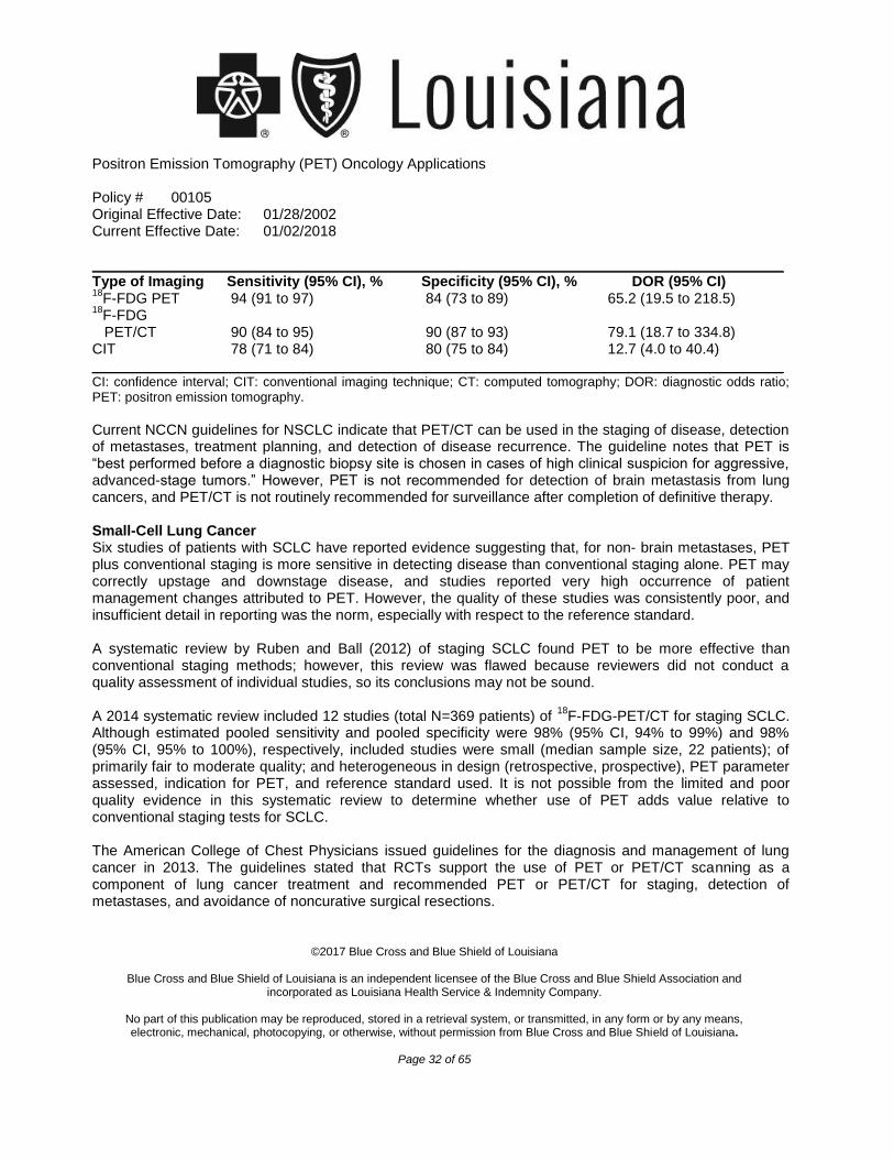
Positron Emission Tomography (PET) Oncology Applications
Policy # 00105 Original Effective Date: 01/28/2002 Current Effective Date: 01/02/2018
©2017 Blue Cross and Blue Shield of Louisiana
Blue Cross and Blue Shield of Louisiana is an independent licensee of the Blue Cross and Blue Shield Association and
incorporated as Louisiana Health Service & Indemnity Company.
No part of this publication may be reproduced, stored in a retrieval system, or transmitted, in any form or by any means, electronic, mechanical, photocopying, or otherwise, without permission from Blue Cross and Blue Shield of Louisiana.
Page 32 of 65
_____________________________________________________________________________________ Type of Imaging Sensitivity (95% CI), % Specificity (95% CI), % DOR (95% CI) 18
F-FDG PET 94 (91 to 97) 65.2 (19.5 to 218.5) 18
F-FDG PET/CT 90 (84 to 95) 90 (87 to 93
84 (73 to 89)
) 79.1 (18.7 to 334.8) CIT 78 (71 to 84) 80 (75 to 84) 12.7 (4.0 to 40.4) _____________________________________________________________________________________ CI: confidence interval; CIT: conventional imaging technique; CT: computed tomography; DOR: diagnostic odds ratio; PET: positron emission tomography.
Current NCCN guidelines for NSCLC indicate that PET/CT can be used in the staging of disease, detection of metastases, treatment planning, and detection of disease recurrence. The guideline notes that PET is “best performed before a diagnostic biopsy site is chosen in cases of high clinical suspicion for aggressive, advanced-stage tumors.” However, PET is not recommended for detection of brain metastasis from lung cancers, and PET/CT is not routinely recommended for surveillance after completion of definitive therapy. Small-Cell Lung Cancer Six studies of patients with SCLC have reported evidence suggesting that, for non- brain metastases, PET plus conventional staging is more sensitive in detecting disease than conventional staging alone. PET may correctly upstage and downstage disease, and studies reported very high occurrence of patient management changes attributed to PET. However, the quality of these studies was consistently poor, and insufficient detail in reporting was the norm, especially with respect to the reference standard. A systematic review by Ruben and Ball (2012) of staging SCLC found PET to be more effective than conventional staging methods; however, this review was flawed because reviewers did not conduct a quality assessment of individual studies, so its conclusions may not be sound. A 2014 systematic review included 12 studies (total N=369 patients) of
18F-FDG-PET/CT for staging SCLC.
Although estimated pooled sensitivity and pooled specificity were 98% (95% CI, 94% to 99%) and 98% (95% CI, 95% to 100%), respectively, included studies were small (median sample size, 22 patients); of primarily fair to moderate quality; and heterogeneous in design (retrospective, prospective), PET parameter assessed, indication for PET, and reference standard used. It is not possible from the limited and poor quality evidence in this systematic review to determine whether use of PET adds value relative to conventional staging tests for SCLC. The American College of Chest Physicians issued guidelines for the diagnosis and management of lung cancer in 2013. The guidelines stated that RCTs support the use of PET or PET/CT scanning as a component of lung cancer treatment and recommended PET or PET/CT for staging, detection of metastases, and avoidance of noncurative surgical resections.
Positron Emission Tomography (PET) Oncology Applications
Policy # 00105 Original Effective Date: 01/28/2002 Current Effective Date: 01/02/2018
©2017 Blue Cross and Blue Shield of Louisiana
Blue Cross and Blue Shield of Louisiana is an independent licensee of the Blue Cross and Blue Shield Association and
incorporated as Louisiana Health Service & Indemnity Company.
No part of this publication may be reproduced, stored in a retrieval system, or transmitted, in any form or by any means, electronic, mechanical, photocopying, or otherwise, without permission from Blue Cross and Blue Shield of Louisiana.
Page 33 of 65
Current NCCN guidelines for SCLC indicate PET can be used in the staging of disease and treatment planning but “is not recommended for routine follow-up.” Section Summary: Lung Cancer Evidence for PET or PET/CT in patients with NSCLC consists of meta-analyses. The meta-analyses have shown that use of PET or PET/CT in patients with lung cancer can aid in the diagnosis, staging, as well as
18 18detecting metastases and recurrence. The evidence supports the use of F-FDG-PET and F- PET/CT for the diagnosis and staging and restaging of NSCLC, but does not support their use for surveillance. Evidence for PET or PET/CT for patients with SCLC consists of systematic reviews and meta-analyses. While these reviews have shown potential benefits in using PET for staging, the quality of the studies was
18 18low. The evidence does not support the use of F-FDG-PET and F-PET/CT for the diagnosis, staging and restaging, or surveillance of SCLC.
18 18LYMPHOMA, INCLUDING HODGKIN DISEASE, AND F-FDG-PET AND F-FDG-PET/CT Of the 14 studies reviewed in the 1999 TEC Assessment, 3 compared PET with anatomic imaging in initial staging and restaging of patients with Hodgkin disease and non-Hodgkin lymphoma. Two of these studies included data from both diseased and nondiseased sites for PET and CT. Both studies found PET to have better overall diagnostic accuracy than CT. The third study addressed detection of diseased sites only and found PET to have a sensitivity similar to that of CT or MRI. Among the 6 studies that reported on concordance between PET and other imaging modalities, PET was discordant with other modalities in 11% to 50% of cases; PET was correct among discordances in 40% to 75% of cases. PET has been reported to affect patient management decisions in 8% to 20% of patients in 5 studies, mainly by correctly upstaging disease, but also by correctly downstaging disease. Thus, when PET is added to conventional imaging, it can provide useful information for selecting effective treatment appropriate to the correct stage of disease. Lymphoma Diagnosis Meta-analyses have reported good sensitivities and specificities with PET/CT in the detection of newly diagnosed Hodgkin lymphoma (2014) and diffuse large B-cell lymphoma (2013). Lymphoma Restaging A 2016 systematic review and meta-analysis by Adams and Kwee evaluated the proportion of false-
18positive lesions at interim and end-of-treatment as detected by F-FDG-PET in patients with lymphoma. The literature search, conducted through January 2016, identified 11 studies (total N=139 patients) for inclusion. Study quality was moderate, as assessed by the QUADAS-2 tool. The weighted summary proportion of false-positive results among all biopsied lesions both during and after completion of treatment
18was 56% (95% CI, 33% to 77%). Subgroup analyses found the following F-FDG-PET false positive proportions for: interim non-Hodgkin lymphoma (83%; 95% CI, 72% to 90%); end-of-treatment non-Hodgkin
Positron Emission Tomography (PET) Oncology Applications
Policy # 00105 Original Effective Date: 01/28/2002 Current Effective Date: 01/02/2018
©2017 Blue Cross and Blue Shield of Louisiana
Blue Cross and Blue Shield of Louisiana is an independent licensee of the Blue Cross and Blue Shield Association and
incorporated as Louisiana Health Service & Indemnity Company.
No part of this publication may be reproduced, stored in a retrieval system, or transmitted, in any form or by any means, electronic, mechanical, photocopying, or otherwise, without permission from Blue Cross and Blue Shield of Louisiana.
Page 34 of 65
lymphoma (31%; 95% CI, 4% to 84%), and end-of-treatment Hodgkin lymphoma (23%; 95% CI, 5% to 65%). We found no studies calculating the false-positive rate for interim Hodgkin lymphoma. A 2015 systematic review by Adams et al focused for the outcomes of patients with Hodgkin lymphoma who
18had negative residual mass after treatment with F-FDG-PET. When a persistent mass is non- FDG-avid, the patient is considered to be in complete remission, though the significance of having a residual mass is unclear. The literature search, conducted through December 2014, identified 5 studies (total N=727 patients) for inclusion. Follow-up of patients in the studies ranged from 1 to 13 years. The pooled relapse proportion was 6.8% (95% CI, 2.6% to 12.5%). Lymphoma Management
18A 2017 systematic review by Adams and Kwee evaluated the prognostic value of F-FDG-PET in patients with refractory or relapsed Hodgkin lymphoma considering autologous cell transplantation. The literature search, conducted through May 2016, identified 11 studies (total N=664 patients) for inclusion. In general, the overall quality of selected studies was poor, based on Quality in Prognosis Studies (QUIPS).
18Pooled sensitivity and specificity of pretransplant F-FDG-PET in predicting treatment failure were 54% (95% CI, 44% to 63%) and 73% (95% CI, 67% to 79%), respectively. Pooled sensitivity and specificity of
18pretransplant F-FDG-PET in predicting death after treatment were 55% (95% CI, 39% to 70%) and 69% (95% CI, 61% to 76%), respectively.
18A 2016 meta-analysis by Adams and Kwee evaluated the prognostic value of F-FDG-PET in patients with aggressive non-Hodgkin lymphoma considering autologous cell transplantation. The literature search, conducted through July 2015, identified 11 studies (total N=745 patients) for inclusion. The overall quality of
18selected studies was moderate, based on QUIPS criteria. Patients with positive pretransplant F-FDG-PET results had progression-free survival (PFS) rates ranging from 0% to 52%. Patients with negative
18pretransplant F-FDG-PET results had PFS rates ranging from 55% to 85%. OS was 17% to 77% in
18 18patients with positive F-FDG-PET results and 78% to 100% in patients with negative F-FDG- PET
18results. Based on 5 studies, pooled sensitivity and specificity of pretransplant F-FDG-PET predicting treatment failure (defined as progressive, residual, or relapsed disease) were 67% (95% CI, 58% to 75%) and 71% (95% CI, 64% to 77%), respectively.
18A 2015 systematic review by Zhu et al evaluated the prognostic value of F-FDG-PET in patients with diffuse B-cell lymphoma treated with rituximab-based immune chemotherapy. The literature search identified 11 studies (N=1081) for inclusion. The pooled hazard ratio comparing PFS of patients with
18 18positive interim F-FDG-PET results and negative interim F-FDG-PET results was 3.0 (95% CI, 2.3 to
183.9). Patients with a negative interim F-FDG-PET result had a higher complete remission rate than
18patients with a positive interim F-FDG-PET result (relative risk, 5.5; 95% CI, 2.6 to 11.8).
Positron Emission Tomography (PET) Oncology Applications
Policy # 00105 Original Effective Date: 01/28/2002 Current Effective Date: 01/02/2018
©2017 Blue Cross and Blue Shield of Louisiana
Blue Cross and Blue Shield of Louisiana is an independent licensee of the Blue Cross and Blue Shield Association and
incorporated as Louisiana Health Service & Indemnity Company.
No part of this publication may be reproduced, stored in a retrieval system, or transmitted, in any form or by any means, electronic, mechanical, photocopying, or otherwise, without permission from Blue Cross and Blue Shield of Louisiana.
Page 35 of 65
Current NCCN guidelines for Hodgkin lymphoma and non-Hodgkin lymphomas indicate that PET/CT may be used in the diagnostic workup, staging, restaging, and evaluating treatment response. The guidelines recommend using the internationally recognized Deauville 5-point PET scale for initial staging and assessment of treatment response. The following PET/CT results are assigned the corresponding scores: 1=no uptake; 2=uptake <mediastinum; 3=uptake >mediastinum but <liver; 4=uptake moderately higher than liver; and 5=uptake markedly higher than liver and/or new lesions. The Deauville PET scores can then be used to determine the course of treatment. Section Summary: Lymphoma, Including Hodgkin Disease
18Evidence for the use of F-FDG-PET/CT in the management of patients with lymphoma consists of systematic reviews and meta-analyses. In patients with lymphoma, PET can provide information for staging
18or restaging. Evidence has also shown that F-FDG-PET/CT can be useful in predicting response to
18 18therapy in patients with lymphoma. The evidence supports the use of F-FDG-PET and F-PET/CT for the diagnosis and staging and restaging of Hodgkin lymphoma and non-Hodgkin lymphoma, but does not support their use for surveillance.
18 18MELANOMA AND F-FDG-PET AND F-FDG-PET/CT Surgical resection for melanoma is limited to those with local disease. Patients with widespread disease are not candidates for resection. Frequently, there is microscopic spread of cancer cells to the proximal lymph nodes. Therefore, patients with a high risk of nodal spread, as assessed by the thickness of the primary melanoma, may be candidates for lymph node sampling, termed sentinel node biopsy. PET scanning has been investigated both as a technique to detect widespread disease as part of an initial staging procedure and to evaluate the status of local lymph nodes to determine the necessity of sentinel node biopsy. To consider PET as a useful alternative to sentinel node biopsy, it must have high sensitivity and specificity when either sentinel node biopsy or lymph node dissection serves as the reference standard. In the only study of this kind, PET had a sensitivity of only 17%, suggesting that PET rarely detects small metastases that can be discovered by sentinel node biopsy. Thus, a 1999 TEC Assessment concluded that PET is not as beneficial as sentinel node biopsy for assessing regional lymph nodes. “The intent of using PET to detect extranodal metastases is to aid in selecting treatment appropriate to the patient’s extent of disease…. It may be inferred from [the evidence] that PET was usually correct when discordant with other modalities. PET affects management in approximately 18% of patients.” In meta-analysis of 9 studies (total N=623 patients), Rodriquez Rivera et al (2014) reported pooled sensitivity and specificity of FDG-PET for detecting systemic metastases in patients with stage III cutaneous melanoma of 89% (95% CI, 65% to 98%) and 89% (95% CI, 77% to 95%), respectively.
Positron Emission Tomography (PET) Oncology Applications
Policy # 00105 Original Effective Date: 01/28/2002 Current Effective Date: 01/02/2018
©2017 Blue Cross and Blue Shield of Louisiana
Blue Cross and Blue Shield of Louisiana is an independent licensee of the Blue Cross and Blue Shield Association and
incorporated as Louisiana Health Service & Indemnity Company.
No part of this publication may be reproduced, stored in a retrieval system, or transmitted, in any form or by any means, electronic, mechanical, photocopying, or otherwise, without permission from Blue Cross and Blue Shield of Louisiana.
Page 36 of 65
Current NCCN guidelines for melanoma indicate that PET/CT can be used for staging and restaging more advanced disease (e.g., stage III) in the presence of specific signs and symptoms. PET/CT is not recommended for stage I or II disease. PET/CT also is listed as an option for surveillance screening for recurrence, every 3 to 12 months (category 2B). Because most recurrences occur within the first 3 years, routine screening for asymptomatic recurrence is not recommended beyond 3 to 5 years. Section Summary: Melanoma
18Evidence for the use of F-FDG-PET/CT in the management of patients with melanoma consists of a TEC Assessment, systematic reviews, and meta-analyses. In patients with melanoma, PET can provide information for staging or restaging in patients with more advanced disease (stage III or higher). The
18 18evidence does not support the use of F-FDG-PET and F-PET/CT for the diagnosis or staging and
18 18restaging of stage I or II melanoma. The evidence supports the use of F-FDG-PET and F-PET/CT for the
18diagnosis and staging and restaging of stage III or IV melanoma. The evidence supports the use of F-
18FDG-PET and F-PET/CT for surveillance of melanoma.
18 18MULTIPLE MYELOMA AND F-FDG-PET AND F-FDG-PET/CT Two systematic reviews, one of which also conducted a meta-analysis, addressed PET for staging of multiple myeloma. Neither compared the diagnostic performance of PET with another imaging modality, so they do not support conclusions about comparative effectiveness. Section Summary: Multiple Myeloma Evidence for the use of PET or PET/CT in the management of patients with multiple myeloma consists of
18 18systematic reviews and a meta-analysis. The evidence does not support the use of F-FDG-PET and F- PET/CT for the diagnosis, staging and restaging, or surveillance of multiple myeloma.
18 18 68 68NEUROENDOCRINE TUMORS AND F-FDG-PET, F-FDG-PET/CT, GA-PET, AND GA-PET/CT Two meta-analyses from the same investigators addressed use of PET in patients with neuroendocrine tumors. One report included patients with thoracic and gastroenteropancreatic neuroendocrine tumors who had
68imaging with PET using gallium -somatostatin receptor radiotracers. The other included studies of
18paragangliomas scanned by PET with fluorine- -dihydroxyphenylalanine. Neither compared PET with another imaging modalities, precluding conclusions about comparative diagnostic performance. Section Summary: Neuroendocrine Tumors Evidence for the use of PET or PET/CT in the management of patients with neuroendocrine tumors consists
18 68of meta-analyses. Two different radiopharmaceuticals were used: F-FDG-PET/CT and Ga-PET/CT The evidence does not support the use of either radiopharmaceutical for the diagnosis, staging and restaging, or surveillance of neuroendocrine tumors.
18 18OVARIAN CANCER AND F-FDG-PET AND F-FDG-PET/CT
Positron Emission Tomography (PET) Oncology Applications
Policy # 00105 Original Effective Date: 01/28/2002 Current Effective Date: 01/02/2018
©2017 Blue Cross and Blue Shield of Louisiana
Blue Cross and Blue Shield of Louisiana is an independent licensee of the Blue Cross and Blue Shield Association and
incorporated as Louisiana Health Service & Indemnity Company.
No part of this publication may be reproduced, stored in a retrieval system, or transmitted, in any form or by any means, electronic, mechanical, photocopying, or otherwise, without permission from Blue Cross and Blue Shield of Louisiana.
Page 37 of 65
For primary evaluation (i.e., suspected ovarian cancer), the ability to rule out malignancy with a high NPV would change management by avoiding unnecessary exploratory surgery. However, available studies have suggested that PET scanning has a poorer NPV compared with other options, including transvaginal ultrasound, Doppler studies, or MRI. Adding PET scanning to ultrasound or MRI did not improve results. Positive predictive value (PPV) is of greatest importance in evaluating patients with known ovarian cancer, either to detect disease recurrence or progression or to monitor response to treatment. Although the 2004 AHRQ systematic review suggested that PET might have value for detecting recurrence when CA125 is elevated and conventional imaging does not clearly show recurrence, this had not been demonstrated in an adequately powered prospective study. A 2008 AHRQ systematic review found that evidence supported the use of PET/CT for detecting recurrent ovarian cancer. Evidence for initial diagnosis and staging of ovarian cancer was inconclusive. A 2017 meta-analysis by Xu et al evaluated the diagnostic value of PET and PET/CT for recurrent or metastatic ovarian cancer. The literature search, conducted through August 2014, identified 64 studies for inclusion: 15 studies (n=657 patients) using PET and 49 studies (n=3065 patients) using PET/CT. The pooled sensitivity and specificity for PET were 89% (95% CI, 86% to 92%) and 90% (95% CI, 84% to 93%), respectively. The pooled sensitivity and specificity for PET/CT were 92% (95% CI, 90% to 93%) and 91% (95% CI, 89% to 93%), respectively. Subgroup analyses were conducted by study region (Asia, Europe, and America). For PET/CT, sensitivities in the Asia and Europe studies were significantly higher compared with the sensitivity in the America studies. A 2013 meta-analysis by Limei et al, included 28 studies (total N=1651 patients) published through December 2012; it evaluated the diagnostic value of PET/CT in suspected recurrent ovarian cancer. Using the Oxford Evidence rating system for quality, 7 studies were considered high quality and 21 were low quality. Reviewers found PET/CT was useful for detecting ovarian cancer recurrence, with pooled sensitivity and specificity of 89% and 75% for the high quality studies and 89% and 93% for the low- quality studies, respectively. American College of Radiology Appropriateness Criteria, also issued in 2013, have indicated that PET/CT is appropriate for detecting and restaging ovarian cancer recurrence. Current NCCN guidelines for ovarian cancer indicate that PET/CT can be appropriate “for indeterminate lesions if results will alter management.” PET/CT may be considered for monitoring patients receiving primary chemotherapy if clinically indicated. PET/CT also can be considered if clinically indicated after complete remission, for follow-up and for monitoring for recurrence. Section Summary: Ovarian Cancer
Positron Emission Tomography (PET) Oncology Applications
Policy # 00105 Original Effective Date: 01/28/2002 Current Effective Date: 01/02/2018
©2017 Blue Cross and Blue Shield of Louisiana
Blue Cross and Blue Shield of Louisiana is an independent licensee of the Blue Cross and Blue Shield Association and
incorporated as Louisiana Health Service & Indemnity Company.
No part of this publication may be reproduced, stored in a retrieval system, or transmitted, in any form or by any means, electronic, mechanical, photocopying, or otherwise, without permission from Blue Cross and Blue Shield of Louisiana.
Page 38 of 65
Evidence for PET and PET/CT for the initial diagnosis of ovarian cancer consists of a 2014 AHRQ systematic review, which reported that the evidence is inconclusive. Evidence for the use of PET and PET/CT for the detection of ovarian cancer recurrence included 2 meta-analyses and a 2008 AHRQ systematic review. Pooled sensitivities and sensitivities support the use of PET and PET/CT for the
18 18detection of recurrent ovarian cancer. The evidence supports the use of F-FDG-PET and F-PET/CT for the diagnosis and staging and restaging of esophageal cancer, but does not support their use for surveillance.
18 18PANCREATIC CANCER AND F-FDG-PET AND F-FDG-PET/CT Both the 2004 AHRQ systematic review and the 1999 TEC Assessment13 focused on 2 clinical applications of PET scanning in patients with known or suspected pancreatic cancer: the use of PET to distinguish between benign or malignant pancreatic masses, and the use of PET as a staging technique in patients with known pancreatic cancer. In terms of distinguishing between benign and malignant disease, the criterion standard is percutaneous or open biopsy. If PET were to be used to allow patients with scans suggesting benign masses to avoid biopsy, a very high NPV would be required. The key statistic underlying the NPV is the false-negative rate. Patients with false-negative results are incorrectly assumed to have benign disease and thus are not promptly treated for pancreatic cancer. Based on the TEC literature review, NPV ranged between 75% and 92%, depending on an underlying prevalence of disease ranging from 50% to 75%. The TEC Assessment concluded that this level of diagnostic performance would not be adequate to recommend against biopsy. The 2004 AHRQ report found that sometimes PET was more accurate than other modalities, but a meta-analysis showed that it is unclear whether PET’s diagnostic performance would surpass decision thresholds for biopsy or laparotomy.14 In both the TEC and AHRQ reviews, data were inadequate to permit conclusions on the role of PET scanning as a technique to stage known pancreatic cancer. In meta-analysis of 9 studies (total N=526 patients), Rijkers et al (2014) reported pooled sensitivity and specificity of FDG-PET/CT for confirming suspected pancreatic cancer of 90% (95% CI, 87% to 93%) and 76% (95% CI, 66% to 84%), respectively. A 2008 AHRQ review for pancreatic carcinoma has suggested that PET/CT can be useful for staging in certain patients when the standard staging protocol is inconclusive. Current NCCN guidelines state that “the role of PET/CT remains unclear… [PET/CT] may be considered after formal pancreatic CT protocol in high-risk patients to detect extra pancreatic metastasis. It is not a substitute for high-quality contrast-enhanced CT.” Section Summary: Pancreatic Cancer Evidence for PET and PET/CT for the initial diagnosis of pancreatic cancer consists of a TEC Assessment, which reported that the NPVs in several studies were inadequate to influence the decision for a biopsy. The use of PET or PET/CT for the staging or restaging of pancreatic cancer was evaluated in an AHRQ
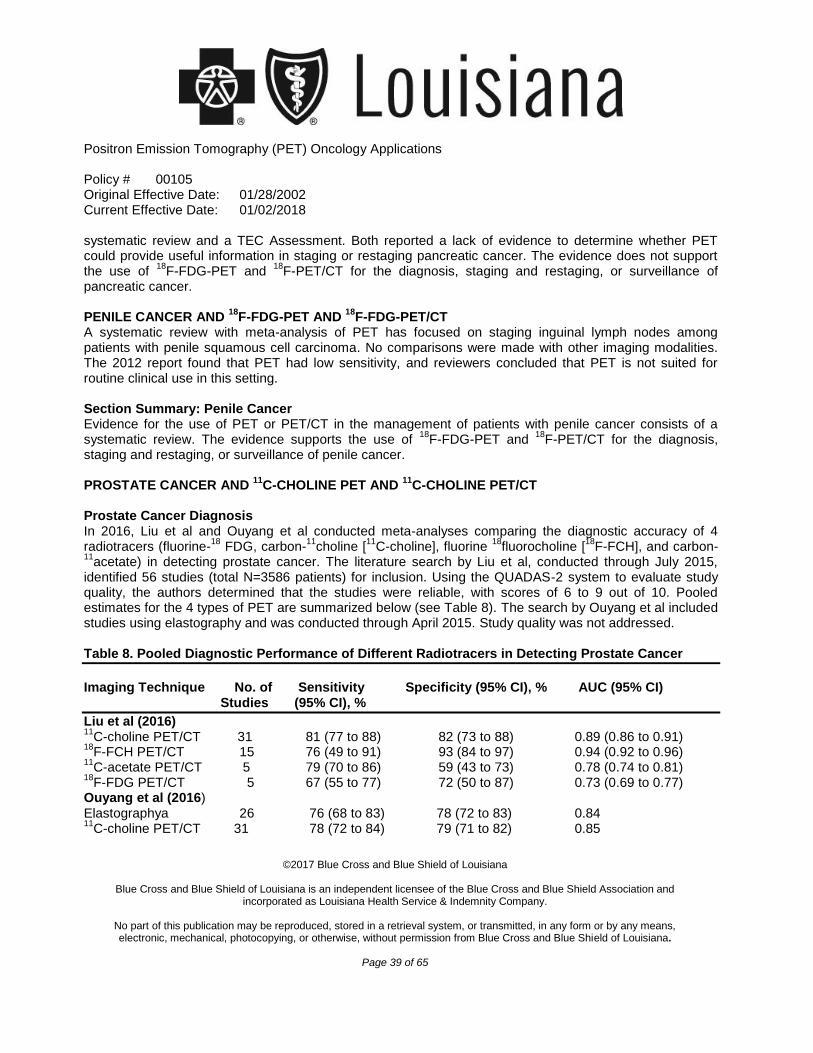
Positron Emission Tomography (PET) Oncology Applications
Policy # 00105 Original Effective Date: 01/28/2002 Current Effective Date: 01/02/2018
©2017 Blue Cross and Blue Shield of Louisiana
Blue Cross and Blue Shield of Louisiana is an independent licensee of the Blue Cross and Blue Shield Association and
incorporated as Louisiana Health Service & Indemnity Company.
No part of this publication may be reproduced, stored in a retrieval system, or transmitted, in any form or by any means, electronic, mechanical, photocopying, or otherwise, without permission from Blue Cross and Blue Shield of Louisiana.
Page 39 of 65
systematic review and a TEC Assessment. Both reported a lack of evidence to determine whether PET could provide useful information in staging or restaging pancreatic cancer. The evidence does not support
18 18the use of F-FDG-PET and F-PET/CT for the diagnosis, staging and restaging, or surveillance of pancreatic cancer.
18 18PENILE CANCER AND F-FDG-PET AND F-FDG-PET/CT A systematic review with meta-analysis of PET has focused on staging inguinal lymph nodes among patients with penile squamous cell carcinoma. No comparisons were made with other imaging modalities. The 2012 report found that PET had low sensitivity, and reviewers concluded that PET is not suited for routine clinical use in this setting. Section Summary: Penile Cancer Evidence for the use of PET or PET/CT in the management of patients with penile cancer consists of a
18 18systematic review. The evidence supports the use of F-FDG-PET and F-PET/CT for the diagnosis, staging and restaging, or surveillance of penile cancer.
11 11PROSTATE CANCER AND C-CHOLINE PET AND C-CHOLINE PET/CT Prostate Cancer Diagnosis In 2016, Liu et al and Ouyang et al conducted meta-analyses comparing the diagnostic accuracy of 4
18 11 11 18 18radiotracers (fluorine- FDG, carbon- choline [ C-choline], fluorine fluorocholine [ F-FCH], and carbon-11
acetate) in detecting prostate cancer. The literature search by Liu et al, conducted through July 2015, identified 56 studies (total N=3586 patients) for inclusion. Using the QUADAS-2 system to evaluate study quality, the authors determined that the studies were reliable, with scores of 6 to 9 out of 10. Pooled estimates for the 4 types of PET are summarized below (see Table 8). The search by Ouyang et al included studies using elastography and was conducted through April 2015. Study quality was not addressed. Table 8. Pooled Diagnostic Performance of Different Radiotracers in Detecting Prostate Cancer
Imaging Technique No. of Sensitivity Specificity (95% CI), % AUC (95% CI) Studies (95% CI), %
Liu et al (2016) 11
C-choline PET/CT 31 81 (77 to 88) 82 (73 to 88) 0.89 (0.86 to 0.91) 18
F-FCH PET/CT 15 76 (49 to 91) 93 (84 to 97) 0.94 (0.92 to 0.96) 11
C-acetate PET/CT 5 79 (70 to 86) 59 (43 to 73) 0.78 (0.74 to 0.81) 18
F-FDG PET/CT 5 67 (55 to 77) 72 (50 to 87) 0.73 (0.69 to 0.77) Ouyang et al (2016) Elastographya 26 76 (68 to 83) 78 (72 to 83) 0.84 11
C-choline PET/CT 31 78 (72 to 84) 79 (71 to 82) 0.85
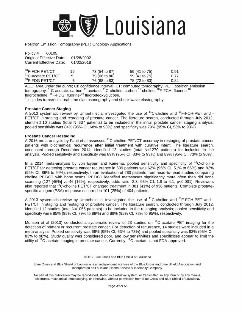
Positron Emission Tomography (PET) Oncology Applications
Policy # 00105 Original Effective Date: 01/28/2002 Current Effective Date: 01/02/2018
©2017 Blue Cross and Blue Shield of Louisiana
Blue Cross and Blue Shield of Louisiana is an independent licensee of the Blue Cross and Blue Shield Association and
incorporated as Louisiana Health Service & Indemnity Company.
No part of this publication may be reproduced, stored in a retrieval system, or transmitted, in any form or by any means, electronic, mechanical, photocopying, or otherwise, without permission from Blue Cross and Blue Shield of Louisiana.
Page 40 of 65
18F-FCH PET/CT 15 73 (54 to 87) 59 (41 to 75) 0.91
11C-acetate PET/CT 5 79 (68 to 86) 59 (41 to 75) 0.77
18F-FDG PET/CT 5 76 (68 to 83) 78 (72 to 83) 0.84
AUC: area under the curve; CI: confidence interval; CT: computed tomography; PET: positron emission 11 11 11 11 18 18
tomography; C-acetate: carbon- acetate; C-choline: carbon- choline; F-FCH: fluorine 18 18
fluorocholine; F-FDG: fluorine- fluorodeoxyglucose. a includes transrectal real-time elastosonography and shear-wave elastography.
Prostate Cancer Staging
11 18A 2013 systematic review by Umbehr et al investigated the use of C-choline and F-FCH-PET and - PET/CT in staging and restaging of prostate cancer. The literature search, conducted through July 2012, identified 10 studies (total N=637 patients) to be included in the initial prostate cancer staging analysis; pooled sensitivity was 84% (95% CI, 68% to 93%) and specificity was 79% (95% CI, 53% to 93%). Prostate Cancer Restaging
11A 2016 meta-analysis by Fanti et al assessed C-choline PET/CT accuracy in restaging of prostate cancer patients with biochemical recurrence after initial treatment with curative intent. The literature search, conducted through December 2014, identified 12 studies (total N=1270 patients) for inclusion in the analysis. Pooled sensitivity and specificity was 89% (95% CI, 83% to 93%) and 89% (95% CI, 73% to 96%).
11In a 2014 meta-analysis by von Eyben and Kairemo, pooled sensitivity and specificity of C-choline PET/CT for detecting prostate cancer recurrence in 609 patients was 62% (95% CI, 51% to 66%) and 92% (95% CI, 89% to 94%), respectively. In an evaluation of 280 patients from head-to-head studies comparing choline PET/CT with bone scans, PET/CT identified metastases significantly more often than did bone scanning (127 [45%] vs 46 [16%], respectively; odds ratio, 2.8; 95% CI, 1.9 to 4.1; p<0.001). Reviewers
11also reported that C-choline PET/CT changed treatment in 381 (41%) of 938 patients. Complete prostate-specific antigen (PSA) response occurred in 101 (25%) of 404 patients.
11 18A 2013 systematic review by Umbehr et al investigated the use of C-choline and F-FCH-PET and - PET/CT in staging and restaging of prostate cancer. The literature search, conducted through July 2012, identified 12 studies (total N=1055 patients) to be included in the restaging analysis; pooled sensitivity and specificity were 85% (95% CI, 79% to 89%) and 88% (95% CI, 73% to 95%), respectively.
11Mohsen et al (2013) conducted a systematic review of 23 studies on C-acetate PET imaging for the detection of primary or recurrent prostate cancer. For detection of recurrence, 14 studies were included in a meta-analysis. Pooled sensitivity was 68% (95% CI, 63% to 73%) and pooled specificity was 93% (95% CI, 83% to 98%). Study quality was considered poor, and low sensitivities and specificities appear to limit the
11 11utility of C-acetate imaging in prostate cancer. Currently, C-acetate is not FDA-approved.
Positron Emission Tomography (PET) Oncology Applications
Policy # 00105 Original Effective Date: 01/28/2002 Current Effective Date: 01/02/2018
©2017 Blue Cross and Blue Shield of Louisiana
Blue Cross and Blue Shield of Louisiana is an independent licensee of the Blue Cross and Blue Shield Association and
incorporated as Louisiana Health Service & Indemnity Company.
No part of this publication may be reproduced, stored in a retrieval system, or transmitted, in any form or by any means, electronic, mechanical, photocopying, or otherwise, without permission from Blue Cross and Blue Shield of Louisiana.
Page 41 of 65
Both a 2009 NCCN report and a 2008 AHRQ systematic review found the evidence insufficient to support the use of PET for any indication in patients with prostate cancer. Reports showed significant overlap between benign prostatic hyperplasia, malignant tumor, local recurrence, and postoperative scarring. PET may have limited sensitivity in detecting distant metastatic disease. The AHRQ report identified only 4 studies of PET for the indications of restaging and recurrence, none of which addressed the effect of PET on management decisions. Prostate Cancer Management
18In 2017, Akin-Akintayo et al evaluated the role of FACBC (anti–1-amino-3-[ F] fluorocyclobutane-1- carboxylic acid or fluciclovine) PET/CT in the management of post-prostatectomy patients with PSA failure being considered for salvage radiotherapy. Forty-two patients who were initially planning radiotherapy due to post-prostatectomy PSA failure underwent fluciclovine PET/CT. Based on the PET/CT results, 17 (40.5%) patients changed a decision relating to the radiotherapy: 2 patients received hormonal therapy rather than radiotherapy when fluciclovine showed extrapelvic disease; 11 patients increased the radiotherapy field from prostate bed only to prostate plus pelvis; and 4 patients reduced the radiotherapy fields from prostate plus pelvis to prostate bed only.
11The European Association of Urology’s 2014 guidelines for prostate cancer have indicated that C- choline PET/CT has limited value unless PSA levels exceed 1.0 ng/mL. In meta-analysis of 14 studies (total N=1667 patients) of radiolabeled choline PET/CT for restaging prostate cancer, Treglia et al (2014) reported a maximum pooled sensitivity of 77% (95% CI, 71% to 82%) in patients with a PSA rate of increase greater than 2 ng/mL per year. Pooled sensitivity was lower for patients with a PSA rate of increase less than 2 ng/mL per year or with a PSA level doubling time of 6 months or less. In meta-analysis of 11 studies (total N=609 patients) of radiolabeled choline PET/CT for staging or restaging prostate cancer, von Eyben et al (2014) reported pooled sensitivity and specificity of 59% (95% CI, 51% to 66%) and 92% (95% CI, 89% to 94%), respectively. Pooled PPV and NPV were 70% and 85%, respectively.
11Current NCCN guidelines for prostate cancer indicate that C-choline PET may be considered for biochemical failure after primary treatment (i.e., radiotherapy or radical prostatectomy), although further study is needed to determine the best use of this imaging modality in patients with prostate cancer. FDG or fluoride PET should not be used routinely for initial assessment or in other settings, due to limited evidence of clinical utility.
11Section Summary: Prostate Cancer and C-CHOLINE
11Evidence for the use of C-choline PET and -PET/CT for diagnosis, staging, and restaging of prostate cancer, consists of meta-analyses. The choice of radiotracer affects the sensitivity and specificity of the
11scans, with most evidence showing that the use of C-choline results in the highest sensitivities and
Positron Emission Tomography (PET) Oncology Applications
Policy # 00105 Original Effective Date: 01/28/2002 Current Effective Date: 01/02/2018
©2017 Blue Cross and Blue Shield of Louisiana
Blue Cross and Blue Shield of Louisiana is an independent licensee of the Blue Cross and Blue Shield Association and
incorporated as Louisiana Health Service & Indemnity Company.
No part of this publication may be reproduced, stored in a retrieval system, or transmitted, in any form or by any means, electronic, mechanical, photocopying, or otherwise, without permission from Blue Cross and Blue Shield of Louisiana.
Page 42 of 65
specificities. Further study is needed to compare PET and PET/CT with other imaging techniques, such as MRI and radionuclide bone scan. One prospective study has looked at the use of FACBC PET/CT in the management of post-prostatectomy patients with PSA failure considering salvage radiotherapy. PET/CT results altered the radiotherapy treatment plans in 40% of the patients. The evidence supports the use of 11
C-choline PET and PET/CT for the diagnosis, staging and restaging, and surveillance of prostate cancer.
68 68PROSTATE CANCER AND GA-PET, AND GA-PET/CT
68A 2016 systematic review by Perera calculated the sensitivity, specificity, and predictive value of Ga- prostate-specific membrane antigen (PSMA) PET in advanced prostate cancer. The literature search, conducted through April 2016, identified 16 studies (total N=1309 patients) for inclusion, though only 11 studies reported histopathologic correlations. Four studies allowed for calculating the predictive ability of 68
Ga-PSMA PET; they showed a pooled sensitivity of 86% (95% CI, 37% to 98%) and a pooled specificity of 68
86% (95% CI, 3% to 100%). The other studies were used to assess Ga-PSMA PET positivity by the amount of radiopharmaceutical injected and for detection of primary and metastatic lesions. Reviewers noted that these analyses were exploratory, because most studies were small, retrospective, from single institutions, and had heterogeneous patient cohorts.
68Section Summary: Prostate Cancer and Ga
68Evidence for the use of Ga in PET consists of a systematic review of small single-institution studies. The CIs of the sensitivity and specificity are wide, indicating uncertainty in the results provided. The evidence
68does not support the use of Ga-PET and -PET/CT for the diagnosis, staging and restaging, and surveillance of prostate cancer.
18 18RENAL CELL CARCINOMA AND F-FDG-PET AND F-FDG-PET/CT
18A 2017 systematic review by Ma et al evaluated the use of FDG-PET or -PET/CT for restaging renal cell carcinoma.120 The literature search, conducted through July 2016, identified 15 studies, mostly retrospective, for inclusion into a meta-analysis. Pooled estimates for sensitivity and specificity were 86% (95% CI, 88% to 93%) and 88% (95% CI, 84% to 91%), respectively. Reviewers concluded that PET showed potential for identifying metastatic or recurrent lesions in patients with renal cell carcinoma, but that more prospective studies would be needed. Current NCCN guidelines for renal cell carcinoma state that “The value of PET in RCC [renal cell carcinoma] remains to be determined. Currently PET alone is not a tool that is standardly used to diagnose kidney cancer or follow for evidence of relapse after nephrectomy.” Section Summary: Renal Cell Carcinoma
18 18The evidence does not support the use of F-FDG-PET and F-PET/CT for the diagnosis, staging and restaging, or surveillance of renal cell carcinoma.
Positron Emission Tomography (PET) Oncology Applications
Policy # 00105 Original Effective Date: 01/28/2002 Current Effective Date: 01/02/2018
©2017 Blue Cross and Blue Shield of Louisiana
Blue Cross and Blue Shield of Louisiana is an independent licensee of the Blue Cross and Blue Shield Association and
incorporated as Louisiana Health Service & Indemnity Company.
No part of this publication may be reproduced, stored in a retrieval system, or transmitted, in any form or by any means, electronic, mechanical, photocopying, or otherwise, without permission from Blue Cross and Blue Shield of Louisiana.
Page 43 of 65
SOFT TISSUE SARCOMA A 2002 AHRQ systematic review on the use of PET for soft tissue sarcoma evaluated 5 indications: distinguishing between benign lesions and malignant soft tissue sarcoma, distinguishing between low- grade and high-grade soft tissue sarcoma, detecting loco-regional recurrence, detecting distant metastases, and evaluating response to therapy. Reviewers found that PET had low diagnostic accuracy in distinguishing low-grade tumors from benign lesions. PET performed better at differentiating high- or intermediate-grade tumors from low-grade tumors; however, it is unclear whether this would impact management decisions and health outcomes. Evidence was insufficient on the comparative diagnostic performance of PET and alternative diagnostic modalities in the diagnosis of soft tissue sarcoma, detection of loco-regional recurrence, detection of distant metastasis, and evaluation of treatment response. A 2012 systematic review by Treglia et al evaluated PET for assessing response to imatinib and other treatments for gastrointestinal stromal tumors. Reviewers selected 19 studies. They concluded there was sufficient evidence that PET/CT can be used to monitor response to imatinib treatment, and that the information can be used to adapt treatment strategies. However, the review had the following limitations: it lacked appraisal of the methodologic quality of individual studies and comparison of decision making and outcomes between PET-guided and non-PET-guided management. Current NCCN guidelines for soft tissue sarcoma state that PET/CT can be useful in determining response to chemotherapy for lesions greater than 3 cm that are firm, deep, and not superficial. The guidelines also state that PET can provide information on imatinib activity after 2 to 4 weeks of therapy when rapid reading of activity is considered necessary; however, long-term PET follow-up is rarely indicated. The guidelines also indicate that PET can be used to assess progression of disease if results from other imaging techniques are inconclusive. Section Summary: Soft Tissue Sarcoma Evidence for the use of PET or PET/CT in patients with soft tissue sarcoma consists of 2 systematic reviews. Results of the ARHQ review showed that PET or PET/CT had low diagnostic accuracy. Another systematic review reported evidence supporting the use of PET/CT in monitoring response to imatinib
18 18treatment. The evidence supports the use of F-FDG-PET and F-PET/CT for the diagnosis and staging and restaging of soft tissue sarcoma, but does not support their use for surveillance.
18 18TESTICULAR CANCER AND F-FDG-PET AND F-FDG-PET/CT The 2004 AHRQ systematic review found 1 prospective study and 4 retrospective studies that generally showed higher sensitivity and specificity for PET compared with CT. However these studies were small in
Positron Emission Tomography (PET) Oncology Applications
Policy # 00105 Original Effective Date: 01/28/2002 Current Effective Date: 01/02/2018
©2017 Blue Cross and Blue Shield of Louisiana
Blue Cross and Blue Shield of Louisiana is an independent licensee of the Blue Cross and Blue Shield Association and
incorporated as Louisiana Health Service & Indemnity Company.
No part of this publication may be reproduced, stored in a retrieval system, or transmitted, in any form or by any means, electronic, mechanical, photocopying, or otherwise, without permission from Blue Cross and Blue Shield of Louisiana.
Page 44 of 65
size and failed to report separate results for patients with and without seminoma. Studies also failed to report separate results by clinical stage of disease. In addition, studies on PET’s ability to discriminate viable tumor and necrosis or fibrosis after treatment of testicular cancer were flawed in 2 main ways. First, most studies did not compare the diagnostic accuracy of PET with other imaging modalities. Second, studies that did compare PET and CT did not state a clear threshold for a positive CT test, making study results difficult to interpret. Therefore, it is uncertain whether use of PET leads to different patient management decisions and health outcomes compared with other imaging modalities. A 2008 AHRQ technology assessment and studies evaluating residual masses in patients after chemotherapy for seminoma support the use of PET. Current NCCN guidelines also support the use of PET to evaluate residual masses that are greater than 3 cm following primary treatment with chemotherapy. The guidelines warn that there is “limited predictive value for PET/CT scan for residual masses.” PET is not recommended for nonseminoma patients. Section Summary: Testicular Cancer Evidence for the use of PET or PET/CT in patients with testicular cancer consists of an AHRQ systematic review of small studies. Results showed that PET or PET/CT can be useful in evaluating residual masses following chemotherapy for seminoma. There is no evidence supporting the use of PET or PET/CT in
18 18nonseminoma patients. The evidence supports the use of F-FDG-PET and F-PET/CT for the diagnosis and staging and restaging of testicular cancer, but does not support their use for surveillance.
18 18THYROID CANCER AND F-FDG-PET AND F-FDG-PET/CT Differentiated
18In 2016, Haslerud et al conducted a systematic review of studies using F-FDG-PET to detect recurrent differentiated thyroid cancer in patients who had undergone ablative therapy. The literature search,
18conducted through December 2014, identified 34 studies (total N=2639 patients) for inclusion: 17 using F-
18FDG-PET/CT, 11 using F-FDG-PET, and 6 using both methods. Study quality was assessed using the
18QUADAS tool. Pooled sensitivity and specificity for F-FDG-PET/CT were 80% (95% CI, 74% to 86%) and
1876% (95% CI, 63% to 85%), respectively. Pooled sensitivity and specificity for F-FDG-PET alone were 77% (95% CI, 63% to 86%) and 76% (95% CI, 60% to 87%), respectively. Combining all 34 studies in the meta-analysis resulted in a pooled sensitivity and specificity of 79% (95% CI, 74% to 84%) and 79% (95% CI, 71% to 85%), respectively. The 2009 NCCN report on PET showed that PET can localize recurrent disease when other imaging tests are negative. Additionally, PET was found to be prognostic in this setting: More metabolically active lesions on PET were strongly correlated with reduced survival. Current NCCN guidelines for thyroid carcinoma
Positron Emission Tomography (PET) Oncology Applications
Policy # 00105 Original Effective Date: 01/28/2002 Current Effective Date: 01/02/2018
©2017 Blue Cross and Blue Shield of Louisiana
Blue Cross and Blue Shield of Louisiana is an independent licensee of the Blue Cross and Blue Shield Association and
incorporated as Louisiana Health Service & Indemnity Company.
No part of this publication may be reproduced, stored in a retrieval system, or transmitted, in any form or by any means, electronic, mechanical, photocopying, or otherwise, without permission from Blue Cross and Blue Shield of Louisiana.
Page 45 of 65
18continue to support the use of F-FDG-PET/CT in thyroid cancer evaluations, such as when iodine-131 imaging is negative and stimulated Tg is greater than 2 to 5 ng/mL. Medullary A meta-analysis of studies on detecting recurrent or metastatic medullary thyroid carcinoma was conducted by Cheng et al (2012). The literature search, conducted through December 2010, identified 15 studies to be
18 18included in the meta-analysis: 8 used F-FDG-PET and 7 used F-FDG-PET/CT. The pooled sensitivity for 18
F-FDG-PET alone in detecting recurrent or metastatic medullary thyroid cancer was 68% (95% CI, 64% to 18
72%). The pooled sensitivity for F-FDG-PET/CT was 69% (95% CI, 64% to 74%). Current NCCN guidelines for medullary thyroid cancer recommend contrast-enhanced CT with or without PET at 2 to 3 months postoperative surveillance. Section Summary: Thyroid Cancer Evidence for the use of PET and PET/CT to diagnose recurrent differentiated and medullary thyroid cancer
18consists of systematic reviews and meta-analyses. Pooled sensitivity and specificity for F-FDG- PET and 18
F-FDG-PET/CT in detecting recurrent differentiated thyroid cancer were comparable, ranging from 76% to 80%. Pooled sensitivity for both PET and PET/CT in detecting recurrent medullary thyroid cancer were also
18 18comparable (68% to 69%). The evidence supports the use of F-FDG-PET and F-PET/CT for the diagnosis and staging and restaging of thyroid cancer, but does not support their use for surveillance.
18 18CANCER OF UNKNOWN PRIMARY AND F-FDG-PET AND F-FDG-PET/CT The 2002 TEC Assessment concluded that FDG-PET met TEC criteria for the workup and management of patients with unknown primaries and a single site of metastatic disease. Specifically, local or regional therapy might be offered to these patients. In this setting, PET scanning might be used to verify the absence of disseminated disease. Regarding this application, the TEC Assessment identified 4 reports of 47 total patients referred for imaging of a single known metastatic site from an unknown primary. In 13 (28%) of these patients, PET scanning identified previously undetected metastases that were confirmed by biopsy. Therefore, the use of PET was found to contribute to optimal decision making regarding the appropriateness of local or regional therapy. Section Summary: Cancer of Unknown Primary
18 18The evidence supports the use of F-FDG-PET and F-PET/CT for the diagnosis, staging and restaging, and surveillance of cancer of unknown primary. CANCER SURVEILLANCE Clinical utility of PET scanning in surveillance (i.e., in performing follow-up PET scans in asymptomatic patients to detect early disease recurrence) is not well-studied. (For this evidence review, a scan is
Positron Emission Tomography (PET) Oncology Applications
Policy # 00105 Original Effective Date: 01/28/2002 Current Effective Date: 01/02/2018
©2017 Blue Cross and Blue Shield of Louisiana
Blue Cross and Blue Shield of Louisiana is an independent licensee of the Blue Cross and Blue Shield Association and
incorporated as Louisiana Health Service & Indemnity Company.
No part of this publication may be reproduced, stored in a retrieval system, or transmitted, in any form or by any means, electronic, mechanical, photocopying, or otherwise, without permission from Blue Cross and Blue Shield of Louisiana.
Page 46 of 65
considered a surveillance scan if performed more than 6 months after therapy [but 12 months for lymphoma].) The 2009 NCCN report stated that “PET as a surveillance tool should only be used in clinical trials.” Additionally, NCCN guidelines for various malignancies often note that PET scans are not recommended in asymptomatic patients. For example, current NCCN guidelines for breast cancer comment that PET scans (as well as many other imaging modalities) provide no advantage in survival or ability to palliate recurrent disease and are not recommended. OTHER ONCOLOGIC APPLICATIONS There are inadequate scientific data to permit conclusions on the role of PET scanning in other malignancies. SUMMARY OF EVIDENCE Bone Sarcoma For individuals who have suspected or diagnosed bone sarcoma and in need of staging or restaging information who receive
18F-FDG-PET or
18F-FDG-PET/CT, the evidence includes systematic reviews and
meta-analyses of many studies. Relevant outcomes are test accuracy and test validity. Pooled analyses have shown that PET or PET/CT can effectively diagnose and stage bone cancer. PET or PET/CT has high sensitivities and specificities in detecting metastases in bone and lymph nodes; however, the tests have low sensitivity in detecting lung metastases. Clinical guidelines include PET and CT to inform management decisions that may offer clinical benefit. The evidence is sufficient to determine that the technology results in a meaningful improvement in the net health outcome. For individuals who are asymptomatic after completing bone sarcoma treatment who receive
18F-FDG- PET
or 18
F-FDG-PET/CT, there is no evidence. Relevant outcomes are test accuracy and test validity. The evidence is insufficient to determine the effects of the technology on health outcomes. Brain Tumors For individuals who have diagnosed brain tumors and in need of staging or restaging information or who have suspected brain cancer or are asymptomatic after completing brain cancer treatment who receive
18F-
FDG-PET, 18
F-FET-PET, or 11
C-methionine PET, the evidence includes several systematic reviews and meta-analyses. Relevant outcomes are test accuracy and test validity. Pooled analyses have shown that PET or PET/CT can be effective in distinguishing brain tumors from normal tissue. Indirect comparisons between the radiotracers
11C-methionine and
18F-FDG have shown that
11C-methionine may have better
diagnostic performance. Clinical guidelines include PET to inform management decisions that may offer clinical benefit. The evidence is sufficient to determine that the technology results in a meaningful improvement in the net health outcome. Breast Cancer For individuals who have diagnosed breast cancer and inconclusive results from other imaging techniques who receive adjunctive
18F-FDG-PET or
18F-FDG-PET/CT for stating or restaging, the evidence includes
Positron Emission Tomography (PET) Oncology Applications
Policy # 00105 Original Effective Date: 01/28/2002 Current Effective Date: 01/02/2018
©2017 Blue Cross and Blue Shield of Louisiana
Blue Cross and Blue Shield of Louisiana is an independent licensee of the Blue Cross and Blue Shield Association and
incorporated as Louisiana Health Service & Indemnity Company.
No part of this publication may be reproduced, stored in a retrieval system, or transmitted, in any form or by any means, electronic, mechanical, photocopying, or otherwise, without permission from Blue Cross and Blue Shield of Louisiana.
Page 47 of 65
meta-analyses. Relevant outcomes are test accuracy and test validity. While studies included in the meta-analyses report variability in estimates of sensitivity and specificity,
18F-FDG-PET or
18F-FDG-PET/CT may
be helpful in situations in which standard staging results are equivocal or suspicious, particularly in patients with locally advanced or metastatic disease. The evidence is sufficient to determine that the technology results in a meaningful improvement in the net health outcome. For individuals who have suspected or diagnosed breast cancer and in need of staging or restaging information who receive
18F-FDG-PET or
18F-FDG-PET/CT, the evidence includes a TEC Assessment,
several systematic reviews, and meta-analyses. Relevant outcomes are test accuracy and test validity. There is no evidence supporting the use of PET in diagnosing breast cancer. The false-negative rates (5.5%-8.5%) using PET in patients with breast cancer can be considered unacceptable, given that breast biopsy can provide more definitive results. PET/CT may be considered for detection of metastases only when results from other imaging techniques are inconclusive. The evidence is insufficient to determine the effects of the technology on health outcomes. For individuals who are asymptomatic after completing breast cancer treatment who receive
18F-FDG- PET
or 18
F-FDG-PET/CT, there is no evidence. Relevant outcomes are test accuracy and test validity. The evidence is insufficient to determine the effects of the technology on health outcomes. Cervical Cancer For individuals who have diagnosed cervical cancer and in need of staging or restaging information who receive
18F-FDG-PET or
18F-PET/CT, the evidence includes an AHRQ report and a meta-analysis. Relevant
outcomes are test accuracy and test validity. Pooled results show that PET can be used for staging or restaging and for detection of recurrent disease. Clinical guidelines include PET and CT to inform management decisions that may offer clinical benefit. The evidence is sufficient to determine that the technology results in a meaningful improvement in the net health outcome. For individuals who have suspected cervical cancer or who are asymptomatic after completing cervical cancer treatment who receive
18F-FDG-PET or
18F-FDG-PET/CT, there is no evidence. Relevant outcomes
are test accuracy and test validity. The evidence is insufficient to determine the effects of the technology on health outcomes. Colorectal Cancer For individuals who have diagnosed CRC and in need of staging or restaging information who receive
18F-
FDG-PET or 18
F-PET/CT, the evidence includes a TEC Assessment and several meta- analyses. Relevant outcomes are test accuracy and test validity. Several pooled analyses evaluating staging or restaging using PET or PET/CT resulted in wide ranges of sensitivities and specificities, from the low 60s to the high 90s. The evidence is insufficient to determine the effects of the technology on health outcomes.
Positron Emission Tomography (PET) Oncology Applications
Policy # 00105 Original Effective Date: 01/28/2002 Current Effective Date: 01/02/2018
©2017 Blue Cross and Blue Shield of Louisiana
Blue Cross and Blue Shield of Louisiana is an independent licensee of the Blue Cross and Blue Shield Association and
incorporated as Louisiana Health Service & Indemnity Company.
No part of this publication may be reproduced, stored in a retrieval system, or transmitted, in any form or by any means, electronic, mechanical, photocopying, or otherwise, without permission from Blue Cross and Blue Shield of Louisiana.
Page 48 of 65
For individuals who have suspected CRC or who are asymptomatic after completing CRC treatment who receive
18F-FDG-PET or
18F-PET/CT, the evidence includes a TEC Assessment and meta-analysis.
Relevant outcomes are test accuracy and test validity. A meta-analysis evaluating the diagnostic accuracy of PET or PET/CT showed a high sensitivity but low specificity. The evidence for the use of PET or PET/CT does not show a benefit over the use of contrast CT in patients with CRC. The evidence is insufficient to determine the effects of the technology on health outcomes. Endometrial Cancer For individuals who have diagnosed endometrial cancer in need of staging or restaging information or who are asymptomatic after completing endometrial cancer treatment who receive
18F-FDG-PET or
18F-
PET/CT, the evidence includes a systematic review and meta-analysis. Relevant outcomes are test accuracy and test validity. Pooled estimates from the meta-analysis showed high sensitivities and specificities for
18F-FDG-PET/CT in detecting lymph node metastases and endometrial cancer recurrence
following treatment. The evidence is sufficient to determine that the technology results in a meaningful improvement in the net health outcome. Esophageal Cancer For individuals who have diagnosed esophageal cancer and in need of staging or restaging information who receive
18F-FDG-PET or
18F-PET/CT, the evidence includes several meta-analyses. Relevant outcomes are
test accuracy and test validity. Pooled estimates have shown high sensitivities and specificities compared to other diagnostic imaging techniques. Clinical guidelines include PET and CT to inform management decisions that may offer clinical benefit. The evidence is sufficient to determine that the technology results in a meaningful improvement in the net health outcome. For individuals who have suspected esophageal cancer or who are asymptomatic after completing esophageal cancer treatment who receive
18F-FDG-PET or
18F-FDG-PET/CT, the evidence includes meta-
analyses. Relevant outcomes are test accuracy and test validity. Pooled analyses showed adequate sensitivities but low specificities. The evidence is insufficient to determine the effects of the technology on health outcomes. Gastric Cancer For individuals who have suspected or diagnosed gastric cancer and in need of staging or restaging information, who receive
18F-FDG-PET or
18F-PET/CT, the evidence includes several meta-analyses.
Relevant outcomes are test accuracy and test validity. Pooled analyses, with sensitivities and specificities ranging from the high 70s to the high 80s, have shown that PET or PET/CT can inform staging or restaging of patients with gastric cancer. Clinical guidelines include PET/CT to inform management decisions that may offer clinical benefit. The evidence is sufficient to determine that the technology results in a meaningful improvement in the net health outcome.
Positron Emission Tomography (PET) Oncology Applications
Policy # 00105 Original Effective Date: 01/28/2002 Current Effective Date: 01/02/2018
©2017 Blue Cross and Blue Shield of Louisiana
Blue Cross and Blue Shield of Louisiana is an independent licensee of the Blue Cross and Blue Shield Association and
incorporated as Louisiana Health Service & Indemnity Company.
No part of this publication may be reproduced, stored in a retrieval system, or transmitted, in any form or by any means, electronic, mechanical, photocopying, or otherwise, without permission from Blue Cross and Blue Shield of Louisiana.
Page 49 of 65
For individuals who are asymptomatic after completing gastric cancer treatment who receive 18
F-FDG- PET or
18F-FDG-PET/CT, the evidence includes meta-analyses. Relevant outcomes are test accuracy and test
validity. Pooled analyses showed low sensitivities and specificities. The evidence is insufficient to determine the effects of the technology on health outcomes. Head and Neck Cancer For individuals who have suspected or diagnosed head and neck cancer who need staging or restaging information who receive
18F-FDG-PET or
18F-PET/CT, the evidence includes a TEC Assessment and
several meta-analyses. Relevant outcomes are test accuracy and test validity. In patients with head and neck cancers, PET and PET/CT are better able to detect local and metastatic disease compared with other imaging techniques. Evidence has also shown that
18F-FDG-PET/CT may be useful in predicting response
to therapy. Two meta-analyses calculated the ability of 18
F-FDG-PET or PET/CT to detect residual or recurrent disease during various stages of treatment and another meta-analysis calculated the ability of positive PET or PET/CT results to predict OS and event-free survival. The evidence is sufficient to determine that the technology results in a meaningful improvement in the net health outcome. For individuals who are asymptomatic after completing head and neck cancer treatment who receive
18F-
FDG-PET or 18
F-FDG-PET/CT, there is no evidence. Relevant outcomes are test accuracy and test validity. The evidence is insufficient to determine the effects of the technology on health outcomes. Non-Small-Cell Lung Cancer For individuals who have suspected NSCLC and inconclusive results from other imaging techniques or who have diagnosed NSCLC and in need of staging or restaging information who receive
18F-FDG-PET or
18F-
PET/CT, the evidence includes several meta-analyses. Relevant outcomes are test accuracy and test validity. Pooled analyses have shown that PET and PET/CT have better diagnostic performance compared with conventional imaging techniques. Clinical guidelines include PET/CT to inform management decisions that may offer clinical benefit. The evidence is sufficient to determine that the technology results in a meaningful improvement in the net health outcome. For individuals who have suspected NSCLC or who are asymptomatic after completing NSCLC treatment who receive
18F-FDG-PET or
18F-FDG-PET/CT, there is no evidence. Relevant outcomes are test accuracy
and test validity. The evidence is insufficient to determine the effects of the technology on health outcomes. Small-Cell Lung Cancer For individuals with diagnosed SCLC and in need of staging or restaging information who receive
18F-FDG-
PET or 18
F-PET/CT, the evidence includes a systematic review and a meta-analysis. Relevant outcomes are test accuracy and test validity. While the quality of the studies was considered low, PET and PET/CT can be considered for staging or restaging in patients with SCLC. Clinical guidelines include PET/CT to
Positron Emission Tomography (PET) Oncology Applications
Policy # 00105 Original Effective Date: 01/28/2002 Current Effective Date: 01/02/2018
©2017 Blue Cross and Blue Shield of Louisiana
Blue Cross and Blue Shield of Louisiana is an independent licensee of the Blue Cross and Blue Shield Association and
incorporated as Louisiana Health Service & Indemnity Company.
No part of this publication may be reproduced, stored in a retrieval system, or transmitted, in any form or by any means, electronic, mechanical, photocopying, or otherwise, without permission from Blue Cross and Blue Shield of Louisiana.
Page 50 of 65
inform management decisions that may offer clinical benefit. The evidence is sufficient to determine that the technology results in a meaningful improvement in the net health outcome. For individuals who have suspected SCLC or who are asymptomatic after completing SCLC treatment who receive
18F-FDG-PET or
18F-FDG-PET/CT, there is no evidence. Relevant outcomes are test accuracy and
test validity. The evidence is insufficient to determine the effects of the technology on health outcomes. Hodgkin and Non-Hodgkin Lymphoma For individuals who have suspected or diagnosed Hodgkin and non-Hodgkin lymphoma in need of staging or restaging information who receive
18F-FDG-PET or PET/CT, the evidence includes a TEC Assessment
and several meta-analyses. Relevant outcomes are test accuracy and test validity. PET and PET/CT have been found to provide useful information in the management of Hodgkin and non-Hodgkin lymphoma. The Deauville 5-point scale was developed based on PET results and can be used for staging and treatment response for patients with lymphoma. Clinical guidelines include PET/CT to inform management decisions that may offer clinical benefit. The evidence is sufficient to determine that the technology results in a meaningful improvement in the net health outcome. For individuals who are asymptomatic after completing Hodgkin lymphoma treatment who receive
18F-
FDG-PET or 18
F-FDG-PET/CT, there is no evidence. Relevant outcomes are test accuracy and test validity. The evidence is insufficient to determine the effects of the technology on health outcomes. For individuals who are asymptomatic after completing non-Hodgkin lymphoma treatment who receive
18F-
FDG-PET or 18
F-FDG-PET/CT, there is no evidence. Relevant outcomes are test accuracy and test validity. The evidence is insufficient to determine the effects of the technology on health outcomes. Melanoma For individuals who have suspected or diagnosed stage I or II melanoma and in need of staging or restaging information who receive
18F-FDG-PET or
18F-PET/CT, the evidence includes a TEC Assessment.
Relevant outcomes are test accuracy and test validity. Evidence has shown PET and PET/CT are not as beneficial as the reference standard (sentinel node biopsy) for assessing regional lymph nodes. The evidence is insufficient to determine the effects of the technology on health outcomes. For individuals who have diagnosed advanced melanoma (stage III or IV) and in need of staging or restaging information who receive
18F-FDG-PET or
18F-PET/CT, the evidence includes a TEC Assessment
and a meta-analysis. Relevant outcomes are test accuracy and test validity. Evidence has shown PET and PET/CT can detect systemic metastases in patients with advanced melanoma. Clinical guidelines include PET/CT for staging or restaging stage III or IV disease and for surveillance. The evidence is sufficient to determine that the technology results in a meaningful improvement in the net health outcome.
Positron Emission Tomography (PET) Oncology Applications
Policy # 00105 Original Effective Date: 01/28/2002 Current Effective Date: 01/02/2018
©2017 Blue Cross and Blue Shield of Louisiana
Blue Cross and Blue Shield of Louisiana is an independent licensee of the Blue Cross and Blue Shield Association and
incorporated as Louisiana Health Service & Indemnity Company.
No part of this publication may be reproduced, stored in a retrieval system, or transmitted, in any form or by any means, electronic, mechanical, photocopying, or otherwise, without permission from Blue Cross and Blue Shield of Louisiana.
Page 51 of 65
For individuals who are asymptomatic after completing melanoma treatment who receive 18
F-FDG-PET or 18
F-FDG-PET/CT, the evidence includes retrospective and observational studies. Relevant outcomes are test accuracy and test validity. At the discretion of the physician, imaging surveillance can be considered every 3 to 12 months. Since recurrences usually occur within 3 years, screening asymptomatic patients beyond 3 to 5 years is not recommended. The evidence is sufficient to determine that the technology results in a meaningful improvement in the net health outcome. Multiple Myeloma For individuals who have suspected or diagnosed multiple myeloma in need of staging or restaging information who receive
18F-FDG-PET or
18F-PET/CT, the evidence includes 2 systematic reviews.
Relevant outcomes are test accuracy and test validity. The evidence did not compare PET or PET/CT with other modalities and, therefore, did not provide comparative effectiveness information. The evidence is insufficient to determine the effects of the technology on health outcomes. For individuals who are asymptomatic after completing multiple myeloma treatment who receive
18F-FDG-
PET or 18
F-FDG-PET/CT, there is no evidence. Relevant outcomes are test accuracy and test validity. The evidence is insufficient to determine the effects of the technology on health outcomes. Neuroendocrine Tumors For individuals who have suspected or diagnosed neuroendocrine tumors and in need of staging or restaging information or who are asymptomatic after completing neuroendocrine tumor treatment who receive
18F-FDG-PET,
18F-PET/CT,
68Ga-PET, or
68Ga-PET/CT, the evidence includes 2 meta-analyses.
Relevant outcomes are test accuracy and test validity. The evidence did not compare PET, PET/CT, Ga-PET, or Ga-PET/CT with other modalities and, therefore, did not provide comparative effectiveness information. The evidence is insufficient to determine the effects of the technology on health outcomes. Ovarian Cancer For individuals who have diagnosed ovarian cancer and in need of staging or restaging information who receive
18F-FDG-PET or
18F-PET/CT, the evidence includes an AHRQ systematic review and several meta-
analyses. Relevant outcomes are test accuracy and test validity. Pooled sensitivities and specificities have supported the use of PET and PET/CT for the detection of recurrent ovarian cancer. Clinical guidelines include PET/CT to inform management decisions that may offer clinical benefit. The evidence is sufficient to determine that the technology results in a meaningful improvement in the net health outcome. For individuals who have suspected ovarian cancer or who are asymptomatic after completing ovarian cancer treatment who receive
18F-FDG-PET or
18F-FDG-PET/CT, there is no evidence. Relevant outcomes
are test accuracy and test validity. The evidence is insufficient to determine the effects of the technology on health outcomes.
Positron Emission Tomography (PET) Oncology Applications
Policy # 00105 Original Effective Date: 01/28/2002 Current Effective Date: 01/02/2018
©2017 Blue Cross and Blue Shield of Louisiana
Blue Cross and Blue Shield of Louisiana is an independent licensee of the Blue Cross and Blue Shield Association and
incorporated as Louisiana Health Service & Indemnity Company.
No part of this publication may be reproduced, stored in a retrieval system, or transmitted, in any form or by any means, electronic, mechanical, photocopying, or otherwise, without permission from Blue Cross and Blue Shield of Louisiana.
Page 52 of 65
Pancreatic Cancer For individuals who have suspected or diagnosed pancreatic cancer and with inconclusive results from other imaging techniques who receive adjunctive
18F-FDG-PET or
18F-FDG-PET/CT for staging or
restaging, the evidence includes a TEC Assessment and a systematic review. Relevant outcomes are test accuracy and test validity. The evidence has shown that PET and PET/CT do not have a high enough NPV to surpass current standard decision thresholds. Therefore PET or PET/CT should only be considered if results from standard staging methods are inconclusive. The evidence is sufficient to determine that the technology results in a meaningful improvement in the net health outcome. For individuals who have suspected or diagnosed pancreatic cancer and in need of staging or restaging information who receive
18F-FDG-PET or
18F-PET/CT, the evidence includes an AHRQ systematic review, a
TEC Assessment, and a meta-analysis published after the review and assessment. Relevant outcomes are test accuracy and test validity. The evidence has shown that PET and PET/CT do not have a high enough NPV to surpass current standard decision thresholds. Therefore PET or PET/CT should only be considered if results from standard staging methods are inconclusive. The evidence is insufficient to determine the effects of the technology on health outcomes. For individuals who are asymptomatic after completing pancreatic cancer treatment who receive
18F-FDG-
PET or 18
F-FDG-PET/CT, there is no evidence. Relevant outcomes are test accuracy and test validity. The evidence is insufficient to determine the effects of the technology on health outcomes. Penile Cancer For individuals who have suspected or diagnosed penile cancer and in need of staging or restaging information who receive
18F-FDG-PET or
18F-PET/CT, the evidence includes a systematic review and a
meta-analysis. Relevant outcomes are test accuracy and test validity. The evidence have shown that PET had a low sensitivity, and no comparisons were made with other modalities. The evidence is insufficient to determine the effects of the technology on health outcomes. For individuals who are asymptomatic after completing penile cancer treatment who receive
18F-FDG-PET
or 18
F-FDG-PET/CT, there is no evidence. Relevant outcomes are test accuracy and test validity. The evidence is insufficient to determine the effects of the technology on health outcomes. Prostate Cancer For individuals who have suspected or diagnosed prostate cancer and in need of staging or restaging information who receive
11C-choline PET or
11C-choline PET/CT, evidence includes several meta- analyses.
Relevant outcomes are test accuracy and test validity. Meta-analyses have reported that the choice of radiotracer affects the sensitivity and specificity of the scans, with most evidence showing that the use of
Positron Emission Tomography (PET) Oncology Applications
Policy # 00105 Original Effective Date: 01/28/2002 Current Effective Date: 01/02/2018
©2017 Blue Cross and Blue Shield of Louisiana
Blue Cross and Blue Shield of Louisiana is an independent licensee of the Blue Cross and Blue Shield Association and
incorporated as Louisiana Health Service & Indemnity Company.
No part of this publication may be reproduced, stored in a retrieval system, or transmitted, in any form or by any means, electronic, mechanical, photocopying, or otherwise, without permission from Blue Cross and Blue Shield of Louisiana.
Page 53 of 65
11C-choline results in the highest sensitivities and specificities compared with
18F-FDG-PET and
11C-
acetate. Of interest is a single study that investigated the use of PET/CT results to inform patient decisions on radiotherapy treatment plans. The study reported that 40% of the patients altered the extent of the treatment planned based on the PET/CT results. The evidence is sufficient to determine that the technology results in a meaningful improvement in the net health outcome. For individuals who are asymptomatic after completing prostate cancer treatment who receive
11C-choline
PET or 11
C-choline PET/CT, there is no evidence. Relevant outcomes are test accuracy and test validity. The evidence is insufficient to determine the effects of the technology on health outcomes. For individuals who have suspected or diagnosed prostate cancer and in need of staging or restaging information who receive
68Ga-PET or
68Ga-PET/CT, the evidence includes a meta-analysis of small single-
institution studies. Relevant outcomes are test accuracy and test validity. The evidence was limited, resulting in estimates with large CIs. The evidence is insufficient to determine the effects of the technology on health outcomes. Renal Cell Carcinoma For individuals who are diagnosed renal cell carcinoma and in need of staging or restaging information who receive
18F-FDG-PET or
18F-PET/CT, the evidence includes a systematic review and meta-analysis.
Relevant outcomes are test accuracy and test validity. The review concluded that PET has potential to detect metastatic or recurrent lesions in patients with renal cell cancer, but that additional prospective studies are needed. The evidence is insufficient to determine the effects of the technology on health outcomes. Soft Tissue Sarcoma For individuals who have diagnosed soft tissue sarcoma and in need of staging or restaging information who receive
18F-FDG-PET or
18F-PET/CT, the evidence includes an AHRQ systematic review. Another
systematic review evaluated PET for assessing response to imatinib. Relevant outcomes are test accuracy and test validity. The review reported that PET had low diagnostic accuracy and there was a lack of studies comparing PET with alternative diagnostic modalities. The evidence is insufficient to determine the effects of the technology on health outcomes. For individuals with diagnosed soft tissue sarcoma and in need of rapid reading of activity following imatinib treatment who receive
18F-FDG-PET or
18F-PET/CT, the evidence includes a systematic review. Relevant
outcomes are test accuracy and test validity. The review concluded that PET/CT can be used to monitor treatment response to imatinib, which can lead to individually adapted treatment strategies. The evidence is sufficient to determine that the technology results in a meaningful improvement in the net health outcome.
Positron Emission Tomography (PET) Oncology Applications
Policy # 00105 Original Effective Date: 01/28/2002 Current Effective Date: 01/02/2018
©2017 Blue Cross and Blue Shield of Louisiana
Blue Cross and Blue Shield of Louisiana is an independent licensee of the Blue Cross and Blue Shield Association and
incorporated as Louisiana Health Service & Indemnity Company.
No part of this publication may be reproduced, stored in a retrieval system, or transmitted, in any form or by any means, electronic, mechanical, photocopying, or otherwise, without permission from Blue Cross and Blue Shield of Louisiana.
Page 54 of 65
For individuals who have soft tissue sarcoma or who are asymptomatic after completing soft tissue sarcoma treatment who receive
18F-FDG-PET or
18F-FDG-PET/CT, the evidence includes a systematic review.
Relevant outcomes are test accuracy and test validity. The review concluded that there was insufficient evidence on the use of PET for detection of loco-regional recurrence. The evidence is insufficient to determine the effects of the technology on health outcomes. Testicular Cancer For individuals with diagnosed testicular cancer in need of staging or restaging information who receive
18F-
FDG-PET or 18
F-PET/CT, the evidence includes an AHRQ systematic review and assessment. Relevant outcomes are test accuracy and test validity. Results have shown that PET or PET/CT can evaluate residual masses following chemotherapy for seminoma. Clinical guidelines include PET/CT to inform management decisions that may offer clinical benefit. There is no evidence supporting the use of PET or PET/CT in nonseminoma patients. The evidence is sufficient to determine that the technology results in a meaningful improvement in the net health outcome. For individuals who have suspected testicular cancer or who are asymptomatic after completing testicular cancer treatment who receive
18F-FDG-PET or
18F-FDG-PET/CT, there is no evidence. Relevant outcomes
are test accuracy and test validity. The evidence is insufficient to determine the effects of the technology on health outcomes. Thyroid Cancer For individuals with diagnosed thyroid cancer and in need of staging or restaging information who receive 18
F-FDG-PET or 18
F-PET/CT, the evidence includes systematic reviews and meta-analyses. Relevant outcomes are test accuracy and test validity. Pooled analyses have shown that PET or PET/CT can effectively detect recurrent differentiated thyroid cancer. Clinical guidelines include PET/CT to inform management decisions that may offer clinical benefit. The evidence is sufficient to determine that the technology results in a meaningful improvement in the net health outcome. For individuals who have suspected thyroid cancer or who are asymptomatic after completing thyroid cancer treatment who receive
18F-FDG-PET or
18F-FDG-PET/CT, there is no evidence. Relevant outcomes
are test accuracy and test validity. The evidence is insufficient to determine the effects of the technology on health outcomes. Unknown Primary and Single-Site Metastatic Disease For individuals with unknown primary and single-site metastatic disease who receive
18F-FDG-PET or
18F-
PET/CT, the evidence includes a TEC Assessment. Relevant outcomes are test accuracy and test validity. Studies reviewed in the Assessment showed that PET identified previously undetected metastases
Positron Emission Tomography (PET) Oncology Applications
Policy # 00105 Original Effective Date: 01/28/2002 Current Effective Date: 01/02/2018
©2017 Blue Cross and Blue Shield of Louisiana
Blue Cross and Blue Shield of Louisiana is an independent licensee of the Blue Cross and Blue Shield Association and
incorporated as Louisiana Health Service & Indemnity Company.
No part of this publication may be reproduced, stored in a retrieval system, or transmitted, in any form or by any means, electronic, mechanical, photocopying, or otherwise, without permission from Blue Cross and Blue Shield of Louisiana.
Page 55 of 65
confirmed by biopsy. PET can contribute to the management of patients with unknown primary. Clinical guidelines include PET/CT to inform management decisions that may offer clinical benefit. The evidence is sufficient to determine that the technology results in a meaningful improvement in the net health outcome.
References 1. Blue Cross and Blue Shield Association, Medical Policy Reference Manual, Draft of “Oncologic Applications of PET Scanning”,
6.01.26, 6:2017. 2. Food and Drug Administration. Positron Emission Tomography (PET). 2016;
http://www.fda.gov/Drugs/DevelopmentApprovalProcess/Manufacturing/ucm085783.htm. 3. CardinalHealth. FDA-Approved Radiopharmaceuticals. 2016;
http://www.cardinalhealth.com/content/dam/corp/web/documents/fact-sheet/CardinalHealth-FDAApprovedRadiopharmaceuticalsandApprovedUses.pdf.
4. Blue Cross Blue Shield Association Technology Evaluation Center (TEC). FDG Positron Emission Tomography for Non-CNS Cancers. Technol Eval Cent Assess. 1997;12(3).
5. Seidenfeld J, Samson DJ, Bonnell CJ, et al. Management of small cell lung cancer. Evid Rep Technol Assess (Full Rep). 2006 Jul 2006(143):1-154.
6. Blue Cross Blue Shield Association Technology Evaluation Center (TEC). FDG Positron Emission Tomography in Melanoma. Technol Eval Cent Assess. 1999;14(27).
7. Blue Cross Blue Shield Association Technology Evaluation Center (TEC). FDG Positron Emission Tomography in Lymphoma. Technol Eval Cent Assess. 1999;14(26).
8. Blue Cross Blue Shield Association Technology Evaluation Center (TEC). FDG Positron Emission Tomography in Colorectal Cancer. Technol Eval Cent Assess. 1999;14(25).
9. Blue Cross Blue Shield Association Technology Evaluation Center (TEC). FDG Positron Emission Tomography in Head and Neck Cancer. Technol Eval Cent Assess. 2000;15(4).
10. Blue Cross Blue Shield Association Technology Evaluation Center (TEC). FDG Positron Emission Tomography to Manage Patients with an Occult Primary Carcinoma and Metastasis outside the Cervical Lymph Nodes. Technol Eval Cent Assess. 2002;17(14).
11. Blue Cross Blue Shield Association Technology Evaluation Center (TEC). FDG Positron Emission Tomography for Evaluating Esophageal Cancer. Technol Eval Cent Assess. 2001;16(21).
12. Wallace MB, Nietert PJ, Earle C, et al. An analysis of multiple staging management strategies for carcinoma of the esophagus: computed tomography, endoscopic ultrasound, positron emission tomography, and thoracoscopy/laparoscopy. Ann Thorac Surg. Oct 2002;74(4):1026-1032. PMID 12400740
13. Westerterp M, van Westreenen HL, Reitsma JB, et al. Esophageal cancer: CT, endoscopic US, and FDG PET for assessment of response to neoadjuvant therapy--systematic review. Radiology. Sep 2005;236(3):841-851. PMID 16118165
14. Blue Cross Blue Shield Association Technology Evaluation Center (TEC). FDG Positron Emission Tomography in Pancreatic Cancer. Technol Eval Cent Assess. 1999;14(28).
15. Matchar DB, Kulasingam SL, Havrilesky L, et al. Positron Emission Testing for Six Cancers (Brain, Cervical, Small Cell Lung, Ovarian, Pancreatic and Testicular) (Contract No. 290-02-0025). Rockville, MD: Agency for Healthcare Research and Quality; 2004.
16. Ioannidis JPA, Lau J. FDG-PET for the Diagnosis and Management of Soft Tissue Sarcoma (Contract No. 290-97-0019). Rockville, MD: Agency for Healthcare Research and Quality; 2002.
17. Blue Cross Blue Shield Association Technology Evaluation Center (TEC). Positron Emission Tomography in Breast Cancer. Technol Eval Cent Assess. 2001;16(5).
18. Blue Cross Blue Shield Association Technology Evaluation Center (TEC). FDG Positron Emission Tomography for Evaluating Breast Cancer. Technol Eval Cent Assess. 2003;18(14).
19. Isasi CR, Moadel RM, Blaufox MD. A meta-analysis of FDG-PET for the evaluation of breast cancer recurrence and metastases. Breast Cancer Res Treat. Mar 2005;90(2):105-112. PMID 15803356
Positron Emission Tomography (PET) Oncology Applications
Policy # 00105 Original Effective Date: 01/28/2002 Current Effective Date: 01/02/2018
©2017 Blue Cross and Blue Shield of Louisiana
Blue Cross and Blue Shield of Louisiana is an independent licensee of the Blue Cross and Blue Shield Association and
incorporated as Louisiana Health Service & Indemnity Company.
No part of this publication may be reproduced, stored in a retrieval system, or transmitted, in any form or by any means, electronic, mechanical, photocopying, or otherwise, without permission from Blue Cross and Blue Shield of Louisiana.
Page 56 of 65
20. Sloka JS, Hollett PD, Mathews M. Cost-effectiveness of positron emission tomography in breast cancer. Mol Imaging Biol. Sep-Oct 2005;7(5):351-360. PMID 16086227
21. Sloka JS, Hollett PD, Mathews M. A quantitative review of the use of FDG-PET in the axillary staging of breast cancer. Med Sci Monit. Mar 2007;13(3):RA37-46. PMID 17325645
22. Podoloff DA, Advani RH, Allred C, et al. NCCN task force report: positron emission tomography (PET)/computed tomography (CT) scanning in cancer. J Natl Compr Canc Netw. May 2007;5 Suppl 1(suppl 1):S1-22; quiz S23-PMID 17509259
23. Podoloff DA, Ball DW, Ben-Josef E, et al. NCCN task force: clinical utility of PET in a variety of tumor types. J Natl Compr Canc Netw. Jun 2009;7 Suppl 2(suppl 2):S1-26. PMID 19555588
24. Ospina MB, Horton J, Seida J, et al. Positron emission tomography for nine cancers (bladder, brain, cervical, kidney, ovarian, pancreatic, prostate, small cell lung, testicular). Technology Assessment Report Project ID: PETC1207. Agency for Healthcare Research and Quality. 2008. PMID 25927134
25. Dunet V, Rossier C, Buck A, et al. Performance of 18F-fluoro-ethyl-tyrosine (18F-FET) PET for the differential diagnosis of primary brain tumor: a systematic review and Metaanalysis. J Nucl Med. Feb 2012;53(2):207-214. PMID 22302961
26. Cheng X, Bao L, Xu Z, et al. (1)(8)F-FDG-PET and (1)(8)F-FDG-PET/CT in the detection of recurrent or metastatic medullary thyroid carcinoma: a systematic review and meta-analysis. J Med Imaging Radiat Oncol. Apr 2012;56(2):136-142. PMID 22498184
27. Cheng X, Li Y, Liu B, et al. 18F-FDG PET/CT and PET for evaluation of pathological response to neoadjuvant chemotherapy in breast cancer: a meta-analysis. Acta Radiol. Jul 2012;53(6):615-627. PMID 22734080
28. Gwynne S, Mukherjee S, Webster R, et al. Imaging for target volume delineation in rectal cancer radiotherapy--a systematic review. Clin Oncol (R Coll Radiol). Feb 2012;24(1):52-63. PMID 22035634
29. Lu YY, Chen JH, Lin WY, et al. FDG PET or PET/CT for detecting intramedullary and extramedullary lesions in multiple myeloma: a systematic review and meta-analysis. Clin Nucl Med. Sep 2012;37(9):833-837. PMID 22889770
30. Ruben JD, Ball DL. The efficacy of PET staging for small-cell lung cancer: a systematic review and cost analysis in the Australian setting. J Thorac Oncol. Jun 2012;7(6):1015-1020. PMID 22534816
31. Sadeghi R, Gholami H, Zakavi SR, et al. Accuracy of 18F-FDG PET/CT for diagnosing inguinal lymph node involvement in penile squamous cell carcinoma: systematic review and meta-analysis of the literature. Clin Nucl Med. May 2012;37(5):436-441. PMID 22475891
32. Treglia G, Castaldi P, Rindi G, et al. Diagnostic performance of Gallium-68 somatostatin receptor PET and PET/CT in patients with thoracic and gastroenteropancreatic neuroendocrine tumours: a meta-analysis. Endocrine. Aug 2012;42(1):80-87. PMID 22350660
33. Treglia G, Cocciolillo F, de Waure C, et al. Diagnostic performance of 18F-dihydroxyphenylalanine positron emission tomography in patients with paraganglioma: a meta-analysis. Eur J Nucl Med Mol Imaging. Jul 2012;39(7):1144-1153. PMID 22358431
34. Treglia G, Mirk P, Stefanelli A, et al. 18F-Fluorodeoxyglucose positron emission tomography in evaluating treatment response to imatinib or other drugs in gastrointestinal stromal tumors: a systematic review. Clin Imaging. May-Jun 2012;36(3):167-175. PMID 22542374
35. van Lammeren-Venema D, Regelink JC, Riphagen, II, et al. (1)(8)F-fluoro-deoxyglucose positron emission tomography in assessment of myeloma-related bone disease: a systematic review. Cancer. Apr 15 2012;118(8):1971-1981. PMID 21887677
36. Wu LM, Hu JN, Hua J, et al. 18 F-fluorodeoxyglucose positron emission tomography to evaluate recurrent gastric cancer: a systematic review and meta-analysis. J Gastroenterol Hepatol. Mar 2012;27(3):472-480. PMID 21916986
37. Barger RL, Jr., Nandalur KR. Diagnostic performance of dual-time 18F-FDG PET in the diagnosis of pulmonary nodules: a meta-analysis. Acad Radiol. Feb 2012;19(2):153-158. PMID 22104289
38. Liu F, Zhang Q, Zhu D, et al. Performance of positron emission tomography and positron emission tomography/computed tomography using fluorine-18-fluorodeoxyglucose for the diagnosis, staging, and recurrence assessment of bone sarcoma: a systematic review and meta-analysis. Medicine (Baltimore). Sep 2015;94(36):e1462. PMID 26356700
39. Treglia G, Salsano M, Stefanelli A, et al. Diagnostic accuracy of (1)(8)F-FDG-PET and PET/CT in patients with Ewing sarcoma family tumours: a systematic review and a meta-analysis. Skeletal Radiol. Mar 2012;41(3):249-256. PMID 22072239
40. Völker T, Denecke T, Steffen I, et al. Positron emission tomography for staging of pediatric sarcoma patients: results of a prospective multicenter trial. J Clin Oncol. Dec 1 2007;25(34):5435-5441. PMID 18048826
Positron Emission Tomography (PET) Oncology Applications
Policy # 00105 Original Effective Date: 01/28/2002 Current Effective Date: 01/02/2018
©2017 Blue Cross and Blue Shield of Louisiana
Blue Cross and Blue Shield of Louisiana is an independent licensee of the Blue Cross and Blue Shield Association and
incorporated as Louisiana Health Service & Indemnity Company.
No part of this publication may be reproduced, stored in a retrieval system, or transmitted, in any form or by any means, electronic, mechanical, photocopying, or otherwise, without permission from Blue Cross and Blue Shield of Louisiana.
Page 57 of 65
41. National Comprehensive Cancer Network (NCCN). NCCN Clinical Practice Guidelines in Oncology: Bone Cancer. Version 2.2017. https://www.nccn.org/professionals/physician_gls/pdf/bone.pdf.
42. Dunet V, Pomoni A, Hottinger A, et al. Performance of 18F-FET versus 18F-FDG-PET for the diagnosis and grading of brain tumors: systematic review and meta-analysis. Neuro Oncol. Mar 2016;18(3):426-434. PMID 26243791
43. Zhao C, Zhang Y, Wang J. A meta-analysis on the diagnostic performance of (18)F-FDG and (11)C-methionine PET for differentiating brain tumors. AJNR Am J Neuroradiol. Jun 2014;35(6):1058-1065. PMID 24029389
44. Deng SM, Zhang B, Wu YW, et al. Detection of glioma recurrence by (1)(1)C-methionine positron emission tomography and dynamic susceptibility contrast-enhanced magnetic resonance imaging: a meta-analysis. Nucl Med Commun. Aug 2013;34(8):758-766. PMID 23670103
45. National Comprehensive Cancer Network (NCCN). NCCN Clinical Practice Guidelines in Oncology: Central Nervous System Cancers. Version 1.2016. https://www.nccn.org/professionals/physician_gls/pdf/cns.pdf.
46. Caldarella C, Treglia G, Giordano A. Diagnostic performance of dedicated positron emission mammography using fluorine-18-fluorodeoxyglucose in women with suspicious breast lesions: a meta-analysis. Clin Breast Cancer. Aug 2014;14(4):241-248. PMID 24472718
47. Hong S, Li J, Wang S. 18FDG PET-CT for diagnosis of distant metastases in breast cancer patients. A meta- analysis. Surg Oncol. Jun 2013;22(2):139-143. PMID 23566435
48. Rong J, Wang S, Ding Q, et al. Comparison of 18 FDG PET-CT and bone scintigraphy for detection of bone metastases in breast cancer patients. A meta-analysis. Surg Oncol. Jun 2013;22(2):86-91. PMID 23726506
49. Xiao Y, Wang L, Jiang X, et al. Diagnostic efficacy of 18F-FDG-PET or PET/CT in breast cancer with suspected recurrence: a systematic review and meta-analysis. Nucl Med Commun. Nov 2016;37(11):1180-1188. PMID 27428888
50. Liu Q, Wang C, Li P, et al. The role of (18)F-FDG PET/CT and MRI in assessing pathological complete response to neoadjuvant chemotherapy in patients with breast cancer: a systematic review and meta-analysis. Biomed Res Int. 2016;2016:3746232. PMID 26981529
51. Sheikhbahaei S, Trahan TJ, Xiao J, et al. FDG-PET/CT and MRI for evaluation of pathologic response to neoadjuvant chemotherapy in patients with breast cancer: a meta-analysis of diagnostic accuracy studies. Oncologist. Aug 2016;21(8):931-939. PMID 27401897
52. Wang Y, Zhang C, Liu J, et al. Is 18F-FDG PET accurate to predict neoadjuvant therapy response in breast cancer? A meta-analysis. Breast Cancer Res Treat. Jan 2012;131(2):357-369. PMID 21960111
53. National Comprehensive Cancer Network (NCCN). NCCN Clinical Practice Guidelines in Oncology: Breast Cancer. Version 2.2017. https://www.nccn.org/professionals/physician_gls/pdf/breast.pdf.
54. Yen TC, See LC, Chang TC, et al. Defining the priority of using 18F-FDG PET for recurrent cervical cancer. J Nucl Med. Oct 2004;45(10):1632-1639. PMID 15471826
55. Chu Y, Zheng A, Wang F, et al. Diagnostic value of 18F-FDG-PET or PET-CT in recurrent cervical cancer: a systematic review and meta-analysis. Nucl Med Commun. Feb 2014;35(2):144-150. PMID 24177043
56. National Comprehensive Cancer Network (NCCN). NCCN Clinical Practice Guidelines in Oncology: Cervical Cancer. Version 1.2016. https://www.nccn.org/professionals/physician_gls/pdf/cervical.pdf.
57. Li C, Lan X, Yuan H, et al. 18F-FDG PET predicts pathological response to preoperative chemoradiotherapy in patients with primary rectal cancer: a meta-analysis. Ann Nucl Med. Jun 2014;28(5):436-446. PMID 24623152
58. Maffione AM, Chondrogiannis S, Capirci C, et al. Early prediction of response by (1)(8)F-FDG PET/CT during preoperative therapy in locally advanced rectal cancer: a systematic review. Eur J Surg Oncol. Oct 2014;40(10):1186-1194. PMID 25060221
59. Memon S, Lynch AC, Akhurst T, et al. Systematic review of FDG-PET prediction of complete pathological response and survival in rectal cancer. Ann Surg Oncol. Oct 2014;21(11):3598-3607. PMID 24802909
60. Jones M, Hruby G, Solomon M, et al. The role of FDG-PET in the initial staging and response assessment of anal cancer: a systematic review and meta-analysis. Ann Surg Oncol. Oct 2015;22(11):3574-3581. PMID 25652048
61. Ye Y, Liu T, Lu L, et al. Pre-operative TNM staging of primary colorectal cancer by (18)F-FDG PET-CT or PET: a meta-analysis including 2283 patients. Int J Clin Exp Med. 2015;8(11):21773-21785. PMID 26885142
62. Rymer B, Curtis NJ, Siddiqui MR, et al. FDG PET/CT can assess the response of locally advanced rectal cancer to neoadjuvant chemoradiotherapy: evidence from meta-analysis and systematic review. Clin Nucl Med. May 2016;41(5):371-375. PMID 26914561
Positron Emission Tomography (PET) Oncology Applications
Policy # 00105 Original Effective Date: 01/28/2002 Current Effective Date: 01/02/2018
©2017 Blue Cross and Blue Shield of Louisiana
Blue Cross and Blue Shield of Louisiana is an independent licensee of the Blue Cross and Blue Shield Association and
incorporated as Louisiana Health Service & Indemnity Company.
No part of this publication may be reproduced, stored in a retrieval system, or transmitted, in any form or by any means, electronic, mechanical, photocopying, or otherwise, without permission from Blue Cross and Blue Shield of Louisiana.
Page 58 of 65
63. Yu T, Meng N, Chi D, et al. Value of (18)F-FDG PET/CT in detecting local recurrent colorectal cancer: a pooled analysis of 26 individual studies. Cell Biochem Biophys. Jun 2015;72(2):443-451. PMID 25737131
64. Maffione AM, Marzola MC, Capirci C, et al. Value of (18)F-FDG PET for predicting response to neoadjuvant therapy in rectal cancer: systematic review and meta-analysis. AJR Am J Roentgenol. Jun 2015;204(6):1261-1268. PMID 26001237
65. Lu YY, Chen JH, Chien CR, et al. Use of FDG-PET or PET/CT to detect recurrent colorectal cancer in patients with elevated CEA: a systematic review and meta-analysis. Int J Colorectal Dis. Aug 2013;28(8):1039-1047. PMID 23407908
66. National Comprehensive Cancer Network (NCCN). NCCN Clinical Practice Guidelines in Oncology: Colon Cancer. Version 2.2017. https://www.nccn.org/professionals/physician_gls/pdf/colon.pdf.
67. National Comprehensive Cancer Network (NCCN). NCCN Clinical Practice Guidelines in Oncology: Rectal Cancer. Version 3.2017. https://www.nccn.org/professionals/physician_gls/pdf/rectal.pdf.
68. Bollineni VR, Ytre-Hauge S, Bollineni-Balabay O, et al. High diagnostic value of 18F-FDG PET/CT in endometrial cancer: systematic review and meta-analysis of the literature. J Nucl Med. Jun 2016;57(6):879-885. PMID 26823564
69. National Comprehensive Cancer Network (NCCN). NCCN Clinical Practice Guidelines in Oncology: Uterine Neoplasms. Version 2.2017. https://www.nccn.org/professionals/physician_gls/pdf/uterine.pdf.
70. Cong L, Wang S, Gao T, et al. The predictive value of 18F-FDG PET for pathological response of primary tumor in patients with esophageal cancer during or after neoadjuvant chemoradiotherapy: a meta-analysis. Jpn J Clin Oncol. Dec 2016;46(12):1118-1126. PMID 27702836
71. Goense L, van Rossum PS, Reitsma JB, et al. Diagnostic performance of (1)(8)F-FDG PET and PET/CT for the detection of recurrent esophageal cancer after treatment with curative intent: a systematic review and meta- analysis. J Nucl Med. Jul 2015;56(7):995-1002. PMID 25952733
72. Shi W, Wang W, Wang J, et al. Meta-analysis of 18FDG PET-CT for nodal staging in patients with esophageal cancer. Surg Oncol. Jun 2013;22(2):112-116. PMID 23478047
73. National Comprehensive Cancer Network (NCCN). NCCN Clinical Practice Guidelines in Oncology: Esophageal Cancer. Version 1.2017. https://www.nccn.org/professionals/physician_gls/pdf/esophageal.pdf. 73. Li P, Liu Q, Wang C, et al. Fluorine-18-fluorodeoxyglucose positron emission tomography to evaluate recurrent gastric cancer after surgical resection: a systematic review and meta-analysis. Ann Nucl Med. Apr 2016;30(3):179-187. PMID 26830546
74. Zou H, Zhao Y. 18FDG PET-CT for detecting gastric cancer recurrence after surgical resection: a meta-analysis. Surg Oncol. Sep 2013;22(3):162-166. PMID 23747134
75. National Comprehensive Cancer Network (NCCN). NCCN Clinical Practice Guidelines in Oncology: Gastric Cancer. Version 1.2017. https://www.nccn.org/professionals/physician_gls/pdf/gastric.pdf.
76. Chen WS, Li JJ, Hong L, et al. Comparison of MRI, CT and 18F-FDG PET/CT in the diagnosis of local and metastatic of nasopharyngeal carcinomas: an updated meta analysis of clinical studies. Am J Transl Res. 2016;8(11):4532-4547. PMID 27904660
77. Wei J, Pei S, Zhu X. Comparison of 18F-FDG PET/CT, MRI and SPECT in the diagnosis of local residual/recurrent nasopharyngeal carcinoma: A meta-analysis. Oral Oncol. Jan 2016;52:11-17. PMID 26547126
78. Cheung PK, Chin RY, Eslick GD. Detecting residual/recurrent head neck squamous cell carcinomas using PET or PET/CT: systematic review and meta-analysis. Otolaryngol Head Neck Surg. Mar 2016;154(3):421-432. PMID 26715675
79. Sheikhbahaei S, Taghipour M, Ahmad R, et al. Diagnostic accuracy of follow-up FDG PET or PET/CT in patients with head and neck cancer after definitive treatment: a systematic review and meta-analysis. AJR Am J Roentgenol. Sep 2015;205(3):629-639. PMID 26295652
80. Sheikhbahaei S, Ahn SJ, Moriarty E, et al. Intratherapy or posttherapy FDG PET or FDG PET/CT for patients with head and neck cancer: a systematic review and meta-analysis of prognostic studies. AJR Am J Roentgenol. Nov 2015;205(5):1102-1113. PMID 26496559
81. Rohde M, Dyrvig AK, Johansen J, et al. 18F-fluoro-deoxy-glucose-positron emission tomography/computed tomography in diagnosis of head and neck squamous cell carcinoma: a systematic review and meta-analysis. Eur J Cancer. Sep 2014;50(13):2271-2279. PMID 25011659
82. Yi X, Fan M, Liu Y, et al. 18 FDG PET and PET-CT for the detection of bone metastases in patients with head and neck cancer. A meta-analysis. J Med Imaging Radiat Oncol. Dec 2013;57(6):674-679. PMID 24283555
Positron Emission Tomography (PET) Oncology Applications
Policy # 00105 Original Effective Date: 01/28/2002 Current Effective Date: 01/02/2018
©2017 Blue Cross and Blue Shield of Louisiana
Blue Cross and Blue Shield of Louisiana is an independent licensee of the Blue Cross and Blue Shield Association and
incorporated as Louisiana Health Service & Indemnity Company.
No part of this publication may be reproduced, stored in a retrieval system, or transmitted, in any form or by any means, electronic, mechanical, photocopying, or otherwise, without permission from Blue Cross and Blue Shield of Louisiana.
Page 59 of 65
83. Gao S, Li S, Yang X, et al. 18FDG PET-CT for distant metastases in patients with recurrent head and neck cancer after definitive treatment. A meta-analysis. Oral Oncol. Mar 2014;50(3):163-167. PMID 24368204
84. National Comprehensive Cancer Network (NCCN). NCCN Clinical Practice Guidelines in Oncology: Head and Neck Cancers. Version 1.2017. https://www.nccn.org/professionals/physician_gls/pdf/head-and-neck.pdf.
85. Ruilong Z, Daohai X, Li G, et al. Diagnostic value of 18F-FDG-PET/CT for the evaluation of solitary pulmonary nodules: a systematic review and meta-analysis. Nucl Med Commun. Jan 2017;38(1):67-75. PMID 27741214
86. Li J, Xu W, Kong F, et al. Meta-analysis: accuracy of 18FDG PET-CT for distant metastasis staging in lung cancer patients. Surg Oncol. Sep 2013;22(3):151-155. PMID 23664848
87. He YQ, Gong HL, Deng YF, et al. Diagnostic efficacy of PET and PET/CT for recurrent lung cancer: a meta- analysis. Acta Radiol. Apr 2014;55(3):309-317. PMID 24081215
88. National Comprehensive Cancer Network (NCCN). NCCN Clinical Practice Guidelines in Oncology: Non-Small Cell Lung Cancer. Version 5.2017. https://www.nccn.org/professionals/physician_gls/pdf/nscl.pdf. 89. Lu YY, Chen JH, Liang JA, et al. 18F-FDG PET or PET/CT for detecting extensive disease in small-cell lung cancer: a systematic review and meta-analysis. Nucl Med Commun. Jul 2014;35(7):697-703. PMID 24694775
89. Silvestri GA, Gonzalez AV, Jantz MA, et al. Methods for staging non-small cell lung cancer: Diagnosis and management of lung cancer, 3rd ed: American College of Chest Physicians evidence-based clinical practice guidelines. Chest. May 2013;143(5 Suppl):e211S-250S. PMID 23649440
90. National Comprehensive Cancer Network (NCCN). NCCN Clinical Practice Guidelines in Oncology: Small Cell Lung Cancer. Version 3.2017. https://www.nccn.org/professionals/physician_gls/pdf/sclc.pdf.
91. Adams HJ, Kwee TC, de Keizer B, et al. Systematic review and meta-analysis on the diagnostic performance of FDG-PET/CT in detecting bone marrow involvement in newly diagnosed Hodgkin lymphoma: is bone marrow biopsy still necessary? Ann Oncol. May 2014;25(5):921-927. PMID 24351400
92. Adams HJ, Kwee TC, de Keizer B, et al. FDG PET/CT for the detection of bone marrow involvement in diffuse large B-cell lymphoma: systematic review and meta-analysis. Eur J Nucl Med Mol Imaging. Mar 2014;41(3):565-574. PMID 24281821
93. Adams HJ, Kwee TC. Proportion of false-positive lesions at interim and end-of-treatment FDG-PET in lymphoma as determined by histology: Systematic review and meta-analysis. Eur J Radiol. Nov 2016;85(11):1963-1970. PMID 27776647
94. Adams HJ, Nievelstein RA, Kwee TC. Outcome of Hodgkin lymphoma patients with a posttreatment 18F-Fluoro-2-Deoxy-d-Glucose positron emission tomography (FDG-PET)-negative residual mass: systematic review and meta-analysis. Pediatr Hematol Oncol. 2015;32(8):515-524. PMID 26561044
95. Adams HJ, Kwee TC. Pretransplant FDG-PET in aggressive non-Hodgkin lymphoma: systematic review and meta-analysis. Eur J Haematol. Apr 2017;98(4):337-347. PMID 27943422
96. Adams HJ, Kwee TC. Prognostic value of pretransplant FDG-PET in refractory/relapsed Hodgkin lymphoma treated with autologous stem cell transplantation: systematic review and meta-analysis. Ann Hematol. Apr 2016;95(5):695-706. PMID 26931115
97. Zhu D, Xu XL, Fang C, et al. Prognostic value of interim (18)F-FDG-PET in diffuse large B cell lymphoma treated with rituximab-based immune-chemotherapy: a systematic review and meta-analysis. Int J Clin Exp Med. 2015;8(9):15340-15350. PMID 26629023
98. National Comprehensive Cancer Network (NCCN). NCCN Clinical Practice Guidelines in Oncology: Hodgkin Lymphoma. Version 1.2017. https://www.nccn.org/professionals/physician_gls/pdf/hodgkins.pdf.
99. National Comprehensive Cancer Network (NCCN). NCCN Clincal Practice Guidelines in Oncology: Non- Hodgkin's Lymphomas. Version 3.2016. https://www.nccn.org/professionals/physician_gls/pdf/nhl.pdf.
100. Rodriguez Rivera AM, Alabbas H, Ramjaun A, et al. Value of positron emission tomography scan in stage III cutaneous melanoma: a systematic review and meta-analysis. Surg Oncol. Mar 2014;23(1):11-16. PMID 24556310
101. National Comprehensive Cancer Network (NCCN). NCCN Clinical Practice Guidelines in Oncology: Melanoma. Version 1.2017. https://www.nccn.org/professionals/physician_gls/pdf/melanoma.pdf.
102. Xu B, Ma J, Jiang G, et al. Diagnostic value of positron emission tomography (PET) and PET/computed tomography in recurrent/metastatic ovarian cancer: A meta-analysis. J Obstet Gynaecol Res. Feb 2017;43(2):378-386. PMID 28150407
103. Limei Z, Yong C, Yan X, et al. Accuracy of positron emission tomography/computed tomography in the diagnosis and restaging for recurrent ovarian cancer: a meta-analysis. Int J Gynecol Cancer. May 2013;23(4):598-607. PMID 23502451
Positron Emission Tomography (PET) Oncology Applications
Policy # 00105 Original Effective Date: 01/28/2002 Current Effective Date: 01/02/2018
©2017 Blue Cross and Blue Shield of Louisiana
Blue Cross and Blue Shield of Louisiana is an independent licensee of the Blue Cross and Blue Shield Association and
incorporated as Louisiana Health Service & Indemnity Company.
No part of this publication may be reproduced, stored in a retrieval system, or transmitted, in any form or by any means, electronic, mechanical, photocopying, or otherwise, without permission from Blue Cross and Blue Shield of Louisiana.
Page 60 of 65
104. Mitchell DG, Javitt MC, Glanc P, et al. ACR appropriateness criteria staging and follow-up of ovarian cancer. J Am Coll Radiol. Nov 2013;10(11):822-827. PMID 24183551
105. National Comprehensive Cancer Network (NCCN). NCCN Clinical Practice Guidelines in Oncology: Ovarian Cancer. Version 1.2016. https://www.nccn.org/professionals/physician_gls/pdf/ovarian.pdf.
106. Rijkers AP, Valkema R, Duivenvoorden HJ, et al. Usefulness of F-18-fluorodeoxyglucose positron emission tomography to confirm suspected pancreatic cancer: a meta-analysis. Eur J Surg Oncol. Jul 2014;40(7):794-804. PMID 24755095
107. National Comprehensive Cancer Network (NCCN). NCCN Clinical Practice Guidelines in Oncology: Pancreatic Adenocarcinoma. Version 1.2017. https://www.nccn.org/professionals/physician_gls/pdf/pancreatic.pdf.
108. Liu J, Chen Z, Wang T, et al. Influence of four radiotracers in PET/CT on diagnostic accuracy for prostate cancer: a bivariate random-effects meta-analysis. Cell Physiol Biochem. 2016;39(2):467-480. PMID 27383216
109. Ouyang Q, Duan Z, Lei J, et al. Comparison of meta-analyses among elastosonography (ES) and positron emission tomography/computed tomography (PET/CT) imaging techniques in the application of prostate cancer diagnosis. Tumour Biol. Mar 2016;37(3):2999-3007. PMID 26415734
110. Umbehr MH, Muntener M, Hany T, et al. The role of 11C-choline and 18F-fluorocholine positron emission tomography (PET) and PET/CT in prostate cancer: a systematic review and meta-analysis. Eur Urol. Jul 2013;64(1):106-117. PMID 23628493
111. Fanti S, Minozzi S, Castellucci P, et al. PET/CT with (11)C-choline for evaluation of prostate cancer patients with biochemical recurrence: meta-analysis and critical review of available data. Eur J Nucl Med Mol Imaging. Jan 2016;43(1):55-69. PMID 26450693
112. von Eyben FE, Kairemo K. Meta-analysis of 11C-choline and 18F-choline PET/CT for management of patients with prostate cancer. Nucl Med Commun. Mar 2014;35(3):221-230. PMID 24240194
113. Mohsen B, Giorgio T, Rasoul ZS, et al. Application of (11) C-acetate positron-emission tomography (PET) imaging in prostate cancer: systematic review and meta-analysis of the literature. BJU Int. Dec 2013;112(8):1062-1072. PMID 23937453
114. Akin-Akintayo OO, Jani AB, Odewole O, et al. Change in salvage radiotherapy management based on guidance with FACBC (Fluciclovine) PET/CT in postprostatectomy recurrent prostate cancer. Clin Nucl Med. Jan 2017;42(1):e22-e28. PMID 27749412
115. Heidenreich A, Bastian PJ, Bellmunt J, et al. EAU guidelines on prostate cancer. Part II: Treatment of advanced, relapsing, and castration-resistant prostate cancer. Eur Urol. Feb 2014;65(2):467-479. PMID 24321502
116. Treglia G, Ceriani L, Sadeghi R, et al. Relationship between prostate-specific antigen kinetics and detection rate of radiolabelled choline PET/CT in restaging prostate cancer patients: a meta-analysis. Clin Chem Lab Med. May 2014;52(5):725-733. PMID 24310773
117. National Comprehensive Cancer Network (NCCN). NCCN Clinical Practice Guidelines in Oncology: Prostate Cancer. Version 2.2017. https://www.nccn.org/professionals/physician_gls/pdf/prostate.pdf.
118. Perera M, Papa N, Christidis D, et al. Sensitivity, specificity, and predictors of positive 68Ga-Prostate-specific membrane antigen positron emission tomography in advanced prostate cancer: a systematic review and meta- analysis. Eur Urol. Dec 2016;70(6):926-937. PMID 27363387
119. Ma H, Shen G, Liu B, et al. Diagnostic performance of 18F-FDG PET or PET/CT in restaging renal cell carcinoma: a systematic review and meta-analysis. Nucl Med Commun. Feb 2017;38(2):156-163. PMID 27824726
120. National Comprehensive Cancer Network (NCCN). NCCN Clincal Practice Guidelines in Oncology: Kidney Cancer. Version 2.2017. https://www.nccn.org/professionals/physician_gls/pdf/kidney.pdf
121. National Comprehensive Cancer Network (NCCN). NCCN Clinical Practice Guidelines in Oncology: Soft Tissue Sarcoma, v2.2017. https://www.nccn.org/professionals/physician_gls/pdf/sarcoma_blocks.pdf.
122. Becherer A, De Santis M, Karanikas G, et al. FDG PET is superior to CT in the prediction of viable tumour in post-chemotherapy seminoma residuals. Eur J Radiol. May 2005;54(2):284-288. PMID 15837411
123. National Comprehensive Cancer Network (NCCN). NCCN Clinical Practice Guidelines in Oncology: Testicular Cancer. Version 2.2016. https://www.nccn.org/professionals/physician_gls/pdf/testicular.pdf.
124. Haslerud T, Brauckhoff K, Reisaeter L, et al. F18-FDG-PET for recurrent differentiated thyroid cancer: a systematic meta-analysis. Acta Radiol. Oct 2016;57(10):1193-1200. PMID 26163534
125. Pace L, Klain M, Salvatore B, et al. Prognostic role of 18F-FDG PET/CT in the postoperative evaluation of differentiated thyroid cancer patients. Clin Nucl Med. Feb 2015;40(2):111-115. PMID 25546215
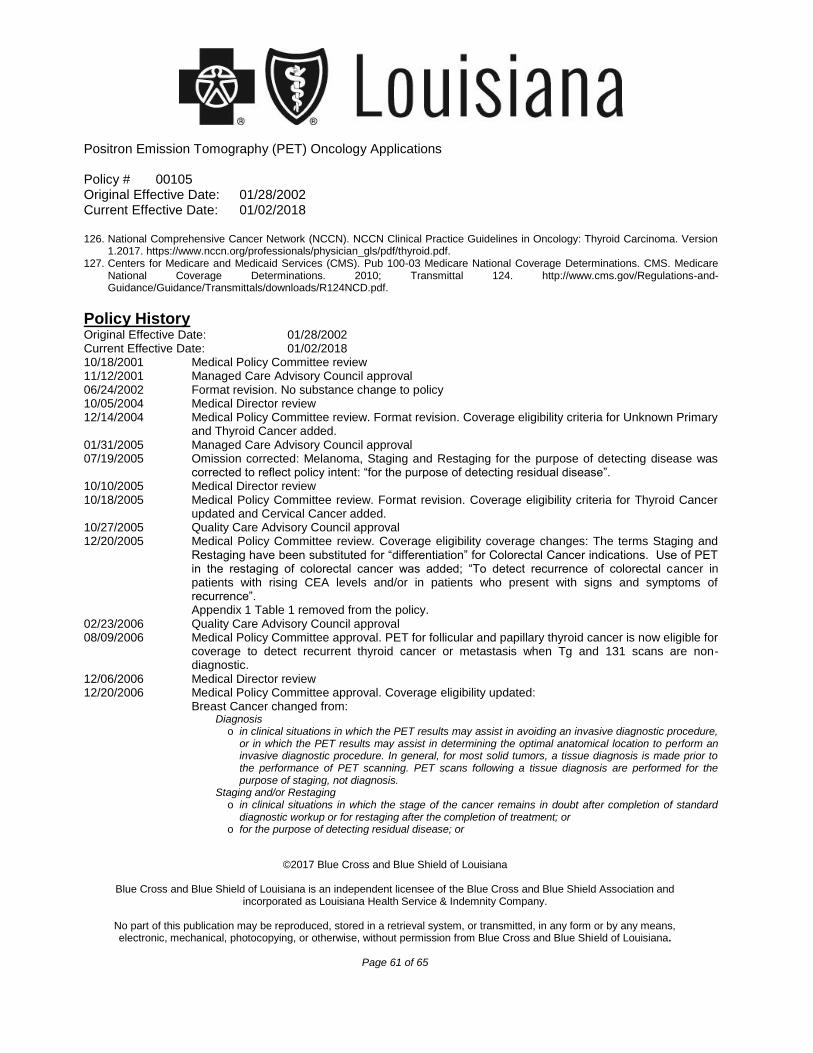
Positron Emission Tomography (PET) Oncology Applications
Policy # 00105 Original Effective Date: 01/28/2002 Current Effective Date: 01/02/2018
©2017 Blue Cross and Blue Shield of Louisiana
Blue Cross and Blue Shield of Louisiana is an independent licensee of the Blue Cross and Blue Shield Association and
incorporated as Louisiana Health Service & Indemnity Company.
No part of this publication may be reproduced, stored in a retrieval system, or transmitted, in any form or by any means, electronic, mechanical, photocopying, or otherwise, without permission from Blue Cross and Blue Shield of Louisiana.
Page 61 of 65
126. National Comprehensive Cancer Network (NCCN). NCCN Clinical Practice Guidelines in Oncology: Thyroid Carcinoma. Version 1.2017. https://www.nccn.org/professionals/physician_gls/pdf/thyroid.pdf.
127. Centers for Medicare and Medicaid Services (CMS). Pub 100-03 Medicare National Coverage Determinations. CMS. Medicare National Coverage Determinations. 2010; Transmittal 124. http://www.cms.gov/Regulations-and- Guidance/Guidance/Transmittals/downloads/R124NCD.pdf.
Policy History Original Effective Date: 01/28/2002 Current Effective Date: 01/02/2018 10/18/2001 Medical Policy Committee review 11/12/2001 Managed Care Advisory Council approval 06/24/2002 Format revision. No substance change to policy 10/05/2004 Medical Director review 12/14/2004 Medical Policy Committee review. Format revision. Coverage eligibility criteria for Unknown Primary
and Thyroid Cancer added. 01/31/2005 Managed Care Advisory Council approval 07/19/2005 Omission corrected: Melanoma, Staging and Restaging for the purpose of detecting disease was
corrected to reflect policy intent: “for the purpose of detecting residual disease”. 10/10/2005 Medical Director review 10/18/2005 Medical Policy Committee review. Format revision. Coverage eligibility criteria for Thyroid Cancer
updated and Cervical Cancer added. 10/27/2005 Quality Care Advisory Council approval 12/20/2005 Medical Policy Committee review. Coverage eligibility coverage changes: The terms Staging and
Restaging have been substituted for “differentiation” for Colorectal Cancer indications. Use of PET in the restaging of colorectal cancer was added; “To detect recurrence of colorectal cancer in patients with rising CEA levels and/or in patients who present with signs and symptoms of recurrence”.
Appendix 1 Table 1 removed from the policy. 02/23/2006 Quality Care Advisory Council approval 08/09/2006 Medical Policy Committee approval. PET for follicular and papillary thyroid cancer is now eligible for
coverage to detect recurrent thyroid cancer or metastasis when Tg and 131 scans are non-diagnostic.
12/06/2006 Medical Director review 12/20/2006 Medical Policy Committee approval. Coverage eligibility updated: Breast Cancer changed from:
Diagnosis o in clinical situations in which the PET results may assist in avoiding an invasive diagnostic procedure,
or in which the PET results may assist in determining the optimal anatomical location to perform an invasive diagnostic procedure. In general, for most solid tumors, a tissue diagnosis is made prior to the performance of PET scanning. PET scans following a tissue diagnosis are performed for the purpose of staging, not diagnosis.
Staging and/or Restaging o in clinical situations in which the stage of the cancer remains in doubt after completion of standard
diagnostic workup or for restaging after the completion of treatment; or o for the purpose of detecting residual disease; or
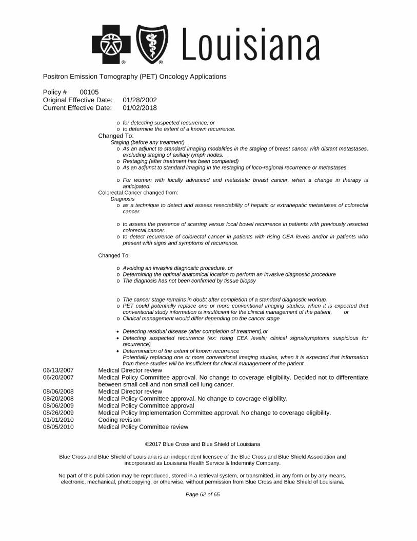
Positron Emission Tomography (PET) Oncology Applications
Policy # 00105 Original Effective Date: 01/28/2002 Current Effective Date: 01/02/2018
©2017 Blue Cross and Blue Shield of Louisiana
Blue Cross and Blue Shield of Louisiana is an independent licensee of the Blue Cross and Blue Shield Association and incorporated as Louisiana Health Service & Indemnity Company.
No part of this publication may be reproduced, stored in a retrieval system, or transmitted, in any form or by any means, electronic, mechanical, photocopying, or otherwise, without permission from Blue Cross and Blue Shield of Louisiana.
Page 62 of 65
o for detecting suspected recurrence; oro to determine the extent of a known recurrence.
Changed To: Staging (before any treatment)
o As an adjunct to standard imaging modalities in the staging of breast cancer with distant metastases,excluding staging of axillary lymph nodes.
o Restaging (after treatment has been completed)o As an adjunct to standard imaging in the restaging of loco-regional recurrence or metastases
Treatment Response Monitoring o For women with locally advanced and metastatic breast cancer, when a change in therapy is
anticipated.Colorectal Cancer changed from:
Diagnosis o as a technique to detect and assess resectability of hepatic or extrahepatic metastases of colorectal
cancer.Restaging
o to assess the presence of scarring versus local bowel recurrence in patients with previously resectedcolorectal cancer.
o to detect recurrence of colorectal cancer in patients with rising CEA levels and/or in patients whopresent with signs and symptoms of recurrence.
06/13/2007 Medical Director review 06/20/2007 Medical Policy Committee approval. No change to coverage eligibility. Decided not to differentiate
between small cell and non small cell lung cancer. 08/06/2008 Medical Director review 08/20/2008 Medical Policy Committee approval. No change to coverage eligibility. 08/06/2009 Medical Policy Committee approval 08/26/2009 Medical Policy Implementation Committee approval. No change to coverage eligibility. 01/01/2010 Coding revision 08/05/2010 Medical Policy Committee review
Changed To: Diagnosis-when PET results may assist in
Staging
Restaging for the purpose of
ooo
Avoiding an invasive diagnostic procedure, orDetermining the optimal anatomical location to perform an invasive diagnostic procedureThe diagnosis has not been confirmed by tissue biopsy
oo
o
The cancer stage remains in doubt after completion of a standard diagnostic workup.PET could potentially replace one or more conventional imaging studies, when it is expected tha t conventional study information is insufficient for the clinical management of the patient, orClinical management would differ depending on the cancer stage
Detecting residual disease (after completion of treatment),or
Detecting suspected recurrence (ex: rising CEA levels; clinical signs/symptoms suspicious fo r recurrence)
Determination of the extent of known recurrencePotentially replacing one or more conventional imaging studies, when it is expected that informat ion from these studies will be insufficient for clinical management of the patient.
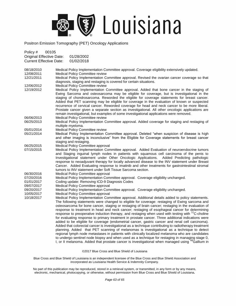
Positron Emission Tomography (PET) Oncology Applications
Policy # 00105 Original Effective Date: 01/28/2002 Current Effective Date: 01/02/2018
©2017 Blue Cross and Blue Shield of Louisiana
Blue Cross and Blue Shield of Louisiana is an independent licensee of the Blue Cross and Blue Shield Association and
incorporated as Louisiana Health Service & Indemnity Company.
No part of this publication may be reproduced, stored in a retrieval system, or transmitted, in any form or by any means, electronic, mechanical, photocopying, or otherwise, without permission from Blue Cross and Blue Shield of Louisiana.
Page 63 of 65
08/18/2010 Medical Policy Implementation Committee approval. Coverage eligibility extensively updated. 12/08/2011 Medical Policy Committee review 12/21/2011 Medical Policy Implementation Committee approval. Revised the ovarian cancer coverage so that
diagnosis, staging and restaging is covered for certain situations. 12/06/2012 Medical Policy Committee review 12/19/2012 Medical Policy Implementation Committee approval. Added that bone cancer in the staging of
Ewing Sarcoma and osteosarcoma may be eligible for coverage, but is investigational in the staging of chondrosarcoma. Reworded the eligible for coverage statements for breast cancer. Added that PET scanning may be eligible for coverage in the evaluation of known or suspected recurrence of cervical cancer. Reworded coverage for head and neck cancer to be more liberal. Prostate cancer given a separate section as investigational. All other oncologic applications are remain investigational, but examples of some investigational applications were removed.
06/06/2013 Medical Policy Committee review 06/25/2013 Medical Policy Implementation Committee approval. Added coverage for staging and restaging of
multiple myeloma. 05/01/2014 Medical Policy Committee review 05/21/2014 Medical Policy Implementation Committee approval. Deleted “when suspicion of disease is h igh
and other imaging is inconclusive” from the Eligible for Coverage statements for breast cancer staging and restaging.
06/25/2015 Medical Policy Committee approval 07/15/2015 Medical Policy Implementation Committee approval. Added Evaluation of neuroendocrine tumors
and Staging inguinal lymph nodes in patients with squamous cell carcinoma of the penis to Investigational statement under Other Oncologic Applications. Added Predicting pathologic response to neoadjuvant therapy for locally advanced disease to the INV statement under Breast Cancer. Added Evaluating response to imatinib and other treatments for gastrointestinal stromal tumors to INV statement under Soft Tissue Sarcoma section.
06/30/2016 Medical Policy Committee approval 07/20/2016 Medical Policy Implementation Committee approval. Coverage eligibility unchanged. 01/01/2017 Coding update: Removing ICD-9 Diagnosis Codes 09/07/2017 Medical Policy Committee approval 09/20/2017 Medical Policy Implementation Committee approval. Coverage eligibility unchanged. 10/05/2017 Medical Policy Committee approval 10/18/2017 Medical Policy Implementation Committee approval. Additional details added to policy statements.
The following statements were changed to eligible for coverage: restaging of Ewing sarcoma and osteosarcoma for bone cancer, staging or restaging of brain cancer; restaging in the evaluation of response to treatment in head and neck cancer; restaging of esophageal cancer for determining
11response to preoperative induction therapy, and restaging when used with testing with C-choline for evaluating response to primary treatment in prostate cancer. Three additional indications were added to be eligible for coverage (endometrial cancer, gastric cancer and renal cell carcinoma). Added that colorectal cancer is investigational as a technique contributing to radiotherapy treatment planning. Added that PET scanning of melanomas is investigational as a technique to detect regional lymph node metastases in patients with clinically localized melanoma who are candidates to undergo sentinel node biopsy and when used as a technique for restaging in managing stage 0,
68I, or II melanoma. Added that prostate cancer is investigational when managed using Gallium in
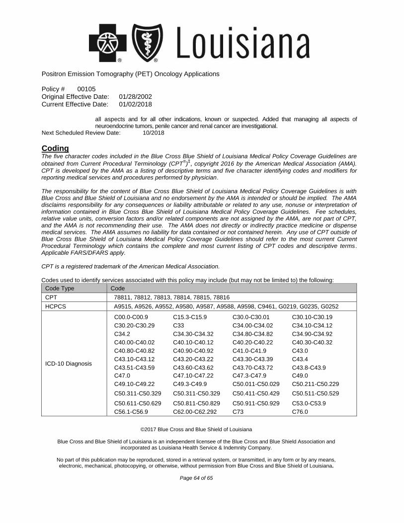
Positron Emission Tomography (PET) Oncology Applications
Policy # 00105 Original Effective Date: 01/28/2002 Current Effective Date: 01/02/2018
©2017 Blue Cross and Blue Shield of Louisiana
Blue Cross and Blue Shield of Louisiana is an independent licensee of the Blue Cross and Blue Shield Association and
incorporated as Louisiana Health Service & Indemnity Company.
No part of this publication may be reproduced, stored in a retrieval system, or transmitted, in any form or by any means, electronic, mechanical, photocopying, or otherwise, without permission from Blue Cross and Blue Shield of Louisiana.
Page 64 of 65
all aspects and for all other indications, known or suspected. Added that managing all aspects of neuroendocrine tumors, penile cancer and renal cancer are investigational.
Next Scheduled Review Date: 10/2018
Coding The five character codes included in the Blue Cross Blue Shield of Louisiana Medical Policy Coverage Guidelines are
® ‡obtained from Current Procedural Terminology (CPT ) , copyright 2016 by the American Medical Association (AMA).
CPT is developed by the AMA as a listing of descriptive terms and five character identifying codes and modifiers for reporting medical services and procedures performed by physician.
The responsibility for the content of Blue Cross Blue Shield of Louisiana Medical Policy Coverage Guidelines is with Blue Cross and Blue Shield of Louisiana and no endorsement by the AMA is intended or should be implied. The AMA disclaims responsibility for any consequences or liability attributable or related to any use, nonuse or interpretation of information contained in Blue Cross Blue Shield of Louisiana Medical Policy Coverage Guidelines. Fee schedules, relative value units, conversion factors and/or related components are not assigned by the AMA, are not part of CPT, and the AMA is not recommending their use. The AMA does not directly or indirectly practice medicine or dispense medical services. The AMA assumes no liability for data contained or not contained herein. Any use of CPT outside of Blue Cross Blue Shield of Louisiana Medical Policy Coverage Guidelines should refer to the most current Current Procedural Terminology which contains the complete and most current listing of CPT codes and descriptive terms. Applicable FARS/DFARS apply. CPT is a registered trademark of the American Medical Association. Codes used to identify services associated with this policy may include (but may not be limited to) the following:
Code Type Code
CPT 78811, 78812, 78813, 78814, 78815, 78816
HCPCS A9515, A9526, A9552, A9580, A9587, A9588, A9598, C9461, G0219, G0235, G0252
ICD-10 Diagnosis
C00.0-C00.9 C15.3-C15.9 C30.0-C30.01 C30.10-C30.19
C30.20-C30.29 C33 C34.00-C34.02 C34.10-C34.12
C34.2 C34.30-C34.32 C34.80-C34.82 C34.90-C34.92
C40.00-C40.02 C40.10-C40.12 C40.20-C40.22 C40.30-C40.32
C40.80-C40.82 C40.90-C40.92 C41.0-C41.9 C43.0
C43.10-C43.12 C43.20-C43.22 C43.30-C43.39 C43.4
C43.51-C43.59 C43.60-C43.62 C43.70-C43.72 C43.8-C43.9
C47.0 C47.10-C47.22 C47.3-C47.9 C49.0
C49.10-C49.22 C49.3-C49.9 C50.011-C50.029 C50.211-C50.229
C50.311-C50.329 C50.311-C50.329 C50.411-C50.429 C50.511-C50.529
C50.611-C50.629 C50.811-C50.829 C50.911-C50.929 C53.0-C53.9
C56.1-C56.9 C62.00-C62.292 C73 C76.0
Positron Emission Tomography (PET) Oncology Applications
Policy # 00105 Original Effective Date: 01/28/2002 Current Effective Date: 01/02/2018
©2017 Blue Cross and Blue Shield of Louisiana
Blue Cross and Blue Shield of Louisiana is an independent licensee of the Blue Cross and Blue Shield Association and
incorporated as Louisiana Health Service & Indemnity Company.
No part of this publication may be reproduced, stored in a retrieval system, or transmitted, in any form or by any means, electronic, mechanical, photocopying, or otherwise, without permission from Blue Cross and Blue Shield of Louisiana.
Page 65 of 65
C78.00-C78.02 C81.00-C81.99 C82.00-C82.99 D03.0
D03.10-D03.339 D03.4 D03.51-D03.72 D03.8-D03.9
*Investigational – A medical treatment, procedure, drug, device, or biological product is Investigational if the effectiveness has not been clearly tested and it has not been incorporated into standard medical practice. Any determination we make that a medical treatment, procedure, drug, device, or biological product is Investigational will be based on a consideration of the following:
A. Whether the medical treatment, procedure, drug, device, or biological product can be lawfully marketed without approval of the U.S. Food and Drug Administration (FDA) and whether such approval has been granted at the time the medical treatment, procedure, drug, device, or biological product is sought to be furnished; or
B. Whether the medical treatment, procedure, drug, device, or biological product requires further studies or clinical trials to determine its maximum tolerated dose, toxicity, safety, effectiveness, or effectiveness as compared with the standard means of treatment or diagnosis, must improve health outcomes, according to the consensus of opinion among experts as shown by reliable evidence, including:
1. Consultation with the Blue Cross and Blue Shield Association technology assessment program (TEC) or other nonaffiliated technology evaluation center(s);
2. Credible scientific evidence published in peer-reviewed medical literature generally recognized by the relevant medical community; or
3. Reference to federal regulations.
**Medically Necessary (or “Medical Necessity”) - Health care services, treatment, procedures, equipment, drugs, devices, items or supplies that a Provider, exercising prudent clinical judgment, would provide to a patient for the purpose of preventing, evaluating, diagnosing or treating an illness, injury, disease or its symptoms, and that are:
A. In accordance with nationally accepted standards of medical practice; B. Clinically appropriate, in terms of type, frequency, extent, level of care, site and duration, and considered effective for the
patient's illness, injury or disease; and C. Not primarily for the personal comfort or convenience of the patient, physician or other health care provider, and not more
costly than an alternative service or sequence of services at least as likely to produce equivalent therapeutic or diagnostic results as to the diagnosis or treatment of that patient's illness, injury or disease.
For these purposes, “nationally accepted standards of medical practice” means standards that are based on credible scientific evidence published in peer-reviewed medical literature generally recognized by the relevant medical community, Physician Specialty Society recommendations and the views of Physicians practicing in relevant clinical areas and any other relevant factors. ‡ Indicated trademarks are the registered trademarks of their respective owners. NOTICE: Medical Policies are scientific based opinions, provided solely for coverage and informational purposes. Medical Policies should not be construed to suggest that the Company recommends, advocates, requires, encourages, or discourages any particular treatment, procedure, or service, or any particular course of treatment, procedure, or service.