polyploid initiation in hawaii tree species a thesis ...value of hawaii’s nursery products is...
TRANSCRIPT

POLYPLOID INITIATION IN HAWAII TREE SPECIES
A THESIS SUBMITTED TO THE GRADUATE DIVISION OF THE UNIVERSITY
OF HAWAIʻI AT MĀNOA IN PARTIAL FULFILLMENT OF THE REQUIREMENTS
FOR THE DEGREE OF
MASTERS OF SCIENCE
IN
TROPICAL PLANT AND SOIL SCIENCE
AUGUST 2014
By
David J. Lingenfelser
Thesis Committee:
Ken Leonhardt, Chairperson
Richard Manshardt
Karen Selph
Keywords: Polyploid, Oryzalin, Hawaii, Landscaping, Trees, Double Chromosomes

ii
ACKNOWLEDGEMENTS
I would like to express my sincere appreciation to Dr. Ken Leonhardt, my
committee chairperson for his support and guidance during my time in graduate school.
I would also like to thank Dr. Richard Manshardt and Dr. Karen Selph for serving
on my committee and for all of their advice.
I am very appreciative of the Monsanto Corporation for providing the funding for
my fellowship.
I would also like to express my thankfulness to my labmate, Amanda Ackerman,
for her friendship and help.
Finally I would like to thank my parents, Frederick Lingenfelser IV and Jean
Lingenfelser, and my brother, Frederick Lingenfelser V for their support and
encouragement.

iii
ABSTRACT
In Hawaii, many plant species commonly used by landscapers yield abundant
large fruits that can injure people/property, attract rats and other pests, create an unsightly
appearance and potentially become invasive. Sterile forms of these landscaping plants
would eliminate their high-maintenance and invasive characteristics. Polyploid forms of
plants, particularly triploids, are often sterile. Autotetraploids also frequently display
some degree of sterility. In addition, polyploids typically exhibit unique physical
characteristics such as thicker leaves, larger organs, and higher levels of chemical
compounds. A project was initiated to create tetraploid forms of eight commonly utilized
species of trees: Thespesia populnea, Calophyllum inophyllum, Clusia rosea, Schefflera
actinophylla, Heritiera littoralis, Jatropha curcas, Plumeria Stenopetala, and Erythrina
sandwicensis. Various concentrations of the dinitroaniline herbicide, Oryzalin (ranging
from 0.01% to 0.5%) were applied to seedling meristems in vivo. Following a period of
vegetative growth, the seedlings were tested for higher ploidy levels. Multiple
autotetraploids and mixoploids have been identified using guard cell measurements and
flow cytometry.
iv
TABLE OF CONTENTS
ACKNOWLEDGEMENTS…………………………………………………..…………ii
ABSTRACT………………………………………………………………..……………iii
LIST OF TABLES………………………………………………………………………vi
LIST OF FIGURES………………………………………………………….....………vii
CHAPTER 1. INTRODUCTION…………….…………………………………………9
1.1 Landscaping Industry in Hawaii……………………………………………9
1.2 Sterility……………………………………………………………………..…9
1.3 Schefflera actinophylla……………………………………………………..16
1.4 Heritiera littoralis…………………………………………………………..17
1.5 Thespesia populnea………………………………………………………....18
1.6 Clusia rosea………………………………………………………………….19
1.7 Calophyllum inophyllum…………………………………………………...20
1.8 Jatropha curcas……………………………………………………………..21
1.9 Plumeria Stenopetala………………………………………..……………...22
1.10 Erythrina sandwicensis…………………………………..……………….22
CHAPTER 2. MATERIALS AND METHODS………………………………………24
2.1 General Methods……………………………………………………………24
2.2 Schefflera actinophylla……………………………………………………..26
2.3 Heritiera littoralis…………………….…………………………………….26
2.4 Thespesia populnea…………………………………………………………27
2.5 Clusia rosea………………………………………………………………….27
2.6 Calophyllum inophyllum…………………………………………………...28
2.7 Jatropha curcas……………………………………………………………..29
2.8 Plumeria Stenopetala……………………………………………………….30
2.9 Erythrina sandwicensis…………………………………………………….30
2.10 Guard Cell Measurement…………………………………………………30
2.11 Flow Cytometry……………………………………………………………31
CHAPTER 3. Results…………………………………………………………………..32
3.1 Schefflera actinophylla……………………………………………………..36
v
3.2 Heritiera littoralis…………………………………………………………..40
3.3 Thespesia populnea…………………………………………………………41
3.4 Clusia rosea………………………………………………………………….44
3.5 Calophyllum inophyllum…………………………………………………...45
3.6 Jatropha curcas……………………………………………………………..47
3.7 Plumeria Stenopetala……………………………………………………….49
3.8 Erythrina sandwicensis…………………………………………………….52
CHAPTER 4. Discussion……………………………………………………………….54
4.1 Schefflera actinophylla……………………………………………………..54
4.2 Heritiera littoralis…………………………………………………………..55
4.3 Thespesia populnea…………………………………………………………56
4.4 Clusia rosea…………………………………………………………………57
4.5 Calophyllum inophyllum…………………………………………………..57
4.7 Jatropha curcas…………………………………………………………….58
4.8 Plumeria Stenopetala………………………………………………………59
4.6 Erythrina sandwicensis…………………………………………………….60
CHAPTER 5. CONCLUSION…………………………………………………………61
LITERATURE………………………………………………………………………….62

vi
LIST OF TABLES
Table 1. Germination Data of all species .......................................................................... 32
Table 2. Polyploid initiation data of all species ................................................................ 35
vii
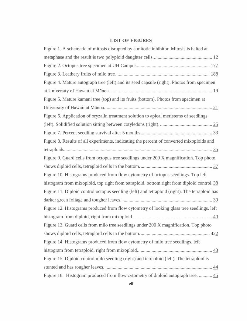
LIST OF FIGURES
Figure 1. A schematic of mitosis disrupted by a mitotic inhibitor. Mitosis is halted at
metaphase and the result is two polyploid daughter cells. ................................................ 12
Figure 2. Octopus tree specimen at UH Campus ............................................................ 177
Figure 3. Leathery fruits of milo tree .............................................................................. 188
Figure 4. Mature autograph tree (left) and its seed capsule (right). Photos from specimen
at University of Hawaii at Mānoa. .................................................................................... 19
Figure 5. Mature kamani tree (top) and its fruits (bottom). Photos from specimen at
University of Hawaii at Mānoa. ........................................................................................ 21
Figure 6. Application of oryzalin treatment solution to apical meristems of seedlings
(left). Solidified solution sitting between cotyledons (right). ........................................... 25
Figure 7. Percent seedling survival after 5 months ........................................................... 33
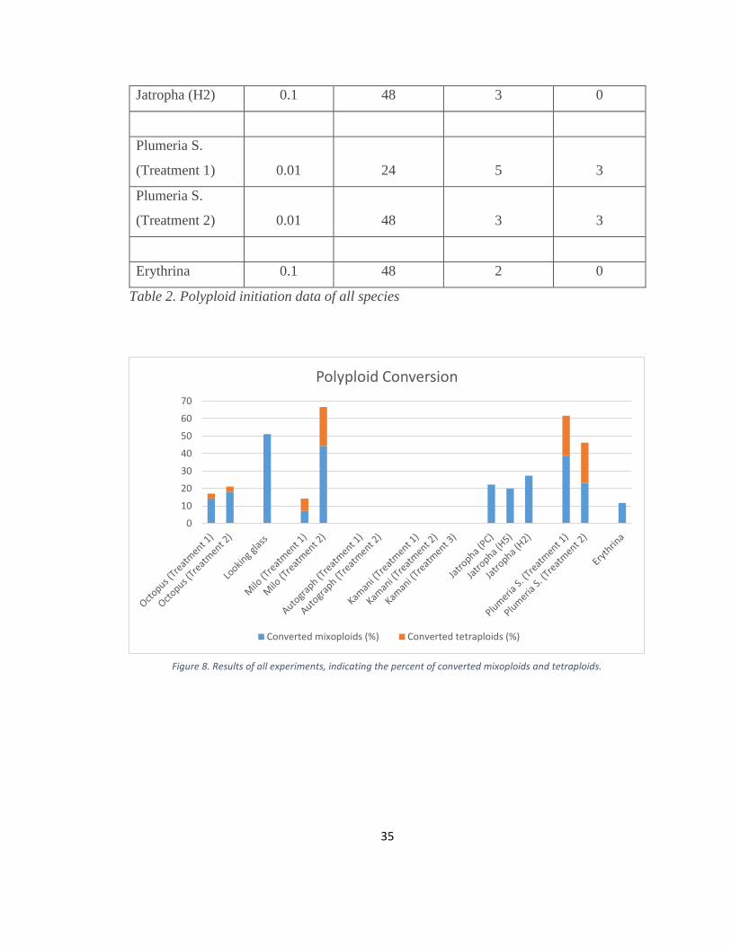
Figure 8. Results of all experiments, indicating the percent of converted mixoploids and
tetraploids. ......................................................................................................................... 35
Figure 9. Guard cells from octopus tree seedlings under 200 X magnification. Top photo
shows diploid cells, tetraploid cells in the bottom. ........................................................... 37
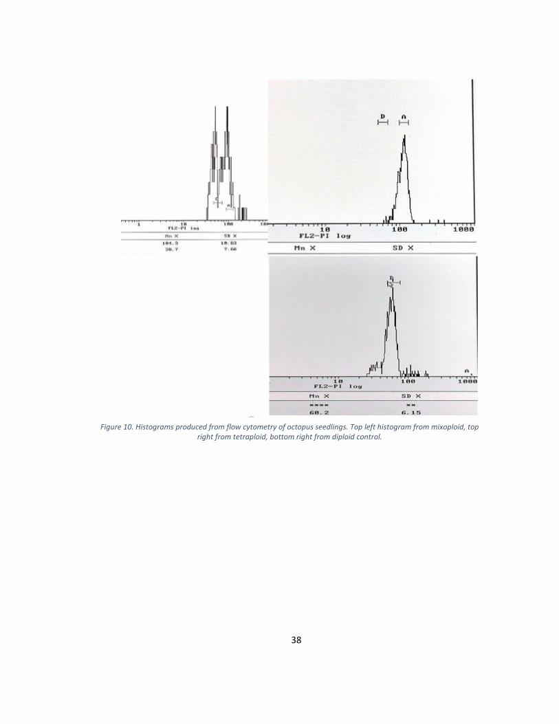
Figure 10. Histograms produced from flow cytometry of octopus seedlings. Top left
histogram from mixoploid, top right from tetraploid, bottom right from diploid control. 38
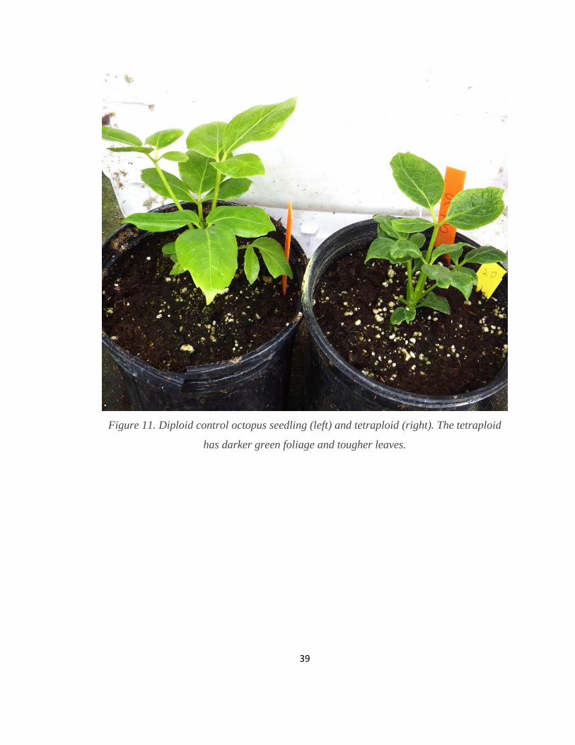
Figure 11. Diploid control octopus seedling (left) and tetraploid (right). The tetraploid has
darker green foliage and tougher leaves. .......................................................................... 39
Figure 12. Histograms produced from flow cytometry of looking glass tree seedlings. left
histogram from diploid, right from mixoploid. ................................................................. 40
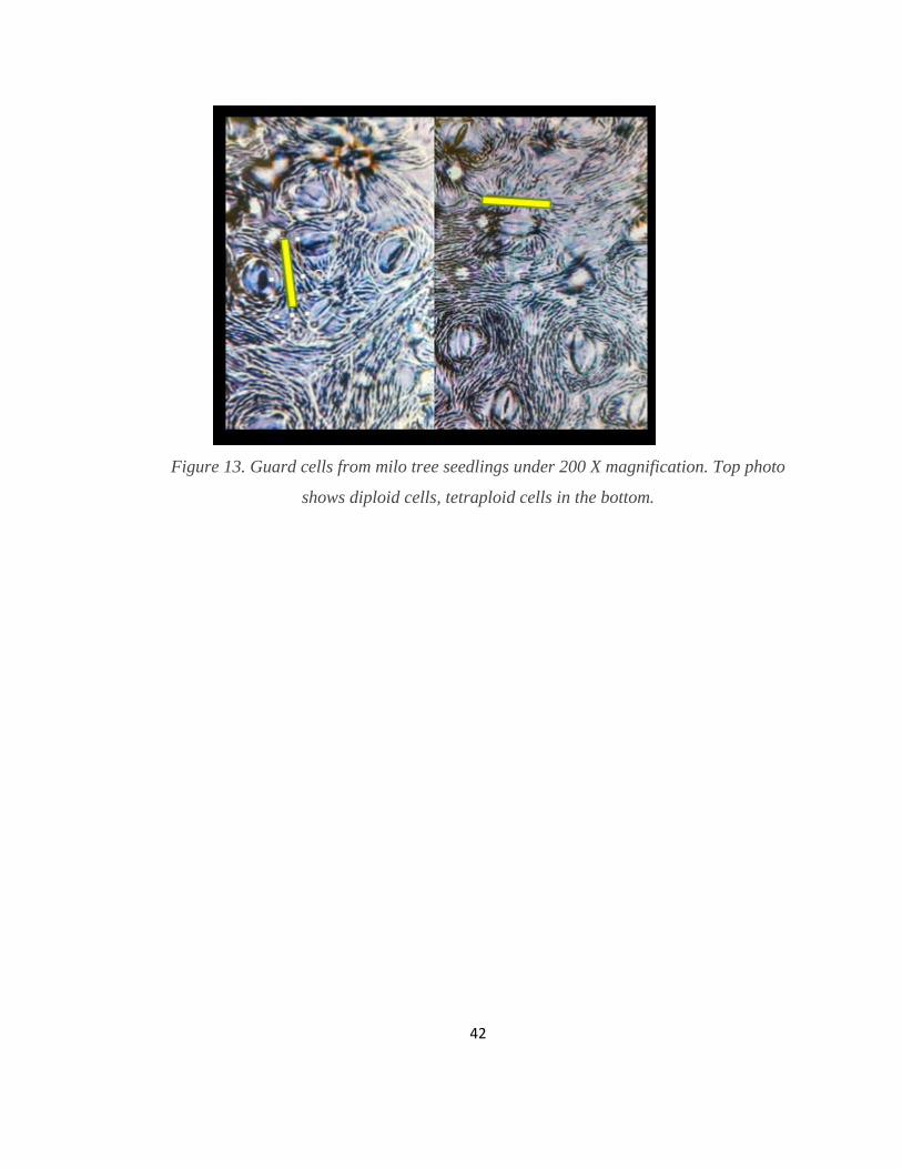
Figure 13. Guard cells from milo tree seedlings under 200 X magnification. Top photo
shows diploid cells, tetraploid cells in the bottom. ......................................................... 422
Figure 14. Histograms produced from flow cytometry of milo tree seedlings. left
histogram from tetraploid, right from mixoploid. ............................................................. 43
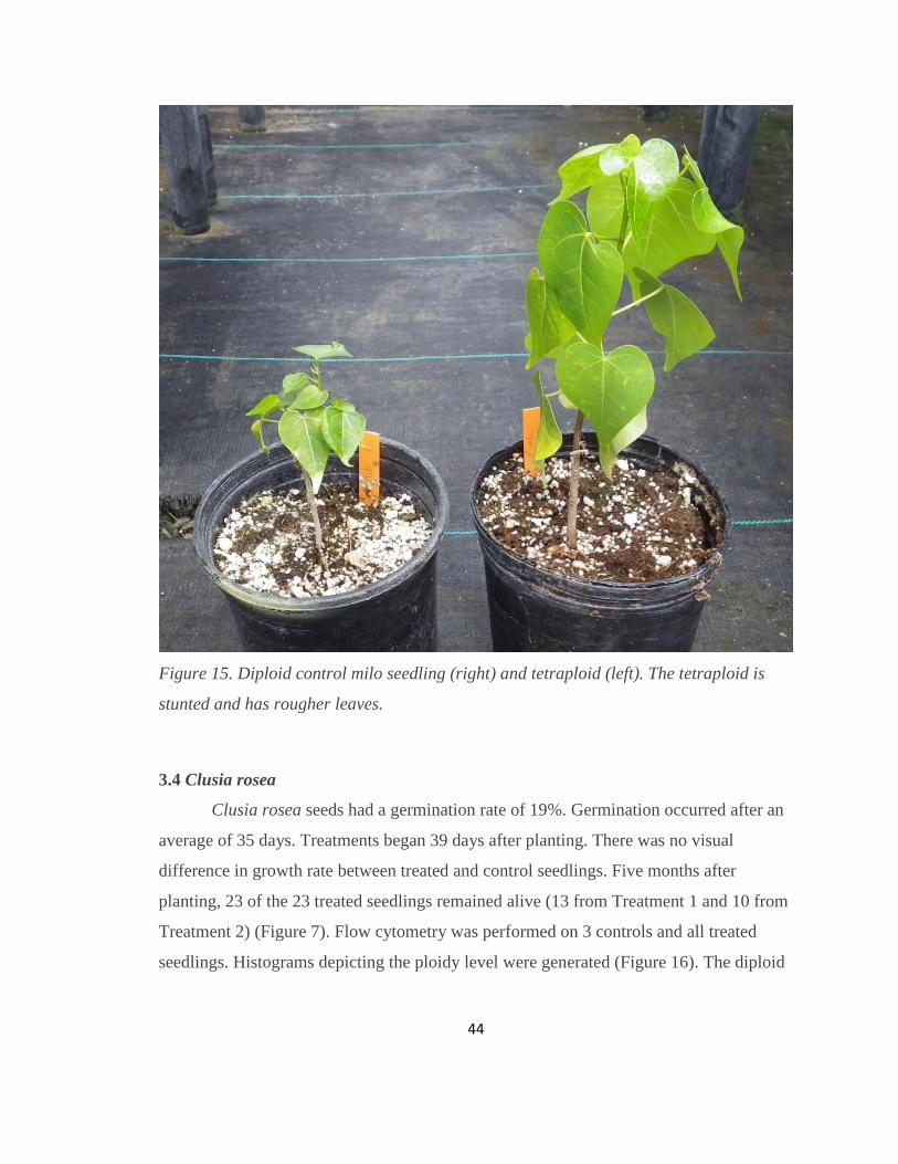
Figure 15. Diploid control milo seedling (right) and tetraploid (left). The tetraploid is
stunted and has rougher leaves. ........................................................................................ 44
Figure 16. Histogram produced from flow cytometry of diploid autograph tree. ........... 45

viii
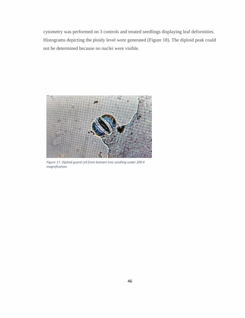
Figure 17. Diploid guard cell from kamani tree seedling under 200 X magnification. .... 46
Figure 18. Histogram produced from flow cytometry of presumably diploid kamani tree.
Location of diploid peak is uncertain due to large quantity of background noise ............ 47
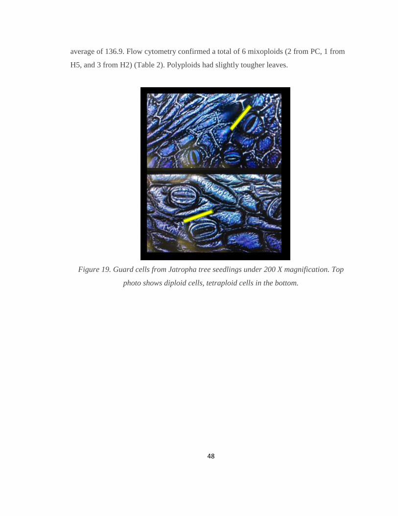
Figure 19. Guard cells from Jatropha tree seedlings under 200 X magnification. Top
photo shows diploid cells, tetraploid cells in the bottom. ................................................. 48
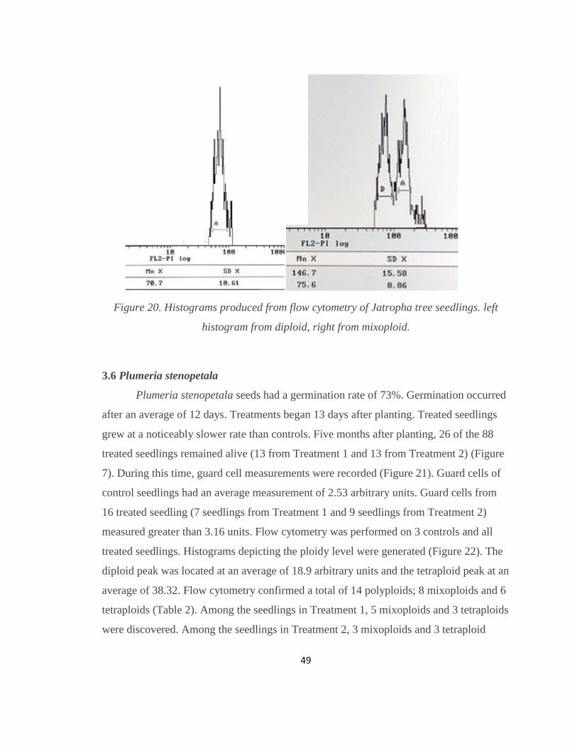
Figure 20. Histograms produced from flow cytometry of Jatropha tree seedlings. left
histogram from diploid, right from mixoploid. ................................................................. 49
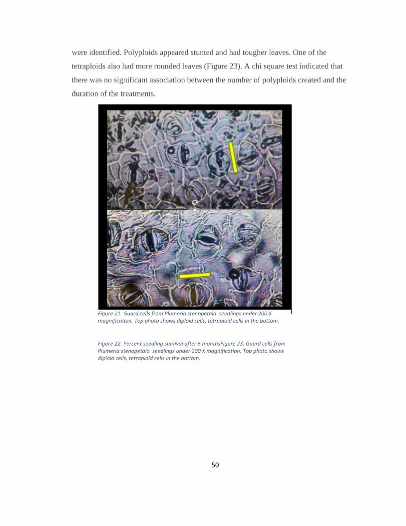
Figure 21. Guard cells from Plumeria stenopetala seedlings under 200 X magnification.
Top photo shows diploid cells, tetraploid cells in the bottom. ......................................... 50
Figure 7. Percent seedling survival after 5 monthsFigure 21. Guard cells from Plumeria
stenopetala seedlings under 200 X magnification. Top photo shows diploid cells,
tetraploid cells in the bottom............................................................................................. 50
Figure 22. Histograms produced from flow cytometry of Plumeria stenopetala seedlings.
Top left histogram from Ddiploid control, top right from mixoploid, bottom right from
tetraploid. .......................................................................................................................... 51
Figure 21. Guard cells from Plumeria stenopetala seedlings under 200 X magnification.
Top photo shows diploid cells, tetraploid cells in the bottom.Figure 22. Histograms
produced from flow cytometry of Plumeria stenopetala seedlings. Top left histogram
from Ddiploid control, top right from mixoploid, bottom right from tetraploid. ............. 51
Figure 23. Guard cells from Erythrina sandwicensis seedlings under 200 X
magnification. Top photo shows diploid cells, tetraploid cells in the bottom. ................. 52
Figure 23. Guard cells from Erythrina sandwicensis seedlings under 200 X
magnification. Top photo shows diploid cells, tetraploid cells in the bottom. ................. 52
Figure 24. Histograms produced from flow cytometry of Erythrina sandwicensis tree
seedlings. Left histogram from diploid control, right from mixoploid. ............................ 53
Figure 24. Histograms produced from flow cytometry of Erythrina sandwicensis tree
seedlings. Left histogram from diploid control, right from mixoploid. ............................ 53
9
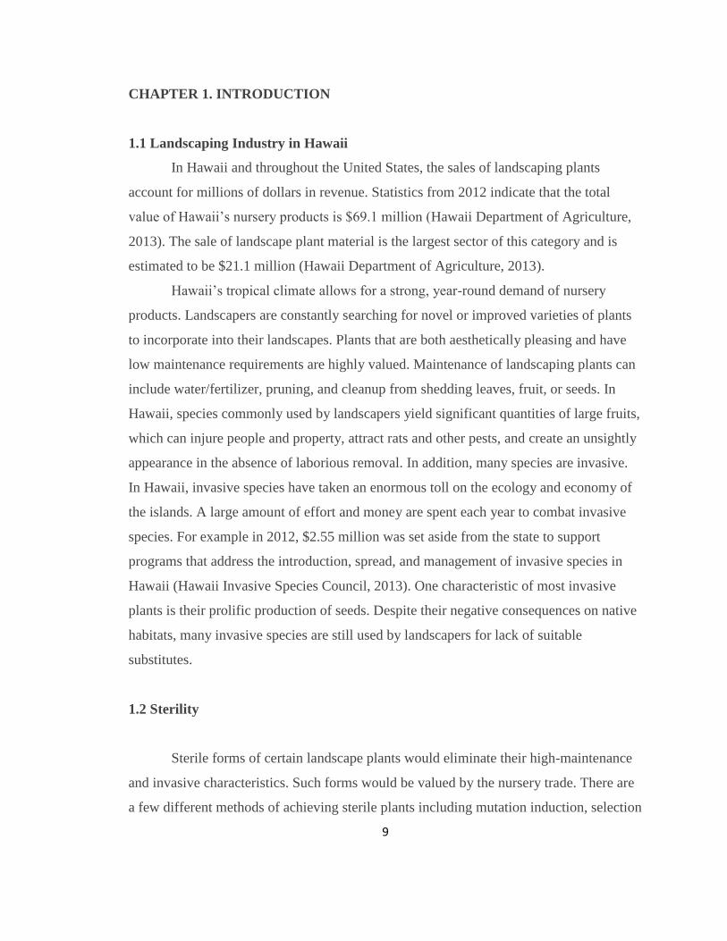
CHAPTER 1. INTRODUCTION
1.1 Landscaping Industry in Hawaii
In Hawaii and throughout the United States, the sales of landscaping plants
account for millions of dollars in revenue. Statistics from 2012 indicate that the total
value of Hawaii’s nursery products is $69.1 million (Hawaii Department of Agriculture,
2013). The sale of landscape plant material is the largest sector of this category and is
estimated to be $21.1 million (Hawaii Department of Agriculture, 2013).
Hawaii’s tropical climate allows for a strong, year-round demand of nursery
products. Landscapers are constantly searching for novel or improved varieties of plants
to incorporate into their landscapes. Plants that are both aesthetically pleasing and have
low maintenance requirements are highly valued. Maintenance of landscaping plants can
include water/fertilizer, pruning, and cleanup from shedding leaves, fruit, or seeds. In
Hawaii, species commonly used by landscapers yield significant quantities of large fruits,
which can injure people and property, attract rats and other pests, and create an unsightly
appearance in the absence of laborious removal. In addition, many species are invasive.
In Hawaii, invasive species have taken an enormous toll on the ecology and economy of
the islands. A large amount of effort and money are spent each year to combat invasive
species. For example in 2012, $2.55 million was set aside from the state to support
programs that address the introduction, spread, and management of invasive species in
Hawaii (Hawaii Invasive Species Council, 2013). One characteristic of most invasive
plants is their prolific production of seeds. Despite their negative consequences on native
habitats, many invasive species are still used by landscapers for lack of suitable
substitutes.
1.2 Sterility
Sterile forms of certain landscape plants would eliminate their high-maintenance
and invasive characteristics. Such forms would be valued by the nursery trade. There are
a few different methods of achieving sterile plants including mutation induction, selection
10
of varieties with reduced fertility, and changing chromosome number to induce sterility.
In my research, I will focus on sterility associated with polyploidy.
A polyploid refers to an organism possessing a higher number of chromosomes
relative to the wild-type. Although polyploidy is a rare phenomenon in animals, it is
fairly common among plants (Ranney, 2006). In fact, the induction of polyploids in
nature is a significant aspect of plant evolution and has been responsible for the
generation of many new species. It is estimated that 30-70% of all flowering plants are of
polyploid origin (Ranney, 2006). Sometimes gametes of two different species will
combine during sexual reproduction. However, since they have unrelated genetic
backgrounds, the gametes might not be able to form pairs of homologous (genotypically
similar) chromosomes. The result of such a cross would result in a sterile progeny.
However, in the rare event of a disruption of mitosis in the meristem, doubling of
chromosomes may occur. This chromosome doubling could restore fertility since it
would allow for the formation of pairs of homologous chromosomes (Kashkush et al.,
2002). The resulting plant is known as an allotetraploid. The fusion of unreduced gametes
between two distinct species would also lead to an allotetraploid. However, that would
also be a very rare event since the formation and fusion of unreduced gametes would
have to occur simultaneously (Hegarty and Hiscock, 2008). In any case, allotetraploids
have four sets of chromosomes; two pairs of homologous versions for each chromosome.
The disruption of mitosis in plant meristems or the fusion of unreduced gametes
within one species can also result in a tetraploid. Both of these examples of tetraploids
are referred to as autotetraploids since they will have 4 sets of homologous chromosomes.
Autotetraploids often display some degree of sterility (Loidl, 1995). This is due to the
fact that instead of two sets of homologous chromosomes, autotetraploids have four
(Loidl, 1995). During the process of meiosis, normally, pairs of homologous
chromosomes segregate and form two separate gametes. However in autotetraploids, the
extra homologous chromosomes can lead to infertility due to uneven pairing.
Triploids, plants with three sets of chromosomes, often display very high levels of
infertility (Jones and Ranney, 2009). Since triploids have an odd number of
chromosomes, it is not possible to divide the chromosomes evenly during meiosis.
11
Triploid plants are generally completely sterile (with the exception of a handful of plants
such as triploid apples and banana, which may produce occasional seeds) (Ranney,
2006). As a result, triploid plants rarely occur naturally. However, people have selected
and bred for triploids to develop many useful cultivars of plants.
Endosperm culture is a direct method of obtaining triploid plants. Endosperm is
the nutritive tissue in angiosperms that generally consists of triploid cells. This tissue is
the product of double fertilization, in which three haploid nuclei- two from the female
and one from the male, fuse together. Therefore plants derived from endosperm should
also be triploid in nature. The first successful proliferation of endosperm was reported in
1962 with endosperm of Santalum album (Thomas and Chaturvedi, 2008). Endosperm
culture typically involves somatic embryogenesis in vitro. Endosperm tissue can be
treated with high levels of auxin to induce callus and then transferred to media with lower
auxin levels and various concentrations and combinations of other plant growth
regulators to induce somatic embryos. Once somatic embryos form, they are generally
transferred to nutritive media without hormones to germinate. Since the 1960s, a variety
of triploid plants have been developed from endosperm culture including kiwi, corn,
coffee, papaya, walnut, citrus, and others.
A triploid can also be developed via hybridization. Once an autotetraploid plant is
developed, a triploid can easily be produced by cross pollinating the tetraploid with the
wild-type diploid. During meiosis, the diploid plant will yield a haploid gamete and the
tetraploid will yield a diploid gamete. The fusion of the haploid and diploid gametes will
produce a triploid zygote. Triploid seeds may display physiological challenges and as a
result, embryo rescue is often required for successful germination (Ranney, 2006).
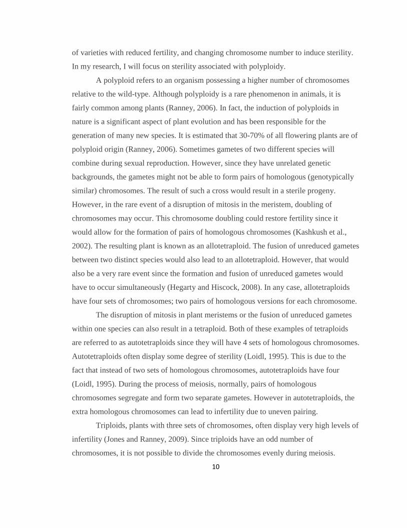
Autotetraploids can occur naturally or can be induced. During mitosis, the
chromosomes within a cell replicate and then cellular division takes place, resulting in
two daughter cells. In nature, it is possible for a disruption in actively dividing
meristematic cells to occur after chromosome replication preventing the cell from
dividing. This would result in a single cell with twice the number of chromosomes
(Figure 1). If all of the cells in the meristem are derived from tetraploid cells, the new
12
growth will be entirely tetraploid. Scientists can also induce this phenomenon using
various chemicals.
A series of chemicals are known to induce polyploid cells in plants.
Conventionally, colchicine has been most widely used by scientists. This compound
occurs naturally as a secondary metabolite in plants of the genus Colchicum. Since the
1930s it was known to be able to inhibit the formation of spindle fibers during cellular
divisions, and therefore stalls mitosis at anaphase. Despite its effectiveness, it is highly
toxic since it is able to effect both plant and animal cells. In addition, it has been known
to promote undesirable mutations in plants (Tuyl et al., 1992). Since the 1930s, other
compounds that act as mitotic inhibitors have been identified. These include; trifluralin,
amiprophos-methyl, and oryzalin. Oryzalin (4-(dipropylamino)-3,5-
dinitrobenzenesulfonamide) was developed as an herbicide to control the spread of
certain ornamental grasses (Tuyl et al., 1992). Oryzalin belongs to the class of
Figure 1. A schematic of mitosis disrupted by a mitotic inhibitor. Mitosis is halted at
metaphase and the result is two polyploid daughter cells.
13
dinitroaniline herbicides (Lehrer et al., 2008). These compounds are bright yellow in
color due to the presence of two nitro groups on the phenyl ring. Dinitroaniline herbicides
are known to inhibit cell division by preventing microtubule synthesis, which regulates
the spindle fibers. During anaphase of mitosis, the spindle fibers are responsible for
separating chromosomes to opposite ends of the cell, which would normally result in the
formation of two daughter cells. Oryzalin more effectively binds to plant tubulins than
colchicine, whereas colchicine is better at binding to animal tubulins (Jones et al., 2008).
As a result, oryzalin can be effective in much lower concentrations than colchicine (van
Tuyl et al., 1990). In addition, many studies have shown oryzalin to be more effective at
inducing polyploidy than colchicine (Ascough et al, 2008).
There are many different methods for delivering the mitotic inhibitor to the plant
to alter its ploidy level. For the chromosome level to be changed, the mitotic inhibitor
must be applied to a region of dividing cells. In a meristem, where cell division takes
place, the chemical will disrupt mitosis and create cells with replicated genomes since
they are initially unable to divide via cytokinesis. In order for the new growth to consist
entirely of polyploid cells, all of the meristematic cells must be exposed to the mitotic
inhibitor during cell division. The most efficient way of accomplishing this is to expose
the meristem to the chemical when it is small and consists of as few cells as possible. To
obtain plants with such primitive meristems, experimenters often use material initiated in
tissue culture. A high success rate has been reported from treating callus, somatic
embryos, and young meristems with mitotic inhibitors in tissue culture (Dhooghe, et al.,
2011). While tissue culture offers some efficiency, it is also a more labor and time-
consuming process. Positive results have been recorded with an assortment of in-vivo
techniques (Ranney, 2006). Applying the mitotic inhibitor to the meristems by soaking
seeds, misting seedlings, placing a cotton swab between cotyledons, or using lanolin or
agar as a solid media, have all been demonstrated to effectively transform diploids to
tetraploids in certain species (Ranney, 2006).
Examples of the use of oryzalin to induce tetraploidy include a study by Lehrer et
al. (2008) that indicated that despite higher mortality rates, oryzalin was more effective at
inducing tetraploid Japanese Barberry, Berberis thunbergii. A concentration of .002%
14
oryzalin doubled the chromosomes of 28% of the treated plants. Another study
(Boonprok et al., 2013) demonstrated that 0.0015% oryzalin applied to seedling
meristems of Jatropha curcas was effective at converting diploids to tetraploids. Väinölä
et al., 2000 showed that oryzalin was more effective than colchicine at creating tetraploid
Rhododendron cultivars. 37.5% of microshoot explants doubled their chromosomes when
.001% oryzalin was applied in vitro for a period of 24 or 48 hours. In another study
(Väinölä, 2000), it was shown that oryzalin was the superior mitotic inhibitor for
Rhododendrons, creating 18.2% polyploids by treating explants with .005% oryzalin for
24 hours. An experiment with Eustoma grandiflorum showed that Colchicine was
effective at creating tetraploids by placing 0.05% aqueous solution between the
cotyledons of young seedlings. (Griesbach and Bhat, 1990). In ornamental ginger,
oryzalin was most effective at inducing polyploids (Sakhanokho et al., 2009). However, a
60 µm oryzalin solution yielded 15% polyploids when applied to embryogenic callus in
vitro. (Sakhanokho et al., 2009). Another study (Eeckhaut, et al., 2004) indicated that 10
µm of oryzalin and trifluralin were equally effective as colchicine, and achieved 5%
polyploid transformation in Spathiphyllum wallisii when applied during anther culture.
Up to 27.1% of Japanese Ceder plants were converted to polyploids when treated with an
aqueous solution of 150 µM oryzalin at their cotyledon stage for 20 days (Contrera,
2012). The dinitroanalines, oryzalin and ethalfluralin, were as effective as colchicine at
inducing polyploidy in watermelon shoots in vitro (Li et al., 1999). Furthermore,
exposure of 10 µM of oryzalin or ethalfluralin for 9 days was effective at inducing
tetraploidy in 50% of the treated plants, while colchicine required 30 days, to produce the
same results (Li, et al., 1999). Hibiscus acetosella seedlings treated with a solution of
agar and 150 µM oryzalin yielded one solid polyploid and one mixoploid. The heated
solution was dropped onto the meristems and applied for three days (Contreras et al.,
2009). Finally, 50% of Platycodon grandiflorus seedlings were transformed to polyploids
when treated with a colchicine and agar solution for 72 hours (Wu et al., 2011). All of
these studies point to oryzalin as an effective agent for inducing polyploidy.
One important aspect of induced polyploidy in a plant, is that one must verify that
all the tissue in the plant is transformed, or at least the germ layer, or that plant may revert

15
to its original ploidy level. Therefore, in order to induce complete polyploidy in a plant,
all of the cells in the shoot meristem must be transformed (Chauvin et al., 2003). A shoot
meristem is divided into three layers; L1, L2, and L3. The L1 is the outermost meristematic
layer, which gives rise to epidermal cells. Beneath that are the L2 and L3 layers, which
give rise to internal plant organs. Cells from the L2 layer generally give rise to the germ
line, and L3 layer cells are known to give rise to the vascular tissue (Sessions et al., 1999).
After treatment with mitotic inhibitors, the meristematic cells of all three layers may or
may not be transformed. If only some of the cells become polyploid, the resulting plant
will be a chimera since it will consist of somatic cells of multiple ploidy levels. Over the
course of its development, it is possible for a chimera to revert to only one ploidy level,
which would negate the treatment effects.
Thus, it is important to verify that treated plants are completely polyploidy.
However, visually it may be difficult to distinguish a chimera from a solid polyploid
because cells that arise from the L1 layer account for much of what can be seen visually.
Polyploidy often results in larger cell size (Leonhardt and Shi, 2007). Measuring guard
cells under a microscope is a common method of estimating ploidy level, since polyploids
will often have larger guard cells (Leonhardt and Shi, 2007). Larger cell size can also
promote thicker leafs and larger fruits and flowers. In addition, polyploid plants may
exhibit stunted growth. However, chimeras, in which only one layer has been transformed,
will display these morphological habits as long as the L1 layer has been transformed. In
order for the polyploid level to be heritable, it is essential for the cells of the L2 layer to be
transformed, as these cells will give rise to the gametes. In addition, sterility cannot be
achieved unless the ploidy of the gametes is altered.
Since we cannot guarantee that visual cues will tell us about the plant’s ploidy
level, we require another method to assess this result. Flow cytometry is a useful tool that
is used to quantify DNA in tissues. This technique can generally allow scientists to
distinguish between solid polyploids and chimeras (Miguel and Leonhardt, 2011).
Sample preparation for flow cytometry involves treating plant tissue with a nuclei
extraction buffer and then staining the nuclei-contaning solution with a DNA-specific
fluorescent dye (Teng, 2007). A flow cytometer then uses laser light to excite the dye in
16
each nuclei and collects the resulting fluorescence signals with photomultiplier tube
detectors at specific wavelengths. The intensity of fluorescence is proportional to the
quantity of DNA in each nuclei. These DNA data, as well as fluorescence data from
other detectors for light scatter (proxy for cell size), are collected as listmode files, and
can be viewed in graphical form. More DNA-dense polyploid nuclei will exhibit a higher
intensity of fluorescence per nuclei. These data are used to estimate the number of nuclei
at each ploidy level. Therefore, chimeric plant tissue can be identified.
The goal of this project was to double the chromosome level, thereby creating
autotetraploids, of eight species of trees. For the following five species, the purpose of
creating tetraploids is to induce sterility: Clusia rosea, Schefflera actinophylla, Heritiera
littoralis, Thespesia populnea, and Calophyllum inophyllum. Eventually, when the
polyploid specimens from this project reach sexual maturity, they will be evaluated.
Since autotetraploids often show some degree of sterility, this project may directly
achieve the desirable outcome. However, if the polyploid specimens continue to be
fertile, they will crossed with their diploid counterparts to create triploids, which are
almost always sterile. For the remaining three species, the objective in doubling the
chromosome number is to improve certain horticultural characteristics: Jatropha curcas,
Plumeria stenopetala, Erythrina sandwicensis. These desirable horticultural
characteristics will be described below.
1.3 Schefflera actinophylla
The octopus tree, Schefflera actinophylla, is an attractive tree native to Australia.
This fast-growing species exhibits upright growth and glossy compound leaves. It gets its
name from its arm-like inflorescences that somewhat resemble the tentacles of an octopus
(Figure 2). It was first introduced to Hawaii in about 1900 (Wagner et al., 1999). Its rapid
growth and prolific seed production led it to become an invasive species in Hawaii and
many other tropical locations. As a result, this species has been listed in the Global
Compendium of Weeds (Randall, 2012). Seeds are naturally distributed by birds and bats
(Gucker, 2011). A diploid octopus trees has a chromosome number of 2n =24 (Yi et al.,
2004). Flowers of this species are hermaphroditic and capable of self-pollination (Brown

17
and Hopkins 1995). Fruits develop approximately six weeks after flowering and seeds
have very high germination rates (Menninger, 1971). A sterile form of the octopus tree
would be useful and landscapers could use it without concern about contributing to its
invasive spread.
1.4 Heritiera littoralis
The looking-glass tree, Heritiera littoralis, is a species of tropical tree that is
native to South East Asia. It has a tall growth habit and can reach heights of 25 m. Leaves
are dark green with a silvery underside. In its native habitats it is harvested for its strong,
high-quality wood. Leaves also have some medicinal value in certain Asian cultures
(Duke et al., 2010). The tree produces numerous clusters of white flowers followed by
large hard fruits. In its natural habitats, fruits are eaten by crabs, monkeys, and wild boars
(Duke et al., 2010). This species undergoes hypogeal germination, meaning the
Figure 2. Octopus tree specimen at UH Campus
18
cotyledons remain underground while the epicotyl rises above the surface. The attractive
appearance of this tree led it to become a popular street tree in Hawaii. However, the
massive quantities of large fruits it produces creates a nuisance and litter problem as a
landscaping tree. A sterile form would benefit landscapers.
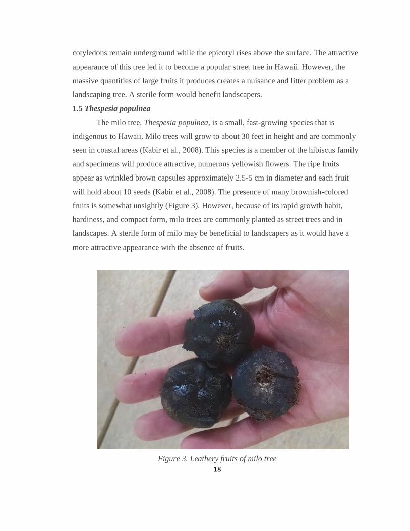
1.5 Thespesia populnea
The milo tree, Thespesia populnea, is a small, fast-growing species that is
indigenous to Hawaii. Milo trees will grow to about 30 feet in height and are commonly
seen in coastal areas (Kabir et al., 2008). This species is a member of the hibiscus family
and specimens will produce attractive, numerous yellowish flowers. The ripe fruits
appear as wrinkled brown capsules approximately 2.5-5 cm in diameter and each fruit
will hold about 10 seeds (Kabir et al., 2008). The presence of many brownish-colored
fruits is somewhat unsightly (Figure 3). However, because of its rapid growth habit,
hardiness, and compact form, milo trees are commonly planted as street trees and in
landscapes. A sterile form of milo may be beneficial to landscapers as it would have a
more attractive appearance with the absence of fruits.
Figure 3. Leathery fruits of milo tree

19
1.6 Clusia rosea
The autograph tree, Clusia rosea, is a medium-sized tree that is native to tropical
America (Starr et al., 2003). This member of the mangosteen family gets its name from
its thick, long-lasting leaves that can be engraved. This species was originally brought to
Hawaii as an ornamental. It is attractive and hardy and as a result, is commonly used in
public and private landscapes. The tree produces large seed capsules which split open
when ripe (Figure 4). The small seeds are dispersed by birds. This species can be either
terrestrial or epiphytic. Seeds often germinate on host plant and compete to eventually
kill the host (Motooka et. al., 2003). Unfortunately, its rapid growth and prolific seed-
producing capabilities has enabled this species to become invasive in Hawaii. A sterile
form of the autograph tree would benefit landscapers since the seed pods make for high
maintenance. A sterile form would also benefit the environment.
Figure 4. Mature autograph tree (left) and its seed capsule (right). Photos from specimen at University of Hawaii at Mānoa.

20
1.7 Calophyllum inophyllum
Kamani, Calophyllum inophyllum, is a medium-sized tree that can be found
throughout the tropics. It is known for its attractive, ornamental appearance and
hardiness. In Hawaii, kamani is commonly found in coastal areas and in urban and
private landscapes. Its tolerance to drought, salt, and wind make it extremely hardy and
rather desirable to the horticulture industry. This species has a lush appearance and
fragrant flowers. This species undergoes hypogeal germination, meaning the cotyledons
remain underground while the epicotyl rises above the surface. In Hawaii, the kamani
tree is known to flower and fruit twice per year (Prabakaran and Britto, 2012). Fruits are
about the size of a golf ball and trees will yield prolific amounts of fruits (Figure 5). The
fallen fruit can become an eyesore and also a trip hazard when it is present on walkways.
A sterile form of kamani may greatly benefit landscapers.

21
Figure 5. Mature kamani tree (top) and its fruits (bottom). Photos from specimen at
University of Hawaii at Mānoa.
1.8 Jatropha curcas
The physic nut, Jatropha curcas, holds a lot of potential as an energy crop. Native
to the tropical Americas, this tree is now grown throughout the world. The tree is fast
growing and somewhat draught tolerant. The seed has a significant oil content, which can
be converted into high-quality biodiesel. Much research has been done to study this
species and a number of cultivars have been identified. In many cases, polyploidy can
22
lead to an increase in fruit and seed size, and produce higher levels of chemical
compounds. Henderson (1977) demonstrated that tetraploid watermelons produced larger
seeds than diploids. Gao et al. (1996) reported that the concentration of major chemical
compounds in tetraploid Salvia miltiorrhiza was higher than their diploid counterparts.
Polyploid cereals have been shown to produce larger seed size and higher protein content
(Dhawan and Lavania, 1996). A tetraploid physic nut may bear seeds of larger size and
higher oil content, and may also be useful for breeding purposes. Piromya and Kermanee
(2013) demonstrated that wool soaked with 5000-6000 ppm colchicine applied to the
meristems successfully doubled the chromosome number in Jatropha. Oliveira et al.
(2013) reported the occurrence of tetraploids and mixoploids when shoot tips were
treated with 0.5 mM colchicine for 96 hours in vitro.
1.9 Plumeria stenopetala
Plumeria are attractive, fragrant flowering trees that originate in Central America
and the Caribbean. In Hawaii, these plants are economically important and contribute
about $500,000 annually to the local flower industry (Nelson, 2009). The plants are used
for both landscaping and for producing leis. However in 1991, a serious pathogen called
plumeria rust became established in the state (Nelson, 2009). This fungal disease attacks
the foliage of plumeria plants and causes considerable decline in growth. Since its
introduction, certain plumeria species have been identified as resistant to the disease.
Among these is Plumeria stenopetala. This species produces thin, white flowers.
Doubling the chromosomes of this species to make a tetraploid variety may be useful in
breeding. If crossed with a diploid susceptible variety, the offspring would be triploid
with 2 sets of alleles coming from the resistant species.
1.10 Erythrina sandwicensis
The wiliwili, Erythrina sandwicensis, is tree in the pea family that is endemic to
the Hawaiian Islands. It has a gnarled growth habit and it makes for an attractive
ornamental tree. It had cultural significance to the ancient Hawaiians, who used the wood
for fishing floats, and the bright orange seeds in leis. One variety of a related species,
23
Erythrina variegata, was commonly used as a windbreak. However in 2005, a new
species of gall wasp, Quadrastichus erythrinae, was discovered in Hawaii (Xu et al.,
2006). It is believed that the wasp originated from East Africa. The wasp parasitizes the
trees and as a result, a large portion of the Erythrina specimens in Hawaii have died.
Polyploids generally have thicker epidermal tissue and this characteristic makes many
plants less susceptible to herbivory (Woodman and Fernandes, 1991). Thicker vascular
tissue, which is derived from the L3 meristematic layer, may also deter the wasp since
that is the tissue that their young feed on. We hypothesize that a parasitic wasp, such as
the gall wasp, may also find a tetraploid Wiliwili less desirable.
24
CHAPTER 2. MATERIALS AND METHODS
2.1 General Methods
In general, seeds of each of the 8 species were first planted into 38-cell treys of a
50/50 mixture of perlite and peat moss. Oryzalin was the sole mitotic inhibitor used in
this project and concentrations ranged from 0.01-0.5%. In most of the treatments,
oryzalin was mixed with Phytagel. Like agar, this gelling agent forms a liquid solution
when heated and solidifies when cooled back to room temperature. Dimethyl sulfoxide
(DMSO) was added to the solution. This is a solvent that dissolves both polar and
nonpolar compounds. It has the ability to penetrate tissue. Treatments were generally
applied to seedlings between the cotyledons as soon as they opened (Figure 6). During
this stage, a drop of the heated treatment solution was placed onto the apical meristem
between the two cotyledons using a dropper. As it cools, it solidifies to remain in place.
Control seedlings were treated with a Phytagel/DMSO solution that lacked oryzalin.
Plastic containers were placed over all seedlings to prevent the gel solution from drying
out. For experiments that had treatment durations of 48 hours or longer, the treatment and
control solutions were removed and reapplied every 24 hours.

25
Two of the species, Heritiera littoralis and Calophyllum inophyllum, used
alternative methods of treatment application. Since the opening of the cotyledons and
emergence of the meristem occurs underground in these species, apical meristems were
not treated with oryzalin. Instead, lateral meristems were treated. Seeds were germinated
and allowed to grow to about 15 cm. At this point, seedlings were cut with a sharp
scissors to about one 2 cm above the first node. Lateral meristems were then treated
immediately after cutting. In Heritiera littoralis, treatment was applied as above using
Phytagel. In Calophyllum inophyllum, treatment was applied as a mist in the absence of
Phytagel.
Treated seedlings were observed every 24 hours and data on germination,
survival, and morphological observations were recorded. After new growth occurred,
guard cell measurements were made to select potential polyploids. However in Heritiera
littoralis, guard cells could not be observed due to the fuzzy underside of the leaves. All
treated seedlings were screened with flow cytometry to determine ploidy level. The
following indicates the specifics of the treatments of each species.
Figure 6. Application of oryzalin treatment solution to apical meristems of seedlings (left). Solidified solution sitting between cotyledons (right).
26
2.2 Schefflera actinophylla
In this experiment, 152 seeds were collected from University of Hawaii at Mānoa
campus of which 32 were randomly designated as controls and 120 were designated for
treatments. The 120 seeds used for treatments were divided into 2 groups of 60 seeds per
treatment.
Treatment #1:
-4 g/L Phytagel
-0.01% oryzalin
-1% dimethyl sulfoxide (DMSO)
Duration = 24 hours.
Treated tissue: apical meristem
Treatment #2
-4 g/L Phytagel
-0.01% oryzalin
-1% dimethyl sulfoxide (DMSO)
Duration = 48 hours.
Treated tissue: apical meristem
2.3 Heritiera littoralis
In this experiment, 150 seeds collected from specimens on Mānoa Road,
Honolulu, Hawaii were planted. Among these, 40 were randomly designated as controls
and 110 were designated as treatments.
Treatment:
-4 g/L Phytagel
-0.5% oryzalin
-1% dimethyl sulfoxide (DMSO)
Duration = 48 hours.
Treated tissue: lateral meristem

27
2.4 Thespesia populnea:
In this experiment, 76 seeds collected from specimens on Lagoon Drive near the
Honolulu International Airport were scarified and planted. Among these, 16 were
randomly designated as controls and 60 were designated for treatments.
Treatment #1
-4 g/L Phytagel
-0.1% oryzalin
-1% dimethyl sulfoxide (DMSO)
Duration = 48 hours.
Treated tissue: apical meristem
This experiment was also repeated using a total of 76 seeds (60 designated for treatments
and 16 for controls) with the following treatment:
Treatment #2
-4 g/L Phytagel
-0.5% oryzalin
-1% dimethyl sulfoxide (DMSO)
Duration = 48 hours.
Treated tissue: apical meristem
2.5 Polyploidization of Clusia rosea
In this experiment, 152 seeds collected from a specimen outside of Bachman Hall,
University of Hawaii, were planted. Among these, 32 were randomly designated as
controls and 120 were designated as treatments. The 120 treatment seeds were divided
into two treatments of 60 seeds per treatment.
Treatment #1:
-4 g/L Phytagel
-0.5% oryzalin

28
-1% dimethyl sulfoxide (DMSO)
Duration = 48 hours.
Treated tissue: apical meristem
Treatment #2
-4 g/L Phytagel
-0.5% oryzalin
-1% dimethyl sulfoxide (DMSO)
Duration = 96 hours.
Treated tissue: apical meristem
2.6 Calophyllum inophyllum
In this experiment, 76 seeds collected from specimens at the University of Hawaii
were scarified and planted. Among these, 16 were randomly designated as controls and
60 were designated as treatment. The 60 treatment seeds were broken up into 2
treatments of 30 seeds per treatment:
Treatment #1:
-0.1% oryzalin
-1% dimethyl sulfoxide (DMSO)
-1mL/L Polysorbate (Tween)
Duration = mist once/day for 2 days
Treated tissue: lateral meristem
Treatment #2
-0.1% oryzalin
-1% dimethyl sulfoxide (DMSO)
-1mL/L Polysorbate (Tween)
Duration = mist once/day for 4 days
Treated tissue: lateral meristem
29
This experiment was also repeated using a total of 76 seeds (60 designated for treatments
and 16 for controls) with the following treatment:
Treatment #3
-0.5% oryzalin
-1% dimethyl sulfoxide (DMSO)
-1mL/L Polysorbate (Tween)
Duration = mist once/day for 20 days
Treated tissue: Lateral meristem
2.7 Jatropha curcas
In this project, seeds of three populations of physic nut were planted. A
total of 38 seeds of each population were planted in an equal mix of perlite and peat
moss. Among those, 8 were designated as controls and 30 designated as treatments.
Treatment:
-4 g/L Phytagel
-0.1% oryzalin
-1% dimethyl sulfoxide (DMSO)
Duration = 48 hours.
Treated tissue: apical meristem
2.8 Plumeria stenopetala
In this experiment, 152 seeds collected from a specimen at the University of
Hawaii Waimanalo Research Station were sown. Among these, 32 were randomly
designated as controls and 120 were designated as treatments. The 120 treatment seeds
were broken up into 2 groups of 60 for 2 treatments
Treatment #1:
-4 g/L Phytagel
-0.01% oryzalin
-1% dimethyl sulfoxide (DMSO)
Duration = 24 hours.

30
Treated tissue: apical meriste
Treatment #2
-4 g/L Phytagel
-0.01% oryzalin
-1% dimethyl sulfoxide (DMSO)
Duration = 48 hours.
Treated tissue: apical meristem
2.9 Erythrina sandwicensis
In this experiment, 152 seeds collected from a specimen at Koko Head
Crater, Oahu, from 2005, were scarified and planted. Among these, 32 were randomly
designated as controls and 120 were designated as treatments.
Treatment:
-4 g/L Phytagel
-0.1% oryzalin
-1% dimethyl sulfoxide (DMSO)
Duration = 48 hours.
2.10 Guard Cell Measurements
Cells of polyploids are typically larger in size due to the presence of additional
DNA (Leonhardt and Shi, 2007). Guard cells make up the stomata of leaves and are
typically found on the undersides of leaves in dicots. To measure the guard cells, one
fully expanded leaf is first removed from a plant. The underside of the leaf is then wiped
to remove any moisture or debris. A light layer of clear nail polish is then applied to the
underside of the leaf so that it covers approximately 1 square cm of surface area. After
allowing it to dry, a piece of transparent tape is then pressed over the nail polish and
removed. The tape lifts the imprint of the guard cells made by the nail polish. The tape is
then stuck to a microscope slide, which is analyzed at 400X magnification. A micrometer
in the microscope ocular is used to take guard cell measurements. Five random guard

31
cells are measured per leaf sample. A potential polyploid is identified if at least 3 of the 5
cells are 1.25 times larger than the average of the control guard cells.
2.11 Flow Cytometry
All flow cytometry samples were prepared using supplies from Cystain PI
Absolute P DNA Staining Kit for Plant Genome Size (Partec; Munster, Germany).
Approximately 2 square cm of leaf tissue is placed in a petri dish. 500 µl of the extraction
buffer is added and the leaf sample is chopped for about one minute using a sharp razor
blade. Razors are used to prepare only two samples and then replaced because it is
essential that the razor is very sharp (Teng, 2007). The solution is then passed through a
fine 50-µm filter into a test tube. 2 mL of the staining solution (consisting of a staining
buffer, propidium iodide and RNAse A) is added. Samples are then stored at 4°C for 12
hours in the dark. After this, they are analyzed by the flow cytometer (Beckman Coulter
XL), and the data files examined for the nuclei fluorescence intensity.

32
CHAPTER 3 RESULTS:
Species Germination
Rate (%)
No. Days to
Germination
No. Days to
Treatment
Schefflera
actinophylla
95 16 18
Heritiera littoralis 93.4 72 111
Thespesia populnea 43 10 12
Clusia rosea 19 35 39
Calophyllum
inophyllum
95 24 27
Jatropha curcas 85 6 7
Plumeria
stenopetala
73 12 13
Erythrina
sandwicensis
82 7 8
Table 1. Germination Data of all species

33
Figure 7. Percent seedling survival after 5 months
34
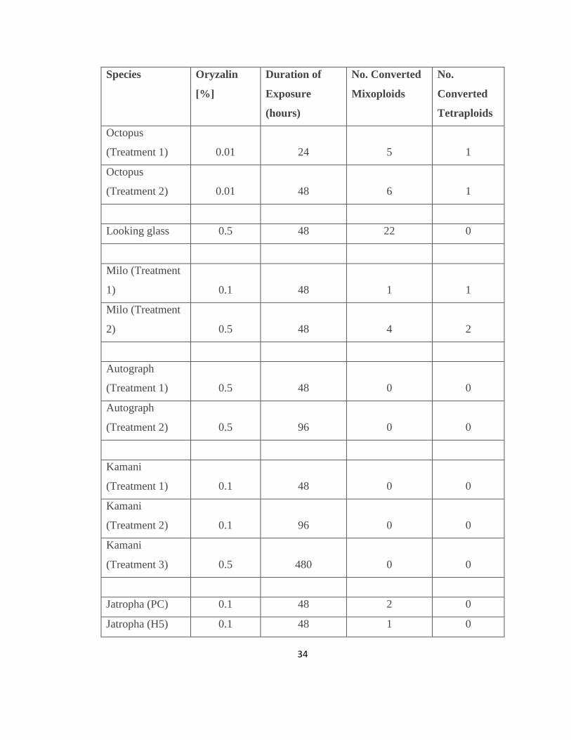
Species Oryzalin
[%]
Duration of
Exposure
(hours)
No. Converted
Mixoploids
No.
Converted
Tetraploids
Octopus
(Treatment 1) 0.01 24 5 1
Octopus
(Treatment 2) 0.01 48 6 1
Looking glass 0.5 48 22 0
Milo (Treatment
1) 0.1 48 1 1
Milo (Treatment
2) 0.5 48 4 2
Autograph
(Treatment 1) 0.5 48 0 0
Autograph
(Treatment 2) 0.5 96 0 0
Kamani
(Treatment 1) 0.1 48 0 0
Kamani
(Treatment 2) 0.1 96 0 0
Kamani
(Treatment 3) 0.5 480 0 0
Jatropha (PC) 0.1 48 2 0
Jatropha (H5) 0.1 48 1 0
35
Jatropha (H2) 0.1 48 3 0
Plumeria S.
(Treatment 1) 0.01 24 5 3
Plumeria S.
(Treatment 2) 0.01 48 3 3
Erythrina 0.1 48 2 0
Table 2. Polyploid initiation data of all species
0
10
20
30
40
50
60
70
Polyploid Conversion
Converted mixoploids (%) Converted tetraploids (%)
Figure 8. Results of all experiments, indicating the percent of converted mixoploids and tetraploids.
36
3.1 Schefflera actinophylla
Schefflera actinophylla seeds germinated at a rate of 95%. Germination time
averaged 16 days. Treatments began 18 days after planting. Treated seedlings grew at a
noticeably slower rate than controls. Five months after planting, 68 of the 114 treated
seedlings remained alive (35 from Treatment 1 and 33 from Treatment 2) (Figure 7).
During this time, guard cell measurements were recorded (Figure 9). Guard cells of
control seedlings had an average measurement of 3.23 arbitrary units. Guard cells from
18 treated seedling (9 seedlings from Treatment 1 and 9 seedlings from Treatment 2)
measured greater than 4 units. Flow cytometry was performed on 3 controls and all
treated seedlings (Figure 10). Flow cytometry histograms depicting the ploidy level were
generated (Figure 6). The diploid peak was located at an average of 58.2 arbitrary units
and the tetraploid peak at an average of 114.8. Flow cytometry confirmed a total of 13
polyploids; 11 mixoploids and 2 tetraploids. Among the seedlings in Treatment 1, 5
mixoploids and one tetraploid were discovered (Table 2). Among the seedlings in
Treatment 2, 6 mixoploids and one tetraploid were identified. Polyploids appeared
stunted and had tougher leaves (Figure 11). A chi square test indicated that there was no
significant association between the number of polyploids created and the duration of the
treatments.

37
Figure 9. Guard cells from octopus tree seedlings under 200 X magnification. Top photo
shows diploid cells, tetraploid cells in the bottom.
38
Figure 10. Histograms produced from flow cytometry of octopus seedlings. Top left histogram from mixoploid, top right from tetraploid, bottom right from diploid control.
39
Figure 11. Diploid control octopus seedling (left) and tetraploid (right). The tetraploid
has darker green foliage and tougher leaves.
40
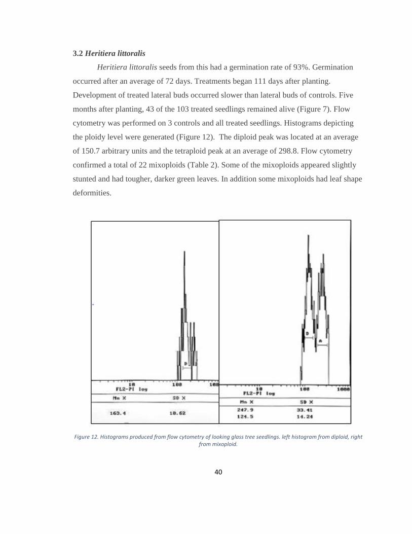
3.2 Heritiera littoralis
Heritiera littoralis seeds from this had a germination rate of 93%. Germination
occurred after an average of 72 days. Treatments began 111 days after planting.
Development of treated lateral buds occurred slower than lateral buds of controls. Five
months after planting, 43 of the 103 treated seedlings remained alive (Figure 7). Flow
cytometry was performed on 3 controls and all treated seedlings. Histograms depicting
the ploidy level were generated (Figure 12). The diploid peak was located at an average
of 150.7 arbitrary units and the tetraploid peak at an average of 298.8. Flow cytometry
confirmed a total of 22 mixoploids (Table 2). Some of the mixoploids appeared slightly
stunted and had tougher, darker green leaves. In addition some mixoploids had leaf shape
deformities.
Figure 12. Histograms produced from flow cytometry of looking glass tree seedlings. left histogram from diploid, right from mixoploid.

41
3.3 Thespesia populnea
Thespesia populnea seeds had a germination rate of 43%. Germination occurred
after an average of 10 days. Treatments began 12 days after planting. Treated seedlings
grew at a noticeably slower rate than controls. Five months after planting, 23 of the 52
treated seedlings remained alive (14 from Treatment 1 and 9 from Treatment 2) (Figure
7). During this time, guard cell measurements were recorded (Figure 13). Guard cells of
control seedlings had an average measurement of 2.89 arbitrary units. Guard cells from
12 treated seedling (4 seedlings from Treatment 1 and 8 seedlings from Treatment 2)
measured greater than 3.6 units. Flow cytometry was performed on 3 controls and all
treated seedlings. Histograms depicting the ploidy level were generated (Figure 14). The
diploid peak was located at an average of 69.3 arbitrary units and the tetraploid peak at an
average of 158.6. Flow cytometry confirmed a total of 8 polyploids; 5 mixoploids and 3
tetraploids. Among the seedlings in Treatment 1, 1 mixoploid and 1 tetraploid were
discovered (Table 2). Among the seedlings in Treatment 2, 4 mixoploids and 2
tetraploids were identified. Polyploids had little difference in morphological
characteristic compared to controls. However, both tetraploids had noticeably darker
green, rougher leaves (Figure 15). A chi square test indicated that there was a significant
association between the number of polyploids created and the concentration of the
treatments. The seedlings treated with 0.5% oryzalin yielded a significantly higher
proportion of polyploids than those treated with 0.1% oryzalin.
42
Figure 13. Guard cells from milo tree seedlings under 200 X magnification. Top photo
shows diploid cells, tetraploid cells in the bottom.

43
Figure 14. Histograms produced from flow cytometry of milo tree seedlings. left histogram from tetraploid, right from mixoploid.
44
Figure 15. Diploid control milo seedling (right) and tetraploid (left). The tetraploid is
stunted and has rougher leaves.
3.4 Clusia rosea
Clusia rosea seeds had a germination rate of 19%. Germination occurred after an
average of 35 days. Treatments began 39 days after planting. There was no visual
difference in growth rate between treated and control seedlings. Five months after
planting, 23 of the 23 treated seedlings remained alive (13 from Treatment 1 and 10 from
Treatment 2) (Figure 7). Flow cytometry was performed on 3 controls and all treated
seedlings. Histograms depicting the ploidy level were generated (Figure 16). The diploid

45
peak was located at an average of 61.5 arbitrary units. All samples were identified as
diploid.
Figure 16. Histogram produced from flow cytometry of diploid autograph tree.
3.5 Calophyllum inophyllum
Calophyllum inophyllum seeds had a germination rate of 95%. Germination
occurred after an average of 24 days. Treatments began 27 days after planting. Treated
seedlings grew at a noticeably slower rate than controls. However, they quickly recovered
vigor after treatments were discontinued. Many of the seedlings from Treatment 3
showed deformed leaf morphology including thicker leaves and various deformed leaf
shapes. Five months after planting, 114 of the 114 treated seedlings remained alive
(Figure 7). During this time, guard cell measurements were recorded (Figure 17). Guard
cells of control seedlings had an average measurement of 2.04 arbitrary units. No
treatments had guard cell measurements greater than an average of 2.55 units. Flow
46
cytometry was performed on 3 controls and treated seedlings displaying leaf deformities.
Histograms depicting the ploidy level were generated (Figure 18). The diploid peak could
not be determined because no nuclei were visible.
Figure 17. Diploid guard cell from kamani tree seedling under 200 X magnification.

47
3.6 Jatropha curcas
Jatropha curcas seeds had a germination rate of 85.6%. Germination occurred
after an average of 6 days. Treatments began 7 days after planting. Treated seedlings
grew at a noticeably slower rate than controls. Five months after planting, 25 of the 77
treated seedlings remained alive (9 from PC, 5 from H5, and 11 from H2) (Figure 7).
During this time, guard cell measurements were recorded (Figure 19). Guard cells of
control seedlings had an average measurement of 3.02 arbitrary units. Guard cells from
10 treated seedling (4 seedlings from PC 1 seedlings from H5 and 4 seedlings from H2)
measured greater than 3.8 units. Flow cytometry was performed on 3 controls and all
treated seedlings. Histograms depicting the ploidy level were generated (Figure 20). The
diploid peak was located at an average of 63.1 arbitrary units and the tetraploid peak at an
. Figure 18. Histogram produced from flow cytometry
of presumably diploid kamani tree. Location of diploid peak is uncertain due to large quantity of
background noise
48
average of 136.9. Flow cytometry confirmed a total of 6 mixoploids (2 from PC, 1 from
H5, and 3 from H2) (Table 2). Polyploids had slightly tougher leaves.
Figure 19. Guard cells from Jatropha tree seedlings under 200 X magnification. Top
photo shows diploid cells, tetraploid cells in the bottom.
49
Figure 20. Histograms produced from flow cytometry of Jatropha tree seedlings. left
histogram from diploid, right from mixoploid.
3.6 Plumeria stenopetala
Plumeria stenopetala seeds had a germination rate of 73%. Germination occurred
after an average of 12 days. Treatments began 13 days after planting. Treated seedlings
grew at a noticeably slower rate than controls. Five months after planting, 26 of the 88
treated seedlings remained alive (13 from Treatment 1 and 13 from Treatment 2) (Figure
7). During this time, guard cell measurements were recorded (Figure 21). Guard cells of
control seedlings had an average measurement of 2.53 arbitrary units. Guard cells from
16 treated seedling (7 seedlings from Treatment 1 and 9 seedlings from Treatment 2)
measured greater than 3.16 units. Flow cytometry was performed on 3 controls and all
treated seedlings. Histograms depicting the ploidy level were generated (Figure 22). The
diploid peak was located at an average of 18.9 arbitrary units and the tetraploid peak at an
average of 38.32. Flow cytometry confirmed a total of 14 polyploids; 8 mixoploids and 6
tetraploids (Table 2). Among the seedlings in Treatment 1, 5 mixoploids and 3 tetraploids
were discovered. Among the seedlings in Treatment 2, 3 mixoploids and 3 tetraploid
50
were identified. Polyploids appeared stunted and had tougher leaves. One of the
tetraploids also had more rounded leaves (Figure 23). A chi square test indicated that
there was no significant association between the number of polyploids created and the
duration of the treatments.
Figure 21. Guard cells from Plumeria stenopetala seedlings under 200 X magnification. Top photo shows diploid cells, tetraploid cells in the bottom.
Figure 22. Percent seedling survival after 5 monthsFigure 23. Guard cells from Plumeria stenopetala seedlings under 200 X magnification. Top photo shows diploid cells, tetraploid cells in the bottom.
51
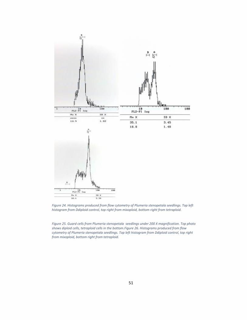
Figure 24. Histograms produced from flow cytometry of Plumeria stenopetala seedlings. Top left histogram from Ddiploid control, top right from mixoploid, bottom right from tetraploid.
Figure 25. Guard cells from Plumeria stenopetala seedlings under 200 X magnification. Top photo shows diploid cells, tetraploid cells in the bottom.Figure 26. Histograms produced from flow cytometry of Plumeria stenopetala seedlings. Top left histogram from Ddiploid control, top right from mixoploid, bottom right from tetraploid.
52
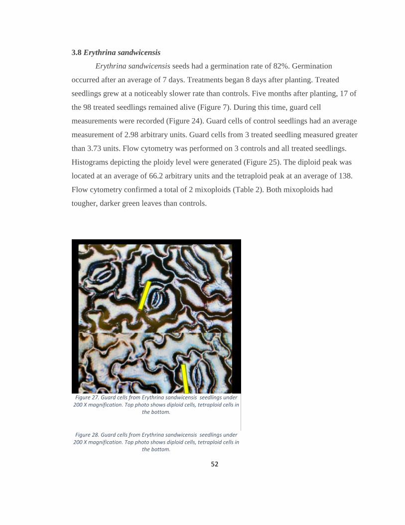
3.8 Erythrina sandwicensis
Erythrina sandwicensis seeds had a germination rate of 82%. Germination
occurred after an average of 7 days. Treatments began 8 days after planting. Treated
seedlings grew at a noticeably slower rate than controls. Five months after planting, 17 of
the 98 treated seedlings remained alive (Figure 7). During this time, guard cell
measurements were recorded (Figure 24). Guard cells of control seedlings had an average
measurement of 2.98 arbitrary units. Guard cells from 3 treated seedling measured greater
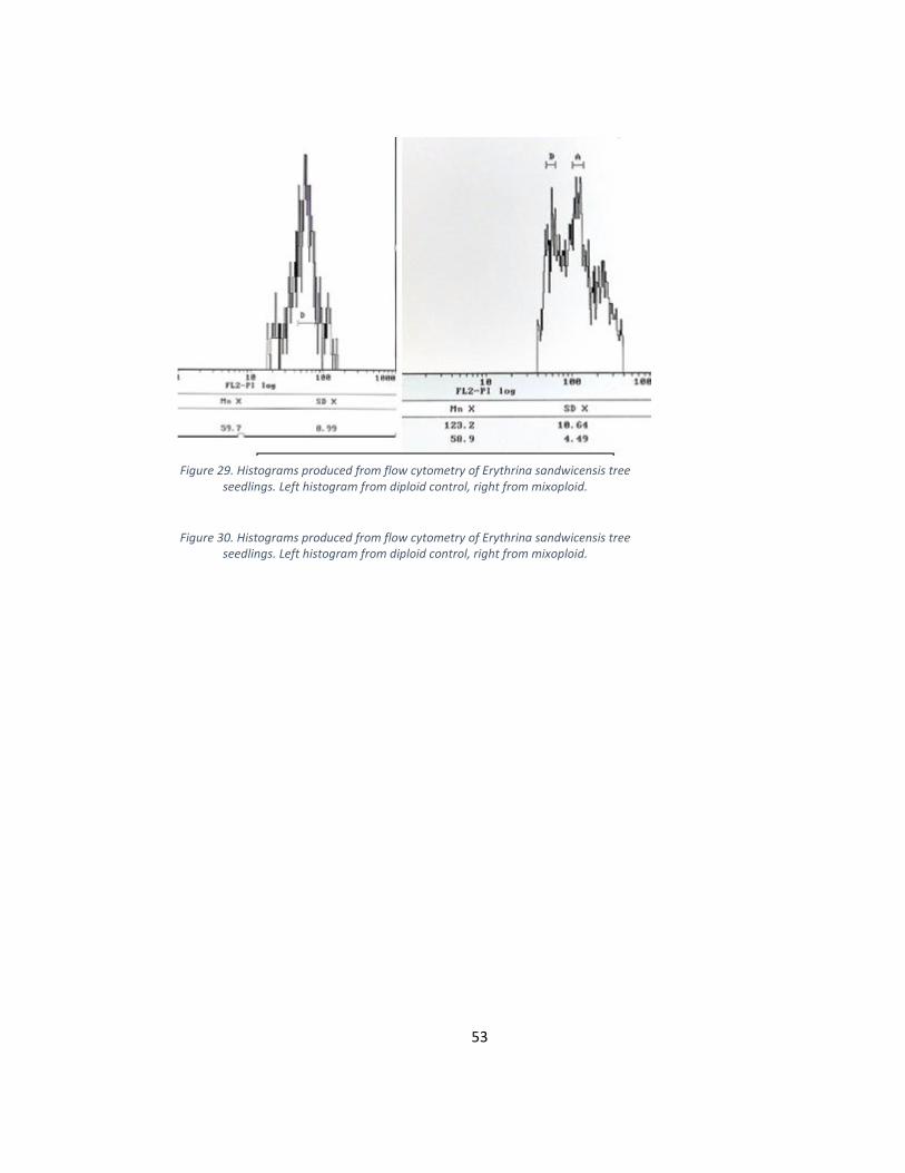
than 3.73 units. Flow cytometry was performed on 3 controls and all treated seedlings.
Histograms depicting the ploidy level were generated (Figure 25). The diploid peak was
located at an average of 66.2 arbitrary units and the tetraploid peak at an average of 138.
Flow cytometry confirmed a total of 2 mixoploids (Table 2). Both mixoploids had
tougher, darker green leaves than controls.
Figure 27. Guard cells from Erythrina sandwicensis seedlings under 200 X magnification. Top photo shows diploid cells, tetraploid cells in
the bottom.
Figure 28. Guard cells from Erythrina sandwicensis seedlings under
200 X magnification. Top photo shows diploid cells, tetraploid cells in the bottom.
53
Figure 29. Histograms produced from flow cytometry of Erythrina sandwicensis tree seedlings. Left histogram from diploid control, right from mixoploid.
Figure 30. Histograms produced from flow cytometry of Erythrina sandwicensis tree
seedlings. Left histogram from diploid control, right from mixoploid.

54
CHAPTER 4. DISCUSSION
4.1 Schefflera actinophylla
Not surprisingly, octopus tree seeds germinated quickly and had high germination
rates. This is one of the traits that make this species invasive. More than half of the
treated seedlings survived their oryzalin application and 19.1% of them became
polyploids. Octopus seedlings had an ideal morphology for polyploid conversion since
the apical meristem was undeveloped upon opening of the cotyledons. Minimally
developed meristems are optimal for conversion because it allows for most of the
dividing, undifferentiated cells to be exposed to the mitotic inhibitor. As a result, the
tissue derived from the treated meristem is more likely to be polyploid. There was no
significant difference in the number of converted seedlings between treatments 1 and 2.
Both treatments were successful in converting to polyploids. This could be a result of
Schefflera actinophylla’s rapid growth rate. In this case, a 24 hour exposure to oryzalin
was sufficient to come in contact with a significant portion of the meristematic cells
during division. The flow cytometry graphs were clean and easy to interpret, indicating
that the leaf tissue of this species contains a minimal amount of secondary compounds
that can be stained by propidium iodide. In some species, such cellular debris can cause
background noise in the flow cytometer results. As it is often observed in polyploids of
other species, Schefflera actinophylla polyploids displayed unique morphological
characteristics such as tougher leaves that were darker green. A sterile octopus tree would
be an excellent addition to the landscaping trees of Hawaii and many other tropical
regions where it is invasive. The tetraploids and chimeric mixoploids developed in this
experiment will continue to be monitored with flow cytometry to observe any reversion
or shift in ploidy level. After the polyploid specimens reach maturity, fertility
observations will be made. If still fertile, polyploid flowers will be cross-pollinated with
diploid flowers and seeds will be planted. If the L2 layer of the original treatments was
converted, the progeny should be triploid and presumably sterile.
55
4.2 Heritiera littoralis
Looking glass seeds had a higher germination rate despite the lengthy amount of
time required to germinate. A large amount, 51.2% of the treated seedlings became
polyploids. However, all 22 of these polyploids were mixoploids; there were no
tetraploids. Looking glass seedlings have hypogeal germination so the cotyledons remain
underground while the epicotyl rises above the surface. Therefore, apical meristems
could not be treated in this experiment because they are very well developed by the time
they emerge from the soil. As a result, lateral buds were treated as soon as the apical
meristems were removed. This method gave access to fairly undeveloped meristem tissue
was sufficient to capture a significant portion of the meristematic cells during their
division. Of all the species used in this project, the looking glass had the highest
polyploid conversion rate. This might suggest that the oryzalin/DMSO solution easily
penetrated to the nuclei of the cells and that the microtubules had a high binding affinity
to oryzalin. However flow cytometry revealed that all of the converted polyploids were
mixoploids. There were no tetraploids. This could indicate that the lateral meristems are
well-enough developed that the treatment was not able to penetrate though all of the
meristematic cell layers. The flow cytometry graphs were clean and easy to interpret. As
it is often observed in polyploids of other species, Heritiera littoralis polyploids
displayed unique morphological characteristics such as tougher leaves that were darker
green in color. In addition, some mixoploids displayed deformed leaf shapes. These are
typical characteristics of polyploids. A looking glass tree would make a terrific addition
to the landscaping trees of Hawaii since it would reduce the amount of fruit litter, which
not only creates an unsightly appearance, but also poses a trip hazard. The chimeric
mixoploids created in this experiment will continue to be monitored with flow cytometry
to observe any reversion or shift in ploidy level. Ideally, at least some of the mixoploids
developed will have been converted in their L2 layer. After the polyploid specimens
reach maturity, fertility observations will be taken. If still fertile, polyploid flowers will
be cross-pollinated with diploid flowers and seeds will be planted. If the L2 layer of the
original treatments was converted, hybrid progeny should be triploid and presumably
sterile.
56
4.3 Thespesia populnea
Germination of Thespesia populnea seeds was relatively low. Despite the
freshness of the seeds and their scarification, less than half of them germinated. In
addition, more than half of the germinated seedlings died after their oryzalin application.
Despite low germination and high seedling mortality, some tetraploids resulted. The
morphology of milo seedlings was ideal for such treatments because the apical meristem
was undeveloped upon opening of the cotyledons. Minimally developed meristems are
optimal for conversion because it allows for most of the dividing, undifferentiated cells to
be exposed to the mitotic inhibitor. As a result, the tissue derived from the treated
meristem is more likely to be polyploid. There was a significant difference in the number
of converted seedlings between Treatments 1 and 2. A 0.5% oryzalin concentration was
more efficient at converting polyploids than a 0.1% concentration. When attempting to
convert diploids to polyploids it is important to consider that the required concentration
of mitotic inhibitor can vary between species. In this case, milo requires a higher
concentration than many other species. The flow cytometry graphs were clean and easy to
interpret, indicating that the leaf tissue of this species contains a minimal amount of small
secondary compounds that can be stained with propidium iodide. Although initial growth
of polyploid seedlings was slower than controls, the polyploids generated in this
experiment generally did not display many morphological differences. The tetraploids
displayed the only visual differences of darker green leaves. One possible explanation for
this observation is that the L1 layer of the mixoploids may not have been doubled in
chromosome number. This cellular layer gives rise to the epidermal tissue, which is the
part of the plant that can be visually observed. If only the L2 and/or L3 layers were
converted there would be fewer observable indications of higher ploidy level. On the
other hand, a tetraploid has all 3 layers of its cellular layers converted to the higher ploidy
level. Therefore, it makes sense for those specimens to have visual differences in leaf
characteristics. A sterile milo tree would be a major improvement to landscaping in
Hawaii. The absence of its brown fruits will enhance its aesthetic qualities and reduce
fruit trash. The tetraploids and chimeric mixoploids will continue to be monitored with
flow cytometry to observe any reversion or shift in ploidy level. After the polyploid
57
specimens reach maturity, fertility observations will be made. If still fertile, polyploid
flowers will be cross-pollinated with diploid flowers and seeds will be planted, with the
expectation of triploid seedlings.
4.4 Clusia rosea
Interestingly, Clusia rosea seeds had a very low germination rates followed by
very slow initial growth of both controls and treatments. These are unusual characteristics
for invasive species, which typically exhibit high germination rates and rapid growth.
Perhaps the seeds came from an inferior tree. Or perhaps the growing conditions were
suboptimal The oryzalin treatments seemed to have little impact on these autograph
seedlings. Most of the seedlings remained alive 5 months after treatments and no diploids
were converted. The flow cytometry graphs were clean and easy to interpret, indicating
that the leaf tissue of this species contains a minimal amount of secondary compounds
that can be stained with propidium iodide Despite the poor results, the seedling did
exhibit an ideal morphology for polyploid conversion because the apical meristems were
undeveloped upon opening of the cotyledons. However, the treatments still failed to have
any effect on ploidy level. One probable explanation is that the failure was a result of the
slow growth rate of autograph seedlings in this experiment. In order for a mitotic
inhibitor to induce its mode of action, the cells must be exposed while actively dividing.
While meristematic tissue is the ideal place to find dividing cells, the cells in these
seedlings may have been dividing a pace too slow to be effected by short exposures to
oryzalin. This observation was made in preliminary trials so longer durations of 48 and
96 hour treatments were used in this experiment. Despite the increased durations of
exposure, oryzalin application may still have been too brief. In future polyploid
conversion attempts for this species, it may be advisable to increase the duration of
exposure to greater than 96 hours, or create an environment for more rapid seedling
growth.
4.5 Calophyllum inophyllum
The germination rate on the kamani seeds was strong and seedlings proved to be
very vigorous even in spite of high concentrations and long durations of oryzalin. None
of the seedling died from toxicity to oryzalin and no obvious polyploids were initiated.
58
Since kamani seeds exhibit hypogeal germination, the apical meristem is very well
developed by the time it emerges from the soil surface and is not suitable for treatment.
Apical meristems were cut to allow for the treatment of undeveloped lateral meristems.
However, the seedlings in this experiment proved to be very resistant to oryzalin. No
potential polyploids were identified from guard cell measurements. Some seedlings
exposed to Treatment 3 did show leaf deformities that are typical of polyploids.
However, because they showed no difference in guard cell measurements, they are
unlikely to be converted. The physical differences might be a result of the plants’
response to long exposures of a foreign chemical. Despite no difference in guard cell
measurements, any seedling displaying morphological differences was tested with flow
cytometry. However, flow cytometry failed to give good results because there was too
much background noise in the graphs. Such noise is typically caused by the presence of
secondary cellular compounds that are also stained by PI. It appears that Calophyllum
inophyllum cells have a high level of these cellular secondary compounds.
All treated seedlings recovered from the herbicide treatment suggesting that
oryzalin has little effect on the cellular division of kamani. Future work might include
testing other mitotic inhibitors such as colchicine.
4.6 Jatropha curcas
Jatropha curcas seeds exhibited a high germination rate and seedlings grew
rapidly. Jatropha seedlings had an ideal morphology for polyploid conversion because the
apical meristem was undeveloped upon opening of the cotyledons. Jatropha seedlings
were relatively sensitive to oryzalin since there was a high mortality rate following
treatments. A good amount of the surviving treated seedlings were converted to
polyploids. However, all of the polyploids were mixoploids. The flow cytometry graphs
were clean and easy to interpret, indicating that the leaf tissue of this species contains a
minimal amount of secondary compounds that can be stained by propidium iodide. The
observation of tougher leaves in some of the mixoploids is typical among polyploid
plants. In this experiment, it suggests that at least the L1 layer, which gives rise to the
epidermal tissue, has been converted to polyploid level. A polyploid Jatropha in which
the L2 layer is converted may be useful to bioenergy researchers. The L2 layer gives rise

59
to the seeds which is the tissue that holds the most oil in this species. Tetraploid tissue
might be larger and therefore yield more oil. The chimeric mixoploids created in this
experiment will continue to be monitored with flow cytometry to observe any reversion
or shift in ploidy level. Ideally, at least some of the mixoploids developed will have been
converted in their L2 layer. After the polyploid specimens reach maturity, fertility
observations, as well as observations on fruit and seed size will be made.
4.7 Plumeria stenopetala
Plumeria stenopetala seeds germinated fairly well and experienced a moderate
growth rate. This species is fairly sensitive to oryzalin. At the low concentration of
0.01%, the oryzalin treatments caused mortality in many of the treated seedlings.
However more than half of the surviving treated seedlings were converted to polyploids.
Both mixoploids and tetraploids were induced. Plumeria seedlings had an ideal
morphology for polyploid conversion due to the fact that the apical meristem was
undeveloped upon opening of the cotyledons. There was no significant difference in the
number of converted seedlings between Treatments 1 and 2. Both treatments were
successful in inducing polyploids. The addition of 24 hours of oryzalin exposure did not
increase the number of converted polyploids, suggesting that cell division in this species
is rapid enough for most cells to undergo a division in a 24 hour period. The flow
cytometry graphs were clean and easy to interpret, indicating that the leaf tissue of this
species contains a minimal amount of secondary compounds that can be stained by
propidium iodide. Many of the converted polyploids in this experiment exhibited
characteristics typical of polyploids including stunted growth, rougher leaves, and even
rounder leaves. A tetraploid Plumeria stenopetala may potentially be useful in breeding
for rust resistance by increasing the copies of resistant genes into susceptible varieties.
The tetraploids and chimeric mixoploids developed in this experiment will continue to be
monitored with flow cytometry to observe any reversion or shift in ploidy level. After the
polyploid specimens reach maturity they will be crossed with varieties of diploid
plumerias that are susceptible to rust, creating triploids that will hopefully have rust
tolerance or resistance
60
4.8 Erythrina sandwicensis
Despite the old age of the Erythrina sandwicensis seeds used in this experiment,
their germination rate was still high. This species proved to be sensitive to oryzalin since
most of the seedlings were dead 5 months after treatment (although some of the mortality
may have been cause by root rot). Erythrina sandwicensis seedlings had a fairly ideal
morphology for polyploid conversion due to the fact that the apical meristem was
somewhat undeveloped upon opening of the cotyledons. No tetraploids were initiated.
However, 2 mixoploids were created. It is apparent that at least the L1 layer of both
mixoploids was converted to tetraploid level because of the increase in guard cell
measurements and also the obvious visual leaf characteristics of tougher, darker green
leaves. In this case, the L1 or L3 layers are the desired layers to convert to polyploid level
because of their potential to make the plant more resistant to the gall wasp. Thicker
tetraploid epidermal leaf tissue may be more difficult for the gall wasp to penetrate and it
also may contain higher levels of secondary defense compound. Tetraploid vascular
tissue arising from the L3 meristematic layer may deter the feeding of newly hatched gall
wasps. The two chimeric mixoploids developed in this experiment will continue to be
monitored with flow cytometry to observe any reversion or shift in ploidy level. After the
polyploid specimens reach maturity, observations will be made to compare their gall
wasp resistance with that of the controls.
61
CHAPTER 5. CONCLUSION
Most of the treatments conducted in this project were successful at initiating
polyploids. With the exception of the autograph tree and kamani, at least some
mixoploids and/or tetraploids were identified from all other species. Oryzalin proved to
be an effective mitotic inhibitor. This chemical should be preferred in conducting future
polyploidy initiation experiments due to its effectiveness, low cost, and relatively low
safety hazard. In addition, using a gelling agent in the treatment solution served as a fast
and easy method of application. Using a gelling agent should be preferred over other vivo
techniques such as application with a soaked cotton swap, mist, or lanolin paste.
There was a large amount of variation between species in mortality rate of treated
seedlings, and also the rate of polyploidy initiation. Some of the seedling death may have
been attributed to negative environmental impacts such as pests and sub-optimal growing
conditions. However the differences in mortality and ploidy conversion rate may also be
due to some toxicity from the oryzalin and differing levels of binding affinity to plant
microtubules.
In addition, the growth rate of the plant may also have an effect on the polyploid
initiation rate. For cells to be converted to polyploids, they must be actively dividing
under the influence of the mitotic inhibitor. Plants with faster growth rates may be more
suitable for these experiments since their meristematic cells are more actively dividing.
Polyploid initiation has an incredible amount of applications in horticulture and
agriculture. In future experiments, specific vivo treatments can be partially based on
seedling characteristics. Ideal species will have relatively undeveloped apical meristems
upon opening of the cotyledons. Robust, vigorous seedlings can be treated with smaller
concentrations and longer exposure, while more petite, slow growing seedlings should be
exposed to lower concentrations and longer exposure.

62
Literature
Ascough, G. D., Van Staden, J., & Erwin, J. E. 2008. Effectiveness of colchicine and
oryzalin at inducing polyploidy in Watsonia lepida NE Brown. HortScience,
43(7), 2248-2251.
Boonprok, T., A. Pongrat, and O. Raksong. 2013. Effects of oryzalin on physic nut
(Jatropha curcas) Khon Kaen Agricultural Journal, 41 (1).
Brown ED, Hopkins MJG, 1995. A test of pollinator specificity and morphological
convergence between nectarivorous birds and rainforest tree flowers in New
Guinea. Oecologia, 103(1):89-100.
Chauvin, J. E., Souchet, C., Dantec, J. P., & Ellisseche, D. 2003. Chromosome doubling
of 2x Solanum species by oryzalin: Method development and comparison with
spontaneous chromosome doubling in vitro. Plant cell, tissue and organ culture,
73(1), 65-73.
Contreras, R. N. (2012). A Simple Chromosome Doubling Technique Is Effective for
Three Species of Cupressaceae. HortScience, 47(6), 712-714.
Contreras, R. N., Ruter, J. M., & Hanna, W. W. 2009. An oryzalin-induced
autoallooctoploid of Hibiscus acetosella ‘Panama Red’. Journal of the
American Society for Horticultural Science, 134(5), 553-559.
Cruden, R. W., & Hermann-Parker, S. M. 1979. Butterfly pollination of Caesalpinia
pulcherrima, with observations on a psychophilous syndrome. The Journal of
Ecology,155-168.
Dhawan O., Lavania U. 1996 Enhancing the productivity of secondary metabolites via
induced polyploidy: a review. Euphytica 87:81-89.
Dhooghe, E., Van Laere, K., Eeckhaut, T., Leus, L., & Van Huylenbroeck, J. 2011.
Mitotic chromosome doubling of plant tissues in vitro. Plant Cell, Tissue and
Organ Culture (PCTOC), 104(3), 359-373.
Duke, N., Kathiresan, K., Salmo III, S.G., Fernando, E.S., Peras, J.R., Sukardjo, S. &
Miyagi, T. 2010. Heritiera littoralis. In: IUCN 2011. IUCN Red List of
Threatened Species. Version 2011.2. <www.iucnredlist.org>. Downloaded on 4
March 2012.

63
Eeckhaut, T. G., Werbrouck, S. P., Leus, L. W., Van Bockstaele, E. J., & Debergh, P. C.
2004. Chemically induced polyploidization in Spathiphyllum wallisii Regel
through somatic embyogenesis
Gao, S. L., Zhu, D. N., Cai, Z. H., & Xu, D. R. 1996. Autotetraploid plants from
colchicine-treated bud culture of Salvia miltiorrhiza Bge. Plant Cell, Tissue and
Organ Culture, 47(1), 73-77.
Greenwood, D. R. 1996. Eocene monsoon forests in central Australia?. Australian
Systematic Botany, 9(2), 95-112.
Griesbach, R. J., & Bhat, R. N. 1990. Colchicine-induced polyploidy in Eustoma
grandiflorum. HortScience, 25(10), 1284-1286.
Gucker CL, 2011. Schefflera actinophylla. Fire Effects Information System., USA: U.S.
Department of Agriculture, Forest Service, Rocky Mountain Research Station,
Fire Sciences Laboratory. http://www.fs.fed.us/database/feis/
Hegarty, M. J., & Hiscock, S. J. 2008. Genomic clues to the evolutionary success of
polyploid plants. Current Biology, 18(10), R435-R444.
Hawaii Department of Agriculture. 2013. Hawaii Flowers and Nursery Products Annual
Summary. United States Department of Agriculture National Agricultural
Statistics Service, DC.
Hawaii Invasive Species Council. 2013. HAWAII INVASIVE SPECIES COUNCIL
APPROVES FISCAL YEAR 2014 BUDGET. <ww.hawaii.gov>
Henderson, W. R. 1977. Effect of cultivar, polyploidy and ‘reciprocal’hybridization on
characters important in breeding triploid seedless watermelon hybrids. J. Amer.
Soc. Hort. Sci, 102(3), 293-277.
Jones, J. R., Ranney, T. G., & Eaker, T. A. 2008. A novel method for inducing
polyploidy in Rhododendron seedlings. J. Amer. Rhododendron Soc, 62(3), 130-
135.
Jones, J., & Ranney, T. G. 2009. Fertility of neopolyploid Rhododendron and occurrence
of unreduced gametes in triploid cultivars. J. Amer. Rhodododendron Soc, 63(3),
131-135.
Kabir, M., Iqbal, M. Z., Shafigh, M., & Faroogi, Z. R. 2008. Reduction in germination

64
and seedling growth of Thespesia populnea L., caused by lead and cadmium
treatments. Pak. J. Bot, 40(6), 2419-2426.
Kashkush, K., Feldman, M., & Levy, A. A. 2002. Gene loss, silencing and activation in a
newly synthesized wheat allotetraploid. Genetics, 160(4), 1651-1659.
Lehrer, J. M., Brand, M. H., & Lubell, J. D. 2008. Induction of tetraploidy in
meristematically active seeds of Japanese barberry (Berberis thunbergii) through
exposure to colchicine and oryzalin. Scientia Horticulturae, 119(1), 67-71.
Leonhardt, K. W., & Shi, X. 2007. Production of tetraploid forms of eight tropical
landscape tree species. In VI International Symposium on New Floricultural
Crops 813 (pp. 133-140)
Li, Y., Whitesides, J. F., & Rhodes, B. 1999. In vitro generation of tetraploid watermelon
with two dinitroanilines and colchicine (Master's thesis, Clemson University).
Loidl, Josef. 1995. Meiotic chromosome pairing in triploid and tetraploid Saccharomyces
cerevisiae. Genetics, 139(4), 1511-1520.
Menninger EA, 1971. Queensland umbrella tree, Brassaia-actinophylla. American
Horticultural Magazine, 50(3):141-142.
Miguel, T. P., & Leonhardt, K. W. 2011. In vitro polyploid induction of orchids using
oryzalin.Scientia Horticulturae, 130(1), 314-319.
Motooka, P., C. Luisa,N. Duane, N. Guy, C. Lincoln. 2003. Weeds of Hawaii‘s Pastures
and Natural Areas; An Identification and Management Guide. College of Tropical
Agriculture and Human Resources, University of Hawaii at Manoa.
Nelson, S. 2009. Plumeria Rust. University of Hawaii Cooperative Extension Network.
Oliveira, S. C., Nunes, A. C. P., Carvalho, C. R., & Clarindo, W. R. 2013. In vitro
polyploidization from shoot tips of Jatropha curcas L.: a biodiesel plant. Plant
Growth Regulation, 69(1), 79-86.
Piromya, R., & Kermanee, P. 2013. Occurrence of tetraploidy in colchicine-treated
physic nut (Jatropha curcas Linn.). Kasetsart Journal, Natural Science, 47(1), 23-
29.
Prabakaran, K., S. Britto 2012. Biology,Agroforestry and Medicinal value

65
Of CalophyllumInophyllumL.(Clusiacea): A Review.International Journal of
Natural Products Research, 1(2): 24-33
Randall RP. 2012. A Global Compendium of Weeds. Perth, Australia: Department of
Agriculture and Food Western Australia, 1124 pp. http://www.agric.wa.gov.au
Ranney, T. G. 2006. Polyploidy: From evolution to new plant development. In Combined
Proceedings International Plant Propagators’ Society (Vol. 56, pp. 137-142).
Sakhanokho, H. F., Rajasekaran, K., Kelley, R. Y., & Islam-Faridi, N. 2009. Induced
Polyploidy in Diploid Ornamental Ginger (Hedychium muluense RM Smith)
Using Colchicine and Oryzalin. HortScience, 44(7), 1809-1814.
Sessions, A., Weigel, D., & Yanofsky, M. F. 1999. The Arabidopsis thaliana
MERISTEM LAYER 1 promoter specifies epidermal expression in meristems
and young primordia. The Plant Journal, 20(2), 259-263.
Starr F., K. Starr, and L. Loope. 2003. Clusia rosea Autograph tree. United States
Geological Survey--Biological Resources Division Haleakala Field Station.
Stéfanie O., A. Nunes, C. Carvalho, and W. Clarindo. 2012. In vitro polyploidization
from shoot tips of Jatropha. Plant Growth Regulation 69 (1): 79-86
Teng, E. S. W. 2007. Foundations for a long term Dracaena breeding program: flower
induction, irradiation, and polyploidization (MS thesis).
Thomas, T. D., & Chaturvedi, R. 2008. Endosperm culture: a novel method for triploid
plant production. Plant Cell, Tissue and Organ Culture, 93(1), 1-14.
Tuyl, J. M., Meijer, B., & van Diën, M. P. 1992. The use of oryzalin as an
alternative for colchicine in in-vitro chromosome doubling of Lilium and Nerine.
In VI International Symposium on Flower Bulbs 325 (pp. 625-630).
Väinölä, A. N. U., & Repo, T. A. P. A. N. I. 2000. Polyploidisation of Rhododendron
cultivars in vitro and how it affects cold hardiness. In IV International Symposium
on In Vitro Culture and Horticultural Breeding 560 (pp. 319-322).
Väinölä, A. (2000). Polyploidization and early screening of Rhododendron hybrids.
Euphytica, 112(3), 239-244.
van Tuyl, J. M., Meijer, B., & van Dien, M. P. 1990. The use of oryzalin as an alternative
for colchicine in in-vitro chromosome doubling of Lilium. Lily Yearbook of the

66
North American Lily Society, Inc., (43), 19-22.
Vanzie-Canton, Susana. 2002. Improved Floral Characteristics of Tetraploid Stephanotis.
(Master's thesis, University of Hawaii).
Wagner WL, Herbst DR, Sohmer SH, 1999. Manual of the flowering plants of Hawaii.
Revised edition. Honolulu, Hawai'i, USA: Bishop Museum Press.
Whistler, W. Arthur 1978. "Vegetation of the montane region of Savai'i". Pacific Science
(University Press of Hawai'i) 32 (1): 89.
Woodman, R. L., & Fernandes, G. W. 1991. Differential mechanical defense: herbivory,
evapotranspiration, and leaf-hairs. Oikos, 11-19.
Wu, Y. X., Yang, F. H., Zhao, X. M., & Yang, W. D. 2011. Identification of tetraploid
mutants of Platycodon grandiflorus by colchicine induction. Caryologia, 64(3),
343-349.
Xu, T., Jacobsen, C. M., Cho, I. K., Hara, A. H., & Li, Q. X. 2006. Application of an
enzyme-linked immunosorbent assay for the analysis of imidacloprid in wiliwili
tree, Erythrina
sandwicensis O. Deg, for control of the wasp Quadrastichus erythrinae. Journal
of agricultural and food chemistry, 54(22), 8444-8449.
Yi T, Lowry PPII, Plunkett GM, Wen J, 2004. Chromosomal evolution in Araliaceae and
closerelatives. Taxon, 53:987-1005.