polymethylated myricetin in trichomes of the wild tomato species
TRANSCRIPT
1
Running title: methylated myricetin in wild tomato trichomes
Corresponding author:
Eran Pichersky
Department of Molecular, Cellular and Developmental Biology, University of Michigan,
Ann Arbor, MI 48109-1048 USA
Tel. 1-734-936 3522
Fax. 1-734-647 0884
E-mail: [email protected]
Plant Physiology Preview. Published on February 22, 2011, as DOI:10.1104/pp.110.169961
Copyright 2011 by the American Society of Plant Biologists
www.plantphysiol.orgon January 31, 2018 - Published by Downloaded from Copyright © 2011 American Society of Plant Biologists. All rights reserved.
2
Polymethylated myricetin in trichomes of the wild tomato species Solanum
habrochaites and characterization of trichome-specific 3′/5′ and 7/4′ myricetin O-
methyltransferases1
Adam Schmidt, Chao Li, Feng Shi, A. Daniel Jones, and Eran Pichersky*
Department of Molecular, Cellular, and Developmental Biology, University of Michigan,
Ann Arbor, MI 48109-1048, USA (A.S., E.P.)
Department of Chemistry, Michigan State University, East Lansing MI 48824 (C.L., F.S.,
A.D.J.)
Department of Biochemistry & Molecular Biology, Michigan State University, East
Lansing MI 48824 (A.D.J.)
*To whom correspondence should be addressed. E-mail: [email protected]; fax 1 734 647-
0884
1This work was supported by National Science Foundation Award DBI-0604336.
The author responsible for distribution of materials integral to the findings presented in
this article in accordance with the policy described in the Instructions for Authors
(www.plantphysiol.org) is: Eran Pichersky ([email protected]).
Keywords: Plant Biochemistry; specialized metabolism; trichomes; Solanaceae;
flavonoids; O-methyltransferases.
www.plantphysiol.orgon January 31, 2018 - Published by Downloaded from Copyright © 2011 American Society of Plant Biologists. All rights reserved.

3
ABSTRACT
Flavonoids are a class of metabolites found in many plant species. They have been
reported to serve several physiological roles such as in defense against herbivores and
pathogens, and in protection against harmful UV radiation. They also serve as precursors
of pigment compounds found in flowers, leaves, and seeds. Highly methylated
derivatives of myricetin, a flavonoid, have been previously reported from a variety of
plants, but an O-methyltransferase responsible for their synthesis have not yet been
identified. Here we show that secreting glandular trichomes (designated type 1 and 4) and
storage glandular trichomes (type 6) on the leaf surface of wild tomato (Solanum
habrochaites acc. LA1777) plants contain 3,7,3′-trimethyl myricetin, 3,7,3′,5′-tetramethyl
myricetin, and 3,7,3′,4′,5′-pentamethyl myricetin, with gland types 1 and 4 containing
several fold more of these compounds than type 6 glands and with the tetramethylated
compound predominating in all three gland types. We have also identified transcripts of
two genes expressed in the glandular trichomes, designated ShMOMT1 and ShMOMT2,
and showed that they encode enzymes capable of methylating myricetin at the 3′ and 5′
and the 7 and 4′ positions, respectively. Both genes are preferentially expressed in
secreting glandular trichome types 1 and 4, and to a lesser degree in storage trichome
type 6, and ShMOMT1 and ShMOMT2 protein levels are correspondingly higher in type
1 and 4 glands compared with type 6 glands.
www.plantphysiol.orgon January 31, 2018 - Published by Downloaded from Copyright © 2011 American Society of Plant Biologists. All rights reserved.

4
INTRODUCTION
Flavonoids constitute a large and structurally diverse family of metabolites synthesized in
plants. The core structure of flavonoids is either a 2-phenylchromen-4-one (flavonoids),
3-phenylchromen-4-one (isoflavonoids), or 4-phenylcoumarin (neoflavonoids). The great
structural diversity of flavonoids stems from the possible substitution on up to 10 carbons
of the core structure. Some common functional group substitutions include
hydroxylation, methylation, sulfonation, methylation, and (iso)prenylation ( Ibrahim and
Anzellotti, 2003). In addition to these core substitutions, hydroxyl functional groups can
be further modified by the addition of a wide range of different sugar moieties, which can
be further modified themselves. Current estimates of the number of structurally distinct,
plant-derived, flavonoids probably exceed 9,000 (Williams and Grayer, 2004). This rich
structural diversity extends well into the functional diversity of flavonoids. They play
crucial roles in plants in pathogen and herbivore defense, protection from harmful UV
radiation, pigmentation of flowers, fruits and seeds. They also act as plant-microbe
signaling molecules, inhibitors in biochemical pathways, and developmental regulators
(reviewed in Buer et al., 2010; Taylor and Grotewold, 2005; Treutter, 2005).
The flavonoid pathway in flowering plants can be traced back to the first plants to
colonize land. The most primitive form of the pathway probably terminated at the
production of flavonols (Rausher, 2008). Dihydroflavonols, the reduced forms of
flavonols, represent an important step in the evolution of the structural and functional
diversity of flavonoids seen in extant flowering plants. All anthocyanins, flavonols, and
derivatives of these come from one of the three dihydroflavonols - dihydrokaempferol,
dihydroquercetin, and dihydromyricetin - the latter being the most highly substituted,
with hydroxyl groups on the 3, 5, 7, 3′, 4′ and 5′ carbons. The enzyme flavonol synthase
(FLS) converts the dihydroflavonoids to their corresponding flavonols; kaempferol,
quercetin, and myricetin, by oxidation of the C2-C3 bond of the C ring (Figure 1).
In plants that synthesize methylated and glycosylated derivatives of myricetin,
levels of unmodified myricetin are generally very low or not detectable (Stevens et al.,
1995 and 1996; Kumar et al., 2009; Michodjehoun-Mestres et al., 2009; Riihinen et al.,
2008; Reynertson et al., 2008). Methylation has been reported at 5 of the 6 available
hydroxyl groups: C3 (C ring), C7 (A ring), and C-3′, -4′, and -5′ of the B ring, but not on
www.plantphysiol.orgon January 31, 2018 - Published by Downloaded from Copyright © 2011 American Society of Plant Biologists. All rights reserved.
5
the C5 (A Ring), from a variety of different families of flowering plants (Stevens et al.,
1995; Kumari et al., 1984; Jung et al., 2003; Jay et al., 1980). Glycosylation of myricetin
occurs consistently at C3 of the C ring, and appears to be reversible in vitro (Modolo et.
al., 2009; Singh et al., 2009; Kumari et al., 1984; Gerats et al., 1983). Glycosylation
renders the flavonoids more water-soluble and facilitates transport into the vacuole,
where they are often stored (reviewed in Vogt and Jones, 2000). Myricetin, myricetin
methyl ethers, and 3-O-glycosylated myricetin derivatives have been reported in leaf
tissues (Braca et al., 2001; Oliveira et al., 2007; Lee et al., 2006; Motta et al., 2005),
fruits (Riihinen et. al., 2008; Gorbatsova et al., 2007; Lako et al., 2007; Le et al., 2007),
flowers (Kumar et. al., 2008; Wu et al., 2008; Liu et al., 2008; Tabart et al., 2006), stems
and bark (Min et al., 2003), and in roots (Ojong et al., 2008).
In plants that synthesize highly methylated flavonols, the process occurs in a
stepwise manner with O-methylation at position 3 being the first step in the process
(Huang et al., 2004; Ibrahim et al., 1987; Thresh and Ibrahim, 1985; Macheix and
Ibrahim, 1984). In Chrysosplenium americanum, methylation of quercetin (Q) proceeds
from 3-methylquercetin (3-MeQ) to 3,7-MeQ to 3,7,4′-MeQ. Several species of the genus
Aeonium accumulate highly methylated quercetin and myricetin. In these species the
methylation pattern appears to follow the same stepwise addition of methyl groups
beginning with position 3. The myricetin methyl ethers that accumulate in the leaves
include 3,7,3′-trimethylmyricetin, 3,7,3′,4′-tetramethylmyricetin, and 3,7,3′,4′,5′-
pentamethylmyricetin (Stevens et al., 1995). To date an enzyme responsible for the
synthesis of polymethylated myricetin in these species has not been identified. An
enzyme isolated from Catharanthus roseus was shown to methylate free myricetin in
vitro, and this reaction was hypothesized to occur in vivo prior to the further
modifications of myricetin into the anthocyanins observed in the plant, but analysis of the
kinetic parameters of the enzyme was not reported (Cacace et al. 2003).
Glandular trichomes are specialized storage and secreting organs that develop on
the surface of areal parts of a wide variety of different plant species (Wagner, 1991;
Schilmiller et al., 2008). They synthesize, store, and secrete specialized metabolites
important to plant defense, and serve as the major source of essential oils (Ambrsio et al.,
2008; Iijima et al., 2004; Croteau et al., 2005). They are a valuable resource for
www.plantphysiol.orgon January 31, 2018 - Published by Downloaded from Copyright © 2011 American Society of Plant Biologists. All rights reserved.
6
elucidating specialized biochemical pathways because they are biochemically highly
active in select pathways, metabolite accumulation is species-specific, and their
metabolites and gene transcripts can be easily extracted and analyzed (Schilmiller et al.,
2008 and 2010). In the Solanum genus, glandular trichomes can be divided into two main
groups, secreting glands and storage glands (Luckwill, 1943; Schilmiller et al., 2010).
The secreting glands (of which there are two types, 1 and 4, with type 4 being shorter) are
supported atop a relatively long multicellular stalk that varies in length, and the gland
itself appears to be unicellular. Droplets rich in specialized metabolites are often
observed on the surface of these glands or on the stalk near the gland. The storage glands,
defined as type 6 glands, are multicellular and sit atop a relatively short multicellular
stalk. The storage glands consist of four cells arranged such that each makes up one
quarter of the round structure.
Here we report the identification of polymethylated myricetin from isolated types
1, 4 and 6 glandular trichomes from the wild tomato Solanum habrochaites. We also
report the identification and the biochemical characterization of two myricetin O-
methyltransferases encoded by transcripts found in the Solanum habrochaites glandular
trichomes, and show that one of them, ShMOMT1, is likely responsible for O-
methylation of the 3′ and 5′ hydroxyl groups and the second, ShMOMT2, is likely
responsible for O-methylation of the 7 and 4′ hydroxyl groups.
RESULTS
Glandular trichomes of Solanum habrochaites (accession LA1777) contain
methylated, non-glycosylated, myricetin
In an initial screen for flavonoids present in leaves of Solanum habrochaites
(accession LA1777), whole leaves were ground and extracted with MTBE, and the
extract analyzed by LC/MS. This analysis revealed that the leaves contain several
glycosylated flavonoids, mostly kampferol diglucoside but also rutin and quercerin
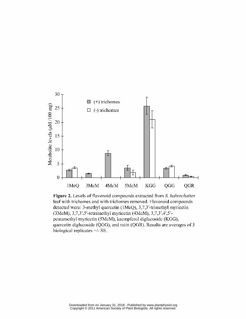
diglucoside (Figure 2). In addition, several non-glycosylated flavonoids were detected,
including 3,7,3′- trimethyl myricetin (3,7,3′-MeM), 3,7,3′,5′- tetramethyl myricetin
(3,7,3′,5′-MeM), 3,7,3′,4′,5′- pentamethyl myricetin (3,7,3′,4′,5′-MeM), and 3- methyl
quercetin (3-MeQ) (Figure 2). When the trichomes were physically removed before the
www.plantphysiol.orgon January 31, 2018 - Published by Downloaded from Copyright © 2011 American Society of Plant Biologists. All rights reserved.

7
leaves were extracted, the levels of kampferol diglucoside, rutin, quercetin diglucoside,
and 3-MeQ detected remained similar, but no 3,7,3′-MeM, 3,7,3′,5′-MeM, and
approximately half the levels of 3,7,3′,4′,5′-MeM, were detected, suggesting that these
compounds were completely or mostly located in the trichomes (Figure 2).
To examine the relative distribution of the non-glycosylated myricetins in specific
types of trichomes, secreting glands and storage glands were collected individually from
leaves of Solanum habrochaites for metabolic profiling (Figure 3 and Supplemental
Figure 1). Secreting glands (types 1 and 4) contained higher levels per gland of all three
methylated myricetins compared to storage glands (type 6). Levels of myricetin
tetramethyl ether (3,7,3′,5′-MeM) were greatest in both secreting and storage gland types
compared to the myricetin trimethyl ether (3,7,3′-MeM) and myricetin pentamethyl ether
(3,7,3′,4′,5′-MeM). However, the levels of all myricetin methyl ethers were 5 to 6 fold
greater in secreting type 1 and 4 glands compared to the corresponding levels in storage
glands. Also, in secreting glands the levels of myricetin pentamethyl ether were slightly
higher than levels of myricetin trimethyl ether, whereas in storage glands levels of the tri-
and penta-methylated myricetins were not significantly different.
Characterization of substrate specificity of ShMOMT1 and ShMOMT2
We have recently constructed EST libraries from the secreting and storage glands
(types 1, 4, and type 6, respectively) of Solanum habrochaites leaves
(http://www.trichome.msu.edu/; McDowell et al., 2011). A bioinformatics search of these
libraries using BLAST sequence comparisons with known O-methyltransferase (OMT)
sequences identified three OMT sequences in S. habrochaites trichomes. All three
cDNAs were expressed in E. coli and the crude extracts were tested for OMT activity
with a battery of substrates (Table I) and [methyl-14C]-S-adenosyl-L-methionine ([14C]-
SAM) as the methyl donor. One cDNA encoded a protein with similarity to plant N-
methyltransferases and had no methylating activity with myricetin, quercetin,
kaempferol, or any other flavonol tested in this investigation (see Table I for list of tested
compounds). Consequently, it was not investigated further. A second cDNA encoded a
protein, subsequently named ShMOMT1, with methylating activity toward myricetin and
quercetin but not kaempferol, suggesting that this protein has 3′/5′ O-methyltransferase
www.plantphysiol.orgon January 31, 2018 - Published by Downloaded from Copyright © 2011 American Society of Plant Biologists. All rights reserved.
8
activity. A third cDNA encoded a protein, subsequently named ShMOMT2, with
methylating activity against all these three flavonols.
ShMOMT1 and ShMOMT2 were further tested with a range of substrates related
to myricetin that could be obtained in sufficient concentrations for these assays.
ShMOMT1 catalyzed the transfer of a methyl group to the 3′ hydroxyl of myricetin as
indicated by co-migration with an authentic standard of 3′-methylmyricetin in radioactive
thin-layer chromatography (RTLC) (Supplemental Figure 2) and by LC/MS
(Supplemental Figure 3) (the 3′ and 5′ hydroxyl positions in this compound are
equivalent, and by convention the product is designated as 3′-methyl myricetin, or
laricitin) and the equivalent position of several other related compounds, including
quercetin, 3-methyl quercetin, and 7-methyl quercetin (i.e. rhamnetin) (Table I). When
the 3′ hydroxyl of the substrate was already methylated, as in laricitin, ShMOMT1
transferred a methyl group to the 5′ hydroxyl, as determined by co-migration with an
authentic standard of 3′,5′-methylmyricetin in RTLC and by LC/MS (Table I and
Supplemental Figures 2 and 3). When both 3′ and 5′ hydroxyls were already
methylated, for example in the substrate 3′,5′-dimethyl myrecitin (i.e. syringetin),
ShMOMT1 could not transfer a methyl group to any other hydroxyl (Table I).
ShMOMT2 transferred a methyl group to the 4′ hydroxyl of kaempferol, but to
the 7 position of quercetin and myricetin (Table I). When the hydroxyl at the 7 position
was already methylated, it transferred a methyl to the 4′ hydroxyl (e.g. with substrate 7-
methyl quercetin), and when the 4′ hydroxyl was already methylated, ShMOMT2
transferred the methyl group to the hydroxyl at the 7 position (e.g. with the substrate
3′,4′,5′-trimethyl myrecitin). It did not transfer a methyl group to any hydroxyl other than
at the 7 or 4′ position (Table I). Radioactive thin-layer chromatography of the reaction
with myricetin revealed a single product that migrated between myricetin and 3′-methyl
myricetin (Supplemental Figure 2). This product was identified by LC/MS as 7-methyl
myricetin. When myricetin was incubated with ShMOMT2 overnight, the product
obtained was 7, 4′- methyl myricetin (Supplemental Figure 4).
The structural relatedness of ShMOMT1 and ShMOMT2 to other OMTs
www.plantphysiol.orgon January 31, 2018 - Published by Downloaded from Copyright © 2011 American Society of Plant Biologists. All rights reserved.

9
The protein encoded by the ShMOMT1 cDNA is 362 amino acids long, with a
calculated molecular mass of 40.7 kD, and it contains all of the recognized plant OMT
domains known or hypothesized to be involved in binding to SAM and metal cofactors
(Ibrahim, 1997) (analysis not shown). ShMOMT1 is most similar (40-49% identity) to a
number of mostly 3′ and 3′/5′ O-methyltransferases (Figure 4), consistent with its
regiospecificity for the 3′ and 5′ positions.
The protein encoded by the ShMOMT2 cDNA is 355 amino acids long, with a
calculated molecular mass of 39.4 kD, and it also contains all of the recognized plant
OMT domains known or hypothesized to be involved in binding to SAM and metal
cofactors (analysis not shown). ShMOMT2 is most similar (29-47% identity) to several
O-methyltransferases identified (with one exception) as specific for the 7 and/or 4′
position (Figure 4), consistent with its regiospecificity for these positions. ShMOMT2 is
only 27% identical to ShMOMT1.
Distribution of ShMOMT1 and ShMOMT2 transcripts and protein in trichome
glands
We used quantitative RT-PCR (qRT-PCR) and Western blot analyses to localize
ShMOMT1 and ShMOMT2 transcript, and ShMOMT1 and ShMOMT2 proteins,
respectively, in the different types of trichome glands. Extracts of collections of
individual types of glands were compared to whole leaf extracts in both types of
experiments. ShMOMT1 transcript levels were 3.5 to 12.5 fold higher in secreting glands
from types 4 and 1 trichomes, respectively, compared to storage glands of type 6
trichomes (Figure 5). ShMOMT2 transcript levels were 2 to 4 fold higher in secreting
glands from types 4 and 1 trichomes, respectively, compared to storage glands of type 6
trichomes (Figure 5). Comparison of transcript levels from leaf tissue with trichomes vs.
leaf tissue from which the trichomes had been mechanically removed indicated that
transcripts of both ShMOMT1 and ShMOMT2 are present exclusively in trichomes
(Figure 5). Protein blot analysis indicated that levels of ShMOMT1 protein were 7 to 8.6
fold higher in secreting glands compared to storage glands, and 1.9 to 2.4 fold higher
compared to whole leaf extracts (Figure 6). Levels of ShMOMT2 protein were 5 to 6.6
www.plantphysiol.orgon January 31, 2018 - Published by Downloaded from Copyright © 2011 American Society of Plant Biologists. All rights reserved.
10
fold higher in secreting glands compared to storage glands, while ShMOMT2 was not
detectable in whole leaf extracts (Figure 6).
Characterization of the kinetic parameters of ShMOMT1 and ShMOMT2
ShMOMT1 and ShMOMT2 were expressed in E. coli BL21 (DE3) cells and the
recombinant proteins were purified to near homogeneity by two successive anion
exchange chromatography steps (Figure 7). The purified ShMOMT1 protein catalyzed
the formation of laricitrin (3′-methyl myricetin) from myricetin with an apparent Km
value of 0.46 µM and an apparent Kcat value of 1.59 s-1. An apparent Km value of 0.21
µM was measured for ShMOMT1 with laricitrin (giving the product syringetin, 3′,5′-
dimethyl myricetin) as the substrate, with an apparent Kcat value of 0.45 s-1. The apparent
Km value for SAM, with myricetin as co-substrate, was 16.64 µM with an apparent Kcat
value of 0.47 s-1 (Table II and Supplemental Figure 5).
Purified ShMOMT2 catalyzed methylation of the 7 hydroxyl group of myricetin,
the 7 hydroxyl group of kaempferide (4′-methyl kaempferol), and the 4′ hydroxyl group
of rhamnetin (7-methyl quercetin). An apparent Km of 1.68 µM was determined for
myricetin with an apparent Kcat value of 7.4x10⁻³ s-1. An apparent Km of 2.27 µM was
determined for kaempferide with an apparent Kcat value of 5.76x10⁻³ s-1. And, an apparent
Km of 2.30 µM was determined for rhamnetin with an apparent Kcat value of 6.40x10⁻³ s-1.
The apparent Km value for SAM with kaempferide as co-substrate was 18.71 µM with an
apparent Kcat value of 1.64x10⁻² s-1 (Table III and Supplemental Figure 6).
Characterization of optimal conditions for catalysis revealed that both ShMOMT1
and ShMOMT2 do not require the addition of Mg2+ or Mn2+ for activity. At levels below
2.5 mM, Mg2+ had little negative effect on activity (≤10%); however, concentrations
above 2.5 mM had increasing inhibitory effects on activity with myricetin. Similarly,
addition of Mn2+ to enzyme assays, using myricetin as substrate, had little negative effect
(≤10%) on activity until levels exceeded 2.5 mM. ShMOMT1 activity with myricetin was
observed in the pH range of 6.0-8.5 with optimal activity observed at pH 7.5. And,
ShMOMT2 activity with myricetin was observed in the pH range of 6.0-9.0 with optimal
activity observed at pH 8.0.
www.plantphysiol.orgon January 31, 2018 - Published by Downloaded from Copyright © 2011 American Society of Plant Biologists. All rights reserved.

11
DISCUSSION
Solanum habrochaites glandular trichomes contain methylated, non-glycosylated
myricetin
Our metabolic profiling of trichome glands from Solanum habrochaites leaf
identified three forms of O-methylated myricetin species: 3,7,3′-MeM, 3,7,3′,5′-MeM,
and 3,7,3′,4′,5′-MeM. These three compounds have previously been shown to accumulate
in tissues of several other plants (Stevens et al., 1995; Dachriyanus et al., 2003;
Ariyanathan et al., 2010), but they have not yet been reported to be present in trichomes.
By isolating individual types of glands, we were able to show that these compounds are
found in three types of glandular trichomes – 1, 4, and 6 (Figure 3), although they are
most abundant in the secreting glands (types 1 and 4).
All of the myricetin methyl ethers that we detected in the glands of glandular
trichomes were methylated at the 3 position (in the C ring). This position is often
glycosylated, and the glycosylated form is then transported to the vacuole (Vogt and
Jones, 2000). We did not find any glycosylated myricetin in the trichomes nor myricetin
species that are not modified at the 3 position, suggesting that the 3-OMT responsible for
this methylation reaction is quite efficient. However, our attempts to detect OMT activity
in crude extracts of glands or whole leaves capable of adding a methylgroup to the 3-
hydroxyl position of myreicetin was unsuccessful, nor could we identify a cDNA
encoding such an enzyme in our EST databases. To our knowledge, no 3-OMTs capable
of methylating myricetin or any other flavonols have been identified from any plant,
although a cellular activity capable of methylating quercetin at the 3 position has been
reported (De Luca and Ibrahim, 1985; Huang et al., 2004).
Our analyses of S. habrochaites leaves with trichomes and leaves with trichomes
removed revealed that 3,7,3′-trimethyl myricetin and 3,7,3′,5′-tetramethyl myricetin were
found in trichome gland cells only, and 3,7,3′,4′,5′-pentamethyl myricetin was found in
both trichomes and the rest of the leaf organ (Figure 2). All other flavonol compounds
were apparently confined mostly to non-trichome leaf cells since their levels did not
decrease significantly when trichomes were removed (Figure 2). The presence of
3,7,3′,4′,5′-pentamethyl myricetin, whose 3,7,3′,5′-tetramethyl myricetin precursor is
found only in the trichomes, outside the trichomes is most likely due to secretion, since
www.plantphysiol.orgon January 31, 2018 - Published by Downloaded from Copyright © 2011 American Society of Plant Biologists. All rights reserved.
12
our analysis indicates that ShMOMT1 and ShMOMT2 are not expressed in non-trichome
leaf cells (Figure 5).
ShMOMT1 is a 3′/5′ myricetin methyltransferase and ShMOMT2 is a 7 and 4′
myricetin methyltransferase
The characterization of the enzymatic properties of ShMOMT1 in vitro showed
that it has high affinity for both myricetin and 3′-methyl myricetin, and its products are
3′-methyl myricetin (laricitrin) and 3′,5′-dimethyl myricetin (syringetin) (Supplemental
Figure 3). In previous studies, OMTs have been identified that can methylate myricetin
at these positions, but in all such cases myricetin was not the best substrate for the
enzyme and the tissue source of the enzyme did not actually contain methylated
myricetin but only related compounds, such as quercetin, kaempferol, tricin, tricetin and
luteolin (Muzac et al., 2000; Lee et al., 2008; Zhou et al., 2006). The catalytic efficiency
of ShMOMT1 with both myricetin and laricitrin are significantly higher than for such
3′,5′-OMTs (Table IV), and these enzymes had higher affinity to the substrates whose
methylation led to the compounds actually observed in the plant.
ShMOMT2 is most similar to some enzymes characterized as 4′
methyltransferases and some characterized as 7 methyltransferases, with one exception
(Figure 4). This exception is Catharanthus roseus flavonol 3′/5′ O-methyltransferase
(Cacace et. al., 2003), which is very similar to Cantharanthus roseus flavonol 4′ O-
methyltransferase (Schröder et. al., 2004) (Figure 4), and may represent a recent case of
gene duplication and divergence. When ShMOMT2 was incubated with kaempferol, a
substrate missing both a 3′ and a 5′-hydroxyl, it added a methyl group to the 4′-hydroxyl
(Table I). However, with either quercetin or myricetin, ShMOMT2 initially added a
methyl group to the 7-hydroxyl, suggesting that 3′ and/or 5′-hydroxyls might inhibit its
activity with the 4′-hydroxyl. This is consistent with the observation that 3,7,3′-trimethyl
myricetin is found in the glands, but no 3,3′,4′-trimethyl myricetin is observed (Figure
3). Thus, it appears that after the 3-hydroxyl is methylated, the next hydroxyls to be
methylated are at the 7 and 3′ position, although which of these two is methylated first
cannot yet be resolved. This is also consistent with what has been shown in
Chrysosplenium americanum, where methylation of quercetin proceeds first to 3-
www.plantphysiol.orgon January 31, 2018 - Published by Downloaded from Copyright © 2011 American Society of Plant Biologists. All rights reserved.

13
methylquercetin, then to 3,7-dimethyl quercetin (De Luca and Ibrahim, 1985). It can be
deduced that the next hydroxyl to be methylated is at the 5′ position, since we see
accumulation of 3,7,3′,5′-tetra methyl myricetin but no 3,7,3′,4′-tetra methyl myricetin,
and also because it appears that ShMOMT1 is not active with a substrate that has a
methyl group at both the 3′,4′ positions (Table I). ShMOMT2 clearly is capable of
methylating the 4′-hydroxyl after it methylated the 7-hydroxyl (tested with 7-methyl
quercetin for lack of 7-methyl myricetin, see Table I, and also by incubating myricetin
with ShMOMT2 for an extended period (>10 h), after which the major product is 7,4′-
myricetin (Supplemental Figure 4)). However, it seems to be less efficient at
methylating the 4′ hydroxyl once the 3′ and/or the 5′ hydroxyls have been methylated
(Table I), consistent with the lower levels of 3,7,3′,4′,5′-pentamethyl myricetin observed
in the trichomes.
A caveat for the kinetic analysis of ShMOMT1 and ShMOMT2 presented here is
that, for lack of availability, we were not able to test them with 3-methylmyricetin or
other combinations of polymethylated myricetin with one methyl group at the 3-position
(for example, 3,7- dimethylmyricetin). However, we did obtain and test both enzymes
with 3-methyl quercetin. The results with ShMOMT1 indicated that it had higher activity
with the 3-methyl quercetin than with quercetin, although ShMOMT2 had lower activity
(Table I). It has been shown for many OMTs that they are regiospecific but not substrate-
specific, meaning that their specificity is determined by the part of the molecule which is
modified by their catalytic activity (Vogt, 2004). However, we note that the turnover rate
of ShMOMT2 with the in vitro substrates tested were substantially lower than the
turnover rates observed for ShMOMT1 (Tables II, III). This might indicate that
ShMOMT2 activity could possibly be rate limiting in the synthesis of polymethylated
myricetins in the trichomes. Alternatively, this enzyme may be more sensitive to the lack
of correct functional groups in the in vitro tested substrates. It is also possible that it may
have additional, non-myricetin related substrates in the cell with which it is more
efficient.
ShMOMT1 and ShMOMT2 are expressed in three different glandular trichomes
www.plantphysiol.orgon January 31, 2018 - Published by Downloaded from Copyright © 2011 American Society of Plant Biologists. All rights reserved.

14
Our data indicate that ShMOMT1 and ShMOMT2 transcripts and proteins are
found in all three types of glandular trichomes in S. habrochaites that are metabolically
active. The levels of the transcripts and proteins in these gland types – 1, 4, and 6 –
correlate well with the amount of methylated myricetin found in them, with type 6 glands
containing an order of magnitude less of each compared with type 1 and 4 glands, with
the exception that type 4 glands have somewhat reduced amounts of both transcripts
compared to type 1 glands. However, the level of ShMOMT1 and ShMOMT2 transcripts
in type 4 glands are still 4-fold and 2-fold higher, respectively, than that found in type 6
glands. In addition to localizing ShMOMT1 and ShMOMT2 in glandular trichomes, we
have detected transcripts of putative genes involved in flavonoid and flavonol
biosynthesis in our EST databases created from isolated trichome glands (types 1, 4, and
6) (Supplemental Table I). Transcripts of both flavonol 3′ hydroxylase and flavonol 3′,5′
hydroxylase, required for the synthesis of myricetin, were detected in these databases,
with highest representation in type 1 glands.
Possible roles of methylated myricetins in tomato glandular trichomes
Flavonoids in general have been hypothesized to as act as UV protectants,
chemical defense compounds, and in plant-insect, plant-microbe, plant-pathogen, and
plant-plant interactions (reviewed by Treutter, 2005). While evidence for some of these
roles (e.g., in plant-microbe interactions) is strong, other roles are still tentative (Treutter,
2005). Furthermore, since flavonoids often occur as a mixture, assigning roles to specific
compounds is difficult. Currently, no physiological function has been postulated
specifically for laricitrin and syringetin in plants, nor for the more highly methylated
myricetins found in the tomato trichomes. Laricitrin and syringetin, but not the more
highly methylated myricetin ethers, are found in red grape and are probably responsible,
along with several other flavonols and methylated derivatives, for the antioxidant potency
of red grapes and wine (Mattivi et al., 2006). However, there is no evidence to support
specific roles for these compounds in grape. Myricetin has also been linked to radical
scavenging activity, xanthine oxidase inhibitory activity, and antioxidant activity in
extracts of Ginkgo leaves and Bridelia ferruginea stem bark (Kobus et al., 2009;
www.plantphysiol.orgon January 31, 2018 - Published by Downloaded from Copyright © 2011 American Society of Plant Biologists. All rights reserved.

15
Cimanga et al., 2001), but in both investigations the tested mixture contained several
other flavonols and methylated derivatives.
Thus, we can only hypothesize that in tomato trichomes the methylated myricetins
contribute to some of the general roles postulated for flavonoids. Their synthesis and
accumulation in glandular trichomes along with their relatively lipophilic nature suggest
that they are likely targeted to the cuticlular space surrounding the secretory cells. In this
location they are well placed to serve roles in chemical defense against herbivores, as UV
protectants, or as radical scavengers to aid in preventing peroxidation of lipids.
METHODS
Plant Material and Growth Conditions
Solanum habrochaites (accession LA1777) seeds were obtained from the C.M.
Rick Tomato Genetics Resource Center (TGRC, University of California at Davis). The
seeds were germinated on sterile filter paper in germination boxes and kept for
approximately 5-7 days before transfer of seedlings to soil. Plants were grown in a
mixture of regular soil:fine sand (3:1, v/v) in a growth chamber under a 14-h light/ 10-h
dark photoperiod. Temperature was maintained at 22°C throughout the light period and
18°C during the dark period.
Gland cells were collected from glandular trichomes by hand with micropipettes
under a dissecting microscope (Leica MZ6). Micropipettes were hand pulled and shaped
from either 9” disposable pasteur pipettes or 1.8 mm X 100 mm capillary tubes. The
micropipettes were approximately 6 cm in length and shaped to taper from one end,
approximately 1.5-2.0 mm, down to approximately 0.25 mm diameter at the opposite
end. Both ends of the pipette were flame sealed to prevent capillary action. Gland cells
were picked from the top of glandular trichome structures using the thin tip of the
micropipette. The cells adhered to the tip until being put into an appropriate buffer for
downstream analyses. Trichomes were removed from leaf material using the same type of
micropipettes, except, they were lightly scraped across the leaf surface in order to remove
the bulk of trichomes without disturbing the leaf surface.
Chemicals
www.plantphysiol.orgon January 31, 2018 - Published by Downloaded from Copyright © 2011 American Society of Plant Biologists. All rights reserved.

16
All chemicals were from Sigma-Aldrich (St. Louis, MO, USA) unless otherwise
noted. Flavonols and methyl flavonols were purchased from Extrasynthese (B.P. 62 -
69730 Genay France) with the exception of the flavonol kaempferol which was
purchased from Indofine Chemical Company, Inc. (12-1 Ilene Court, Belle Mead, NJ
08502). Deuterium labeled S-adenosyl-L- methionine was purchased from C/D/N
isotopes (Quebec, Canada). And, methanol, 88% formic acid, and acetonitrile were
purchased from VWR Scientific (West Chester, PA).
Metabolic profiling of leaf and trichome gland cells and metabolite identification
Approximately 50 mg fresh weight of leaf material were extracted in 100 µL of
ice-cold acetonitrile:isopropanol:water (3:3:2 v/v/v) at room temperature overnight.
Samples were evaporated to near dryness and resuspended in 50% methanol (v/v) for
LC/MS analysis. For leaf material with trichomes removed, a glass probe (described
previously) was used to gently scrape trichomes from the surface of the leaf prior to
extraction with the 3:3:2 solvent mixture.
A total of 50 gland cells from each type of glandular trichome (types 1, 4, and 6)
were collected with micropipettes and extracted in 50 µL of ice-cold
acetonitrile:isopropanol:water (3:3:2 v/v/v). Samples were stored overnight at -20°C,
evaporated to near dryness and resuspended in 50% methanol (v/v) for LC/MS analysis.
Samples were analyzed on a QTRAP™ 3200 mass spectrometer from Applied
Biosystems/MDS Sciex (Concord, Ontario, Canada) coupled to a Shimadzu UFLC LC-
20AD system and SIL-HTc autosampler. Separation was achieved with a Thermo Beta-
basic C18 column (150 mm × 1.0 mm, 5 μm) at 30 C. The mobile phases were, (A) 0.5%
formic acid, (B) 0.5% formic acid in 60% methanol+ 40% acetonitrile. A 15 min reverse
phase gradient at a flow rate of 0.100 mL/min was used for separation. The linear
gradient elution program was as follows: 10% B for 0.3 min, 40% B and linear increase
to 100% from 0.31 to 8.5 min, followed by an isocratic hold at 100% B for 2.5 min. At 11
min. B was returned to 10% and the column was equilibrated for 4 min before the next
injection.
The mass spectrometer was operated in the positive ion mode with a
TurboIonSpray source. Enhanced product ion (MS/MS) scanning was accomplished with
www.plantphysiol.orgon January 31, 2018 - Published by Downloaded from Copyright © 2011 American Society of Plant Biologists. All rights reserved.

17
dynamic fill time and was used for ion detection at 40 V collision energy (CE). The other
ionization parameters were as follows; curtain gas (CUR) 10, ion source gas 1 (GS1) 12,
ion source gas 2 (GS2) 30, source temperature (TEM) 400°C, entrance potential (EP) 10
V, CAD high; IS voltage 5500 V. The mass spectrometer and the HPLC system were
controlled by Analyst 1.4.2 software from Applied Biosystems/MDS Sciex.
All flavonol glycosides observed in leaf dip extracts were glycosylated in the 3-
position judged by the high abundance of radical anion aglycone fragment ions in
negative ion MS/MS spectra (Cuyckens and Claeys, 2005). Authentic standards of most
aglyconic polymethylated myricetin metabolites were not available from commercial
sources. Owing to the substantial number of methylated isomers, their low levels in plant
tissues, and their co-elution with other metabolites, comparisons to ultraviolet spectra of
standards were not feasible, nor were sufficient amounts of purified metabolites available
for detailed NMR structure determination. In view of these limitations, position of
methyl groups in methylated myricetins were assigned based on co-elution of plant
metabolites with authentic standards when available produced semi-synthetically from
standards of myricetin or methylated myricetins when possible, predictions of relative LC
retention times based on the ease of formation of intramolecular hydrogen bonds in some
isomers, and on MS/MS product ion spectra that showed positional isomer-selective
differences in fragmentation behavior. In the latter case, ion structure assignments were
aided through enzymatic synthesis of individual O-d3-methylated myricetin derivatives
from d3-S-adenosylmethionine. Product ion MS/MS spectra of [M+H]+ ions derived from
methylated myricetins yielded evidence for position-selective fragmentation chemistry.
Abundances of fragments arising from loss of a methyl radical (-15 Da) relative to
[M+H]+ ions varied among methylated myricetins, and relative yields of these fragments
decreased based on methyl position as 3 > 4′ > 3′ = 5′ >> 7. Assignments of myricetins
methylated on the A-ring (either the 5- or 7-positions) were facilitated by observations of
characteristic fragment ion masses observed in the MS/MS product ion spectra. In the
absence of methylation in these positions, the fragment ion derived from the A-ring group
(designated as 1,3A+) appears at m/z 153, but when either of the 5- or 7-positions are
methylated, this fragment mass shifts upward in mass by 14 Da to appear at m/z 167. We
considered methylation at the 5-position unlikely because this is a rare metabolite, and
www.plantphysiol.orgon January 31, 2018 - Published by Downloaded from Copyright © 2011 American Society of Plant Biologists. All rights reserved.
18
none of the metabolites gave MS/MS fragments suggestive of two methyl groups on the
A-ring. One additional feature, the loss of 16 Da from the [M+H]+ precursor, was shown
using deuterium labeling to specifically occur when at least two methyl ether groups were
present on the B-ring (3’, 4’, or 5’ positions). The combinations of these features in the
MS/MS spectra allow us to use a process of elimination to generate unambiguous
evidence for the assignments of methyl group positions in methylated myricetin
metabolites.
RNA isolation
Total RNA was extracted from 100 mg fresh weight of young leaf material or
young leaf material from which trichomes had been removed. Tri Reagent (Molecular
Research Center, Inc.) was used in accordance with the manufacturer’s instructions to
extract total RNA from leaf and from leaf with trichomes removed. First-strand cDNA
was synthesized with SuperScript II reverse transcriptase (Invitrogen) using an anchored
poly-T primer supplied by the manufacturer.
Quantitative RT-PCR
Total RNA from young leaf material and young leaf material with trichome
removed were extracted as described above then treated with DNase using the DNA-free
kit (Ambion). Superscript II Reverse Transcriptase (Invitrogen) and an anchored poly-T
primer were used for first-strand cDNA synthesis. A negative control sample was run in
parallel without reverse transcriptase added to the reaction mixture. All samples were
normalized to the amplification of a Solanum lycopersicum actin gene (accession:
BT013707). Quantitative expression analysis was performed using the StepOnePlus Real-
Time PCR System (Applied Biosystems). The Fast Sybr Green Master Mix (Applied
Biosystems) reagent was used according to the manufacturers’ instructions in preparation
of the qPCR reactions. The cycling conditions were: 40X 15 sec/95°C, 30 sec/60°C, 30
sec/72°C. Cycling was followed by a melting stage that ramped up from 55 to 95°C with
an increasing gradient of 0.5°C, and a 10-s pause at each temperature. The entire
experiment was performed in triplicate starting with total RNA isolation from gland cells,
leaves, or leaves with trichomes removed. The threshold cycle (Ct) values from each
www.plantphysiol.orgon January 31, 2018 - Published by Downloaded from Copyright © 2011 American Society of Plant Biologists. All rights reserved.

19
experiment were averaged and the relative expression level of ShMOMT1 in each tissue
was calculated using the comparative Ct method (Schmittgen and Livak, 2008). The
results were expressed relative to expression levels of ShMOMT1 or ShMOMT2 in leaf
material with trichomes.
Isolation, Expression, and purification of recombinant ShMOMT1
The full-length ShMOMT1 and ShMOMT2 ORF’s were cloned from cDNA
made from S. habrochaites leaf RNA. Tri Reagent (Molecular Research center, Inc.) was
used to extract total RNA from approximately 100 mg of material and SuperScript II
Reverse Transcriptase (Invitrogen) was used to synthesize first-strand cDNA. ShMOMT1
sequence was amplified using KOD Hot Start DNA polymerase (Novagen) from first-
strand cDNA and ligated into the pGEM-T Easy vector (Promega), grown in Escherichia
coli Top 10 cells, and full-length cDNAs were verified by DNA sequencing. The full-
length ORF was amplified from the pGEM-T Easy vector using KOD Hot Start DNA
Polymerase (Novagen), gel-purified using MinElute (Qiagen), and inserted into the
pEXP5-CT/TOPO expression vector (invitrogen) with the native stop codon intact. The
correct pEXP5-CT/TOPO construct was verified by DNA sequencing, isolated using
QIAprep Spin Miniprep kit (Qiagen) and transformed into E.coli BL21(DE3)pLysS cells
(Invitrogen). A colony carrying the correct construct was isolated and grown in LB
medium containing 100 μg/mL ampicillin and 50 μg/mL chloramphenicol at 37°C to an
OD600 of 0.5-0.8. Cultures were induced with 1 mM isopropylthio-β-galactoside and
grown at 18°C for an additional 4 hours.
Induced cultures were pelleted by centrifugation, resuspended in 1/10 volume
lysis buffer (50 mM Tris, 10 mM NaCl, 1 mM EDTA, 10% glycerol, 14 mM β-
mercaptoethanol, pH 8.0), and lysed at 4°C by sonication. The cell lysate was cleared by
centrifugation, and the supernatant was partially purified with DE53 anion exchanger
(Whatman International, Ltd.). ShMOMT1 and ShMOMT2 were each purified by anion-
exchange chromatography on an HiTrap Q HP column (GE Healthcare). A linear
gradient of (10 - 1000 mM) NaCl in lysis buffer was used for the initial purification on
the DE53 anion exchanger, and a linear gradient of (250 - 500 mM) NaCl in lysis buffer
was used for the second round of purification on the HiTrap Q HP anion exchanger.
www.plantphysiol.orgon January 31, 2018 - Published by Downloaded from Copyright © 2011 American Society of Plant Biologists. All rights reserved.

20
ShMOMT1 eluted in the 400 - 500 mM and ShMOMT2 eluted in the 300-400 mM
fractions from the DE53 anion exchanger and in the 350 - 400 mM and the 300 - 350 mM
fractions from the HiTrap Q HP anion exchanger, respectively. The active fractions were
identified by radiochemical enzyme assays as described above, using myricetin as
substrate. SDS-PAGE was used to visualize the degree of homogeneity of the active
fractions.
Enzyme assays and Product Identification
Radiochemical enzyme assays consisted of 50 mM Tris-HCl (pH 7.5, ShMOMT1
or pH 8.0, ShMOMT2), 5 µg of recombinant ShMOMT1 or ShMOMT2, 250 µM of
substrate dissolved in 1:1 mixture DMSO:ddH20, and 200 μM SAM (Perkin Elmer
Instruments), in a final volume of 50 µL. Assays were incubated at room temperature for
30 minutes and stopped by the addition of 2 N HCl. Reaction products were extracted
with 200 µL of ethyl acetate and counted in a scintillation counter (model LS6500,
Beckman Coulter, Fullerton, CA). Kinetic analyses were carried out within the linear
range of reaction velocity by adjusting the concentration of recombinant protein in the
assay. Raw data (counts per minute [cpm]) were converted to picokatals as previously
described in D’ Auria et al. (2002).
To produce ample product for LC/MS analyses, enzyme assays were performed
using nonradiolabeled SAM and 10 fold reaction volumes. A Continuous Extraction
Assay Method (CEAM) was designed to optimize product accumulation in these assays.
Reactions were performed in 1.5 mL glass vials (Supelco Analytical, 27080-U) in a final
volume of 500 µL (aq). A layer of 100% ethyl acetate (500 μL) was carefully applied
over the aqueous assay volume to serve as a non-polar extraction phase. The reactions
were set up on ice and mixed briefly before addition of the ethyl acetate layer. Reactions
were sealed with a screw-top septum, and incubated overnight at room temperature. The
ethyl acetate was removed, evaporated, and the residue resuspended in 50 µL,
ethanol:ddH2O (1:1).
Metabolite identities were determined using TLC following the method of Owens
and McIntosh (2009) with the following modifications, Polygram Sil G/UV254 plastic
sheets (Macherey-Nagel Inc.) and running buffer of toluene:ethylformate:formic acid
www.plantphysiol.orgon January 31, 2018 - Published by Downloaded from Copyright © 2011 American Society of Plant Biologists. All rights reserved.

21
(5:4:1, v/v/v), and by LC-MS using three different criteria: accurate mass, measured with
time-of-flight mass spectrometry; retention time comparison with authentic standards;
and comparison of mass spectra fragmentation patterns (see section Metabolic profiling
of leaf and trichome gland cells and chemical identification above, and Supplemental
Figures 1, 3, and 4).
Protein Blot Analysis
Total protein was extracted from collections of 500 gland cells from each of the
different types of glandular trichomes (type 1, 4, and 6) in 50 µL of SDS-PAGE sample
buffer (100 mM Tris, 2% SDS, 5% ß-mercaptoethanol, 15% glycerol, 0.1% bromophenol
blue). Total protein extraction from leaves followed the protocol given in Dudareva et al.
1996. Polyclonal antibodies to ShMOMT1 or ShMOMT2 were generated at Cocalico
Biologicals (Reamstown, PA) in rabbit from recombinant ShMOMT1 or ShMOMT2
protein (Supplemental Figure 7). Anti-α-tubulin was from Sigma-Aldrich and served as
an internal control to standardize samples from gland cells and leaves. All antibodies
(anti-ShMOMT1, anti-ShMOMT2, and anti- α-tubulin) were used at a 1:3,000 dilution
and incubated with gel blots for 1 h. All other conditions of the protein gel blots were
performed as described previously (Dudareva et al., 1996)
ACKNOWLEGMENTS
We would like to express our thanks to Drs. Robert Last and Anthony Schillmiller, and to
Ms. Jeongwoon Kim (Michigan State University) for sharing their results with us prior to
publication, and for helpful advice.
REFERENCES
Ambrsio SR, Oki Y, Heleno VCG, Chaves JS, Nascimento P, Lichston JE,
Constantino MG, Varanda EM, Da Costa FB (2008) Constituents of glandular
trichomes of Tithonia diversifolia: Relationships to herbivory and antifeedant
activity. Phytochemistry 69: 2052-2060
Ariyanathan S, Saraswathy A, Rajamanickam GV, Connolly JD (2010) Polyphenols
www.plantphysiol.orgon January 31, 2018 - Published by Downloaded from Copyright © 2011 American Society of Plant Biologists. All rights reserved.

22
from the roots of Plumbago rosea. Indian Journal of Chemistry Section B-
Organic Chemistry Including Medicinal Chemistry 49: 386-389
Braca A, Bilia AR, Mendez J, Morelli I (2001) Myricetin glycosides from Licania
densiflora. Fitoterapia 72: 182-185
Buer CS, Imin N, Djordjevic MA (2010) Flavonoids: New roles for old molecules.
Journal of Integrative Plant Biology 52: 98-111
Cacace S, Schroder G, Wehinger E, Strack D, Schmidt J, Schroder J (2003) A
flavonol O-methyltransferase from Catharanthus roseus performing two
sequential methylations. Phytochemistry 62: 127-137
Croteau RB, Davis EM, Ringer KL, Wildung MR (2005) (-)-Menthol biosynthesis and
molecular genetics. Naturwissenschaften 92: 562-577
Cuyckens F, M Claeys (2005) Determination of the glycosylation site in flavonoid
mono-O-glycosides by collision-induced dissociation of electrospray-generated
deprotonated and sodiated molecules. J Mass Spectrom 40: 364-372
Dachriyanus, Fahmi R, Sargent MV, Skelton BW, White AH (2004) 5-Hydroxy-3,3
',4 ',5 ',7-pentamethoxyflavone (combretol). Acta Crystallographica Section E-
Structure Reports Online 60: O86-O88
D’ Auria JC, Chen F, Pichersky E (2002) Characterization of an acyltransferase
capable of synthesizing benzylbenzoate and other volatile esters in flowers and
damaged leaves of Clarkia breweri. Plant Physiology 130: 466-476
De Luca V, Ibrahim RK (1985) Enzymatic synthesis of polymethylated flavonols in
Chrysosplenium Americanum. 1. Partial purification and some properties of S-
adenosyl-L-methionine flavonol 3-O-methyltransferases, 6-O-methyltransferases,
7-O-methyltransferases, and 4′-O-methyltransferases. Archives of Biochemistry
and Biophysics 238: 596-605
Dudareva N, Cseke L, Blanc VM, Pichersky E (1996) Evolution of floral scent in
Clarkia: Novel patterns of S-linalool synthase gene expression in the C. breweri
flower. Plant Cell 8: 1137-1148
Felsenstein J (1985) Confidence limits on phylogenies: An approach using the bootstrap.
Evolution 39:783-791.
Gerats AGM, Wallroth M, Donkerkoopman W, Groot SPC, Schram AW (1983) The
www.plantphysiol.orgon January 31, 2018 - Published by Downloaded from Copyright © 2011 American Society of Plant Biologists. All rights reserved.

23
genetic control of the enzyme UDP-glucose - 3-O-flavonoid-glucosyltransferase
in flowers of Petunia hybrida. Theoretical and Applied Genetics 65: 349-352
Gorbatsova J, Lougas T, Vokk R, Kalijurand M (2007) Comparison of the contents of
various antioxidants of sea buckthorn berries using CE. Electrophoresis 28: 4136-
4142
Huang TS, Anzellotti D, Dedaldechamp F, Ibrahim RK (2004) Partial purification,
kinetic analysis, and amino acid sequence information of a flavonol 3-O-
methyltransferase from Serratula tinctoria. Plant Physiology 134: 1366-1376
Ibrahim RK (1997) Plant O-methyltransferase signatures. Trends in Plant Science 2:
249-250
Ibrahim RK (2005) A forty-year journey in plant research: Original contributions to
flavonoid biochemistry. Canadian Journal of Botany-Revue Canadienne De
Botanique 83: 433-450
Ibrahim RK, De Luca V, Khouri H, Latchinian L, Brisson L, Charest PM (1987)
Enzymology and compartmentation of polymethylated flavonol glucosides in
Chrysosplenium americanum. Phytochemistry 26: 1237-1245
Iijima Y, Gang DR, Fridman E, Lewinsohn E, Pichersky E (2004) Characterization of
geraniol synthase from the peltate glands of sweet basil. Plant Physiology 134:
370-379
Jay M, Voirin B, Hasan A, Gonnet JF, Viricel MR (1980) Chemotaxonomic study of
vascular plants. 43. Chemosystematic experiment on tribe Loteae. Biochemical
Systematics and Ecology 8: 127-132
Joshi CP, Chiang VL (1998) Conserved sequence motifs in plant S-adenosyl-L-
methionine-dependent methyltransferases. Plant Molecular Biology 37: 663-674
Jung HA, Kim JE, Chung HY, Choi JS (2003) Antioxidant principles of Nelumbo
nucifera stamens. Archives of Pharmacal Research 26: 279-285
Kumar A, Malik AK, Tewary DK (2009) A new method for determination of myricetin
and quercetin using solid phase microextraction-high performance liquid
chromatography-ultra violet/visible system in grapes, vegetables and red wine
samples. Analytica Chimica Acta 631: 177-181
Kumar N, Bhandari P, Singh B, Gupta AP, Kaul VK (2008) Reversed phase-HPLC
www.plantphysiol.orgon January 31, 2018 - Published by Downloaded from Copyright © 2011 American Society of Plant Biologists. All rights reserved.
24
for rapid determination of polyphenols in flowers of rose species. Journal of
Separation Science 31: 262-267
Kumari GNK, Rao LJM, Rao NSP (1984) Myricetin methyl ethers from Solanum
Pubescens. Phytochemistry 23: 2701-2702
Lako J, Trenerry VC, Wahlqvist M, Wattanapenpaiboon N, Sotheeswaran S,
Premier R (2007) Phytochemical flavonols, carotenoids and the antioxidant
properties of a wide selection of Fijian fruit, vegetables and other readily
available foods. Food Chemistry 101: 1727-1741
Le K, Chiu F, Ng K (2007) Identification and quantification of antioxidants in Fructus
lycii. Food Chemistry 105: 353-363
Lee TH, Liu DZ, Hsu FL, Wu WC, Hou WC (2006) Structure-activity relationships of
five myricetin galloylglycosides from leaves of Acacia confusa. Botanical
Studies: An International Journal 47: 37-43
Lee YJ, Kim BG, Chong Y, Lim Y, Ahn JH (2008) Cation dependent O-
methyltransferases from rice. Planta 227: 641-647
Liu Y, Li WJ, Ling XM, Lai XY, Li YZ, Zhang QY, Zhao YY (2008) Simultaneous
determination of the active ingredients in Abelmoschus manihot (L.) Medicus by
CZE. Chromatographia 67: 819-823
Luckwill, L. (1943) The genus Lycopersicon: An historical, biological, and taxonomic
survey of the wild and cultivated tomatoes. PhD thesis. Aberdeen University,
Aberdeen.
Mattivi F, Guzzon R, Vrhovsek U, Stefanini M, Velasco R (2006) Metabolite profiling
of grape: Flavonols and anthocyanins. Journal of Agricultural and Food
Chemistry 54: 7692-7702
McDowell ET, Kapteyn J, Schmidt A, Li C, Kang J, Descour A, Shi F, Larson M,
Schilmiller A, An L, Jones AD, Pichersky E, Soderlund CA and Gang DR
(2011) Comparative functional genomic analysis of Solanum glandular trichome
types. Plant Physiology (DOI:10.1104/pp.110.167114).
Michodjehoun-Mestres L, Souquet JM, Fulcrand H, Bouchut C, Reynes M,
Brillouet JM (2009) Monomeric phenols of cashew apple (Anacardium
occidentale L.). Food Chemistry 112: 851-857
www.plantphysiol.orgon January 31, 2018 - Published by Downloaded from Copyright © 2011 American Society of Plant Biologists. All rights reserved.
25
Min BS, Lee SY, Kim JH, Lee JK, Kim TJ, Kim DH, Kim YH, Joung H, Lee HK,
Nakamura N, Miyashiro H, Hattori M (2003) Anti-complement activity of
constituents from the stem-bark of Juglans mandshurica. Biological &
Pharmaceutical Bulletin 26: 1042-1044
Modolo LV, Li LN, Pan HY, Blount JW, Dixon RA, Wang XQ (2009) Crystal
structures of glycosyltransferase UGT78G1 reveal the molecular basis for
glycosylation and deglycosylation of (iso)flavonoids. Journal of Molecular
Biology 392: 1292-1302
Motta LB, Kraus JE, Salatino A, Salatino MLF (2005) Distribution of metabolites in
galled and non-galled foliar tissues of Tibouchina pulchra. Biochemical
Systematics and Ecology 33: 971-981
Muzac I, Wang J, Auzellotti D, Zhang H, Ibrahim RK (2000) Functional expression
of an Arabidopsis cDNA clone encoding a flavonol 3'-O-methyltransferase and
characterization of the gene product. Archives of Biochemistry and Biophysics
375: 385-388
Ojong PB, Njiti V, Guo ZB, Gao M, Besong S, Barnes SL (2008) Variation of
flavonoid content among sweetpotato accessions. Journal of the American Society
for Horticultural Science 133: 819-824
Oliveira I, Sousa A, Valentao P, Andrade PB, Ferreira I, Ferreres F, Bento A,
Seabra R, Estevinho L, Pereira JA (2007) Hazel (Corylus avellana L.) leaves as
source of antimicrobial and antioxidative compounds. Food Chemistry 105: 1018-
1025
Owens DK and McIntosh CA (2009) Identification, recombinant expression, and
biochemical characterization of a flavonol 3-O-glucosyltransferase clone from
Citrus paradise. Phytochemistry 70: 1382-1391
Rausher MD (2008) The evolution of flavonoids and their genes. In E Grotewold, ed,
Science of Flavonoids, Springer, New York, pp 175-211.
Reynertson KA, Yang H, Jiang B, Basile MJ, Kennelly EJ (2008) Quantitative
analysis of antiradical phenolic constituents from fourteen edible Myrtaceae
fruits. Food Chemistry 109: 883-890
Riihinen K, Jaakola L, Karenlampi S, Hohtola A (2008) Organ-specific distribution of
www.plantphysiol.orgon January 31, 2018 - Published by Downloaded from Copyright © 2011 American Society of Plant Biologists. All rights reserved.

26
phenolic compounds in bilberry (Vaccinium myrtillus) and 'northblue' blueberry
(Vaccinium corymbosum x V. angustifolium). Food Chemistry 110: 156-160
Saitou N and Nei M (1987) The neighbor-joining method: A new method for
reconstructing phylogenetic trees. Molecular Biology and Evolution 4:406-425.
Schilmiller A, Shi F, Kim J, Charbonneau AL, Holmes D, Jones AD, Last RL (2010)
Mass spectrometry screening reveals widespread diversity in trichome specialized
metabolites of tomato chromosomal substitution lines. Plant Journal 62: 391-403
Schilmiller AL, Last RL, Pichersky E (2008) Harnessing plant trichome biochemistry
for the production of useful compounds. Plant Journal 54: 702-711
Schmittgen TD, Livak KJ (2008) Analyzing real-time PCR data by the comparative CT
method. Nature Protocols 3: 1101-1108
Schröder G, Wehinger E, Lukacin R, Wellmann F, Seefelder W, Schwab W,
Schröder J (2004) Flavonoid methylation: a novel 4′-O-methyltransferase from
Catharanthus roseus, and evidence that partially methylated flavanones are
substrates of four different flavonoid dioxygenases. Phytochemistry 65: 1085-
1094
Singh AP, Luthria D, Wilson T, Vorsa N, Singh V, Banuelos GS, Pasakdee S (2009)
Polyphenols content and antioxidant capacity of eggplant pulp. Food Chemistry
114: 955-961
Stevens JF, Hart H, Elema ET, Bolck A (1996) Flavonoid variation in Eurasian sedum
and Sempervivum. Phytochemistry 41: 503-512
Stevens JF, Hart HT, Wollenweber E (1995) The Systematic and evolutionary
significance of exudate flavonoids in Aeonium. Phytochemistry 39: 805-813
Tabart J, Kevers C, Pincemail J, Defraigne JO, Dommes J (2006) Antioxidant
capacity of black currant varies with organ, season, and cultivar. Journal of
Agricultural and Food Chemistry 54: 6271-6276
Tamura K, Dudley J, Nei M & Kumar S (2007) MEGA4: Molecular Evolutionary
Genetics Analysis (MEGA) software version 4.0. Molecular Biology and
Evolution 24:1596-1599.
Taylor LP, Grotewold E (2005) Flavonoids as developmental regulators. Current
Opinion in Plant Biology 8: 317-323
www.plantphysiol.orgon January 31, 2018 - Published by Downloaded from Copyright © 2011 American Society of Plant Biologists. All rights reserved.

27
Thresh K, Ibrahim RK (1985) Are Spinach chloroplasts involved in flavonoid O-
methylation. Zeitschrift Fur Naturforschung C-A Journal of Biosciences 40: 331-
335
Vogt T (2004) Regiospecificity and kinetic properties of a plant natural product O-
methyltransferase are determined by its N-terminal domain. FEBS Letters 561:
159-162
Vogt T, Jones P (2000) Glycosyltransferases in plant natural product synthesis:
Characterization of a supergene family. Trends in Plant Science 5: 380-386
Wagner GJ (1991) Secreting glandular trichomes - More than just hairs. Plant
Physiology 96: 675-679
Wu JH, Huang CY, Tung YT, Chang ST (2008) Online RP-HPLC-DPPH screening
method for detection of radical-scavenging phytochemicals from flowers of
Acacia confusa. Journal of Agricultural and Food Chemistry 56: 328-332
Zhou JM, Gold ND, Martin VJJ, Wollenweber E, Ibrahim RK (2006) Sequential O-
methylation of tricetin by a single gene product in wheat. Biochimica Et
Biophysica Acta-General Subjects 1760: 1115-1124
Zuckerkandl E & Pauling L (1965) Evolutionary divergence and convergence in
proteins. In V Bryson, HJ Vogel, eds, Evolving Genes and Proteins, Academic
Press, New York, pp. 97-166.
www.plantphysiol.orgon January 31, 2018 - Published by Downloaded from Copyright © 2011 American Society of Plant Biologists. All rights reserved.

28
Table I. Relative activity of ShMOMT1 with flavonol and O-methyl flavonol substrates1.
Substrate Relative Activity2
(%)
ShMOMT1
Product
RelativeActivity2
(%)
ShMOMT2
Product
Flavonols
Kaempferol3
Quercetin4
Myricetin5
≤1
51
100
--
3′-methyl quercetin
3′-methyl myricetin
117
119
100
4′-methyl kaempferol
7-methyl quercetin
7-methyl myricetin11
O-methyl flavonols
4′-methyl kaempferol6
3,7,4′-trimethyl kaempferol
3′-methyl quercetin7
3-methyl quercetin
7-methyl quercetin8
3,7,3′,4′-tetramethyl quercetin
3′-methyl myricetin9
3′,5′-dimethyl myricetin10
3′,4′,5′ trimethyl myricetin
≤ 1
≤ 1
≤ 1
61
70
≤ 1
64
≤ 1
≤ 1
--
--
--
3,3′-dimethyl quercetin
7,3′-dimethyl quercetin
--
3′,5′-dimethyl myricetin
--
--
105
3
23
19
120
3
5
11
36
7,4′-dimethyl kaempferol
--
3′,4′-dimethyl quercetin
3,7-dimethyl quercetin
7,4′-dimethyl quercetin
--
3′,4′-dimethyl myricetin
7,3′,5′-trimethyl myricetin
7,3′,4′,5′-tetramethyl
myricetin
1 Additional substrates tested include; caffeic acid, eugenol, isoeugenol, chavicol, orcinol, and several different flavones, isoflavones, and anthocyanidins. Substrate concentration was 200 µM, and incubation time was 30 min. 2 100% relative activity represents total methylating activity; ShMOMT1 3.4 nmol min-1 mg-1, ShMOMT2 0.02 nmol min-1 mg-1, of the recombinant proteins assayed with myricetin as substrate. 3-5 Structures in figure 1. 6 Kaempferide 7 Isorhamnetin 8 Rhamnetin 9 Laricitrin 10 Syringetin 11 After overnight incubation (>10 h), the main product is 7, 4′-dimethylmyricetin.
www.plantphysiol.orgon January 31, 2018 - Published by Downloaded from Copyright © 2011 American Society of Plant Biologists. All rights reserved.

29
Table II. Kinetic parameters of ShMOMT1 with myricetin, laricitrin, and [14C]-SAM as substrates. Km and Kcat ± S.E. Values are averages of three assays.
Substrates Km (µM) Kcat (s-1) Kcat/Km (µM-1 s-1)
Myricetin1 0.46 ± 0.05 1.59 ± 0.15 3.46 ± 0.38
Laricitrin2 0.21 ± 0.04 0.45 ± 0.01 2.14 ± 0.27
SAM3 16.64 ± 2.10 0.47 ± 0.06 0.03 ± 4.0 x10⁻³ 1Structure in figure 1. 23′- methyl myricetin. 3Adenosyl-L-methionine, S-[methyl-14C]-
www.plantphysiol.orgon January 31, 2018 - Published by Downloaded from Copyright © 2011 American Society of Plant Biologists. All rights reserved.

30
Table III. Kinetic parameters of ShMOMT2 with myricetin, kaempferide, rhamnetin, and [14C]-SAM as substrates. Km and Kcat ± S.E. Values are averages of three assays.
Substrates Km (µM) Kcat (s-1) Kcat/Km (µM-1 s-1)
Myricetin1 1.68 ± 0.23 7.40x10⁻³ ± 9.03x10⁻⁴ 4.41x10⁻³ ± 5.74x10⁻⁴
4’-methylkaempferol2 2.27 ± 0.37 5.76x10⁻³ ± 8.89x10⁻⁴ 2.53x10⁻³ ± 3.27x10⁻⁴
7-methylquercetin3 2.30 ± 0.20 6.40x10⁻³ ± 5.22x10⁻⁴ 2.78x10⁻³ ± 3.06x10⁻⁴
SAM4 18.71 ± 2.51 1.64x10⁻² ± 2.33x10⁻³ 8.75x10⁻⁴ ± 1.73x10⁻⁴ 1Structure in figure 1. 2Kaempferide 3Rhamnetin 4Adenosyl-L-methionine, S-[methyl-14C]-
www.plantphysiol.orgon January 31, 2018 - Published by Downloaded from Copyright © 2011 American Society of Plant Biologists. All rights reserved.
31
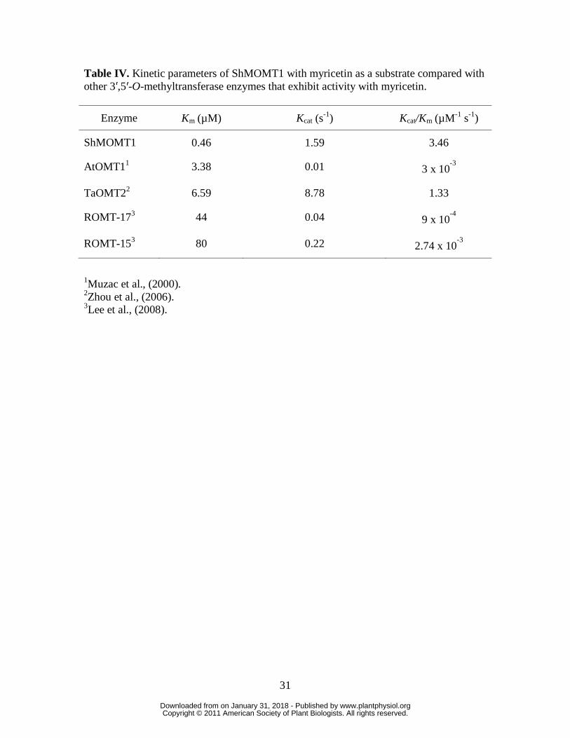
Table IV. Kinetic parameters of ShMOMT1 with myricetin as a substrate compared with other 3′,5′-O-methyltransferase enzymes that exhibit activity with myricetin.
Enzyme Km (µM) Kcat (s-1) Kcat/Km (µM-1 s-1)
ShMOMT1 0.46 1.59 3.46
AtOMT11 3.38 0.01 3 x 10⁻³
TaOMT22 6.59 8.78 1.33
ROMT-173 44 0.04 9 x 10⁻⁴
ROMT-153 80 0.22 2.74 x 10⁻³
1Muzac et al., (2000). 2Zhou et al., (2006). 3Lee et al., (2008).
www.plantphysiol.orgon January 31, 2018 - Published by Downloaded from Copyright © 2011 American Society of Plant Biologists. All rights reserved.

32
FIGURE LEGENDS
Figure 1. Generic flavonol structure showing the lettering system for the three rings and
the numbering system for the carbons. Addition of hydroxyl groups at the 3′ position or 3′
and 5′ positions designate quercetin and myricetin, respectively.
Figure 2. Levels of flavonoid compounds extracted from S. habrochaites leaf with
trichomes and with trichomes removed. Flavonoid compounds detected were: 3-methyl
quercetin (1MeQ), 3,7,3′-trimethyl myricetin (3MeM), 3,7,3′,5′-tetramethyl myricetin
(4MeM), 3,7,3′,4′,5′-pentamethyl myricetin (5MeM), kaempferol diglucoside (KGG),
quercetin diglucoside (QGG), and rutin (QGR). Results are averages of 3 biological
replicates +/- SE.
Figure 3. Levels of O-methylated myricetin compounds measured in extracts from 50
glandular trichomes from S. habrochaites leaf. O-Methylated myricetin compounds
detected were: (3,7,3′)-trimethyl myricetin; (3,7,3′,5′)- tetramethyl myricetin;
(3,7,3′,4′,5′)- pentamethyl myricetin. Results are averages of 3 biological replicates +/-
SE.
Figure 4. Analysis of the level of relatedness of the ShMOMT1-encoded protein and
ShMOMT2-encoded protein to other plant O-methyltransferases. For each protein,
descriptions in parentheses indicate the chemical class of the preferred substrate, the
position(s) that are O-methylated by the enzyme, and the accession number of the
sequence. The evolutionary history was inferred using the Neighbor-Joining method
(Saitou and Nei, 1987). The percentage of replicate trees in which the associated taxa
clustered together in the bootstrap test (1000 replicates) are shown next to the branches
(Felsenstein, 1985). The evolutionary distances were computed using the Poisson
correction method and are in the units of the number of amino acid substitutions per site
(Zuckerkandl and Pauling, 1965). Phylogenetic analyses were conducted in MEGA4
(Tamura et al., 2007).
www.plantphysiol.orgon January 31, 2018 - Published by Downloaded from Copyright © 2011 American Society of Plant Biologists. All rights reserved.

33
Figure 5. Relative levels of ShMOMT1 and ShMOMT2 transcript measured in
trichomes, leaves, and leaves with trichomes removed by qRT-PCR. Results are averages
of 3 biological replicates +/- SE.
Figure 6. Relative levels of ShMOMT1 and ShMOMT2 protein measured in trichomes.
Levels were determined by quantitative Western blot analysis with anti-ShMOMT1 or
anti-ShMOMT2 and anti-α-tubulin. Results are averages of 3 biological replicates +/- SE.
Figure 7. Purification of E. coli-produced ShMOMT1 (panel A) and ShMOMT2 (panel
B). Lanes: 1, non-induced crude extract E. coli cells carrying the ShMOMT1 or
ShMOMT2 expression vector but not induced with IPTG; 2, induced crude extract; 3,
The fractions eluting from DE53 column (Panel A, 2 µg; Panel B, 10 µg) with the highest
MOMT activity; 4, The fractions eluting from the HiTrapQ column (Panel A, 0.25 µg;
Panel B, 5 µg ) with the highest MOMT activity. SDS-PAGE visualized with Coomaise
Brilliant Blue.
www.plantphysiol.orgon January 31, 2018 - Published by Downloaded from Copyright © 2011 American Society of Plant Biologists. All rights reserved.

R 3'O
R1
2'3'
4'OH
5'8
OOH
B5
6'2
78
O 1OH
R2
36
2A
C3
45
6OH
OOH
Kae
mpf
erol
(R1
=R
2=
H)
Kae
mpf
erol
(R1
= R
2 =
H)
Que
rcet
in (R
1 =
OH
, R2
= H
) Q
(,
)M
yric
etin
(R1
= R
2 =
OH
)
Fi1
Gi
fll
tt
hi
Figu
re 1
.Gen
eric
flav
onol
stru
ctur
e sh
owin
g th
ele
tterin
gsy
stem
fort
heth
ree
rings
and
the
the
lette
ring
syst
em fo
r the
thre
e rin
gs a
nd th
e nu
mbe
ring
syst
em fo
r the
car
bons
. Add
ition
of
hydr
oxyl
gro
ups a
t the
3′ p
ositi
on o
r 3′
and
5′
itid
it
tid
iti
posi
tions
des
igna
te q
uerc
etin
and
myr
icet
in,
resp
ectiv
ely.
resp
ectiv
ely.
www.plantphysiol.orgon January 31, 2018 - Published by Downloaded from Copyright © 2011 American Society of Plant Biologists. All rights reserved.
www.plantphysiol.orgon January 31, 2018 - Published by Downloaded from Copyright © 2011 American Society of Plant Biologists. All rights reserved.

www.plantphysiol.orgon January 31, 2018 - Published by Downloaded from Copyright © 2011 American Society of Plant Biologists. All rights reserved.

Gly
cyrr
hiza
echi
nata
(Iso
flava
none
, 4’,
AB
0916
84)
100
Lotu
s jap
onic
us(I
sofla
vano
ne, 4
’, A
B09
1686
)98
jp
(,
,)
Med
icag
osa
tiva
(Iso
flavo
ne, 7
, U97
125)
98ed
icag
osa
tiva
(so
avo
e,7,
U97
5)
Gly
cyrr
hiza
echi
nata
(Iso
flavo
ne7
AB
0916
85)
100
96G
lycy
rrhi
zaec
hina
ta(I
sofla
vone
, 7, A
B09
1685
)
ShM
OM
T2(F
ll
7/4’
)
100
ShM
OM
T2 (F
lavo
nol,
7/4’
)
Ch
h(F
ll
4’A
AR
0241
9)89
Cat
hara
nthu
sros
eus(
Flav
onol
, 4’,
AA
R02
419)
8983
Cat
hara
nthu
sros
eus(
Flav
onol
, 3’/5
’, AY
1275
68)
99
Men
tha
pipe
rita
(Fla
vono
id, 7
, AY
3374
57)
Men
tha
pipe
rita
(Fla
vono
id, 4
’, AY
3374
61)
100
ShM
OM
T1 (F
lavo
nol,
3’/5
’ )59
(,
)Ar
abid
opsi
s tha
liana
(Fla
vono
l, 3’
, NP2
0022
7)59
Men
tha
pipe
rita
(Fla
vono
id3’
AY33
7460
)
p(
,,
)
Men
tha
pipe
rita
(Fla
vono
id, 3
, AY
3374
60)
Triti
cum
aest
ivum
(Fla
vono
id3’
4’5’
AB
B03
907
)98
Mdi
ti(C
ffi
id3
AA
B46
623)
Triti
cum
aest
ivum
(Fla
vono
id, 3
,4,5
, AB
B03
907
)80
Ch
l(F
ll
3’U
1679
3)
Med
icag
osa
tiva
(Caf
feic
acid
, 3, A
AB
4662
3)97
Chr
ysos
ople
nium
amer
ican
um(F
lavo
nol,
3’, U
1679
3)95
Chr
ysos
ople
nium
amer
ican
um(F
lavo
nol,
3’/5
’, U
1679
4)99
Ory
zasa
tiva
(Fla
vono
id, 3
’/5’,
0010
6103
1)
Figu
re 4
.Ana
lysi
s of t
he le
vel o
f rel
ated
ness
of t
he S
hMO
MT1
-enc
oded
pro
tein
and
gu
e.
ays
sot
eev
eo
eat
edes
sot
eSh
Oe
code
dp
ote
ad
ShM
OM
T2-e
ncod
ed p
rote
in to
oth
er p
lant
O-m
ethy
ltran
sfer
ases
. For
eac
h pr
otei
n, d
escr
iptio
ns
in p
aren
thes
es in
dica
te th
e ch
emic
al c
lass
of t
he p
refe
rred
subs
trate
, the
pos
ition
(s) t
hat a
re O
-m
ethy
late
dby
the
enzy
me
and
the
acce
ssio
nnu
mbe
roft
hese
quen
ceTh
eev
olut
iona
ryhi
stor
ym
ethy
late
d by
the
enzy
me,
and
the
acce
ssio
n nu
mbe
r of t
he se
quen
ce. T
he e
volu
tiona
ry h
isto
ry
was
infe
rred
usi
ng th
e N
eigh
bor-J
oini
ng m
etho
d (S
aito
u an
d N
ei, 1
987)
. The
per
cent
age
of
gg
g(
,)
pg
repl
icat
e tre
es in
whi
ch th
e as
soci
ated
taxa
clu
ster
ed to
geth
er in
the
boot
stra
p te
st (1
000
repl
icat
es) a
re sh
own
next
to th
e br
anch
es (F
else
nste
in, 1
985)
. The
evo
lutio
nary
dis
tanc
es w
ere
com
pute
dus
ing
the
Pois
son
corr
ectio
nm
etho
dan
dar
ein
the
units
ofth
enu
mbe
rofa
min
oac
idco
mpu
ted
usin
g th
e Po
isso
n co
rrec
tion
met
hod
and
are
in th
e un
its o
f the
num
ber o
f am
ino
acid
su
bstit
utio
ns p
er si
te (Z
ucke
rkan
dl a
nd P
aulin
g, 1
965)
. Phy
loge
netic
ana
lyse
s wer
e co
nduc
ted
p(
g,)
yg
yin
MEG
A4
(Tam
ura
et a
l., 2
007)
.
www.plantphysiol.orgon January 31, 2018 - Published by Downloaded from Copyright © 2011 American Society of Plant Biologists. All rights reserved.

www.plantphysiol.orgon January 31, 2018 - Published by Downloaded from Copyright © 2011 American Society of Plant Biologists. All rights reserved.

www.plantphysiol.orgon January 31, 2018 - Published by Downloaded from Copyright © 2011 American Society of Plant Biologists. All rights reserved.

www.plantphysiol.orgon January 31, 2018 - Published by Downloaded from Copyright © 2011 American Society of Plant Biologists. All rights reserved.