polymerase exchange on single dna molecules reveals
TRANSCRIPT

Polymerase exchange on single DNA molecules revealsprocessivity clamp control of translesion synthesisJames E. Katha, Slobodan Jergicb, Justin M. H. Heltzelc,d, Deena T. Jacobe, Nicholas E. Dixonb, Mark D. Suttonc,d,Graham C. Walkere, and Joseph J. Loparoa,1
aDepartment of Biological Chemistry and Molecular Pharmacology, Harvard Medical School, Boston, MA 02115; bCentre for Medical and Molecular Bioscience,University of Wollongong, Wollongong, NSW 2522, Australia; cDepartment of Biochemistry and dWitebsky Center for Microbial Pathogenesis andImmunology, School of Medicine and Biomedical Sciences, University at Buffalo, State University of New York, Buffalo, NY 14214; and eDepartment ofBiology, Massachusetts Institute of Technology, Cambridge, MA 02139
Edited by Richard D. Kolodner, Ludwig Institute for Cancer Research, La Jolla, CA, and approved April 24, 2014 (received for review November 10, 2013)
Translesion synthesis (TLS) by Y-family DNA polymerases alleviatesreplication stalling at DNA damage. Ring-shaped processivity clampsplay a critical but ill-defined role in mediating exchange betweenY-family and replicative polymerases during TLS. By reconstitutingTLS at the single-molecule level, we show that the Escherichia coliβ clamp can simultaneously bind the replicative polymerase (Pol) IIIand the conserved Y-family Pol IV, enabling exchange of the twopolymerases and rapid bypass of a Pol IV cognate lesion. Further-more, we find that a secondary contact between Pol IV and β limitsPol IV synthesis under normal conditions but facilitates Pol III dis-placement from the primer terminus following Pol IV induction dur-ing the SOS DNA damage response. These results support a role forsecondary polymerase clamp interactions in regulating exchangeand establishing a polymerase hierarchy.
single-molecule techniques | DNA replication | DNA repair |lesion bypass | DinB
Despite the action of several DNA repair pathways, unrepaireddamage is encountered by replicative DNA polymerases,
which stall at DNA-distorting lesions. Translesion synthesis (TLS),most notably by Y-family polymerases, is one pathway that alle-viates such roadblocks. In TLS, a Y-family polymerase switcheswith a stalled replicative polymerase, synthesizes across fromand past the lesion, and then, switches back to allow resumptionof normal synthesis (1). The ability of Y-family polymerases tobypass damaged DNA comes at the cost of lower fidelity, requiringcareful regulation of polymerase exchange (2).Processive synthesis by DNA polymerases requires their teth-
ering to the protein-binding cleft of a ring-shaped processivityclamp by a conserved clamp-binding motif (CBM). Canonicalclamps, such as the bacterial β and eukaryotic proliferating cellnuclear antigen (PCNA), are multimeric, with a binding cleft oneach protomer. Biochemical experiments with bacterial (3, 4) andeukaryotic (5) proteins have suggested that clamps can simulta-neously bind multiple DNA polymerases during active DNAsynthesis, serving as a molecular toolbelt (6). This multivalencymay facilitate rapid polymerase exchange and lesion bypass. How-ever, it remains unclear if and when large multisubunit replicativepolymerases can accommodate Y-family polymerases on the clamp.Furthermore, most organisms have multiple Y-family polymerasesand many other clamp-binding proteins—at least 10 in Escherichiacoli (7) and over 50 in humans (8). It is consequently an openquestion how the correct polymerase is selected at a DNA lesion (1).To further elucidate the role of processivity clamps in poly-
merase trafficking, we studied the E. coli replicative polymerase,the polymerase (Pol) III heterotrimer αeθ, and the Y-family PolIV, which individually bind the dimeric β clamp. Pol IV, encodedby the gene dinB, is widely conserved across the three domains oflife, and it is the homolog of human Pol κ (9). In addition to itsfunction in lesion bypass (10), E. coli Pol IV is required for themechanistically controversial phenomenon of stress-inducedmutagenesis (11, 12), which is proposed to occur by its prefer-ential synthesis at double-strand break intermediates (13, 14),and involved in reactive oxygen species-mediated antibiotic
lethality by its incorporation of oxidized nucleotides into thegenome (15).Interactions of Pol IV with β and their implications for the
toolbelt model have generated widespread interest (4, 16). Thestructure of the C-terminal little finger domain of Pol IV bound toβ revealed that Pol IV can simultaneously interact with the cleftand the rim, a secondary site of β near its dimer interface, whichpositions the Pol IV catalytic domain well away from the DNArunning through the center of the clamp (17). Although this po-tential inactive binding mode for Pol IV has been interpreted asevidence for the toolbelt model, Pol III was shown to containa second weak CBM in its e exonuclease subunit (18) in addition tothe CBM in its α catalytic subunit that has a strong affinity for theβ clamp. A recent study proposed that Pol III would, therefore,occlude Pol IV from clamp binding during replication, only ac-commodating simultaneous binding after a lesion-induced stall (19).Previous efforts to reconstitute this model system for poly-
merase exchange have involved stalling Pol III on β at a primerterminus by nucleotide omission to synchronize a populationof molecules and simulate a lesion-induced block (3, 4). Thesestudies were not able to resolve an exchange back to Pol III afterPol IV synthesis. To bypass these limitations and elucidate themolecular mechanism of exchange between Pol III and Pol IV,we developed a single-molecule assay to observe the whole TLSreaction, quantifying polymerase exchange and bypass at site-specific DNA lesions. Here, we show that Pol III and Pol IV cansimultaneously bind β during active synthesis, enabling rapidlesion bypass, and report a previously unidentified inactive
Significance
DNA damage can be a potent block to replication. One path-way to bypass damage is translesion synthesis (TLS) by spe-cialized DNA polymerases. These conserved TLS polymeraseshave higher error rates than replicative polymerases, requiringcareful regulation of polymerase exchange. By reconstitutingthe full polymerase exchange reaction at the single-moleculelevel, we show how distinct sets of binding sites on theβ processivity clamp regulate exchange between the Escher-ichia coli replicative polymerase (Pol) III and the TLS Pol IV. Atlow concentrations, Pol IV binds β in an inactive binding mode,promoting rapid bypass of a cognate DNA lesion, whereas athigh concentrations (corresponding to SOS damage responselevels), association at a low-affinity sites facilitates Pol IIIdisplacement.
Author contributions: J.E.K. and J.J.L. designed research; J.E.K. performed research; J.E.K.,S.J., J.M.H.H., D.T.J., N.E.D., M.D.S., and G.C.W. contributed new reagents/analytic tools;J.E.K. and J.J.L. analyzed data; and J.E.K., S.J., N.E.D., M.D.S., G.C.W., and J.J.L. wrotethe paper.
The authors declare no conflict of interest.
This article is a PNAS Direct Submission.1To whom correspondence should be addressed. E-mail: [email protected].
This article contains supporting information online at www.pnas.org/lookup/suppl/doi:10.1073/pnas.1321076111/-/DCSupplemental.
www.pnas.org/cgi/doi/10.1073/pnas.1321076111 PNAS | May 27, 2014 | vol. 111 | no. 21 | 7647–7652
BIOCH
EMISTR
Y

binding mode for Pol III. We also observe that, at high con-centrations (corresponding to up-regulated levels during theSOS DNA damage response), Pol IV occupies a secondarycontact on β, promoting dissociation of Pol III. These resultssupport a model in which secondary contacts between proc-essivity clamps and Y-family polymerases establish a hierarchyfor polymerase selection.
ResultsSingle-Molecule Assay to Measure DNA Polymerase Activity.We usedan assay that exploits the differential elasticity of ssDNA anddsDNA to observe primer extension on individual DNA mole-cules within a microfluidic flow cell (20, 21). Each primedssDNA substrate, constructed from a 7.2-kb phage M13 genome(Fig. S1A), was coupled to a micrometer-scale bead. Laminarflow of buffer through the flow cell exerts a constant force of∼3 pN on the bead and by extension, uniformly throughout theDNA tether. At this low force, ssDNA is entropically coiled,whereas dsDNA is stretched to nearly its crystallographic length(Fig. S1C); conversion of ssDNA to dsDNA causes motion of thebead in the direction of buffer flow and can be tracked with highaccuracy in space (σ ∼ 70 bp) and time (0.5 s) to determine theamount of DNA synthesized (Fig. 1A). DNA synthesis beganafter the introduction of the components required to reconstituteprocessive synthesis—polymerase(s), β, the clamp loader com-plex τ3δδ′χψ, and nucleotides.Using this technique, we characterized primer extension by Pol
III and Pol IV. Synthesis by either polymerase occurred in dis-crete steps of processive synthesis interspersed by pauses (Fig. 1Band Fig. S1 E and F). Distributions for the processivity (Fig. 1 Cand D) and rate (Fig. 2A) of each synthesis step were generatedfrom a large number of events; the data were in agreement withprevious single-molecule experiments for Pol III (22) and bulkdata for Pol IV (23). Pauses between synthesis steps were ex-ponentially distributed, consistent with a single rate-limiting step,and we observed that increasing the concentration of Pol IIIfrom 5 to 30 nM reduced the pause length (Fig. S2 A–C, timeconstant τ decreases from 19.7 to 12.4 s). Biophysical and structuraldata suggest that only one Pol III binds the clamp dimer (4, 18, 24,25), arguing that pauses observed during synthesis result fromstochastic dissociation of Pol III from the clamp and the diffusion-limited recruitment of a new polymerase from solution (22).In contrast, results from structural and biophysical experi-
ments suggest that two Pol IV molecules may simultaneouslybind to the dimeric β (4, 17). To test this model, we purified
a mutant clamp with a single binding cleft, β+/βC (26). Althoughincreasing the concentration of Pol IV from 5 to 30 nM alsodecreased pauses between Pol IV synthesis steps (Fig. S2 D–F, τdecreases from 58.9 to 16.8 s), pausing was not affected by theuse of β+/βC (Fig. S2G). The Pol IV processivity, however, droppedalmost in one-half in experiments with β+/βC (Fig. S3). Together,these data imply that two Pol IV molecules can occupy β si-multaneously but that exchange between the two occurs ona timescale faster than our resolution, increasing the apparentprocessivity. Similar to Pol III, the concentration-dependentpauses observed result from recruitment of a Pol IV molecule tothe clamp from solution.
Observation of Pol III–Pol IV Exchange and Lesion Bypass. The dra-matically different rates of the two polymerases (Fig. 2A) enableassignment of synthesis events to either Pol III or Pol IV. We,therefore, performed primer extension with a mixture of Pol III(5 nM) and Pol IV (30 nM). This ratio was chosen to approxi-mate that found in cells during exponential growth (2), withconcentrations reduced (from about 20 nM for Pol III and 300 nMfor Pol IV) so that distinct synthesis events could be resolved. Ifthe fraction of active protein differs for each one, then the molarratio of active polymerases will be shifted by a constant factor.Under these conditions, Pol III performed 78% of DNA syn-thesis (Fig. S4), likely because of stronger interactions with β (2);however, one or more Pol IV events were also observed in 75%of trajectories (Fig. 2B), exchanging with Pol III.To observe polymerase exchange in the physiological context of
a DNA lesion, we adapted a protocol (27) to generate single-stranded M13 substrates with a site-specific N2-furfuryl-dG ad-duct: a cognate lesion for Pol IV (Fig. S1B). N2-furfuryl-dG isa minor groove lesion that is efficiently and accurately bypassedby Pol IV and an analog of the primary adduct formed by theantibiotic nitrofurazone, an agent to which Pol IV KO strains aresignificantly sensitive (10).Although the lesion strongly blocked Pol III in ensemble
synthesis in the absence of the clamp (Fig. S5A), we found that itblocked only 65% of trajectories in single-molecule experiments(Fig. 3A and Fig. S5B). Previous studies have shown that Pol IIIlesion bypass efficiency is strongly promoted by β (28) and in-creased dNTP levels, which bias polymerase over exonucleaseactivity (29); we, indeed, observed that higher dNTP levels in-creased bypass (Fig. S5B). The addition of both polymerasesto the primer extension reaction alleviated the block at the N2-furfuryl-dG position (Fig. 3A) and revealed polymerase exchangeat the lesion site and bypass by Pol IV (Fig. 3B).
Cover Slip
Objective
Flow
DNA
Inlet Outlet
Flowcell ssDNA
primerForce
DNA synthesis
0 100 200 300 400 0
1000
2000
3000
4000
5000
6000
7000
Time (s)
DN
A sy
nthe
sis
(bp)
500
Pol III
Pol IV
600
A B
0
0.5
1 x 10-3 x 10-3
0 0
0.5
1
1.5
Pro
babi
lity
dens
ity (b
p-1)
Processivity (bp) Processivity (bp) 0 1000 2000 3000 4000 5000 300 600 900 1200 1500 1800
C D Pol IV processivity = 625 ± 39 bp (N = 86)
Pol III processivity = 957 ± 33 bp (N = 470)
Full length
Fig. 1. A single-molecule primer extension assay.(A) Under an applied force of ∼3 pN, ssDNA is en-tropically collapsed, whereas dsDNA is extended tonearly its crystallographic length. Primer extensionresults in motion of tethered beads in the directionof flow. (B) Representative trajectories of synthesisby Pol III and Pol IV on individual DNA molecules(Fig. S1 E and F). (C and D) Processivity distributionsfor (C) Pol III (5 nM) and (D) Pol IV (30 nM); valuesrepresent means ± SEMs.
7648 | www.pnas.org/cgi/doi/10.1073/pnas.1321076111 Kath et al.

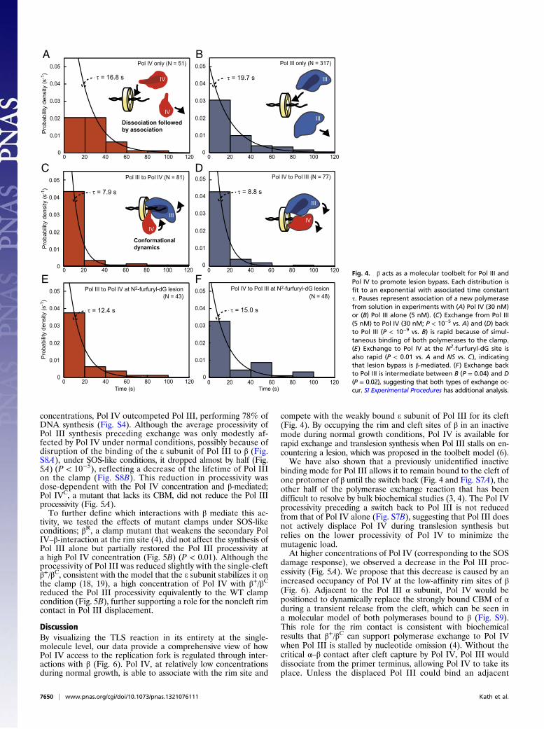
Kinetics of Polymerase Exchange Support the Toolbelt Model. Theobservation of exchange from Pol III to Pol IV and back isuniquely accessible in our single-molecule reconstitution andpermits us to investigate the role of β in polymerase trafficking.In the toolbelt model, polymerase exchange is only limited by thetimescale of conformational changes of Pol III and Pol IV si-multaneously bound to β. In an alternative model, in which stericeffects prevent concurrent binding, exchange requires dissocia-tion of the first polymerase followed by recruitment of the sec-ond from solution, which would be sensitive to protein dilution.Quantifying exchange by measuring the time between the ter-mination of synthesis by one polymerase and the subsequentinitiation of synthesis by the other allows us to distinguish be-tween these two models.The time for exchange from Pol III to Pol IV on undamaged
DNA (Fig. 4C) was more rapid than the diffusion-limited re-cruitment time of Pol IV from solution (Fig. 4A and Fig. S2)seen in exponential fits and a statistical comparison of thetwo datasets (P < 10−5) (SI Experimental Procedures). Fur-thermore, reducing the concentration of Pol IV in exchangeexperiments from 30 to 15 nM did not affect the timescale ofexchange (Fig. S6), whereas the same dilution increased pausetimes in experiments with Pol IV alone (Fig. 4A and Fig. S2E).These data argue that exchange during active synthesis occursbetween two polymerases bound to the clamp. Our observationof β-mediated exchange implies that the second Pol III CBM inthe e subunit does not exclude Pol IV from binding the clampin the absence of a lesion-induced stall in contrast to a previoussuggestion (19); rather, Pol IV can compete with e for a cleft,allowing Pol IV to bind β while Pol III is synthesizing DNA.Importantly, we observed that exchange back to Pol III after
Pol IV synthesis was also more rapid than the recruitment timeof Pol III from solution (Fig. 4 B and D) (P < 10−9). Further-more, exchange from Pol IV to Pol III in the presence of thesingle-cleft β+/βC (Fig. S7A, τ = 27.3 s) was slower than inexperiments with the WT clamp (P < 10−3), closely matching therecruitment time of Pol III from solution [not significant (NS) vs.Fig. 4B]. These data show that Pol III can bind the opposing cleft
of β in an inactive conformation while Pol IV is carrying outsynthesis and that eliminating the second cleft abolishes rapidexchange. The fact that the Pol IV processivity preceding exchangeto Pol III matches that of Pol IV on β+/βC (Figs. S3B and S7B)shows that a single Pol IV is bound to β in the presence of Pol IIIand strongly implies that Pol III does not displace Pol IV duringthe exchange back but takes over after Pol IV stochasticallyreleases DNA.To test if polymerase exchange can occur in the physiological
context of Pol III encountering a DNA lesion, we collected datafor exchange from Pol III to Pol IV within the experimentalresolution (±200 bp) of the N2-furfuryl-dG position (Fig. 3B).These exchange times (Fig. 4E) matched the times for exchangeon undamaged DNA (NS vs. Fig. 4C) and were more rapid thanrecruitment of Pol IV from solution (P < 0.01 vs. Fig. 4A), in-dicating that Pol IV was bound to β when Pol III encountered thelesion. The switch back to Pol III after the lesion (Fig. 4F) wasintermediate between rapid β-mediated exchange and recruitmentfrom solution, suggesting a mixture of these two types of exchange.
Binding of Pol IV at a Secondary Site on β Reduces the Pol IIIProcessivity. Our results support a model in which Pol IV, ata ratio to Pol III consistent with normal growth, can bind β in aninactive conformation, thereby promoting rapid bypass of DNAlesions encountered during synthesis. During the SOS DNAdamage response, however, the cellular concentration of PolIV increases roughly 10-fold, whereas Pol III levels remainconstant (2). To test if an increased ratio of Pol IV to Pol IIIalters polymerase exchange, we performed primer extensionexperiments with 5 nM Pol III and 300 nM Pol IV. At these
0 100 200 300 400 500 600 0 0.01 0.02 0.03 0.04 0.05 0.06 0.07
Rate (bp/s)
Pro
babi
lity
dens
ity (b
p-1 s
)
Pol IV (N = 86)
Pol III (x20) (N = 470)
A
0 50 100 150 200 250 3000
1000
2000
3000
4000
350
Pol III synthesis Pol IV synthesis Pause
Time (s)
DN
A S
ynth
esis
(bp)
B
Rate = 222 ± 6.9 bp/s
Rate = 10.8 ± 1.2 bp/s
Fig. 2. Observing exchange between Pol III and Pol IV. (A) Rate distributionsfor Pol III (blue) and Pol IV (red); values represent means ± SEMs. Probabilitydensities for Pol III are multiplied by 20 to facilitate comparison. (B) Sampletrajectories of rapid exchange between Pol III and Pol IV.
A x 10-4
5
4
3
2
1
0 1600 3200 4800 6400Total synthesis (bp)
Pro
babi
lity
dens
ity (b
p-1) Pol III only
Pol III and Pol IV
DN
A S
ynth
esis
(bp)
Time (s)0 50 100 150 200 250 300
± 200 bp of lesion
40003500300025002000
10001500
B
Pol IVPol III
Pol III
8000
Lesion Full length
Fig. 3. A single-molecule reconstitution of polymerase exchange and by-pass at a DNA lesion. (A) An N2-furfuryl-dG lesion at ∼3,150 bp blocksprocessive synthesis by Pol III (5 nM; n = 69) but is bypassed when Pol IV(30 nM) is added (n = 175). (B) Rapid exchange from Pol III to Pol IV and backis observed at the lesion site.
Kath et al. PNAS | May 27, 2014 | vol. 111 | no. 21 | 7649
BIOCH
EMISTR
Y
concentrations, Pol IV outcompeted Pol III, performing 78% ofDNA synthesis (Fig. S4). Although the average processivity ofPol III synthesis preceding exchange was only modestly af-fected by Pol IV under normal conditions, possibly because ofdisruption of the binding of the e subunit of Pol III to β (Fig.S8A), under SOS-like conditions, it dropped almost by half (Fig.5A) (P < 10−5), reflecting a decrease of the lifetime of Pol IIIon the clamp (Fig. S8B). This reduction in processivity wasdose-dependent with the Pol IV concentration and β-mediated;Pol IVC, a mutant that lacks its CBM, did not reduce the Pol IIIprocessivity (Fig. 5A).To further define which interactions with β mediate this ac-
tivity, we tested the effects of mutant clamps under SOS-likeconditions; βR, a clamp mutant that weakens the secondary PolIV–β-interaction at the rim site (4), did not affect the synthesis ofPol III alone but partially restored the Pol III processivity ata high Pol IV concentration (Fig. 5B) (P < 0.01). Although theprocessivity of Pol III was reduced slightly with the single-cleftβ+/βC, consistent with the model that the e subunit stabilizes it onthe clamp (18, 19), a high concentration of Pol IV with β+/βCreduced the Pol III processivity equivalently to the WT clampcondition (Fig. 5B), further supporting a role for the noncleft rimcontact in Pol III displacement.
DiscussionBy visualizing the TLS reaction in its entirety at the single-molecule level, our data provide a comprehensive view of howPol IV access to the replication fork is regulated through inter-actions with β (Fig. 6). Pol IV, at relatively low concentrationsduring normal growth, is able to associate with the rim site and
compete with the weakly bound e subunit of Pol III for its cleft(Fig. 4). By occupying the rim and cleft sites of β in an inactivemode during normal growth conditions, Pol IV is available forrapid exchange and translesion synthesis when Pol III stalls on en-countering a lesion, which was proposed in the toolbelt model (6).We have also shown that a previously unidentified inactive
binding mode for Pol III allows it to remain bound to the cleft ofone protomer of β until the switch back (Fig. 4 and Fig. S7A), theother half of the polymerase exchange reaction that has beendifficult to resolve by bulk biochemical studies (3, 4). The Pol IVprocessivity preceding a switch back to Pol III is not reducedfrom that of Pol IV alone (Fig. S7B), suggesting that Pol III doesnot actively displace Pol IV during translesion synthesis butrelies on the lower processivity of Pol IV to minimize themutagenic load.At higher concentrations of Pol IV (corresponding to the SOS
damage response), we observed a decrease in the Pol III proc-essivity (Fig. 5A). We propose that this decrease is caused by anincreased occupancy of Pol IV at the low-affinity rim sites of β(Fig. 6). Adjacent to the Pol III α subunit, Pol IV would bepositioned to dynamically replace the strongly bound CBM of αduring a transient release from the cleft, which can be seen ina molecular model of both polymerases bound to β (Fig. S9).This role for the rim contact is consistent with biochemicalresults that β+/βC can support polymerase exchange to Pol IVwhen Pol III is stalled by nucleotide omission (4). Without thecritical α–β contact after cleft capture by Pol IV, Pol III woulddissociate from the primer terminus, allowing Pol IV to take itsplace. Unless the displaced Pol III could bind an adjacent
0
0.01
0.02
0.03
0.04
0.05
0 20 40 60 80 100 120
0
0.01
0.02
0.03
0.04
0.05
0 20 40 60 80 100 120
0
0.01
0.02
0.03
0.04
0.05
0 20 40 60 80 100 120
0
0.01
0.02
0.03
0.04
0.05
0 20 40 60 80 100 120
0
0.01
0.02
0.03
0.04
0.05
0 20 40 60 80 100 120
0
0.01
0.02
0.03
0.04
0.05
0 20 40 60 80 100 120
Pro
babi
lity
dens
ity (s
-1)
AP
roba
bilit
y de
nsity
(s-1
)ytisned
ytilibaborP
(s-1
)
C
E
B
D
F
Dissociation followedby association
III
IV
Conformationaldynamics
Pol IV only (N = 51) Pol III only (N = 317)
III
III
III
Pol IV to Pol III (N = 77)
III
III
IV
Pol III to Pol IV (N = 81)
Pol III to Pol IV at N2-furfuryl-dG lesion Pol IV to Pol III at N2-furfuryl-dG lesion
Time (s) Time (s)
(N = 43) (N = 48)
IV
IVτ = 16.8 s τ = 19.7 s
τ = 12.4 s
τ = 7.9 s
τ = 15.0 s
τ = 8.8 s
Fig. 4. β acts as a molecular toolbelt for Pol III andPol IV to promote lesion bypass. Each distribution isfit to an exponential with associated time constantτ. Pauses represent association of a new polymerasefrom solution in experiments with (A) Pol IV (30 nM)or (B) Pol III alone (5 nM). (C) Exchange from Pol III(5 nM) to Pol IV (30 nM; P < 10−5 vs. A) and (D) backto Pol III (P < 10−9 vs. B) is rapid because of simul-taneous binding of both polymerases to the clamp.(E) Exchange to Pol IV at the N2-furfuryl-dG site isalso rapid (P < 0.01 vs. A and NS vs. C), indicatingthat lesion bypass is β-mediated. (F) Exchange backto Pol III is intermediate between B (P = 0.04) and D(P = 0.02), suggesting that both types of exchange oc-cur. SI Experimental Procedures has additional analysis.
7650 | www.pnas.org/cgi/doi/10.1073/pnas.1321076111 Kath et al.

unoccupied protomer or is stabilized by additional interactions, itwould likely dissociate from the clamp entirely.The requirement of the Pol IV CBM indicates that binding at
rim sites is not sufficient for a reduction of the Pol III lifetime onDNA and that Pol IV must also compete for the cleft bound byα. Shared contacts, such as a single cleft of β during competitionbetween Pol III and Pol IV bound at additional sites on β, havebeen proposed to be important in facilitating dissociation andsubunit exchange in multiprotein complexes upon transientcontact release (30). This phenomenon has also been observed inthe dynamic processivity of phage T4 and T7 replication, whereadditional polymerases are able to associate with moving repli-somes and undergo exchange on a timescale faster than that ofstochastic dissociation of the synthesizing polymerase (31–33),and the facilitated dissociation of the E. coli DNA-binding pro-tein Fis by nucleoid proteins (34). Secondary contacts, such asthe rim site for Pol IV shown here, play an important role inorienting proteins to exploit transient changes in occupancy ofthese shared sites, resulting in binding partner exchange.During coordinated leading and lagging strand replication,
a displaced Pol III may remain associated with the replisomethrough additional contacts with the clamp loader complex.These contacts, however, do not seem to prevent Pol IV fromaccessing β; previous biochemical experiments with a fullyreconstituted replisome have shown that Pol IV can replace PolIII (35). Furthermore, overexpression of Pol IV beyond SOSlevels in cells has been shown to arrest replication and inducetoxicity because of unregulated access of Pol IV to the replica-tion fork (15, 35, 36). Removing the CBM residues alleviatesPol IV toxicity, whereas mutating the rim-contacting residues
partially alleviates it (16). These data are explained by ourmodel: contacts with the rim site and subsequently, the cleftprovide a molecular path for Pol IV to displace Pol III from theprimer terminus after SOS induction.A putative interaction with the rim site could also position the
other E. coli Y-family polymerase, Pol V, on the clamp when it isexpressed later in the SOS damage response (37). This bindingactivity would create a hierarchy for access to the primer ter-minus, a view of the toolbelt model in which clamp–polymeraseinteractions do more than merely increase the local polymeraseconcentration at the DNA template. During normal growthconditions, Pol III, which is preferentially loaded to the primerterminus (38), performs the majority of DNA synthesis. Pol IV isable to simultaneously bind β in an inactive mode, although it iscurrently unclear how other β-interacting proteins would in-fluence its occupancy in vivo. After SOS induction, the rim sitepositions Pol IV to preferentially bind a cleft of β when itbecomes available. Such a competitive advantage would ensuretimely access of Y-family polymerases to the primer templateand is likely to be important with several proteins competing foran open cleft.Noncleft contacts may play a similar role in regulating access
to the DNA template in other domains of life. PCNA plays a keyrole in coordinating the handoff of DNA intermediates betweena polymerase, flap endonuclease, and ligase during both Okazakifragment maturation and long-patch base excision repair in eukar-yotes (39). Structural studies have revealed that Flap endonuclease-1,Polymerase β, and Ligase I bind overlapping but distinct regionsof DNA intermediates, which may facilitate displacement duringhandoff (40). In the archaeon Sulfolobus solfataricus, the threeenzymes each bind distinct monomers of the heterotrimeric PCNAduring Okazaki fragment maturation (41). However suggestive, itremains unknown if the homotrimeric eukaryotic PCNA can si-multaneously bind any combination of these three proteins.Eukaryotic TLS is regulated, in part, by ubiquitination of PCNA;
all four human Y-family polymerases have ubiquitin-binding do-mains (1). Structural data of monoubiquitinated PCNA and itsconformations support the model that the secondary ubiquitin siteallows the TLS polymerase Pol η to bind an occupied clamp andposition it to compete for the cleft on transient dissociation of thereplicative polymerase (42, 43). In contrast to bacteria, where theoccupancy of the rim site is controlled by polymerase concentration,analogous sites in eukaryotes are introduced by posttranslationalmodification. We anticipate that the single-molecule approachesdescribed here will serve as powerful tools to elucidate the roleof these interactions in translesion synthesis.
A
B
500600700
400
900800
Pol
III p
roce
ssiv
ity (b
p)
0 50 100 150 200 250 300 [Pol IV] (nM)[IV]/[III]0 10 20 30 40 50 60
200
400
600
800
1000
1200
0 nM Pol IV 300 nM Pol IV
Pol
III p
roce
ssiv
ity (b
p)
β+/βC
βR
βWT
1000
300200100
0
Pol IVPol IVC
Fig. 5. Binding of Pol IV at a secondary site on β reduces the Pol III proc-essivity. (A) The Pol III processivity decreases with increasing concentrationsof Pol IV (●) but is unaffected by Pol IVC, a mutant lacking its CBM (▲). (B)Reduction in the Pol III processivity with βWT (black bars) also occurs with thesingle-cleft mutant clamp β+/βC (gray bars; NS) but is partially alleviated byβR, a clamp with a weakened Pol IV-interacting rim interface (cross-hatchedbars; P < 0.01). N ranges from 71 to 470 for A and B, respectively. Valuesrepresent means ± SEMs.
β clamp
cleft
rim Pol IV
Pol III
Normal growth conditionsRatio of [Pol IV] to [Pol III] ~10:1
SOS damage-induced conditionsRatio of [Pol IV] to [Pol III] ~100:1
WEAK
(3) capture of cleft from α
(2) transient dissociation of α CBM
IIIα
ε
IV
(1) transient association at rim sites
(3) exchange and TLS
N2-dG lesion
(1) increased occupancy at rim sites
(4) Pol III dissociates, Pol IV binds primer
(2) capture of cleft from
STRONG
Fig. 6. A model for how the Pol IV occupancy at rim sites and competitionwith Pol III subunits dictate polymerase exchange at different Pol IV con-centrations. The small θ subunit of Pol III, which binds e, is not representedfor clarity.
Kath et al. PNAS | May 27, 2014 | vol. 111 | no. 21 | 7651
BIOCH
EMISTR
Y
Experimental ProceduresProteins and Buffers. Pol III core (αeθ), Pol IV, β (WT and mutants), and otherE. coli replisome components were expressed and purified as described in SIExperimental Procedures. Experiments were performed in replication buffer[50 mM Hepes-KOH, pH 7.9, 12 mM Mg(OAc)2, 80 mM KCl, 0.1 mg mL−1 BSA]with 5 mM DTT, 1 mM ATP, 760 μM dNTPs, 15 nM τ3δδ′χψ, 30 nM clamp (β, βR,or β+/βC as dimers), and the indicated concentrations of Pol III and/or Pol IV;60 μM dNTPs were used for single-molecule lesion bypass experiments. Single-stranded DNA-binding (SSB) protein was excluded from primer extensionexperiments, because SSB protein extends ssDNA at low force, reducing thecontrast with dsDNA that is used to observe replication (44).
ssDNA Constructs. DNA constructs were generated from circular M13 ssDNAwith either M13mp18 ssDNA (New England Biolabs) or purified M13mp7(L2)containing a site-specific N2-furfuryl-dG lesion and constructed using a pre-viously described protocol (27). M13mp7(L2) phage stock was a gift fromJohn Essigmann (Massachusetts Institute of Technology, Cambridge, MA).Additional details on substrate construction and a list of oligonucleotidesused (Table S1) can be found in SI Experimental Procedures.
Single-Molecule Experiments. Single-molecule primer extension experimentswere performed in custom microfluidic flow cells constructed with function-alized glass coverslips as described previously (45). Statistical comparisons weremade between full datasets with the two-tailed Wilcoxon rank sum test usingthe Matlab function ranksum. A significance level of P = 0.05 was used. Ad-ditional details on the experimental approach and data analysis are describedin SI Experimental Procedures.
ACKNOWLEDGMENTS. We thank Jamie Foti for providing purified Pol IV,Deyu Li for assistance with purifying the N2-furfuryl-dG oligonucleotide, andJohannes Walter and Seungwoo Chang for helpful discussions and criticalreading of the manuscript. This work was funded by National Institutes ofHealth Grants T32 GM008313 (to J.E.K.), R01 GM066094 (to M.D.S.), R01CA021615 (to G.C.W.), and P30ES002019 (to G.C.W.); a National ScienceFoundation Graduate Research Fellowship (to J.E.K.); the Smith FamilyAward in Biomedical Excellence (to J.J.L.); the Stuart Trust Fellows Award(to J.J.L.); and Australian Research Council Grant DP0877658, including anAustralian Professorial Fellowship (to N.E.D.). G.C.W. is an American CancerSociety Professor.
1. Sale JE, Lehmann AR, Woodgate R (2012) Y-family DNA polymerases and their role intolerance of cellular DNA damage. Nat Rev Mol Cell Biol 13(3):141–152.
2. Sutton MD (2010) Coordinating DNA polymerase traffic during high and low fidelitysynthesis. Biochim Biophys Acta 1804(5):1167–1179.
3. Indiani C, McInerney P, Georgescu R, Goodman MF, O’Donnell M (2005) A sliding-clamp toolbelt binds high- and low-fidelity DNA polymerases simultaneously.Mol Cell19(6):805–815.
4. Heltzel JM, Maul RW, Scouten Ponticelli SK, Sutton MD (2009) A model for DNApolymerase switching involving a single cleft and the rim of the sliding clamp. ProcNatl Acad Sci USA 106(31):12664–12669.
5. Zhuang Z, et al. (2008) Regulation of polymerase exchange between Pol eta and Poldelta by monoubiquitination of PCNA and the movement of DNA polymerase holo-enzyme. Proc Natl Acad Sci USA 105(14):5361–5366.
6. Pagès V, Fuchs RP (2002) How DNA lesions are turned into mutations within cells?Oncogene 21(58):8957–8966.
7. Wijffels G, et al. (2004) Inhibition of protein interactions with the β2 sliding clamp ofEscherichia coli DNA polymerase III by peptides from β2-binding proteins. Bio-chemistry 43(19):5661–5671.
8. Moldovan GL, Pfander B, Jentsch S (2007) PCNA, the maestro of the replication fork.Cell 129(4):665–679.
9. Ohmori H, et al. (2001) The Y-family of DNA polymerases. Mol Cell 8(1):7–8.10. Jarosz DF, Godoy VG, Delaney JC, Essigmann JM, Walker GC (2006) A single amino
acid governs enhanced activity of DinB DNA polymerases on damaged templates.Nature 439(7073):225–228.
11. McKenzie GJ, Lee PL, Lombardo MJ, Hastings PJ, Rosenberg SM (2001) SOS mutatorDNA polymerase IV functions in adaptive mutation and not adaptive amplification.Mol Cell 7(3):571–579.
12. Slechta ES, et al. (2003) Adaptive mutation: General mutagenesis is not a pro-grammed response to stress but results from rare coamplification of dinB with lac.Proc Natl Acad Sci USA 100(22):12847–12852.
13. Ponder RG, Fonville NC, Rosenberg SM (2005) A switch from high-fidelity to error-prone DNA double-strand break repair underlies stress-induced mutation. Mol Cell19(6):791–804.
14. Pomerantz RT, Kurth I, Goodman MF, O’Donnell ME (2013) Preferential D-loop ex-tension by a translesion DNA polymerase underlies error-prone recombination. NatStruct Mol Biol 20(6):748–755.
15. Foti JJ, Devadoss B, Winkler JA, Collins JJ, Walker GC (2012) Oxidation of the guaninenucleotide pool underlies cell death by bactericidal antibiotics. Science 336(6079):315–319.
16. Wagner J, Etienne H, Fuchs RP, Cordonnier A, Burnouf D (2009) Distinct β-clamp in-teractions govern the activities of the Y family PolIV DNA polymerase. Mol Microbiol74(5):1143–1151.
17. Bunting KA, Roe SM, Pearl LH (2003) Structural basis for recruitment of translesionDNA polymerase Pol IV/DinB to the β-clamp. EMBO J 22(21):5883–5892.
18. Jergic S, et al. (2013) A direct proofreader-clamp interaction stabilizes the Pol IIIreplicase in the polymerization mode. EMBO J 32(9):1322–1333.
19. Toste Rêgo A, Holding AN, Kent H, Lamers MH (2013) Architecture of the Pol III-clamp-exonuclease complex reveals key roles of the exonuclease subunit in processiveDNA synthesis and repair. EMBO J 32(9):1334–1343.
20. Wuite GJ, Smith SB, Young M, Keller D, Bustamante C (2000) Single-molecule studiesof the effect of template tension on T7 DNA polymerase activity. Nature 404(6773):103–106.
21. Maier B, Bensimon D, Croquette V (2000) Replication by a single DNA polymerase ofa stretched single-stranded DNA. Proc Natl Acad Sci USA 97(22):12002–12007.
22. Tanner NA, et al. (2008) Single-molecule studies of fork dynamics in Escherichia coliDNA replication. Nat Struct Mol Biol 15(2):170–176.
23. Wagner J, Fujii S, Gruz P, Nohmi T, Fuchs RP (2000) The β clamp targets DNA poly-merase IV to DNA and strongly increases its processivity. EMBO Rep 1(6):484–488.
24. Lamers MH, Georgescu RE, Lee S-G, O’Donnell M, Kuriyan J (2006) Crystal structure ofthe catalytic α subunit of E. coli replicative DNA polymerase III. Cell 126(5):881–892.
25. Ozawa K, et al. (2013) Proofreading exonuclease on a tether: The complex betweenthe E. coli DNA polymerase III subunits α, epsilon, θ and β reveals a highly flexiblearrangement of the proofreading domain. Nucleic Acids Res 41(10):5354–5367.
26. Scouten Ponticelli SK, Duzen JM, Sutton MD (2009) Contributions of the individualhydrophobic clefts of the Escherichia coli β sliding clamp to clamp loading, DNAreplication and clamp recycling. Nucleic Acids Res 37(9):2796–2809.
27. Delaney JC, Essigmann JM (2006) Assays for determining lesion bypass efficiency andmutagenicity of site-specific DNA lesions in vivo. Methods Enzymol 408:1–15.
28. Tomer G, Reuven NB, Livneh Z (1998) The β subunit sliding DNA clamp is responsiblefor unassisted mutagenic translesion replication by DNA polymerase III holoenzyme.Proc Natl Acad Sci USA 95(24):14106–14111.
29. Gon S, Napolitano R, Rocha W, Coulon S, Fuchs RP (2011) Increase in dNTP pool sizeduring the DNA damage response plays a key role in spontaneous and induced-mu-tagenesis in Escherichia coli. Proc Natl Acad Sci USA 108(48):19311–19316.
30. Ha T (2013) Single-molecule approaches embrace molecular cohorts. Cell 154(4):723–726.
31. Yang J, Zhuang Z, Roccasecca RM, Trakselis MA, Benkovic SJ (2004) The dynamicprocessivity of the T4 DNA polymerase during replication. Proc Natl Acad Sci USA101(22):8289–8294.
32. Hamdan SM, et al. (2007) Dynamic DNA helicase-DNA polymerase interactions assureprocessive replication fork movement. Mol Cell 27(4):539–549.
33. Loparo JJ, Kulczyk AW, Richardson CC, van Oijen AM (2011) Simultaneous single-molecule measurements of phage T7 replisome composition and function reveal themechanism of polymerase exchange. Proc Natl Acad Sci USA 108(9):3584–3589.
34. Graham JS, Johnson RC, Marko JF (2011) Concentration-dependent exchange accel-erates turnover of proteins bound to double-stranded DNA. Nucleic Acids Res 39(6):2249–2259.
35. Indiani C, Langston LD, Yurieva O, GoodmanMF, O’Donnell M (2009) Translesion DNApolymerases remodel the replisome and alter the speed of the replicative helicase.Proc Natl Acad Sci USA 106(15):6031–6038.
36. Uchida K, et al. (2008) Overproduction of Escherichia coli DNA polymerase DinB (PolIV) inhibits replication fork progression and is lethal. Mol Microbiol 70(3):608–622.
37. Beuning PJ, Sawicka D, Barsky D, Walker GC (2006) Two processivity clamp inter-actions differentially alter the dual activities of UmuC. Mol Microbiol 59(2):460–474.
38. Downey CD, McHenry CS (2010) Chaperoning of a replicative polymerase ontoa newly assembled DNA-bound sliding clamp by the clamp loader. Mol Cell 37(4):481–491.
39. Chapados BR, et al. (2004) Structural basis for FEN-1 substrate specificity and PCNA-mediated activation in DNA replication and repair. Cell 116(1):39–50.
40. Tsutakawa SE, et al. (2011) Human flap endonuclease structures, DNA double-baseflipping, and a unified understanding of the FEN1 superfamily. Cell 145(2):198–211.
41. Beattie TR, Bell SD (2012) Coordination of multiple enzyme activities by a single PCNAin archaeal Okazaki fragment maturation. EMBO J 31(6):1556–1567.
42. Freudenthal BD, Gakhar L, Ramaswamy S, Washington MT (2010) Structure of mon-oubiquitinated PCNA and implications for translesion synthesis and DNA polymeraseexchange. Nat Struct Mol Biol 17(4):479–484.
43. Tsutakawa SE, et al. (2011) Solution X-ray scattering combined with computationalmodeling reveals multiple conformations of covalently bound ubiquitin on PCNA.Proc Natl Acad Sci USA 108(43):17672–17677.
44. Fu H, Le S, Chen H, Muniyappa K, Yan J (2013) Force and ATP hydrolysis dependentregulation of RecA nucleoprotein filament by single-stranded DNA binding protein.Nucleic Acids Res 41(2):924–932.
45. Tanner NA, van Oijen AM (2010) Visualizing DNA replication at the single-moleculelevel. Methods Enzymol 475:259–278.
7652 | www.pnas.org/cgi/doi/10.1073/pnas.1321076111 Kath et al.

Supporting InformationKath et al. 10.1073/pnas.1321076111SI Experimental ProceduresProteins and Buffers. Escherichia coli proteins were purified fromoverproducing strains as previously described and untaggedunless otherwise noted: polymerase (Pol) IV (1) and Pol IVC
(ΔC5) (2); the Pol III holoenzyme subunits α, δ, and δ′ (3);e and θ (4); the WT clamp β (5), N-terminally his6- and heartmuscle kinase-tagged βR (E93K-L98K), and β+/βC, a stable dimerformed from N-terminally Myc-tagged β and his6- and heartmuscle kinase-tagged β(ΔC5) (6); and τ and refolded ψ withinthe χψ-complex (7). The Pol III αeθ core and clamp loader as-sembly with the stoichiometry τ3δδ′χψ were each reconstitutedand purified following reported protocols (7).The quality of purified protein was determined by comparing
the activities of multiple preparations. A fraction of inactive ormisfolded polymerases, inevitable in purification, would eitherfail to bind the clamp and not be observed in synthesis or bind andresult in termination events or extended pauses. Because ourconclusions depend on the reduction of pause times because ofpolymerase exchange, they are unlikely to be affected by inactiveprotein. If the fractions of active protein in the Pol III and Pol IVpreparations used differed, the molar ratio of active Pol IV toactive Pol III would differ from the ratio determined by proteinconcentration (Fig. 5) by a constant factor throughout this workand similarly not affect our conclusions.
Synthesis of an N2-furfuryl-dG–Containing Oligonucleotide. The 20-mer oligonucleotide containing the N2-furfuryl-dG lesion wasconstructed as previously described (8). A 20-mer oligonucleo-tide containing a fluoro substituent at the N2 position of guanine(X) was purchased from Chemgenes: 5′-CTA CCT XTG GACGGC TGC GA-3′. A fraction of the product was found tocontain a hydroxyl group in place of the fluorine, the result ofNaOH treatment to separate the oligonucleotide from the resinduring manufacture. The fluorine was displaced with a fufurylgroup by treating the oligonucleotide with fufurylamine, leavingthe contaminating N2-hydroxyl-dG unaffected. Fufurylaminetreatment was followed by HPLC and MALDI-TOF MS as de-scribed (8) to remove the hydroxyl-containing oligonucleotide toobtain a pure 20-mer with the N2-fufuryl-dG lesion.
M13 ssDNA with a Site-Specific DNA Lesion. ssDNA with a site-specific DNA lesion was constructed using M13mp7(L2), a mu-tant phage that contains an EcoRI site within a stable hairpin in itsgenome (Fig. S1B). Phage stock was a gift from John Essigmann(Massachusetts Institute of Technology, Cambridge, MA). Aprotocol to purify phage genomes and ligate a lesion-containingoligonucleotide at the digested EcoRI site (9) was adapted forthis study with the following modifications. After PEG pre-cipitation, the isolated phage pellet was extracted two or threetimes with 25:24:1 phenol:chloroform:isoamyl alcohol and onetime with pure chloroform. DNA in the final aqueous layer wasethanol-precipitated and redissolved in 10 mM Tris (pH 8.5)buffer to ∼2 μg μL−1; 100 μg M13mp7(L2) DNA was linearized ina 100-μL digestion reaction with 40 U EcoRI-HF (New EnglandBiolabs) and 1× Buffer 4 at 23 °C for 8 h and purified with se-quential phenol:chloroform:isoamyl alcohol and chloroform ex-tractions and an ethanol precipitation. Then, it was dissolved in100 μL Tris buffer. Purification of linear ssDNA prevented deg-radation in later steps.Thirty picomoles 5′-phosphorylated oligonucleotide insert 5′-
CTA CCT XTG GAC GGC TGC GA-3′ (X = N2-furfuryl-dG orthe dG control) was ligated at 16 °C overnight into 20 pmol
purified linear ssDNA using annealed scaffold oligonucleotides5′-AAA ACG ACG GCC AGT GAA TTT CGC AGC CGT CC-3′ and 5′-GGT AGA CTG AAT CAT GGT CAT AGC-3′ (25pmol each) and 800 U T4 DNA ligase (New England Biolabs) ina 60-μL reaction. To remove the scaffold oligonucleotides, un-ligated linear M13 DNA, and excess insert, the mixture wassubsequently treated at 37 °C for 4 h with 18 U T4 DNA poly-merase and 80 U exonuclease I (New England Biolabs). TheDNA was finally purified by sequential phenol:chloroform:iso-amyl alcohol and chloroform extractions, ethanol-precipitated,and dissolved in 50 μL 10 mM Tris buffer to obtain the lesion-containing (or control) phage ssDNA.The progress of DNA construction was monitored by taking
samples at each step and separating them on a 0.8% Tris-acetate-EDTA agarose gel stained with ethidium bromide (Fig. S5C). Asa control to guarantee that EcoRI completely linearized thehairpin-containing M13mp7(L2) ssDNA, which if uncut, couldcontaminate lesion-containing DNA, a mock ligation reactionwas performed with digested ssDNA and scaffolds but withoutthe insert. The mock reaction was then treated with T4 DNApolymerase and Exo I, which together degrade linear but notcircular ssDNA. Degradation was nearly complete, showing ef-ficient digestion of the hairpin (Fig. S5C).
Single-Molecule DNA Substrates. Linear end-labeled single-moleculeDNA substrates were constructed using circular 7.2-kb M13 phagegenomes (Fig. S1A). Experiments on undamaged DNA used sub-strates generated from M13mp18 ssDNA (New England Biolabs).Sixteen microliters this DNA (250 ng μL−1) was annealed in 20 μLwith 1 μMoligonucleotide mp18-SalI (Table S1) by heating to 65 °Cfor 10 min and slowly cooling to room temperature. Ten microlitersannealed DNA was linearized at the dsDNA region with 10 U SalI(New England Biolabs) and 1× Buffer 3 in 50 μL at 37 °C for 1 h.Forty microliters restriction digest reaction was mixed to a finalvolume of 55 μL with 30 nM end-labeled oligonucleotides M13-5′-biotin and phosphorylated M13-3′-dig and 30 nM scaffolding oli-gonucleotides mp18-scaffold-1 and mp18-scaffold-2 (Table S1). Thescaffolds were annealed to the linearized phage DNA and the end-labeled oligonucleotides by heating to 65 °C for 20 min and coolingto room temperature, also inactivating SalI; scaffold-mediated li-gation was subsequently performed overnight at 16 °C with 400 UDNA ligase (New England Biolabs). The reaction was stopped byheat-inactivating ligase at 65 °C for 10 min and adding EDTA (20mM final) to the cooled mixture. The stock solution (final substrateconcentration ∼ 5 nM) was stored at 4 °C.Single-molecule DNA substrates containing a site-specific le-
sion (Fig. S1B) were prepared similarly from lesion-containingM13mp7(L2) DNA (see above). ssDNA containing the N2-furfuryl-dG lesion or the control dG was annealed with mp7L2-AlwNI(Table S1) and digested with 20 U AlwNI (New England Biolabs) in1× Buffer 4 at 37 °C for 1 h. Linearized DNA was ligated toM13-5′-biotin and phosphorylated M13-3′-dig using the scaffolding oligo-nucleotides mp7L2-scaffold-1 and mp7L2-scaffold-2 (Table S1).The annealed oligonucleotide mp18-scaffold-1 (for the un-
damaged substrate) or mp7L2-scaffold-1 (for the lesion-containingsubstrate) near the 3′ terminus of the linear M13 template servedas the primer for DNA synthesis. For the lesion-containing sub-strate, the primer terminus is situated ∼3,150 nt from the N2-furfuryl-dG site.
Single-Molecule Flow Stretching Experiments. Single-molecule ex-periments were performed at 23 °C using custom microfluidic
Kath et al. www.pnas.org/cgi/content/short/1321076111 1 of 10

flow cells with glass coverslips as described previously (10). Insummary, primed end-labeled M13 bacteriophage ssDNA wasattached to the glass surface of the flow cell by a biotin-streptavidinlinkage on one end and a micrometer-scale bead (tosyl-activated,2.8-μm diameter; Dynal) by a digoxigenin–antidigoxigenin inter-action on the other end. A magnet exerts a weak force of ∼1 pN tolift the paramagnetic bead off the surface, and laminar flow ofbuffer through the flow cell exerts a constant force of ∼3 pN on thebead and by extension, uniformly throughout the DNA tether. Atthis low force, ssDNA is entropically coiled, whereas dsDNA isstretched to nearly its crystallographic length (Fig. S1C). Conver-sion of ssDNA to dsDNA during primer extension can, therefore,be observed and tracked by extension of the DNA tether andmovement of the coupled bead.Glass coverslips were functionalized with a ratio of biotinylated
PEG succinimidyl valerate:methyl-PEG succinimidyl valerate(Laysan Bio) of 0.75%:15% (wt/vol) in 0.1 M NaHCO3 (pH 8.2)(10). Dried coverslips, stored under vacuum, were stable forseveral months.Before an experiment, the functionalized coverslip surface was
incubated with 0.2 mg mL−1 streptavidin (Sigma) in PBS for 30min and then washed and incubated with blocking buffer (20 mMTris·HCl, pH 7.5, 50 mM NaCl, 2 mM EDTA, 0.2 mg mL−1
BSA, 0.005% Tween 20) for an additional 30 min; 2–4 μL M13substrate stock (∼5 nM; see above) was diluted with 500 μLblocking buffer and drawn into the flow cell at 0.025 mL min−1
with a syringe pump (Harvard Apparatus 11 Plus), allowingbinding of DNA by the 5′-biotinylated ends to immobilizedstreptavidin sites. A stock of α-digoxigenin–functionalized poly-styrene beads (tosyl-activated, 2.8-μm diameter; Dynal) wasprepared as previously described (10); 2 μL bead stock was di-luted with 500 μL blocking buffer and drawn into the flow cell at0.025 mL min−1 to specifically bind the 3′-digoxigenin–labeledDNA substrates. Excess beads and DNA were removed from theflow cell by washing with 1 mL blocking buffer (>100 volumes) at0.035 mL min−1.Immediately before the synthesis reaction, ∼150 μL replication
buffer was introduced to exchange buffer. A solution of 500 μLreplication buffer with replication proteins and nucleotides wasadded at 0.015 mL min−1. A magnet exerting a weak force of ∼1 pNwas used to lift the tethered paramagnetic beads off the surface;laminar flow at this rate through the flow cell exerts a constantforce of ∼3 pN on the tether. After 2 min to allow the flow tostabilize, several hundred beads were visualized using dark-fieldmicroscopy through a 10× objective (Olympus) and imaged witha QIClick CCD camera (Q-Imaging). Data were recorded for2,750 frames at 2 Hz using the software package Micro-Manager(www.micro-manager.org).Primer extension on each individual molecule was observed by
the motion of its bead in the direction of flow as the entropicallycoiled ssDNA was converted to extended dsDNA as previouslyindicated. Synthesis was not observed when dNTPs were excluded.
Single-Molecule Data Analysis. Individual beads were fit to 2DGaussians and tracked with high accuracy (σ ∼ 20 nm) using thesoftware package DiaTrack (Semasopht); beads nonspecificallystuck to the surface were used to subtract drift uniformly from alltrajectories. Raw data for bead displacement in nanometers wereconverted into the number of base pairs synthesized using a cal-ibration factor of 3.9 bp nm−1 determined by dividing the sub-strate length (7,249 bp) by the differential extension of ssDNAand dsDNA at the flow rate used (1,848 nm) (Fig. S1C).To confirm the calibration factor and show the ability to ob-
serve a site-specific block of replication in the single-moleculeprimer extension assay, the dideoxy chain-terminated oligonu-cleotide 5′-GCT AAC GAG CGT CTT TCC AGA GCC TAATTT GCC AGT TA-ddC-3′ (Integrated DNA Technologies) wasannealed onto the M13mp18 single-molecule substrate ∼3,000 bp
from the primer terminus. Synthesis was performed with T7DNA polymerase exo−, a gift from Charles Richardson (HarvardMedical School, Boston); primer extension terminated at theexpected location (Fig. S1D).Single-molecule trajectories were selected where the tethered
DNA length increased in the direction of flow (y) but not thetransverse direction (x). Trajectories that had a rapid simulta-neous jump in both x and y represented sticking or unsticking ofthe bead to the surface of the flow cell and were excluded fromanalysis.Synthesis trajectories were fit to segmented lines, with each
segment corresponding to a Pol III event, a Pol IV event, or apause using custom MATLAB code (Mathworks). Initial esti-mates for boundaries between segments were selected manually.The middle 80% of each region was then fit to a line, and newsegment boundaries were determined from the intersection be-tween adjacent segments. The processivity and rate for a segmentare defined as the rise and slope, respectively.Statistically significant synthesis events were defined as having
a processivity greater than 3σ of the trajectory’s noise (deter-mined for individual trajectories but generally ∼200 bp); eventswere otherwise defined as pauses. A cutoff of 45 bp s−1 was usedto assign significant synthesis events to Pol III (faster) or Pol IV(slower). This cutoff captures 93% of Pol III events and 95% ofPol IV events in experiments with individual polymerases.Single-molecule data were binned to generate distributions
and normalized to integrated counts, generating probabilitydensities to facilitate comparisons. Fits of normalized histo-grams to one-term exponentials of the form A × exp(−x/λ) forprocessivities (Fig. 1 and Figs. S3 and S7B) and A × exp(−t/τ)for pauses (Fig. 4 and Figs. S2, S6, and S7A) were determinedusing the MATLAB command fit, which generated the expo-nential fit constant (τ or λ).Statistical comparisons were made between full datasets with
the two-tailed Wilcoxon rank sum test using the MATLABfunction ranksum and a significance level of P = 0.05.
Comparisons of Pause and Exchange Data. The slow rate of Pol IV(10.8 bp s−1 on average) implies that it takes ∼3σ/(10 bp s−1) or∼20 s to distinguish a Pol IV event from a pause. This lowerbound for detection leads to undersampling of pauses less than20 s for experiments with Pol IV alone and introduces noise inthe determination of Pol III and Pol IV exchange times; how-ever, these times are not similarly undersampled, becauseexchange is observed as a change in polymerase rate. We,therefore, binned the times in Fig. 4A and Fig. S2 D–G by 20-sintervals and when fitting curves to these distributions, excludedthe first bin.For statistical comparisons between full datasets in Fig. 4 and
Figs. S6 and S7A, the Bonferroni correction for multiple samplecomparisons was used to determine significance.To exclude the possibility that exchange from Pol III to Pol IV
in Fig. 4 C and E is only observed as more rapid because ofundersampling of Pol IV pauses in Fig. 4A, we simulated un-biased pause times expected from Fig. 4A using the fit constantof 16.8 s and the MATLAB exponential random number gen-erator exprnd. One hundred independent datasets were ge-nerated, each with n = 51 to match the dataset in Fig. 4A; allproduced pause time distributions that were slower than theexchange data from Pol III to Pol IV on undamaged DNA (P <10−5 for each vs. Fig. 4C) and at the N2-furfuryl-dG lesion (P <0.01 for each vs. Fig. 4E).As expected, a similar analysis simulating unbiased data from
pause times for Pol IV in experiments with the single-cleft clamp,β+/βC (Fig. S2G, τ = 17.9 s, n = 165), produced pause distribu-tions slower than exchange data from Pol III to Pol IV (P < 10−8
for each vs. Fig. 4C and P < 10−3 for each vs. Fig. 4E); therefore,the equally valid comparison of exchange data with Pol IV
Kath et al. www.pnas.org/cgi/content/short/1321076111 2 of 10

pauses with β+/βC does not affect our conclusion that Pol III andPol IV simultaneously bind β and rapidly exchange.Because pauses between rapid Pol III events are not similarly
undersampled, this analysis was not required for Fig. 4 B, D, andF, and the statistical comparisons reported were from the datadisplayed in Fig. 4.
Bulk Primer Extension Reactions. Running start bulk primer ex-tension reactions (Fig. S5A) were performed using the template5′-CTA CCT XTG GAC GGC TGC GA-3′ (X = N2-furfuryl-dGor the dG control) annealed with the 5′-32P-phosphorylatedprimer 5′-TCG CAG CCG T-3′ in replication buffer with either50 nM Pol III or Pol IV; 30-μL reactions containing the primertemplate (20 nM final) were initiated by adding dNTPs (250 μM)and incubating at 37 °C. At the indicated times, 3 μL each re-action was added to 10 μL stop buffer (50 mM Tris·HCl, pH 7.5,25 mM EDTA, 0.5% SDS). The control reaction lacking dNTPswas stopped after 15 min. Samples were separated on a 12%
(wt/vol) urea-PAGE gel, and the dried gel was exposed toa phosphor screen and imaged with a Personal Molecular Im-ager (BioRad).
Modeling of the Structure of a Pol III–Pol IV-β Complex. Fig. S9,prepared in PyMOL (Schrödinger), was based on a previouslypublished model of the closed Pol III αeθ complex bound toDNA and β derived from structures of individual subunits andconstraints from NMR, small-angle X-ray scattering, and cross-linking (11). The structure of Pol IV (Protein Data Bank IDcode 4IR9) (12) was docked onto the rim site of β using thecocrystal structure of the Pol IV little finger domain bound to β(Protein Data Bank ID code 1UNN) (13). Docking resulted ina minor clash with the fingers domain of the Pol III α-subunit atresidue 95 of Pol IV but no other clashes. The clamp-bindingmotif of Pol IV is disordered in the Pol IV structure (12) butwould be positioned to replace the α–clamp-binding motif ona transient release from the cleft.
1. Beuning PJ, Simon SM, Godoy VG, Jarosz DF, Walker GC (2006) Characterization ofEscherichia coli translesion synthesis polymerases and their accessory factors.MethodsEnzymol 408:318–340.
2. Maul RW, Ponticelli SK, Duzen JM, Sutton MD (2007) Differential binding ofEscherichia coli DNA polymerases to the β-sliding clamp. Mol Microbiol 65(3):811–827.
3. Wijffels G, et al. (2004) Inhibition of protein interactions with the β2 sliding clampof Escherichia coli DNA polymerase III by peptides from β2-binding proteins.Biochemistry 43(19):5661–5671.
4. Hamdan S, et al. (2002) Hydrolysis of the 5′-p-nitrophenyl ester of TMP by theproofreading exonuclease (e) subunit of Escherichia coli DNA polymerase III. Biochemistry41(16):5266–5275.
5. Oakley AJ, et al. (2003) Flexibility revealed by the 1.85 Å crystal structure of the βsliding-clamp subunit of Escherichia coli DNA polymerase III. Acta Crystallogr D 59(Pt7):1192–1199.
6. Scouten Ponticelli SK, Duzen JM, Sutton MD (2009) Contributions of the individualhydrophobic clefts of the Escherichia coli β sliding clamp to clamp loading, DNAreplication and clamp recycling. Nucleic Acids Res 37(9):2796–2809.
7. Tanner NA, et al. (2008) Single-molecule studies of fork dynamics in Escherichia coliDNA replication. Nat Struct Mol Biol 15(2):170–176.
8. Jarosz DF, Godoy VG, Delaney JC, Essigmann JM, Walker GC (2006) A single aminoacid governs enhanced activity of DinB DNA polymerases on damaged templates.Nature 439(7073):225–228.
9. Delaney JC, Essigmann JM (2006) Assays for determining lesion bypass efficiency andmutagenicity of site-specific DNA lesions in vivo. Methods Enzymol 408:1–15.
10. Tanner NA, van Oijen AM (2010) Visualizing DNA replication at the single-moleculelevel. Methods Enzymol 475:259–278.
11. Ozawa K, et al. (2013) Proofreading exonuclease on a tether: The complex betweenthe E. coli DNA polymerase III subunits α, epsilon, θ and β reveals a highly flexiblearrangement of the proofreading domain. Nucleic Acids Res 41(10):5354–5367.
12. Sharma A, Kottur J, Narayanan N, Nair DT (2013) A strategically located serine residueis critical for the mutator activity of DNA polymerase IV from Escherichia coli. NucleicAcids Res 41(9):5104–5114.
13. Bunting KA, Roe SM, Pearl LH (2003) Structural basis for recruitment of translesionDNA polymerase Pol IV/DinB to the β-clamp. EMBO J 22(21):5883–5892.
Kath et al. www.pnas.org/cgi/content/short/1321076111 3 of 10

Fig. S1. A single-molecule primer extension assay. (A) A schematic of the construction of single-molecule substrates from M13 phage genomes: the circularssDNA is linearized with a restriction enzyme and end-labeled using scaffold-mediated ligation. (B) A schematic of the generation of ssDNA containing a site-specific lesion: an internal hairpin is cleaved with EcoRI, and a chemically synthesized, lesion-containing oligonucleotide is ligated with scaffolds to recircularizethe molecule. Excess primers and scaffolds are removed using T4 DNA polymerase and exonuclease I. (C) The differential extensions of ssDNA and dsDNA atincreasing flow rates. dsDNA was generated from the single-molecule substrate stock using ϕ29 DNA polymerase (New England Biolabs). The conversion factor(3.9 bp nm−1) used to calculate DNA synthesis from bead displacement was determined by dividing the total substrate length, 7,249 bp, by the difference in theextension of dsDNA and ssDNA at 0.015 mL min−1 (1,848 nm). (D) Annealing a 3′-dideoxy–terminated oligonucleotide onto the ssDNA substrate at the +3,000-bpposition blocks synthesis by T7 DNA polymerase exo− in 95% of trajectories (n = 91), confirming the conversion factor. The predicted location of the oligo-nucleotide block is marked with a dotted red line, and the full length of the substrate is marked with a dotted black line. Example trajectories for (E) Pol III (5 nM)and (F) Pol IV (30 nM). Initial time points are shifted for clarity; note different scales of axes.
Kath et al. www.pnas.org/cgi/content/short/1321076111 4 of 10

Fig. S2. Effects of polymerase concentration and the number of clefts on pausing. Pauses between polymerase synthesis events in single-molecule trajectoriesare exponentially distributed, suggesting a single rate-limiting step, and inversely related with concentration; increasing Pol III from (A) 5 to (B) 12 and (C) 30 nMor Pol IV from (D) 5 to (E) 15 and (F) 30 nM reduces these pause times. These data demonstrate that pauses observed during synthesis by Pol III or Pol IV alonerepresent dissociation of a polymerase followed by the diffusion-limited recruitment of another from solution. Association times (pauses) of Pol III are shorterbecause of a greater Ka for clamp binding measured in surface plasmon resonance experiments (1). Pauses observed in 30 nM Pol IV experiments with (G) thesingle-cleft clamp, β+/βC, are not significantly different from pauses observed in experiments with the WT clamp, βWT. Observed pauses are, therefore, not causedby switching between two Pol IV molecules potentially bound to the same β dimer; τ represents the exponential constant for fits to pause distributions. The firstbins of D–G are undersampled because of limits on the experimental resolution and therefore, were not used for fits.
1. Heltzel JM, Maul RW, Scouten Ponticelli SK, Sutton MD (2009) A model for DNA polymerase switching involving a single cleft and the rim of the sliding clamp. Proc Natl Acad Sci USA106(31):12664–12669.
Kath et al. www.pnas.org/cgi/content/short/1321076111 5 of 10

Fig. S3. Effect of the number of clefts available on Pol IV processivity. The observed processivity of Pol IV is greater in experiments with (A) βWT than with (B)β+/βC (P < 10−3), supporting structural data that two Pol IV molecules can bind a single β (1)—the apparent processivity is artificially increased by rapid, un-resolvable switches between two polymerases bound to the clamp; λ represents the exponential constant for fits to processivity distributions. The first bins areundersampled because of limits on the experimental resolution and therefore, were not used for fits.
1. Bunting KA, Roe SM, Pearl LH (2003) Structural basis for recruitment of translesion DNA polymerase Pol IV/DinB to the β-clamp. EMBO J 22(21):5883–5892.
Fig. S4. Effects of increasing concentrations of Pol IV on competition with Pol III. (A) Example trajectories at a low and high ratio of Pol IV to Pol III withassignments. At high concentrations of Pol IV, individual pauses are no longer observable. (B) Distributions for the percent of synthesis completed by Pol III inindividual trajectories in the presence of low (30 nM Pol IV, average of 78% Pol III synthesis; n = 92 trajectories) and high (300 nM Pol IV, 22% Pol III synthesis;n = 53) levels of Pol IV.
Kath et al. www.pnas.org/cgi/content/short/1321076111 6 of 10

Fig. S5. Distributive and processive synthesis on templates containing a site-specific N2-furfuryl-dG lesion. (A) A time course for distributive synthesis (in theabsence of β) by Pol III or Pol IV on an oligonucleotide template with an N2-furfuryl-dG (left side) or control dG (right side) base at the +3 position from theprimer terminus. The lesion is a strong block for Pol III but rapidly bypassed by Pol IV. Arrows denote the location of the labeled 10-mer primer and the fullyextended 20-mer product. Degradation of the primer by Pol III for the −dNTP control and on the lesion template is incomplete because of the short primer andthe large footprint of the Pol III core. (B) The percentage of trajectories that bypasses the lesion site in single-molecule experiments. Bypass is defined assynthesis past the site plus 3σ of the noise (3,350 bp). Different conditions include lesion and control substrates, low and high dNTP concentrations, and with orwithout Pol IV. (C) Successive steps of preparation of lesion-containing M13 ssDNA; each lane of the agarose gel contains 700 ng DNA or the equivalent amountof the reaction unless otherwise specified. Lanes are numbered 1–10 from the left side: lane 1, 2-log ladder (5 μL; New England Biolabs); lane 2, M13mp18ssDNA (New England Biolabs); lane 3, purified M13mp7L2 ssDNA; lane 4, after EcoRI digestion and subsequent purification; lane 5, after scaffold-mediatedligation with lesion-containing insert; lane 6, after treatment with T4 DNA polymerase and exonuclease I; lane 7, purified N2-furfuryl-dG M13mp7L2. Treat-ment of mock-ligated (using scaffolds but no insert) substrate shows that digestion of the hairpin is nearly complete: lane 8, M13mp7L2 after the mock li-gation; lane 9, after treatment with T4 polymerase and exonuclease I; lane 10, the same but loading 10 times the amount as lane 9.
Kath et al. www.pnas.org/cgi/content/short/1321076111 7 of 10

Fig. S6. Reducing the Pol IV concentration from 30 to 15 nM results in a timescale of exchange with Pol III statistically indistinguishable from Fig. 4C, despitethe same dilution increasing the association time in experiments with Pol IV alone (Fig. S2 E and F). Exchange from (A) Pol III (5 nM) to Pol IV (15 nM) is morerapid than pauses in experiments with 15 nM Pol IV (P < 10−5 vs. Fig. S2E); exchange from (B) Pol IV to Pol III remains rapid (P < 10−7 vs. Fig. 4A).
Fig. S7. Stoichiometry of Pol III and Pol IV on the clamp during rapid exchange. (A) Exchange from Pol IV (5 nM) to Pol III (30 nM) is diffusion-limited (NS vs.Fig. 4B and P < 10−3 vs. Fig. 4D) in experiments with the single-cleft clamp, β+/βC, further showing that Pol III requires access to the free cleft to bind the clampduring Pol IV synthesis. (B) The processivity of Pol IV events preceding exchange to Pol III is less than the processivity of Pol IV alone (30 nM) with β (P < 10−3 vs.Fig. S3A) and not significantly different from Pol IV with β+/βC (Fig. S3B), showing that a single Pol IV, at low concentrations, binds the clamp during exchangewith Pol III.
Kath et al. www.pnas.org/cgi/content/short/1321076111 8 of 10

Fig. S8. Effects of Pol IV at low and high concentrations on Pol III stability. (A) Pol III processivity is modestly reduced by the absence of the e–β contact andsimilarly reduced by the presence of Pol IV on the clamp, suggesting that Pol IV can compete with e for a cleft. The average Pol III processivity in the followingexperiments (left to right): Pol III only (5 nM) in experiments with the WT clamp, βWT; Pol III only (5 nM) with the single-cleft clamp, β+/βC, which eliminatesbinding by the weaker e exonuclease subunit clamp-binding motif; and Pol III (5 nM) events that precede Pol IV (30 nM) events with the WT cleft. A similarreduction in Pol III processivity was previously observed in single-molecule leading strand experiments when the e clamp-binding motif was weakened (1). (B)The average lifetime of Pol III on the clamp for individual synthesis events decreases with increasing concentrations of Pol IV. The lifetime for each event wasdetermined by dividing the processivity by the rate.
1. Jergic S, et al. (2013) A direct proofreader-clamp interaction stabilizes the Pol III replicase in the polymerization mode. EMBO J 32(9):1322–1333.
Kath et al. www.pnas.org/cgi/content/short/1321076111 9 of 10

Fig. S9. Pol III and Pol IV bound to β (yellow), with Pol IV positioned at the rim site to capture the Pol III α clamp-binding motif during a transient release fromits cleft. This model shows that concurrent binding of the two polymerases to the same protomer of β is consistent with structural data for Pol III and Pol IV andtheir clamp-binding modes. SI Experimental Procedures has details of model construction.
Table S1. Oligonucleotides (Integrated DNA Technologies) usedto construct single-molecule substrates
Names Sequences (5′ to 3′)
mp18-SalI CTGCAGGTCGACTCTAGA
mp18-scaffold-1 GCGGGCAATATGTACCTCTAGAGGATCCCC
mp18-scaffold-2 ATGCCTGCAGGTCGAACTATGCGACTGGAC
mp7L2-AlwNI AGCGCAGTCTCTGAATTTAC
mp7L2-scaffold-1 GCGGGCAATATGTACTCTCTGAATTTACCG
mp7L2-scaffold-2 GAATGGAAAGCGCAGACTATGCGACTGGAC
M13-3′-dig GTACATATTGCCCGCAAAAAA-Dig
M13-5′-biotin BioTEG-GTCCAGTCGCATAGT
Kath et al. www.pnas.org/cgi/content/short/1321076111 10 of 10