polymerase chain reaction for detection of food borne bacterial pathogens … · 2019-11-15 ·...
TRANSCRIPT

An-Najah National University
Faculty of Graduate Studies
Polymerase chain reaction for detection of food
borne bacterial pathogens in meat products in
Jenin district-Palestine
By
Bara'a Radwan Mahmoud Alqarem
Supervisor
Dr. Ghaleb Adwan
This Thesis is Submitted in Partial Fulfillment of the requirements for
the Degree of Master in Biological Sciences, Faculty of Graduate
Studies, An-Najah National University, Nablus - Palestine.
2014

iii
Dedication
To all of my family, my mother, my father, my husband and my sons, to
the souls of the martyrs and my precious nation.

iv
Acknowledgements
Foremost, I would like to express my sincere gratitude to my supervisor Dr.
Ghaleb Adwan for the continuous support, constant encouragement,
constructive comments, indispensable guidance throughout this work, for
his valuable criticism of my study and research, for his patience,
motivation, enthusiasm, and immense knowledge. His guidance
enlightened me throughout the research and the writing of this thesis.
My thanks for all who made the lab environment friendly for working, the
technicians in Department of Biology and Biotechnology at An-Najah
National University for their help, support and cooperation.
A special thanks goes to my husband for his continuous help and support. I
also would like to thank all of my family for the support during my first
and second year of M. Sc. degree.
v
اإلقرار
:أنا الموقع أدناه، مقدم الرسالة التي تحمل العنوان
Polymerase chain reaction for detection of food borne
bacterial pathogens in meat products in Jenin district-
Palestine
أقز تأى ها شولت عليَ ُذٍ الزسالح إًّوا ُْ ًتاج جِذي الخاص، تاستثٌاء ها تّوت اإلشارج إليَ
حيثوا ّرد، ّأّى ُذٍ الزسالح ككل، أّ أّي جزء هٌِا لن يقّذم هي قثل لٌيل أّي درجح أّ لقة علوّي
.لذٓ أّي هؤسسح تعليويح أّ تحثيح أخزٓ
Declaration
The work provided in this thesis, unless otherwise referenced, is the
researchers own work, and has not been submitted elsewhere for any other
degree or qualification.
Student's name: اسم الطالب:
Signature: التوقيع:
Date: التاريخ:
vi
List of Contents
No Subject Page
Dedication iii
Acknowledgement iv
List of contents vi
List of tables viii
List of figures ix
List of abbreviations xi
Abstract xiii
Chapter One: Introduction 1
1.1 General background 1
Chapter Two: Literature Review 5
2.1 Overview of food-borne illness 5
2.1.1 Salmonella 9
2.1.2 Escherichia coli 9
2.1.3 Staphylococcus aureus 10
2.2 Molecular analysis methods 14
2.3 Aims of the study 15
Chapter Three: Materials and Methods 17
3.1 Sample collection 17
3.2 Media preparation 18
3.2.1 Tryptone Soya Broth-Yeast Extarct (TSBYE) media 18
3.2.2 Xylose-Lysine Deoxycholate (XLD) Agar 18
3.2.3 MacConkey Agar 18
3.2.4 Mannitol Salt Agar (MSA) 19
3.3 Food sample preparation bacterial culturing 19
3.4 DNA extraction 20
3.5 Detection of food pathogens by PCR 20
3.5.1 Detection of E. coli Mdh gene 20
3.5.2 Detection of femA (S. uareus) and 1.8-kb HindIII
DNA fragment(Salmonella spp)
21
3.5.3 Detection of staphylococcal enterotoxin (sea-see) genes 22
3.5.4 Detection of E. coli pathotypes 22
Chapter Four: Results 25
4.1 Bacterial enumeration and cultural characterization 25

vii
4.2 Detection of E. coli by PCR 26
4.3 Detection of S. spp and S. aureus by PCR 27
4.4 Detection of staphylococcal enterotoxin (sea-see) genes 28
4.5 Detection of E. coli pathotypes 29
Chapter Five: Discussion 31
References 37
ب الملخص

viii
List of Tables
No. Table Title Page
1. Some pathogenic microorganisms responsible for
foodborne illness (Velusamy et al., 2010).
11
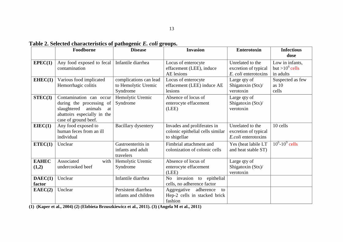
2. Selected characteristics of pathogenic E. coli groups. 13
3. Target genes for PCR amplification, amplicon size,
primer sequences and annealing temperature.
24
4. Prevalence of E. coli pathotypes coinfection in 3 types
of meat samples.
30

ix
List of Figures
No. Figure Title Page
1. Distribution of samples collection localities in Jenin
governorate. 17
2. Multiplex PCR profiles 27

x
List of Diagrams
No. Diagram Title Page
1. Morphological characterization on different
selective media; MacConkey agar, XLD Agar and
MSA.
25
2. Prevalence of E. coli in beef, chicken and turkey
meat.
26
3. Distribution of Salmonella and S. aureus in three
types of meat products using PCR. 28
4. Distribution of staphylococcal enterotoxin genes
(sea-see) in meat samples.
29
5. Prevalence of E. coli pathotypes EHEC, EPEC,
EAEC, DAEC and ETEC with uni-infection in 3
types of meat samples .
30

xi
List of Abbreviations
DNA: Deoxyribonucleic acid
E.coli: Escherichia coli
PCR: Polymerase chain reaction
dNTPs: Deoxyribonucleotide triphosphates
EDTA: Ethelendiaminetetraacetic acid
Taq DNA polymerase: Thermus aquaticus DNA polymerase
H2S:Hydrogen Sulfide
MgCl2: Magnesium chloride
afa: a fimbrial adhesins
EHEC: Enterohemorrhagic E. coli
EPEC: Enteropathogenic E. coli
EAEC: Enteroaggregative E. coli
ETEC: Enterotoxigenic E. coli
DAEC: Diffusely adherent E. coli
EIEC : Enteroinvasive E. coli
HUS: Hemolytic Uremic Syndrome
S. aureus: Staphylococcus aureus
SEs :Staphylococcal enterotoxins
SEa : Staphylococcal enterotoxin a
SEb : Staphylococcal enterotoxin b
SEc : Staphylococcal enterotoxin c
SEd : Staphylococcal enterotoxin d
SEe : Staphylococcal enterotoxin e
LEE: Locus of enterocyte effacement
Qty: Quantity
Stx :Shigatoxin gene
TSBYE: Tryptone Soya Broth-Yeast Extarct
XLD: Xylose-Lysine Deoxycholate
MSA: Mannitol Salt Agar
mdh : Malate dehydrognase gene

xii
LT: heat-labile enterotoxin
ST : Heat-stable enterotoxin
VT: Verocytotoxin
Eae: Attaching and effacing gene
BFP: Bundle-forming pilus
CFU: Colony forming unit
FemA: factor essential for expression of methicillin resistance

xiii
Polymerase chain reaction for detection of foodborne
bacterial pathogens in meat products in Jenin district-
Palestine By
Bara'a Radwan Mahmoud Alqarem
Supervisor
Dr. Ghaleb Adwan
Abstract
Foodborne diseases occur worldwide, including those acquired
through the consumption of contaminated meat. This study was conducted
to investigate the prevalence of enterotoxigenic Staphylococcus aureus,
Salmonella and Escherichia coli pathotypes contamination in 40 samples of
fresh (n=35) and frozen (n=5) beef, turkey and chicken using multiplex
PCR. The meat samples were purchased from local markets in Jenin
district, Palestine. Results of this research showed that the total mesophilic
aerobic bacterial counts ranged between 4.3 log10 to 5.7 log10 cfu/g for
frozen meat and 6.95 log10 to 7.78 log10 cfu/g for fresh meat. Results
showed that the prevalence of S. aureus, Salmonella and E. coli was 30%,
25% and 95%, respectively. It was found that 75% of S. aureus strains
were enterotoxigenic. Two samples of non S. aureus (FemA-) were
toxigenic one of them was sec+ and the other was see
+. These results
showed that 89.5% (34/38) of meat samples contaminated with E. coli were
belonged to enterohemorrhagic E. coli (EHEC), enteropathogenic E. coli
(EPEC), enteroaggregative E. coli (EAEC), enterotoxigenic E. coli
(ETEC), diffuse adherent E. coli (DAEC) pathotypes. A total of 21%

xiv
(8/38) of meat samples contaminated with E. coli were coinfected with
these pathotypes.
It is recommended to establish a permanent program for surveillance
of microbial contamination with all foodborne pathogens. This will
generate and provide data with statistical and epidemiological value, and
can be used for estimating the exposure of consumers to foodborne
pathogens and evaluating the effects of control measures on the
contamination of food.

Chapter One
Introduction
1.1 General background
Food is considered the most important energy source for humans and
animals, may be easily contaminated with pathogens if not handled
appropriately (Mead et al., 1999). The World Health Organization (WHO)
defines foodborne illnesses as diseases, usually either toxic or infectious in
nature, caused by agents that enter the human body through the process of
food ingestion. The presence of living microorganisms in food is a natural
and unavoidable occurrence. Cooking generally kills most of pathogenic
microorganisms, but undercooked foods, processed ready-to-eat foods, and
minimally processed foods can contain harmful bacteria that are serious
health threats.
bacteria, parasites, toxins, metals, and prions
The symptoms of foodborne illness range from
mild gastroenteritis to life-threatening neurologic, hepatic, and renal
syndromes (Mead et al., 1999).
The spectrum of food-borne diseases is changing in constant rate.
For example, a century ago, typhoid fever, tuberculosis and cholera were
common food-borne diseases. Improvements in food safety, that included
pasteurization of milk, safe canning, and disinfection of water supplies
have eliminated those diseases. Today, other food-borne infections have
taken their place, including some that have only recently been discovered.

2
Moreover, many emerging strains have been found to possess a
combination of genetic traits that enhance virulence to cause disease in
humans. This stands to that other pathogenic bacteria will become the
subject of legislation in the future. The major problem in some pathogens
such as E. coli O157:H7, is their ability to survive for long period under
adverse conditions (Wilkes et al., 2005; Uhlich et al.,2010, Van et al.,
2011). In addition, some salmonella serotypes have been reported to resist
some therapeutic antibiotics (Antunes et al., 2003).
The foundational knowledge of stress adaptation and long-term
survival of some bacterial pathogens must be taken in to consideration.
Investigation of the survival of foodborne pathogens would be beneficial to
our understanding of transmission of these pathogens and the potential
sources of foodborne illness (Allen et al., 2008; Shen et al., 2011).
Food safety is a global health goal and the food-borne diseases take a
major crisis on health. The detection and enumeration of microbial
pathogens in food and on surfaces that come into contact with food is the
major step in the prevention and recognition of problems related to health
and any integrated food safety program. Both food industrial companies
and government authorities use chemical and microbiological analysis to
monitor and control the state of contamination and the quality at all times
and analyze trends so as to assess and detect emerging risks (López-
Campos et al., 2013).

3
Recently, an increasing number of countries in the Eastern
Mediterranean region have moved to improve, update and strengthen their
systems and infrastructure for food safety. These countries have adopted
an approach based on risk management to monitor and control the safety of
domestically produced and imported food or drafted new food legislation.
Some countries have well-functioning foodborne surveillance systems and
reporting mechanisms. Given the strong reliance of the Eastern
Mediterranean region on food imports, ensuring the safety and quality of
imported food is a recognized concern throughout the region. Many
countries in the region have embarked on unifying food safety activities
from farm to fork. They have established or are establishing food and drug
authorities, which will cover food laws and regulations, food control
management, foodborne disease surveillance and investigation systems,
inspection services, recall and tracking systems, food monitoring
laboratories, and information and education activities for the consumers
themselves (Elmi, 2004).
Microbiological analysis of foods is based on the detection of
microorganisms by conventional and standard bacterial detection methods
such as culture and colony counting methods, immunology-based methods
and DNA-based methods, may need up to several hours or even a few days
to obtain the results (Velusamy et al., 2010; López-Campos et al., 2012).
Due to the perishable nature of most food products, there is an increased
demand for the development of rapid, specific, sensitive, accurate and
field-applicable methods to detect microorganisms. In particular, tests that

4
can be sensitive and completed within short time that would enable
processors to take quick corrective actions when contaminants are detected.
The use of DNA based methods in microbial diagnostic has greatly
enhanced the ability to investigate and quantify particularly pathogenic
bacteria in both food and water. Many of these molecular techniques have
been accepted and implemented in standard protocols for detection and
quantification of the most important pathogens (Beneduce et al., 2007).

5
Chapter Two
Literature review
2.1. Overview of food-borne illness:
The WHO reported in 2005 alone, that 1.8 million people died from
diarrhoeal diseases and a high proportion of these cases can be referred to
contamination of food and drinking water (WHO, 2008). Of the known
pathogens that cause foodborne illness Aeromonas hydrophila, Bacillus
cereus, Brucella spp, Campylobacter spp, Clostridium spp, Escherichia
coli, Listeria monocytogenes, Mycobacterium bovis, Salmonella spp,
Shigella spp, Staphylococcus aureus, Vibrio spp and Yersinia
enterocolitica. There are possibilities of other newly emerging foodborne
diseases (WHO, 2008). A number of factors can explain the emergence of
new foodborne pathogens such as new animal feeding practices, changes in
animal husbandry, increase in international trade, changes in the agronomic
process, changes in food technology, increase in travel, changes in lifestyle
and consumer demands and increase in susceptible populations (Elmi,
2004). In developed countries it is estimated that up to a third of the
population are affected by microbiological foodborne illnesses each year
(De Guisti et al., 2007). In the United States, foodborne diseases caused by
31 known pathogens are responsible for an estimated 9.4 million episodes
of foodborne illness, that 5.5 million (59%) foodborne illnesses were
caused by viruses, 3.6 million (39%) by bacteria, and 0.2 million (2%) by
parasites. Foodborne infections caused 56 000 hospitalizations, and 1300
deaths each year (Scallan et al., 2011). The CDC estimates that 47.8
6
million Americans (roughly 1 in 6 people) are sickened by foodborne
disease every year (CDC, 2011). It is estimated that 130 million Europeans
(WHO 2000), and 5.4 million Australians are annually affected by episodes
of foodborne disease (Hall et al., 2005).
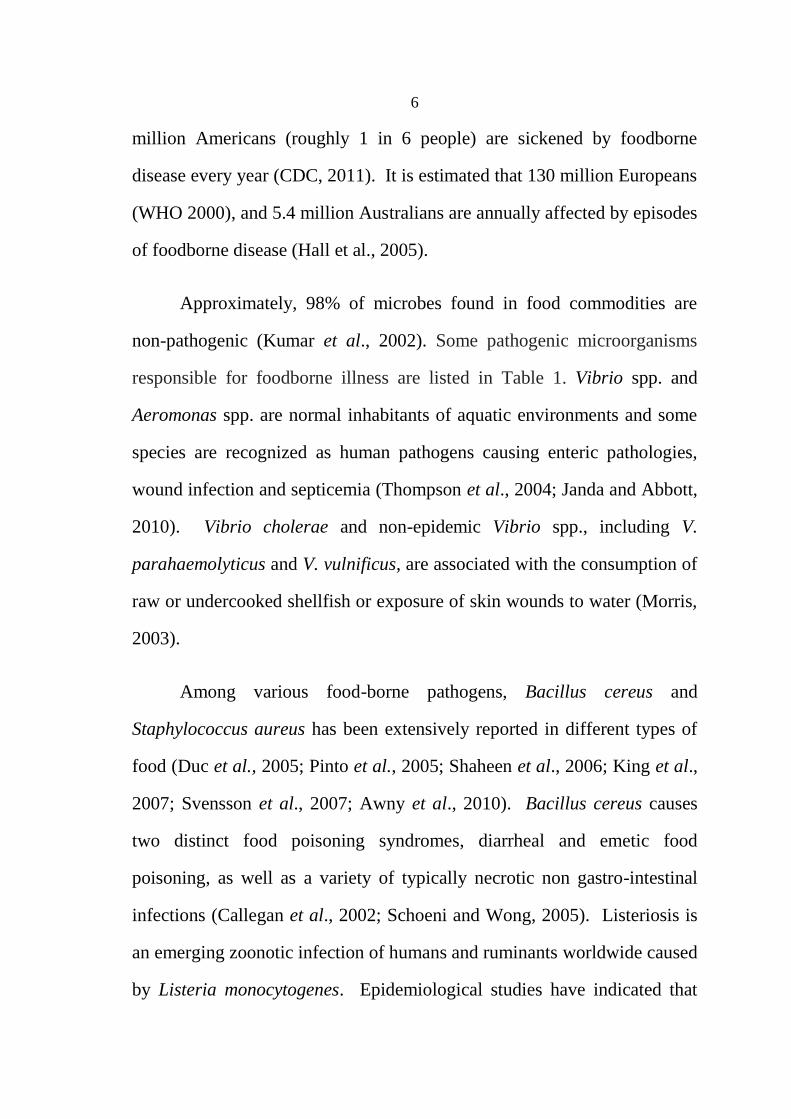
Approximately, 98% of microbes found in food commodities are
non-pathogenic (Kumar et al., 2002). Some pathogenic microorganisms
responsible for foodborne illness are listed in Table 1. Vibrio spp. and
Aeromonas spp. are normal inhabitants of aquatic environments and some
species are recognized as human pathogens causing enteric pathologies,
wound infection and septicemia (Thompson et al., 2004; Janda and Abbott,
2010). Vibrio cholerae and non-epidemic Vibrio spp., including V.
parahaemolyticus and V. vulnificus, are associated with the consumption of
raw or undercooked shellfish or exposure of skin wounds to water (Morris,
2003).
Among various food-borne pathogens, Bacillus cereus and
Staphylococcus aureus has been extensively reported in different types of
food (Duc et al., 2005; Pinto et al., 2005; Shaheen et al., 2006; King et al.,
2007; Svensson et al., 2007; Awny et al., 2010). Bacillus cereus causes
two distinct food poisoning syndromes, diarrheal and emetic food
poisoning, as well as a variety of typically necrotic non gastro-intestinal
infections (Callegan et al., 2002; Schoeni and Wong, 2005). Listeriosis is
an emerging zoonotic infection of humans and ruminants worldwide caused
by Listeria monocytogenes. Epidemiological studies have indicated that
7
both epidemic and sporadic cases of human listeriosis occur following
consumption of contaminated food. Since 1980s, the incidence has risen
steadily including large outbreaks making listeriosis one of a major public
health issue, leading in intensified surveillance and control of Listeria
monocytogenes in food industry, which contributed to a decrease of human
listeriosis cases. Unlike infection with other common foodborne
pathogens, listeriosis is associated with the highest case fatality rate ranges
from 24% to 52% despite adequate antimicrobial treatment (Oevermann et
al., 2010).
Some foodborne diseases are well recognized, but are considered
emerging because they have recently become more common. Though there
are various foodborne pathogens that have been identified for foodborne
illness, Campylobacter, Salmonella, Listeria monocytogenes, and
Escherichia coli O157:H7 have been generally found to be responsible for
majority of food-borne outbreaks (Alocilja and Radke, 2003; Chemburu et
al., 2005). Data on foodborn illnesses in USA showed that, most (58%)
illnesses were caused by norovirus, followed by nontyphoidal Salmonella
spp. (11%), Clostridium perfringens (10%), and Campylobacter spp. (9%).
Leading causes of hospitalization were nontyphoidal Salmonella spp.
(35%), norovirus (26%), Campylobacter spp. (15%), and Toxoplasma
gondii (8%). Leading causes of death were nontyphoidal Salmonella spp.
(28%), T. gondii (24%), L. monocytogenes (19%), and norovirus (11%)
(Scallan et al., 2011).

8
In Palestine, a total of 250 stool samples were collected during an
outbreak from symptomatic and asymptomatic patients in northern
Palestine in 1999. A total of 176 (70.4%) were identified as Shiga toxigenic
Escherichia coli (STEC), of the 176 STEC isolates, 124 (70.5%) were of
serotype O157 (Adwan et al., 2002). Also, 14.4% raw beef samples in
northern Palestine during 2001 were contaminated with STEC (Adwan and
Adwan, 2004). In addition, 150 children less than 5 years of age suffering
from acute gastroenteritis and diarrhea were investigated for various
enteropathogens by conventional and molecular techniques. Bacterial
enteropathogens were detected in 17.3% of the diarrheal samples. Shigella
spp was the most common bacterial pathogen (6.0%), followed by
Campylobacter coli/jejuni (4.7%), Escherichia coli O157:H7 (4.7%), and
Salmonella spp (2.0%) (Abu Elamreen et al., 2007).
Various meat or animal products such as beef, eggs, dairy, fish, and
poultry are important reservoirs for many of the food-borne pathogens and
have been associated with largest number of foodborne diseases outbreaks
during 2009–2010. Salmonella in sprouts and vine-stalk vegetables are
responsible for foodborne diseases outbreaks. The large number of
outbreaks caused by unpasteurized dairy products is consistent with
findings that more outbreaks occur in states that permit the sale of
unpasteurized dairy products (MMWR, 2013).

9
2.1.1 Salmonella
Salmonella serotypes are ubiquitous enteric bacteria and etiological
agents of food-borne gastroenteritis (salmonellosis), causing typhoid and
paratyphoid fevers. There are more than 2500 serovars of Salmonella and
all are potential pathogens (Chattopadhyay et al., 2013). Biologically,
Salmonella is very similar to E. coli in that is a Gram-negative facultative
anaerobe that colonizes the intestinal tracts mainly human or animal host.
The bacteria are transmitted to humans through consumption of
contaminated food of animal origin, mainly meat, poultry, eggs and milk
(Jay et al., 2005). The symptoms of Salmonella infection usually appear
12–72 hours after infection, and include fever, abdominal pain, diarrhea,
nausea and sometimes vomiting (Scallan et al., 2011).
2.1.2 Escherichia coli
capable of causing intestinal disease. These bacteria include strains
of enterohemorrhagic E. coli (EHEC), enteropathogenic E. coli (EPEC),
enteroaggregative E. coli (EAEC), enterotoxigenic E. coli (ETEC), diffuse
adherent E. coli (DAEC), and enteroinvasive E. coli (EIEC) (Cocolin et al.,
2000). Transmission of E. coli pathotypes appears to be mainly caused by
consumption of foods of animal source, especially ground beef and milk.
In addition, other foods implicated in outbreaks include turkey sandwiches,
potatoes, roast beef, dry sausage, yogurt and apple cider (Deng et al.,
1996). Selected characteristics of each one of the groups is presented in
Table 2.

10
Escherichia coli O157:H7 is a gram-negative, flagellated, rod-shaped
bacteria. The cell wall contains the “O” antigen, and the “H” represents
flagellar antigen. E. coli O157:H7 is specifically adapted for survival in the
gastrointestinal tract of host organisms (Jay et al., 2005). It is most
common as a causative agent of Hemolytic Uremic Syndrome (HUS). The
virulence factor is due to production of shiga-like toxins, responsible for
HUS which attack renal cells, causing lysis, that leading to bloody stool.
Also, this pathogen is able to survive in a wide range of conditions
including lower temperatures associated with storage of meat products.
Scallan et al. (2011) reported that E. coli O157:H7 could be responsible for
over 60,000 cases of illness each year and being responsible for up to 20
deaths on average.
2.1.3 Staphylococcus aureus
S. aureus is recognized worldwide as a frequent cause of foodborne
disease in dairy foods, which produces a spectrum of proteins and virulence
factors that are thought to contribute to its pathogenicity. Poultry, meat and
egg products as well as milk and milk products have been reported as
common foods that may cause staphylococcal food poisoning.
Staphylococcal enterotoxins (SEs) are recognized agents of staphylococcal
food poisoning syndrome, with sequelae such as shock, and may be
involved in other types of infections in humans and animals (Adwan et al.,
2005; 2008; 2013).

11
Table 1. Some pathogenic microorganisms responsible for foodborne illness (Velusamy et al., 2010). Pathogen Associated foods
(reported food
contaminants)
Infective dose a
(no. of
organisms)
Incubation period b
Symptoms Name of the
disease
Campylobacter
jejuni
Raw milk, and raw or
under-cooked meat,
poultry or shellfish
400–500 2 to 5 days Fever, headache, and muscle
pain followed by diarrhea,
abdominal pain and nausea
Campylobacteriosis
Salmonella spp. Raw/undercooked eggs,
poultry, and meat; raw milk
and dairy products; seafood;
chocolate; salad and spices
15–20 12 to 24 h Stomach pain, diarrhea,
nausea, chills, fever, and
headache
Salmonellosis
E.. coli Raw/undercooked eggs,
poultry, and meat; raw milk
and dairy products; seafood;
and leafy vegetables
<10 2 to 4 days Stomach pain, diarrhea,
nausea, chills, fever, and
headache
Hemorrhagic
colitis
L. monocytogenes Soft cheese, raw milk,
improperly processed ice
cream, raw leafy vegetables;
raw meat and poultry
<1000 2 days to 3 weeks Fever, chills, headache,
backache, sometimes
abdominal pain and
diarrhea
Listeriosis
Bacillus cereus Meats, milk, vegetables,
fish, rice, pasta, and cheese
30 min to 15 h Diarrhea, abdominal
cramps, nausea, and
vomiting
Bacillus cereus
food
poisoning
Clostridium
botulinum
Improperly canned foods,
garlic in oil, and
vacuumpackaged
and tightly
wrapped food
< nano grams 12–36 h Double vision, droopy
eyelids, trouble speaking
and swallowing, and
difficulty breathing
Foodborne
botulism
Clostridium
perfringens
Undercooked meats,
meat products, and gravies
>10 8 8–22 h Abdominal cramps and
diarrhea
Perfringens food
poisoning
Shigella Salads, raw vegetables, <10 12–50 h Abdominal pain, cramps, Shigellosis

12
dairy products, and poultry fever, vomiting, and diarrhea
containing blood and mucus
Yersinia
enterocolitica
Meat (mostly pork),
oysters, fish, and raw milk
Unknown 1–3 days Diarrhea and/or vomiting;
fever and abdominal pain
Yersiniosis
Vibrio
parahaemolyticus
Raw, improperly cooked,
or cooked, recontaminated
fish and shellfish, and
oysters
> 1 million 4 h–4 days Diarrhea, abdominal cramps,
nausea, vomiting, headache,
fever, and chills
V.
parahaemolyticus
associated
gastroenteritis
Vibrio vulnificus Raw or recontaminated
oysters, clams, and crabs
<100 <16 h Diarrhea, and wound
infections
Syndrome called
“primary
septicemia a Infective dose: the amount of agent that must be consumed to give rise to symptoms of foodborne illness.
b Incubation period: the delay between consumption of a contaminated food and appearance of the first symptoms of illness.
13
Table 2. Selected characteristics of pathogenic E. coli groups. Foodborne Disease Invasion Enterotoxin
Infectious
dose
EPEC(1) Any food exposed to fecal
contamination
Infantile diarrhea Locus of enterocyte
effacement (LEE), induce
AE lesions
Unrelated to the
excretion of typical
E. coli enterotoxins
Low in infants,
but >106
cells
in adults
EHEC(1)
Various food implicated
Hemorrhagic colitis
complications can lead
to Hemolytic Uremic
Syndrome
Locus of enterocyte
effacement (LEE) induce AE
lesions
Large qty of
Shigatoxin (Stx)/
verotoxin
Suspected as few
as 10
cells
STEC(3)
Contamination can occur
during the processing of
slaughtered animals at
abattoirs especially in the
case of ground beef.
Hemolytic Uremic
Syndrome
Absence of locus of
enterocyte effacement
(LEE)
Large qty of
Shigatoxin (Stx)/
verotoxin
EIEC(1)
Any food exposed to
human feces from an ill
individual
Bacillary dysentery Invades and proliferates in
colonic epithelial cells similar
to shigellae
Unrelated to the
excretion of typical
E.coli enterotoxins
10 cells
ETEC(1)
Unclear Gastroenteritis in
infants and adult
travelers
Fimbrial attachment and
colonization of colonic cells
Yes (heat labile LT
and heat stable ST)
106-10
9 cells
EAHEC
(1,2)
Associated with
undercooked beef
Hemolytic Uremic
Syndrome
Absence of locus of
enterocyte effacement
(LEE)
Large qty of
Shigatoxin (Stx)/
verotoxin
DAEC(1)
factor
Unclear Infantile diarrhea No invasion to epithelial
cells, no adherence factor
EAEC(2)
Unclear Persistent diarrhea
infants and children
Aggregative adherence to
Hep-2 cells in stacked brick
fashion
(1) (Kaper et al., 2004) (2) (Elzbieta Brzuszkiewicz et al., 2011). (3) (Angela M et al., 2011)
14
2.2. Molecular analysis methods:
Detection, isolation and identification of different types of microbial
pathogens contaminating food would be time consuming and expensive if
done in a conventional way. Usually, detection of pathogenic bacteria
using conventional methods is largely based on cultivation procedures,
which use enrichment broths followed by isolation of colonies on selective
media, biochemical identification and confirmation of pathogenicity of the
isolates. However, these methods have several limitations, such as
dependency on enrichment and selective culture, difficulty of quantitative
analysis and long culture time. The development of rapid and reliable
detection methods for food-borne pathogens is ongoing to complement or
replace culture-based approaches and bypass some of their intrinsic biases
and their own limitations. Examples of these methods include biosensors
(e.g., bioluminescence biosensor, impedimetry, piezoelectric biosensors,
etc), immunological methods, and nucleic acid based assays (Mandal et al.,
2010). PCR offers distinct advantages over culture and other standard
methods such as specificity, sensitivity, rapidity, accuracy and capacity to
detect small amounts of target nucleic acid in a sample. PCR based
methods are used in the detection of wide range of pathogens like S.
aureus, L. monocytogenes, Salmonella, B. cereus, E. coli O157: H7,
Yersinia enterocolitica, C. jejuni (Velusamy et al., 2010). Multiplex PCR
is very useful technique as it allows the simultaneous detection of several
pathogens by introducing different primers to amplify DNA regions coding
for specific genes of each bacterial strain targeted (Touron et al., 2005). A
15
multiplex PCR method was developed to rapidly detect different food
bacterial pathogens (Wang et al., 1997; Cocolin et al., 2000; Beneduce et
al., 2007; kim et al., 2007; Jeshveen et al., 2012; Kawasaki et al., 2009).
Examples of multiplex PCR technique for the simultaneous detection
pathogens include multiplex PCR assay for rapid and simultaneous
detection of E. coli O157:H7, Salmonella and Shigella (Li et al., 2005),
simultaneous detection of E. coli O157:H7, Salmonella, S. aureus, L.
monocytogenes, and V. parahaemolyticus (Kim et al., 2007), simultaneous
detection of bacteria of the genus Listeria, L. monocytogenes, and major
serotypes and epidemic clones of L. monocytogenes (Chen and Knabel,
2007), simultaneous detection of E. coli O157: H7 and L. monocytogenes
(Mukhopadhyay and Mukhopadhyay, 2007), simultaneous detection of
Listeria, Salmonella and E. coli pathogens (Tavakoli et al., 2010),
simultaneous detection of Salmonella spp., L. monocytogenes, Escherichia
coli O157:H7, and S. aureus (Kawasaki et al., 2009; 2012). In spite of its
advantages, from an industrial point of view routine detection of microbes
using PCR can be expensive and complicated, requiring skilled workers to
carry out the tests.
2.3 Aims of the study:
Food safety is a global health goal and the foodborne diseases take a
major crisis on health. Salmonella spp, E. coli, and S. aureus are major
foodborne pathogens that represent a permanent challenge to the meat
industry. These bacterial species are considered dangerous pathogens with
their ability to cause diseases in humans and animals. Detection and

16
identification of microbial pathogens in food is the solution to the
prevention and recognition of problems related to health and safety. This
study aimed to detect enterotoxigenic S. aureus, Salmonella spp and E. coli
pathotypes from meat products (fresh and frozen) using PCR technique and
to enumerate bacterial cells in these food samples. The meat samples
included in this study were collected from local markets in Jenin district,
Palestine.

17
Chapter Three
Materials and Methods
3.1. Collection of samples
Forty meat samples, included fresh (35 samples: 13 beef; 13 chicken
and 9 turkey) and frozen (5 samples: 2 beef ; 2 chicken and 1 turkey) were
purchased randomly during May-June 2014, from different localities in
Jenin governorate as shown in (Figure 1). These samples were transferred
in container, under aseptic conditions provided with ice bags within few
hours to the Microbiology Laboratory, Department of Biology at An-Najah
National University-Nablus, Palestine.
Figure 1. Distribution of samples collection localities in Jenin governorate.
18
3.2. Media preparation
3.2.1 Tryptone Soya Broth-Yeast Extarct (TSBYE) media:
TSBYE was prepared according to the following formula; Tryptone
Soya Broth (Oxoid) (30 g) and Yeast Extarct (Acumedia) (6 g) were
suspended together in one liter of distilled water, mixed well. The broth
was then distributed into flasks to have 90 ml each. The flasks were
autoclaved at 121°C for 15 minutes, allowed to cool and then stored at 4ºC.
3.2.2 Xylose-Lysine Deoxycholate (XLD) Agar:
XLD Agar (Oxoid, UK) was prepared according to manufacturer's
instructions labeled on the bottle. In a 2 L flask, 1 L of deionized water
was mixed with 56.7 g XLD Agar, heated and stirred until the agar
dissolved. The solution allowed to boil for 1 minute, and then autoclaved
at 121°C for 15 minutes. After that it was allowed to cool to about 50°C,
the agar was poured into sterile Petri dishes to have 25-30 ml each that was
left overnight at room temperature. The following morning the Petri dishes
were turned upside down and stored at 4ºC.
3.2.3 MacConkey Agar:
MacConkey agar (HIMEDIA) was prepared according to the
manufacturer's instructions labeled on the bottle. A 1L flask containing
500 ml deionized water and 25 g MacConkey agar was heated and stirred
until the agar dissolved. The solution was allowed to boil for 1min, and
then was autoclaved at 121°C for 15 min. After that it was allowed to cool,

19
and the agar was poured into sterile Petri dishes to have 20 ml that was
covered and left overnight. The following morning the Petri dishes were
turned upside down and stored at 4°C.
3.2.4 Mannitol Salt Agar (MSA):
BBLTM
Mannitol agar (BD) was prepared according to the
manufacturer's instructions labeled on the bottle. In a 1 L flask, 500 ml
deionized water were heated and mixed with 55.5 g MSA until the agar
dissolved. The solution was allowed to boil for 1 minute, and then
autoclaved at 121°C for 15 minutes. After that it was allowed to cool to
about 50°C, and poured into sterile Petri dishes to have 20 ml each, then
left overnight at room temperature. The following morning the Petri dishes
were turned upside down and stored at 4°C.
3.3. Food sample preparation bacterial culturing
Of each food sample purchased from local food stores, 10 g were
homogenized in 90 ml TSBYE medium, then the suspension was mixed
well. Five of serial decimal dilutions of samples with sterile normal saline
were in duplicates on nutrient agar. The plates were then incubated at 37°C
for 24h before colonies were counted. Then 5 ml of TSBYE was incubated
at 37°C/18-24h and used for DNA extraction and subcultured on XLDA,
MSA and MacConkey.

20
3.4. DNA extraction:
DNA was prepared for PCR according to the method described
previously with some modifications (Adwan et al., 2013). Briefly, 1.5 ml of
cells from overnight TSBYE broth were centrifuged, the pellet was washed
twice with 1 ml of 1X Tris-EDTA buffer (10 mM Tris-HCl, 1 mM EDTA
[pH 8.0]), then resuspended in 0.5 ml of sterile distilled H2O, and was
boiled for 10-15 min. The cells then immediately were incubated on ice for
10 min. The debris was pelleted by centrifugation at 11,500 X g for 5 min.
DNA was extracted from the supernatant using phenol-chloroform method,
then DNA was precipitated using 96% cold ethanol. The nucleic acid pellet
was washed with 70% cold ethanol, dried and then resuspended in 300 μl
TE (Tris 10 mM, EDTA 1 mM, pH 8), DNA concentration was determined
using a spectrophotometer and the samples were stored at -20ºC until use
for further DNA analysis.
3.5. Detection of food pathogens by PCR
3.5.1 Detection of E. coli Mdh gene:
E. coli were identified by PCR with specific primers for malate
dehydrognase gene (mdh) as described previously (Hsu et al. 2007). Primer
nucleotide sequences and expected size of amplicon are presented in Table
3. The PCR reaction mix (25 μL) was performed using 12.5 μL of PCR
premix with MgCl2 (ReadyMixTM
Taq PCR Reaction Mix with MgCl2, Sigma),
0.4 μM of each primer, and 2 μL DNA template. DNA amplification was
performed using thermal cycler (Mastercycler Personal, Eppendorf)

21
according to the following thermal conditions: initial denaturation for 2
min at 94°C followed by 30 cycles at 94°C for 1 min for denaturation,
annealing at 59°C for 30 s and extension at 72°C for 1 min. Final extension
was carried out at 72°C for 5 min. The amplified products were examined
by (2%) agarose gel electrophoresis to determine the size of amplified
fragment for each isolate. A DNA ladder of 100 bp was also included in
all gels (100bp DNA ladder RTU, GeneDireX). Negative control was
included in these experiments.
3.5.2 Detection of femA (S. aureus) and 1.8-kb HindIII DNA fragment
(Salmonella spp.):
The primers targeted a Salmonella species-specific sequence within a
1.8-kb HindIII DNA fragment, and the S. aureus femA gene and expected
sizes of amplicons are presented in Table 3. PCR was performed as
described previously with some modification (Kawasaki et al., 2012). The
PCR reaction mix (25 μL) was performed using 12.5 μL of PCR premix
with MgCl2 (ReadyMixTM
Taq PCR Reaction Mix with MgCl2, Sigma), 0.4 μM
of each primer, and 2 μL DNA template. DNA amplification was
performed using thermal cycler (Mastercycler Personal, Eppendorf)
according to the following thermal conditions: initial denaturation for 2
min at 94°C followed by 40 cycles at 94°C for 20 s for denaturation,
annealing at 56°C for 30 s and extension at 72°C for 30 s. Final extension
will be carried out at 72°C for 5 min. The amplified products were
examined by 1.5% agarose gel electrophoresis to determine the size of
amplified fragment for each isolate. A DNA ladder of 100bp was also

22
included in all gels (100bp DNA ladder RTU, GeneDireX). Negative
control was included in these experiments.
3.5.3. Detection of staphylococcal enterotoxin (sea-see) genes:
Primer nucleotide sequences and expected sizes of amplicons are
presented in Table 3. The PCR reaction mix (25 μL) was performed using
12.5 μL of PCR premix with MgCl2 (ReadyMixTM
Taq PCR Reaction Mix
with MgCl2, Sigma), 0.4 μM of each primer, and 2 μL DNA template.
DNA amplification was performed using thermal cycler (Mastercycler
Personal, Eppendorf) as the following thermal conditions: initial
denaturation for 2 min at 94°C followed by 30 cycles at 94°C for 1 min for
denaturation, annealing at 55°C for 1 min and extension at 72°C for 2 min.
Final extension was carried out at 72°C for 5 min. The amplified products
were examined by 1.5% agarose gel electrophoresis to determine the size
of amplified fragment for each isolate. A DNA ladder of 100bp was also
included in all gels (100bp DNA ladder RTU, GeneDireX). Negative
control was included in these experiments.
3.5.4. Detection of E. coli pathotypes:
The targeted genes from different E. coli pathotypes including
enterohemorrhagic E. coli (EHEC), enteropathogenic E. coli (EPEC),
enteroaggregative E. coli (EAEC), enterotoxigenic E. coli (ETEC) and
diffusely adherent E. coli (DAEC) were amplified using oligonucleotide
primer pairs and expected sizes of amplicons are listed in Table 3. These

23
genes were detected with some modifications as described previously
(Gómez-Duarte et al., 2009). The PCR reaction mix (25 μL) was
performed using 12.5 μL of PCR premix with MgCl2 (ReadyMixTM
Taq PCR
Reaction Mix with MgCl2, Sigma), 0.4 μM of each primer and 2 μL DNA
template. DNA amplification was performed using thermal cycler
(Mastercycler Personal, Eppendorf) according to the following thermal
conditions: initial denaturation for 2 min at 94°C followed by 40 cycles at
92°C for 30 s for denaturation, annealing at 59°C for 30 s and extension at
72°C for 30 s. Final extension was carried out at 72°C for 5 min. The
amplified products were examined by (2%) agarose gel electrophoresis to
determine the size of amplified fragment for each isolate. A DNA ladder
of 100bp was also included in all gels (100bp DNA ladder RTU,
GeneDireX).

24
Table 3. Target genes for PCR amplification, amplicon size, primer sequences and annealing temperature. Organism Gene target Oligonucleotide sequence (5→3)
* Amplicon
Size (bp)
Annealing
temperature
Reference Primer
mix
E. coli Mdh Mdh1 ACT GAA AGG CAA ACA GCC AGG C
mdh2 CGT TCT GTT CAA ATG CGC TCA GG
392 59°C Hsu et al. 2007 1
Salmonella
spp.
HindIII DNA
fragment
TS-11 GTCACGGAAGAAGAGAAATCCGTACG
TS-5 GGGAGTCCAGGTTGACGGAAAATTT
375 56°C Tsen et al. 1994 2
S. aureus FemA Fem F TATGAGTTAAAGCTTGCTGAAGGTT
Fen R TTACCAGCATTACCTGTAATCTCG
296 56°C Kawasaki et al., 2012 2
ETSA sea
SEA-3 CCTTTGGAAACGGTTAAAACG
SEA-4 TCTGAACCTTCCCATCAAAAAC
127
55°C Becker et al. 1998 3
ETSA seb
SEB-1 TCGCATCAAACTGACAAACG
SEB-4 GCAGGTACTCTATAAGTGCCTGC
477 55°C Becker et al. 1998 3
ETSA sec
SEC-3 CTCAAGAACTAGACATAAAAGCTAGG
SEC-4 TCAAAATCGGATTAACATTATCC
271
55°C Becker et al. 1998 3
ETSA sed
SED-3 CTAGTTTGGTAATATCTCCTTTAAACG
SED-4 TTAATGCTATATCTTATAGGGTAAACATC
319
55°C Becker et al. 1998 3
ETSA see
SEE-3 CAGTACCTATAGATAAAGTTAAAACAAGC
SEE-2 TAACTTACCGTGGACCCTTC
178
55°C Becker et al. 1998 3
EHEC VT VTcom-u GAGCGAAATAATTTATATGTG
VTcom-d TGATGATGGCAATTCAGTAT
518 59°C Gómez-Duarte et al., 2009 4
EHEC,
EPEC
eae eae1 CTGAACGGCGATTACGCGAA
eae2 CGAGACGATACGATCCAG
917 59°C Gómez-Duarte et al., 2009 4
EPEC bfpA BFP1 AATGGTGCTTGCGCTTGCTGC
BFP2 GCCGCTTTATCCAACCTGGTA
326 59°C Gómez-Duarte et al., 2009 4
EAEC aggR aggRks1 GTATACACAAAAGAAGGAAGC
aggRksa2 ACAGAATCGTCAGCATCAGC
254 59°C Gómez-Duarte et al., 2009 4
ETEC LT LT1 GCACACGGAGCTCCTCAGTC
LT2 TCCTTCATCCTTTCAATGGCTTT
218 59°C Gómez-Duarte et al., 2009 5
ETEC ST ST1 GCTAAACCAGTAGAG(C)TCTTCAAAA
ST2 CCCGGTACAG(A)GCAGGATTACAACA
147 59°C Gómez-Duarte et al., 2009 5
DAEC daaE daaE1 GAACGTTGGTTAATGTGGGGTAA
daaE2 TATTCACCGGTCGGTTATCAGT
542 59°C Gómez-Duarte et al., 2009 5
*All the primers used in this study were synthesized by Sigma-Aldrich (Israel).

25
Chapter Four
Results
4.1. Bacterial enumeration and cultural characterization
The total aerobic bacterial counts ranged between 4.3 log10 to 5.7 log10
cfu/g for frozen meat and 6.95 log10 to 7.78 log10 cfu/g for fresh meat.
Results of bacterial culture on MacConkey agar showed that 95% (38/40)
of samples were lactose fermenter with bright pink color colonies. On
XLD Agar, 22.5% (9/40) of samples had colonies with black centers due to
H2S production. Results also showed that 65% (26/40) of samples
subcultured on MSA were mannitol fermenter and had a yellow color due
to acid production. Results of cultural characterization on different
selective media; MacConkey agar, XLD Agar and MSA are represented in
diagram 1.
Diagram 1. Morphological characterization on different selective and differential media;
MacConkey agar, XLD Agar and MSA.

26
4.2. Detection of E. coli by PCR
All of the 40 DNA samples were tested using primers specific for
malic acid dehydrogenase (mdh) gene for E. coli, 95% (38/40) generated
the expected PCR products with size equals to 392 bp. The prevalence of
E. coli was 100%, 93.3% and 90% in beef, chicken and turkey meat,
respectively. Results about prevalence of E. coli using PCR in three types
of meat are presented in diagram 2 and Figure 2.
Diagram 2. Prevalence of E. coli in beef, chicken and turkey meat.

27
Figure 1: Multiplex PCR profiles. Lane L contained ladder (100bp DNA ladder RTU,
GeneDireX), lane 1 represents E. coli target gene (mdh); lane 2 represent Salmonella spp.
target gene (HindIII DNA fragment); lane 3 shows S. aureus target gene (FemA); lane 4
belonged to pathogenic E. coli groups target genes (VT for EHEC and bfpA for EPEC and eae
for both); lanes 5 represents EAEC target gene (aggR); lanes 6-9 belonged to staphylococcal
enterotoxin genes of (sed, sec, see and sea, respectively); lane 10 represents ETEC target genes
(LT and ST).
4.3. Detection of Salmonella spp. and S. aureus by PCR.
PCR assay was carried out for the DNA from 40 meat samples to detect
the presence of Salmonella spp. and S. aureus in three types of meat
samples. The size of PCR products produced by specific primers for
Salmonella spp. and S. aureus were 375 bp and 296 bp, respectively. The
prevalence of Salmonella spp., S. aureus and both in forty meat samples
was 17.5% and 22.5% and 7.5%, respectively. The distribution of
Salmonella spp. and S. aureus in three types of meat products is presented
in diagram 3 and Figure 1.

28
Diagram 3. Distribution of Salmonella spp and S. aureus in three types of meat products
using PCR.
4.4. Detection of staphylococcal enterotoxin (sea-see) genes
PCR assay was conducted on DNA from all meat samples. Only
staphylococcal enterotoxin genes were detected from samples contaminated
with S. aureus. The prevalence of staphylococcal enterototoxin genes
among S. aureus isolates was 25%, 0.0%, 0%, 8.3% and 25% for sea, seb,
sec, sed and see, respectively. In addition, 5% of samples were coinfected
with sea and see. Two beef samples one was sec+ and the other see
+, while
these were FemA- and non mannitol fermenter. Prevalence of
staphylococcal enterototoxin genes among S. aureus isolates in meat
samples are presented in diagram 4 and Figure 2.

29
Diagram 4. Distribution of staphylococcal enterotoxin genes (sea-see) in meat samples.
* Two samples one had sec+ and another had see
+ but FemA
- and non mannitol
fermenter.
4.5. Detection of E. coli pathotypes
The PCR assay was used for detecting the presence of pathogenic
groups of E. coli. Detection of VT and eae genes for EHEC, bfpA and eae
genes of EPEC, aggR gene for EAEC, daaE gene DAEC and LT and/or ST
for ETEC. Amplification of these genes produced PCR products of 518 bp
and 917 bp, 326 bp and 917 bp, 254 bp, 542 bp and 218 bp and/or 147 bp
for EHEC, EPEC, EAEC, DAEC and ETEC, respectively. These results
showed that 89.5% (34/38) of meat samples contaminated with E. coli
belonged to E. coli pathotypes tested in this research. The total prevalence
of uni-infcted samples with EHEC, EPEC, EAEC, DAEC and ETEC was
0%, 0%, 5%, 0% and 60 % respectively, while 21% (8/38) of meat samples

30
contaminated with E. coli were coinfected with these pathotypes.
Prevalence of E. coli pathotypes groups in 3 types of meat are presented in
diagram 5, table 4 and figure 1.
Diagram 5. Prevalence of E. coli pathotypes EHEC, EPEC, EAEC, DAEC and ETEC with
uni-infection in 3 types of meat samples .
Table 4. Prevalence of E. coli pathotypes coinfection in 3 types of meat
samples.
E. coli pathotypes Source and number of samples EHEC EPEC ETEC EAEC Beef Chicken Turkey Total
(VT++eae
+) (bfpA
++eae
+) ST
+ LT
+ aggR
+
+ - + - - 1 1 0 2
- + + + - 1 0 0 1
- - + + + 0 1 0 1
+ - + - + 1 0 0 1
+ - - - + 1 0 0 1
+ + - - - 2 0 0 2
+: presence of gene; -: absence of gene

31
Chapter Five
Discussion
Conventional detection of pathogenic bacteria is largely based on
cultivation procedures, which use enrichment broths followed by isolation
of colonies on selective media, biochemical identification and confirmation
of pathogenicity of the isolates. Since many human pathogenic bacteria
can be found in the contaminated food including meat, the specific
detection of these pathogenic microorganisms in food is often suggested to
assess a more accurate human health risk. Development of new techniques
including molecular assays are used to complement or replace culture-
based approaches and bypass some of their intrinsic biases and their own
limitations. PCR is one of these techniques and is considered a sensitive
detection method for specific pathogens and multiplex PCR assay may
provide a useful tool for rapid and specific detection of pathogens in food.
This is an important step towards the control and prevention of food-borne
epidemics (Awny et al., 2010). Numerous studies were published about
PCR detection of different food-borne pathogens (Li et al., 2005; Beneduce
et al., 2007; kim et al., 2007; Jeshveen et al., 2012; Kawasaki et al., 2009).
Results showed heavy bacteriological load in different meat types
with a total viable counts ranging from 4.3 log10 to 5.7 log10 cfu/g for
frozen meat and 6.95 log10 to 7.78 log10 cfu/g for fresh meat. The presence
of a high number of viable bacteria is considered as an indicator of the
short expected shelf life of meat. Total bacterial viable counts in different
32
types of meats ranges from 5.6 log10 CFU/g to 9 log10 CFU/g (Eisel et al.,
1997; Bhandare et al., 2007; Arain et al., 2010; Awny et al., 2010;
Abdellah et al., 2013; Anihouvi et al., 2013). The presence of high count of
viable bacteria organisms from 6 log10 CFU/g to 9 log10 CFU/g, as an
indication of open-air meat spoilage (Eribo and Jay, 1985). Therefore, it is
considered that fresh meat that contains 5 log10 CFU/g to 6 log10 CFU/g of
background organisms are inherently safer than those that contain less
bioload; however, this hypothesis applies only to harmless bacteria (Jay,
1996).
A wide spectrum of pathogens play a role in foodborne disease. Most
of them have a zoonotic origin and have reservoirs in healthy food animals
from which they spread to an increasing variety of foods. Therefore, foods
of animal origin are considered major vehicles for the transmission of
human foodborne infections (Todd, 1997). Salmonella and S. aureus are
the most common and frequent pathogens responsible for food poisoning
and food related infections (Costa et al., 2012; Aydin et al., 2011).
According to WHO (WHO, 2006), 25% of the diarrhea in foodborne illness
is caused by food infected with E. coli.
S. aureus food poisoning is one of the most economically important
food-borne pathogen worldwide. Results of this research showed that 30%
of meat samples were contaminated with S. aureus, and 75% (9/12) were
toxigenic. The prevalence of S. aureus in different food ranged from
12.12% to 50.8% (Adwan et al., 2005; Awny et al., 2010; Vázquez-

33
Sánchez et al., 2012; EI-Jakee et al., 2013). The prevalence of
enterotoxigenic S. aureus in different food samples reported for countries
such as Brazil, the USA, Switzerland, South Korea, Poland, Slovakia,
China, France, Palestine, Egypt, Brasil, Thailand, Spain and Japan has
ranged from 4.7% to 100% (Adwan et al., 2005; Pelisser et al., 2009;
Awny et al., 2010; Vázquez-Sánchez et al., 2012; EI-Jakee et al., 2013;
Akbar and Anal, 2013; Anihouvi et al., 2013). Two samples which had
sec+ or see
+ but FemA
- and non mannitol fermenter, these were
Staphylococcus coagulase-negative. It was reported that, coagulase-
negative as well as coagulase-positive staphylococci are capable of
genotypic and phenotypic enterotoxigenicity (Veras et al., 2008; Podkowik
et al., 2013). In the present study, detection of toxin genes by PCR allows
the determination of potentially enterotoxigenic pathogen irrespective of
whether the strain produces the toxin or not. For this reason, PCR may be
considered more sensitive than immunological methods that determine SE
production (Adwan et al., 2005). The most common types of
staphylococcal enterotoxins are SEA to SEE. Isolates carrying toxin genes
sea to see are responsible for 95% of staphylococcal food poisoning
outbreaks (Bergdoll, 1983). SEA is the most common enterotoxin
recovered from food poisoning outbreaks (Balaban and Rasooly, 2000).
The remaining staphylococcal food-borne disease outbreaks may therefore
be associated with other newly identified SEs (MacLauchlin et al., 2000;
Rosec and Gigaud, 2002; Omoe et al., 2002). Staphylococcal enterotoxins
are thermostable and also resistant to gastrointestinal proteases such as

34
pepsin, explaining its ability to remain active after ingestion. Therefore,
the presence of S. aureus in food can be considered a potential health risk
(Adwan et al., 2006).
Among various food-borne pathogens, Salmonella serotypes are the
most common bacteria responsible for food-borne gastroenteritis. There
are more than 2500 serovars of Salmonella and all are considered as
pathogenic. Results of this research showed that 25% of meat samples
were contaminated with Salmonella. The prevalence of Salmonella in
different food ranged from 1.56% to 100% (Zhao et al. 2001; Rivera-
Betancourt et al., 2004; Busani et al., 2005; Cohen et al. 2007; Bhandare et
al., 2007; Miranda et al. 2009; Moussa et al. 2010; Aftab et al., 2012;
Elmanama et al., 2013; Iyer et al., 2013; Anihouvi et al., 2013; Adeyanju
and Ishola, 2014). Salmonella is regarded as a zero tolerance organism in
foods and should not be present in food, thus the Salmonella testing of food
samples is mandatory (Chattopadhyay et al., 2013). However, Salmonella
is ubiquitous in nature, and can be found in the digestive tracts of different
animals, poultry products, eggs, milk products and seafood. Raw chicken
meat is known to be the major source for Salmonella food poisoning (Chen
et al., 2008).
E. coli has been implicated as an agent of diarrheal disease.
Diarrheagenic strains of E. coli can be divided into five main categories on
the basis of distinct epidemiological and clinical features, specific virulence
factors, and association with certain serotypes: EAEC, EHEC, EIEC,

35
EPEC, DAEC and ETEC (Nguyen et al., 2005; Gómez-Duarte et al., 2006).
The prevalence of diarrheagenic E. coli was 22.5% among children
suffering from diarrhea (Nguyen et al., 2005; Hien et al., 2007). Results of
this research showed that 95% (38/40) of meat samples were contaminated
with E. coli, and 89.5% (34/38) of these meat samples contaminated with
E. coli were diarrheagenic. The prevalence of E. coli in different food
ranged from 9.1% to 100% (Zhao et al 2001; Rivera-Betancourt et al.,
2004; Cohen et al. 2007; Lee et al. 2009; Saikia and Joshi 2010; Ukut et al.
2010; Biswas et al., 2010; Abdellah et al., 2013; Iyer et al., 2013;
Adeyanju and Ishola, 2014). High prevalence of E. coli and/or
diarrheagenic E. coli can be explained due to that E. coli isolates are part
of the normal enteric flora in these animals. A total of 39 pathogenic E.
coli isolates from the three meat types (fresh beef, poultry and pork) were
categorized into three virulence groups, comprise of ETEC (43.6%),
EHEC (35.9%), and EPEC (20.5%) ( Lee et al. 2009).
The prevalence of foodborne pathogens from food samples differs
among studies. This could be due in part to several factors such as
differences in the reservoir in the various countries or ecological origin of
strains, the sensitivity of detection methods, detected genes and number of
samples, and type of samples analyzed (whole birds versus steaks; fresh
versus frozen), time of sample collection and type of storage (Zhao et
al.2001; Adwan et al., 2005). The poor hygiene and sanitation prevailing in
the abattoirs as well as the shops, unwashed carcasses, transportation and
display at butcher shops encourage microbial contaminations and growth.

36
The higher microbial load in the shops further enhances the chances of
early meat spoilage (Bhandare et al, 2007).
Several factors could have close relationship with the heavy
bacteriological load in different meat types studied in this research. The
main factor is the climate surrounded the studied region. In the current
study Jenin government is considered the lowest city from the sea level in
the north of Palestine, which considered the main reason for the high
temperature and humidity in the region, where the suitable condition for
high microbial growth. In addition, the direct exposure of meat to the open
air in most shops are studied is considered another important factor that
leads to the high microbial load.
Therefore, it is recommended to establish a permanent program for
surveillance of microbial contamination with all food-borne pathogens.
This may generate and provide data with statistical and epidemiological
value. These data can be used for estimating the exposure of consumers to
foodborne pathogens and evaluating the effects of control measures on the
contamination of food. It is well known that contamination of food items
that are usually consumed after cooking represents a low threat from a
public health point of view; however, more attention should be paid to the
contamination of ready-to-eat products. Consumption of undercooked
meat products and cross-contamination during food handling and
preparation must be avoided to ensure food safety at home and in the food
service industry.

37
References
Abdellah E, Fouzia RF, Bouchra O. Prevalence and antibiogram study
of Escherichia coli and Staphylococcus aureus in turkey meat in
Morocco. Pharmaceut Anal Acta 2013; 4(9).
Abu Elamreen FH, Abed AA, Sharif FA. Detection and identification
of bacterial enteropathogens by polymerase chain reaction and
conventional techniques in childhood acute gastroenteritis in Gaza,
Palestine. Int J Infect Dis 2007;11(6):501-507.
Adeyanju GT, Ishola O. Salmonella and Escherichia coli
contamination of poultry meat from a processing plant and retail
markets in Ibadan, Oyo State, Nigeria. Springer Plus 2014, 3:139.
Adwan G, Adwan K, Jarrar N, Salameh Y. Prevalence of seg, seh and
sei genes among clinical and nasal swab of Staphylococcus aureus
isolates. Br Microbiol Res J 2013; 3(2): 139-149.
Adwan G., Abu-Shanab B., Adwan K. Enterotoxigenic Staphylococcus
aureus in raw milk in the North of Palestine. Turk J of Biol 2005;
29:229-232.
Adwan G., Abu-Shanab B., Adwan K., Odeh M. Enterotoxigenecity of
S. aureus isolates recovered from chronic urogenital tract infection in
North of Palestine. Pak J Med Sci 2008; 24 (2): 246-250.

38
Adwan GM, Abu-ShanabBA, Adwan KM, Jarrar NR. Toxigenicity of
Staphylococcus aureus isolates from Northern Palestine. Emirates Med J
2006; 24(2):127-129.
Adwan GM, Adwan KM. Isolation of shiga toxigenic Escherichia coli
from raw beef in Palestine. Int J Food Microbiol 2004; 97:81-84.
Adwan K, Abu-Hasan N, Essawi T, Bdir M. Isolation and
characterisation of Shiga toxigenic Escherichia coli strains from
northern Palestine. J Med Microbiol 2002; 51(4):332-335.
Aftab M, Rahman A, M. Qureshi MS, Akhter S, Sadique U, Sajid U,
Zaman S. Level of Salmonella in beef of slaughtered cattle at Peshawar.
J Anim Plant Sci 2012; 22(2 Suppl.):24-27.
Akbar A, Anal AK. Prevalence and antibiogram study of Salmonella
and Staphylococcus aureus in poultry meat. Asian Pac J Trop Biomed.
2013; 3(2):163-168.
Allen, K.J., D. Lepp, R.C. McKellar, M.W. Griffiths. Examination of
stress and virulence gene expression in Escherichia coli O157:H7 using
targeted microarray analysis. Food. Path. and Dis. 2008;. 5:437-447.
Alocilja EC, Radke SM. Market analysis of biosensors for food safety.
Biosens Bioelectron 2003; 18:841–846.
Amira Souii, Mouna Ben Nejma, Amel Elfray Rhim, Maha Mastouri,
Besma Bel Hadj Jrad, Mohamed Makhlouf Mohamed Nour. Molecular

39
identification of four Salmonella serovars isolated from food in Tunisia
based on the sequence of the ribosomal RNA genes. African J. of
Microbiology Research 2012; 6(35): 6454-6461.
Angela M. Valadez, Chitrita Debroy, Edward Dudley, Catherine N.
Cutter1. Multiplex PCR Detection of Shiga Toxin–Producing
Escherichia coli Strains Belonging to Serogroups O157, O103, O91,
O113, O145, O111, and O26 Experimentally Inoculated in Beef
Carcass Swabs, Beef Trim, and Ground Beef. J. of Food Protection
2011;74(2): 228–239.
Anihouvi DGH, KAyodé APP, Anihouvi VB, Azokpota P, Kotchoni SO,
Hounhouigan DJ. Microbial contamination associated with the
processing of tchachanga , a roasted meat product. Afr J Biotechnol
2013;12(18):2449-2455.
Antunes, P., C.Réu, J.C. Sousa, L. Peixe, and N. Pestana. Incidence of
Salmonella from poultry products and their susceptibility to
antimicrobial agents. Int J Food Microbiol 2003; 82:97-103.
Arain MA, Rajput IR, Khaskheli M, Faraz S, Devrajani K, Fazlani SA.
Evaluation of microbial quality of goat meat at local market of Tando
Jam. Pak J Nutr 2010; 9(3): 287-290.
Awny NM, Abou Zeid AAM, Abdo MA. Prevalence of toxigenic
bacteria in some Egyptian food. Proceeding of Fifth Scientific
Environmental Conference. Zagazig Uni. 2010; 107-124.

40
Aydin A, Sudagidan M, Muratoglu K. Prevalence of staphylococcal
enterotoxins, toxin genes and genetic-relatedness of foodborne
Staphylococcus aureus strains isolated in the Marmara Region of
Turkey. Int J Food Microbiol 2011;148: 99-106.
Balaban N, Rasooly A. Staphylococcal enterotoxins. Int J Food
Microbiol 2000; 61(1): 1-10.
Becker K, Roth R, Peters G. Rapid and specific detection of toxigenic
Staphylococcus aureus: use of two multiplex PCR enzyme
immunoassays for amplification and hybridization of staphylococcal
enterotoxin genes, exfoliative toxin genes and toxic shock syndrome
toxin 1 gene. J Clin Microbiol 1998; 36(9): 2548-53.
Beneduce L, Fiocco D, Spano G. Development of PCR-based
molecular tools for the detection of emerging food- and water-borne
pathogenic bacteria. In: Communicating current research and
educational topics and trends in applied microbiology (ed. A. Mendez-
Vilas) 2007; 569 – 576.
Bergdoll MS. Enterotoxins., Adlam C. ed. Sataphylococci and
staphylococcal infections. In Easton CSF 1983; 559-598..
Bhandare SG, Sherikar AT, Paturkar AM, Waskar VS, Zende RJ. A
Comparison of microbial contamination on sheep/goat carcasses in a
modern Indian abattoir and traditional meat shops. Food Control 2007;
18(7): 854-858.

41
Biswas S, Parvez MAK, Shafiquzzaman M, Nahar S, M N Rahman M.
Isolation and characterization of Escherichia coli in ready-to-eat foods
vended in Islamic University, Kushtia. J bio-sci 2010;18: 99-103.
Brzuszkiewicz E., Thürmer A., Schuldes J., Leimbach A., Liesegang H.,
Meyer, F.-D., Boelter J., Petersen H., Gottschalk G., Daniel R. Genome
sequence analyses of two isolates from the recent Escherichia coli
outbreak in Germany reveal the emergence of a new pathotype:
Entero-Aggregative-Haemorrhagic Escherichia coli(EAHEC). Archives
of Microbiology 2011; 193: 883-891.
Busani L, Cigliano A, Taioli E, Caligiuri V, Chiavacci L, Di Bella C,
Battisti A, Duranti A, Gianfranceschi M, Nardella MC, Ricci A, Rolesu S,
Tamba M, Marabelli R, Caprioli A, Italian Group of Veterinary
Epidemiology. Prevalence of Salmonella enterica and Listeria
monocytogenes contamination in foods of animal origin in Italy. J Food
Prot 2005; 68(8): 1729-1733.
Callegan MC, Cochran DC, Kane ST, Gilmore MS, Gominet M,
Lereclus D. Contribution of membrane-damaging toxins to Bacillus
endophthalmitis pathogenesis. Infect Immun 2002; 70: 5381–5389.
CDC, CDC Estimates of Foodborne Illnessin the United States: CDC
2011 Estimates: Findings. CDC 2011.
http://www.cdc.gov/foodborneburden/2011-foodborne stimates.html.

42
Chattopadhyay S, Kaur A, Jain S, Singh H. Sensitive detection of food-
borne pathogen Salmonella by modified PAN fibers-immunoassay.
Biosens Bioelectron 2013; 45: 274-80.
Chemburu S,Wilkins E, Abdel-Hamid I. Detection of pathogenic
bacteria in food samples using highly-dispersed carbon particles.
Biosens Bioelectron 2005; 21: 491–499.
Chen W-T, Tsal S-J, Shih D Y-C, Wang Y-G C, Chu-Ying Lou Chyr C-
Y L. An improved pre-culture procedure for the detection of
Salmonella in frozen chicken meat by Duplex Polymerase Chain
Reaction. J Food Drug Anal 2008; 6(5): 83-90.
Chen Y, Knabel SJ. Multiplex PCR for simultaneous detection of
bacteria of the genus Listeria, Listeria monocytogenes and major
serotypes and epidemic clones of L. monocytogenes. Appl Environ
Microbiol 2007; 73: 6299–6304.
Cocolin L, Manzano M, Cantoni C, Comi G. A multiplex-PCR method
to detect enterohemorrhagic (EHEC) and enteropathogenic (EPEC)
Escherichia coli in artificially contaminated foods. Int J Hyg Environ
Health 2000; 203(2): 159-164.
Cohen N, Ennaji H, Bouchrif B, Hassar M, Karib H. Comparative
study of microbiological quality of raw poultry meat at various seasons
and for different slaughtering processes in Casablanca (Morocco). J
Appl Poult Res 2007;16:502–508.

43
Costa LF, Paixão TA, Tsolis RM, Bäumler AJ, Santos RL.
Salmonellosis in cattle: advantages of being an experimental model.
Res Vet Sci 2012;93:1-6.
Cuiwei Zhao et al. Prevalence of Campylobacter spp., Escherichia coli
and Salmonella Serovars in Retail Chicken, Turkey, Pork and Beef
from the Greater Washington, D.C., Area. Applied And Environmental
Microbiology 2001;67(12):5431–5436.
De Giusti M, De Medici D, Tufi D, Marzuillo C, Boccia A.
Epidemiology of emerging foodborne pathogens. Ital J Public Health
2007; 4(1):24-31.
Deng MY, Cliver DO, Day SP, Fratamico PM. Enterotoxigenic
Escherichia coli detected in foods by PCR and an enzyme-linked
oligonucleotide probe. Int J Food Microbiol 1996;30(3):217-29.
Duc LH, Dong TC, Logan NA, Sutherland AD, Taylor J, Cutting SM.
Cases of emesis associated with bacterial contamination of an infant
breakfast cereal product. Int J. Food Microbiol 2005; 102:245-251.
EI-Jakee J, Marouf SA, Ata NS, Abdel-Rahman EH, Abd El-Moez SI,
Samy AA, Walaa E. El-Sayed WE. Rapid Method for Detection of
Staphylococcus aureus Enterotoxins in Food. Global Veterinaria
2013(3): 335-341.

44
Eisel WG, Linton RH, Muriana PM. A survey of microbial levels for
incoming raw beef, environmental sources, and ground beef in a red
meat processing plant. Food Microbiol 1997;14, 273–282.
Elmanama AA, El kahlout KAE, Elnakhalaa SR. Occurrence of
Salmonella in fresh and frozen meat in the Gaza Strip. J Purity Utility
Reaction Environ 2013;2(6):142-152.
Elmi M. Food safety: current situation, unaddressed issues and the
emerging priorities. East Mediterr Health J 2004;10(6):794-800.
Eribo BE, Jay JM. Incidence of Acinetobacter spp. and Other Gram-
Negative, Oxidase-Negative Bacteria in fresh and spoiled ground beef.
App Env Microbiol 1985;49: 256-257.
Gómez-Duarte OG, Bai J, Newell E. Detection of Escherichia coli,
Salmonella spp., Shigella spp., Yersinia enterocolitica, Vibrio cholerae,
and Campylobacter spp. enteropathogens by 3-reaction multiplex
polymerase chain reaction. Diagn Microbiol Infect Dis 2009;63(1):1-9.
Hall G, Kirk MD, Becker N, Gregory JE, Unicomb L, Millard G,
Stafford R, Lalor K; OzFoodNet Working Group. Estimating foodborne
gastroenteritis, Australia. Emerg Infect Dis 2005;11(8):1257-1264.
Hien BT, Trang do T, Scheutz F, Cam PD, Mølbak K, Dalsgaard A.
Diarrhoeagenic Escherichia coli and other causes of childhood
diarrhoea: a case-control study in children living in a wastewater-use
area in Hanoi, Vietnam. J Med Microbiol 2007;56(Pt 8):1086-1096.

45
Hsu SC, Chiu TH, Pang JC, Hsuan-Yuan CH, Chang GN, Tsen HY.
Characterisation of antimicrobial resistance patterns and class 1
integrons among Escherichia coli and Salmonella enterica serovar
Choleraesuis strains isolated from humans and swine in Taiwan. Int J
Antimicrob Agents 2006; 27:383–391.
Iyer A, Kumosani T, Yaghmoor S, Barbour E, Azhar E, Harakeh S.
Escherichia coli and Salmonella spp. in meat in Jeddah, Saudi Arabia.
J Infect Dev Ctries. 2013;7(11):812-818.
Janda JM, Abbott SL. The Genus Aeromonas: Taxonomy,
pathogenicity, and infection. Clin Microbiol Rev 2010; 23:35-73.
Jay JM. Microorganisms in fresh ground meats: the relative safety of
products with low versus high numbers. Meat Sci 1996;43: 59-66.
Jeshveen SS, Chai LC, Pui C F, Son R. Optimization of multiplex PCR
conditions for rapid detection of Escherichia coli O157:H7 virulence
genes. Int Food Res J 2012; 19(2): 461-466.
Kaper J. B., Nataro J. P., Mobley H. L. Pathogenic Escherichia coli.
Nature Reviews. Microbiology 2004; 2(2):123-140.
Kawasaki S, Fratamico PM, Horikoshi N, Okada Y, Takeshita K,
Sameshima T, Kawamoto S. Evaluation of a multiplex PCR system for
simultaneous detection of Salmonella spp., Listeria monocytogenes, and
Escherichia coli O157:H7 in foods and in food subjected to freezing.
Foodborne Pathog. Dis. 2009;6(1):81-89.

46
Kawasaki S, Kusano K, Arai R, Komeda T, Kamisaki-Horikoshi N,
Kawamoto S. Multiplex PCR detection of Salmonella spp., Listeria
monocytogenes, Escherichia coli O157:H7, and Staphylococcus aureus
in processed foods using [TA10] simultaneous growth broth. J Food
Agr Environ 2012;10 (3 and 4): 261-266.
Kim JS, Lee GG, Park JS, Jung YH, Kwak HS, Kim SB, Nam YS,
Kwon ST. A novel multiplex PCR assay for rapid and simultaneous
detection of five pathogenic bacteria: Escherichia coli O157:H7,
Salmonella, Staphylococcus aureus, Listeria monocytogenes, and Vibrio
parahaemolyticus. J Food Prot 2007;70:1656-1662.
King NJ, Whyte R., Hudson J.A. Presence and significance of Bacillus
cereus in dehydrated potatoes. J Food Prot 2007;70(2): 514-520.
Kumar SH, Iddya K, Karunasagar I. Molecular methods for rapid and
specific detection of pathogens in seafood. Aquacult Asia 2002;3: 34-37.
Lee GY, Jang HI, Hwang IG, Rhee MS. Prevalence and classification
of pathogenic Escherichia coli isolated from fresh beef, poultry, and
pork in Korea. Int J Food Microbiol 2009;134(3):196-200.
Li Y, Zhuang S, Mustapha A. Application of a multiplex PCR for the
simultaneous detection of Escherichia coli O157:H7, Salmonella and
Shigella in raw and ready-to-eat meat products. Meat Sci
2005;71(2):402-406.

47
López-Campos G, Martínez-Suárez JV, Aguado-Urda M., Victoria
López-Alonso. Detection, Identification, and Analysis of Foodborne
Pathogens. In: Microarray Detection and Characterization of Bacterial
Foodborne Pathogens. Springer Briefs in Food, Health and Nutrition
2012; 13-32.
Mac Lauchlin J, Narayanan GL, Mithani V, O'Neill G. The detection of
enterotoxins and toxic shock syndrome toxin genes in Staphylococcus
aureus by polymerase chain reaction. J Food Prot 2000;63: 479-488.
Mandal PK, Biswas AK, Choi K, Pal UK. Methods for rapid detection
of foodborne pathogens: An overview. Am J Food Technol 2010; 6(2):
87-102.
Mead PS, Slutsker L, Dietz V, McCaig LF, Bresee JS, Shapiro C, Griffin
PM, Tauxe RV. Food-related illness and death in the United States.
Emerg Infect Dis 1999; 5(5):607-625.
Miranda JM, Mondraón AC, Martinez B, Guarddon M, Rodriguez JA.
Prevalence and antimicrobial resistance patterns of Salmonella from
different raw foods in Mexico. J Food Prot 2009; 72(5): 966-971.
Morbidity and Mortality Weekly Report (MMWR). Surveillance for
foodborne disease outbreaks-United States, 2009–2010. MMWR 2013;
62(03):41-47.

48
Morris JG Jr. Cholera and other types of vibriosis: a story of human
pandemics and oysters on the half shell. Clin Infect Dis 2003; 37:272-
280.
Moussa IM, Gassem MA, Al-Doss AA, Sadik WAM, Mawgood AA
(2010). Using molecular techniques for rapid detection of Salmonella
serovars in frozen chicken and chicken products collected from
Riyadh, Saudi Arabia. Afr J Biotechnol 2010; 9(5):612–619.
Mukhopadhyay A, Mukhopadhyay UK. Novel multiplex PCR
approaches for the simultaneous detection of human pathogens:
Escherichia coli O157:H7 and Listeria monocytogenes. J Microbiol
Methods 2007; 68:193–200.
Naravaneni R, Jamil K. Rapid detection of food-borne pathogens by
using molecular techniques. J Med Microbiol 2005;54(Pt 1):51-54.
Nguyen TV, Le Van P, Le Huy C, Gia KN, Weintraub A. Detection and
characterization of diarrheagenic Escherichia coli from young children
in Hanoi, Vietnam. J Clin Microbiol. 2005;43(2):755-760.
Oevermann A, Zurbriggen A, Vandevelde M. 2010. Rhombencephalitis
caused by Listeria monocytogenes in humans and ruminants: a zoonosis
on the rise?. Interdiscip Perspect Infect Dis 2010; 632513.
Omoe K, Ishikawa M, Shimoda Y, Hu DL, Ueda S, Shinagawa K.
Detection of seg, seh, and sei genes in Staphylococcus aureus isolates
and determination of the enterotoxin productivities of S. aureus

49
isolates harboring seg, seh, or sei genes. J Clin Microbiol 2002;40: 857-
862.
Pelisser MR, Klein CS, Ascoli KR, Zotti TR, Arisi AC. Ocurrence of
Staphylococcus aureus and multiplex PCR detection of classic
enterotoxin genes in cheese and meat products. Braz J Microbiol
2009;40(1):145-148.
Pinto B, Chenoll E, Aznar R. Identification and typing of foodborne
Staphylococcus aureus by PCR-based techniques. Syst Appl microbial
2005;37:4012–4019.
Pod kowik M, Park JY, Seo KS, Bystroń J, Bania J. Enterotoxigenic
potential of coagulase-negative staphylococci. Int J Food Microbiol
2013;163(1):34-40.
Rivera-Betancourt M, Shackelford SD, Arthur TM, Westmoreland KE,
Bellinger G, Rossman M, Reagan JO, Koohmaraie M. Prevalence
of Escherichia coli O157:H7, Listeria monocytogenes, and Salmonella in
two geographically distant commercial beef processing plants in the
United States. J Food Prot 2004;67(2):295-302.
Rosec JP, Gigaud O. Staphylococcal enterotoxin genes of classical
and new types detected by PCR in France. Int J Food Microbiol
2002;77: 61-70.

50
Saikia P, Joshi SR . Retail market poultry meats of North-East India-
a microbiological survey for pathogenic contaminants. Res J Microbiol
2010;5(1):36–43.
Scallan E, Hoekstra RM, Angulo FJ, Tauxe RV, Widdowson MA, Roy
SL, Jones JL, Griffin PM. Foodborne illness acquired in the United
States-major pathogens. Emerg Infect Dis 2011;17:7-15.
Schoeni JL, Wong ACL. B. cereus food poisoning and its toxins. J
Food Prot 2005;68:636–648.
Shaheen R, Andersson MA, Apetroaie C, Schulz A, Ehling-Schulz M,
Ollilainen VM, Salkinoja-Salonen MS. Potential of selected infant food
formulas for production of Bacillus cereus emetic toxin, cerulide. Int J
Food Microbiol 2006;107(3): 287-294.
Shen, C., I. Geornaras, K.E. Belk, G.C. Smith, J.N. Sofos. Thermal
inactivation of acid, cold, heat, starvation, and desiccation stress-
adapted Escherichia coliO157:H7 in moisture-enhanced nonintact beef.
J. Food Prot. 2011;74:531-538.
Svensson B, Monthan A, Guinebretiere HM, Nguyen C, Christiansson
A. Toxin production potential and the detection of toxin genes among
strains of Bacillus cereus group isolated along the dairy production
chain. Int Dairy J 2007;17:1201-1208.
Tavakoli HR, Najafi A, Ahmadi A. Rapid specific and concurrent
detection of Listeria, Salmonella and Escherichia coli pathogens by

51
multiplex PCR in Iranian food. Afr J Microbiol Res 2010; 4(23):2503-
2507.
Thompson FL, Lida T, Swing J. Biodiversity of vibrios. Microbiol Mol
Biol Rev 2004;68:403-431.
Todd, E. C. Epidemiology of foodborne diseases: a worldwide review.
World Health Stat Q. 1997;50:30-50.
Touron A, Berthe T, Pawlak B, Petit F. Detection of Salmonella in
environmental water and sediment by a nested-multiplex polymerase
chain reaction assay. Res Microbiol 2005; 156(4):541-553.
Tsen H. Y., Liou J. W., Lin C. K. Possible use of a polymerase chain
reaction method for specific detection of Salmonella in beef. J Ferment
Bioeng 1994;77:137-143.
Uhlich, G.A., D.P. Rogers, D.A. Mosier. Escherichia coli serotype
O157:H7 retention on solid surfaces and peroxide resistance is
enhanced by dual-strain biofilm formation. Foodborne Path. Dis.
2010;7:935-943.
Ukut OE, Okonko IO, Ikpoh IS, Nkang AO, Udeze AO,Babalola
TA,Mejeha OK, Fajobi EA. Assessment of bacter iological quality of
fresh meats sold in Calabar metropolis, Nigeria. Electron J Environ
Agric Food Chem 2010;9(1):89-100.

52
van Elsas, J.D., AV., Semenov, R Costa, J.T. Trevors. Survival of
Escherichia coli in the environment: fundamental and public health
aspects. ISME J. 2011; 5:173-183.
Vázquez-Sánchez D, López-Cabo M, Saá-Ibusquiza P, Rodríguez-
Herrera JJ. Incidence and characterization of Staphylococcus aureus in
fishery products marketed in Galicia (Northwest Spain). Int J Food
Microbiol 2012;157(2):286-296.
Velusamy V, Arshak K, Korostynska O, Oliwa K, Adley C. An
overview of foodborne pathogen detection: in the perspective of
biosensors. Biotechnol Adv 2010;28(2):232-254.
Veras JF, do Carmo LS, Tong LC, Shupp JW, Cummings C, Dos Santos
DA, Cerqueira MM, Cantini A, Nicoli JR, Jett M. A study of the
enterotoxigenicity of coagulase-negative and coagulase-positive
staphylococcal isolates from food poisoning outbreaks in Minas Gerais,
Brazil. Int J Infect Dis 2008;12(4):410-415.
Wang RF, Cao WW, Cerniglia CE. A universal protocol for PCR
detection of 13 species of foodborne pathogens in foods. J Appl
Microbiol 1997;83(6):727-736.
WHO. WHO Global Salm Surv Progess Report 2000 - 2005. WHO
2006.

53
Wilks, S.A., H. Michels, C.W. Keevil. The survival of Escherichia coli
O157 on a range of metal surfaces. Int. J. Food Microbiol. 2005;105:445-
454.
World Health Organization. Foodborne disease outbreaks: Guidelines
for investigation and control. WHO. Geneva: World Health Organization;
2008.
World Health Organization. Foodborne disease: a focus on health
education, 2000. WHO. Geneva. World Health Organization; 2000.

جامعة النجاح الوطنية
كمية الدراسات العميا
استخدام تقنية تفاعالت البممرة المتسمسمة لمكشف عن مسببات االمراض البكتيرية فمسطين -في منتجات المحوم في محافظة جنين
اعداد
براء رضوان محمود القرم
اشراف
غالب عدوان. د
درجة الماجستير في برنامج العموم الحصول عمىقدمت هذه األطروحة استكمااًل لمتطمبات
. فمسطين-بكمية الدراسات العميا، في جامعة النجاح الوطنية، في نابمس الحياتية،2014

ب
استخدام تقنية تفاعالت البممرة المتسمسمة لمكشف عن مسببات االمراض البكتيرية في منتجات فمسطين-المحوم في محافظة جنين
اعداد براء رضوان محمود القرم
اشرافغالب عدوان. د
الممخص
تحدث األمراض المنقولة عن طريق األغذية في جميع أنحاء العالم، من خالل استيالك وقد أجريت ىذه الدراسة لمبحث في انتشار كل من المكورات العنقودية الذىبية . المحوم المموثة
عينة من 40السالمونيال واإلشريكية القولونية بأنواعيا الممرضة في ،المنتجة لمسموم المعويةوالتي اشتممت عمى لحوم البقر والديك الرومي (5= ن )والمجمدة (35= ن )المحوم الطازجة
تم شراء عينات المحوم من األسواق . باستخدام تقنية تفاعالت البممرة المتسمسمة المركبة، والدجاجوأظيرت نتائج ىذا البحث أن العدد الكمى لمبكتيريا اليوائية . المحمية في محافظة جنين، فمسطين
غرام لعينات المحوم المجمدة و / log10 5.7 cfu إلى log10 4.3 متوسطة الحرارة تراوحت بين log10 6.95 إلى log10 7.78 cfu /وأظيرت النتائج أن معدل . غرام لعينات المحوم الطازجة
٪ 95٪ و 25٪، 30السالمونيال واإلشريكية القولونية كان ،انتشار بكتريا المكورة العنقودية الذىبية وجدت . ٪ من البكتيريا العنقودية الذىبية كانت منتجة لمسموم المعوية75وقد وجد أن . عمى التوالي
+secاحداىا ، منتجة لمسموم المعوية (-FemA)عينتين من غير البكتيريا العنقودية الذىبية من عينات المحوم المموثة (34/38)٪ 89.5وأظيرت ىذه النتائج أن . +seeواالخرى كانت
االشريكية القولونية ، بالبكتيريا االشريكية القولونية ينتمي إلى كل من االنواع الممرضة التالية االشريكية ، (EPEC)، االشريكية القولونية العوية الممرضة (EHEC)المسببة لمنزف المعوي
واالشريكية (ETEC)، االشريكية القولونية السمية المعوية (EAEC)القولونية المعوية المتحوصمة من عينات (8/38)٪ 21و قد اظيرت الدراسة ايضا ان ما مجموعو .(DAEC)القولونية المنتشرة
. المحوم المموثة باالشريكية القولونية احتوت تموت مشترك بيذه االنواع الممرضة

ج
فمن المستحسن وضع برنامج دائم لمراقبة التموث الميكروبي لكل مسببات األمراض المنقولة . بالغذاء
ويمكن استخداميا لتقدير . ىذا قد يسعى بتوليد وتوفير بيانات ذو قيمة احصائية ووبائيةالتعرض لمسببات األمراض المنقولة عن طريق األغذية لممستيمكين وتقييم آثار تدابير الرقابة عمى
.تموث األغذية