polymerase chain reaction amplifying mycobacterial dna
TRANSCRIPT

1
Polymerase chain reaction amplifying mycobacterial
DNA from aspirates obtained by endoscopic ultrasound allows
accurate diagnosis of mycobacterial disease in HIV-positive
patients with abdominal lymphadenopathy
Nieuwoudt M1,2, Lameris R1, Corcoran C3, Rossouw TM4 , Slavik T5,6, Du Plessis J1, Omoshoro-Jones JAO7, Stivaktas PI8,9, Potgieter F10, Van der
Merwe SW1
1Hepatology and GI research laboratory, Department of Immunology, University of Pretoria
2DST/NRF Centre of Excellence in Epidemiological Modelling and Analysis (SACEMA),
Stellenbosch University, South Africa 3Department of Molecular pathology, Ampath laboratories, Pretoria
4Department of Family Medicine, University of Pretoria
5 Department of Pathology, Ampath laboratories
6Department of Anatomical pathology, University of Pretoria
7Department of surgery, Chris Hani-Baragwanath Academic hospital, University of the
Witwatersrand, South Africa 8MRC Unit of Inflammation and Immunity, Department of Immunology, University of Pretoria
9Tshwane Academic Division of the National Health Laboratory Service
10Interventional GI-unit,
Pretoria East hospital, Pretoria, South Africa
Corresponding author current affiliation: Prof. SW van der Merwe MBChB, MSc, PhD [email protected] Tel. + 32 16 34 42 99 Fax. + 32 16 34 43 87 UZ Leuven | campus Gasthuisberg | Herestraat 49 | B - 3000 Leuven |
2
Abstract
Abdominal lymphadopathy in Human Immunodeficiency Virus (HIV) infection remains a diagnostic challenge. We performed a prospective cohort study recruiting thirty-one symptomatic HIV+ patients with abdominal lymphadenopathy assessing diagnostic yield of endoscopic ultrasound (EUS) fine needle aspiration (FNA). Mean age was 38 years, 52% were female, mean CD4 count and viral load were 124 cells/pl, and 4 log respectively. EUS confirmed additional mediastinal nodes in 26 %. Porta- hepatis was the most common abdominal site. EUS FNA was subjected to cytology, culture and polymerase chain reaction (PCR) analysis. Mycobacterial infections were confirmed in 67.7% and 31% had reactive lymphadenopathy. Cytology and culture had low sensitivity whereas PCR identified 90% of mycobacterial infections. Combining appearance of EUS FNA and cytology a diagnostic algorithm was developed to indicate when analysis with PCR would be useful. PCR performed on an EUS guided aspirate was highly accurate in confirming mycobacterial disease and determining genotypic drug resistance.
Key words: HIV, Abdominal lymphadenopathy, Endoscopic ultrasound, Tuberculosis
3
Background
Tuberculosis is a frequent dominant opportunistic infection in HIV-infected individuals and continues
to be a leading cause of mortality in this patient population. While pulmonary tuberculosis can often
be reliably diagnosed with imaging and sputum analysis, extra-pulmonary disease may often remain
obscure. Tuberculosis lymphadenitis is now the most common extra-pulmonary manifestation of
tuberculosis in both HIV positive and negative individuals in the USA (Fiske et al. 2010; Heller et al.
2010)
Isolated abdominal lymphadenopathy especially presents a diagnostic challenge in HIV- infected
patients. The dysfunctional immune response in HIV may obscure signs of infection which may lead
to atypical clinical presentations (Bos et al. 2013; Sinkala et al. 2009). In endemic areas,
Mycobacterium tuberculosis (MTB) accounts for the majority of cases of lymphadenopathy but needs
to be differentiated from infections by atypical mycobacteria, other bacterial species, fungi and
parasites, conditions such as reactive lymphadenopathy and various malignancies (Giordani et al.
2013; Radin 1995; Sarma et al. 2010). Imaging studies alone fail to distinguish between different
etiologies (Dos Santos et al. 2008), with the consequence of inaccurate diagnoses and empirical
treatment decisions (Mendelson 2007; Navani et al. 2011). Due to the emergence of multidrug
resistant tuberculosis MDR every effort should be made establishing the correct diagnosis and
determine drug susceptibilities of the mycobacterium before commencing anti-tuberculosis therapy
(Abbadi et al. 2009; WHO 2010).
Various methods have been described to obtain tissue samples from enlarged abdominal lymph
nodes that vary in complexity and invasiveness. In general, percutaneous, endoscopic and surgical
techniques are available (Gupta and Madoff 2007; Uzunkoy et al.2004; Yasurfuku et al. 2007).
Although surgical lymph node biopsy has been considered the gold standard (Bhutani and Logrono
2005), its invasive nature and related morbidity preclude its general use. Percutaneous-guided
4
biopsies are often not feasible if lesions are in close proximity to major vessels or where there are
overlying organs.
Endoscopic ultrasound (EUS)- guided fine needle aspiration (FNA) is a minimally invasive procedure
that can be performed under conscious sedation, enabling the sampling of different lymph node
stations in the mediastinum and abdomen in a single session. EUS-FNA has become an established
procedure in obtaining tissue from suspicious gastrointestinal and mediastinal lesions and has a low
complication rate (Al-Haddad et al. 2009; Bhutani and Logrono 2005; Saftoiu and Vilmann 2009).
The role of endoscopic ultrasound in the assessment of abdominal lymphnodes in HIV infected
individuals has not been adequately studied. We hypothesized that in HIV, due to an attenuated
dysfunctional immune response, the typical cytological findings associated with mycobacterial
disease such as granuloma formation and the presence of acid fast bacilli may be absent or altered.
This study examined the diagnostic efficacy of EUS-FNA in a population of advanced HIV-infected
patients and the safety of the procedure in this setting. After obtaining EUS FNA samples the
accuracy of various downstream diagnostic modalities were compared and the impact that the
results of these investigations had on treatment decisions was examined.
Materials and methods
Study population
A multicenter prospective cohort study was performed and symptomatic HIV-1–infected individuals
with abdominal lymphadenopathy were recruited between September 2009 to February 2012 from
the HIV Comprehensive Care Clinic and from the Infectious Diseases Units, at Steve Biko Academic
and Pretoria East Hospitals, Gauteng, South Africa. These tertiary referral centres specialize in
advanced HIV care and therapeutic endoscopy. All symptomatic HIV infected patients seen in follow-
5
up were screened by ultrasonography or computed tomography for the presence of abdominal
lymph nodes.
Patients were included in the study if abdominal lymph nodes were larger than 1 cm were present
and if a microbiological diagnosis could not be established through standard examination of sputum,
fluid, blood or superficial lymph node fine needle aspiration. Patients who were symptomatic despite
at least three months of empiric treatment were also included. The most common symptoms
observed in such patients were fever, weight loss and night sweats.
A presumptive diagnosis, based on the clinical and radiological information, was formulated by the
infectious disease specialist. The endoscopist and pathologist were blinded to this. By comparing the
presumptive diagnosis to the final diagnosis the impact of EUS FNA on decision making was
examined. Due to the previously described absence of a diagnostic gold standard in such patients, a
diagnostic composite was employed that included cytological analysis, culture and PCR. This was
done as per the NICE guidelines (Nice Clinical Guidelines 2006). This protocol followed the principles
of the Declaration of Helsinki and was approved by the University of Pretoria Ethics Committee.
Written informed consent was obtained from each participant.
EUS-FNA technique
The procedure was performed under conscious sedation by expert endoscopists (FP, JOJ, SvdM) with
more than 10 years experience in echoendoscopy performing more than 500 procedures per year.
Linear array echoscopic ultrasound (Pentax Hitachi 7500) using a standard frequency setting of
7.5Mhz and standard 22-gauge needles (Cook Endoscopy, Limerick, Ireland) were used. The location
of lymph nodes (celiac, porta-hepatis, retroperitoneal, para-aortic, mediastinal), number of nodes,
echogenicity (hypo-echoic, hyper-echoic, isodense), form (round, matted, elongated), the absence or
presence of necrosis were prospectively recorded. In addition tissue elastography was performed

6
and the lymph node classified as hard or soft. FNA was performed by trans-esophageal, gastric or
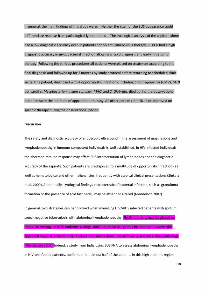
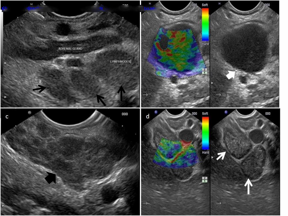
duodenal approach depending on the location of the lymph node groups (figure 1 a-d).
The aspirate was characterized as ‘bloody’ or ‘yellow’ and smears were prepared and fixed on glass
slides for cytological analysis by the gastroenterologist. No pathologist was present in endoscopy
suite. Additional aspirates were placed in three sterile tubes containing 500 µL of physiological saline
for culture, PCR and flow cytometry. Post-procedure, patients were observed until stable and were
monitored telephonically after 24 hours and at day 7 for possible procedure-related complications.
Patients were followed for 3 months following the procedure to correlate endoscopic ultrasound
findings with clinical outcome before returning to scheduled clinic visits.
Cytology
Fine needle aspiration material was subjected to Papanicolaou staining (to evaluate morphology), as
well as Giemsa, Ziehl-Neelsen (detection of acid fast bacilli [AFB]), periodic acid-Schiff and Warthin-
Starry stains. The cytological specimens were interpreted by an expert pathologist (TS) blinded to the
presumptive diagnosis and endoscopic findings.
PCR technique
DNA was extracted from the aspirate using QIAamp® DNA Mini kits (Qiagen, Hilden, Germany).
Detection of Mycobacterial DNA was performed using the Roche LightCycler® Mycobacterium PCR kit
(Roche Diagnostics, Mannheim, Germany) allowing amplification and differentiation of
Mycobacterium tuberculosis (MTB) complex from Mycobacterium avium (MAC) and Mycobacterium
kansasii by means of a post-PCR melt analysis. Genotypic rifampicin and isoniazid susceptibility
testing was performed on MTB positive samples (Genotype® MTBDRplus assay, Hain Lifesciences,
Nehren, Germany).
7
Culture
Mycobacteria were isolated from the aspirated samples using BacT/ALERT liquid culture media
(bioMérieux, Marcy-l’Etoile, France). Identification of positive cultures and genotypic rifampicin and
isoniazid susceptibility testing were then performed. Non-tuberculous mycobacteria from positive
cultures were identified using the GenoType® Mycobacterium CM reverse line probe assay (Hain
Lifesciences, Nehren, Germany). Fungi were isolated from the biopsy samples by inoculation onto
Sabouraud dextrose agar and incubation at 25oC for up to 3 weeks. Identification of isolates was
performed by standard phenotypic methods (Zeng et al. 2007).
Flow cytometry
Fine-needle aspiration specimens were washed twice with RPMI 1640 tissue culture medium,
centrifuged at 250 g for 10 minutes. Enumeration of the cells was performed using Flow-Count™
Fluorospheres (Beckman Coulter, Miami, FL, USA) and the cells were then resuspended in RPMI 1640
tissue culture medium. Immunophenotyping was performed on a Beckman Coulter Cytomics FC500
(Beckman Coulter, Miami, FL, USA). Monoclonal antibodies were used to identify aberrant B and T-
cell populations.
Statistics
All data were entered into a Microsoft Excel 2010 spreadsheet and descriptive statistics calculated.
The accuracy of the diagnostic methods was assessed using two approaches. Firstly, due to the high
burden of tuberculosis (TB) observed in this study, the ability of the methods to identify TB, and
secondly, their ability to identify mycobacterial infections of any kind. That is, the results of the
presumptive diagnoses and the individual diagnostic methods were binary classified in terms of their
identifying any mycobacteria or TB in particular. In the TB-only analysis, we controlled for those
patients undergoing prior anti-tubercular treatment (ATT) or in whom a lymphoma was found to be
8
the final diagnosis. Using Statistix 9 (Analytical software, Tallahassee, Florida, USA) comparisons were
made by means of cross tabulation, two-tailed Fischer exact tests and the calculation of positive and
negative predictive values, between the results of each of the methods versus the final diagnosis. A p
< 0.05 value was considered significant.
Results
EUS-FNA assessment was conducted on 31 HIV-1-infected patients with pathologically enlarged
abdominal lymph nodes. All patients had advanced HIV disease. The characteristics and the findings
for the patients studied are given in Table 1, with more detailed individual patient findings given in
the supplementary text.
EUS detected enlarged lymph nodes in all patients. Para-aortic and porta-hepatis nodes were the
most common locations found in 20 (64,5 %) and 17 (54,8 %) of patients respectively, while 8 (25,8
%) of patients had additional mediastinal nodes. The mean diameter of the nodes were 28mm (15-
63mm). The endoscopic ultrasound appearance of lymph nodes was highly variable including
evidence of necrosis (inhomogenous with central hypo-echogenicity) (32%), hypoechoic (48%), iso
and hyper-echoic appearances (20%) (figure 1 a-d). Tissue elastography was not useful in separating
reactive lymph nodes from tuberculous lymphadenitis. Extra-nodal lesions were also found in 7 (22.5
%) of patients including: an infected sub-diaphragmatic multi-loculated cyst (5,6cm x 2,3cm) that was
drained, one peri-pancreatic cold abscess and pericardial effusions. Only one pericardial effusion was
considered clinically important and was aspirated under EUS guidance where PCR and culture
confirmed MTB. The procedure was found to be safe in this patient population and no immediate or
post-procedural (48 hours) complications were reported.
Cytology yielded the following results in patients with a final diagnosis of a mycobacterial infection:
AFB were detected in 38%, culture was positive in 67% and PCR in 90%. The mean time to positive
culture was 25 days (13-49). Overall MTB was diagnosed in 17/31 (54.8%) patients, MAC in 5/31
9
(16.1%) and mixed TB/MAC in 1/31 (3%) of patients. In all but two MTB PCR positive patients, there
was sufficient target DNA to perform direct drug sensitivity genotyping, leading to two patients being
diagnosed with drug resistant tuberculosis. In all cases the final diagnosis was based on a
combination of at least 2 of the following: cytology, culture and PCR. In one case, flow cytometry was
unable to characterize a lymphoma and a surgical excision biopsy was required for confirmation of
the subtype.
The accuracy of the diagnostic methods relative to identifying TB is given in Table 2.1. In the total
patient group cytology, culture and PCR were all independently significantly associated with a
Final_TB diagnosis. When the ATT-naïve patient group was compared, only culture and PCR remained
significantly associated with the Final_TB diagnosis. The accuracy of the diagnostic methods relative
to identifying any mycobacterial infection is given in Table 2.2. A PCR (+) for mycobacteria, cytology-
with a finding of necrosis and a positive culture of mycobacteria were all independently significantly
associated with a Final_diagnosis of Mycobacteria. A finding of AFB was not. When cytology, AFB and
culturing were combined into a composite variable, the resulting variable’s diagnostic accuracy
increased to nearly that of PCRs’ and was significantly associated with a Final_mycobateria diagnosis.
Following EUS-FNA aspirate investigations, the final diagnosis changed from the presumptive
diagnosis in a total of 13/31 (41.9%) patients and made additional diagnoses in 4/31 (12.9%)
patients. The use of EUS-FNA therefore resulted in a change in the management of 17/31 (54.8%)
patients. In addition 7/31 (23%) patients were diagnosed with a fungal infection. Fungal species
identified included Candida Albicans (57% of 4/7), Candida Glabrata (29% of 2/7) and Candida Krusi
(14% of 1/7). Forty three percent (3/7) were PCR and culture positive and 29% (2/7) were culture
and cytology positive. Fungal infections were only considered clinically relevant if the sample that
yielded the positive result was obtained from the stomach or duodenum. It however remains difficult
to differentiate mucosal contaminants from invasive infections, at least to regional lymph nodes in
this population.
10
In general, the main findings of this study were: i. Neither the size nor the EUS appearance could
differentiate reactive from pathological lymph nodes ii. The cytological analysis of the aspirate alone
had a low diagnostic accuracy even in patients not on anti-tuberculous therapy. iii. PCR had a high
diagnostic accuracy in mycobacterial infection allowing a rapid diagnosis and early initiation of
therapy. Following the various procedures all patients were placed on treatment according to the
final diagnosis and followed up for 3 months by study protocol before returning to scheduled clinic
visits. One patient, diagnosed with 4 opportunistic infections, including Cytomegalovirus (CMV), MTB
pericarditis, Mycobacterium avium complex (MAC) and C. Glabrata, died during the observational
period despite the initiation of appropriate therapy. All other patients stabilized or improved on
specific therapy during the observational period.
Discussion
The safety and diagnostic accuracy of endoscopic ultrasound in the assessment of mass lesions and
lymphadenopathy in immuno-competent individuals is well established. In HIV-infected individuals
the aberrant immune response may affect EUS interpretation of lymph nodes and the diagnostic
accuracy of the aspirate. Such patients are predisposed to a multitude of opportunistic infections as
well as hematological and other malignancies, frequently with atypical clinical presentations (Sinkala
et al. 2009). Additionally, cytological findings characteristic of bacterial infection, such as granuloma
formation or the presence of acid fast bacilli, may be absent or altered (Mendelson 2007).
In general, two strategies can be followed when managing HIV/AIDS infected patients with sputum
smear negative tuberculosis with abdominal lymphadenopathy. Firstly, patients may be placed on
empirical therapy. In MTB endemic settings, anti-tubercular drugs may be initiated however this
approach may risk adverse drug reactions and interactions, hepatotoxicity and miss other pathology
(Mendelson 2007). Indeed, a study from India using EUS FNA to assess abdominal lymphadenopathy
in HIV-uninfected patients, confirmed that almost half of the patients in this high endemic region
11
were found to have non-tubercular pathology (Dhir et al. 2011) Similarly, our study confirmed that
31% of HIV infected patients with enlarged nodes, even in a high endemic area such as South Africa,
had reactive lymphadenopathy. This implies that empirical ATT, prescribed solely on the basis of the
presence of enlarged lymph nodes, may be associated with considerable treatment-related risks and
should consequently be avoided.
Secondly, a tissue sample may be obtained allowing treatment decisions to be based on a laboratory
confirmed diagnosis. Multiple sampling modalities are available including percutaneous FNA and
minimally invasive surgery; neither has been extensively investigated in an HIV-positive population.
Percutaneous ultrasound-guided FNA in HIV-positive patients has been reported to have a high
technical success rate and diagnostic accuracy but only allows assessment of large, superficial nodes
(Veerapand et al 2004). In patients with overlying organs, where multiple lymph nodes sites are
affected, or smaller and deeper nodes, percutaneous sampling becomes challenging (Gupta and
Madoff 2007). In this study we report that endoscopic ultrasound is safe and effective in evaluating
abdominal lymphadenopathy and obtaining tissue samples in HIV infected patients.
Classic EUS findings that have been proposed to differentiate between pathological and reactive
nodes, include size greater than 1 cm, a round shape, hypo-echoic texture and sharp margins
(Catalano et al. 1994). These findings were common in our study and not useful in differentiating
between tuberculous lymphadenitis and reactive lymphadenopathy. Recent studies of mediastinal
MTB in HIV-negative patients have found the presence of hypo-echoic nodes in the majority of
patients (Song et al. 2010), and that the lymph node appearance alone could not reliably
differentiate tuberculous nodes from nodes due to sarcoidosis (Fritscher-Ravens et al. 2011). In our
study the EUS appearance of lymph nodes in HIV-infected patients was highly variable, including a
hypo-echoic texture, particularly when nodes smaller than 2cm were observed. Nodes also
demonstrated central breakdown, extensive necrosis and with a matted or hyper-echoic appearance
(Figure 1).
12
In HIV-uninfected patients, FNA aspirates obtained by EUS are routinely analyzed by cytology. A large
study from India assessing mediastinal lymph nodes by EUS FNA based on cytology only, found an
overall diagnostic yield of 93% (Puri et al.2010). Navani and coworkers (2011) showed, using
endobronchial ultrasound-guided transbronchial needle aspiration, cytological findings consistent
with tuberculosis in 86% of cases. Yet culture was only positive in 47% of patients implying that
treatment decisions were based mainly on cytological findings without the opportunity to
differentiate MTB from MAC or to assess drug resistance. In our study, findings typically associated
with mycobacterial disease such as granuloma formation were often absent, most likely due to HIV-
associated immunosuppression. We found the presence of necrosis on cytology to be predictive of a
positive mycobacterial culture and PCR. Additionally, although highly specific for mycobacterial
disease, microscopy in the form of Ziehl-Nielsen staining for acid-fast bacilli (AFB), cannot reliably
differentiate between MTB and MAC nor identify drug resistant strains.
Culture has historically been considered the gold standard in identifying and differentiating
mycobacterium species but may take up to six weeks and is therefore of little value in early patient
management, particularly in immune-compromised patients prone to rapid progression and early
mortality (Mendelson 2007). In our study mycobacterial culture was positive in only 67% of patients
with the mean time to a positive culture of 25 days that would considerably delay initiation of
effective therapy. A recent study in patients with isolated mediastinal lymphadenopathy suggests
that culture yields may be lower in HIV positive patients (Navani et al. 2011). In that study in a
subgroup of 17 HIV positive patients only 36% had positive mycobacterial cultures. Collectively the
published data suggest that FNA subjected to cytology and culture only, is inadequate in obtaining a
final diagnosis in the majority of HIV positive patients with abdominal lymphadenopathy. In contrast
PCR amplifying mycobacterial DNA had a diagnostic accuracy of 94%. Combining all modalities we
could show that the final diagnosis changed from the presumptive diagnosis as formulated by the
infectious disease specialist leading to a change in the management in 42% of patients.
13
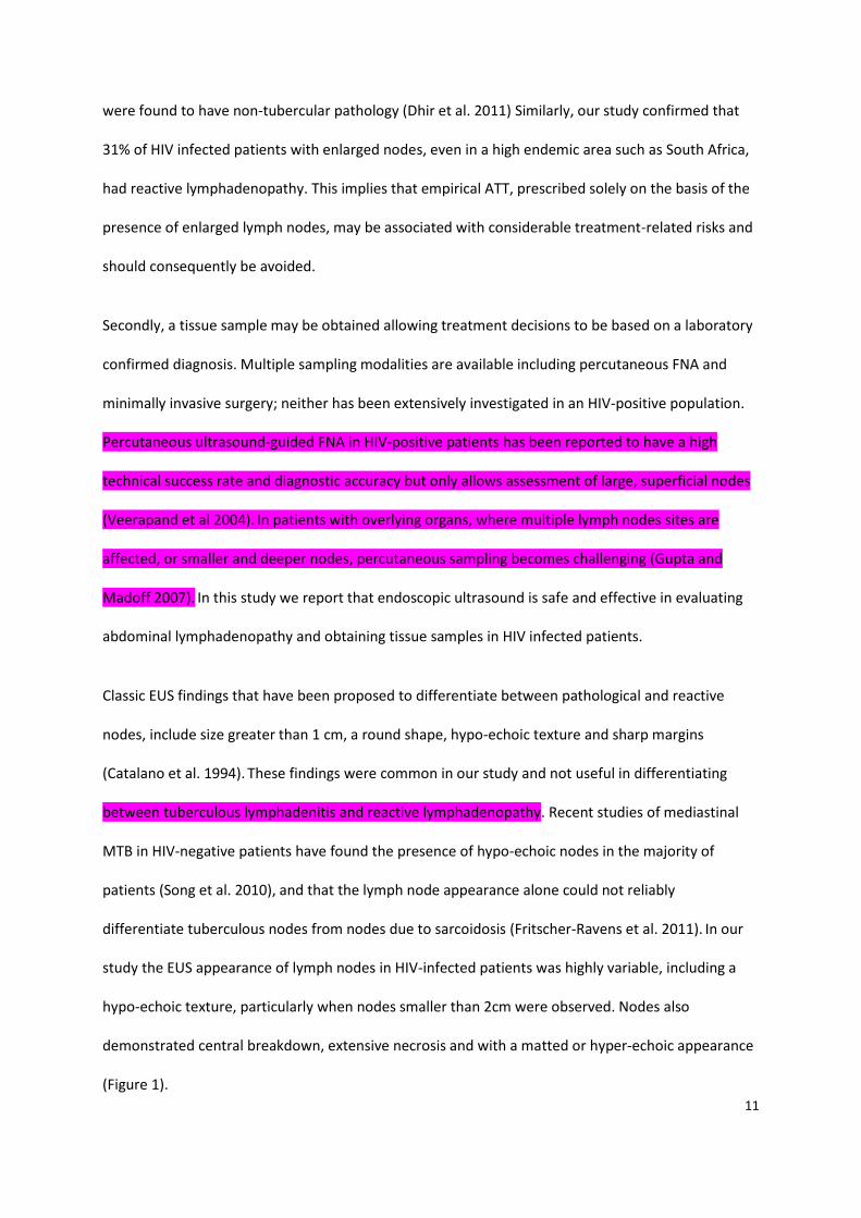
Based on the calculated positive and negative predictive values of the various post EUS-FNA
modalities we defined a diagnostic algorithm (figure 2). Our results demonstrated that the presence
of a yellow aspirate in combination with caseous necrosis on cytology had a PPV of 96% whereas a
bloody aspirate in the absence of caseous necrosis had a 77% NPV which can be used to determine in
which patients additional PCR should be performed. PCR remains imperative to differentiate MDR-TB
strains and MTB from MAC.
Our detailed prospective analysis which assessed the diagnostic accuracy of EUS guided FNA was
performed on a relatively small sample size of patients with advanced HIV infection. The results of
our study need to be validated in a larger cohort of patients. We also realize that endoscopic
ultrasound and PCR may not be readily available in many resource limited settings. This implies that
in such settings that percutaneous ultrasound-guided FNA relying on cytology may remain an
important diagnostic tool. Irrespective, our study provides a clear description of the value of EUS and
the utilization of PCR in the management of patients with advanced HIV with abdominal
lymphadenopathy.
In summary, in addition to analyses of EUS-FNA by cytology and culture, polymerase chain reaction
amplification of mycobacterial DNA was found to be invaluable in providing a final diagnosis and
guiding therapy. If mycobacterial infection was confirmed by PCR, sequencing and genotypic drug
susceptibility testing could also be performed, enabling early diagnosis and the prompt initiation of
appropriate therapy.
Acknowledgment
We would like to thank Dr CF Schluter, Dr F Kinkel and Dr Kgomo, Dr. Sieling for their contributions to
the study and in the care of our patients.
Conflict of interest
14
Potential competing interests: None. Financial support: South African Gastroenterological Society
(SAGES)/ Astra Zeneca Fellowship in Gastroenterology awarded to Schalk van der Merwe.
Figure legends
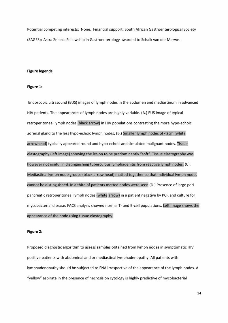
Figure 1:
Endoscopic ultrasound (EUS) images of lymph nodes in the abdomen and mediastinum in advanced
HIV patients. The appearances of lymph nodes are highly variable. (A.) EUS image of typical
retroperitoneal lymph nodes (black arrow) in HIV populations contrasting the more hypo-echoic
adrenal gland to the less hypo-echoic lymph nodes; (B.) Smaller lymph nodes of <2cm (white
arrowhead) typically appeared round and hypo-echoic and simulated malignant nodes. Tissue
elastography (left image) showing the lesion to be predominantly “soft”. Tissue elastography was
however not useful in distinguishing tuberculous lymphadenitis from reactive lymph nodes. (C).
Mediastinal lymph node groups (black arrow head) matted together so that individual lymph nodes
cannot be distinguished. In a third of patients matted nodes were seen (D.) Presence of large peri-
pancreatic retroperitoneal lymph nodes (white arrow) in a patient negative by PCR and culture for
mycobacterial disease. FACS analysis showed normal T- and B-cell populations. Left image shows the
appearance of the node using tissue elastography.
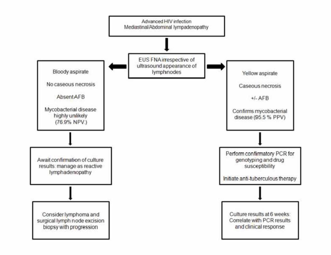
Figure 2:
Proposed diagnostic algorithm to assess samples obtained from lymph nodes in symptomatic HIV
positive patients with abdominal and or mediastinal lymphadenopathy. All patients with
lymphadenopathy should be subjected to FNA irrespective of the appearance of the lymph nodes. A
“yellow” aspirate in the presence of necrosis on cytology is highly predictive of mycobacterial
15
disease. These samples should be subjected to further analysis with culture and PCR to differentiate
MTB from MAC and exclude genotypic resistance. If the aspirate is “bloody” and necrosis is not
detected on cytology mycobacterial disease is highly unlikely. Such a sample should be subjected to
culture and the patient carefully followed. In event of progression an excision lymph node biopsy
should be performed to exclude lymphoma.
References:
Abbadi SH, Sameaa GA, Morlock G, Cooksey RC. Molecular identification of mutations associated
with anti-tuberculosis drug resistance among strains of Mycobacterium tuberculosis. Int J Infect Dis,
2009;13(6):673-8
16
Al-Haddad M, Savabi MS, Sherman S, McHenry L, Leblanc J, Cramer H, Emerson R, O'Neil J, Khashab
M, Dewitt J. Role of endoscopic ultrasound-guided fine-needle aspiration with flow cytometry to
diagnose lymphoma: a single center experience. J Gastroenterol Hepatol. 2009 Dec;24(12):1826-33
Barnard M, Albert H, Coetzee G, O’Brien R, Bosman E. Rapid molecular screening for multidrug-
resistant tuberculosis in a high-volume public health laboratory in South Africa. Am J Respir Crit Care
Med. 2008 Apr 1;177(7):787-92
Bhutani MS, Logroño R. Endoscopic ultrasound-guided fine-needle aspiration cytology for diagnosis
above and below the diaphragm. J Clin Ultrasound 2005;33:401-11.
Bos JC, Smalbraak L, Macome AC, Gomes E, van Leth F, Prins JM.TB diagnostic process management
of patients in a referral hospital in Mozambique in comparison with the 2007 WHO
recommendations for the diagnosis of smear-negative pulmonary TB and extrapulmonary TB. Int
Health. 2013 Oct 14. [Epub ahead of print]
Catalano MF, Sivak MV Jr, Rice T, Gragg LA, Van Dam J. Endosonographic features predictive of lymph
node metastasis. Gastroinstest Endosc 1994;40(4):442-6.
Dhir V, Mathew P, Bhandari S, Bapat M, Kwek A, Doctor V, Maydeo A. Endosonography-guided fine
needle aspiration cytology of intra-abdominal lymph nodes with unknown primary in a tuberculosis
endemic region. J Gastroenterol Hepatol. 2011 Dec;26(12):1721-4
Dos Santos RP, Scheid KL, Willers DM, Goldani LZ. Comparative radiological features of disseminated
disease due to Mycobacterium tuberculosis vs non-tuberculosis mycobacteria among AIDS patients
in Brazil. BMC Infect Dis, 2008;29;8:24.
Fiske CT, Griffin MR, Erin H, Warkentin J, Lisa K, Arbogast PG, Sterling TR. Black race, sex, and
extrapulmonary tuberculosis risk: an observational study. BMC Infect Dis. 2010; 22:10:16.
17
Fritscher-Ravens A, Ghanbari A, Topalidis T, Pelling M, Kon OM, Patel K, Arlt A, Bhowmik A (2011)
Granulomatous mediastinal adenopathy: Can endoscopic ultrasound guided fine needle aspiration
differentiate between tuberculosus and sarcoidosis? Endoscopy.2011 Nov;43(11):955-61
Giordani MT, Brunetti E, Binazzi R, Benedetti P, Stecca C, Goblirsch S, Heller T. Extrapulmonary
mycobacterial infections in a cohort of HIV-positive patients: ultrasound experience from Vicenza,
Italy. Infection 2013;41:409–414
Gupta S, Madoff DC. Image-guided percutaneous needle biopsy in cancer diagnosis and staging. Tech
Vasc Interv Radiol 2007; 10(2):88-101.
Heller T, Goblirsch S, Wallrauch C, Lessells R,Brunetti E. Abdominal tuberculosis: sonographic
diagnosis and treatment response in HIV-positive adults in rural South Africa . Int J Infect Dis.2010
Sep;14:108-12
Mendelson M. Diagnosing tuberculosis in HIV-infected patients: challenges and future prospects. Br
Med Bull 2007;81-82:149-65.
National Institute for Health and Clinical Excellence (NICE). NICE guidelines on Tuberculosis 2006.
http://www.nice.org.uk/CG33
Navani N, Molyneaux PL, Breen RA, Connell DW, Jepson A, Nankivell M, Brown JM, Morris-Jones S,
Ng B, Wickremasinghe M, Lalvani A, Rintoul RC, Santis G, Kon OM, Janes SM. Utility of endobronchial
ultrasound-guided transbronchial needle aspiration in patients with tuberculous intrathoracic
lymphadenopathy: a multicentre study. Thorax. 2011 Oct; 66(10):889-93.
Puri R, Vilmann P, Sud R, Kumar M, Taneja S, Verma K, Kaushik N. Endoscopic ultrasound-guided fine-
needle aspiration cytology in the evaluation of suspected tuberculosis in patients with isolated
mediastinal lymphadenopathy. Endoscopy. 2010 Jun;42(6):462-7
18
Radin R. HIV infection: analysis in 259 consecutive patients with abnormal abdominal CT findings.
Radiology.1995; 197(3):712-22.
Săftoiu A, Vilmann P.Role of endoscopic ultrasound in the diagnosis and staging of pancreatic cancer.
J Clin Ultrasound. 2009 Jan;37(1):1-17
Sarma PK, Chowhan AK, Agrawal V, Agarwal V .Fine needle aspiration cytology in HIV-related
lymphadenopathy: experience at a single centre in north India. Cytopathology, 2010;21(4):234-9
Sinkala E, Gray S, Zulu I, Mudenda V, Zimba L, Vermund SH, Drobniewski F, Kelly P. Clinical and
ultrasonographic features of abdominal tuberculosis in HIV positive adults in Zambia. BMC Infect Dis,
2009 Apr 17;9:44.
Song HJ, Park YS, Seo DW, Jang SJ, Choi KD, Lee SS, Lee GH, Jung HY, Kim JH(2010) Diagnosis of
mediastinal tuberculosis by using EUS-guided needle sampling in a geographic region with an
intermediate tuberculosis burden. Gastrointest Endosc. 2010 Jun;71(7):1307-13
Uzunkoy A, Harma M, Harma M. Diagnosis of abdominal tuberculosis: experience from 11 cases and
review of the literature. World J Gastroenterol 2004;10:3647-9.
Veerapand P, Chotimanvijit R, Laohasrisakul N, Muennooch W. Percutaneous ultrasound-guided fine
needle aspiration of abdominal lymphadenopathy in AIDS patients.J Med Assoc Thai. 2004
Apr;87(4):400-4.
World Health Organization Multidrug and extensively drug-resistant TB (M/XDR-TB): 2010 Global
report on surveillance and response. 2010
http://whqlibdoc.who.int/publications/2010/9789241599191_eng.pdf
19
asufuku K, Nakajima T, Chiyo M, Sekine Y, Shibuya K, Fujisawa T. Endobronchial ultrasonography:
urrent status and future directions. J Thorac Oncol 2007;2:970-9.
eng X, Kong F, Halliday C, Chen S, Lau A, Playford G, Sorrell TC. Reverse Line Blot Hybridization Assay
or Identification of Medically Important Fungi from Culture and Clinical Specimens. J Clin Microbiol
007;(9):2872-80.
Supplementary text
Polymerase chain reaction amplification of mycobacterial and fungal DNA
The MTBDRplus assay is a PCR-reverse line probe assay that detects mutations associated with
rifampicin resistance within an 81-bp ‘hot spot’ region of the rpoB gene. In addition, it detects
mutations in codon 315 of the katG gene and in the inhA promoter region that are associated with
INH resistance. This assay has been shown to have a sensitivity of >95% and >90% for detecting
rifampicin and INH resistance respectively in South African TB isolates, as compared to the gold
standard of phenotypic drug susceptibility testing (Barnard et al. 2008).
A broad range fungal PCR targeting the conserved internal transcribed spacer region 2 (ITS-2) of the
fungal ribosomal RNA gene was used to analyze each sample for the presence of fungal DNA. This
PCR incorporated primers flanking the fungal ITS-2 region as described by Zeng et al (2007). The real-
time PCR was performed with a 50µl reaction mixture containing 10µl extracted DNA, 25µl SensiMix
dT (containing reaction buffer, heat-activated Taq DNA polymerase, dNTPs, 3mM MgCl2) [Quantace,
London, United Kingdom], 1x Sybr Green (Quantace, London, United Kingdom), 25pmol of primers
its3Sb (5’ GTGAATCATCGARTCTTTGAACG-3’; positions 271-279) and its4Ab (5’-
GTTGGTTTCTTTTCCTCCGCTTAT TGATATGC-3’; positions 710-741). Primer positions refer to Candida
albicans sequences corresponding to Genbank accession numbers AF455524 and L28817
respectively. Amplification was performed on a RotorGene 6000 thermal cycler (Corbett Research,
Sydney, Australia), with the following conditions: 1 cycle of 94°C for 10 minutes, 30 cycles of 95 °C for
15s, 50°C for 30s, and 72°C for 30s. The presence of specific amplified product was determined by
means of a post-PCR melt analysis. DNA sequences were compared with sequences from GenBank,
EMBL, and DJB database using the gapped BLASTN 2.0.5 program.
Individual patient diagnostic findings
Pat.
Presumptive diagnosis
Medi-cation
EUS-FNA Biopsy examination
Echo-appearance
Sample
AFB
Cytology
PCR
Culture
Final Diagnosis
1 MTB Non Hypo-echoic nodes
Bloody Negative atypical lymphoid proliferation
negative No growth Non Hodgkins lymphoma1
2 MTB
lymphoma
ATT Hypo-echoic nodes
Yellow Positive caseous necrosis
MTB (Sensitive)
C. Albicans
MTB
C. Albicans
MTB
C. albicans
3 MTB AB Hypo-echoic nodes
Bloody Negative Non specific reactive
Negative MTB (Sensitive)
MTB
4 MTB ART Hypo-echoic nodes
Pericardial effusion
Bloody
fluid
Positive candida spp MAC
MTB (Sensitive)2
C. glabrata
MAC
Co-infection MAC, MTB2
C. glabrata
5 MTB AB Hypo-echoic nodes
Bloody Positive Non specific reactive
MAC MAC MAC
6 MDR-MTB
lymphoma
ART
MAC
Central breakdown
Yellow Positive AFB positive
Caseous necrosis
MTB (Rifampicin resistant)
No growth Resistant MTB
7 MDR-MTB ART
ATT
Central breakdown
Yellow Negative Non specific reactive
MTB (Sensitive)
No growth MTB
8 MDR-MTB ART
ATT
Central breakdown
Yellow Negative Caseous necrosis
MTB (Sensitive)
No growth MTB
9 MTB AB Central breakdown
Yellow Negative Caseous necrosis
MTB (Sensitive)
MTB MTB
10 MDR-MTB ART
ATT
Central breakdown
Multi-loculated cyst
Bloody
Cyst fluid
Negative Non specific reactive
negative No growth
α-hemolytic strep + coagulase neg staph3
Reactive lymph-adenopathy
Infected multi-loculated cyst
11 MTB
Lymphoma
Non Central breakdown
Bloody Negative Non specific reactive
negative no growth Reactive lymph-adenopathy
12 MTB AB Central breakdown
Yellow Positive Granulomas
Caseous necrosis
MTB (Sensitive)
C. Albicans
MTB MTB
C. Albicans
13 MDR-MTB ATT Hypo-echoic nodes
Yellow Positive Caseous necrosis
MTB (Sensitive)
MTB MTB
14 MTB
Lymphoma
AB Hypo-echoic nodes
Bloody Negative Candida spp MTB MTB (Sensitive)
C. Albicans
MTB
C. Albicans
15 Hospital acquired infection
ART Hypo-echoic
Peri-pancreatic cold abscess
Yellow4 Negative Caseous necrosis4
MTB4 No growth MTB4
16 Resistant MAC
MTB
ART
MAC
Hyper-echoic Yellow Positive Caseous necrosis
MAC C. Albicans
MAC
MAC
C. Albicans
17 MTB ART Central breakdown
Bloody Negative Non specific reactive
MTB (sensitive)
MTB MTB
18 MTB ART Hypo-echoic Bloody Negative Non specific reactive
negative No growth Reactive lympha-denopathy
19 Reactive ART Hypo-echoic Bloody Negative Non specific reactive
negative No growth Reactive lymph-adenopathy
20 MTB ART Hypo-echoic Bloody Negative Non specific reactive
negative MAC MAC
21 MTB Non Central breakdown
Yellow Negative Non specific reactive
MTB (sensitive)
No growth MTB
22 MTB Non Hypo-echoic Yellow Negative Caseous necrosis
MTB (sensitive)
No growth MTB
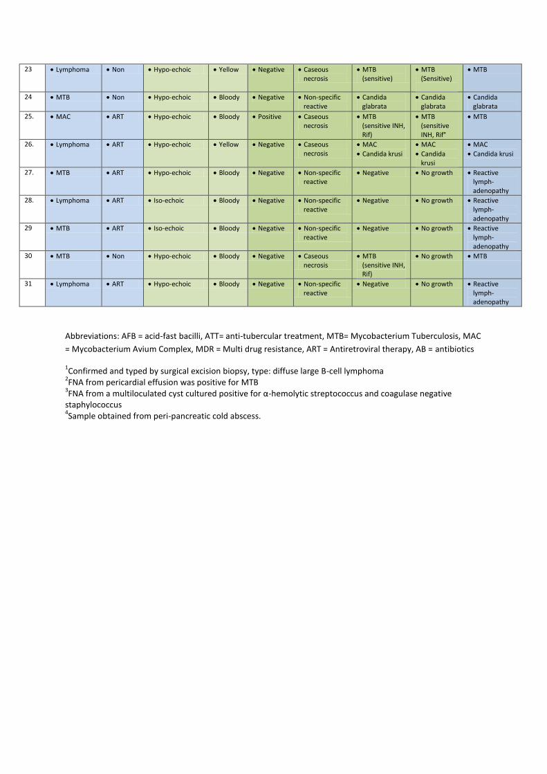
23 Lymphoma Non Hypo-echoic Yellow Negative Caseous necrosis
MTB (sensitive)
MTB (Sensitive)
MTB
24 MTB Non Hypo-echoic Bloody Negative Non-specific reactive
Candida glabrata
Candida glabrata
Candida glabrata
25. MAC ART Hypo-echoic Bloody Positive Caseous necrosis
MTB (sensitive INH, Rif)
MTB (sensitive INH, Rif°
MTB
26. Lymphoma ART Hypo-echoic Yellow Negative Caseous necrosis
MAC
Candida krusi
MAC
Candida krusi
MAC
Candida krusi
27. MTB ART Hypo-echoic Bloody Negative Non-specific reactive
Negative No growth Reactive lymph-adenopathy
28. Lymphoma ART Iso-echoic Bloody Negative Non-specific reactive
Negative No growth Reactive lymph-adenopathy
29 MTB ART Iso-echoic Bloody Negative Non-specific reactive
Negative No growth Reactive lymph-adenopathy
30 MTB Non Hypo-echoic Bloody Negative Caseous necrosis
MTB (sensitive INH, Rif)
No growth MTB
31 Lymphoma ART Hypo-echoic Bloody Negative Non-specific reactive
Negative No growth Reactive lymph-adenopathy
Abbreviations: AFB = acid-fast bacilli, ATT= anti-tubercular treatment, MTB= Mycobacterium Tuberculosis, MAC
= Mycobacterium Avium Complex, MDR = Multi drug resistance, ART = Antiretroviral therapy, AB = antibiotics
1Confirmed and typed by surgical excision biopsy, type: diffuse large B-cell lymphoma
2FNA from pericardial effusion was positive for MTB
3FNA from a multiloculated cyst cultured positive for α-hemolytic streptococcus and coagulase negative
staphylococcus 4Sample obtained from peri-pancreatic cold abscess.
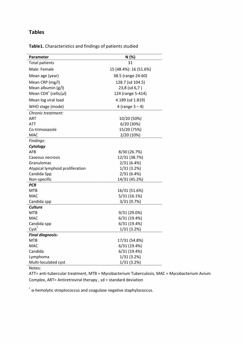
Tables
Table1. Characteristics and findings of patients studied
Parameter N (%)
Total patients 31
Male: Female 15 (48.4%): 16 (51.6%)
Mean age (year) 38.5 (range 24-60)
Mean CRP (mg/l) Mean albumin (g/l)
128.7 (sd 104.5) 23,8 (sd 6,7 )
Mean CD4+ (cells/μl) 124 (range 5-414)
Mean log viral load 4.189 (sd 1.819)
WHO stage (mode) 4 (range 3 – 4)
Chronic treatment: ART ATT Co-trimoxazole MAC
10/20 (50%) 6/20 (30%)
15/20 (75%) 2/20 (10%)
Findings:
Cytology AFB Caseous necrosis Granulomas Atypical lymphoid proliferation Candida Spp Non-specific
8/30 (26.7%)
12/31 (38.7%) 2/31 (6.4%) 1/31 (3.2%) 2/31 (6.4%)
14/31 (45.2%)
PCR MTB MAC Candida spp
16/31 (51.6%) 5/31 (16.1%) 3/31 (9.7%)
Culture MTB MAC Candida spp Cyst*
9/31 (29.0%) 6/31 (19.4%) 6/31 (19.4%) 1/31 (3.2%)
Final diagnosis:
MTB MAC Candida Lymphoma Multi-loculated cyst
17/31 (54.8%) 6/31 (19.4%) 6/31 (19.4%) 1/31 (3.2%) 1/31 (3.2%)
Notes: ATT= anti-tubercular treatment, MTB = Mycobacterium Tuberculosis, MAC = Mycobacterium Avium
Complex, ART= Antiretroviral therapy , sd = standard deviation
* α-hemolytic streptococcus and coagulase negative staphylococcus.
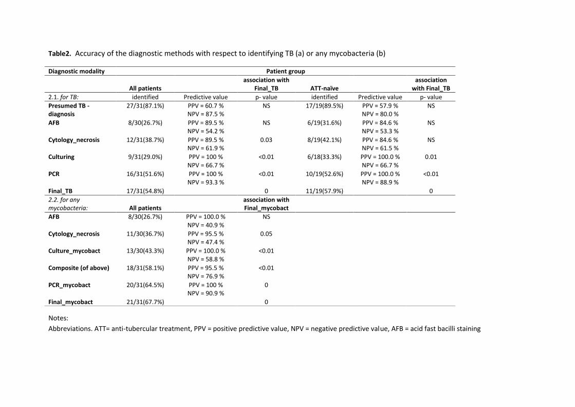
Table2. Accuracy of the diagnostic methods with respect to identifying TB (a) or any mycobacteria (b)
Notes:
Abbreviations. ATT= anti-tubercular treatment, PPV = positive predictive value, NPV = negative predictive value, AFB = acid fast bacilli staining
Diagnostic modality Patient group
All patients
association with Final_TB
ATT-naïve
association with Final_TB
2.1. for TB: identified Predictive value p- value identified Predictive value p- value
Presumed TB - diagnosis
27/31(87.1%) PPV = 60.7 % NPV = 87.5 %
NS 17/19(89.5%) PPV = 57.9 % NPV = 80.0 %
NS
AFB 8/30(26.7%) PPV = 89.5 % NPV = 54.2 %
NS 6/19(31.6%) PPV = 84.6 % NPV = 53.3 %
NS
Cytology_necrosis 12/31(38.7%) PPV = 89.5 % NPV = 61.9 %
0.03 8/19(42.1%) PPV = 84.6 % NPV = 61.5 %
NS
Culturing 9/31(29.0%) PPV = 100 % NPV = 66.7 %
<0.01 6/18(33.3%) PPV = 100.0 % NPV = 66.7 %
0.01
PCR 16/31(51.6%) PPV = 100 % NPV = 93.3 %
<0.01 10/19(52.6%) PPV = 100.0 % NPV = 88.9 %
<0.01
Final_TB 17/31(54.8%) 0 11/19(57.9%) 0
2.2. for any mycobacteria:
All patients
association with Final_mycobact
AFB 8/30(26.7%) PPV = 100.0 % NPV = 40.9 %
NS
Cytology_necrosis 11/30(36.7%) PPV = 95.5 % NPV = 47.4 %
0.05
Culture_mycobact 13/30(43.3%) PPV = 100.0 % NPV = 58.8 %
<0.01
Composite (of above) 18/31(58.1%) PPV = 95.5 % NPV = 76.9 %
<0.01
PCR_mycobact 20/31(64.5%) PPV = 100 % NPV = 90.9 %
0
Final_mycobact 21/31(67.7%) 0
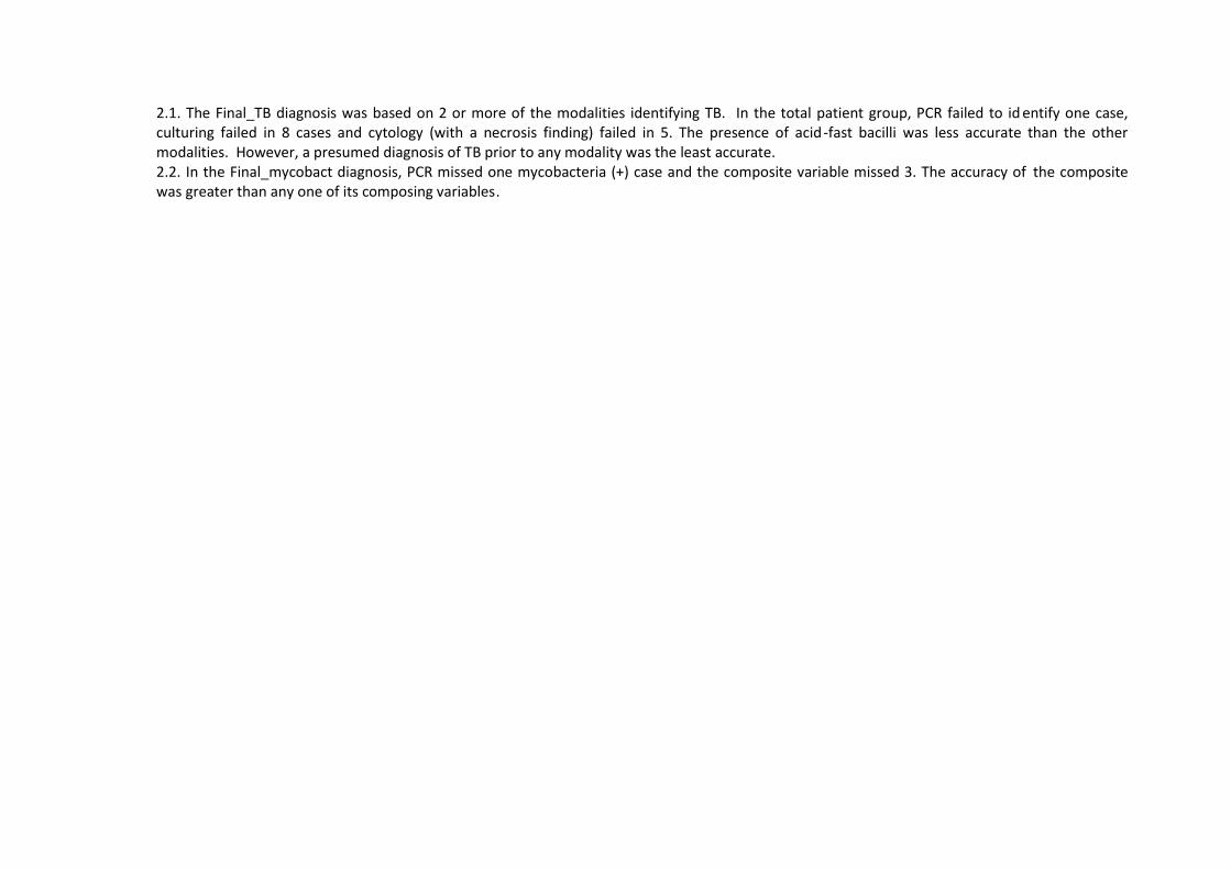
2.1. The Final_TB diagnosis was based on 2 or more of the modalities identifying TB. In the total patient group, PCR failed to id entify one case, culturing failed in 8 cases and cytology (with a necrosis finding) failed in 5. The presence of acid-fast bacilli was less accurate than the other modalities. However, a presumed diagnosis of TB prior to any modality was the least accurate.
2.2. In the Final_mycobact diagnosis, PCR missed one mycobacteria (+) case and the composite variable missed 3. The accuracy of the composite was greater than any one of its composing variables.
FigureClick here to download Figure: UMB Final Nieuwoudt_figure 1 with arrows (2).pptx