polyfacts - home | polysciences
TRANSCRIPT

Peripheral blood smear (peripheral blood film) is a glass microscope slide coated with a thin layer of venous blood on one side that has been air dried and fixed with methanol. A Romanowsky stain variant such as StainRITE™ Wright Stain (Cat. #24986) is used primarily to stain peripheral blood smears and bone marrow aspirates and examined under a microscope.
Microscope examination of the peripheral blood smear is only one consideration, the second and third are peripheral blood counts and red blood cell (RBC) indices, respectively. These three tools evaluate hematological disorders as well. Peripheral blood smears are stained with StainRITE™ Wright Giemsa (Cat. #24985), StainRITE™ Wright Stain (Cat. #24986) and StainRITE™ MGG (Cat. #24981). These
stains are utilized to identify different blood disorders in RBC, white blood cells (WBC) and platelet population of cells. Morphologic abnormalities of peripheral blood cells are discovered by microscopic examination with the oil immersion lens of well-prepared peripheral blood smear films stained with Wright’s stain. In the interpretation of the morphology of RBCs, an examiner concentrates on areas of red cells that are singly spread out. The peripheral blood smear is very important because it validates and supplements information provided by automated hematology analyzers (“blood cell counters”). Blood cell counters are precise with the classification of abnormal cells, however, it is required that a trained microscopist, peripheral blood smear and light microscope be used when a specimen is flagged by the hematology analyzer in order to obtain an accurate conclusion.
www.polysciences.com
News | V iews | Ins ights
PolyFactsBioSciencesVol. 5 | No. 1
Featured Product ····················· 1
Use the Wright Stain: High Quality StainRITE™ Ready-to-Use Stains for Hematology
Science Solutions ···················· 4
Fix Lymphoreticular Myeloid, Lymph Nodes and Bone Marrow Tissues
Product Spotlight ···················· 5
Gold Standard Series: Gill’s Hematoxylins #1, 2 & 3
Additional Lab Products for Hematology Staining ·············· 5
Flash Dip FNA / H. Pylori KitSudan Black B, C.I. 26150Terry’s Polychrome Methylene BluePeriodic Acid Schiff’s (PAS) StainLeishman Stain
INSIDE THIS ISSUE
. . . . Continued on page 2
Use the Wright Stain High Quality StainRITE™ Ready-to-Use Stains for Hematology
StainRITE™ May-Grünwald Giemsa Stain, 100x (Cat. #24981)
StainRITE™ Wright Stain, 100x (Cat. #24986)
StainRITE™ Wright Giemsa, Stain, 100x(Cat. #24985)
• Sterilized lancet or needle• 20 clean microscope slides and coverslips• Canada balsam• 95% ethyl or methyl alcohol
• Distilled water• Giemsa stain with buffer and protocol• Microscope 200x or greater• Immersion Oil
Peripheral Blood Smear Preparation
In addition to providing cell counts and graphical displays of the information recovered, these instruments also provide a warning that atypical cells were found and provide a presumptive identification of the abnormality. The instrument operator reviews the information from each specimen, to determine if a smear preparation and light microscopy will be necessary. If a specimen is not flagged by the blood cell count analyzer and does not require a microscopic evaluation the information is immediately released to the clinician.
The wedge slide, (“push slide”) technique developed by Maxwell Wintrobe remains the standard method for the preparation of peripheral blood smears (films). The following procedure is used to prepare a peripheral smear.
Blood Smear Preparation Materials

Taking the blood
Cleanse finger with a sterile lancet and puncture fingertip. Keep all the materials needed ready and protected from dust, particularly the clean microscope slides.
• Place a 1”x 3” glass microscope slide with a frosted end on a flat surface (usually the counter top of a laboratory bench).
• Attach label on the slide or write patient name, specimen identification number and date of preparation on the frosted surface.
• Place 2-3mm drop of blood approximately 1/4” from the frosted slide with a wooden applicator stick or glass capillary tube.
• Hold the slide by the narrow side between thumb and forefinger of one hand at the end farthest from the frosted end.
• Grasp a second slide (“spreader slide”) between thumb and forefinger of the other hand at the frosted end.
• Place edge of the spreader slide on the lower slide in front of the drop of blood (side farthest from the frosted end).
• Pull spreader slide toward the frosted end until it touches the drop of blood. Permit the blood to spread by capillary motion until it almost reaches the edges of the spreader slide.
• Push spreader slide forward at a 30º angle with a rapid even motion. Let the weight of the slide do the work or place a small drop of blood near an end of a slide.
• Bring edge of another slide in contact with the drop and allow the drop to bank evenly behind the spreader.
• The angle between the two slides has to be 30º - 40º. Push to the left in a smooth quick motion.
• The smear should cover about half the slide. • It is important that the quantity of blood is not excessive otherwise
the red cells could hide the leukocytes. • If you succeed in making a gradual transition from thick to thin in
your smear, you should get a zone with a satisfactory distribution of cells. With a single drop of blood, you can make several smears.
• Perform multiple smears. They are not always successful but with multiple attempts one well prepared smear should be achieved.
• To avoid producing clots, you must make each smear with fresh blood straight after having deposited it.
• It is useful to be helped by another person where one deposits the blood and the other makes the smears.
• With a microscope, observe to check if smears are properly made. • Red cells must not overlap each other, nor be so scarce that they
are too spread out.
Notes: If you observe the smear as it is after fixing, you will see very little because all cells are very transparent. The erythrocytes are slightly visible, but the leukocytes are too pale, almost invisible and you will not see anything inside them. To be able to observe and recognize the different kinds of leukocyte, you must stain them.
2
. . . . Use the Wright Stain, continued
drop ofblood
ADVANCEMENT
ADHESION
30˚- 40˚APPROACHING
StainRITE™ Stain ResultsStainRITE™ Wright-Giemsa Stain Solution(Cat. #24985)
StainRITE™ Wright Stain Solution(Cat. #24986)
StainRITE™ May-Grünwald Stain Solution(Cat. #24981)
Erythrocytes
PolymorphonuclearNeutrophils
Eosinophils Cytoplasm
Basophils
Lymphocytes and Monocytes
Platelets
light pink to moderate purple, not grey or blue
blue to dark blue to purple nuclei reddish purple lilac granules, pale pink cytoplasm
blue to dark blue to purple nuclei, red to orange-red granules, blue cytoplasm
purple to dark blue to black nuclei, purple granules
dark purple nuclei, sky blue cytoplasm
violet to purple granules
yellowish red
dark blue nuclei, reddish lilac granulates, pale pink cytoplasm
blue nuclei, red to orange-red granules,blue cytoplasm
purple to dark blue nuclei,dark purple granules
dark purple nuclei, sky blue cytoplasm
violet to purple granules
light pink to moderate purple, not grey or blue
blue to dark blue to purple nuclei, reddish purple lilac granules, pale pink cytoplasm
blue to dark blue to purple nuclei, red to orange-red granules, blue cytoplasm
purple to dark blue to black nuclei, purple granules
dark purple nuclei, sky blue cytoplasm
violet to purple granules
Featured StainRITE™ Ready-to-Use Hematology Stains
Cat. # Description Size
24985249842498624989249812503225038
StainRITE™ Wright-Giemsa Stain SolutionStainRITE™ Wright-Giemsa Stain Phosphate Buffer pH 6.8StainRITE™ Wright Stain SolutionStainRITE™ Wright Stain Phosphate Buffer pH 6.8StainRITE™ May-Grünwald Stain SolutionStainRITE™ May-Grünwald Stain Phosphate Buffer pH 7.2StainRITE™ Giemsa Stain (for May-Grünwald)
1L, 10L1L, 4L1L, 4L, 20L1L, 4L1L, 4L1L, 4L100ml, 400ml
How are StainRITE™ Blood Stains Used?
A peripheral blood smear was once prepared on nearly everyone who had a complete blood count (CBC) performed. In smaller labs blood cell counts are still done manually, while larger labs use automated blood cell counting instruments. If the presence of abnormal WBCs, RBCs or platelets is suspected a blood smear examined by a trained eye is still the best method for definitively evaluating and identifying immature and abnormal cells.
There are many diseases, disorders and deficiencies that effect the blood cells function, lifespan, type and amount produced. Although usually only normal mature cells are released into the bloodstream, circumstances can force the bone marrow to release immature and/or malformed cells into the circulation.
When is a Blood Smear Ordered?
A Blood smear is primarily ordered to evaluate blood cell populations when there’s a differential in the CBC performed with an automated blood cell counter indicating the presence of abnormal or immature cells. It may also be performed when a doctor suspects a deficiency, disease or disorder that is affecting blood cell production, such as anemias, abnormal production of cells or increased cell destruction. A blood smear may also be ordered when a patient is being treated or monitored for a blood cell-related disease.
What is a Bone Marrow Aspirate / Biopsy?
Bone marrow biopsy and aspiration procedures provide information about the status and capability for blood cell production. It is not routinely ordered, in fact the majority of people will never have one done. A bone marrow aspiration and/or biopsy is ordered to help evaluate blood cell production, diagnose leukemia, bone marrow disorder and a variety of stages of cancer that have spread into the marrow and to determine the cause of severe anemia. Severe anemia occurs due to decreased RBC production, increased loss, abnormal RBC production and/or to a vitamin or mineral deficiency or excess. Conditions that affect the bone marrow can affect the number, mixture and maturity of the cells and its fibrous structure.
A bone marrow sample may also be evaluated and cultured for the presence of microorganisms such as fungus, bacteria and mycobac-teria (such as that which causes Tuberculosis, TB) when the patient has a fever of an unknown origin. Additional marrow testing may be ordered when it is suspected that the patient has a chromosomal abnormality or disorder associated with iron storage that may cause iron to accumulate in the marrow. In histology a Trichrome Stain (Cat. #24205, #25088), Retic Stain (Cat. #25093) and Iron Stain (Cat. #24199) are also done.
When a person is being treated for cancer, a bone marrow aspiration/biopsy may be ordered to evaluate the response to therapy. It can determine whether suppressed marrow function is beginning to return to normal.
A CBC and reticulocyte are frequently ordered along with the bone marrow aspiration/biopsy. The results are used to help evaluate cell production in the marrow and compared to current cell populations in the blood.
When is a Bone Marrow Aspirate / Biopsy Ordered?
Bone marrow aspiration/biopsy may be ordered as a diagnostic procedure, for example when one of the following blood disorders is suspected:
• Aplastic Anemia • Acute Leukemia • Myelodysplastic Syndrome • Chronic Myelogenous Leukemia • Myelofibrosis and Essential Thrombocythemia • Multiple Myeloma • Severe Thrombocytopenia and/or anemia
and/or neutropenia
It may be ordered when staging certain cancers. Staging is a careful and thorough examination and classification of how far the cancer has spread and what body organs are involved. These may include:
• Hodgkin’s and Non-Hodgkin’s Lymphomas• Small Cell Carcinoma of the Lung (not frequently done anymore)
It may be ordered for culturing in some cases, such as:
• Persistent fever is present in HIV/AIDS or other patients with a weak immune system, sometimes a PAS, GMS, Gram Stain is done in histology
• In patients suspected of having infectious diseases such as Brucellosis or Typhoid Fever
A bone marrow biopsy and aspiration may also be ordered at intervals when a person is being treated for a cancer to evaluate whether marrow function is being suppressed, and if it is, when its function begins to recover.
. . . . Use the Wright Stain, continued
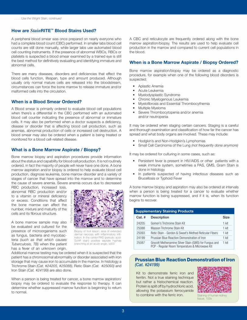
Biopsy of foot lesion: area of extended dermal necrosis with inflammatory infil-tration. In the center PAS (periodic acid-Schiff stain) positive septate hyphae branching at an acute angle, x400
Phot
o Co
urte
sy o
f ww
w.cd
c.go
v
Supplementary Staining Products
Cat. # Description Size
2420525088250932419925087
Gomori’s Trichrome Stain KitMasson Trichrome Stain KitRetic Stain - Gordon & Sweet’s Method Reticular FibersPrussian Blue Reaction Demonstration of IronGrocott Methenamine Silver Stain (GMS) for Fungus and PCP - Regular Room Temperature & Microwave Kit
1 kit1 kit1 kit1 kit1 kit
Kit to demonstrate ferric iron and ferritin. Not a true staining technique but rather a histochemical reaction. Protein is split off by hydrochloric acid, allowing the potassium ferrocyanide to combine with the ferric iron. Staining of human kidney
tissue, 100x
Prussian Blue Reaction Demonstration of Iron(Cat. #24199)
3

400 Valley Road Warrington, PA 18976
PolyFactsVol. 5 | No. 1 BioSciences
U.S. Corporate Headquarters | 400 Valley Road, Warrington, PA 18976 | 1(800) 523-2575 / (215) 343-6484 | Fax 1(800) 343-3291 | [email protected]
Polysciences Europe GmbH | Handelsstrasse 3 D-69214 Eppelheim, Germany | +(49) 6221-765767 | Fax +(49) 6221-764620 | [email protected]
Visit www.polysciences.com/newcat
POLYSCIENCES
C ATA L O G
Processing & EmbeddingStaining, Histology, Cytology
Special Stains & ReagentsElectrophoresis Products
Microbiology ProductsPlus many others..
StainRITE™ Get it done Wright!
Let us feature your article in the next PolyFacts issue. Showcase your research and experience using
Polysciences products.
If you have any suggestions or want to be a part of PolyFacts future
issues please email us at: [email protected]
70119
BE OUR N E X TFEATURED AUTHOR
Request a
FREE copy!
Ready-to-Use Stains for Hematologywww.polysciences.com/stainrite

• Save Time - Decalcify in as little as 3 hours!• Ready-to-Use - for human and animal tissue• Recommended for IHC, special stains and routine stains• Enhanced nuclear detail• Environmentally safe and biodegradable
Super Decalcifier I - Delicate is recommended for mildly calcified specimens such as bone marrow biopsies (core), and specimens that require fast di-agnostic work such as immunohistochemistry, cytochemistry and special stain procedures. Ready-to-Use formula produces enhanced nuclear detail in both human and animal tissue. Environmentally safe and biodegradable.
Human Degenerative Joint Disease, decalcified with Super Decalcifier I: Delicate and stained with H & E, 10X
Super Decalcifier I: Delicate
Cat. # Description Size
24888 Super Decalcifier I: Delicate 500ml, 1L, Case (6 x liter)
Fix Lymphoreticular Myeloid, Lymph Nodes and Bone Marrow TissuesFixation is the most influential factor in the long series of steps between grossing the tissue and coverslipping the stained slide. Nearly all the steps can be reversed if a problem occurs. Tissue can be reverse processed and reprocessed. Most stains can be removed and restained to improve intensity. The one step in which errors are permanent and can not be undone is fixation. Understanding the importance of fixation is crucial to producing quality slides and interpreting results. For over 45 years, Polysciences, Inc. has set the standard for consistent, high quality fixatives and reagents. Our Acetic Acid Formalin for Bone Marrow and Lymph Nodes Fixative is designed to fix lymphoreticular myeloid, lymph nodes and bone marrow tissues.
Science Solutions
Procedure for bone marrow biopsies using Polysciences’ Acetic Acid Formalin for Bone Marrow & Lymph Node Fixative
Aspirate1. Allow aspirate to clot in petri dish2. Transfer clot to specimen container filled with 10% NBF3. Process as usual
Bone Core Fixation1. Immediately fix in Acetic Acid Formalin for Bone Marrow and Lymph Node
Fixative (Cat. #24910) for a minimum of one hour2. Fix overnight if brought in later than 2 hours before end of work day
Decalcify first thing in the morning
Bone Core Decalcification1. Place fixed core and label in a plastic cassette. Super Decalcifier II:
Heavy Duty (Cat. #24887) - will discolor metal. Decalcify in Deli-Cal Block Solution (Cat. #24900) for 10-15 minutes. Do not leave periodically check
2. Test for decalcification by gently checking for pliability3. Rinse in running tap water for approximately 2 minutes
Processing1. If biopsy arrived in afternoon, process with the rest of the specimens2. If the biopsy is decalcified in the morning, hand process in 120cc
plastic specimen cups, 15 minutes each container, start with 70% ethanol / speed process through processor, 15 minutes each station with heat and vacuum, starting with the first dehydrant station
Microtoming1. Cut both aspirate and bone cores at 2-3 microns.2. Place 3 levels on one slide. Cut an extra slide of the last level and
set aside on the back of the water bath in case a special stain is requested
Staining1. Hematoxylin approximately 5 minutes. Bone core 1 minute
(check under the microscope before counterstaining)2. Eosin for 10 dips, dehydrate, clear and coverslip
Note: Over decalcification will result in poor or indifferent histological detail and staining characteristics.Reference: Adapted from Becky Scholes, HTL,MT(ASCP) H.I.S.T.O., The Official Newsletter of the Iowa Society for Histotechnology, Tech Tip
Cat. # Description Size
24910 Acetic Acid Formalin for Bone Marrow and Lymph Node Fixative
500ml, 1L
Gold Standard Series: Acetic Acid Formalin for Bone Marrow & Lymph Node Fixative
Designed to fix lymphoreticular myeloid, lymph nodes and bone marrow tissues.
4

5
Product SpotlightGold Standard Series Gill’s HematoxylinsGill’s Hematoxylin #1, for Cytology
Formulated for routine cytology staining. This single strength formula optimally stains gynecological and non-gynecological specimens. Can be used for staining filters and membranes. General purpose nuclear stain, progressive type. Used with hematoxylin and eosin staining.
Gill’s Hematoxylin #2, double strengthfor Histology & Cytology
Used when a stronger or darker nuclear stain is required for cytology or immuno-histochemistry (IHC) conterstaining. Gill’s #2 formulation is a double strength mixture and stains darker and more quickly than Gill’s #1. General purpose nuclear stain, progressive type. Used with hematoxylin and eosin staining.
Gill’s Hematoxylin #3, triple strengthfor Histology
Appropriate for routine histology staining where a darker nuclear stain is desired. This is a triple strength formula requiring shorter staining times. Formula is very stable and does not require filtering prior to each use. General purpose nuclear stain, progressive type. Used with hematoxylin and eosin staining.
Leishman StainFor staining bone marrow and blood smears to identify leucocytes, malarial parasites and trypanosomas.
Sudan Black B, C.I. 26150, CertifiedThis stain is specific for lipids. Sudan black B stains a variety of lipids, including neutral fat, phospholipids and sterols. Normal granulocyte precursors show increased sudanophilia that corresponds roughly to the number of granules. Promyelocytes contain a few sudanophilic granules and mature polymorphonuclear neutrophils contain large numbers of sudanophilic granules. Eosinophil granules are also sudanophilic, especially on the edge. Monocytes may be unstained or may contain a few discrete sudanophilic granules. Macrophages often contain sudanophilic material. In general, Sudan black B stain is similar to the peroxidase reaction in distinguishing acute lymphocytic leukemia from acute myelogenous leukemia.
Flash Dip FNA / H. Pylori Kit
High quality, rapid turnaround staining kit, serves many purposes in a cost conscious laboratory environment. Flash Dip is an optimal hematology stain for differential assessment of rapid blood smears, as well as for detecting H. pylori microorganism. Useful in applications such as: Fine Needle Aspirations, Frozen Sections, Blood Smears, Cytological Specimens, Bone Marrow Biopsies, Touch Preps and Microorganism Detection. Interprets result in less than 15 seconds!
Terry’s Polychrome Methylene Blue 2% AqueousExcellent dye for nuclear and nucleolar details. Can be used to demonstrate erythrocyte alterations/inclusions and some erythrocyte parasites, as well as to visualize reticulocytes. Methylene blue is different from that in Romanowsky stains. In order to stain a slide with methylene blue a drop of stain is placed on a coverslip. The coverslip is then placed on an unfixed blood smear. An alternative method is to mix some stain with fresh blood before a blood smear is made.
For our full line of Hematoxylin Eosin Bluing products, visit www.polysciences.com/hematox
Gill’s Hematoxylin Products
Cat. # Description Size
242422424324244
Gill’s Hematoxylin #1 Gill’s Hematoxylin #2, double strengthGill’s Hematoxylin #3, triple strength
500ml, 1000ml500ml, 1000ml500ml, 1000ml
Pancreas, Gill’s Hematoxylin #3and Scott’s Eosin Y, 100x
Breast, Gill’s Hematoxylin #2, 100x
Cheek, Gill’s Hematoxylin #1, 100X
Additional Lab Products for Hematology Staining
Periodic Acid Schiff’s (PAS) StainThis reaction stains polysaccharides. Periodic acid oxidizes carbohydrates and similar compounds to aldehydes. Aldehydes can then react with the Schiff reagent (leuko-fuchsin) to release fuchsin and stain the cellular components containing oxidizable compounds. A variety of intracellular compounds react with the PAS reagents, but in the blood and marrow cells glycogen is the most abundant compound, as evidenced by amylase digestion that blocks the staining. In blood from samples, the cytoplasm of polymorphonuclear leukocytes stains intensely pink or red, with granular appearance in some cells. Monocyte cytoplasm stains faintly pink and may contain fine or coarse granules. Erythrocytes do not stain. Platelets stain intensely. In the normal marrow, the earliest granulocyte precursors do not stain. Cytoplasmic staining increases with increasing maturity of the granulocytic cells. The PAS reaction is helpful in the diagnosis of some cases of acute lymphoblastic leukemia and some subtypes of acute myeloid leukemia.
Additional Lab Products for Hematology Staining
Cat. # Description Size
2460625008099782420025001
Flash Dip / H.Pylori Stain KitSudan Black B, C.I. 26150, CertifiedTerry’s Polychrome Methylene Blue 2% aqueousPeriodic Acid Schiff’s (PAS) StainLeishman Stain
250ml, 500ml25g470ml1 kit25g