poly(acrylic) acid-coated silver nanoparticles for antibacterial … · 2018-12-10 · textiles. 1...
TRANSCRIPT
Poly(acrylic) acid-coated Silver Nanoparticles for Antibacterial Textile Finishing
E. Falletta*, M. Bonini*, E. Fratini*, A. Lo Nostro**, A. Becheri*, P. Lo Nostro*, P. Baglioni*
*Department of Chemistry and CSGI, University of Florence, 50019 Sesto Fiorentino (Firenze), Italy, [email protected]
**Department of Public Health, University of Florence, 50134 Firenze, Italy, [email protected]
ABSTRACT
The synthesis of silver nanoparticles was achieved by reducing AgNO3 with NaBH4 or upon exposure to UV radiation. Poly(acrylic) acid specimen of various molecular weights were used as templating materials. Nanoparticle dispersions were analyzed through Dynamic Light Scattering, UV-VIS Absorption and Small-Angle X-Ray Scattering. The results indicate that the nanoparticle size depends on the reduction method and on the molecular weight of the polymer.
Different textile fabrics (cotton and wool) were treated with the Ag nanoparticle dispersion, and their antimicrobial activity was tested against Staphylococcus aureus for biomedical applications. Keywords: Silver nanoparticles, antimicrobial activity, textiles.
1 INTRODUCTION
Silver-based solutions and clusters possess relevant bactericide and fungicide properties with a broad spectrum, with low toxicity levels against mammalian cells. In fact, silver deposits in human cells in vivo are reported to be biocompatible [1].
Due to their larger contact area, silver nanoparticles can also be supported on suitable materials and keep a significant bactericide activity [2], which is crucial for biomedical applications.
In this work silver nanoparticles with a narrow size distribution were synthesized and used for cotton and wool fabric finishing.
2 EXPERIMENTAL SECTION
2.1 Matherials
AgNO3 was purchased from Riedel-de-Haën, poly(acrylates) (Mw= 1200, 15000; sodium salts, solutions) and NaBH4 were obtained from Aldrich. Wool and cotton samples were a kind gift from Grado Zero Espace (Italy).
2.2 Synthesis
Silver nanosols were prepared from AgNO3 as Silver source and poly(acrylates) (PAA) as templating material
[3]. Two different reduction methods were used: NaBH4 reduction and UV-exposure. In order to clarify the role of the polymer length over the size of the obtained nanoparticles, two PAA molecular weights were used: 1200 and 15000 (from now on referred to as PAA1200 and PAA15000). Synthesis was performed by keeping a constant [PAA]/[Ag+] ratio. NaBH4 reductions were performed by adding a NaBH4 ice-cold solution to the solutions containing PAA and Ag+ under vigorous stirring. UV reductions were performed with an Oriel lamp, operating with a water filter. Solutions were exhaustively irradiated while being continuously mixed with a magnetic stirrer.
2.3 Textile treatment
Nanoparticle uptake on textiles: The fabric samples were conditioned at constant relative humidity (33%) and temperature (20° C). The wool and cotton samples (5 cm 5 cm) were soaked for 15 min in the Ag nanoparticles dispersion, under magnetic stirring. The clothes were then squeezed to remove the excess dispersion, rinsed and dried in an oven at 130º C for 15 min at atmospheric pressure (dry heat).
Plating of loaded textiles. For antimicrobial tests, a suspension of Staphylococcus aureus ATCC 25923 was prepared with a turbidity of 0.5 Mc Farland units (about 5·106 UFC/mL). 100 µL of the suspension were spread on TSA (Tryptone-Soya-Agar growth medium from Oxoid, Milan, Italy) in a Petri dish and the fabric sample was adjusted on the top of the medium. The dishes were incubated at 37° C in a thermostat at room pressure in aerobic atmosphere for 24 h. At the end of the incubation, the inhibition rings were evaluated.
2.4 Physico-chemical characterization
UV-VIS Absorption. Absorbance spectra were collected with a Perkin-Elmer Lambda 5 spectrophotometer for solutions of chemicals, and with a Perkin-Elmer Lambda 35, equipped with a 60 mm integrating sphere, for fabrics.
DLS. Light scattering experiments were carried out on a Brookhaven Instruments apparatus (BI 9000AT correlator card and BI 200SM goniometer). The light source was the second harmonic of a diode-pumped Coherent Innova Nd:YAG laser (λ = 532nm), linearly polarized in the vertical direction and impinging on the sample with 60 mW power. The signal was detected by an EMI 9863B/350
NSTI-Nanotech 2007, www.nsti.org, ISBN 1420063766 Vol. 4, 2007412
photomultiplier. The laser long-term power stability was ±0.5%. The light scattered from the sample was collected at 90° with respect to the incident laser light radiation. To obtain the size distribution of the scattering objects, the autocorrelation functions were Laplace inverted using the CONTIN routine.
Small Angle X-ray Scattering. SAXS measurements were carried out with a Nanoviewer (Rigaku), equipped with a Mercury Charge Coupled Device detector (CCD) containing 1024 1024 pixels of width 68 mm. Cu-Kα radiation at λ = 1.542 Å was emitted by a Micromax007 X-ray rotating anode (Rigaku), operating at a maximum power of 0.8 kW with a circular focal spot size of 70 mm. X-rays were conditioned using the Confocal Max-Flux Mirror (Rigaku/Osmic) in order to totally remove the Cu-Kβ component maintaining the high flux and symmetry of the rotating anode source. X-ray collimation was performed through a 3-point collimation system. The sample-to-detector distance was about 605 mm. The volume between the sample and the detector was kept under vacuum during the measurements to minimize scattering from the air. The Q-range was calibrated using silver behenate, which is known to have a well-defined lamellar structure (d = 58.48 Å). Scattering curves were monitored in a Q-range from 0.01 to 0.6 Å-1. The samples were filled into a 1 mm quartz capillary using a syringe. The temperature was controlled by a Peltier device, with an accuracy of ± 0.1° C. The working temperature was 20° C. All 2D spectra were corrected for the dark current; the dezinger procedure was applied to all images in order to remove spurious signals. The empty cell contribution (quartz capillary) was removed using the relative sample/empty transmissions. Finally, 2-D spectra were azimuthally averaged in order to obtain the correspondent 1-D scattering intensity distribution.
3 RESULTS AND DISCUSSION
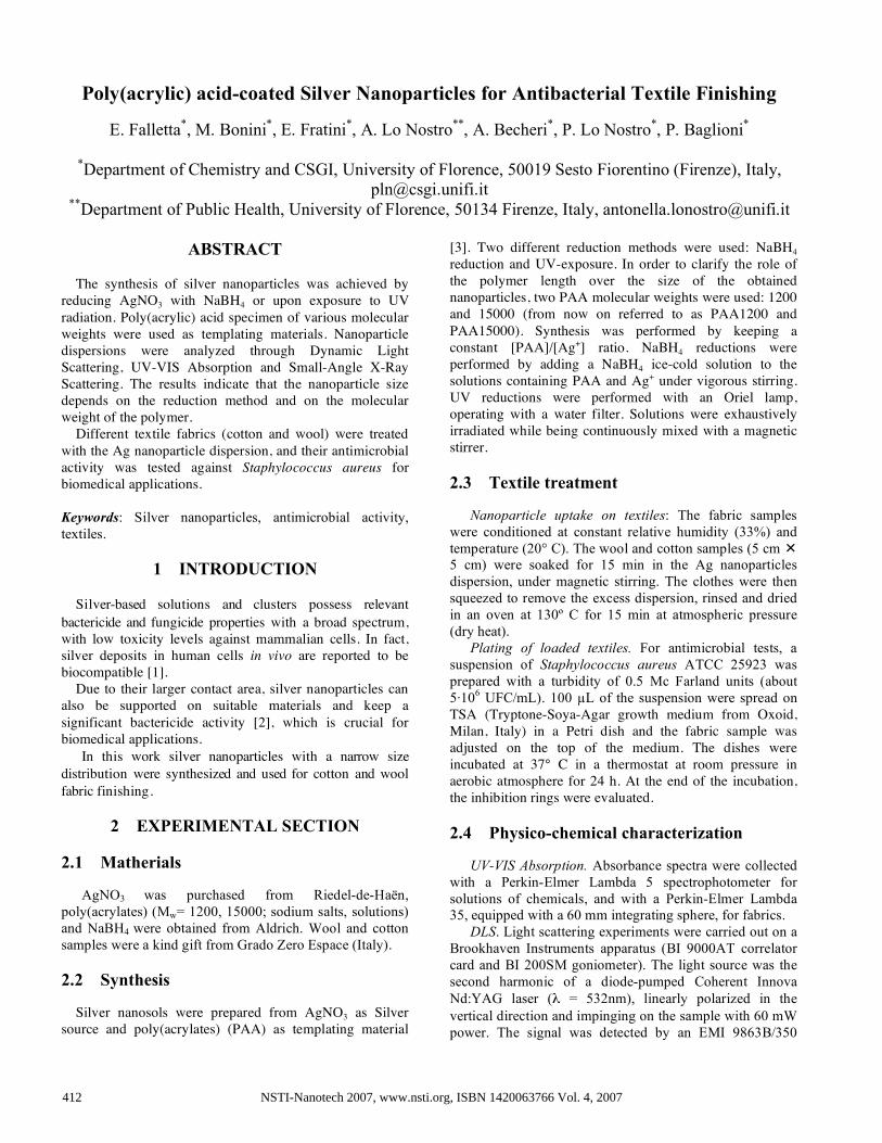
UV-Visible Absorption. UV-VIS spectra (Fig.1) show an
absorption maximum at about 400 nm due to the surface plasmon resonance, a typical behavior exhibited by metallic nanoparticles. In the case of silver nanoparticles an absorption peak at such wavelength means that the nanoparticles size is in the range of a few nanometers. The spectra also show an inter-particle plasmon interaction peak at about 600-700 nm, due to the presence of aggregates in solution.
DLS. The size-distribution obtained from the
autocorrelation function fittings are in agreement with the UV-VIS absorption results, and confirm the presence of aggregates in solution. Moreover, the fitting shows two aggregate populations: the first one ranging between 20 and 50 nm in size, and the second with particles larger than 100 nm.
Fig.1: UV-Visible Absorption spectra of Ag nanoparticle dispersions obtained with the PAA 1200 (full line) and
15000 (dotted line) SAXS. The results show that the size of the nanoparticles
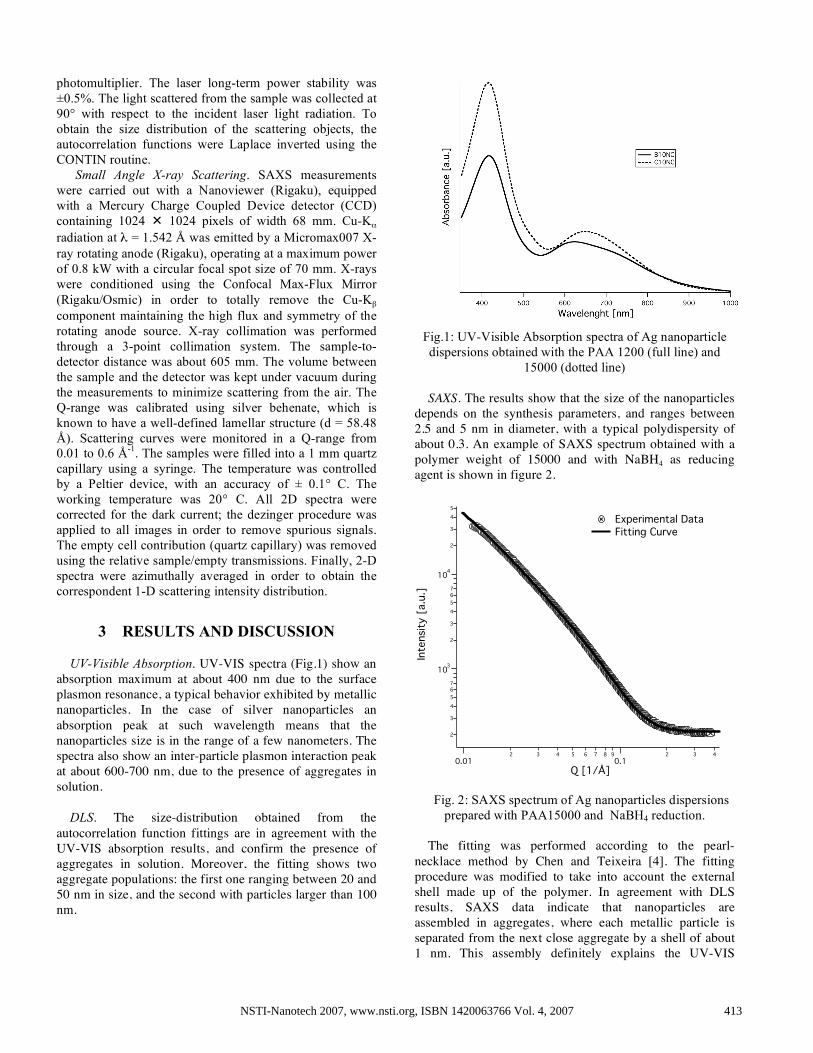
depends on the synthesis parameters, and ranges between 2.5 and 5 nm in diameter, with a typical polydispersity of about 0.3. An example of SAXS spectrum obtained with a polymer weight of 15000 and with NaBH4 as reducing agent is shown in figure 2.
2
3
4
5
67
103
2
3
4
5
67
104
2
3
4
5
Inte
nsi
ty [
a.u.]
0.012 3 4 5 6 7 8 9
0.12 3 4
Q [1/Å]
Experimental Data Fitting Curve
Fig. 2: SAXS spectrum of Ag nanoparticles dispersions
prepared with PAA15000 and NaBH4 reduction.
The fitting was performed according to the pearl-necklace method by Chen and Teixeira [4]. The fitting procedure was modified to take into account the external shell made up of the polymer. In agreement with DLS results, SAXS data indicate that nanoparticles are assembled in aggregates, where each metallic particle is separated from the next close aggregate by a shell of about 1 nm. This assembly definitely explains the UV-VIS
NSTI-Nanotech 2007, www.nsti.org, ISBN 1420063766 Vol. 4, 2007 413
absorbance spectra: in fact, the peak at lower wavelength can be ascribed to the surface plasmon resonance, typical of ultra-small metallic nanoparticles, while the peak at higher wavelength is due to the mutual interaction of the surface plasmons of closely located nanoparticles.
UV-VIS Absorption on Textiles. Untreated cotton and wool samples show no absorption peak between 400 and 800 nm, while after the treatment with silver dispersions a peak at around 400 nm appears (Fig. 3), showing that silver nanoparticles have been successfully deposited on the fabric.
Fig.3: Integrating sphere UV-VIS Absorption spectra of untreated (dotted line) and Ag-loaded B10NC (full line).
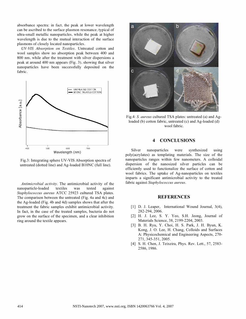
Antimicrobial activity. The antimicrobial activity of the
nanoparticle-loaded textiles was tested against Staphylococcus aureus ATCC 25923 cultured TSA plates. The comparison between the untreated (Fig. 4a and 4c) and the Ag-loaded (Fig. 4b and 4d) samples shows that after the treatment the fabric samples exhibit antimicrobial activity. In fact, in the case of the treated samples, bacteria do not grow on the surface of the specimen, and a clear inhibition ring around the textile appears.
Fig.4: S. aureus cultured TSA plates: untreated (a) and Ag-
loaded (b) cotton fabric, untreated (c) and Ag-loaded (d) wool fabric.
4 CONCLUSIONS Silver nanoparticles were synthesized using
poly(acrylates) as templating materials. The size of the nanoparticles ranges within few nanometers. A colloidal dispersion of the nanosized silver particles can be efficiently used to functionalize the surface of cotton and wool fabrics. The uptake of Ag-nanoparticles on textiles imparts a significant antimicrobial activity to the treated fabric against Staphylococcus aureus.
REFERENCES [1] D. J. Leaper, International Wound Journal, 3(4),
282-294, 2006. [2] H. J. Lee, S. Y. Yeo, S.H. Jeong, Journal of
Materials Science, 38, 2199-2204, 2003. [3] B. H. Ryu, Y. Choi, H. S. Park, J. H. Byun, K.
Kong, J. O. Lee, H. Chang, Colloids and Surfaces A: Physicochemical and Engineering Aspects, 270-271, 345-351, 2005.
[4] S. H. Chen, J. Teixeira, Phys. Rev. Lett., 57, 2583-2586, 1986.
NSTI-Nanotech 2007, www.nsti.org, ISBN 1420063766 Vol. 4, 2007414