pnas-1970-benjamin-394-9
DESCRIPTION
gxdchfvjgbTRANSCRIPT

Proceedings of the National Academy of SciencesVol. 67, No. 1, pp. 394-399, September 1970
Host Range Mutants of Polyoma Virus*
Thomas L. Benjamin
PUBLIC HEALTH RPSEARCH INSTITUTE OF THE CITY OF NEW YORK, INC., NEW YORK, N.Y. 10016
Communicated by James D. Watson, June 29, 1970
Abstract. A line of polyoma-transformed mouse cells has been isolated whichis fully susceptible to lytic infection by polyoma virus. This line has been usedto select virus mutants which have lost most or all of their ability to grow in theuntransformed parental line while retaining the ability to grow in the transformedderivative. These virus mutants are also defective in their ability to transformcells of rat or hamster origin. Since the DNA extracted from the mutants hasthe same host range as the whole virus, the mutants appear to be blocked at someintracellular step which is required both for the completion of virus developmentin mouse cells and for transformation in rat or hamster cells.
It now seems clear that neoplastic transformation of mammalian cells bythe small DNA-containing tumor viruses, polyoma and simian virus 40(SV40), is accompanied by persistence of viral genes, and that some of thesegenes continue to be expressed in transformed cells in the absence of in-fectious virus. 2 However, the nature of those gene functions which continueto be expressed by the cell-associated viral genome, and the role such functionsmight have in directing the transformed state, are unknown. The DNA contentof these viruses is small (3 X 10⁶ daltons), corresponding to perhaps 5-8 genes;radiobiological evidence indicates that only a fraction (50-60%) of the smallgenetic content is required to cause the neoplastic response compared to that re-quired for virus reproduction.3 Thus, whatever these viruses do to cause trans-formation they do by putting into play only a small number of discrete functions.To further understand this process, it is crucial first to identify what these func-tions are, and second to determine how the relevant viral functions interact withthe host cell to alter its growth.The search for conditionally lethal mutants of these viruses offers a promising
approach for studying the biochemical changes involved in transformation.Temperature-sensitive mutants of polyoma virus have already been isolatedand studied.46 The existence of another class of polyoma mutant showing aconditional lethality of the host range type has been predicted7-namely, mu-tants blocked in their growth in normal cells, but still able to grow in cells pre-viously transformed by polyoma. This type of host range selection was chosenbecause it seemed possible that the survival of the mutants in the permissive(transformed) cell would be based on complementation of the conditionallydefective viral function(s) by the homologous function(s) of the cell-associatedviral genome; thus, if any viral genes expressed in the transformed cell are re-quired for the initiation or maintenance of the transformed state (and also for
394

VOL. 67, 1970 HOST RANGE MUTANTS OF POLYOAMA VIRUS 395
virus reproduction), then mutants defective in these crucial function(s) wouldbe found. The present communication describes the isolation and preliminarycharacterization of such mutants.
Materials and Methods. Cell lines: The 3T3 line8 of Swiss mouse embryofibroblast served as a normal line from which the polyoma-transformed line 3T3-Py-3was isolated (see Results and Discussion).Mutagenesis: (a) Hydroxylamine: Hydroxylamine was used on a purified stock
of small plaque virus by mixing 0.4 ml of the virus in Tris-buffered saline and 0.1 ml of2 M NH20H + 2M NaCl, pH 7.0, incubating at 450C for 90 min, and dialyzing against alarge volume of buffer at 4VC. The survival of plaque-forming units after this treatmentwas about 5%. Aliquots of the hydroxylamine-treated virus were then passed once atlow multiplicity through 3T3-Py-3 to allow segregation of possible mutational hetero-zygotes. These lysates were then used for plaque isolations.
(b) Nitrosoguanidine: N-methyl-N'-nitro-N-nitrosoguanidine was added 17 hr afterinfection of a baby mouse kidney culture with small plaque polyoma virus at a final con-centration of 314 1ug/ml. Receptor-destroying enzyme and anti-viral antiserum werepresent from the third to seventeenth hr to remove residual virus from the inoculum.The total yield of virus 50 hr after infection was about 3% of the untreated control. Thecrude lysate from the NG-treated culture was used directly for plaque isolations.Plaque screening and mutant isolations: Appropriate dilutions of the mutagen-
ized lysates were plated on sub-confluent monolayers of 3T3-Py-3, and single plaqueswere picked on the fifth to eighth day. Each plaque isolate was then tested for itsability to produce plaques on confluent monolayers of 3T3, or in some instances onmouse embryo secondary cultures. Those isolates were kept which failed to producedistinct plaques after 10-14 days of incubation; three further sequential single-plaqueisolations on 3T3-Py-3 were carried out with each such isolate. Wild type unselectedplaques on 3T3-Py-3 from a nonmutagenized virus stock were kept as controls. Stocksof all virus isolates were grown on 3T3-Py-3. All isolations, screening, and growth ofstocks were done at 37.0-38.5'C unless stated otherwise in the text.
Results and Discussion. Isolation and characterization of polyoma-trans-formed 3T3 lines: A cell line with the following properties was required as apermissive host for the isolation of virus mutants: (1) transformed by polyomaand virus-free, (2) highly susceptible to productive reinfection by polyoma, and(3) capable of being used in a plaque assay. Whereas polyoma transformantsof nonpermissive hosts (rat or hamster) are known to remain nonpermissive,rare transformants which arise after infection of the lytic host (mouse) mightretain susceptibility to lytic infection. Previous reports9'10 showing polyoma-transformed mouse cells to have increased resistance to reinfection might beexplained on the basis of a strong selection for viral resistance which accompaniedtheir isolation rather than an intrinsic "immunity."
Suitable transformants were therefore sought in the following way. 3T3 cellsexposed to polyoma at a multiplicity of 10-20 plaque-forming units (PFU)/cellwere first washed to remove unadsorbed virus and then plated in agar suspensioncultures." High-titer rabbit anti-viral antiserum and receptor-destroyingenzyme were added to the soft agar layer to protect the rare emerging trans-formants from being reinfected and killed by the large amounts of virus beingreleased by the majority of cells. After 9 days in the agar, a number of smallclones were picked and re-cloned in liquid media containing antiserum and re-ceptor-destroying enzyme. Clones with transformed morphology were grown,tested for the presence of infectious virus, and re-cloned if necessary until virus-
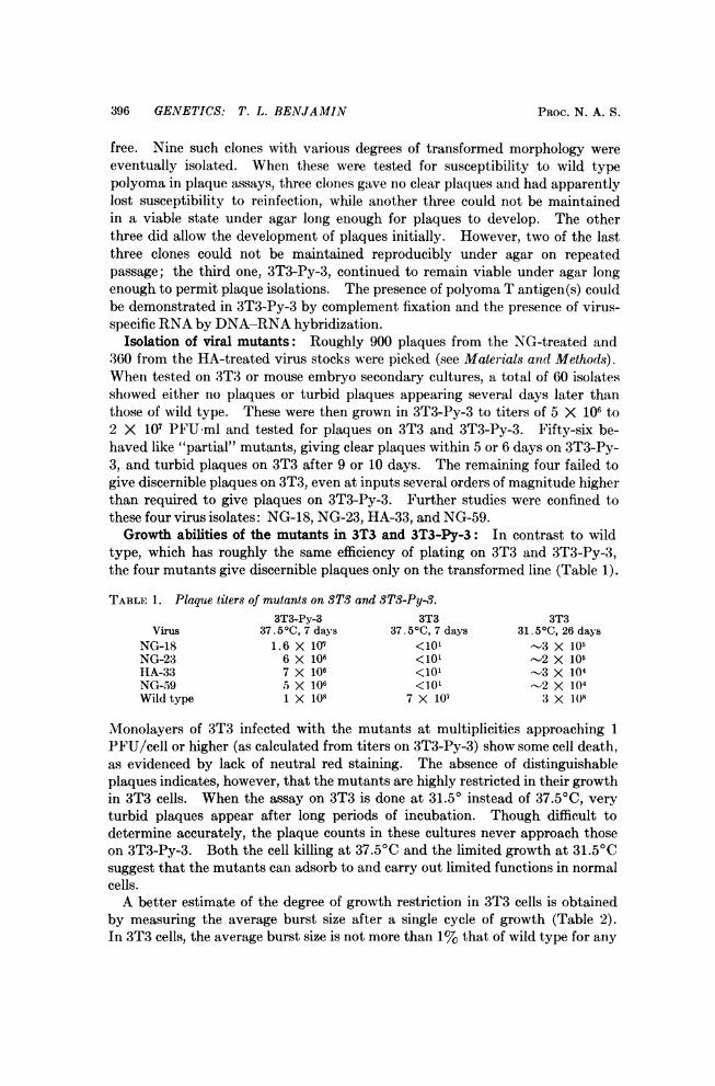
396 GENETICS: T. L. BENJAMIN PROC. N. A. S.
free. Nine such clones with various degrees of transformed morphology wereeventually isolated. When these were tested for susceptibility to wild typepolyoma in plaque assays, three clones gave no clear plaques and had apparentlylost susceptibility to reinfection, while another three could not be maintainedin a viable state under agar long enough for plaques to develop. The otherthree did allow the development of plaques initially. However, two of the lastthree clones could not be maintained reproducibly under agar on repeatedpassage; the third one, 3T3-Py-3, continued to remain viable under agar longenough to permit plaque isolations. The presence of polyoma T antigen(s) couldbe demonstrated in 3T3-Py-3 by complement fixation and the presence of virus-specific RNA by DNA-RNA hybridization.
Isolation of viral mutants: Roughly 900 plaques from the NG-treated and360 from the HA-treated virus stocks were picked (see Materials and Methods).When tested on 3T3 or mouse embryo secondary cultures, a total of 60 isolatesshowed either no plaques or turbid plaques appearing several days later thanthose of wild type. These were then grown in 3T3-Py-3 to titers of 5 X 10⁶ to2 X 107 PFU'ml and tested for plaques on 3T3 and 3T3-Py-3. Fifty-six be-haved like "partial" mutants, giving clear plaques within 5 or 6 days on 3T3-Py-3, and turbid plaques on 3T3 after 9 or 10 days. The remaining four failed togive discernible plaques on 3T3, even at inputs several orders of magnitude higherthan required to give plaques on 3T3-Py-3. Further studies were confined tothese four virus isolates: NG-18, NG-23, HA-33, and NG-59.Growth abilities of the mutants in 3T3 and 3T3-Py-3: In contrast to wild
type, which has roughly the same efficiency of plating on 3T3 and 3T3-Py-3,the four mutants give discernible plaques only on the transformed line (Table 1).
TABLE 1. Plaque titers of mutants on ST3 and 3T3-Py-3.3T3-Py-3 3T3 3T3
Virus 37.50C, 7 days 37.51C, 7 days 31.50C, 26 daysNG-18 1.6 X 10⁷ <10' -3 X 10⁵NG-23 6 X 10⁶ <101 -2 X 10⁶HA-33 7 X 10⁶ <101 -3 X 10⁴NG-59 5 X 10⁶ <101 -2 X 10⁴Wild type 1 X 10⁸ 7 X 10⁷ 3 X 1(8
Monolayers of 3T3 infected with the mutants at multiplicities approaching 1PFU/cell or higher (as calculated from titers on 3T3-Py-3) show some cell death,as evidenced by lack of neutral red staining. The absence of distinguishableplaques indicates, however, that the mutants are highly restricted in their growthin 3T3 cells. When the assay on 3T3 is done at 31.50 instead of 37.50C, veryturbid plaques appear after long periods of incubation. Though difficult todetermine accurately, the plaque counts in these cultures never approach thoseon 3T3-Py-3. Both the cell killing at 37.50C and the limited growth at 31.50Csuggest that the mutants can adsorb to and carry out limited functions in normalcells.A better estimate of the degree of growth restriction in 3T3 cells is obtained
by measuring the average burst size after a single cycle of growth (Table 2).In 3T3 cells, the average burst size is not more than 1%, that of wild type for any

VOL. 67, 1970 HOST RANGE MUTANTS OF POLYOMA VIRUS 397
TABLE 2. Average burst sizes of mutants on 3TS and STS-Py-3.*Virus 3T3 3T3-Py-3
NG-18 0.63 4.2 X 10²NG-23 1.4 4.5 X 10²HA-33 0.19 6.0 X 10²NG-59 2.1 2.0 X 10²Wild type 3.0 X 10² 5.1 X 10²
* Cells were infected at a multiplicity of approximately 0.1 PFU/cell and harvested 48 hr later.Cells and media were sonicated, and the virus was assayed on 3T3-Py-3. The numbers given areratios of output to input virus.
of the mutants, while in 3T3-Py-3, the mutants give values approximately thesame as wild type and several hundred times higher than in 3T3. If 3T3 cellsinfected by the mutants are incubated for a time equivalent to two wild typegrowth cycles, essentially the same results are obtained.To test directly whether the reduced burst sizes in 3T3 could be due to failure
in adsorption, penetration, or uncoating, infections were done with the DNA'sextracted from two of the mutants (Table 3). The results show the same host
TABLE 3. DNA infectivity of mutants.*Virus yields
DNA Input (PFU) 3T3 3T3-Py-3NG-18 (9 X 10²) 3 X 10² 3 X 10⁴+ DNaset 0 0
HA-33 (2.2 X 10³) 4 X 10³ 1.6 X 10⁶+ DNase 0 0
Wild type (1.1 X 10³) 1.6 X 10⁶ 2 X 10⁶+ DNase 0 0
* Cultures were first washed with 2 ml Hanks buffer containing 200 ;&g/ml DEAE-Dextran. TheDNA, in 0.1 ml Hanks buffer containing 750 ;g/ml DEAE-Dextran, was added for 20 min at roomtemperature. The cultures were then washed with Hanks and fed. DNA input titers were measuredon 3T3-Py-3 in the same way. Virus yields 44 hr after infection with the DNA were measured on3T3-Py-3.
t DNase treatment was carried out with 0.05 ;sg/ml enzyme for 10 min at 37.50C.
range pattern of infectivity as in experiments with whole virus. That the DNAfrom the mutants does give rise in 3T3 cells to some infectious virus can be ex-plained on the basis of incomplete loss of function (leakiness), or by the en-capsidation of input DNA with a bypass of essential early function(s), or both.It is clear in any case that the restricted growth of the mutants in 3T3 cells isnot due simply to a failure in any step prior to uncoating.
Since the mutants were isolated on dividing cells of 3T3-Py-3 and screened forabsence of growth on stationary cells of 3T3, the possibility existed of havingselected mutants requiring an actively dividing cell in which to grow but beingindifferent to the transformed state of its host. This was tested by comparingvirus growth in exponential and stationary 3T3 cells (Table 4). A comparisonof results between the mutants and wild type shows that 3T3 cells in exponentialgrowth do not become permissive for the mutants.
Since the mutants can propagate stably in 3T3-Py-3, recombination with thecell-associated viral genes (marker rescue) occurs only infrequently if at all, andcannot be a necessary step in the growth of the mutants.

398 GENETICS: T. L. BENJAMIN PROC. N. A. S.
TABLE 4. Growth of the mutants in dividing and nondividing STS cells.-- Total virus per culture*
Input virus (PFU) 3T3, day 1 3T3, day 4NG-18 (1.2 X 10⁵) 1.5 X 10⁵ 6.4 X 10³NG-23 (6 X 10⁴) 1.2 X 10⁵ 8.4 X 10⁴HA-33 (5 X 10⁴) 7.0 X 10³ 7.5 X 10²NG-59 (5 X 10⁴) 1.6 X 10⁵ 1. 5 X 10⁴Wild type (2 X 10⁵) 2. 1 X 10⁸ 2.4 X 10⁷
* Cultures of 3T3 in 60-mm dishes containing 3.5 X 10⁶ cells in exponential growth on day 1 and1.9 X 106 nondividing cells on Day 4 were infected and harvested 48 hr later. Cells and media weresonicated and the virus was assayed on 3T3-Py-3.
Properties of the virus particles: The mutant virus particles have propertiesassociated with the capsid which are not detectably different from those of wildtype particles. Both mutant and wild type viruses have PFU:HAU ratios inthe range of 2-4 X 10⁴. The mutants possess neutralization antigen(s) similarto wild type, as shown by their inactivation by antiserum prepared againstwild type particles.
Transformation: All four of the mutants fail to transform rat embryo fibro-blasts (Table 5). The frequency of transformation is lower by a factor of at
TABLE 5. Transformation of RECI-3.*Transforming Efficiency of
PFU in Transformants units wild typeVirus inoculum per culture per PFU (%)
NG-18 5 X 10⁶ 0 <2.0 X 10-' 0.07NG-23 9 X 10⁵ 0 <1.1 X 10⁻⁶ 0.4HA-33 3 X 10⁶ 0 <3.3 X 10-' 0.12NG-59 7 X 10⁵ 0 <1.4 X 10⁻⁶ 0.52Wild type 3 X 10⁵ 80 2.7 X 10⁻⁴ 100
* REC1-3 is an established line of rat embryo fibroblast. The agar suspension method was usedto measure transformation."
least 200 compared to wild type. Similar results were also obtained using theBHK line of baby hamster kidney cells.11
Basis of the host range properties of the mutants: A plausible explanationin general terms for the host range properties of the mutants can be based oncomplementation-the transformed cell expressing (either continuously or uponinfection) some function(s) homologous to that of the virus and required by thethe latter both to grow in and to transform normal cells. The positive correla-tion in all four cases studied between the selected property of transformed celldependence in virus growth and the predicted (unselected) property of beingunable to cause transformation suggests that the cell-associated function(s)which allows the mutants to grow is also implicated somehow in the transformedstate. This function(s) could be associated with any of the "integrated" viralgenes themselves, or with some cellular gene(s) which is expressed specificallyin polyoma-transformed cells, or perhaps generally in transformed cells ofdiverse origin. Further clarification should come from experiments now inprogress on the abilities of other cell types (including spontaneously or SV40-transformed lines and phenotypic revertants of polyoma-transformed lines) tosupport, the growth of the mutants.

VOL. 67, 1970 HOST RANGE MUTANTS OF POLYOMA VIRUS 399
I would like to thank Dr. J. Piatigorsky for stimulating discussions, and D. Tidaback andS. Ching for experimental assistance. Portions of this work were carried out in the Division ofBiology at the California Institute of Technology, at the Institute for Biomedical Research,and at the Public Health Research Institute of the City of New York, Inc.
Abbreviation: PFU, plaque-forming units; HAU, hemagglutination units.* The work was partly supported by U.S. Public Health Service grant CA 11473-01 of the
National Cancer Institute.1 Benjamin, T. L., J. Mol. Biol., 16, 359 (1966).2Westphal, H., and R. Dulbecco, Proc. Nat. Acad. Sci. USA, 59, 1158 (1968).3 Benjamin, T. L., Proc. Nat. Acad. Sci. USA, 54, 121 (1965).4Fried, M., Proc. Nat. Acad. Sci., USA, 53, 486 (1965).6 Eckhart, W., Virology, 38, 120 (1968).6 DiMayorca, G., J. Callender, G. Marin, and R. Giordano, Virology, 38, 126 (1968).7Benjamin, T. L., in Subviral Carcinogenesis, 1st Int. Symp. Tumor Viruses, Nagoya, Japan,
(1968) p. 62.8 Todaro, G., and H. Green, J. Cell Biol., 17, 299 (1963).9 Vogt, M., and R. Dulbecco, Proc. Nat. Acad. Sci. USA, 46, 365 (1960).10 Hellstrbm, I., K. E. Hellstrom, and H. 0. Sjogren, Virology, 16, 282 (1962).1 Macpherson, I., and L. Montagnier, Virology, 23, 291 (1964).