plos biology the liganding of glycolipid transfer protein...
TRANSCRIPT
The Liganding of Glycolipid Transfer ProteinIs Controlled by Glycolipid Acyl StructureLucy Malinina
1[, Margarita L. Malakhova
2[, Alex T. Kanack
2, Min Lu
3, Ruben Abagyan
4, Rhoderick E. Brown
2*,
Dinshaw J. Patel1*
1 Structural Biology Program, Memorial Sloan-Kettering Cancer Center, New York, New York, United States of America, 2 Hormel Institute, University of Minnesota, Austin,
Minnesota, United States of America, 3 Department of Biochemistry, Weill Medical College of Cornell University, New York, New York, United States of America, 4 Department
of Molecular Biology, The Scripps Research Institute, La Jolla, California, United States of America
Glycosphingolipids (GSLs) play major roles in cellular growth and development. Mammalian glycolipid transferproteins (GLTPs) are potential regulators of cell processes mediated by GSLs and display a unique architecture amonglipid binding/transfer proteins. The GLTP fold represents a novel membrane targeting/interaction domain amongperipheral proteins. Here we report crystal structures of human GLTP bound to GSLs of diverse acyl chain length,unsaturation, and sugar composition. Structural comparisons show a highly conserved anchoring of galactosyl- andlactosyl-amide headgroups by the GLTP recognition center. By contrast, acyl chain chemical structure and occupancy ofthe hydrophobic tunnel dictate partitioning between sphingosine-in and newly-observed sphingosine-out ligand-binding modes. The structural insights, combined with computed interaction propensity distributions, suggest aconcerted sequence of events mediated by GLTP conformational changes during GSL transfer to and/or frommembranes, as well as during GSL presentation and/or transfer to other proteins.
Citation: Malinina L, Malakhova ML, Kanack AT, Lu M, Abagyan R, et al. (2006) The liganding of glycolipid transfer protein is controlled by glycolipid acyl structure. PLoS Biol4(11): e362. DOI: 10.1371/journal.pbio.0040362
Introduction
Glycosphingolipids (GSLs) are ubiquitous components ofeukaryotic plasma membranes and mediate numerous bio-logical functions, from differentiation and proliferation toinvasive adhesion, neurodegeneration, and apoptosis [1,2].The importance of GSLs and their metabolites in processessuch as tissue development is illustrated by the lethal effect ofdisruption of GSL biosynthesis during embryonic develop-ment [3]. Whereas the bulk of GSLs reside in the outer leafletof the plasma membrane where their sugar headgroupsproject into the extracellular environment, increasing evi-dence suggests that GSLs also localize to membranes ofintracellular organelles (e.g., nucleus and mitochondria) [4–6].Vesicular trafficking plays a dominant role in distributingGSLs intracellularly after synthesis in the Golgi. However, anonvesicular pathway also exists, possibly involving glycolipidtransfer proteins (GLTPs) [7,8].
Based on their ability to selectively accelerate the inter-membrane transfer of glycolipids, GLTPs were discovered inthe cytosolic extracts of bovine spleen and porcine brain, andlater in a wide variety of tissues [9–11]. The 23–24-kDa GLTPsdisplay absolute specificity for glycolipids [12], are highlyconserved among mammals [13], and include plant and fungalorthologs that have been implicated in programmed celldeath responses [14,15]. Recently, we determined the first x-ray structure of human GLTP in both the GSL-free andlactosylceramide (LacCer)-bound forms [16]. In addition todisplaying a novel architecture that defines GLTP as thefounding member of a new protein superfamily [17,18](see http: / /supfam.org/SUPERFAMILY/cgi-bin/scop.cgi?sunid¼110004 and http://scop.mrc-lmb.cam.ac.uk/scop-1.69/data/scop.b.b.bja.b.b.A.html; accessed 12 September 2006)with a novel protein fold for membrane interaction and forlipid binding/transfer [19–27], the structure of the 18:1
LacCer-GLTP complex revealed the basis for GSL bindingspecificity by GLTP. The liganding site consists of a sugarheadgroup recognition center that anchors the ceramide-linked sugar to the protein surface and a hydrophobic tunnelthat accommodates the hydrocarbon chains of ceramide.Comparative structural analyses, including crystallographicB-factor distributions of apo-GLTP and the LacCer-GLTPcomplex, suggest that liganding of the glycolipid most likelyoccurs via an adaptive recognition process [16]. A cleft-likegating mechanism, involving conformational changes to twointerhelical loops and one a helix, appears to facilitate entryand exit of the lipid chains in the membrane-associated statewhen the GSL headgroup is attached to the sugar headgrouprecognition center.An important feature of the GLTP hydrophobic tunnel is
its ability to expand to accommodate the GSL ceramidemoiety. In the case of the 18:1 LacCer-GLTP complex, the 18-carbon oleoyl and sphingosine chains of LacCer reside side byside within the hydrophobic channel [16]. However, naturallyoccurring mammalian glycolipids typically have acyl chainswith lengths ranging from 16 to 26 carbons with occasional
Academic Editor: Fred Hughson, Princeton University, United States of America
Received October 20, 2005; Accepted August 29, 2006; Published October 24,2006
DOI: 10.1371/journal.pbio.0040362
Copyright: � 2006 Malinina et al. This is an open-access article distributed underthe terms of the Creative Commons Attribution License, which permits unrestricteduse, distribution, and reproduction in any medium, provided the original authorand source are credited.
Abbreviations: GalCer, galactosylceramide; GLTP, glycolipid transfer protein; GSL,glycosphingolipid; LacCer, lactosylceramide; ODA, optimal docking area
* To whom correspondence should be addressed. E-mail: [email protected] (REB);[email protected] (DJP)
[ These authors contributed equally to this work.
PLoS Biology | www.plosbiology.org November 2006 | Volume 4 | Issue 11 | e3620001
PLoS BIOLOGY
monounsaturation. To test the conformational propertiesand accommodation limits of the GLTP hydrophobic tunnel,we synthesized glycolipids (LacCer or galactosylceramide[GalCer]) containing short acyl chains (e.g., octanoyl anddodecanoyl), medium unsaturated acyl chains (e.g., linoleoyl),as well as long, physiologically relevant unsaturated acylchains (e.g., nervonyl), and we structurally characterized theircomplexes with GLTP. To elucidate conformational changescaused by the sugar headgroup binding in the GLTP, we alsostructurally characterized the complex between GLTP andthe model compound n-hexyl-b-D-glycoside, which, unlikeGSLs, has a single, very short hydrocarbon chain lacking anamide linkage.
The present structural studies provide definitive evidencefor a novel GSL-GLTP complexation mode characterized by adifference in the accommodation of the ceramide lipid chainsbut not the sugar headgroups. Chief among the differences isbending of the sphingosine chain, causing outward projectionfrom the hydrophobic tunnel. Comparative analysis of allGSL-GLTP structures reveals that the hydrophobic tunnelconsists of two functionally distinct compartments, enablingGLTP to accommodate GSL acyl chains of different lengthsand conformational restrictions. Equally importantly, ourfindings suggest a concerted sequence of events during GSLliganding, in which the sphingosine chain is the last part ofthe glycolipid to enter GLTP and the first part to leave GLTPduring interaction with membranes. Interaction propensitydistribution computations indicate a potential GLTP inter-face for interactions with other proteins and/or membranes,which coincide with a region of the proposed GLTP gatemediating GSL binding and release.
Results
Nonpolar Nature of GLTP Hydrophobic Channel PromotesOccupancy by Nonpolar Lipids
Our original structural characterization of GLTP revealedtwo noteworthy features of the hydrophobic tunnel thatencapsulates the GSL hydrocarbon chains [16]. First, thetunnel is highly nonpolar. No water molecules reside in thechannel, which is lined by the side chains of nonpolarphenylalanine, leucine, isoleucine, and alanine residues,together with a few valine and proline residues. Second, alarge portion of the tunnel is conformationally adaptable andexpands during GSL acquisition to accommodate the hydro-carbon chains. Moreover, localized in the lower part of thehydrophobic tunnel of apo-GLTP (i.e., glycolipid-free GLTP)is an extraneous hydrocarbon molecule that contains at leastsix carbon atoms and is clearly observable in electron densitymaps (Figure S1). Consistent with this finding is a recentreport of decanoic acid (ten-carbon chain) within thehydrophobic tunnels of two glycolipid-free bovine GLTPstructures [28]. It is not clear if the extraneous hydrocarbon isacquired during heterologous expression in Escherichia coli orsubsequent crystallization. However, the situation is notunusual for GSL binding proteins [22] and also has beenfrequently observed among other lipid binding/transferproteins [20].
The extraneous hydrocarbon is displaced from the hydro-phobic tunnel when human GLTP forms a complex with 18:1LacCer (Figure S2). A similar displacement of decanoic acid
from the hydrophobic tunnel also seems to occur whenbovine GLTP complexes with ganglioside GM3 [28].
Structure of the 24:1 GalCer-GLTP Complex ContainingLong Acyl Chain GSLTo determine how GSLs with long and physiologically
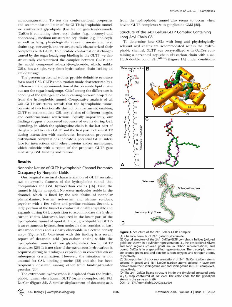
relevant acyl chains are accommodated within the hydro-phobic channel, GLTP was cocrystallized with GalCer con-taining a nervonoyl acyl chain (24-carbon chain with a cis-15,16 double bond, 24:1D15cis) (Figure 1A) under conditions
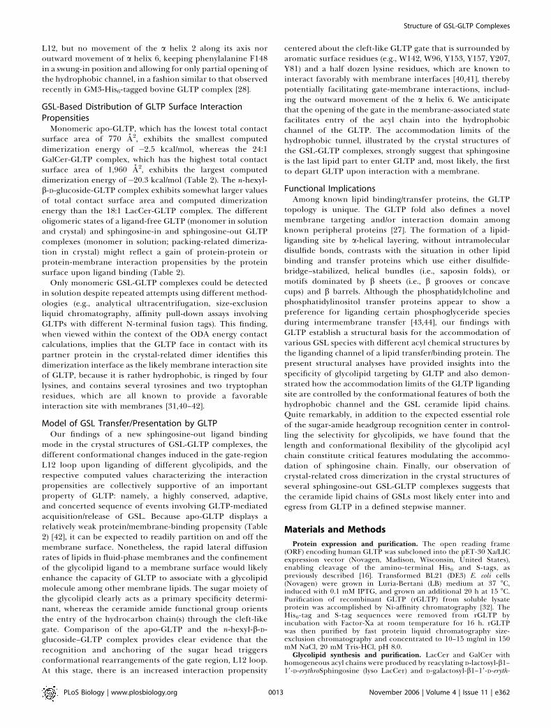
Figure 1. Structure of the 24:1 GalCer-GLTP Complex
(A) Chemical formula of 24:1 galactosylceramide.(B) Crystal structure of the 24:1 GalCer-GLTP complex. a helices (coloredgold) are shown in a cylinder representation, 310 helices (colored silver)and loop regions (colored gold) are in ribbon representations, andbound GalCer is in a space-filling representation. The glycolipid atomsare colored green, red, and blue for carbon, oxygen, and nitrogen atoms,respectively.(C) Superposition of stick representations of 24:1 GalCer (carbon atomscolored in green) and 18:1 LacCer (carbon atoms colored in lavender)derived from their sphingosine-out and sphingosine-in GLTP complexes,respectively.(D) The 24:1 GalCer ligand structure inside the simulated annealed omit2Fo-Fc map contoured at 1r level. The color code for the glycolipidatoms is the same as in (B).DOI: 10.1371/journal.pbio.0040362.g001
PLoS Biology | www.plosbiology.org November 2006 | Volume 4 | Issue 11 | e3620002
Structure of GSL-GLTP Complexes
described previously [16] for complexation of GLTP withLacCer containing an oleoyl acyl chain (18:1D9cis) (FigureS2A). The crystal structure of 24:1 GalCer bound to GLTP(Figure 1) was refined to an R factor (measure of goodness offit) of 18.0% at 1.85 A resolution (Table 1) (see Materials andMethods), enabling construction of an uninterrupted andclear electron density map for the sugar-amide headgroupand both ceramide lipid chains (Figure 1D). The overall a-helical topology of the 24:1 GalCer-GLTP complex (Figure1B) and the conformational features of its glycolipid-bindingregion are very similar to that reported previously for ourstructure of the 18:1 LacCer-GLTP complex (Figure S2B) [16]and recently confirmed in bovine His6-tagged GLTP [28]. Twolong a helices (helices 8 and 4) intertwine to form a super-helix, with both being noticeably bent (Figure 1B). One ofthese helices (helix 4) interacts directly with 24:1 GalCer, withone segment supporting the polar headgroup and theremainder supporting the nonpolar lipid chains.
Galactose Versus Lactose in the GLTP Recognition CenterIn the 24:1 GalCer-GLTP complex, the galactose sugar
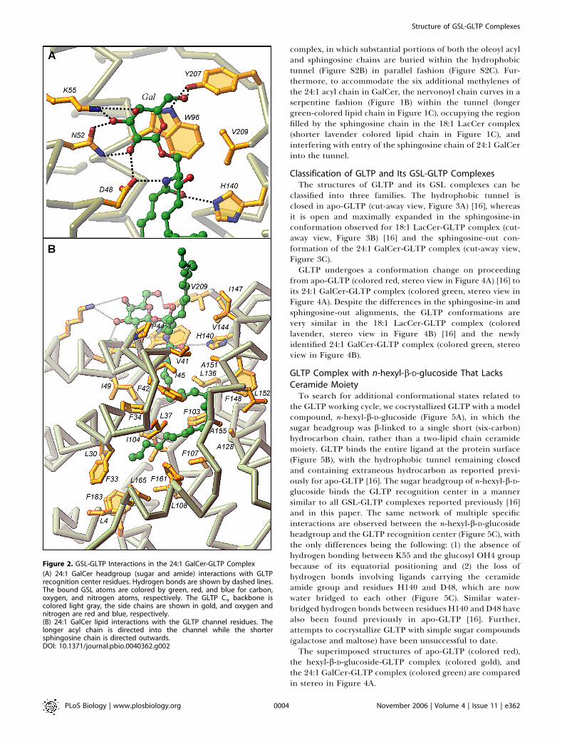
headgroup is anchored within the GLTP recognition center,which is located on the protein surface, through a network ofhydrogen bonding and hydrophobic interactions (Figure 2A).The hydrogen bonding involves D48, N52, and K55 of a helix2 and Y207 near the C terminus. The stacking of W96 (a helix4) against the B face of the galactose ring (Figure 2A) issimilar to that with the glucose ring in 18:1 LacCer complex[16]. The only difference in the liganding between the single
sugar headgroup of 24:1 GalCer and the two-sugar headgroupof 18:1 LacCer involves K55, which hydrogen bonds with theOH3 and OH4 hydroxyls of the initial sugar ring attached tothe ceramide in 24:1 GalCer (Figure 2A), rather thanhydrogen bonding with the OH3 hydroxyl of the secondsugar residue in the lactose of the 18:1 LacCer [16].Orientation of the ceramide amide group of 24:1 GalCer is
controlled by a pair of hydrogen bonds involving D48 andH140, with alignment of the initial segment of the sphingoidbase being facilitated by van der Waals contacts with V209(Figure 2A). A similar situation occurs in the 18:1 LacCer-GLTP complex [16]. The interactions are important becausethey result in a conserved and oriented entry of the GSLceramide lipid tails into the hydrophobic channel of GLTP.
Accommodation of a Long Acyl Chain GSL by GLTPAccommodation of the ceramide lipid tails within the
hydrophobic channel in the 24:1 GalCer-GLTP complex(Figure 1B) differs strikingly from that observed in the 18:1LacCer-GLTP complex (Figure S2B) [16]. Only the longnervonoyl acyl chain is inserted into the hydrophobic tunnel,which is lined by the nonpolar side chains of multiplephenylalanine, leucine, isoleucine, and alanine residues inthe 24:1 GalCer-GLTP complex (longer green-colored lipidchain in Figures 1B and 2B). The sphingosine chain of 24:1GalCer bends sharply at carbon 6, and projects nearlyorthogonally away from the cleft-like gate of the tunnel(shorter green-colored lipid chain in Figures 1B and 2B). Suchan arrangement contrasts with that of the 18:1 LacCer-GLTP
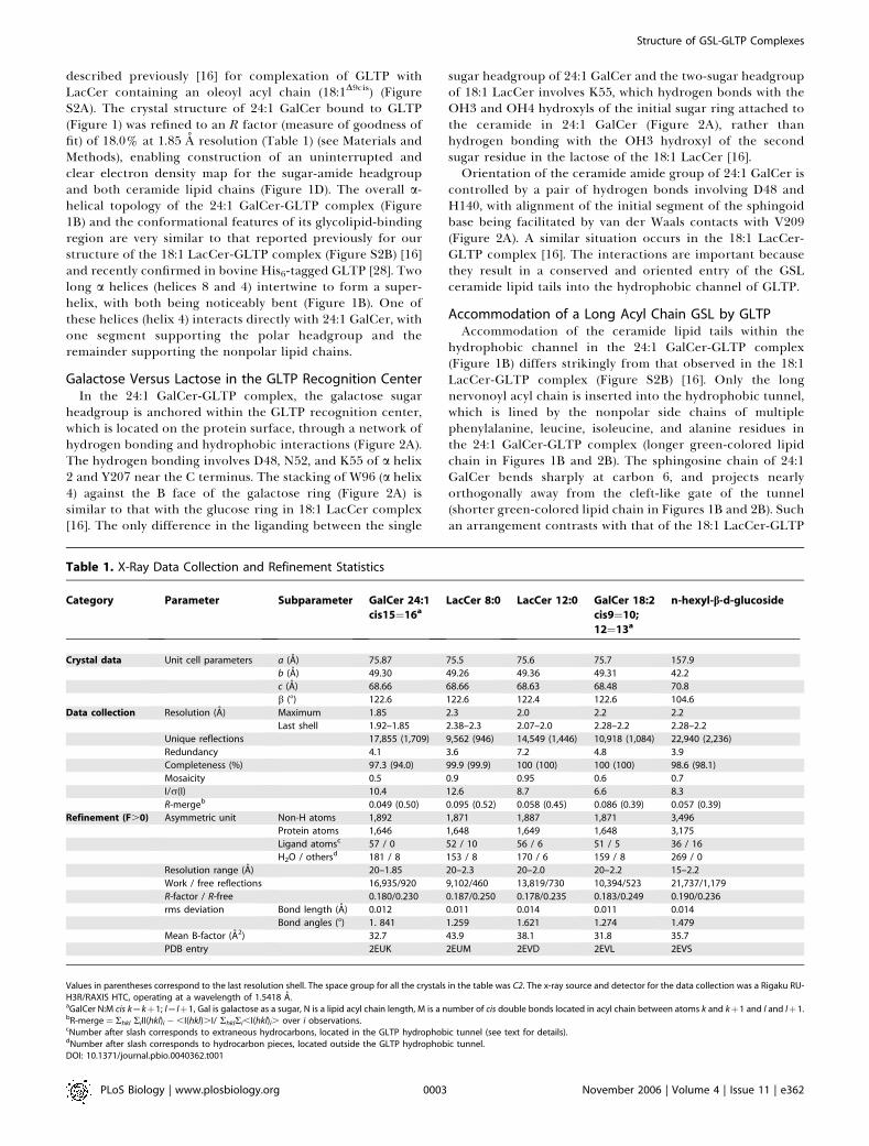
Table 1. X-Ray Data Collection and Refinement Statistics
Category Parameter Subparameter GalCer 24:1
cis15¼16aLacCer 8:0 LacCer 12:0 GalCer 18:2
cis9¼10;
12¼13a
n-hexyl-b-d-glucoside
Crystal data Unit cell parameters a (A) 75.87 75.5 75.6 75.7 157.9
b (A) 49.30 49.26 49.36 49.31 42.2
c (A) 68.66 68.66 68.63 68.48 70.8
b (8) 122.6 122.6 122.4 122.6 104.6
Data collection Resolution (A) Maximum 1.85 2.3 2.0 2.2 2.2
Last shell 1.92–1.85 2.38–2.3 2.07–2.0 2.28–2.2 2.28–2.2
Unique reflections 17,855 (1,709) 9,562 (946) 14,549 (1,446) 10,918 (1,084) 22,940 (2,236)
Redundancy 4.1 3.6 7.2 4.8 3.9
Completeness (%) 97.3 (94.0) 99.9 (99.9) 100 (100) 100 (100) 98.6 (98.1)
Mosaicity 0.5 0.9 0.95 0.6 0.7
I/r(I) 10.4 12.6 8.7 6.6 8.3
R-mergeb 0.049 (0.50) 0.095 (0.52) 0.058 (0.45) 0.086 (0.39) 0.057 (0.39)
Refinement (F.0) Asymmetric unit Non-H atoms 1,892 1,871 1,887 1,871 3,496
Protein atoms 1,646 1,648 1,649 1,648 3,175
Ligand atomsc 57 / 0 52 / 10 56 / 6 51 / 5 36 / 16
H2O / othersd 181 / 8 153 / 8 170 / 6 159 / 8 269 / 0
Resolution range (A) 20–1.85 20–2.3 20–2.0 20–2.2 15–2.2
Work / free reflections 16,935/920 9,102/460 13,819/730 10,394/523 21,737/1,179
R-factor / R-free 0.180/0.230 0.187/0.250 0.178/0.235 0.183/0.249 0.190/0.236
rms deviation Bond length (A) 0.012 0.011 0.014 0.011 0.014
Bond angles (8) 1. 841 1.259 1.621 1.274 1.479
Mean B-factor (A2) 32.7 43.9 38.1 31.8 35.7
PDB entry 2EUK 2EUM 2EVD 2EVL 2EVS
Values in parentheses correspond to the last resolution shell. The space group for all the crystals in the table was C2. The x-ray source and detector for the data collection was a Rigaku RU-H3R/RAXIS HTC, operating at a wavelength of 1.5418 A.aGalCer N:M cis k¼kþ1; l¼ lþ1, Gal is galactose as a sugar, N is a lipid acyl chain length, M is a number of cis double bonds located in acyl chain between atoms k and kþ1 and l and lþ1.bR-merge¼ Rhkl RiII(hkl)i �,I(hkl).I/ RhklRi,I(hkl)i. over i observations.cNumber after slash corresponds to extraneous hydrocarbons, located in the GLTP hydrophobic tunnel (see text for details).dNumber after slash corresponds to hydrocarbon pieces, located outside the GLTP hydrophobic tunnel.DOI: 10.1371/journal.pbio.0040362.t001
PLoS Biology | www.plosbiology.org November 2006 | Volume 4 | Issue 11 | e3620003
Structure of GSL-GLTP Complexes
complex, in which substantial portions of both the oleoyl acyland sphingosine chains are buried within the hydrophobictunnel (Figure S2B) in parallel fashion (Figure S2C). Fur-thermore, to accommodate the six additional methylenes ofthe 24:1 acyl chain in GalCer, the nervonoyl chain curves in aserpentine fashion (Figure 1B) within the tunnel (longergreen-colored lipid chain in Figure 1C), occupying the regionfilled by the sphingosine chain in the 18:1 LacCer complex(shorter lavender colored lipid chain in Figure 1C), andinterfering with entry of the sphingosine chain of 24:1 GalCerinto the tunnel.
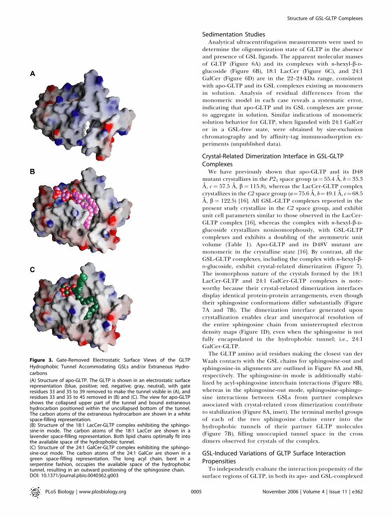
Classification of GLTP and Its GSL-GLTP ComplexesThe structures of GLTP and its GSL complexes can be
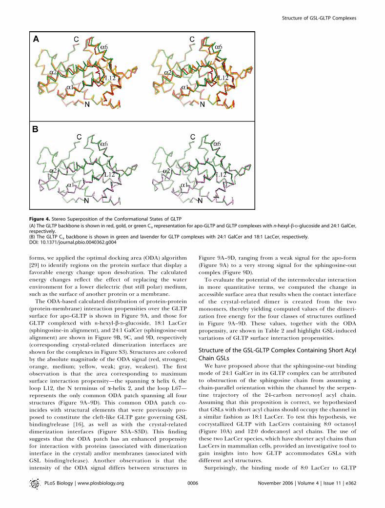
classified into three families. The hydrophobic tunnel isclosed in apo-GLTP (cut-away view, Figure 3A) [16], whereasit is open and maximally expanded in the sphingosine-inconformation observed for 18:1 LacCer-GLTP complex (cut-away view, Figure 3B) [16] and the sphingosine-out con-formation of the 24:1 GalCer-GLTP complex (cut-away view,Figure 3C).GLTP undergoes a conformation change on proceeding
from apo-GLTP (colored red, stereo view in Figure 4A) [16] toits 24:1 GalCer-GLTP complex (colored green, stereo view inFigure 4A). Despite the differences in the sphingosine-in andsphingosine-out alignments, the GLTP conformations arevery similar in the 18:1 LacCer-GLTP complex (coloredlavender, stereo view in Figure 4B) [16] and the newlyidentified 24:1 GalCer-GLTP complex (colored green, stereoview in Figure 4B).
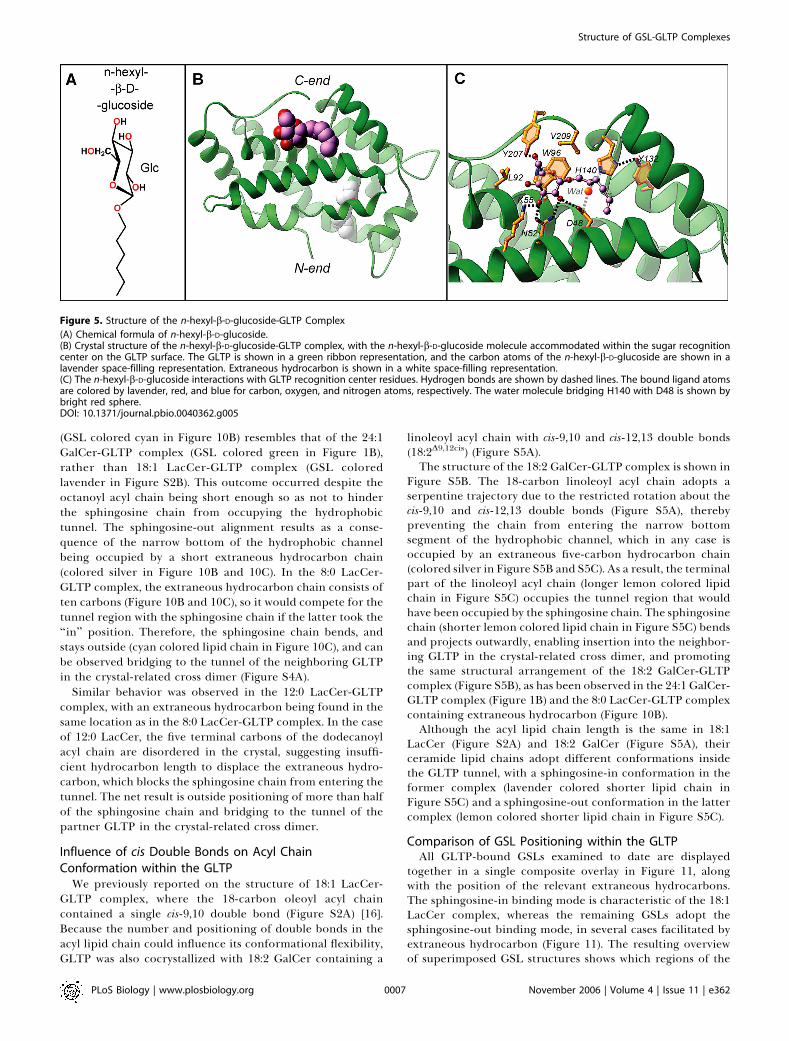
GLTP Complex with n-hexyl-b-D-glucoside That LacksCeramide MoietyTo search for additional conformational states related to
the GLTP working cycle, we cocrystallized GLTP with a modelcompound, n-hexyl-b-D-glucoside (Figure 5A), in which thesugar headgroup was b-linked to a single short (six-carbon)hydrocarbon chain, rather than a two-lipid chain ceramidemoiety. GLTP binds the entire ligand at the protein surface(Figure 5B), with the hydrophobic tunnel remaining closedand containing extraneous hydrocarbon as reported previ-ously for apo-GLTP [16]. The sugar headgroup of n-hexyl-b-D-glucoside binds the GLTP recognition center in a mannersimilar to all GSL-GLTP complexes reported previously [16]and in this paper. The same network of multiple specificinteractions are observed between the n-hexyl-b-D-glucosideheadgroup and the GLTP recognition center (Figure 5C), withthe only differences being the following: (1) the absence ofhydrogen bonding between K55 and the glucosyl OH4 groupbecause of its equatorial positioning and (2) the loss ofhydrogen bonds involving ligands carrying the ceramideamide group and residues H140 and D48, which are nowwater bridged to each other (Figure 5C). Similar water-bridged hydrogen bonds between residues H140 and D48 havealso been found previously in apo-GLTP [16]. Further,attempts to cocrystallize GLTP with simple sugar compounds(galactose and maltose) have been unsuccessful to date.The superimposed structures of apo-GLTP (colored red),
the hexyl-b-D-glucoside-GLTP complex (colored gold), andthe 24:1 GalCer-GLTP complex (colored green) are comparedin stereo in Figure 4A.
Figure 2. GSL-GLTP Interactions in the 24:1 GalCer-GLTP Complex
(A) 24:1 GalCer headgroup (sugar and amide) interactions with GLTPrecognition center residues. Hydrogen bonds are shown by dashed lines.The bound GSL atoms are colored by green, red, and blue for carbon,oxygen, and nitrogen atoms, respectively. The GLTP Ca backbone iscolored light gray, the side chains are shown in gold, and oxygen andnitrogen are red and blue, respectively.(B) 24:1 GalCer lipid interactions with the GLTP channel residues. Thelonger acyl chain is directed into the channel while the shortersphingosine chain is directed outwards.DOI: 10.1371/journal.pbio.0040362.g002
PLoS Biology | www.plosbiology.org November 2006 | Volume 4 | Issue 11 | e3620004
Structure of GSL-GLTP Complexes
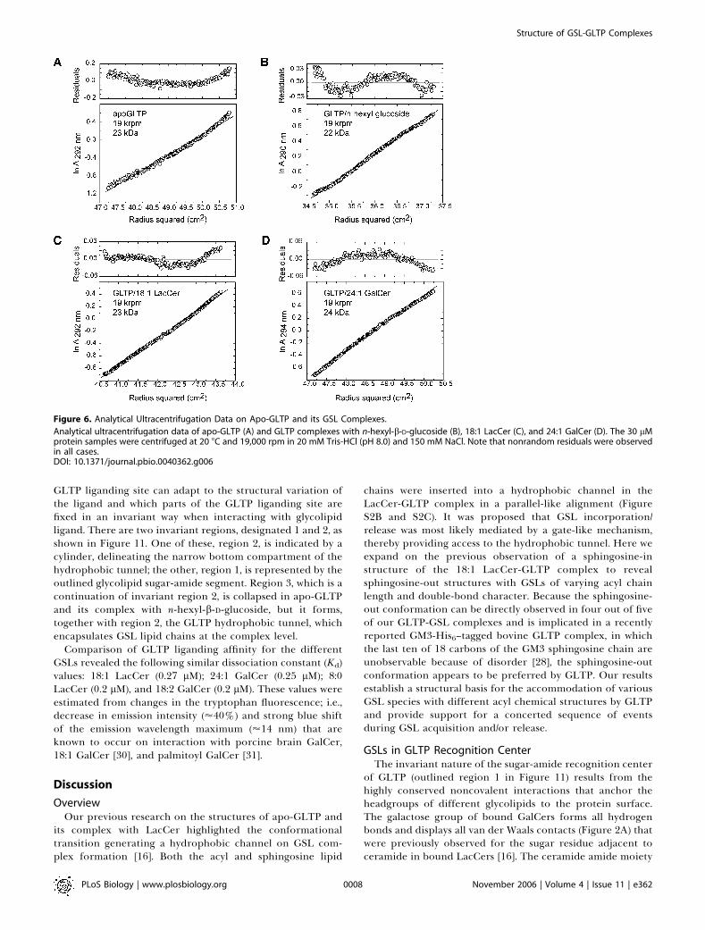
Sedimentation StudiesAnalytical ultracentrifugation measurements were used to
determine the oligomerization state of GLTP in the absenceand presence of GSL ligands. The apparent molecular massesof GLTP (Figure 6A) and its complexes with n-hexyl-b-D-glucoside (Figure 6B), 18:1 LacCer (Figure 6C), and 24:1GalCer (Figure 6D) are in the 22–24-kDa range, consistentwith apo-GLTP and its GSL complexes existing as monomersin solution. Analysis of residual differences from themonomeric model in each case reveals a systematic error,indicating that apo-GLTP and its GSL complexes are proneto aggregate in solution. Similar indications of monomericsolution behavior for GLTP, when liganded with 24:1 GalCeror in a GSL-free state, were obtained by size-exclusionchromatography and by affinity-tag immunoadsorption ex-periments (unpublished data).
Crystal-Related Dimerization Interface in GSL-GLTPComplexesWe have previously shown that apo-GLTP and its D48
mutant crystallizes in the P21 space group (a¼ 55.4 A, b¼ 35.3A, c ¼ 57.5 A, b ¼ 115.8), whereas the LacCer-GLTP complexcrystallizes in the C2 space group (a¼75.6 A, b¼49.1 A, c¼68.5A, b ¼ 122.5) [16]. All GSL-GLTP complexes reported in thepresent study crystallize in the C2 space group, and exhibitunit cell parameters similar to those observed in the LacCer-GLTP complex [16], whereas the complex with n-hexyl-b-D-glucoside crystallizes nonisomorphously, with GSL-GLTPcomplexes and exhibits a doubling of the asymmetric unitvolume (Table 1). Apo-GLTP and its D48V mutant aremonomeric in the crystalline state [16]. By contrast, all theGSL-GLTP complexes, including the complex with n-hexyl-b-D-glucoside, exhibit crystal-related dimerization (Figure 7).The isomorphous nature of the crystals formed by the 18:1LacCer-GLTP and 24:1 GalCer-GLTP complexes is note-worthy because their crystal-related dimerization interfacesdisplay identical protein-protein arrangements, even thoughtheir sphingosine conformations differ substantially (Figure7A and 7B). The dimerization interface generated uponcrystallization enables clear and unequivocal resolution ofthe entire sphingosine chain from uninterrupted electrondensity maps (Figure 1D), even when the sphingosine is notfully encapsulated in the hydrophobic tunnel; i.e., 24:1GalCer-GLTP.The GLTP amino acid residues making the closest van der
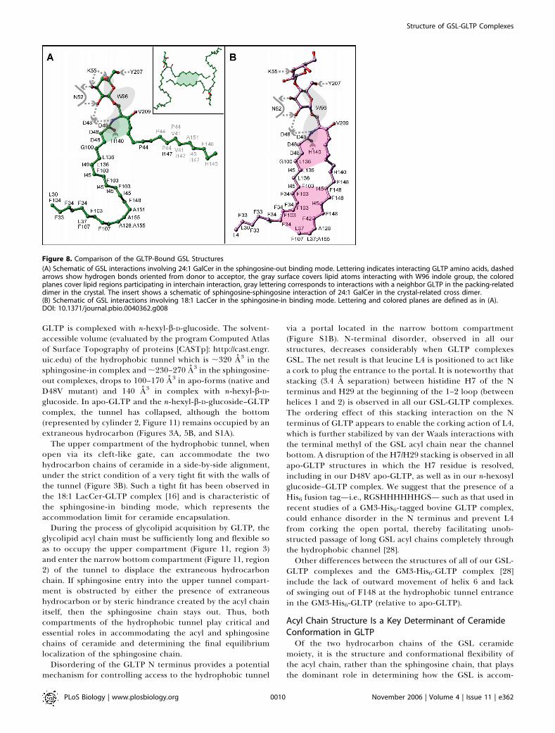
Waals contacts with the GSL chains for sphingosine-out andsphingosine-in alignments are outlined in Figure 8A and 8B,respectively. The sphingosine-in mode is additionally stabi-lized by acyl-sphingosine interchain interactions (Figure 8B),whereas in the sphingosine-out mode, sphingosine-sphingo-sine interactions between GSLs from partner complexesassociated with crystal-related cross dimerization contributeto stabilization (Figure 8A, inset). The terminal methyl groupsof each of the two sphingosine chains enter into thehydrophobic tunnels of their partner GLTP molecules(Figure 7B), filling unoccupied tunnel space in the crossdimers observed for crystals of the complex.
GSL-Induced Variations of GLTP Surface InteractionPropensitiesTo independently evaluate the interaction propensity of the
surface regions of GLTP, in both its apo- and GSL-complexed
Figure 3. Gate-Removed Electrostatic Surface Views of the GLTP
Hydrophobic Tunnel Accommodating GSLs and/or Extraneous Hydro-
carbons
(A) Structure of apo-GLTP. The GLTP is shown in an electrostatic surfacerepresentation (blue, positive; red, negative; gray, neutral), with gateresidues 33 and 35 to 39 removed to make the tunnel visible in (A), andresidues 33 and 35 to 45 removed in (B) and (C). The view for apo-GLTPshows the collapsed upper part of the tunnel and bound extraneoushydrocarbon positioned within the uncollapsed bottom of the tunnel.The carbon atoms of the extraneous hydrocarbon are shown in a whitespace-filling representation.(B) Structure of the 18:1 LacCer-GLTP complex exhibiting the sphingo-sine-in mode. The carbon atoms of the 18:1 LacCer are shown in alavender space-filling representation. Both lipid chains optimally fit intothe available space of the hydrophobic tunnel.(C) Structure of the 24:1 GalCer-GLTP complex exhibiting the sphingo-sine-out mode. The carbon atoms of the 24:1 GalCer are shown in agreen space-filling representation. The long acyl chain, bent in aserpentine fashion, occupies the available space of the hydrophobictunnel, resulting in an outward positioning of the sphingosine chain.DOI: 10.1371/journal.pbio.0040362.g003
PLoS Biology | www.plosbiology.org November 2006 | Volume 4 | Issue 11 | e3620005
Structure of GSL-GLTP Complexes
forms, we applied the optimal docking area (ODA) algorithm[29] to identify regions on the protein surface that display afavorable energy change upon desolvation. The calculatedenergy changes reflect the effect of replacing the waterenvironment for a lower dielectric (but still polar) medium,such as the surface of another protein or a membrane.
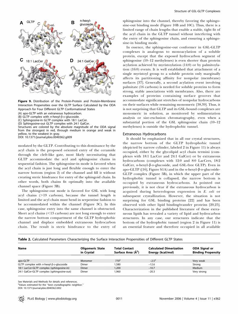
The ODA-based calculated distribution of protein-protein(protein-membrane) interaction propensities over the GLTPsurface for apo-GLTP is shown in Figure 9A, and those forGLTP complexed with n-hexyl-b-D-glucoside, 18:1 LacCer(sphingosine-in alignment), and 24:1 GalCer (sphingosine-outalignment) are shown in Figure 9B, 9C, and 9D, respectively(corresponding crystal-related dimerization interfaces areshown for the complexes in Figure S3). Structures are coloredby the absolute magnitude of the ODA signal (red, strongest;orange, medium; yellow, weak; gray, weakest). The firstobservation is that the area corresponding to maximumsurface interaction propensity—the spanning a helix 6, theloop L12, the N terminus of a-helix 2, and the loop L67—represents the only common ODA patch spanning all fourstructures (Figure 9A–9D). This common ODA patch co-incides with structural elements that were previously pro-posed to constitute the cleft-like GLTP gate governing GSLbinding/release [16], as well as with the crystal-relateddimerization interfaces (Figure S3A–S3D). This findingsuggests that the ODA patch has an enhanced propensityfor interaction with proteins (associated with dimerizationinterface in the crystal) and/or membranes (associated withGSL binding/release). Another observation is that theintensity of the ODA signal differs between structures in
Figure 9A–9D, ranging from a weak signal for the apo-form(Figure 9A) to a very strong signal for the sphingosine-outcomplex (Figure 9D).To evaluate the potential of the intermolecular interaction
in more quantitative terms, we computed the change inaccessible surface area that results when the contact interfaceof the crystal-related dimer is created from the twomonomers, thereby yielding computed values of the dimeri-zation free energy for the four classes of structures outlinedin Figure 9A–9D. These values, together with the ODApropensity, are shown in Table 2 and highlight GSL-inducedvariations of GLTP surface interaction propensities.
Structure of the GSL-GLTP Complex Containing Short AcylChain GSLsWe have proposed above that the sphingosine-out binding
mode of 24:1 GalCer in its GLTP complex can be attributedto obstruction of the sphingosine chain from assuming achain-parallel orientation within the channel by the serpen-tine trajectory of the 24-carbon nervonoyl acyl chain.Assuming that this proposition is correct, we hypothesizedthat GSLs with short acyl chains should occupy the channel ina similar fashion as 18:1 LacCer. To test this hypothesis, wecocrystallized GLTP with LacCers containing 8:0 octanoyl(Figure 10A) and 12:0 dodecanoyl acyl chains. The use ofthese two LacCer species, which have shorter acyl chains thanLacCers in mammalian cells, provided an investigative tool togain insights into how GLTP accommodates GSLs withdifferent acyl structures.Surprisingly, the binding mode of 8:0 LacCer to GLTP
Figure 4. Stereo Superposition of the Conformational States of GLTP
(A) The GLTP backbone is shown in red, gold, or green Ca representation for apo-GLTP and GLTP complexes with n-hexyl-b-D-glucoside and 24:1 GalCer,respectively.(B) The GLTP Ca backbone is shown in green and lavender for GLTP complexes with 24:1 GalCer and 18:1 LacCer, respectively.DOI: 10.1371/journal.pbio.0040362.g004
PLoS Biology | www.plosbiology.org November 2006 | Volume 4 | Issue 11 | e3620006
Structure of GSL-GLTP Complexes
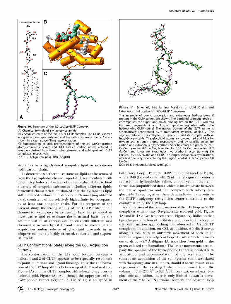
(GSL colored cyan in Figure 10B) resembles that of the 24:1GalCer-GLTP complex (GSL colored green in Figure 1B),rather than 18:1 LacCer-GLTP complex (GSL coloredlavender in Figure S2B). This outcome occurred despite theoctanoyl acyl chain being short enough so as not to hinderthe sphingosine chain from occupying the hydrophobictunnel. The sphingosine-out alignment results as a conse-quence of the narrow bottom of the hydrophobic channelbeing occupied by a short extraneous hydrocarbon chain(colored silver in Figure 10B and 10C). In the 8:0 LacCer-GLTP complex, the extraneous hydrocarbon chain consists often carbons (Figure 10B and 10C), so it would compete for thetunnel region with the sphingosine chain if the latter took the‘‘in’’ position. Therefore, the sphingosine chain bends, andstays outside (cyan colored lipid chain in Figure 10C), and canbe observed bridging to the tunnel of the neighboring GLTPin the crystal-related cross dimer (Figure S4A).
Similar behavior was observed in the 12:0 LacCer-GLTPcomplex, with an extraneous hydrocarbon being found in thesame location as in the 8:0 LacCer-GLTP complex. In the caseof 12:0 LacCer, the five terminal carbons of the dodecanoylacyl chain are disordered in the crystal, suggesting insuffi-cient hydrocarbon length to displace the extraneous hydro-carbon, which blocks the sphingosine chain from entering thetunnel. The net result is outside positioning of more than halfof the sphingosine chain and bridging to the tunnel of thepartner GLTP in the crystal-related cross dimer.
Influence of cis Double Bonds on Acyl ChainConformation within the GLTP
We previously reported on the structure of 18:1 LacCer-GLTP complex, where the 18-carbon oleoyl acyl chaincontained a single cis-9,10 double bond (Figure S2A) [16].Because the number and positioning of double bonds in theacyl lipid chain could influence its conformational flexibility,GLTP was also cocrystallized with 18:2 GalCer containing a
linoleoyl acyl chain with cis-9,10 and cis-12,13 double bonds(18:2D9,12cis) (Figure S5A).The structure of the 18:2 GalCer-GLTP complex is shown in
Figure S5B. The 18-carbon linoleoyl acyl chain adopts aserpentine trajectory due to the restricted rotation about thecis-9,10 and cis-12,13 double bonds (Figure S5A), therebypreventing the chain from entering the narrow bottomsegment of the hydrophobic channel, which in any case isoccupied by an extraneous five-carbon hydrocarbon chain(colored silver in Figure S5B and S5C). As a result, the terminalpart of the linoleoyl acyl chain (longer lemon colored lipidchain in Figure S5C) occupies the tunnel region that wouldhave been occupied by the sphingosine chain. The sphingosinechain (shorter lemon colored lipid chain in Figure S5C) bendsand projects outwardly, enabling insertion into the neighbor-ing GLTP in the crystal-related cross dimer, and promotingthe same structural arrangement of the 18:2 GalCer-GLTPcomplex (Figure S5B), as has been observed in the 24:1 GalCer-GLTP complex (Figure 1B) and the 8:0 LacCer-GLTP complexcontaining extraneous hydrocarbon (Figure 10B).Although the acyl lipid chain length is the same in 18:1
LacCer (Figure S2A) and 18:2 GalCer (Figure S5A), theirceramide lipid chains adopt different conformations insidethe GLTP tunnel, with a sphingosine-in conformation in theformer complex (lavender colored shorter lipid chain inFigure S5C) and a sphingosine-out conformation in the lattercomplex (lemon colored shorter lipid chain in Figure S5C).
Comparison of GSL Positioning within the GLTPAll GLTP-bound GSLs examined to date are displayed
together in a single composite overlay in Figure 11, alongwith the position of the relevant extraneous hydrocarbons.The sphingosine-in binding mode is characteristic of the 18:1LacCer complex, whereas the remaining GSLs adopt thesphingosine-out binding mode, in several cases facilitated byextraneous hydrocarbon (Figure 11). The resulting overviewof superimposed GSL structures shows which regions of the
Figure 5. Structure of the n-hexyl-b-D-glucoside-GLTP Complex
(A) Chemical formula of n-hexyl-b-D-glucoside.(B) Crystal structure of the n-hexyl-b-D-glucoside-GLTP complex, with the n-hexyl-b-D-glucoside molecule accommodated within the sugar recognitioncenter on the GLTP surface. The GLTP is shown in a green ribbon representation, and the carbon atoms of the n-hexyl-b-D-glucoside are shown in alavender space-filling representation. Extraneous hydrocarbon is shown in a white space-filling representation.(C) The n-hexyl-b-D-glucoside interactions with GLTP recognition center residues. Hydrogen bonds are shown by dashed lines. The bound ligand atomsare colored by lavender, red, and blue for carbon, oxygen, and nitrogen atoms, respectively. The water molecule bridging H140 with D48 is shown bybright red sphere.DOI: 10.1371/journal.pbio.0040362.g005
PLoS Biology | www.plosbiology.org November 2006 | Volume 4 | Issue 11 | e3620007
Structure of GSL-GLTP Complexes
GLTP liganding site can adapt to the structural variation ofthe ligand and which parts of the GLTP liganding site arefixed in an invariant way when interacting with glycolipidligand. There are two invariant regions, designated 1 and 2, asshown in Figure 11. One of these, region 2, is indicated by acylinder, delineating the narrow bottom compartment of thehydrophobic tunnel; the other, region 1, is represented by theoutlined glycolipid sugar-amide segment. Region 3, which is acontinuation of invariant region 2, is collapsed in apo-GLTPand its complex with n-hexyl-b-D-glucoside, but it forms,together with region 2, the GLTP hydrophobic tunnel, whichencapsulates GSL lipid chains at the complex level.
Comparison of GLTP liganding affinity for the differentGSLs revealed the following similar dissociation constant (Kd)values: 18:1 LacCer (0.27 lM); 24:1 GalCer (0.25 lM); 8:0LacCer (0.2 lM), and 18:2 GalCer (0.2 lM). These values wereestimated from changes in the tryptophan fluorescence; i.e.,decrease in emission intensity (’40%) and strong blue shiftof the emission wavelength maximum (’14 nm) that areknown to occur on interaction with porcine brain GalCer,18:1 GalCer [30], and palmitoyl GalCer [31].
Discussion
OverviewOur previous research on the structures of apo-GLTP and
its complex with LacCer highlighted the conformationaltransition generating a hydrophobic channel on GSL com-plex formation [16]. Both the acyl and sphingosine lipid
chains were inserted into a hydrophobic channel in theLacCer-GLTP complex in a parallel-like alignment (FigureS2B and S2C). It was proposed that GSL incorporation/release was most likely mediated by a gate-like mechanism,thereby providing access to the hydrophobic tunnel. Here weexpand on the previous observation of a sphingosine-instructure of the 18:1 LacCer-GLTP complex to revealsphingosine-out structures with GSLs of varying acyl chainlength and double-bond character. Because the sphingosine-out conformation can be directly observed in four out of fiveof our GLTP-GSL complexes and is implicated in a recentlyreported GM3-His6–tagged bovine GLTP complex, in whichthe last ten of 18 carbons of the GM3 sphingosine chain areunobservable because of disorder [28], the sphingosine-outconformation appears to be preferred by GLTP. Our resultsestablish a structural basis for the accommodation of variousGSL species with different acyl chemical structures by GLTPand provide support for a concerted sequence of eventsduring GSL acquisition and/or release.
GSLs in GLTP Recognition CenterThe invariant nature of the sugar-amide recognition center
of GLTP (outlined region 1 in Figure 11) results from thehighly conserved noncovalent interactions that anchor theheadgroups of different glycolipids to the protein surface.The galactose group of bound GalCers forms all hydrogenbonds and displays all van der Waals contacts (Figure 2A) thatwere previously observed for the sugar residue adjacent toceramide in bound LacCers [16]. The ceramide amide moiety
Figure 6. Analytical Ultracentrifugation Data on Apo-GLTP and its GSL Complexes.
Analytical ultracentrifugation data of apo-GLTP (A) and GLTP complexes with n-hexyl-b-D-glucoside (B), 18:1 LacCer (C), and 24:1 GalCer (D). The 30 lMprotein samples were centrifuged at 20 8C and 19,000 rpm in 20 mM Tris-HCl (pH 8.0) and 150 mM NaCl. Note that nonrandom residuals were observedin all cases.DOI: 10.1371/journal.pbio.0040362.g006
PLoS Biology | www.plosbiology.org November 2006 | Volume 4 | Issue 11 | e3620008
Structure of GSL-GLTP Complexes

is also similarly positioned in all complexes (Figure 11)through a pair of hydrogen-bonding interactions with D48and H140, with their alignment facilitated by hydrophobiccontacts with V209, as noted for the 24:1 GalCer-GLTPcomplex (Figure 2A). Thus, all hydrogen bonds and van derWaals interactions found between the GLTP recognitioncenter and the sugar-amide headgroup in GSL are conserved,except for hydrogen-bond interactions involving the K55 e-amino group (Figure 2A) [16]. Point mutations support thekey role of W96, H140, D48, and N52 in the recognition andanchoring of the sugar-amide moieties by GLTP [16,26],because mutations W96A and H140L result in almostcomplete inactivation, and mutations D48V and N52I resultin significant inactivation. On the other hand, despite thehydrogen bonding by residues K55 and Y207 with theglycolipid headgroups in all complexes, mutations K55I andY207L retain almost full transfer activity, as measured byboth galactosyl- and lactosylceramide transfer-binding assays[16,32]. These findings lead us to propose two categories ofinteractions: (1) primary, which are invariant and involve therecognition and anchoring of the first sugar and amidegroup; and (2) secondary, which serve a less essentialsupporting role and provide flexibility for binding of morecomplex sugar headgroups. Examples of the latter are K55and Y207, which appear likely to play roles in the liganding ofother GSLs known to be transferred by GLTP [12,32,33].Lysine K55 appears to be a good candidate to interactspecifically with negatively charged glycolipid head groups,such as sulfatide or gangliosides; the phenyl ring of Y207appears to be a good candidate for undergoing a stackinginteraction with extended and/or branched sugars of complexGSLs. This stacking interaction would be analogous to that ofW96 with the initial sugar of GalCer (Figure 2A) and LacCer[16], observed in our GSL-GLTP complexes.The all–a-helical conformation of GLTP does not prevent
its sugar-amide recognition center from sharing two funda-mental features of the liganding centers of many othercarbohydrate binding proteins including all known galactose-specific binding proteins, which display a þ b or all-bconformations. The two fundamental features are: (1) hydro-gen bond donor/acceptor residues arranged so as to facilitateinteractions with hydroxyl groups along the perimeter of thepyranose ring and (2) the presence of an aromatic residue, i.e.tryptophan, against which the sugar stacks [34,35]. Ourobservation of decreased glycolipid transfer activity in theW96F mutant of GLTP [16] is consistent with the idea that thelarger stacking platform provided by tryptophan (comparedto phenylalanine or tyrosine) facilitates positional orienta-tion of the sugar for optimal hydrogen bonding interactionswith the perimeter residues [36]. It is also noteworthy that allhydrogen bond classes observed in previous protein-carbo-hydrate interactions [34] are evident when GLTP interactswith the GSL sugar-amide. Among the classes are bidendatehydrogen bonding involving N52, bifurcated hydrogen bondsinvolving D48 (and K55 in case of galactose), as well as thepropensity of the OH2 and OH3 sugar hydroxyl groups andamino acid side chains (e.g., Y207) to form cooperativehydrogen bonds.
Two Compartments within the GLTP Hydrophobic TunnelThe upper part of the hydrophobic tunnel (region 3 in
Figure 11) is collapsed in apo-GLTP (Figure 3A) and when
Figure 7. Crystal-Related Dimerization of GLTP Complexed with Different
GSL Ligands
(A) Crystal-related dimer in the structure of the 18:1 LacCer-GLTP [16]. TheGLTPs are shown in a gold ribbon representation, and the carbon atomsof the LacCers are shown in a lavender space-filling representation.(B) Crystal-related cross dimer in the structure of the 24:1 GalCer-GLTPcomplex. The GLTPs are shown in a gold ribbon representation, and thecarbon atoms of the GalCers are shown in a green space-fillingrepresentation.(C) Crystal-related dimer in the structure of the n-hexyl-b-D-glucoside-GLTP complex. The GLTPs are shown in a green ribbon representation,while the carbon atoms of the n-hexyl-b-D-glucoside are shown in alavender space-filling representation. Extraneous hydrocarbon is shownin a white space-filling representation.DOI: 10.1371/journal.pbio.0040362.g007
PLoS Biology | www.plosbiology.org November 2006 | Volume 4 | Issue 11 | e3620009
Structure of GSL-GLTP Complexes
GLTP is complexed with n-hexyl-b-D-glucoside. The solvent-accessible volume (evaluated by the program Computed Atlasof Surface Topography of proteins [CASTp]: http://cast.engr.uic.edu) of the hydrophobic tunnel which is ;320 A3 in thesphingosine-in complex and ;230–270 A3 in the sphingosine-out complexes, drops to 100–170 A3 in apo-forms (native andD48V mutant) and 140 A3 in complex with n-hexyl-b-D-glucoside. In apo-GLTP and the n-hexyl-b-D-glucoside–GLTPcomplex, the tunnel has collapsed, although the bottom(represented by cylinder 2, Figure 11) remains occupied by anextraneous hydrocarbon (Figures 3A, 5B, and S1A).
The upper compartment of the hydrophobic tunnel, whenopen via its cleft-like gate, can accommodate the twohydrocarbon chains of ceramide in a side-by-side alignment,under the strict condition of a very tight fit with the walls ofthe tunnel (Figure 3B). Such a tight fit has been observed inthe 18:1 LacCer-GLTP complex [16] and is characteristic ofthe sphingosine-in binding mode, which represents theaccommodation limit for ceramide encapsulation.
During the process of glycolipid acquisition by GLTP, theglycolipid acyl chain must be sufficiently long and flexible soas to occupy the upper compartment (Figure 11, region 3)and enter the narrow bottom compartment (Figure 11, region2) of the tunnel to displace the extraneous hydrocarbonchain. If sphingosine entry into the upper tunnel compart-ment is obstructed by either the presence of extraneoushydrocarbon or by steric hindrance created by the acyl chainitself, then the sphingosine chain stays out. Thus, bothcompartments of the hydrophobic tunnel play critical andessential roles in accommodating the acyl and sphingosinechains of ceramide and determining the final equilibriumlocalization of the sphingosine chain.
Disordering of the GLTP N terminus provides a potentialmechanism for controlling access to the hydrophobic tunnel
via a portal located in the narrow bottom compartment(Figure S1B). N-terminal disorder, observed in all ourstructures, decreases considerably when GLTP complexesGSL. The net result is that leucine L4 is positioned to act likea cork to plug the entrance to the portal. It is noteworthy thatstacking (3.4 A separation) between histidine H7 of the Nterminus and H29 at the beginning of the 1–2 loop (betweenhelices 1 and 2) is observed in all our GSL-GLTP complexes.The ordering effect of this stacking interaction on the Nterminus of GLTP appears to enable the corking action of L4,which is further stabilized by van der Waals interactions withthe terminal methyl of the GSL acyl chain near the channelbottom. A disruption of the H7/H29 stacking is observed in allapo-GLTP structures in which the H7 residue is resolved,including in our D48V apo-GLTP, as well as in our n-hexosylglucoside–GLTP complex. We suggest that the presence of aHis6 fusion tag—i.e., RGSHHHHHHGS— such as that used inrecent studies of a GM3-His6-tagged bovine GLTP complex,could enhance disorder in the N terminus and prevent L4from corking the open portal, thereby facilitating unob-structed passage of long GSL acyl chains completely throughthe hydrophobic channel [28].Other differences between the structures of all of our GSL-
GLTP complexes and the GM3-His6-GLTP complex [28]include the lack of outward movement of helix 6 and lackof swinging out of F148 at the hydrophobic tunnel entrancein the GM3-His6-GLTP (relative to apo-GLTP).
Acyl Chain Structure Is a Key Determinant of CeramideConformation in GLTPOf the two hydrocarbon chains of the GSL ceramide
moiety, it is the structure and conformational flexibility ofthe acyl chain, rather than the sphingosine chain, that playsthe dominant role in determining how the GSL is accom-
Figure 8. Comparison of the GLTP-Bound GSL Structures
(A) Schematic of GSL interactions involving 24:1 GalCer in the sphingosine-out binding mode. Lettering indicates interacting GLTP amino acids, dashedarrows show hydrogen bonds oriented from donor to acceptor, the gray surface covers lipid atoms interacting with W96 indole group, the coloredplanes cover lipid regions participating in interchain interaction, gray lettering corresponds to interactions with a neighbor GLTP in the packing-relateddimer in the crystal. The insert shows a schematic of sphingosine-sphingosine interaction of 24:1 GalCer in the crystal-related cross dimer.(B) Schematic of GSL interactions involving 18:1 LacCer in the sphingosine-in binding mode. Lettering and colored planes are defined as in (A).DOI: 10.1371/journal.pbio.0040362.g008
PLoS Biology | www.plosbiology.org November 2006 | Volume 4 | Issue 11 | e3620010
Structure of GSL-GLTP Complexes
modated by the GLTP. Contributing to this dominance by theacyl chain is the proposed oriented entry of the ceramidethrough the cleft-like gate, most likely necessitating thatGLTP accommodate the acyl and sphingosine chains insequential fashion. The sphingosine-in mode is favored whenthe acyl chain is just long and flexible enough to enter thenarrow bottom (region 2) of the channel and fill it withoutcreating steric hindrance for entry of the sphingoid chain. Inother words, both chains fit optimally into the availablechannel space (Figure 3B).
The sphingosine-out mode is favored for GSL with longacyl chains (.19 carbons), because the tunnel length islimited and the acyl chain must bend in serpentine fashion tobe accommodated within the channel (Figure 3C). In thiscase, sphingosine entry into the same channel is obstructed.Short acyl chains (,13 carbons) are not long enough to enterthe narrow bottom compartment of the GLTP hydrophobicchannel and displace embedded extraneous hydrocarbonchain. The result is steric hindrance to the entry of
sphingosine into the channel, thereby favoring the sphingo-sine-out binding mode (Figure 10B and 10C). Thus, there is alimited range of chain lengths that enable a stable, tight fit ofthe acyl chain in the GLTP tunnel without interfering withthe entry of the sphingosine chain, and ensuring a sphingo-sine-in binding mode.In essence, the sphingosine-out conformer in GSL-GLTP
complexes is analogous to monoacylation of a solubleprotein, except that the exposed hydrocarbon segment ofsphingosine (10–12 methylenes) is even shorter than proteinacylation achieved by myristoylation (14:0) or by palmitoyla-tion (16:0) events. It is well established that attachment of asingle myristoyl group to a soluble protein only marginallyaffects its partitioning affinity for nonpolar (membrane)surfaces [37]. Generally, a second acylation event involvingpalmitate (16 carbons) is needed for soluble proteins to formstrong, stable associations with membranes. Also, there areexamples of proteins containing surface grooves thataccommodate significant stretches of nonpolar hydrocarbonson their surfaces while remaining monomeric [38,39]. Thus, itis not surprising that GLTP and its GSL-bound complexes aremonomeric in solution, as monitored by sedimentationanalysis or size-exclusion chromatography, even when asubstantial portion of the GSL sphingosine chain (10–12methylenes) is outside the hydrophobic tunnel.
Extraneous HydrocarbonsIt should be emphasized that in all our crystal structures,
the narrow bottom of the GLTP hydrophobic tunnel(depicted by narrow cylinder, labeled 2 in Figure 11) is alwaysoccupied, either by the glycolipid acyl chain termini (com-plexes with 18:1 LacCer and 24:1 GalCer) or by extraneoushydrocarbons (complexes with 12:0 and 8:0 LacCers, 18:2GalCer, n-hexyl-b-D-glucoside, and GSL-free GLTP). Even inapo-GLTP ([16], Figure S1A) and in the n-hexyl-b-D-glucoside-GLTP complex (Figure 5B), in which the upper part of thehydrophobic tunnel is collapsed, the narrow bottom isoccupied by extraneous hydrocarbons. As pointed outpreviously, it is not clear if the extraneous hydrocarbon isacquired during heterologous expression in E. coli orsubsequent crystallization. However, the situation is notsurprising for GSL binding proteins [22] and has beenobserved with other lipid binding/transfer proteins [20,25].Characterization in the published literature of these extra-neous lipids has revealed a variety of lipid and hydrocarbonstructures. In any case, our structures indicate that thebottom of the hydrophobic tunnel (region 2 in Figure 11) isan essential feature and therefore occupied in all available
Table 2. Calculated Parameters Characterizing the Surface Interaction Propensities of Different GLTP States
Name Oligomeric State
in Crystal
Total Contact
Surface Area (A2)
Calculated Dimerization
Energy (kcal/mol)
ODA Signal or
Binding Propensity
apo-GLTP Monomer 770a –2.5a Very weak
GLTP complex with n-hexyl-b-D-glucoside Dimer 1,580 –12.6 Strong
18:1 LacCer-GLTP complex (sphingosine-in) Dimer 1,200 –10.7 Medium
24:1 GalCer-GLTP complex (sphingosine-out) Dimer 1,960 –20.3 Very strong
See Materials and Methods for details and references.aValues estimated for the ‘‘best crystallographic neighbor.’’DOI: 10.1371/journal.pbio.0040362.t002
Figure 9. Distribution of the Protein-Protein and Protein-Membrane
Interaction Propensities over the GLTP Surface Calculated by the ODA
Approach for Four Different GLTP Conformational States
(A) apo-GLTP with an extraneous hydrocarbon.(B) GLTP complex with n-hexyl-b-D-glucoside.(C) Sphingosine-in GLTP complex with 18:1 LacCer.(D) Sphingosine-out GLTP complex with 24:1 GalCer.Structures are colored by the absolute magnitude of the ODA signalfrom the strongest in red, through medium in orange and weak inyellow, to the weakest in gray.DOI: 10.1371/journal.pbio.0040362.g009
PLoS Biology | www.plosbiology.org November 2006 | Volume 4 | Issue 11 | e3620011
Structure of GSL-GLTP Complexes
structures by a tightly-fitted nonpolar lipid or extraneoushydrocarbon chain.
To determine whether the extraneous lipid can be removedfrom the hydrophobic channel, apo-GLTP was incubated withb-methylcyclodextrin because of its established ability to binda variety of nonpolar substances including different lipids.Structural characterization showed that the extraneous lipidstill remained within the hydrophobic channel (unpublisheddata), consistent with a relatively high affinity for occupancyby at least one nonpolar chain. For the purposes of thepresent study, the strong affinity of the GLTP hydrophobicchannel for occupancy by extraneous lipid has provided aninvestigative tool to evaluate the structural basis for theaccommodation of various GSL species with different acylchemical structures by GLTP and a tool to show that theacquisition and/or release of glycolipid proceeds in anadaptive manner via highly oriented, concerted, and sequen-tial events.
GLTP Conformational States along the GSL AcquisitionPathway
The conformation of the L12 loop, located between ahelices 1 and 2 of GLTP, appears to be especially responsiveto point mutations and ligand binding. Thus, the conforma-tion of the L12 loop differs between apo-GLTP (colored red,Figure 4A) and the GLTP complex with n-hexyl-b-D-glucoside(colored gold, Figure 4A), even though the upper part of thehydrophobic tunnel (segment 3, Figure 11) is collapsed in
both cases. Loop L12 in the D48V mutant of apo-GLTP [16],where D48 (located on a helix 2) of the recognition center isreplaced by hydrophobic valine, adopts yet another con-formation (unpublished data), which is intermediate betweenthe native apo-form and the complex with n-hexyl-b-D-glucoside. Taken together, these data indicate that events inthe GLTP headgroup recognition center contribute to theconformation of the L12 loop.A comparison of the conformation of the L12 loop in GLTP
complexes with n-hexyl-b-D-glucoside (colored gold, Figure4A) and 24:1 GalCer (colored green, Figure 4A), indicates thatligand-sugar attachment facilitates adoption by this loop ofthe conformation approaching that observed in GSL-GLTPcomplexes. In addition, on GSL acquisition, a helix 2 movesalong its axis, with an outwards movement of both its N-terminal segment and adjacent loop L12, while a helix 6 movesoutwards by ’2.7 A (Figure 4A, transition from gold to thegreen-colored conformations). The latter movements accom-pany the opening of the hydrophobic tunnel associated withacquisition and accommodation of the acyl chain. Thesubsequent acquisition of the sphingosine chain associatedwith the sphingosine-in complex, should it occur, results in anexpansion of the existing hydrophobic tunnel from thevolume of 230–270 A3 to 320 A3. In contrast, on n-hexyl-b-D-glucoside acquisition, there is only limited outwards move-ment of the a helix 2 N-terminal segment and adjacent loop
Figure 11. Schematic Highlighting Positions of Lipid Chains and
Extraneous Hydrocarbons in GSL-GLTP Complexes
The assembly of bound glycolipids and extraneous hydrocarbons, ifpresent in the GLTP tunnel, are shown. The bordered segment labeled 1encompasses the sugar- and amide-binding site on the GLTP, whereasbordered segments 2 and 3 span lipid-binding sites within thehydrophobic GLTP tunnel. The narrow bottom of the GLTP tunnel isschematically represented by a transparent cylinder, labeled 2. Thesegment labeled 3 is collapsed in apo-GLTP and its complex with n-hexyl-b-D-glucoside. The glycolipid atoms are colored red and blue foroxygen and nitrogen atoms, respectively, and by specific colors forcarbon and extraneous hydrocarbons. Specific colors are green for 24:1GalCer, cyan for 8:0 LacCer, lavender for 18:1 LacCer, lemon for 18:2GalCer, and silver for extraneous hydrocarbons accompanying 8:0LacCer, 18:2 LacCer, and apo-GLTP. The longest extraneous hydrocarbon,which is the only one entering the region labeled 3, accompanies 8:0LacCer.DOI: 10.1371/journal.pbio.0040362.g011
Figure 10. Structure of the 8:0 LacCer-GLTP Complex
(A) Chemical formula of 8:0 lactosylceramide.(B) Crystal structure of the 8:0 LacCer-GLTP complex. The GLTP is shownin a gold ribbon representation, and the carbon atoms of the LacCer areshown in a cyan space-filling representation.(C) Superposition of stick representations of the 8:0 LacCer (carbonatoms colored in cyan) and 18:1 LacCer (carbon atoms colored inlavender) derived from their sphingosine-out and sphingosine-in GLTPcomplexes, respectively.DOI: 10.1371/journal.pbio.0040362.g010
PLoS Biology | www.plosbiology.org November 2006 | Volume 4 | Issue 11 | e3620012
Structure of GSL-GLTP Complexes
L12, but no movement of the a helix 2 along its axis noroutward movement of a helix 6, keeping phenylalanine F148in a swung-in position and allowing for only partial opening ofthe hydrophobic channel, in a fashion similar to that observedrecently in GM3-His6-tagged bovine GLTP complex [28].
GSL-Based Distribution of GLTP Surface InteractionPropensities
Monomeric apo-GLTP, which has the lowest total contactsurface area of 770 A2, exhibits the smallest computeddimerization energy of �2.5 kcal/mol, whereas the 24:1GalCer-GLTP complex, which has the highest total contactsurface area of 1,960 A2, exhibits the largest computeddimerization energy of�20.3 kcal/mol (Table 2). The n-hexyl-b-D-glucoside-GLTP complex exhibits somewhat larger valuesof total contact surface area and computed dimerizationenergy than the 18:1 LacCer-GLTP complex. The differentoligomeric states of a ligand-free GLTP (monomer in solutionand crystal) and sphingosine-in and sphingosine-out GLTPcomplexes (monomer in solution; packing-related dimeriza-tion in crystal) might reflect a gain of protein-protein orprotein-membrane interaction propensities by the proteinsurface upon ligand binding (Table 2).
Only monomeric GSL-GLTP complexes could be detectedin solution despite repeated attempts using different method-ologies (e.g., analytical ultracentrifugation, size-exclusionliquid chromatography, affinity pull-down assays involvingGLTPs with different N-terminal fusion tags). This finding,when viewed within the context of the ODA energy contactcalculations, implies that the GLTP face in contact with itspartner protein in the crystal-related dimer identifies thisdimerization interface as the likely membrane interaction siteof GLTP, because it is rather hydrophobic, is ringed by fourlysines, and contains several tyrosines and two tryptophanresidues, which are all known to provide a favorableinteraction site with membranes [31,40–42].
Model of GSL Transfer/Presentation by GLTPOur findings of a new sphingosine-out ligand binding
mode in the crystal structures of GSL-GLTP complexes, thedifferent conformational changes induced in the gate-regionL12 loop upon liganding of different glycolipids, and therespective computed values characterizing the interactionpropensities are collectively supportive of an importantproperty of GLTP: namely, a highly conserved, adaptive,and concerted sequence of events involving GLTP-mediatedacquisition/release of GSL. Because apo-GLTP displays arelatively weak protein/membrane-binding propensity (Table2) [42], it can be expected to readily partition on and off themembrane surface. Nonetheless, the rapid lateral diffusionrates of lipids in fluid-phase membranes and the confinementof the glycolipid ligand to a membrane surface would likelyenhance the capacity of GLTP to associate with a glycolipidmolecule among other membrane lipids. The sugar moiety ofthe glycolipid clearly acts as a primary specificity determi-nant, whereas the ceramide amide functional group orientsthe entry of the hydrocarbon chain(s) through the cleft-likegate. Comparison of the apo-GLTP and the n-hexyl-b-D-glucoside–GLTP complex provides clear evidence that therecognition and anchoring of the sugar head triggersconformational rearrangements of the gate region, L12 loop.At this stage, there is an increased interaction propensity
centered about the cleft-like GLTP gate that is surrounded byaromatic surface residues (e.g., W142, W96, Y153, Y157, Y207,Y81) and a half dozen lysine residues, which are known tointeract favorably with membrane interfaces [40,41], therebypotentially facilitating gate-membrane interactions, includ-ing the outward movement of the a helix 6. We anticipatethat the opening of the gate in the membrane-associated statefacilitates entry of the acyl chain into the hydrophobicchannel of the GLTP. The accommodation limits of thehydrophobic tunnel, illustrated by the crystal structures ofthe GSL-GLTP complexes, strongly suggest that sphingosineis the last lipid part to enter GLTP and, most likely, the firstto depart GLTP upon interaction with a membrane.
Functional ImplicationsAmong known lipid binding/transfer proteins, the GLTP
topology is unique. The GLTP fold also defines a novelmembrane targeting and/or interaction domain amongknown peripheral proteins [27]. The formation of a lipid-liganding site by a-helical layering, without intramoleculardisulfide bonds, contrasts with the situation in other lipidbinding and transfer proteins which use either disulfide-bridge–stabilized, helical bundles (i.e., saposin folds), ormotifs dominated by b sheets (i.e., b grooves or concavecups) and b barrels. Although the phosphatidylcholine andphosphatidylinositol transfer proteins appear to show apreference for liganding certain phosphoglyceride speciesduring intermembrane transfer [43,44], our findings withGLTP establish a structural basis for the accommodation ofvarious GSL species with different acyl chemical structures bythe liganding channel of a lipid transfer/binding protein. Thepresent structural analyses have provided insights into thespecificity of glycolipid targeting by GLTP and also demon-strated how the accommodation limits of the GLTP ligandingsite are controlled by the conformational features of both thehydrophobic channel and the GSL ceramide lipid chains.Quite remarkably, in addition to the expected essential roleof the sugar-amide headgroup recognition center in control-ling the selectivity for glycolipids, we have found that thelength and conformational flexibility of the glycolipid acylchain constitute critical features modulating the accommo-dation of sphingosine chain. Finally, our observation ofcrystal-related cross dimerization in the crystal structures ofseveral sphingosine-out GSL-GLTP complexes suggests thatthe ceramide lipid chains of GSLs most likely enter into andegress from GLTP in a defined stepwise manner.
Materials and Methods
Protein expression and purification. The open reading frame(ORF) encoding human GLTP was subcloned into the pET-30 Xa/LICexpression vector (Novagen, Madison, Wisconsin, United States),enabling cleavage of the amino-terminal His6 and S-tags, aspreviously described [16]. Transformed BL21 (DE3) E. coli cells(Novagen) were grown in Luria-Bertani (LB) medium at 37 8C,induced with 0.1 mM IPTG, and grown an additional 20 h at 15 8C.Purification of recombinant GLTP (rGLTP) from soluble lysateprotein was accomplished by Ni-affinity chromatography [32]. TheHis6-tag and S-tag sequences were removed from rGLTP byincubation with Factor-Xa at room temperature for 16 h. rGLTPwas then purified by fast protein liquid chromatography size-exclusion chromatography and concentrated to 10–15 mg/ml in 150mM NaCl, 20 mM Tris-HCl, pH 8.0.
Glycolipid synthesis and purification. LacCer and GalCer withhomogeneous acyl chains were produced by reacylating D-lactosyl-b1–19-D-erythroSphingosine (lyso LacCer) and D-galactosyl-b1–19-D-eryth-
PLoS Biology | www.plosbiology.org November 2006 | Volume 4 | Issue 11 | e3620013
Structure of GSL-GLTP Complexes
roSphingosine (lyso GalCer or psychosine) (Avanti Polar Lipids,Alabaster, Alaska, United States) with the desired fatty acyl residue asdescribed previously [45,46]. Briefly, the N-hydroxy succinimide esterof the desired fatty acid was prepared, recrystallized, and reacted withlyso-glycolipid. Reacylation was performed at 60 8C under nitrogen for6–8 h in the presence of the catalyst, N-ethyldiisopropylamine.Following reacylation, the glycolipid was purified by flash columnchromatography and crystallized from CHCl3/CH3OH using �20 8Cacetone. Using the preceding approach, N-dodecanoyl lactosylsphin-gosine (12:0 LacCer), N-cis-9-octadecenoyl lactosylsphingosine(18:1D9cis LacCer or N-oleoyl LacCer), N-cis-15-tetracosenoyl galacto-syl-sphingosine (24:1D15cis GalCerorN-nervonoylGalCer), andN-cis-N-cis-9,12-octadecenoyl galactosyl-sphingosine (18:2D9,12cis GalCer or N-linoleoyl GalCer) were prepared. N-octanoyl lactosylsphingosine (8:0LacCer) was obtained fromAvanti Polar Lipids. Acyl homogeneities ofthe glycolipid derivatives were confirmed by quantitative release,methylation, and analysis of the fatty acyl residues via capillary gaschromatography. Glycolipid purity was confirmed by thin layerchromatography. n-hexyl-b-D-glucoside was obtained from Anatrace(Maumee, Ohio, United States).
Sedimentation equilibrium analysis. Analytical ultracentrifugationmeasurements were carried out on a Beckman XL-A (BeckmanCoulter, Fullerton, California, United States) analytical ultracentri-fuge equipped with an An-60 Ti rotor (Beckman Coulter) at 20 8C.His6-tagged GLTP and His6-tagged GLTP complexes were loaded atinitial concentration of 7, 15, and 30 lM and analyzed at rotor speedof 19,000 and 22,000 rpm. Data were acquired at wavelengths 290–294nm and processed simultaneously with a nonlinear least-squaresfitting routine [47]. Solvent density and protein partial specificvolume were calculated according to solvent and protein composi-tion, respectively [48]. We estimated the partial specific volume ofGSLs as 0.98 mL/g at 20 8C.
Fluorescence measurements. Steady-state fluorescence measure-ments were performed using a SPEX Fluoromax instrument (Instru-ments S.A., Inc., Edina, New Jersey, United States). Excitation andemission bandpasses were 5 nm and the cuvette holder was temper-ature controlled to 37 60.1 8C (Thermo-Neslab, Portsmouth, NewHampshire, United States). To eliminate contributions from aminoacid residues other than W and to minimize absorbance byacrylamide, the excitation wavelength was 295 nm. Emission spectrawere recorded from 310 to 420 nm using GLTP concentrations (1 lM)with an optical density of less than 0.1 at 295 nm to avoid the innerfilter effect. Measurements were performed under constant stirringby addition of small defined aliquots of GSL dissolved in ethanol (1mM) to a fixed volume of protein. Rapid equilibration of thefluorescence emission signal was observed (2–3 min) with noadditional change observed by additional incubation up to 20 min.
Crystallization and x-ray data collection. Crystals of glycolipid-GLTP complexes were obtained by cocrystallization of protein withglycolipid using the hanging drop technique. Protein samples (10–15mg/ml) were mixed with glycolipid dispersion (0.4 mM in 20%–40%EtOH), and then with well solution at ratio 1:1:1 resulting in a pHnear 6.0. The well solution contained 15%–20% (weight/volume) PEG3350 (or PEG 8000), 50 mM KH2PO4, pH 4.5. Single crystals appearedin 2 wk to 3 mo, depending on glycolipid type. All GSL-GLTP crystalswere isomorphous and belonged to the space group C2, with unit cellparameters indicated in Table 1. GLTP complex with n-hexyl-b-D-glycoside, which also crystallized in the C2 space group, ischaracterized by different unit cell (Table 1) and two molecules inthe asymmetric unit (AU), in contrast with GSL-GLTP crystals, whichall have one molecule in AU. Apo-GLTP crystals belonged to the P21space group [16,49]. Crystals were transferred into well solutionincluding 20% glycerol as cryoprotectant, next mounted in a fiberloop and flash-frozen in a cold nitrogen stream for x-ray datacollection. All data sets were collected on a Rigaku RU-H3R X-raygenerator (Rigaku Americas, The Woodlands, Texas, United States)equipped with a RAXIS-HTC detector. Data collection statistics arepresented in Table 1.
Structure determination and refinement. Because the GSL-GLTPcrystals reported in this paper are isomorphous to crystals of the 18:1LacCer-GLTP complex [16], the coordinate set of this crystalstructure (with ligand omitted) was used as an initial model for the24:1 GalCer-GLTP complex (Table 1, column 1). The 24:1 GalCer wasadded with the help of the difference electron density map. Themodel was refined in REFMAC [50] at 1.85 A resolution to a final R-factor/R-free of 0.180/0.230 (Table 1). The automatic procedure ARP/wARP was used to add solvent molecules to a final model [51]. Thesimulated annealed omit 2Fo-Fc map for the ligand region is shown inFigure 1D. After appropriate deletions, the model was applied as theinitial one for all other GSL-GLTP complexes, which were refined at
an appropriate resolution to R factors shown in Table 1. Thestructure of GLTP complex with n-hexyl-b-D-glucoside was deter-mined by the molecular replacement (MR) method via the AMoReprogram [52], and then refined in REFMAC. The model used in theMR search was native apo-GLTP.
Evaluating general protein-protein interface propensity. Theprotocols for the ODA computations [29] are as follows: a mesh ofpoints, located 3 A above the protein surface, was generated with anaverage distance of 5 A between them. For series of spheres centeredat each point and characterized by a variable radius, changing from 5A to 20 A, the product of the atomic solvent accessible areas by theatomic ODA densities [29] was calculated. The optimal radius wasthen selected and the best desolvation value was assigned to theresidues. The method was previously validated on 66 nonhomologousprotein pairs involved in nonobligate protein-protein complexes ofknown structure.
Estimating the binding energy for GLTP complexes. The bindingenergies between two monomers with and without ligands werecalculated using the method described in [53]. The binding energyvalues were calculated using an empirical formula consisting of threemain contributions: (1) the electrostatic free energy differenceevaluated using the boundary element solution of the Poisson equation[54], (2) the surface energydifference, and (3) the entropic loss estimate.The parameters used in calculations, including the constant C¼7 kcal/mol, the surface energydensity 0.03 kcal/A2, and the interface dielectricconstant of 8, were optimized previously on a diverse set of protein-peptide and protein-protein complexes. The expected accuracy wasshown to amount to 2.5 kcal/mol.
Calculating the contact surface areas. The contact surface area wascalculated using the fast implementation of the Shrake and Rupleyalgorithm implemented in the ICM program [55], using the proberadius of 1.4 A and the van der Waals radii as follows: C aliphatic,1.95 A; C aromatic, 1.8 A; N, 1.7 A; OH, 1.6 A; O, 1.4 A; SH, 2.0 A; andS, 1.8 A. The contact surface area was calculated as Area(A)þArea(B)� Area(AB).
Supporting Information
Figure S1. Structure of Apo-GLTP
(A) Structure of the apo-GLTP [16], with a helices shown in a goldcylinder representation, whereas 310 helices are shown in silverribbon representation. Extraneous hydrocarbon is shown in a silverspace-filling representation. (B) The portal of the apo-GLTP formwith no residues removed from the GLTP electrostatic representa-tion. The extraneous hydrocarbon end seen from the portal is shownin a silver space-filling representation.
Found at DOI: 10.1371/journal.pbio.0040362.sg001 (6.0 MB TIF).
Figure S2. Structure of the 18:1 LacCer-GLTP Complex
(A) Chemical formula of 18:1 lactosylceramide. (B) Crystal structureof the 18:1 LacCer-GLTP complex [16]. The GLTP is shown in a goldribbon representation, whereas the carbon atoms of the LacCers areshown in a lavender-colored space-filling representation. (C) Stickrepresentation of the sphingosine-in conformation of the 18:1LacCer (carbon atoms colored in lavender) in the complex.
Found at DOI: 10.1371/journal.pbio.0040362.sg002 (2.2 MB TIF).
Figure S3. Protein-Protein and Protein-Membrane InteractionPropensities Calculated by the ODA Approach for Three DifferentGLTP Complexes for Views Emphasizing Crystal-Related Dimeriza-tion
(A) Complex of GLTP with n-hexyl-b-D-glucoside. (B) Sphingosine-inGLTP complex with 18:1 LacCer. (C) Sphingosine-out GLTP complexwith 24:1 GalCer. Structures are colored by the absolute magnitude ofthe ODA signal from the strongest in red, through medium in orange,weak in yellow, to the weakest in gray.
Found at DOI: 10.1371/journal.pbio.0040362.sg003 (4.4 MB TIF).
Figure S4. Crystal-Related Cross Dimerization of GLTP Complexedwith Different GSL Ligands
(A) Crystal-related cross dimer in the structure of the 8:0 LacCer-GLTP complex. The GLTPs are shown in a gold ribbon representa-tion, whereas the carbon atoms of the LacCers are shown in a cyanspace-filling representation. (B) Crystal-related cross dimer in thestructure of the 18:2 GalCer-GLTP complex. The GLTPs are shown ina gold ribbon representation, whereas the carbon atoms of theGalCers are shown in a lemon-colored space-filling representation.
PLoS Biology | www.plosbiology.org November 2006 | Volume 4 | Issue 11 | e3620014
Structure of GSL-GLTP Complexes
Found at DOI: 10.1371/journal.pbio.0040362.sg004 (5.3 MB TIF).
Figure S5. Structure of the 18:2 GalCer-GLTP Complex
(A) Chemical formula of 18:2 galactosylceramide. (B) The crystalstructure of the 18:2 GalCer-GLTP complex. The GLTP is shown in agold cylinder representation, whereas the carbon atoms of theGalCers are shown in a lemon-colored space-filling representation.(C) Superposition of stick representations of the 18:2 GalCer (carbonatoms colored in lemon) and 18:1 LacCer (carbon atoms colored inlavender) derived from their sphingosine-out and sphingosine-inGLTP complexes, respectively. Arrows point out the positions of cisdouble bonds in acyl chains.
Found at DOI: 10.1371/journal.pbio.0040362.sg005 (2.1 MB TIF).
Accession Numbers
The coordinates and diffraction amplitudes from this study havebeen deposited in the Protein Data Bank (http://www.rcsb.org/pdb/)with the following accession numbers: 24:1 GalCer-GLTP complex(2EUK); 8:0 LacCer-GLTP complex (2EUM); 12:0 LacCer-GLTPcomplex (2EVD); 18:2 GalCer-GLTP complex (2EVL); n-hexyl-b-D-glucoside-GLTP complex (2EVS); and D48V mutant apo-GLTP(2EVT). The previously deposited coordinates for apo-GLTP and18:1 LacCer-GLTP have accession numbers 1SWX and 1SX6,
respectively[16]. The GenBank (http://www.ncbi.nlm.nih.gov/Genbank/) accession numbers for open reading frame encodinghuman GLTP are AF209704, AY372530, AY372531, and AY372532.
Acknowledgments
We thank Alex Teplov for technical support during x-ray datacollection and Dr. Xin-Min Li and Helen Pike for synthesizing andpurifying several of the glycolipids.
Author contributions. LM, MLM, REB, and DJP conceived anddesigned the experiments. LM was responsible for x-ray datacollection, analysis and structure determination. MLM was respon-sible for the biochemistry and crystallization, with biochemicalassistance from ATK. ML was responsible for the ultracentrifugationexperiments. RA was responsible for the ODA calculations. REB andDJP supervised and insured implementation of the project. LM, REB,and DJP wrote the paper with contributions from MLM, ML, and RA.
Funding. This research was supported by the Abby RockefellerMauze Trust and the Dewitt Wallace and Maloris Foundations (DJP),the Hormel Foundation (REB), NIH/NIGMS GM45928 (REB), andNIH/NCI CA121493 (to DJP and REB).
Competing interests. The authors have declared that no competinginterests exist.
References1. Hakomori SI (2002) The glycosynapse. Proc Natl Acad Sci U S A 99: 225–
232.2. Dwek RA, Butters TD, Platt FM, Zitzmann N (2002) Targeting glycosylation
as a therapeutic approach. Nat Rev Drug Discov 1: 65–75.3. Yamashita T, Wada R, Sasaki T, Deng C, Bierfreund U, et al. (1999) A vital
role for glycosphingolipid synthesis during development and differ-entiation. Proc Natl Acad Sci U S A 96: 9142–9147.
4. Kozireski-Chuback D, Wu G, Ledeen RW (1999) Upregulation of nuclearGM1 accompanies axon-like, but not dendrite-like, outgrowth in NG108–15 cells. J Neurosci Res 55: 107–118.
5. Rippo MR, Malisan F, Ravagnan L, Tomassini B, Condo I, et al. (2000) GD3ganglioside directly targets mitochondria in a bcl-2-controlled fashion.FASEB J 14: 2047–2054.
6. Morales A, Colell A, Mari M, Garcia-Ruiz C, Fernandez-Checa JC (2004)Glycosphingolipids and mitochondria: Role in apoptosis and disease.Glycoconj J 20: 579–588.
7. Sasaki T (1990) Glycolipid transfer protein and intracellular traffic ofglucosylceramide. Experientia 46: 611–616.
8. Warnock DE, Lutz MS, Blackburn WA, Young WW Jr., Baenziger JU (1994)Transport of newly synthesized glucosylceramide to the plasma membraneby a non-Golgi pathway. Proc Natl Acad Sci U S A 91: 2708–2712.
9. Metz RJ, Radin NS (1982) Purification and properties of a cerebrosidetransfer protein. J Biol Chem 257: 12901–12907.
10. Abe A, Sasaki T (1985) Purification and some properties of the glycolipidtransfer protein from pig brain. J Biol Chem 260: 11231–11239.
11. Brown RE, Jarvis KL, Hyland KJ (1990) Purification and characterization ofglycolipid transfer protein from bovine brain. Biochim Biophys Acta 1044:77–83.
12. Yamada K, Abe A, Sasaki T (1985) Specificity of the glycolipid transferprotein from pig brain. J Biol Chem 260: 4615–4621.
13. Lin X, Mattjus P, Pike HM, Windebank AJ, Brown RE (2000) Cloning andexpression of glycolipid transfer protein from bovine and porcine brain. JBiol Chem 275: 5104–5110.
14. Brodersen P, Petersen M, Pike HM, Olszak B, Skov S, et al. (2002) Knockoutof Arabidopsis accelerated-cell-death11 encoding a sphingosine transferprotein causes activation of programmed cell death and defense. GenesDev 16: 490–502.
15. Mattjus P, Turcq B, Pike HM, Molotkovsky JG, Brown RE (2003) Glycolipidintermembrane transfer is accelerated by HET-C2, a filamentous fungusgene product involved in the cell-cell incompatibility response. Biochem-istry 42: 535–542.
16. Malinina L, Malakhova ML, Teplov A, Brown RE, Patel DJ (2004) Structuralbasis for glycosphingolipid transfer specificity. Nature 430: 1048–1053.
17. Gough J, Karplus K, Hughey R, Chothia C (2001) Assignment of homologyto genome sequences using a library of hidden Markov models thatrepresent all proteins of known structure. J Mol Biol 313: 903–919.
18. Murzin AG, Brenner SE, Hubbard T, Chothia C (1995) SCOP: a structuralclassification of proteins database for the investigation of sequences andstructures. J Mol Biol 247: 536–540.
19. Gadola SD, Zaccai NR, Harlos K, Shepherd D, Castro-Palomino JC, et al.(2002) Structure of human CD1b with bound ligands at 2.3 A, a maze foralkyl chains. Nat Immunol 3: 721–726.
20. Hamilton JA (2004) Fatty acid interactions with proteins: What X-raycrystal and NMR solution structures tell us. Prog Lipid Res 43: 177–199.
21. Roderick SL, Chan WW, Agate DS, Olsen LR, Vetting MW, et al. (2002)
Structure of human phosphatidylcholine transfer protein in complex withits ligand. Nat Struct Biol 9: 507–511.
22. Wright CS, Zhao Q, Rastinejad F (2003) Structural analysis of lipidcomplexes of GM2-activator protein. J Mol Biol 331: 951–964.
23. Tilley SJ, Skippen A, Murray-Rust J, Swigart PM, Stewart A, et al. (2004)Structure-function analysis of human phosphatidylinositol transfer proteinalpha bound to phosphatidylinositol. Structure 12: 317–326.
24. Soccio RE, Breslow JL (2003) StAR-related lipid transfer (START) proteins:Mediators of intracellular lipid metabolism. J Biol Chem 278: 22183–22186.
25. Yoder MD, Thomas LM, Tremblay JM, Oliver RL, Yarbrough LR, et al.(2001) Structure of a multifunctional protein. Mammalian phosphatidyli-nositol transfer protein complexed with phosphatidylcholine. J Biol Chem276: 9246–9252.
26. Zajonc DM, Elsliger MA, Teyton L, Wilson IA (2003) Crystal structure ofCD1a in complex with a sulfatide self antigen at a resolution of 2.15 A. NatImmunol 4: 808–815.
27. Cho W, Stahelin RV (2005) Membrane-protein interactions in cell signalingand membrane trafficking. Annu Rev Biophys Biomol Struct 34: 119–151.
28. Airenne TT, Kidron H, Nymalm Y, Nylund M, West G, et al. (2006)Structural evidence for adaptive ligand binding of glycolipid transferprotein. J Mol Biol 355: 224–236.
29. Fernandez-Recio J, Totrov M, Skorodumov C, Abagyan R (2005) Optimaldocking area: A new method for predicting protein-protein interactionsites. Proteins 58: 134–143.
30. Li X-M, Malakhova ML, Lin X, Pike HM, Chung T, et al. (2004) Humanglycolipid transfer protein: Probing conformation using fluorescencespectroscopy. Biochemistry 43: 10285–10294.
31. West G, Nylund M, Slotte JP, Mattjus P (2006) Membrane interaction andactivity of the glycolipid transfer protein. Biochim Biophys Acta (E-pub 7July 2006).
32. Malakhova ML, Malinina L, Pike HM, Kanack AT, Patel DJ, et al. (2005)Point mutational analysis of the liganding site in human glycolipid transferprotein. Functionality of the complex. J Biol Chem 280: 26312–26320.
33. Brown RE, Stephenson FA, Markello T, Barenholz Y, Thompson TE (1985)Properties of a specific glycolipid transfer protein from bovine brain.Chem Phys Lipids 38: 79–93.
34. Quiocho FA, Vyas NK (1999) Atomic interactions between protein/enzymesand carbohydrates. In: Hecht SM, editor. Bioorganic Chemistry: Carbohy-drates. New York: Oxford University Press. pp 441–457.
35. Sujatha MS, Balaji PV (2004) Identification of common structural featuresof binding sites in galactose-specific proteins. Proteins: Struct FuncBioinform 55: 44–65.
36. Sujatha MS, Sasidhar YU, Balaji PV (2004) Energetics of galactose- andglucose-aromatic amino acid interactions: Implications for binding ingalactose-specific proteins. Protein Sci 13: 2502–2514.
37. Murray D, Ben-Tal N, Honig B, McLaughlin S (1997) Electrostaticinteraction of myristoylated proteins with membranes: Simple physics,complicated biology, Structure 5: 985–989.
38. Chakravarty B, Gu Z, Chirala SS, Wakil SJ, Quiocho FA (2004) Human fattyacid synthase: Structure and substrate selectivity of the thioesterasedomain, Proc Natl Acad Sci U S A 101: 15567–15572.
39. vandenBergB,BlackPN,Clemons JrWM,RapoportTA (2004)Crystal structureof the long-chain fatty acid transporter FadL, Science 304: 1506–1509.
40. White SH, Wimley WC (1998) Hydrophobic interactions of peptides withmembrane interfaces. Biochim Biophys Acta 1376: 339–352.
41. Killian JA, von Heijne G (2000) How proteins adapt to a membrane-waterinterface. Trends Biochem Sci 25: 429–434.
PLoS Biology | www.plosbiology.org November 2006 | Volume 4 | Issue 11 | e3620015
Structure of GSL-GLTP Complexes
42. Rao CS, Chung T, Pike HM, Brown RE (2005) Glycolipid transfer proteininteraction with bilayer vesicles: Modulation by changing lipid composi-tion. Biophys J 89: 4017–4028.
43. Kasurinen J, van Paridon PA, Wirtz KW, Somerharju P (1990) Affinity ofphosphatidylcholine molecular species for the bovine phosphatidylcholineand phosphatidylinositol transfer proteins. Properties of the sn-1 and sn-2acyl binding sites. Biochemistry 29: 8548–8554.
44. Hunt AN, Skippen AJ, Koster G, Postle AD, Cockcroft S (2004) Acyl chain-based molecular selectivity for HL60 cellular phosphatidylinositol and ofphosphatidylcholine by phosphatidylinositol transfer protein alpha. Bio-chim Biophys Acta 1686: 50–60.
45. Li XM, Momsen MM, Brockman HL, Brown RE (2002) Lactosylceramide:Effect of acyl chain structure on phase behavior and molecular packing.Biophys J 83: 1535–1546.
46. Smaby JM, Kulkarni VS, Momsen M, Brown RE (1996) The interfacial elasticpacking interactions of galactosylceramides, sphingomyelins, and phos-phatidylcholines. Biophys J 70: 868–877.
47. Johnson ML, Correia JJ, Yphantis DA, Halvorson HR (1981) Analysis of datafrom the analytical ultracentrifuge by nonlinear least-squares techniques.Biophys J 36: 575–588.
48. Laue TM, Shah BD, Ridgeway TM, Pelletier SL (1992) Computer-aidedinterpretation of analytical sedimentation data for proteins. In: Harding
SE, Rowe AJ, Horton JC, editors. Analytical ultracentrifugation inbiochemistry and polymer science. Cambridge (United Kingdom): RoyalSociety of Chemistry. pp. 90–125.
49. West G, Nymalm Y, Airenne TT, Kidron H, Mattjus P, et al. (2004)Crystallization and x-ray analysis of bovine glycolipid transfer protein. ActaCrystallogr D Biol Crystallogr 60: 703–705.
50. Murshudov GN, Vagin AA, Dodson EJ (1997) Refinement of macro-molecular structures by the maximum-likelihood method. Acta CrystallogrD Biol Crystallogr 53: 240–255.
51. Lamzin VS, Wilson KS (1993) Automated refinement of protein models.Acta Crystallogr D Biol Crystallogr 49: 129–147.
52. Navaza J (1997) AMoRe: an automated package for molecular replacement.Acta Crystallogr D Biol Crystallogr A50: 157–163.
53. Schapira M, Totrov M, Abagyan R (1999) Prediction of the binding energyfor small molecules, peptides and proteins. J Mol Recognit 12: 177–190.
54. Totrov M, Abagyan R (2001) Rapid boundary element solvation electro-statics calculations in folding simulations: Successful folding of a 23-residuepeptide. Biopolymers 60: 124–133.
55. Abagyan R, Totrov M, Kuznetsov DN (1994) ICM - a new method forprotein modeling and design. Applications to docking and structureprediction from the distorted native conformation. J Com Chem 15: 488–506.
PLoS Biology | www.plosbiology.org November 2006 | Volume 4 | Issue 11 | e3620016
Structure of GSL-GLTP Complexes