platelet rna signatures for the detection of cancer - springer · platelet rna signatures for the...
TRANSCRIPT

Platelet RNA signatures for the detection of cancer
Nik Sol1,2 & Thomas Wurdinger2,3,4
Published online: 5 July 2017# The Author(s) 2017. This article is an open access publication
Abstract Platelets are equipped with RNA processing ma-chineries, such as pre-mRNA splicing, pre-miRNA process-ing, and mRNA translation. Since platelets are devoid of anucleus, most RNA transcripts in platelets are derived frommegakaryocytes during thrombocytogenesis. However, plate-lets can also ingest RNA molecules during circulation and/orinteraction with other cell types. Since platelets were first de-scribed by Bizzozero in 1881, their well-established role inhemostasis and thrombosis has been intensively studied.However, in the past decades, the list of biological processesin which platelets play an important role keeps expanding. Inthis review, we discuss how platelet RNA biomarker signa-tures can be altered in the presence of cancer.
Keywords Platelets . Transcriptome . mRNA . Splicing .
Tumor-educated platelets . Liquid biopsy . Biomarkers
1 Platelets and cancer in brief
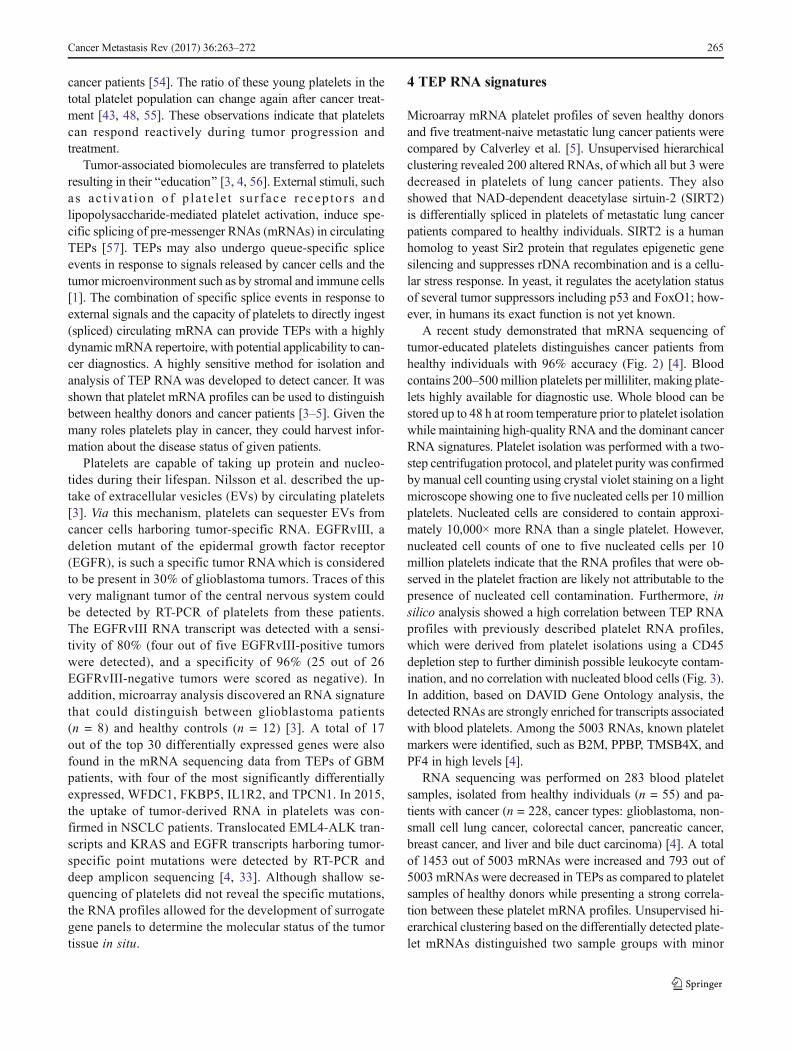
Platelets are implicated in tumor biology and metastasis(Fig. 1) [1, 2], and tumor cells can, directly and indirectly,impose changes on platelet RNA and protein content [3–5].As a result, these tumor-educated platelets (TEPs) have analtered function and can in various ways promote tumorcell survival and metastasis, as well as other hallmarks ofcancer [6]. Tumor cells interact with platelets indirectly viadifferent signaling molecules or directly via different re-ceptors, mainly the platelet activation receptor P-selectin[7–10]. Upon activation, platelets can release severalgrowth and pro-angiogenic factors like ANGPT1, PDGF,BFGF, EGF, HGF, IGF1, TGFβ, VEGF-A, and VEGF-C[11, 12]. Platelets can release these factors at a metastaticniche, thereby providing a pro-tumoral growth microenvi-ronment [1].
Calver ley et a l . showed that NAD-dependentdeacetylase sirtuin-2 (SIRT2) is differentially spliced inplatelets of metastatic lung cancer patients. This gene playsa role in epigenetic silencing, concluding that plateletscould induce growth and progression of tumors by releas-ing epigenetic silencers [5]. Further, platelets alter cancercells to evade the detection by the immune system, bytransferring MHC class I proteins to the tumor cells,resulting in protection against natural killer cells [13].Platelets can also mechanically protect cancer cells fromdestruction via NK cells by forming a cell–fibrin–plateletaggregate surrounding circulating tumor cells (CTC) orarrested tumor cells. This physical shield avoids cell-mediated immune detection and supports cancer cell sur-vival [14–17]. Such protective properties of platelets areimportant for cancer metastasis by promoting cancer cellsurvival in the circulation [18, 19].
* Nik [email protected]
Thomas [email protected]
1 Department of Neurology, VU University Medical Center,Amsterdam, The Netherlands
2 Brain Tumor Center Amsterdam, VU University Medical Center,Amsterdam, The Netherlands
3 Department of Neurosurgery, VU University Medical Center,Amsterdam, The Netherlands
4 Department of Neurology, Massachusetts General Hospital andNeuroscience Program, Harvard Medical School, Boston, MA, USA
Cancer Metastasis Rev (2017) 36:263–272DOI 10.1007/s10555-017-9674-0

2 Liquid biopsies and cancer detection
Liquid biopsies have been introduced as a potential gamechanger in cancer management, with blood tests emerging asa minimally invasive, safe, and sensitive alternative or com-plementary approach for tissue biopsies [20–22]. Blood rep-resents a rich source of information through which solid can-cers (and their subtypes) can be detected, identified and clas-sified, and matched to a specific therapy [23–30]. Targetedrisk-based screening based on a person’s individual risk ofcancer is envisioned to be the anti-cancer strategy of the fu-ture. Current clinical oncology practice relies on the removalof tumor tissue through biopsies for analysis of tumor-linkedgenetic alterations. Although the use of tumor tissue biopsiesis the current gold standard for cancer diagnosis and representsan essential tool in cancer management, it has become appar-ent that the information acquired from a single biopsy pro-vides a spatially and temporally limited snapshot of a(metastatic) tumor and often fails to reflect the heterogeneityof the disease [22, 31, 32]. Moreover, tumor biopsies are in-vasive which poses a limitation for repeated sampling (neededfor monitoring treatment response and resistance to targetedtherapies). Liquid biopsies could provide a potential revolu-tion in cancer diagnostics as a minimally invasive method fordetecting and monitoring diseases, complementary to currenttissue biopsy approaches. Liquid biopsies can therefore pro-vide an accurate and comprehensive spatiotemporal snapshot
of the tumor and its microenvironment on multiple levels, andenable (1) early detection (screening), (2) prognosis for theindividual patient: stage and spread, (3) identification of newtargets for personalized treatment, (4) pre-treatment classifica-tion for personalized therapy/prediction of response to thera-py, (5) early therapy response monitoring, Breal-time^ assess-ment of treatment effectiveness, and (6) follow-up and earlydetection of recurrence of the disease and its metastases.Currently, blood-based biopsy measurements focus on evalu-ation of biomarker biosources, including circulating tumorDNA (ctDNA), circulating tumor cells (CTCs), extracellularvesicles (EVs; exosomes, microvesicles, microparticles,oncosomes), and tumor-educated platelets (TEPs) [3, 4,33–38].
3 Tumor-educated platelets
Platelets have long been considered as a potential diagnostictool in cancer. Several studies have shown that a simple plate-let count already harbors potential clinical relevant informa-tion [39–44]. Besides platelet counts, the size of platelets [44,45] and platelet protein markers, such as P-selectin, are usedfor blood-based cancer diagnostics and prognostics [44,46–51]. Platelets can interact with cancer cells in various waysleading to platelet hyper-reactivity [52, 53]. Furthermore,there may be an increase in young reticulated platelets in
Megakaryocyte
DNA
RNA molecule
RNA-binding protein
Blood platelet
Tumor-educated platelet
Protein
mRNA molecule
Microvesicles
Red blood cells
White blood cells
Tumor cells
miRNA
Stromal cells
Endothelial cells
AAAAAA
AAAAAA
Extracellular queues
A
Primary tumor
Metastases
B Tumor-educated platelet
Circulating platelet
C
D
Fig. 1 Schematic representation of tumor-mediated education of plateletsand the megakaryocyte leading to metastasis. (a) Megakaryocytes in thebone marrow and lungs sort specific RNA and proteins into plateletprecursors. (b) Circulating platelets contain a variety of RNA transcriptsand proteins. During their 7–10-day lifespan, platelets interact withimmune cells, cancer cells, and stromal cells. These direct interactions aswell as distant cell signaling, for instance, via vesicle-mediated
communication in whole blood, changes the content of the platelet andplatelet function. (c) This process leads to the development of tumor-educated platelets. Next, tumor-educated platelets can influence the processof metastasis formation by stimulating or blocking immune cells, endothe-lial cells, stromal cells, and cancer cells, either by direct cell-to-cell contactor by releasing extracellular queues. (d) Finally, metastasis could affect thesorting of specific RNA and proteins of megakaryocytes into platelets
264 Cancer Metastasis Rev (2017) 36:263–272
cancer patients [54]. The ratio of these young platelets in thetotal platelet population can change again after cancer treat-ment [43, 48, 55]. These observations indicate that plateletscan respond reactively during tumor progression andtreatment.
Tumor-associated biomolecules are transferred to plateletsresulting in their Beducation^ [3, 4, 56]. External stimuli, suchas ac t iva t ion o f p la te l e t su r face recep to r s andlipopolysaccharide-mediated platelet activation, induce spe-cific splicing of pre-messenger RNAs (mRNAs) in circulatingTEPs [57]. TEPs may also undergo queue-specific spliceevents in response to signals released by cancer cells and thetumor microenvironment such as by stromal and immune cells[1]. The combination of specific splice events in response toexternal signals and the capacity of platelets to directly ingest(spliced) circulating mRNA can provide TEPs with a highlydynamic mRNA repertoire, with potential applicability to can-cer diagnostics. A highly sensitive method for isolation andanalysis of TEP RNAwas developed to detect cancer. It wasshown that platelet mRNA profiles can be used to distinguishbetween healthy donors and cancer patients [3–5]. Given themany roles platelets play in cancer, they could harvest infor-mation about the disease status of given patients.
Platelets are capable of taking up protein and nucleo-tides during their lifespan. Nilsson et al. described the up-take of extracellular vesicles (EVs) by circulating platelets[3]. Via this mechanism, platelets can sequester EVs fromcancer cells harboring tumor-specific RNA. EGFRvIII, adeletion mutant of the epidermal growth factor receptor(EGFR), is such a specific tumor RNAwhich is consideredto be present in 30% of glioblastoma tumors. Traces of thisvery malignant tumor of the central nervous system couldbe detected by RT-PCR of platelets from these patients.The EGFRvIII RNA transcript was detected with a sensi-tivity of 80% (four out of five EGFRvIII-positive tumorswere detected), and a specificity of 96% (25 out of 26EGFRvIII-negative tumors were scored as negative). Inaddition, microarray analysis discovered an RNA signaturethat could distinguish between glioblastoma patients(n = 8) and healthy controls (n = 12) [3]. A total of 17out of the top 30 differentially expressed genes were alsofound in the mRNA sequencing data from TEPs of GBMpatients, with four of the most significantly differentiallyexpressed, WFDC1, FKBP5, IL1R2, and TPCN1. In 2015,the uptake of tumor-derived RNA in platelets was con-firmed in NSCLC patients. Translocated EML4-ALK tran-scripts and KRAS and EGFR transcripts harboring tumor-specific point mutations were detected by RT-PCR anddeep amplicon sequencing [4, 33]. Although shallow se-quencing of platelets did not reveal the specific mutations,the RNA profiles allowed for the development of surrogategene panels to determine the molecular status of the tumortissue in situ.
4 TEP RNA signatures
Microarray mRNA platelet profiles of seven healthy donorsand five treatment-naive metastatic lung cancer patients werecompared by Calverley et al. [5]. Unsupervised hierarchicalclustering revealed 200 altered RNAs, of which all but 3 weredecreased in platelets of lung cancer patients. They alsoshowed that NAD-dependent deacetylase sirtuin-2 (SIRT2)is differentially spliced in platelets of metastatic lung cancerpatients compared to healthy individuals. SIRT2 is a humanhomolog to yeast Sir2 protein that regulates epigenetic genesilencing and suppresses rDNA recombination and is a cellu-lar stress response. In yeast, it regulates the acetylation statusof several tumor suppressors including p53 and FoxO1; how-ever, in humans its exact function is not yet known.
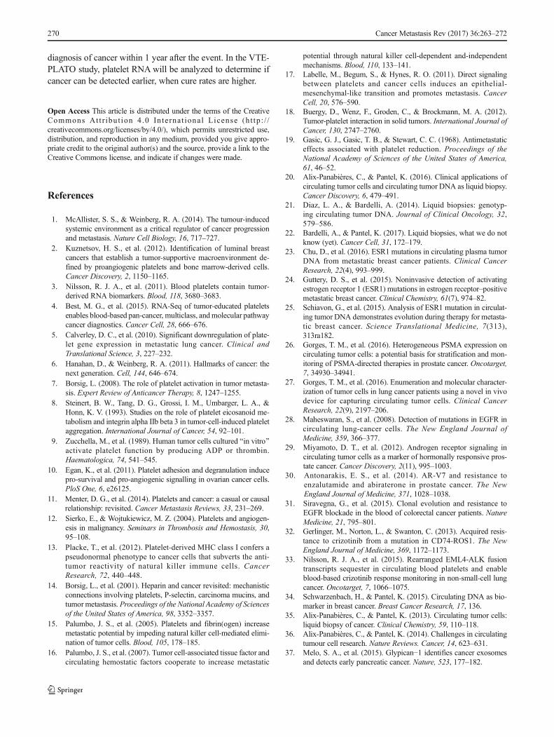
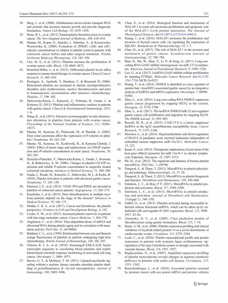
A recent study demonstrated that mRNA sequencing oftumor-educated platelets distinguishes cancer patients fromhealthy individuals with 96% accuracy (Fig. 2) [4]. Bloodcontains 200–500million platelets per milliliter, making plate-lets highly available for diagnostic use. Whole blood can bestored up to 48 h at room temperature prior to platelet isolationwhile maintaining high-quality RNA and the dominant cancerRNA signatures. Platelet isolation was performed with a two-step centrifugation protocol, and platelet purity was confirmedby manual cell counting using crystal violet staining on a lightmicroscope showing one to five nucleated cells per 10 millionplatelets. Nucleated cells are considered to contain approxi-mately 10,000× more RNA than a single platelet. However,nucleated cell counts of one to five nucleated cells per 10million platelets indicate that the RNA profiles that were ob-served in the platelet fraction are likely not attributable to thepresence of nucleated cell contamination. Furthermore, insilico analysis showed a high correlation between TEP RNAprofiles with previously described platelet RNA profiles,which were derived from platelet isolations using a CD45depletion step to further diminish possible leukocyte contam-ination, and no correlation with nucleated blood cells (Fig. 3).In addition, based on DAVID Gene Ontology analysis, thedetected RNAs are strongly enriched for transcripts associatedwith blood platelets. Among the 5003 RNAs, known plateletmarkers were identified, such as B2M, PPBP, TMSB4X, andPF4 in high levels [4].
RNA sequencing was performed on 283 blood plateletsamples, isolated from healthy individuals (n = 55) and pa-tients with cancer (n = 228, cancer types: glioblastoma, non-small cell lung cancer, colorectal cancer, pancreatic cancer,breast cancer, and liver and bile duct carcinoma) [4]. A totalof 1453 out of 5003 mRNAs were increased and 793 out of5003 mRNAs were decreased in TEPs as compared to plateletsamples of healthy donors while presenting a strong correla-tion between these platelet mRNA profiles. Unsupervised hi-erarchical clustering based on the differentially detected plate-let mRNAs distinguished two sample groups with minor
Cancer Metastasis Rev (2017) 36:263–272 265

Pla
tele
t exp
ress
ion
TEP + HC expression, Best et al.
A B C D E
Meg
akar
yocy
teex
pres
sion
B-c
ell e
xpre
ssio
n
CD
4 T-
cells
expr
essi
on
CD
8 T-
cells
expr
essi
on
HGF
Gra
nulo
cyte
expr
essi
on
J
Mem
ory
T-ce
llsex
pres
sion
Mon
ocyt
eex
pres
sion
K L M
I
Nat
ural
Kill
er c
ells
expr
essi
onr2 =0.711 r2 =0.825 r2 =0.654 r2 =0.759 r2 =0.713
r2 r275.0= 2 r823.0= 2 r813.0= 2 =0.303
r2 r013.0= 2 r822.0= 2 r004.0= 2 =0.238
Fig. 3 Correlation plots of TEP RNA signatures with other (a) nucleatedblood cells. Correlation plots between platelets sequenced by Best et al.compared with platelet RNA expression levels from different studies andcompared with RNA expression from different blood cells. (a) Plateletsfrom Bray et al. [82]. (b) Poly A selected RNAs from platelets from
Kissopoulou et al. [83]. (c) Ribosomal RNA-depleted platelets fromKissopoulou et al. [83]. (d) Platelets from Rowley et al. [84]. (e)Platelets from Simon et al. [85]. (f) Megakaryocyte [86]. (g) MemoryT-cells [87]. (h) CD4 T-cells [87]. (i) CD8 T-cells [87]. (j) Granulocytes[87]. (k) B-cells [87]. (l) Monocytes [87]. (m) Natural killer cells [87]
-1.5 1.50
Healthy (n = 55)Cancer (n = 228)
p<0.0001Fisher’s exact test
Patient
20
10
0
25 200 1000 4000
[FU]
Blood draw
Plateletisolation
RNA extraction
cDNA synthesisand
amplificationRNA-seq
preparation
A
B
Fig. 2 ThromboSeq workflow. (a) TEP mRNA sequencing workflow;blood draw is performed on patients, and from a single 6-ml EDTA-coated tube, platelets are isolated. RNA extraction is done according tomanufacturer’s protocol using the mirVana RNA isolation kit (LifeTechnologies). Next, mRNA is amplified using SMARTer Ultra low in-put RNA kit (Clontech). Samples are prepared for sequencing on the
Hiseq 2500 Illumina platform using the Truseq Nano DNA SamplePrep Kit (Illumina). After each step, quality control was performed bycritical inspection of Bioanalyzer profiles (Agilent). (b) Heatmap ofunsupervised clustering of platelet mRNA profiles of healthy donors(red, n = 55) and patients with cancer (gray, n = 228)
266 Cancer Metastasis Rev (2017) 36:263–272

overlap. Using the different mRNA profiles of cancer patientsand healthy donors, it was possible to develop a predictivealgorithmwith high accuracy in separating healthy individualsfrom cancer patients. The RNA profiles allowed for supportvector machine (SVM) classification, enabling the ability tocorrectly classify whether the patient has cancer or not (accu-racy: 96%), which tumor type is present (accuracy 71%), andwhich molecular mutational subtype the tumor has (accuracy85–95%). Interestingly, all 39 patients with early-stage, non-metastasized cancer were correctly identified as cancer pa-tients [4]. These promising data show that the TEP cancerclassification platform deserves thorough follow-up evalua-tion experiments, profiling of additional prospectively collect-ed patient cohorts, cancer (molecular) subtypes, and other(inflammatory) diseases, and further training and developmentof the cumulative SVM algorithms. This patient cohort includ-ed six (heterogenic) tumor types, i.e., non-small cell lung car-cinoma (n = 60), colorectal cancer (n = 41), glioblastoma(n = 39), pancreatic cancer (n = 35), hepatobiliary cancer(n = 14), and breast cancer (n = 39).
Although it was not possible to measure significant differ-ences between localized and metastasized tumors, it is notexcluded that a larger sample set of localized and metastasizedsamples from the same cancer type will have the power to doso. Since the TEP profiles closely resemble the different tumortypes as determined by their organ of origin, regardless ofsystemic dissemination, it was possible to develop amulticlass algorithm predicting the primary tumor locationand separate tumors based upon their mutational status [4].Different primary tumors showed different RNA profiles,making it possible to determine malignant lesions as a primarytumor or as a metastasis, for instance on chest imaging.Resemblance in platelet RNA profiles from different primarytumors is partly explained by similar driver mutations. KRASfor instance is a driver mutation in many cancer types. Thismutation and others leave a specific signature in platelets mak-ing it possible to select patients for different targeted therapies.Furthermore, when sequenced deep enough, the specific driv-er mutations can be found in platelets.
This thromboSeq technique shows the potential of plateletsas liquid biopsy biosource. Besides mRNA, platelets also con-tain non-coding and small RNAs. Analysis of differentiallyexpressed non-coding RNAs revealed 20 genes of which 16were upregulated in TEPs (Table 1). These 20 non-codingRNAs showed a tumor-specific profile. Interestingly, one ofthe downregulated RNAs is Metastasis Associated LungAdenocarcinoma Transcript 1 (MALAT1) [4]. This non-coding RNA is retained in the nucleus where it acts as a tran-scriptional regulator of numerous genes, including somegenes involved in cancer metastasis. Its upregulation in mul-tiple cancerous tissues has been associated with the prolifera-tion and metastasis of tumor cells [58–60]. Growth arrest-specific transcript 5 (GAS5), another downregulated non-
coding RNA, is involved in cellular proliferation, and itsdownregulation has been shown to be pro-cancerous in sever-al tumor types [61, 62]. Both SNHG5 (small nucleolar RNAhost gene) and SNHG8 appear to play a role in gastric cancerby regulating migration and proliferation [63–65].Lymphocytic leukemia 1 and 2 (DLEU1 and DLEU2) arefrequently deleted in several hematological cancers, and can-cer susceptibility candidate 15 (CASC15) has a role in theformation of neuroblastoma [66–68]. Given the function thatthese non-coding RNAs seem to have in cancer, it is interest-ing to further investigate non-coding RNA expression inplatelets from cancer patients.
Of the non-coding RNAs, microRNAs are of special inter-est. Ple et al. described in 2012, 532 different micro-RNAs inplatelets [69]. Increasing work suggests an important role forplatelet microRNAs in platelet biogenesis and function[70–73]. In cardiovascular disease, nine differentiallyexpressed microRNAs were found comparing patients withmyocardial infarction with healthy controls [74]. This showsthe potential of microRNAs in platelets as a diagnostic tool.Although microRNAs play an important role in cancer, theirexpression is mostly studied in tissue and exosomes.MicroRNA expression in TEPs needs further research to de-termine their diagnostic power.
DAVID and CAGE gene ontology algorithms were appliedto the diagnostic RNA panels of tumor-educated platelets.This revealed a downregulation of RNAs involved in RNAmetabolism and RNA splicing. Interestingly, there was a cor-relation to platelet activation, platelet and vesicle transport,cytoskeleton activation, and ATP signaling. These programspotentially reveal a hyperactive state of tumor-educated plate-lets matching the hyper-reactivity found in a functional anal-ysis of platelets from certain cancer patients. This hyperactiv-ity seems to be different in cancer patients compared to pa-tients with non-cancerous inflammatory diseases. Microarraystudies performed on platelets from different non-cancerousinflammatory diseases identified 22 differentially expressedgenes [75–78]. Expression levels of these genes in TEPs ap-peared to be randomly expressed compared to platelets ofhealthy donors, suggesting that platelet RNA in patients withnon-cancerous disease is different from patients with cancer[4]. More studies comparing TEPs with platelets from patientswith non-cancerous inflammatory disease and healthy indi-viduals could give us more insight into the function of plate-lets in different diseases including cancer. To gain more in-sight, the collection has been initiated of relevant patient sam-ples from multiple non-cancerous clinical conditions, such asmultiple sclerosis, inflammatory bowel disease, chronic ob-structive pulmonary disease, cardiovascular disease, pancrea-titis, and premalignant lesions (e.g., pancreatic intra-epithelialneoplasms). These cohorts may improve the strength ofthromboSeq in the clinical practice by not only comparinghealthy from cancer but separating patients with similar
Cancer Metastasis Rev (2017) 36:263–272 267

Tab
le1
Differentially
expressednon-coding
RNAsbetweenhealthydonorplateletsandTEPs
Ensem
blgene
IDHGNCsymbolDescriptio
nChrom
osom
eBand
Strand
Startpositio
nEnd
positio
nCoding
gene
size
logF
ClogC
PM
PValue
FDR
ENSG
00000251562
MALAT1
Metastasis-associated
lung
adenocarcinomatranscript
1(non-protein
coding)[Source:
HGNCSy
mbol;Acc:29665]
11q13.1
165265233
65273940
8708
1.9466091726
3.3594222629
2.22E−1
81.36E−1
6
ENSG
00000234741
GAS5
Growth
arrest-specific5
(non-protein
coding)[Source:
HGNCSy
mbol;Acc:16355]
1q25.1
−1173833038
173838020
3631
1.2965700312
6.0667784925
6.46E−1
83.75E−1
6
ENSG
00000253394
LIN
C00534
Longintergenicnon-protein
coding
RNA534[Source:
HGNCSy
mbol;Acc:43643]
8q21.3
191233716
91581546
1354
−1.27419487426.9347392686
6.66E−1
83.84E−1
6
ENSG
00000233093
LIN
C00892
Longintergenicnon-protein
coding
RNA892[Source:
HGNCSy
mbol;Acc:48578]
Xq26.3
1135721702
135724588
1918
−0.97991764575.6522502356
9.05E−15
3.17E−1
3
ENSG
00000237803
LIN
C00211
Longintergenicnon-protein
coding
RNA211[Source:
HGNCSy
mbol;Acc:37459]
2p22.2
−138053390
38103417
2595
−0.83694183246.0623819217
3.19E−1
41.02E−1
2
ENSG
00000224805
LIN
C00853
Longintergenicnon-protein
coding
RNA853[Source:
HGNCSy
mbol;Acc:43716]
1p33
147644922
47646011
661
−0.80771607925.7337580102
1.59E−1
34.57E−1
2
ENSG
00000269893
SNHG8
Smalln
ucleolar
RNAhostgene
8(non-protein
coding)[Source:
HGNCSy
mbol;Acc:33098]
4q26
1119199864
119200978
923
1.3323081879
3.4509009322
9.35E−1
22.03E−1
0
ENSG
00000222041
LIN
C00152
Longintergenicnon-protein
coding
RNA152[Source:
HGNCSy
mbol;Acc:28717]
2p11.2
187754887
87906324
3125
−0.73602990767.5625603844
2.60E−11
5.10E−1
0
ENSG
00000203875
SNHG5
Smalln
ucleolar
RNAhostgene
5(non-protein
coding)[Source:
HGNCSy
mbol;Acc:21026]
6q14.3
−186370710
86388451
2206
1.1865024192
6.4001568217
5.55E−1
11.03E−0
9
ENSG
00000174365
SNHG11
Smalln
ucleolar
RNAhostgene
11(non-protein
coding)
[Source:HGNCSy
mbol;
Acc:25046]
20q11.23
137075221
37079564
2813
−1.27086769414.2727590978
9.28E−1
11.65E−0
9
ENSG
00000176124
DLEU1
Deleted
inlymphocyticleukem
ia1(non-proteincoding)[So
urce:
HGNCSy
mbol;Acc:13747]
13q14.2
150656307
51297372
9926
−0.80005768284.3696475017
8.62E−10
1.29E−0
8
ENSG
00000253819
LIN
C01151
Longintergenicnon-protein
coding
RNA1151
[Source:
HGNCSy
mbol;Acc:49471]
8q24.13
−1123682624
123706345
571
−0.71258732495.4683635079
4.38E−0
95.72E−0
8
ENSG
00000172965
MIR4435–1HG
mIR4435-1
hostgene
(non-protein
coding)[Source:
HGNCSy
mbol;Acc:35163]
2q13
−1111953927
112252677
8500
−0.55433852598.5284464047
2.25E−0
82.55E−0
7
ENSG
00000232065
LIN
C01063
Longintergenicnon-protein
coding
RNA1063
[Source:
HGNCSy
mbol;Acc:49092]
3q29
−1196358369
196359458
348
−0.89600164043.6110058313
3.23E−0
83.55E−0
7
ENSG
00000215483
LIN
C00598
13q14.11
−141025131
41055143
1037
−0.86015852393.7626854672
1.02E−0
68.05E−0
6
268 Cancer Metastasis Rev (2017) 36:263–272

clinical symptoms that could either have a benign or malig-nant disease.
5 Future directions
The assessment of the main and alternative approaches fordevelopment and implementation of liquid biopsies in theclinical setting requires a strongly interdisciplinary effort witha wide range of scientific and technology competencies to leada radical breakthrough with transformative impact.Combining TEPs with other biosources may enable next-generation liquid biopsy tests for the detection and specifica-tion of cancer. Key will be to get the most out of the individualbiosources using the most sensitive techniques, includinghigh-intensity sequencing (perhaps including detection ofnovel epigenetic and epitranscriptomic features). The newestfunctional assays may be used to interrogate the differentbiosources on a functional level, in particular CTCs, TEPs,and EVs [79–81]. Moreover, it may be of interest to combinenucleic acid-based biomarkers with functional assays throughstate-of-the-art approaches such as targeted NGS for detectingcancer-specific modifications of nucleic acids (RNA/DNA).Detection and usage of cancer-driven alterations of the bio-logical properties of RNA molecules (e.g., nucleotides addi-tions, editing, and modifications) as cancer-specific featuresmay boost a critical new dimension of biomarker discovery.Combining isolation procedures of EVs and ctDNA, TEPsand ctDNA, and CTCs and TEPs have been considered asnext-generation biomarker troves [22]. Hence, strong quanti-tative computational analysis is essential. Through self-learning algorithms, biomarker files can be interrogated tocalculate which combination of biomarkers yields the highestsensitivity and specificity. To this end, several computationalaspects may need to be taken into consideration, e.g., (i) hier-archically structured and secure data storage and access, (ii)standardized bioinformatics protocols including automatedquality controls and primary data analysis, and (iii) innovativemachine learning models using different biosource combina-tions in order to obtain diagnostic synergy and to provide abiological rationale for the functionality of the detectedbiomarkers.
The thromboSeq technique should be tested in multipleclinical trials before it can be used in a clinical setting. Thesestudies should focus on cancer detection, treatment responseprediction, prognostics, or monitoring of disease load. Severalstudies regarding early detection of cancer have currently im-plemented platelet RNA analysis in their protocols, e.g., thePLATO-VTE study (ClinicalTrials.gov identifier:NCT02739867) is focused on early detection of cancer inpatients with an unprovoked symptomatic pulmonaryembolism and/or distal or proximal deep vein thrombosis ofthe leg. These patients have a 5 to 10% chance of getting theT
able1
(contin
ued)
Ensem
blgene
IDHGNCsymbolDescriptio
nChrom
osom
eBand
Strand
Startpositio
nEnd
positio
nCoding
gene
size
logF
ClogC
PM
PValue
FDR
Longintergenicnon-protein
coding
RNA598[Source:
HGNCSy
mbol;Acc:42770]
ENSG
00000237854
LIN
C00674
Longintergenicnon-protein
coding
RNA674[Source:
HGNCSy
mbol;Acc:44355]
17q24.2
166098049
66111659
734
−0.78849071335.1088601129
2.63E−0
61.89E−0
5
ENSG
00000250334
LIN
C00989
Longintergenicnon-protein
coding
RNA989[Source:
HGNCSy
mbol;Acc:48918]
4q21.21
180413570
80497614
2231
−0.44601065399.2584443266
5.38E−0
63.62E−0
5
ENSG
00000214194
LIN
C00998
Longintergenicnon-protein
coding
RNA998[Source:
HGNCSy
mbol;Acc:48953]
7q31.1
−1112756773
112758668
1896
−0.51535400794.2695677376
7.12E−0
53.57E−0
4
ENSG
00000272168
CASC
15Cancersusceptibilitycandidate15
(non-protein
coding)[Source:
HGNCSy
mbol;Acc:28245]
6p22.3
121665003
22214734
11,161
−0.57588741275.1721135489
1.63E-04
7.36E−0
4
ENSG
00000231607
DLEU2
Deleted
inlymphocyticleukem
ia2(non-proteincoding)[So
urce:
HGNCSy
mbol;Acc:13748]
13q14.2
−150601269
50699856
2728
−0.45869980336.4034037922
1.02E-04
4.91E−0
4
Cancer Metastasis Rev (2017) 36:263–272 269
diagnosis of cancer within 1 year after the event. In the VTE-PLATO study, platelet RNAwill be analyzed to determine ifcancer can be detected earlier, when cure rates are higher.
Open Access This article is distributed under the terms of the CreativeCommons At t r ibut ion 4 .0 In te rna t ional License (h t tp : / /creativecommons.org/licenses/by/4.0/), which permits unrestricted use,distribution, and reproduction in any medium, provided you give appro-priate credit to the original author(s) and the source, provide a link to theCreative Commons license, and indicate if changes were made.
References
1. McAllister, S. S., & Weinberg, R. A. (2014). The tumour-inducedsystemic environment as a critical regulator of cancer progressionand metastasis. Nature Cell Biology, 16, 717–727.
2. Kuznetsov, H. S., et al. (2012). Identification of luminal breastcancers that establish a tumor-supportive macroenvironment de-fined by proangiogenic platelets and bone marrow-derived cells.Cancer Discovery, 2, 1150–1165.
3. Nilsson, R. J. A., et al. (2011). Blood platelets contain tumor-derived RNA biomarkers. Blood, 118, 3680–3683.
4. Best, M. G., et al. (2015). RNA-Seq of tumor-educated plateletsenables blood-based pan-cancer, multiclass, andmolecular pathwaycancer diagnostics. Cancer Cell, 28, 666–676.
5. Calverley, D. C., et al. (2010). Significant downregulation of plate-let gene expression in metastatic lung cancer. Clinical andTranslational Science, 3, 227–232.
6. Hanahan, D., & Weinberg, R. A. (2011). Hallmarks of cancer: thenext generation. Cell, 144, 646–674.
7. Borsig, L. (2008). The role of platelet activation in tumor metasta-sis. Expert Review of Anticancer Therapy, 8, 1247–1255.
8. Steinert, B. W., Tang, D. G., Grossi, I. M., Umbarger, L. A., &Honn, K. V. (1993). Studies on the role of platelet eicosanoid me-tabolism and integrin alpha IIb beta 3 in tumor-cell-induced plateletaggregation. International Journal of Cancer, 54, 92–101.
9. Zucchella, M., et al. (1989). Human tumor cells cultured Bin vitro^activate platelet function by producing ADP or thrombin.Haematologica, 74, 541–545.
10. Egan, K., et al. (2011). Platelet adhesion and degranulation inducepro-survival and pro-angiogenic signalling in ovarian cancer cells.PloS One, 6, e26125.
11. Menter, D. G., et al. (2014). Platelets and cancer: a casual or causalrelationship: revisited. Cancer Metastasis Reviews, 33, 231–269.
12. Sierko, E., & Wojtukiewicz, M. Z. (2004). Platelets and angiogen-esis in malignancy. Seminars in Thrombosis and Hemostasis, 30,95–108.
13. Placke, T., et al. (2012). Platelet-derived MHC class I confers apseudonormal phenotype to cancer cells that subverts the anti-tumor reactivity of natural killer immune cells. CancerResearch, 72, 440–448.
14. Borsig, L., et al. (2001). Heparin and cancer revisited: mechanisticconnections involving platelets, P-selectin, carcinoma mucins, andtumor metastasis. Proceedings of the National Academy of Sciencesof the United States of America, 98, 3352–3357.
15. Palumbo, J. S., et al. (2005). Platelets and fibrin(ogen) increasemetastatic potential by impeding natural killer cell-mediated elimi-nation of tumor cells. Blood, 105, 178–185.
16. Palumbo, J. S., et al. (2007). Tumor cell-associated tissue factor andcirculating hemostatic factors cooperate to increase metastatic
potential through natural killer cell-dependent and-independentmechanisms. Blood, 110, 133–141.
17. Labelle, M., Begum, S., & Hynes, R. O. (2011). Direct signalingbetween platelets and cancer cells induces an epithelial-mesenchymal-like transition and promotes metastasis. CancerCell, 20, 576–590.
18. Buergy, D., Wenz, F., Groden, C., & Brockmann, M. A. (2012).Tumor-platelet interaction in solid tumors. International Journal ofCancer, 130, 2747–2760.
19. Gasic, G. J., Gasic, T. B., & Stewart, C. C. (1968). Antimetastaticeffects associated with platelet reduction. Proceedings of theNational Academy of Sciences of the United States of America,61, 46–52.
20. Alix-Panabières, C., & Pantel, K. (2016). Clinical applications ofcirculating tumor cells and circulating tumor DNA as liquid biopsy.Cancer Discovery, 6, 479–491.
21. Diaz, L. A., & Bardelli, A. (2014). Liquid biopsies: genotyp-ing circulating tumor DNA. Journal of Clinical Oncology, 32,579–586.
22. Bardelli, A., & Pantel, K. (2017). Liquid biopsies, what we do notknow (yet). Cancer Cell, 31, 172–179.
23. Chu, D., et al. (2016). ESR1 mutations in circulating plasma tumorDNA from metastatic breast cancer patients. Clinical CancerResearch, 22(4), 993–999.
24. Guttery, D. S., et al. (2015). Noninvasive detection of activatingestrogen receptor 1 (ESR1) mutations in estrogen receptor–positivemetastatic breast cancer. Clinical Chemistry, 61(7), 974–82.
25. Schiavon, G., et al. (2015). Analysis of ESR1 mutation in circulat-ing tumor DNA demonstrates evolution during therapy for metasta-tic breast cancer. Science Translational Medicine, 7(313),313ra182.
26. Gorges, T. M., et al. (2016). Heterogeneous PSMA expression oncirculating tumor cells: a potential basis for stratification and mon-itoring of PSMA-directed therapies in prostate cancer. Oncotarget,7, 34930–34941.
27. Gorges, T. M., et al. (2016). Enumeration and molecular character-ization of tumor cells in lung cancer patients using a novel in vivodevice for capturing circulating tumor cells. Clinical CancerResearch, 22(9), 2197–206.
28. Maheswaran, S., et al. (2008). Detection of mutations in EGFR incirculating lung-cancer cells. The New England Journal ofMedicine, 359, 366–377.
29. Miyamoto, D. T., et al. (2012). Androgen receptor signaling incirculating tumor cells as a marker of hormonally responsive pros-tate cancer. Cancer Discovery, 2(11), 995–1003.
30. Antonarakis, E. S., et al. (2014). AR-V7 and resistance toenzalutamide and abiraterone in prostate cancer. The NewEngland Journal of Medicine, 371, 1028–1038.
31. Siravegna, G., et al. (2015). Clonal evolution and resistance toEGFR blockade in the blood of colorectal cancer patients. NatureMedicine, 21, 795–801.
32. Gerlinger, M., Norton, L., & Swanton, C. (2013). Acquired resis-tance to crizotinib from a mutation in CD74-ROS1. The NewEngland Journal of Medicine, 369, 1172–1173.
33. Nilsson, R. J. A., et al. (2015). Rearranged EML4-ALK fusiontranscripts sequester in circulating blood platelets and enableblood-based crizotinib response monitoring in non-small-cell lungcancer. Oncotarget, 7, 1066–1075.
34. Schwarzenbach, H., & Pantel, K. (2015). Circulating DNA as bio-marker in breast cancer. Breast Cancer Research, 17, 136.
35. Alix-Panabières, C., & Pantel, K. (2013). Circulating tumor cells:liquid biopsy of cancer. Clinical Chemistry, 59, 110–118.
36. Alix-Panabières, C., & Pantel, K. (2014). Challenges in circulatingtumour cell research. Nature Reviews. Cancer, 14, 623–631.
37. Melo, S. A., et al. (2015). Glypican−1 identifies cancer exosomesand detects early pancreatic cancer. Nature, 523, 177–182.
270 Cancer Metastasis Rev (2017) 36:263–272
38. Skog, J., et al. (2008). Glioblastoma microvesicles transport RNAand proteins that promote tumour growth and provide diagnosticbiomarkers. Nature Cell Biology, 10, 1470–1476.
39. Stone, R. L., et al. (2012). Paraneoplastic thrombocytosis in ovariancancer. The New England Journal of Medicine, 366, 610–618.
40. Mantur, M., Koper, O., Snarska, J., Sidorska, A., & Kruszewska-Wnorowska, K. (2008). Evaluation of {PDGF}-{AB} and {sP}-selectin concentrations in relation to platelet count in patients withcolorectal cancer before and after surgical treatment. PolskieArchiwum Medycyny Wewnętrznej, 118, 345–350.
41. Cho, M. S., et al. (2012). Platelets increase the proliferation ofovarian cancer cells. Blood, 120, 4869–4872.
42. Bottsford-Miller, J., et al. (2015). Differential platelet levels affectresponse to taxane-based therapy in ovarian cancer.Clinical CancerResearch, 21, 602–610.
43. Ryningen, A., Apelseth, T., Hausken, T., & Bruserud, Ø. (2006).Reticulated platelets are increased in chronic myeloproliferativedisorders, pure erythrocytosis, reactive thrombocytosis and priorto hematopoietic reconstitution after intensive chemotherapy.Platelets, 17, 296–302.
44. Matowicka-Karna, J., Kamocki, Z., Polińska, B., Osada, J., &Kemona, H. (2013). Platelets and inflammatory markers in patientswith gastric cancer. Clinical & Developmental Immunology, 2013,1–6.
45. Wang, R., et al. (2015). Electron cryotomography reveals ultrastruc-ture alterations in platelets from patients with ovarian cancer.Proceedings of the National Academy of Sciences, 112, 14266–14271.
46. Mantur, M., Kemona, H., Pietruczuk, M., & Wasiluk, A. (2002).Does renal carcinoma affect the expression of P-selectin on plate-lets? Neoplasma, 49, 243–245.
47. Mantur, M., Kemona, H., Kozłowski, R., & Kemona-Chetnik, I.(2003). Effect of tumor stage and nephrectomy on CD62P expres-sion and sP-selectin concentration in renal cancer. Neoplasma, 50,262–265.
48. Dymicka-Piekarska, V., Matowicka-Karna, J., Osada, J., Kemona,H., & Butkiewicz, A. M. (2006). Changes in platelet CD 62P ex-pression and soluble P-selectin concentration in surgically treatedcolorectal carcinoma. Advances in Medical Sciences, 51, 304–308.
49. Osada, J., Rusak, M., Kamocki, Z., Dabrowska, M. I., & Kedra, B.(2010). Platelet activation in patients with advanced gastric cancer.Neoplasma, 57, 145–150.
50. Peterson, J. E., et al. (2012). VEGF, PF4 and PDGF are elevated inplatelets of colorectal cancer patients. Angiogenesis, 15, 265–273.
51. Kamińska, J., et al. (2014). Does thrombopoiesis in multiple mye-loma patients depend on the stage of the disease? Advances inMedical Sciences, 59, 166–171.
52. Meikle, C. K. S., et al. (2017). Cancer and thrombosis: the plateletperspective. Frontiers in Cell and Development Biology, 4, 147.
53. Cooke, N. M., et al. (2013). Increased platelet reactivity in patientswith late-stage metastatic cancer. Cancer Medicine, 2, 564–570.
54. Angénieux, C., et al. (2016). Time-dependent decay of mRNA andribosomal RNA during platelet aging and its correlation with trans-lation activity. PloS One, 11, e0148064.
55. Balduini, C. L., et al. (1999). Relationship between size and thiazoleorange fluorescence of platelets in patients undergoing high-dosechemotherapy. British Journal of Haematology, 106, 202–207.
56. Nilsson, R. J. A., et al. (2010). Rearranged EML4-ALK fusiontranscripts sequester in circulating blood platelets and enableblood-based crizotinib response monitoring in non-small-cell lungcancer. Oncotarget, 1, 1066–1075.
57. Brown, G. T., & McIntyre, T. M. (2011). Lipopolysaccharide sig-naling without a nucleus: kinase cascades stimulate platelet shed-ding of proinflammatory IL-1β-rich microparticles. Journal ofImmunology, 186, 5489–5496.
58. Chen, S., et al. (2016). Biological function and mechanism ofMALAT-1 in renal cell carcinoma proliferation and apoptosis: roleof the MALAT-1–Livin protein interaction. The Journal ofPhysiological Sciences. doi:10.1007/s12576-016-0486-8.
59. Huang, J., et al. (2016). MALAT1 promotes the proliferation andinvasion of thyroid cancer cells via regulating the expression ofIQGAP1. Biomedicine & Pharmacotherapy, 83, 1–7.
60. Chen, D., et al. (2017). The role of MALAT-1 in the invasion andmetastasis of gastric cancer. Scandinavian Journal ofGastroenterology, 52, 790–796.
61. Bian, D., Shi, W., Shao, Y., Li, P., & Song, G. (2017). Long non-coding RNA GAS5 inhibits tumorigenesis via miR-137 in melano-ma. American Journal of Translational Research, 9, 1509–1520.
62. Luo, G., et al. (2017). LncRNAGAS5 inhibits cellular proliferationby targeting P27Kip1. Molecular Cancer Research. doi:10.1158/1541-7786 MCR-16-0331.
63. Huang, T., et al. (2016). SNHG8 is identified as a key regulator ofepstein-barr virus(EBV)-associated gastric cancer by an integrativeanalysis of lncRNA and mRNA expression.Oncotarget, 7, 80990–81002.
64. Zhao, L., et al. (2016). Long non-coding RNA SNHG5 suppressesgastric cancer progression by trapping MTA2 in the cytosol.Oncogene, 35, 5770–5780.
65. Zhao, L., et al. (2017). The lncRNA SNHG5/miR-32 axis regulatesgastric cancer cell proliferation and migration by targeting KLF4.The FASEB Journal, 31, 893–903.
66. Russell, M. R., et al. (2015). CASC15-S is a tumor suppressorlncRNA at the 6p22 neuroblastoma susceptibility locus. CancerResearch, 75, 3155–3166.
67. Morenos, L., et al. (2014). Hypermethylation and down-regulationof DLEU2 in paediatric acute myeloid leukaemia independent ofembedded tumour suppressor miR-15a/16-1. Molecular Cancer,13, 123.
68. Kasar, S., et al. (2014). Therapeutic implications of activation of thehost gene (Dleu2) promoter for miR-15a/16-1 in chronic lympho-cytic leukemia. Oncogene, 33, 3307–3315.
69. Plé, H., et al. (2012). The repertoire and features of human plateletmicroRNAs. PloS One, 7, e50746.
70. Dangwal, S., & Thum, T. (2013). MicroRNAs in platelet physiolo-gy and pathology. Hämostaseologie, 33, 17–20.
71. Dangwal, S., & Thum, T. (2012). MicroRNAs in platelet biogenesisand function. Thrombosis and Haemostasis, 108, 599–604.
72. Edelstein, L. C., & Bray, P. F. (2011). MicroRNAs in platelet pro-duction and activation. Blood, 117, 5289–5296.
73. Edelstein, L. C., et al. (2013). MicroRNAs in platelet produc-tion and activation. Journal of Thrombosis and Haemostasis,11(Suppl 1), 340–350.
74. Gidlöf, O., et al. (2013). Platelets activated during myocardial in-farction release functional miRNA, which can be taken up by en-dothelial cells and regulate ICAM1 expression. Blood, 121, 3908–3917, S1-26.
75. Gnatenko, D. V., et al. (2009). Class prediction models ofthrombocytosis using genetic biomarkers. Blood, 115, 7–14.
76. Healy, A. M., et al. (2006). Platelet expression profiling and clinicalvalidation of myeloid-related protein-14 as a novel determinant ofcardiovascular events. Circulation, 113, 2278–2284.
77. Lood, C., et al. (2010). Platelet transcriptional profile and proteinexpression in patients with systemic lupus erythematosus: up-regulation of the type I interferon system is strongly associated withvascular disease. Blood, 116, 1951–1957.
78. Raghavachari, N., et al. (2007). Amplified expression profilingof platelet transcriptome reveals changes in arginine metabolicpathways in patients with sickle cell disease. Circulation, 115,1551–1562.
79. Rauschenberger, L., et al. (2016). Exosomal particles secretedby prostate cancer cells are potent mRNA and protein vehicles
Cancer Metastasis Rev (2017) 36:263–272 271

for the interference of tumor and tumor environment. Prostate,76, 409–424.
80. Moscardó, A., Latorre, A., Santos, M. T., Bonanad, S., & Vallés, J.(2015). Platelet function in malignant hematological disorders.Current Opinion in Oncology, 27, 522–531.
81. Pantel, K., & Alix-Panabieres, C. (2016). Functional studies onviable circulating tumor cells. Clinical Chemistry, 62, 328–334.
82. Bray, P. F., et al. (2013). The complex transcriptional landscape ofthe anucleate human platelet. BMC Genomics, 14, 1.
83. Kissopoulou, A., Jonasson, J., Lindahl, T. L., & Osman, A.(2013). Next generation sequencing analysis of human platelet
PolyA+ mRNAs and rRNA-depleted total RNA. PloS One, 8,e81809.
84. Rowley, J. W., et al. (2011). Genome-wide RNA-seq analysis ofhuman and mouse platelet transcriptomes. Blood, 118, e101–e111.
85. Simon, L. M., et al. (2014). Human platelet microRNA-mRNAnetworks associated with age and gender revealed by integratedplateletomics. Blood, 123, e37–e45.
86. Chen, L., et al. (2014). Transcriptional diversity during lineagecommitment of human blood progenitors. Science, 345, 1251033.
87. Hrdlickova, B., et al. (2014). Expression profiles of long non-coding RNAs located in autoimmune disease-associated regionsreveal immune cell-type specificity. Genome Medicine, 6, 88.
272 Cancer Metastasis Rev (2017) 36:263–272