platelet lysate-based pro-angiogenic nanocoatings · full length article platelet lysate-based...
TRANSCRIPT

Acta Biomaterialia xxx (2015) xxx–xxx
Contents lists available at ScienceDirect
Acta Biomaterialia
journal homepage: www.elsevier .com/locate /actabiomat
Full length article
Platelet lysate-based pro-angiogenic nanocoatings
http://dx.doi.org/10.1016/j.actbio.2015.12.0281742-7061/� 2015 Acta Materialia Inc. Published by Elsevier Ltd. All rights reserved.
⇑ Corresponding author at: University of Minho, 3B’s Research Group,Avepark – Parque de Ciência e Tecnologia, Zona Industrial da Gandra, 4805-017Barco – Guimarães, Portugal.
E-mail address: [email protected] (J.F. Mano).
Please cite this article in press as: S.M. Oliveira et al., Platelet lysate-based pro-angiogenic nanocoatings, Acta Biomater. (2015), http://dx.doi.org/10actbio.2015.12.028
Sara M. Oliveira, Rogério P. Pirraco, Alexandra P. Marques, Vítor E. Santo, Manuela E. Gomes, Rui L. Reis,João F. Mano ⇑3B’s Research Group – Biomaterials, Biodegradable and Biomimetics, Avepark – Parque de Ciência e Tecnologia, Zona Industrial da Gandra, 4805-017 Barco – Guimarães, PortugalICVS/3B’s – PT Government Associate Laboratory, Braga/Guimarães 4805-017, Portugal
a r t i c l e i n f o
Article history:Received 2 September 2015Received in revised form 10 December 2015Accepted 15 December 2015Available online xxxx
Keywords:Layer-by-layer assemblingInstructive surfacesPlatelet lysateGrowth factorsAngiogenesisEndothelial cells
a b s t r a c t
Human platelet lysate (PL) is a cost-effective and human source of autologous multiple and potentpro-angiogenic factors, such as vascular endothelial growth factor A (VEGF A), fibroblast growth factorb (FGF b) and angiopoietin-1. Nanocoatings previously characterized were prepared by layer-by-layerassembling incorporating PL with marine-origin polysaccharides and were shown to activate humanumbilical vein endothelial cells (HUVECs). Within 20 h of incubation, the more sulfated coatings inducedthe HUVECS to the form tube-like structures accompanied by an increased expression of angiogenic-associated genes, such as angiopoietin-1 and VEGF A. This may be a cost-effective approach to modify2D/3D constructs to instruct angiogenic cells towards the formation of neo-vascularization, driven bymultiple and synergistic stimulations from the PL combined with sulfated polysaccharides.
Statement of Significance
The presence, or fast induction, of a stable and mature vasculature inside 3D constructs is crucial for newtissue formation and its viability. This has been one of the major tissue engineering challenges, limitingthe dimensions of efficient tissue constructs. Many approaches based on cells, growth factors, 3Dbioprinting and channel incorporation have been proposed. Herein, we explored a versatile technique,layer-by-layer assembling in combination with platelet lysate (PL), that is a cost-effective source of manypotent pro-angiogenic proteins and growth factors. Results suggest that the combination of PL withsulfated polyelectrolytes might be used to introduce interfaces onto 2D/3D constructs with potentialto induce the formation of cell-based tubular structures.
� 2015 Acta Materialia Inc. Published by Elsevier Ltd. All rights reserved.
1. Introduction
The development of tissue engineering constructs containing afunctional and mature pre-vasculature is still a major challenge[1–3]. In the absence of such a network, the viability and regenera-tion potential of thick constructs will be compromised due to thelimitation of nutrients and cell debris diffusion. In order to over-come this issue, researchers have been recurring either tomaterial-based and cell-based approaches aiming to create an ade-quate vasculature inside engineered constructs. Material-basedapproaches have been focusing on the development of cellular oracellular 3D organized vessel-like structures through microfabrica-tion and customized cell seeding methodologies [4,5]. On the other
hand, cell-based approaches have been centered the instructionand activation of the involved angiogenic cells (e.g., endothelialand pericytes) to lead their cellular assembling into stable cellulartubular networks (i.e., tubulogenesis). The specific instruction ofendothelial cells (EC) towards the formation of stable tube-likestructures (TLS) has been extensively investigated [6,7]. Naturalor synthetic extracellular cues such as collagen, fibrin, growth fac-tors (GFs) or similar epitopes, are known to activate specific integ-rins and tyrosine kinase receptors, efficiently promoting angiogeniccells activation and formation of TLS. However, most of thoseinstructive cues/constructs are frequently obtained from animal-origin and costly sources, or need complicated procedures. The for-mation of neo-vessels involves a complex crosstalk between severalcell types, platelets releasates, extracellular matrix and theirsecreted pro and anti-angiogenic factors. Vascular endothelialgrowth factor (VEGF) and fibroblast growth factor b (FGFb) are con-sidered the most potent angiogenic GFs being frequently used to
.1016/j.

2 S.M. Oliveira et al. / Acta Biomaterialia xxx (2015) xxx–xxx
prepare angiogenic biomaterials. The angiogenesis is initiated andregulated by several cells types, GFs and other bioactive proteinsand environmental cues such as hypoxia [8]. The ECs are activated,proliferate, migrate and, in the final stage, their tubular structuresare stabilized by pericytes, smooth vascular cells and synthetizedECM [8].
Recent works have been highlighting the importance of the pro-vision of multiple GFs in order to achieve better networks regard-ing size features and stability [1,9–12]. This has been explored bythe combination of multiple recombinant GFs or other cell typesable to provide such bioactive moieties to the EC. Platelets, naturalplayers in the healing process, are very attractive sources of multi-ple GFs, metalloproteinases and other potent regulators of angio-genesis [13].
The ability of platelets derivatives to stimulate ECs prolifera-tion, migration and enhance in vitro and in vivo angiogenesis hasbeen recently reported [9,14–20]. These features has been mainlyattributed to platelet-rich-plasma (PRP). PRP has been mixed withbiomaterials [9,18,19], adsorbed onto scaffolds [20], used asPRP-gel [17] or by itself [14–16]. Moreover, reports have shownthat PRP, used as extract in GFs-reduced Matrigel, can promotethe formation of tube-like structures (TLS) of ECs within less than24 h, which reinforces its angiogenic potential [14–16]. However,Matrigel or other similar rich basement membranes are from ani-mal sarcoma origin, thus are not considered a suitable option forhuman application [21].
Herein, we propose the reconstruction of angiogenic nanobase-ment membranes-like constructs by using platelet lysate (PL) – as asource of multiple angiogenic factors-, marine-origin polysacchar-ides – as stabilizers -, and layer-by-layer assembling (LbL) – for acontrolled assembling – Fig. 1a. PL was obtained by lysing humanplatelet concentrates by freezing-thawing cycles – Fig. 1b.
LbL is a simple and versatile technique comprising thealternated deposition of polyelectrolytes (PEs) interacting byelectrostatic, or other types of interactions, and can be performedunder mild conditions [22–24]. In order to achieve an efficient ECactivation mediated by GFs, their stability, conformation anddensity presented to the cells must be adequate. Moreover, the typeof binding between the GF and their stabilizer will affect theintracellular signal transduction [25] – Fig. 1c. Under this context,
Fig. 1. Schematic representation of the approach followed. (a) PL preparation. (b) Layer-endothelial cells (ECs) during 20 h on the nanocoatings and expected interaction throug
Please cite this article in press as: S.M. Oliveira et al., Platelet lysate-based pro-aactbio.2015.12.028
several PEs were assembled with PL, and in order to preliminarilyassess the pro-angiogenic potential of the nanocoatings, humanumbilical vein ECs (HUVECs) adhesion, proliferation, morphologyand gene expression were analyzed.
2. Materials and methods
2.1. Materials
Medium molecular weight chitosan (Chi), with a degree ofdeacetylation of 80% (Sigma Aldrich, MKBB0566), was purified bya re-precipitation method. Briefly, Chi powder was dissolved in2% (v/v) acetic acid solution with 1% (w/v) concentration. The mix-ture was stirred overnight at room temperature. The impuritieswere removed by four filtration cycles. Then, Chi was precipitatedby addition of 1 M NaOH while stirring. Final steps consisted onwashing Chi with distilled water until reaching a neutral pH andon Chi dehydration rising with ethanol–water mixtures withincreasing ethanol content (20–100% v/v). Chi was freeze-driedfor 3 days and ground. j- (Sigma–Aldrich, 22048), i- (Fluka,22045), k-carrageenan (Car; Sigma–Aldrich, 22049), sodiumheparin (Hep; Sigma–Aldrich, H3149), sodium alginate (Alg; SigmaAldrich, 250 cP), and poly(ethyleneimine) solution (PEI; Sigma–Aldrich, P3143) were used as received.
2.2. Materials preparation
2.2.1. Preparation of platelet lysatePlatelet concentrates were obtained from different platelet col-
lections performed at Instituto Português do Sangue (IPS, Porto,Portugal), under a previously established cooperation protocol.The components were obtained using the Trima Accel� AutomatedBlood Collection System. All the platelet products were biologicallyqualified according to the Portuguese legislation. The plateletcount was performed at the IPS using the COULTER� LH 750Hematology Analyzer and the sample volume adjusted to1 million platelet lL�1. The collected samples were subject to threerepeated temperature cycles (frozen with liquid nitrogen at�196 �C and heated at 37 �C) and frozen at �20 �C until furtheruse. The remaining platelets were eliminated by centrifugation at
by-Layer assembling onto tissue culture polystyrene (TCPS) surfaces. (c) Culture ofh VEGF, FGF, Angiopoietin-1 and other receptors.
ngiogenic nanocoatings, Acta Biomater. (2015), http://dx.doi.org/10.1016/j.

S.M. Oliveira et al. / Acta Biomaterialia xxx (2015) xxx–xxx 3
1400g for 10 min. Aliquots of Platelet lysate (PL) were stored at�20 �C until final use. This procedure has not jeopardized growthfactors bioactivity [26].
2.2.2. Polyelectrolytes solutionsj-, i-, k-Car, Hep and Alg were prepared in 1 M Tris HCl 40 nM
NaCl pH 7.4 at a concentration of 0.5 mg mL�1. Chi was dissolved insodium acetate buffer at a concentration of 0.5 mg mL�1. PL was10-fold diluted with Tris HCl buffer for the multilayers with Car,Hep and Alg, or with 1 M sodium acetate 40 mM NaCl pH 6 to becombined with Chi.
2.2.3. Coatings preparation in 48-well platesIn order to prepare coatings with 6 bilayers of the pairs Alg/PL,
Chi/PL, jCar/PL, iCar/PL, kCar/PL and Hep/PL, all 48-well plateswere modified with 0.5 mL of 0.5% (w/v) PEI solution to confer apositive surface charge. Then, the solution was removed and thewells were extensively rinsed with distilled water in order toremove the unbound PEI. LbL assembling was started by theadsorption of the negative PE. In the case of Chi, an Alg layer wasfirst adsorbed which was followed by Chi. The adsorption timesand volumes used were: 4 min and 0.5 mL for the polysaccharidessolutions; 0.5 mL and 10 min for the PL solution; intermediate ris-ing steps x2 for 30 s using the respective buffers. The sequence wasrepeated 1, 3 or 6 times. The well plates were let to air-dry over-night and then sterilized using a UV light for 40 min.
2.3. Cell behavior assessment
2.3.1. HUVECs isolationHuman umbilical cords obtained after caesarean sections from
healthy donors were provided by Hospital de S. Marcos, Braga, Por-tugal. They were delivered in transport buffer, containing 0.14 MNaCl, 0.004 MKCI and 0.011 M glucose in 0.001 M phosphate bufferat pH 7.4. Human umbilical cord vein endothelial cells (ECs)(HUVECS) were isolated as described in literature by Jaffe andothers [1].
Biological samples were provided under a protocol approved bythe Hospitals Ethical Committees and the 3B’s Research Group.Cells were expanded using M199 supplemented with 50 lg/mLendothelial cell growth supplement (ECGS, BDBiosciences),50 lg/mL of heparin, 3.4 ll/mL Gibco� GlutaMAXTM (Life Technolo-gies) and 20% fetal bovine serum (FBS). Cells were cultured at37 �C, 5%CO2, 99% humidity and medium was exchanged every2–3 days. HUVECs from two different donors and between passage4 and 7 were used in the experiments.
2.3.2. Cell cultureTo proceed with the cell seeding, expanded cells were harvested
by trypsinization and filtered with a 100 lm cell strainer toremove possible cell aggregates. Different cell densities wereprepared: 20,000 cells/mL with 0% FBS, 20,000 cells/mL with10% FBS, 20,000 cells/mL with 20% FBS for cell adhesion andproliferation quantification; 100,000 cells/mL, 200,000 cell/mL,300,000 cells/mL with 10% FBS for cell morphology studies. Avolume of 500 lL of cell suspension was dispensed into each well.Well-plates were incubated for 20 h. After that, medium wasreplaced with fresh one with 10% FBS, ECGS and sodium heparinfor proliferation quantification. Medium without ECGS and sodiumheparin was used as control.
For FGF/VEGF blockage test, M199 medium was supplementedwith 10% FBS and DMSO or FGF/VEGF Receptor Tyrosine KinaseInhibitor (PD173074, Santa Cruz Biotechnology), dissolved inDMSO. According with the manufacturer, it is (N-[2-[[4-(Diethyl-amino)butyl]amino]-6-(3,5-dimethoxyphenyl)pyrido[2,3-d]pyrimi-
Please cite this article in press as: S.M. Oliveira et al., Platelet lysate-based pro-aactbio.2015.12.028
din-7-yl]-N0-(1,1-dimethylethyl)urea) a potent inhibitor of manyVEGF and FGF receptors.
HUVECs (50,000 cells/500 uL) were seeded onto 1 cm2-coatedwell plates and cultured for 20 h in medium supplemented with150 nM (0.0075% in DMSO) or 200 nM (0.01% in DMSO) of inhibitorand only DMSO (0.0075% and 0.01%).
2.3.3. Cytoskeleton stainingAfter 20 h or 4 h in culture, samples were gently rinsed twice
with sterile PBS and then fixed with formalin 10% (v/v) during20 min. Cells were permeabilized with 0.5 mL of Triton 0.2% (v/v)in PBS during 2 min and then rinsed with PBS. Samples wereincubated in the dark with 100 lL of (1:100) Phalloidin-TRITC(Sigma–Aldrich) solution for 30 min and then washed with PBS.For cell nuclei staining, well plates were incubated in the darkfor 5 min with 100 lL 4,6-diamino-2-phenyindole dilactate (DAPI,Sigma–Aldrich) diluted 1:1000 in PBS. Samples were observedusing an inverted Axio Observer Fluorescence Inverted Microscope(Zeiss, Germany) and random images recorded for analysis.
2.4. Image analysis
2.4.1. Angiogenesis analyzerAngiogenesis Analyzer, a toolset of Image J software, allowed
the analysis of cellular networks.The total length of the tube-like structures, number of nodes
and of master nodes, number of meshes and of master mesheswere quantified on cytoskeleton fluorescence images of HUVECsafter 20 h of culture. A node was defined as pixels that have at least3 neighbors, corresponding to a bifurcation. A junction was consid-ered a node or fused nodes. The segments correspond to elementsthat were limited by two junctions/nodes while the branches wereelements delimited by a junction and one extremity. The mastersegments were considered pieces of three, delimited by twojunctions, but not exclusively implicated with one branch (masterjunctions). The master junctions linked at least 3 master segments.The meshes were defined as areas enclosed by the segments ormaster segments.
2.4.2. Cell profilerCell Profiler [43] allowed analysing the morphological changes
of HUVECs when cultured for 20 h on coatings, in the presence orabsence of inhibitors. was Eccentricity, form factor and majorand minor axis length features available with Cell Profiler analysiswere used. The values of eccentricity is defined as the ratio of thedistance between the foci of the considered ellipse and its majoraxis length. The values vary between 0 and 1. Values equal to zeroare actually circles while ellipses with eccentricity of 1 are lines.The form factor was calculate as 4p(Cell Area)/(Cell Perimeter)2,where 1 represent a perfect circular cell. The major and minor axislength (in pixels) correspond to the major and minor axis of theellipse, respectively. The images used for Cell Profiler analyseswere the same as for Angiogenesis Analyzer.
2.5. dsDNA quantification
In order to quantify cell attachment after 20 h and proliferationup to 4 days in culture, dsDNA was quantified using the Quant-iTTM
PicoGreen� dsDNA assay kit (Molecular Probes/Invitrogen) follow-ing the manufacturer instructions. After the incubation periods,the well plates were gently rinsed once with sterile PBS. Then,1 mL of ultra-pure sterile water added and kept at �80 �C untilquantification. For the quantification, samples were defrosted atroom temperature and the fluorescence was read in a microplatereader at an excitation wavelength of 485 nm and emissionwavelength of 528 nm. A standard curve was created by varying
ngiogenic nanocoatings, Acta Biomater. (2015), http://dx.doi.org/10.1016/j.

4 S.M. Oliveira et al. / Acta Biomaterialia xxx (2015) xxx–xxx
the concentration of standard dsDNA standard from0 to 2 mg mL�1.dsDNA values of the sampleswere read off from the standard graph.At least five specimens were measured per each sample.
2.6. Real time RT-PCR
The quantification of angiogenic gene expression (Table 1) of theHUVECS cultured on the coatings and on TPCS during 20 h, was per-formed using quantitative PCR by a two-step fluorogenic assayusing the PerfeCtaTM SYBR� Green System (Quanta Biosciences).
The total RNA was extracted using the TRI� Reagent(Sigma–Aldrich), following the manufacturer’s instructions andthen quantified using Nanodrop� ND-100 spectrophotometer(thermo Scientific). First-strand complementary DNA (cDNA) wassynthetized using 1 lg RNA of each sample and the qScriptTMcDNA Synthesis Kit (Quanta Biosciences) for a 20 lL reaction. Theobtained cDNA was used as a template for the amplification ofthe target genes using aMasterCycler EP Gradient detection System(Eppendorf) thermocycler and the PerfeCtaTM SYBR�Green Systemkit following the manufacturers’ instructions. The Livak method,2�DDCt, was used to evaluate the relative expression of each targetgene.DCt was calculated by the difference between the Ct values ofthe target gene and the b-actin or GAPDH endogenous housekeep-ing gene. DDCt was obtained by subtracting the DCt of the calibra-tor sample (TCPS) to the DCt of the sample. The results arerepresented as 2�DDCt and as gene expression relative to TCPS.
2.7. Statistical analysis
The normality of the data was verified with Shapiro–Wilk test.All data was then statistically analyzed using non-parametric tests.The unpaired one-tailed t-test with Welch’s correction for non-parametric data was used (p 6 0.05).
3. Results and discussion
In the natural ECM, glycosaminoglycans presenting variousmolecular arrangements, as well as different sulfation degrees(SD), bind and stabilize GFs mainly by electrostatic interactions[27–29]. Heparin, or synthetic heparin-analogue ending sulfategroups, are widely used to stabilize and attract GFs [30–31]. Sincethey present high affinity and the interactions are mainly electro-static, the conformation and bioactivity of the GFs is usually pre-served. Natural resources, including those from marine-origin,offer a wide range of PEs with several molecular properties andSD that may affect the incorporation of GFs from PL onto thecoatings [32–34]. Therefore, marine-origin polysaccharides were
Table 1Sequences and melting temperature of the angiogenic genes analyzed.
Gene Primer sequence (Forward, Reverse 50–30) Tm (�C)
b-actin ACTGGAACGGTGAAGGTGAC 59.5AGAGAAGTGGGGTGGCTTTT
GAPDH ACAGTCAGCCGCATC 58.4GACAAGCTTCCCGTTCTCAG
Integrin b3 ACCAGTAACCTGCGGATTGG 59.4TCCGTGACACACTCTGCTTC
Integrin am CCGATTCCAAACTGGGAGCA 59.4GGCCACTGAAGATGGAGCAT
Integrin a5 TGGCCTTCGGTTTACAGTCC 59.4GGAGAGCCGAAAGGAAACCA
VEGFA GACAGATCACAGGTACAGGG 58.4AGAAGCAGGTGAGAGTAAGC
FGFb GAGCAAATCTGCCCTGCTCA 59.4TCCCGCATACTCTGGAGACA
Angiopoietin-1 GAAGGGAACCGAGCCTATTC 58.4GGGCACATTTGCACATACAG
Please cite this article in press as: S.M. Oliveira et al., Platelet lysate-based pro-aactbio.2015.12.028
considered convenient and cost-effective sources of PEs to attract,stabilize, buildup and tune PL nanocoatings prepared by LbLassembling [35]. We hypothesize that such combination of PEsand PL could be an adequate strategy to deconstruct fundamentalstructural and functional aspects of the extra-cellular matrixtowards the design of clinically relevant tissue engineering con-structs able to stimulate angiogenesis [36]. PEs with differentcharge and functional groups were combined with PL, namely:alginate (Alg; �1) and chitosan (Chi; +1), as non-sulfated PEs;and j-, i-, and k-carrageenan (jCar, iCar, kCar; �1, �2, �3 respec-tively), as sulfated PEs; along with heparin (Hep; �3), as a controlsulfated PE. In a previous work [26], the capability of these PEs tosequester fibroblast growth factor b (FGFb), vascular endothelialgrowth factor A (VEGF A) and platelet derived growth factor(PDGF) from PL during the assembling was analyzed. It has beenobserved that the content of those GFs in the nanocoatings(30–50 thick with 6 bilayers) was highly influenced by the natureof the PE. Briefly, the sulfated PEs and Hep were able to adsorbedhigher levels of GFs; however, higher SD did not imply enhancedincorporation. Namely, PDGF adsorption was decreased with theincrease of SD, while the highest contents of VEGF A reached weredetected on Hep and iCar. Interestingly, iCar was able to adsorb ahigh amount of all the measured GFs. Consequently, higher SDrepresented nanocoatings with increased VEGF A/PDGF andFGFb/PDGF ratios suggesting them to be more adequate for themorphogenic activation of EC.
3.1. Endothelial cells adhesion
The HUVECs were seeded onto coatings of PL with 6 bilayers ofAlg/PL, Chi/PL, jCar/PL, iCar/PL, kCar/PL and Hep/PL, in the absenceof both EC growth supplement (ECGS) and Hep, and with variedconcentrations of fetal bovine serum (FBS, 0%, 10% and 20%) upto 20 h – Fig. 2a.
In absence of serum, the adhesion of HUVECs was limited andthe number of cells in all the coatings and in the tissue culturepolystyrene (TCPS) was similar – Fig. 2a. Comparing to TCPS, with10% FBS, cell adhesion was not significantly affected by the pre-sence of the multilayers with exception of Alg and Chi (p < 0.05)– Fig. 2b. Increasing the content of FBS to 20% has also not alteredmuch the adhesion of HUVECS relatively to TCPS – Fig. 2c. None-theless, in presence of serum, HUVECS tended to adhere more oniCar/PL6 (p < 0.05). On the other hand, the Chi/PL and Alg/PL nano-coatings showed a tendency to impair cell adhesion (p < 0.05),independently on the concentration of FBS.
3.2. Endothelial cells morphology
The ability of those multilayers to activate ECs towards the for-mation of tube-like structures (TLS) was also assessed. HUVECswere seeded with a density of 100,000 cells/cm2 in presence of10% FBS and their morphology was observed after 20 h of incuba-tion. Fig. 3 and Fig. S1 show the HUVECs morphology on all theassessed conditions. k/PL6 and Hep/PL6 induced considerablyhigher cell cohesion, forming branching anastomosing tubes-likewith multicentric junctions giving rise to a network of TLS –Fig. 4. No clear TLS were observed in Alg/PL6, Chi/PL6 nor i/PL6.Although Chi and Alg PEs significantly adsorb PL [26], the natureor stability of their pro-angiogenic cues were not enough to inducethe cellular assembling. In the case of i/PL, TLS structures wereobserved under other cell density conditions (53,000 cells/cm2) –Fig. S2. The ECs highly adhered onto i/PL6, which is believed toconsequently inhibit TLS formation. This might be caused by VEcadherin complexationwith VEGF receptor, which inhibits its phos-phorylation by VEGF and consequently the formation of TLS [37].
ngiogenic nanocoatings, Acta Biomater. (2015), http://dx.doi.org/10.1016/j.

1 Bilayer 3 Bilayers
50,000 cells
6 Bilayers
150,000 cells100,000 cells
100,
000
cells
6 bi
laye
rs
400µm 400µm 400µm
400µm 400µm 400µm
a
b
Fig. 3. EC morphology onto several k/PL multilayers, after 20 h of culture, in presence of 10% FBS and absence of ECGS and Hep. (a) Morphology on multilayers with differentnumber of bilayers: k/PL1, k/PL3 and k/PL6. (b) Effect of seeding density on the morphology of HUVECs on k/PL6 multilayers. (nuclei: blue, actin fibers: orange). (Forinterpretation of the references to colour in this figure legend, the reader is referred to the web version of this article.)
/PL/PL
/PL
Hep/PL
Alg/PLChi/P
LTCPS
0.00
0.01
0.02
0.03
0.04
0.05
0.060% FBS
dsD
NA
(µg
cm
-2)
/PL/PL
/PL
Hep/PL
Alg/PLChi/P
LTCPS
0.00
0.01
0.02
0.03
0.04
0.05
0.0610% FBS
*#
dsD
NA
(µg
cm
-2)
/PL/PL
Hep/PL
Alg/PLTCPS
0.00
0.01
0.02
0.03
0.04
0.05
0.06
*
20% FBS
dsD
NA
(µg
cm
-2)
(a) (b) (c)
Fig. 2. HUVECs adhesion after 20 h in culture in absence of ECGS and: (a) 0% FBS, (b) 10% FBS, (c) 20% FBS. All significances are indicated with: ⁄ (different to TCPS), # (differentto all) and bars (pair is different), (p < 0.05, n = 6; mean ± sem).
100 µm 100 µm 100 µm
Cytoskeleton Nuclei Merged
Fig. 4. Representative image of HUVECS assembled into a tube-like structure after20 h in culture (nuclei: blue, actin fibers: orange). (For interpretation of thereferences to colour in this figure legend, the reader is referred to the web version ofthis article.)
S.M. Oliveira et al. / Acta Biomaterialia xxx (2015) xxx–xxx 5
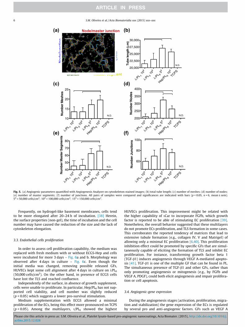
The number of bilayers forming the nanocoatings could alsoaffect the density of instructive proteins presented to the cells.Thereby, the cell seeding density of HUVECs and the number ofbilayers of the k/PL pair were also varied. Respectively, 50,000,100,000 and 150,000 cells/cm2 were seeded on k/PL6, and100,000 cells/cm2 seeded on nanocoatings prepared with 1, 3 and6 bilayers (k/PL1, k/PL3 and k/PL6) – Fig. 3. Total tube-length,
Please cite this article in press as: S.M. Oliveira et al., Platelet lysate-based pro-aactbio.2015.12.028
number of meshes, number of nodes and master junctions formedwere quantified on fluorescence images (5�) using AngiogenesisAnalyzer for Image J – Fig. 5. A single bilayer of k/PL was sufficientto promote the formation of a network with 100,000 cells, whichtotal tube length could be increased with increasing number oflayers. With 6 bilayers, a lower cell density has shown to be moreadequate in obtaining a better network formation than with fewerlayers. This indicates that cell adhesion and TLS are dependent onthe number of bilayers and increasing the number of layers allowsa decrease of the required cell density.
Besides the total tube length, the number of nodes and mesheswere also influenced by the number of layers. It was observed atendency of increase all the features with the increasing of thenumber of layers. In general, Hep/PL6 (100,000 cells) and k/PL6(50,000 cells) have shown similar results, though a higher numberof total nodes was observed on kCar.
Cell Profiler was used to analyze cell form factor (i.e., round-ness), eccentricity (i.e., elongation), major axis (i.e., cell length)and minor axis (i.e., cell width) – Fig. S3. This analysis revealed thatunder the tested conditions, the EC morphology was significantlychanged when seeded on the multilayers in comparison to TCPS.While on TCPS cells show the normal cobblestone-like morphol-ogy, they become rounder on the instructive multilayers (form fac-tor closer to 1). However, on the multilayers that successfullyinduced TLS, the cells elongation factor was similar to TCPS. Bothwidth and length decreased on the multilayers, thus not alteringsignificantly the elongation relatively to TCPS.
ngiogenic nanocoatings, Acta Biomater. (2015), http://dx.doi.org/10.1016/j.
Node/master junction
Nod
e/ju
nctio
n
mesh
master segmentbranch
400µm
/PL 1
104
/PL 3
104
/PL 6
54
Hep/P
L 6 1
04
TCPS20,000
22,500
25,000
27,500
30,000
Tu
be
len
gth
(µm
)
N.D.
/PL 1
104
/PL 3
104
/PL 6
54
Hep/P
L 6 1
04
TCPS
60
80
100
N.D.Nu
mb
er J
un
ctio
ns
/PL 1
104
/PL 3
104
/PL 6
54
Hep/P
L 6 1
04
TCPS40
50
60
70
80
N.D.
Nu
mb
er M
aste
r S
egm
ents
/PL 1
104
/PL 3
104
/PL 6
54
Hep/P
L 6 1
04
TCPS10
15
20
25
Nu
mb
er M
esh
es
N.D.
/PL 1
104
/PL 3
104
/PL 6
54
Hep/P
L 6 1
04
TCPS200
250
300
350
400
N.D.Nu
mb
er N
od
es
(a) (b)
(c) (d)
(e) (f)
Fig. 5. (a) Angiogenic parameters quantified with Angiogenesis Analyzer on cytoskeleton-stained images; (b) total tube length; (c) number of meshes; (d) number of nodes;(e) number of master segments; (f) number of junctions. All pairs of samples were compared and significances are indicated with bars (p < 0.05, n = 6, mean ± sem).54 = 50,000 cells/cm2; 104 = 100,000 cells/cm2; 154 = 150,000 cells/cm2.
6 S.M. Oliveira et al. / Acta Biomaterialia xxx (2015) xxx–xxx
Frequently, on hydrogel-like basement membranes, cells tendto be more elongated after 20–24 h of incubation. [38] Herein,the surface properties (non-gel), the time of incubation and the cellnumber may have caused the reduction of the size and the lack ofcytoskeleton elongation.
3.3. Endothelial cells proliferation
In order to assess cell proliferation capability, the medium wasreplaced with fresh medium with or without ECGS-Hep and cellswere incubated for more 3 days – Fig. 6a and b. Morphology wasobserved after 4 days in culture – Fig. 6c. Even though theinitial media was changed, removing possible released GFs,HUVECs kept some cell alignment after 4 days in culture on kPL6(50,000 cells/cm2). On the other hand, in presence of ECGS cellshave lost the TLS and reached confluence.
Independently of the surface, in absence of growth supplement,cells were unable to proliferate. In particular, Hep/PL6 has not sup-ported cell viability, and cell number was slightly reduced(p < 0.05) which suggests a lower pro-survival stimulation.
Medium supplementation with ECGS allowed a minimalproliferation of the ECs, being this effect more pronounced on TCPS(p < 0.05). Among the multilayers, i/PL6 showed the highest
Please cite this article in press as: S.M. Oliveira et al., Platelet lysate-based pro-aactbio.2015.12.028
HUVECs proliferation. This improvement might be related withthe higher capability of iCar to incorporate FGFb, which growthfactor is reported to be able of stimulating EC proliferation [39].Nonetheless, the overall behavior suggested that these multilayersdo not promote ECs proliferation, and TLS formation in some cases.This corroborates the reported tendency of matrices that lead toextensive tubule formation (e.g., collagen IV, V and Matrigel) ofallowing only a minimal EC proliferation [6,40]. This proliferationinhibition effect could be promoted by specific GFs that are simul-taneously capable of eliciting the formation of TLS and inhibit ECproliferation. For instance, transforming growth factor beta 1(TGF-b1) induces angiogenesis through VEGF A-mediated apopto-sis [41]. TGF-b1 is one of the multiple GF that can be found in PL.The simultaneous presence of TGF-b1 and other GFs, rather thanonly promoting angiogenesis or mitogenesis (e.g., by FGFb andVEGF A, PDGF), could both elicit angiogenesis and impair prolifera-tion or cell apoptosis.
3.4. Angiogenic-gene expression
During the angiogenesis stages (activation, proliferation, migra-tion and stabilization) the gene expression of the ECs is regulatedby several pro and anti-angiogenic factors. GFs such as VEGF A
ngiogenic nanocoatings, Acta Biomater. (2015), http://dx.doi.org/10.1016/j.

(a) (b) (c)
Fig. 6. Proliferation of HUVECs cultured with 10% FBS and in absence (a), or presence of ECGS-hep (b), after adhesion for 20 h (in absence of ECGS-hep and with 10% FBS). Allsignificances are indicate with: ⁄ (different to TCPS), # (different to all, after 4 days), (different to Chi/PL and Alg/PL) and bars (pair is different), (p < 0.05, n = 6; mean ± sem).(c) HUVECs (k/PL6 50,000 cells/cm2) morphology after 4 days in culture showing some remaining TLS when cultured in absence of ECGS-hep (10%FBS), while in presence ofECGS-hep (10%FBS) cells had disassembled, proliferated and reached confluence.
S.M. Oliveira et al. / Acta Biomaterialia xxx (2015) xxx–xxx 7
and FGFb activate ECs and promote their proliferation. Integrinsuch as a5, am, b3 play important roles during EC migration whileangiopoietin-1, PDGF and TGF-b regulate maturation and vesselsstabilization [8,42,43]. Herein, gene expression of VEGFA, FGFb,integrins (a5, am and b3) and angiopoietin-1 were quantified after20 h of culture – Fig. 7.
In accordance with the literature, the angiogenic-gene expres-sion alterations during TLS formation are expected to be of smallmagnitude (<2-fold) or even negative, as related to TCPS [42,44].Indeed, for the majority of conditions, gene expression was similaror significantly lower than TCPS with the exception of theexpression of VEGFA and angiopoietin-1. Regarding the expressionof integrins, k/PL6 (50,000 cells), k/PL3 (100,000 cells), i/PL6(100,000 cells) and Hep/PL6 (100,000 cells) have, in general, shownlower or similar expression to TCPS (p < 0.05). Exogenous FGFb isknown to be able to promote angiogenesis, both in vivo and in vitro,by up-regulating the expression of VEGFA and the endogenousVEGFA in ECs [45]. However, the expression of FGFb was decreasedon i/PL6 (100,000 cells), k/PL3 (100,000 cells), k/PL6 (50,000 cells),k/PL6 (100,000 cells), with exception of Hep/PL6.
0.0
0.5
1.0
1.5
* *
Inte
Fold
var
iatio
n
0.00
0.25
0.50
0.75
1.00
1.25
1.50
* *
*
Integrin
Fold
var
iatio
n
0.0
0.5
1.0
1.5
2.0
2.5
**
Fold
var
iatio
n
0
1
2
3
4*
** *
* *
*
*P=0.066
Angiopoietin-1
Fold
var
iatio
n
Alg/PL 6 1
04
Car/P
L 6 104
Car/P
L 1 104
Car/P
L 3 104
Car/P
L 6 54
Car/P
L 6 104
Car/P
L 6 154
Hep/P
L 6 104
TCPS
Alg/PL 6 1
04
Car/P
L 6 104
Car/P
L 1 104
Car/P
L 3 104
Car/
Alg/PL 6 1
04
Car/P
L 6 104
Car/P
L 1 104
Car/P
L 3 104
Car/P
L 6 54
Car/P
L 6 104
Car/P
L 6 154
Hep/P
L 6 104
TCPS
Alg/PL 6 1
04
Car/P
L 6 104
Car/P
L 1 104
Car/P
L 3 104
Car/P
VE
Fig. 7. Gene expression-fold variation of Angiopoietin-1, VEGF A, FGFb and integrins am,housekeeping b-actin gene or GAPHD (in case of VEGFA) and calculated by the Livak meidentified with ⁄ (p < 0.05, n = 8, mean ± sem).
Please cite this article in press as: S.M. Oliveira et al., Platelet lysate-based pro-aactbio.2015.12.028
Both VEGFA and angiopoietin-1 are strong pro-angiogenicfactors with distinct functions and bidirectional dependent: oneup-regulates the other. While VEGFA causes vascular permeability,angiopoietin-1 stabilizes the blood vessels and avoids plasma leak-age induced by VEGFA [46,47]. Although being frequently relatedwith different angiogenesis stages, the simultaneously stimulationof ECs with VEGFA and angiopoietin-1 has previously shown asynergistic improvement of angiogenesis [48].
Recently, it has been reported that PRP contains high amounts ofangiopoietin-1 (�300fold more than VEGFA) [16]. The same studyhas shown that angiopoietin-1 and its respective cell receptor(Tie2) are crucial in promoting angiogenesis when using a prepara-tion of 250-fold diluted PRP.
Both the expression of VEGF A and angiopoietin-1 weresimultaneously increased with exception of TCPS, Alg/PL6 and i/PL6. Angiopoietin-1 was increased even on the multilayers notpromoting TLS which suggests that surface VEGFA or FGFb (whichprimarily up-regulates VEGFA [45]) might have up-regulatedangiopoietin-1 [49]. If a significant amount of angiopoietin-1 hadbeen incorporated in the coatings, this could up-regulate VEGFA
*
grin v
0.0
0.5
1.0
1.5
**
**
Fold
var
iatio
n
**
**
0.0
0.5
1.0
1.5 *
*
* *
*
Integrin
Fold
var
iatio
n
PL 6 54
Car/P
L 6 104
Car/P
L 6 154
Hep/P
L 6 104
TCPS
Alg/PL 6 1
04
Car/P
L 6 104
Car/P
L 1 104
Car/P
L 3 104
Car/P
L 6 54
Car/P
L 6 104
Car/P
L 6 154
Hep/P
L 6 104
TCPS
L 6 54
Car/P
L 6 104
Car/P
L 6 154
Hep/P
L 6 104
TCPS
Alg/PL 6 1
04
Car/P
L 6 104
Car/P
L 1 104
Car/P
L 3 104
Car/P
L 6 54
Car/P
L 6 104
Car/P
L 6 154
Hep/P
L 6 104
TCPS
GF A FGFb
a5, b3 relatively to TCPS. The expression of theses genes was normalized against thethod (2�DDCt). Samples were compared with the control (TPCS) and differences are
ngiogenic nanocoatings, Acta Biomater. (2015), http://dx.doi.org/10.1016/j.

Fig. 8. Cell morphology of HUVECs seeded on k/PL6 and TPCs in presence of VEGF/FGF receptor kinase inhibitor (or DMSO) inhibiting the formation of TLS. (cytoskeleton:orange; nuclei: blue). (For interpretation of the references to colour in this figure legend, the reader is referred to the web version of this article.)
8 S.M. Oliveira et al. / Acta Biomaterialia xxx (2015) xxx–xxx
expression and VEGFA endogenous content, and consequently,indirectly stimulate the formation of TLS [48,50,51].
3.5. Blockage of FGF/VEGF tyrosine kinase receptor
HUVECs were cultured in the presence of a FGF/VEGF tyrosinekinase receptor inhibitor to understand whether the morphogenicchanges were driven by those pro-angiogenic GFs. This compoundcould block the interaction of FGF and VEGF from the nanocoating,or the soluble form, with their respective cell receptors (e.g., FGFR2and VEGFR2). A condition that was shown to induce TLS wasselected: 50,000 cells/cm2 onto k/PL6. Cells were seeded with asupplier’s recommended range of concentrations of inhibitor dis-solved with DMSO (150 nM and 200 nM, containing 0.0075% v/vand 0.01% v/v DMSO, respectively) or only with DMSO (0.0075%v/v and 0.01% v/v of DMSO – named DMSO 150 and DMSO 200)during 20 h. The use of DMSO as a solvent has diminished theobserved cellular density and made cells more elongated – Fig. 8.However, the ability of HUVECS to be more cohesive and to alignwas further reduced with the presence of the inhibitor and withits increased concentration. No TLS were observed on the surfaces.Cell Profiler analysis has not revealed significant cell shape differ-ences between k/PL6 (50,000 cells) with inhibitor and only withDMSO, both for 150 nM and for 200 nM – Fig. S3. DMSO is reportedas being able to decrease cell adhesion even at low concentrations(1.55% v/v) [52]. Concentrations higher than 1% v/v have also beenreported to impair the formation of TLS on Matrigel [53]. Thereby,one cannot exclude the inhibition of TLS and changes in cellmorphology to be in part caused by DMSO. Nonetheless, thereare indications that at least at some extent, FGF/VEGF hasmediated the formation of TLS.
The PL coatings assembled with the more sulfated polysacchar-ides were able to induce a pro-angiogenic profile in HUVECs. Thisstrategy may be a cost-effective way to introduce pro-angiogenicinterfaces on surfaces or 3D scaffolds.
4. Conclusions
There is still a current need to develop cost effective cell-interfaces able to promote angiogenesis and the formation of stablevasculature.
PL is a source of several pro-angiogenic and other proteinsinvolved in the angiogenesis from the earliest to the maturationphases. Herein, PL was incorporated in layer-by-layer assemblednanocoatings with varied polysaccharides and number of layers.
Please cite this article in press as: S.M. Oliveira et al., Platelet lysate-based pro-aactbio.2015.12.028
The nanocoatings prepared with the more sulfated polysaccharideselicited the formation of tube-like structures in ECs within 20 h ofincubation. These morphogenic changes were accompanied by dif-ferences in gene expressions, mainly higher VEGFA and higherangiopoietin-1. Future work must be performed in order to clarifythe rule of PL as well as the in vivo behavior of the developedstructures.
Layer-by-Layer assembling including PL might be a simplemethodology to introduce and tune cost-effective pro-angiogenicinterfaces in 2D/3D biomaterials.
Acknowledgments
The research leading to these results has received funding fromEuropean Union’s Seventh Framework Program (FP7/2007-2013)under grant agreement nª REGPOT-CT2012-316331 – POLARISand FP7-KBBE-2010-4-266033 – SPECIAL. This work was alsosupported by the European Research Council grant agreementERC-2012-ADG-20120216-321266 for the project ComplexiTE.Portuguese Foundation for Science and Technology is gratefullyacknowledged for fellowship of Sara M. Oliveira (SFRH/BD/70107/2010). The researcher contract of R.P. Pirraco through RL3-TECT-NORTE-01-0124-FEDER-000020, co-financed by North PortugalRegional Operational Program (ON.2-O Novo Norte), under theNational Strategic Reference Framework, through the EuropeanRegional Development Fund is also acknowledged.
Appendix A. Supplementary data
Supplementary data associated with this article can be found, inthe online version, at http://dx.doi.org/10.1016/j.actbio.2015.12.028.
References
[1] F.A. Auger, L. Gibot, D. Lacroix, The pivotal role of vascularization in tissueengineering, Annu. Rev. Biomed. Eng. 15 (2013) 177–200.
[2] E.C. Novosel, C. Kleinhans, P.J. Kluger, Vascularization is the key challenge intissue engineering, Adv. Drug Delivery Rev. 63 (2011) 300–311.
[3] M.I. Santos, R.L. Reis, Vascularization in bone tissue engineering: physiology,current strategies, major hurdles and future challenges, Macromol. Biosci. 10(2010) 12–27.
[4] Y.J. Blinder, D.J. Mooney, S. Levenberg, Engineering approaches for inducingblood vessel formation, Curr. Opin. Chem. Eng. 3 (2014) 56–61.
[5] L.E. Bertassoni, M. Cecconi, V. Manoharan, M. Nikkhah, J. Hjortnaes, A.L.Cristino, G. Barabaschi, D. Demarchi, M.R. Dokmeci, Y. Yang, A. Khademhosseini,Hydrogel bioprinted microchannel networks for vascularization of tissueengineering constructs, Lab Chip 14 (2014) 2202–2211.
ngiogenic nanocoatings, Acta Biomater. (2015), http://dx.doi.org/10.1016/j.

S.M. Oliveira et al. / Acta Biomaterialia xxx (2015) xxx–xxx 9
[6] C.A. Staton, M.W. Reed, N.J. Brown, A critical analysis of current in vitro and invivo angiogenesis assays, Int. J. Exp. Pathol. 90 (2009) 195–221.
[7] M.W. Irvin, A. Zijlstra, J.P. Wikswo, A. Pozzi, Techniques and assays for thestudy of angiogenesis, Exp. Biol. Med. (Maywood) 239 (2014) 1476–1488.
[8] X.M. vanWijk, T.H. van Kuppevelt, Heparan sulfate in angiogenesis: a target fortherapy, Angiogenesis 17 (2014) 443–462.
[9] M. Matsui, Y. Tabata, Enhanced angiogenesis by multiple release of platelet-rich plasma contents and basic fibroblast growth factor from gelatin hydrogels,Acta Biomater. 8 (2012) 1792–1801.
[10] G. Sufen, Y. Xianghong, C. Yongxia, P. Qian, bFGF and PDGF-BB have asynergistic effect on the proliferation, migration and VEGF release ofendothelial progenitor cells, Cell Biol. Int. 35 (2011) 545–551.
[11] G. Sun, Y.I. Shen, S. Kusuma, K. Fox-Talbot, C.J. Steenbergen, S. Gerecht,Functional neovascularization of biodegradable dextran hydrogels withmultiple angiogenic growth factors, Biomaterials 32 (2011) 95–106.
[12] Q. Sun, E.A. Silva, A. Wang, J.C. Fritton, D.J. Mooney, M.B. Schaffler, P.M.Grossman, S. Rajagopalan, Sustained release of multiple growth factors frominjectable polymeric system as a novel therapeutic approach towardsangiogenesis, Pharm. Res. 27 (2010) 264–271.
[13] K. Stellos, S. Kopf, A. Paul, J.U. Marquardt, M. Gawaz, J. Huard, H.F. Langer,Platelets in Regeneration. Seminars in Thrombosis and Hemostasis, ThiemeMedical Publishers, 2010. p. 175–84.
[14] S.C. Bir, J. Esaki, A. Marui, K. Yamahara, H. Tsubota, T. Ikeda, R. Sakata,Angiogenic properties of sustained release platelet-rich plasma:characterization in-vitro and in the ischemic hind limb of the mouse, J. Vasc.Surg. 50 (2009) 870–879. e2.
[15] N. Kakudo, N. Morimoto, S. Kushida, T. Ogawa, K. Kusumoto, Platelet-richplasma releasate promotes angiogenesis in vitro and in vivo, Med. Mol.Morphol. 47 (2014) 83–89.
[16] T. Mammoto, A. Jiang, E. Jiang, A. Mammoto, Platelet rich plasma extractpromotes angiogenesis through the angiopoietin1-Tie2 pathway, Microvasc.Res. 89 (2013) 15–24.
[17] B. Zhou, J. Ren, C. Ding, Y. Wu, D. Hu, G. Gu, J. Li, Rapidly in situ formingplatelet-rich plasma gel enhances angiogenic responses and augments earlywound healing after open abdomen, Gastroenterol. Res. Pract. 2013 (2013).926764.
[18] Y. Man, P. Wang, Y. Guo, L. Xiang, Y. Yang, Y. Qu, P. Gong, L. Deng, Angiogenicand osteogenic potential of platelet-rich plasma and adipose-derived stem cellladen alginate microspheres, Biomaterials 33 (2012) 8802–8811.
[19] F. Findikcioglu, K. Findikcioglu, R. Yavuzer, N. Lortlar, K. Atabay, Effect ofpreoperative subcutaneous platelet-rich plasma and fibrin glue application onskin flap survival, Aesthetic Plast. Surg. 36 (2012) 1246–1253.
[20] J. Leotot, L. Coquelin, G. Bodivit, P. Bierling, P. Hernigou, H. Rouard, N.Chevallier, Platelet lysate coating on scaffolds directly and indirectly enhancescell migration, improving bone and blood vessel formation, Acta Biomater. 9(2013) 6630–6640.
[21] E. Polykandriotis, A. Arkudas, R.E. Horch, U. Kneser, G. Mitchell, To matrigel ornot to matrigel, Am. J. Pathol. 172 (2008) 1441–1442.
[22] J. Borges, J.F. Mano, Molecular interactions driving the layer-by-layer assemblyof multilayers, Chem. Rev. 114 (2014) 8883–8942.
[23] R.R. Costa, J.F. Mano, Polyelectrolyte multilayered assemblies in biomedicaltechnologies, Chem. Soc. Rev. 43 (2014) 3453–3479.
[24] S.M. Oliveira, T.H. Silva, R.L. Reis, J.F. Mano, Nanocoatings containing sulfatedpolysaccharides prepared by layer-by-layer assembly as models to study cell-material interactions, J. Mater. Chem. B 1 (2013) 4406–4418.
[25] K. Gaengel, C. Betsholtz, Endocytosis regulates VEGF signalling duringangiogenesis, Nat. Cell Biol. 15 (2013) 233–235.
[26] S.M. Oliveira, V.E. Santo, M.E. Gomes, R.L. Reis, J.F. Mano, Layer-by-layerassembled cell instructive nanocoatings containing platelet lysate,Biomaterials 48 (2015) 56–65.
[27] J. Kreuger, D. Spillmann, J.P. Li, U. Lindahl, Interactions between heparansulfate and proteins: the concept of specificity, J. Cell Biol. 174 (2006) 323–327.
[28] G.S. Schultz, A. Wysocki, Interactions between extracellular matrix and growthfactors in wound healing, Wound Repair Regen. 17 (2009) 153–162.
[29] S.K. Nigam, K.T. Bush, Growth factor-heparan sulfate ‘‘switches” regulatingstages of branching morphogenesis, Pediatr. Nephrol. 29 (2014) 727–735.
[30] T.H. Nguyen, S.H. Kim, C.G. Decker, D.Y. Wong, J.A. Loo, H.D. Maynard, Aheparin-mimicking polymer conjugate stabilizes basic fibroblast growthfactor, Nat. Chem. 5 (2013) 221–227.
[31] T.N. Vo, F.K. Kasper, A.G. Mikos, Strategies for controlled delivery of growthfactors and cells for bone regeneration, Adv. Drug Delivery Rev. 64 (2012)1292–1309.
[32] T.H. Silva, A. Alves, B.M. Ferreira, J.M. Oliveira, L.L. Reys, R.J.F. Ferreira, R.A.Sousa, S.S. Silva, J.F. Mano, R.L. Reis, Materials of marine origin: a review on
Please cite this article in press as: S.M. Oliveira et al., Platelet lysate-based pro-aactbio.2015.12.028
polymers and ceramics of biomedical interest, Int. Mater. Rev. 57 (2012) 276–307.
[33] K. Senni, J. Pereira, F. Gueniche, C. Delbarre-Ladrat, C. Sinquin, J. Ratiskol, G.Godeau, A.M. Fischer, D. Helley, S. Colliec-Jouault, Marine polysaccharides: asource of bioactive molecules for cell therapy and tissue engineering, Mar.Drugs 9 (2011) 1664–1681.
[34] J.F. Mano, G.A. Silva, H.S. Azevedo, P.B. Malafaya, R.A. Sousa, S.S. Silva, L.F.Boesel, J.M. Oliveira, T.C. Santos, A.P. Marques, N.M. Neves, R.L. Reis, Naturalorigin biodegradable systems in tissue engineering and regenerativemedicine: present status and some moving trends, J. R. Soc. Interface 4(2007) 999–1030.
[35] S.M. Oliveira, R.L. Reis, J.F. Mano, Assembling human platelet lysate intomultiscale 3D scaffolds for bone tissue engineering, ACS Biomater. Sci. Eng. 1(2015) 2–6.
[36] J.F. Mano, Designing biomaterials for tissue engineering based on thedeconstruction of the native cellular environment, Mater. Lett. 141 (2015)198–202.
[37] M.G. Lampugnani, F. Orsenigo, M.C. Gagliani, C. Tacchetti, E. Dejana, Vascularendothelial cadherin controls VEGFR-2 internalization and signaling fromintracellular compartments, J. Cell Biol. 174 (2006) 593–604.
[38] P.J. Stahl, T.R. Chan, Y.I. Shen, G. Sun, S. Gerecht, S. Yu, Capillary network-likeorganization of endothelial cells in PEGDA scaffolds encoded with angiogenicsignals via triple helical hybridization, Adv. Funct. Mater. 24 (2014) 3213–3225.
[39] A. Sahni, C.W. Francis, Stimulation of endothelial cell proliferation by FGF-2 inthe presence of fibrinogen requires avb32004, Blood 104 (2004) 3635–3641.
[40] J.A. Madri, S.K. Williams, Capillary endothelial cell cultures: phenotypicmodulation by matrix components, J. Cell Biol. 97 (1983) 153–165.
[41] G. Ferrari, B.D. Cook, V. Terushkin, G. Pintucci, P. Mignatti, Transforminggrowth factor-beta 1 (TGF-b1) induces angiogenesis through vascularendothelial growth factor (VEGF)-mediated apoptosis, J. Cell. Physiol. 219(2009) 449–458.
[42] R. Mammadov, B. Mammadov, S. Toksoz, B. Aydin, R. Yagci, A.B. Tekinay, M.O.Guler, Heparin mimetic peptide nanofibers promote angiogenesis,Biomacromolecules 12 (2011) 3508–3519.
[43] K.P. Claffey, Molecular profiling of angiogenic markers: a step towardsinterpretive analysis of a complex biological function, Am. J. Pathol. 161(2002) 7–11.
[44] A.D. Grove, V.V. Prabhu, B.L. Young, F.C. Lee, V. Kulpa, P.J. Munson, E.C. Kohn,Both protein activation and gene expression are involved in early vasculartube formation in vitro, Clin. Cancer Res. 8 (2002) 3019–3026.
[45] G. Seghezzi, S. Patel, C.J. Ren, A. Gualandris, G. Pintucci, E.S. Robbins, R.L.Shapiro, A.C. Galloway, D.B. Rifkin, P. Mignatti, Fibroblast growth factor-2(FGF-2) induces vascular endothelial growth factor (VEGF) expression in theendothelial cells of forming capillaries: an autocrine mechanism contributingto angiogenesis, J. Cell Biol. 141 (1998) 1659–1673.
[46] J. Gavard, V. Patel, J.S. Gutkind, Angiopoietin-1 prevents VEGF-inducedendothelial permeability by sequestering Src through mDia, Dev. Cell 14(2008) 25–36.
[47] S.P. Ngok, R. Geyer, M. Liu, A. Kourtidis, S. Agrawal, C. Wu, H.R. Seerapu, L.J.Lewis-Tuffin, K.L. Moodie, D. Huveldt, R. Marx, J.M. Baraban, P. Storz, A.Horowitz, P.Z. Anastasiadis, VEGF and Angiopoietin-1 exert opposing effectson cell junctions by regulating the Rho GEF Syx, J. Cell Biol. 199 (2012) 1103–1115.
[48] T.I. Koblizek, C. Weiss, G.D. Yancopoulos, U. Deutsch, W. Risau, Angiopoietin-1induces sprouting angiogenesis in vitro, Curr. Biol. 8 (1998) 529–532.
[49] M. Hangai, T. Murata, N. Miyawaki, C. Spee, J.I. Lim, S. He, D.R. Hinton, S.J. Ryan,Angiopoietin-1 upregulation by vascular endothelial growth factor in humanretinal pigment epithelial cells, Invest. Ophthalmol. Vis. Sci. 42 (2001) 1617–1625.
[50] W.H. Zhu, A. MacIntyre, R.F. Nicosia, Regulation of angiogenesis by vascularendothelial growth factor and angiopoietin-1 in the rat aorta model: distincttemporal patterns of intracellular signaling correlate with induction ofangiogenic sprouting, Am. J. Pathol. 161 (2002) 823–830.
[51] S. Kanda, H. Kanetake, Y. Miyata, Role of Src in angiopoietin 1-inducedcapillary morphogenesis of endothelial cells: Effect of chronic hypoxia on Srcinhibition by PP2, Cell. Signal. 19 (2007) 472–480.
[52] N. Eter, M. Spitznas, DMSO mimics inhibitory effect of thalidomide onchoriocapillary endothelial cell proliferation in culture, Br. J. Ophthalmol. 86(2002) 1303–1305.
[53] K. Koizumi, Y. Tsutsumi, Y. Yoshioka, M. Watanabe, T. Okamoto, Y. Mukai, S.Nakagawa, T. Mayumi, Anti-angiogenic effects of dimethyl sulfoxide onendothelial cells, Biol. Pharm. Bull. 26 (2003) 1295–1298.
ngiogenic nanocoatings, Acta Biomater. (2015), http://dx.doi.org/10.1016/j.