platelet indices in pregnancy induced hypertension · pdf fileplatelet indices in pregnancy...
TRANSCRIPT
Bhavana Thakur et al; Platelet indices in PIH
J Cont Med A Dent September-December 2016 Volume 4 Issue 3 20
ORIGINAL ARTICLE
Platelet Indices in Pregnancy Induced Hypertension
Thakur Bhavana 1, Kulkarni Vishal 2, Thakur Prashant 3
1. Assistant Professor, Department of Pathology, Pt Jawaharlal Nehru Medical College, Raipur Chhattisgarh. 2. Associate Professor, Department of Pathology, Government Medical College, Rajnandgaon. Chhattisgarh.
3. Consultant Pediatric Cardiologist, Ramkrishna Care Hospital, Raipur, Chhattisgarh.
Abstract Preeclampsia is a syndrome with both maternal and fetal manifestations. Haematological abnormalities such as thrombocytopenia and decrease in some plasma clotting factors may develop in pre-eclamptic women. The platelet count has an association at prediction of increasing grade of pregnancy induced hypertension (PIH). There is an inverse relationship between the severity of PIH and platelet count. The platelet indices of Mean Platelet Volume (MPV) and platelet distribution width (PDW) too are in consistent relationship with PIH. The greater MPV values suggest the increase grade of PIH of preeclampsia, severe preeclampsia and eclampsia. PDW too can suggest the PIH for its severity especially in the groups of preeclampsia, severe preeclampsia and eclampsia and the risk of consumptive coagulopathy. Thus, investigations with baseline complete blood cell count including platelet count and platelet indices is necessary in patients with a hypertensive disorder of pregnancy. Keywords: Pregnancy Induces Hypertension, Platelet Count, Platelet Indices
Address for correspondence: Dr. Bhavana Thakur, C-129/5, Near Devi Lakshmi Hospital, Tagore Nagar, Raipur, Chhattisgarh. Email: [email protected]
DOI:10.18049/jcmad/332 Received on :25/10/2016 Revised :31/10/2016 Accepted : 03/11/2016
Introduction
The most serious consequence for the mother and baby result from preeclampsia and eclampsia. These are associated with vasospasm, pathologic vascular lesions in multiple organ system, increased platelet activation and subsequent activation of coagulation system in the microvasculature.1
Hypercoagulability is a constant accompaniment of hypertensive disease of pregnancy and particularly pre-eclampsia.2Haematological abnormalities such as thrombocytopenia and decrease in some plasma clotting factors may develop in pre-eclamptic women. Thus, coagulation testing is common in these patients for evidence of DIC and HELLP (Hemolysis, Elevated Liver Enzymes, Low Platelets) syndrome.3 Preeclampsia is a syndrome with both maternal and fetal manifestations,4 the pathogenesis of which lie vasospasm, pathologic vascular lesions in multiple organ system, increased
platelet activation and subsequent activation of coagulation system in the microvasculature.1
In the recent years interest has also been focused in the haemostatic abnormalities that are associated with many pregnancy-related disorders including hypertensive disease complicating pregnancy.2 The major adverse outcomes of pre-eclampsia and eclampsia include central nervous system injuries such as seizures (eclampsia), ischemic heart disease, stroke, type II diabetes, and venous thromboembolism hemorrhagic and ischemic strokes, hepatic damage, HELLP syndrome, renal dysfunction as well as increased frequency of cesarean delivery, preterm delivery, and abruptio placentae, in comparison with women without history of the disease.5 Thus, coagulation testing with baseline complete blood cell count including platelet count and platelet indices probably sufficient in patients with a hypertensive disorder of pregnancy is common in these patients for evidence of DIC and HELLP.3, 6
Bhavana Thakur et al; Platelet indices in PIH
J Cont Med A Dent September-December 2016 Volume 4 Issue 3 21
AIM & Objectives
To study Platelet count, Platelet indices in cases of Pregnancy induced hypertension and to correlate and compare the values with Normotensive pregnant subjects.
Materials & Methods
The present prospective case control study was carried out in the Department of Pathology of Jawaharlal Nehru Medical College and Acharya Vinoba Bhave Rural Hospital at Datta Meghe Institute of Medical Sciences, Sawangi (Meghe), Wardha from August 2011 to July 2013. The present study comprised pregnant women admitted in Obstetrics and Gynaecology Department of the hospital. The preliminary data in regards to name, age, sex, registration number, obstetric, menstrual, and family history, general and systemic examination and investigations were recorded in a proforma after getting informed consent from the patients. The study was been conducted on two groups of pregnant women: Group I: Control: 50 normal healthy women in the 2nd and 3rd trimester of pregnancy. Group II: Included 150 pregnant women which were further divided into subgroups of: 1) Gestational hypertension, 2) Preeclampsia,3) Severe preeclampsia and 4) Eclampsia. Normal healthy women who developed hypertension for the first time during pregnancy after 20 weeks of gestation were included in PIH category.7. The further categorization was done according to following diagnostic criteria: 1. Gestational hypertension4- It is defined by
the blood pressure elevation of greater than 140 mm Hg systolic or 90 mm Hg diastolic in a previously normotensive women for the first time after mid pregnancy, but in whom proteinuria is not identified.
2. Preeclampsia4 - It is defined by hypertension (blood pressure greater than 140 mm Hg systolic or 90 mm Hg diastolic) associated with proteinuria > 0.3g/l in a 24 hour urine collection or 1+ dipstick or greater in random urine collection, after 20
weeks of gestation in a previously normotensive women.
3. Severe Preeclampsia8 – This condition was categorised if systolic blood pressure was >160 mm Hg and diastolic blood pressure> 110 mm Hg.
4. Eclampsia4 - The onset of convulsions in women with pre-eclampsia that cannot be attributed to other causes is termed as eclampsia.
Subjects with haemorrhagic disorders, Sepsis, functional uterine bleeding, placental abruption or previa, diabetes, respiratory, circulatory, renal and hepatic disorders, known cases of hypertension and subjects taking drugs which can affect platelet count were excluded from the study. Hemoglobin estimation, platelet count was done by Automated Haematologyanalyser, Sysmex Corp. KX.21, Japan. (Normal range of platelet count was considered to be1.5-4.0 lacs/mm3). Platelet indices like Mean platelet volume (MPV) and platelet distribution width (PDW) were estimated with reference range of the laboratory for MPV being 6.5-11.0 fl and for PDW being 10.0-18.0 fl. The peripheral blood smear (PS) of the cases were stained by Leishman’s stain. Platelets were studied for their adequacy and morphology. The results were analysed statistically to draw comparison between the Groups. The statistical data was processed using Microsoft Excel to draw the values of significance for Group II and its subgroups. Tests of significance applied were Chi square and ‘Z’ test for statistical analysis to suggest the relation between the observed abnormal value of chosen laboratory tests in the study with hypertensive disorders of pregnancy and its importance in antenatal care.
Results
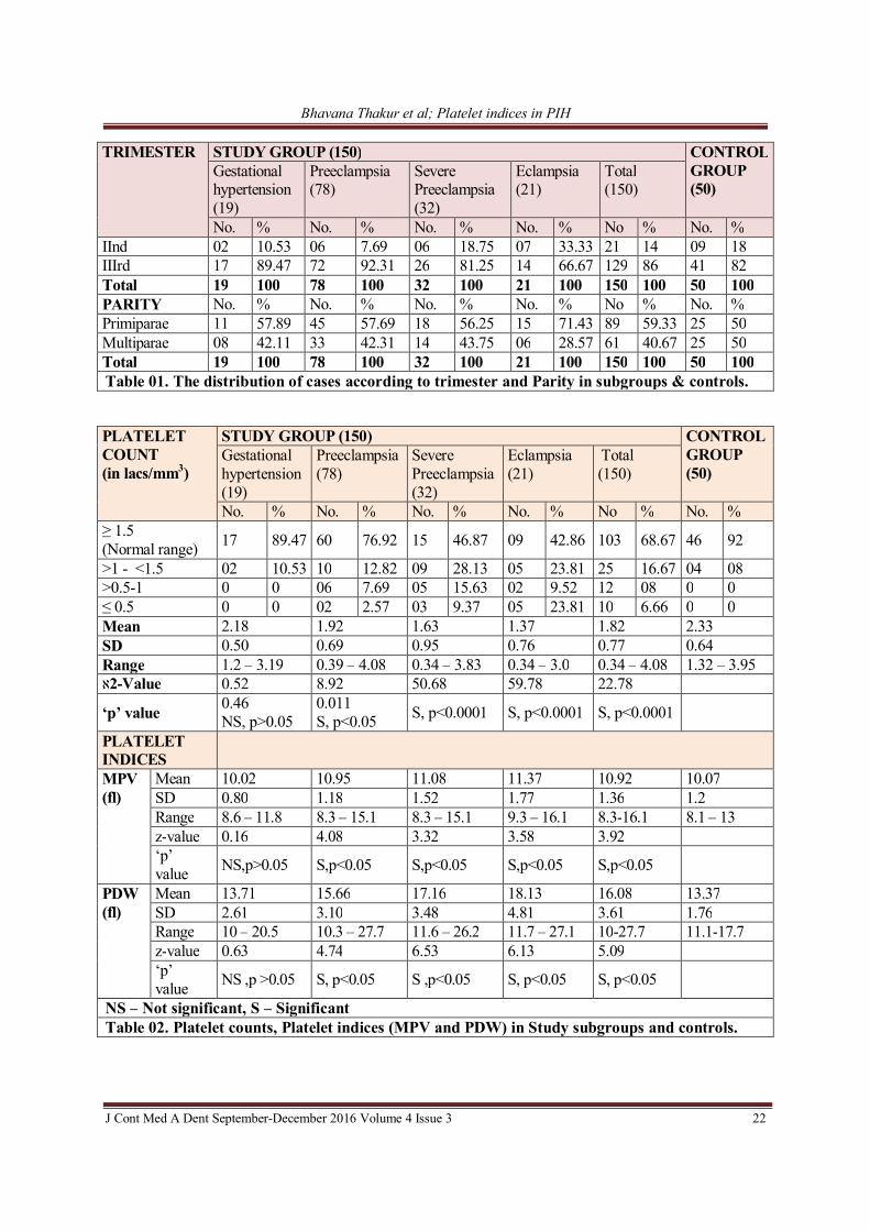
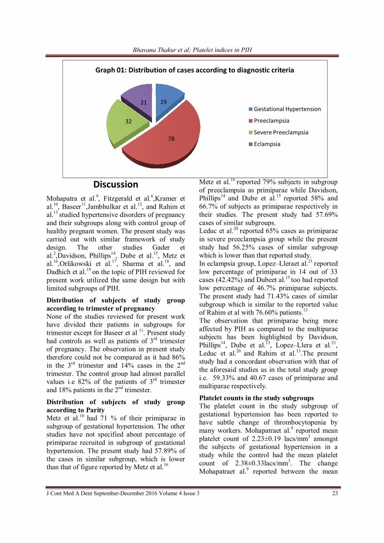
The distribution of cases in subgroups according to diagnostic criteria is depicted in Graph 01. Table 01 shows the distribution of cases according to trimester and Parity in subgroups & controls. The platelet counts and indices according to the subgroups is depicted in Table No. 02.
Bhavana Thakur et al; Platelet indices in PIH
J Cont Med A Dent September-December 2016 Volume 4 Issue 3 22
TRIMESTER STUDY GROUP (150) CONTROL GROUP (50)
Gestational hypertension (19)
Preeclampsia (78)
Severe Preeclampsia (32)
Eclampsia (21)
Total (150)
No. % No. % No. % No. % No % No. % IInd 02 10.53 06 7.69 06 18.75 07 33.33 21 14 09 18 IIIrd 17 89.47 72 92.31 26 81.25 14 66.67 129 86 41 82 Total 19 100 78 100 32 100 21 100 150 100 50 100 PARITY No. % No. % No. % No. % No % No. % Primiparae 11 57.89 45 57.69 18 56.25 15 71.43 89 59.33 25 50 Multiparae 08 42.11 33 42.31 14 43.75 06 28.57 61 40.67 25 50 Total 19 100 78 100 32 100 21 100 150 100 50 100 Table 01. The distribution of cases according to trimester and Parity in subgroups & controls.
PLATELET COUNT (in lacs/mm3)
STUDY GROUP (150) CONTROL GROUP (50)
Gestational hypertension (19)
Preeclampsia (78)
Severe Preeclampsia (32)
Eclampsia (21)
Total (150)
No. % No. % No. % No. % No % No. % ≥ 1.5 (Normal range) 17 89.47 60 76.92 15 46.87 09 42.86 103 68.67 46 92
>1 - <1.5 02 10.53 10 12.82 09 28.13 05 23.81 25 16.67 04 08 >0.5-1 0 0 06 7.69 05 15.63 02 9.52 12 08 0 0 ≤ 0.5 0 0 02 2.57 03 9.37 05 23.81 10 6.66 0 0 Mean 2.18 1.92 1.63 1.37 1.82 2.33 SD 0.50 0.69 0.95 0.76 0.77 0.64 Range 1.2 – 3.19 0.39 – 4.08 0.34 – 3.83 0.34 – 3.0 0.34 – 4.08 1.32 – 3.95 Value 0.52 8.92 50.68 59.78 22.78-2א
‘p’ value 0.46 NS, p>0.05
0.011 S, p<0.05 S, p<0.0001 S, p<0.0001 S, p<0.0001
PLATELET INDICES
MPV (fl)
Mean 10.02 10.95 11.08 11.37 10.92 10.07 SD 0.80 1.18 1.52 1.77 1.36 1.2 Range 8.6 – 11.8 8.3 – 15.1 8.3 – 15.1 9.3 – 16.1 8.3-16.1 8.1 – 13 z-value 0.16 4.08 3.32 3.58 3.92 ‘p’ value NS,p>0.05 S,p<0.05 S,p<0.05 S,p<0.05 S,p<0.05
PDW (fl)
Mean 13.71 15.66 17.16 18.13 16.08 13.37 SD 2.61 3.10 3.48 4.81 3.61 1.76 Range 10 – 20.5 10.3 – 27.7 11.6 – 26.2 11.7 – 27.1 10-27.7 11.1-17.7 z-value 0.63 4.74 6.53 6.13 5.09 ‘p’ value NS ,p >0.05 S, p<0.05 S ,p<0.05 S, p<0.05 S, p<0.05
NS – Not significant, S – Significant Table 02. Platelet counts, Platelet indices (MPV and PDW) in Study subgroups and controls.
Bhavana Thakur et al; Platelet indices in PIH
J Cont Med A Dent September-December 2016 Volume 4 Issue 3 23
Discussion
Mohapatra et al.9, Fitzgerald et al.6,Kramer et al.10, Baseer11,Jambhulkar et al.12, and Rahim et al.13 studied hypertensive disorders of pregnancy and their subgroups along with control group of healthy pregnant women. The present study was carried out with similar framework of study design. The other studies Gader et al.2,Davidson, Phillips14, Dube et al.15, Metz et al.16,Orlikowski et al.17, Sharma et al.18, and Dadhich et al.19 on the topic of PIH reviewed for present work utilized the same design but with limited subgroups of PIH.
Distribution of subjects of study group according to trimester of pregnancy None of the studies reviewed for present work have divided their patients in subgroups for trimester except for Baseer et al 11. Present study had controls as well as patients of 3rd trimester of pregnancy. The observation in present study therefore could not be compared as it had 86% in the 3rd trimester and 14% cases in the 2nd trimester. The control group had almost parallel values i.e 82% of the patients of 3rd trimester and 18% patients in the 2nd trimester.
Distribution of subjects of study group according to Parity Metz et al.16 had 71 % of their primiparae in subgroup of gestational hypertension. The other studies have not specified about percentage of primiparae recruited in subgroup of gestational hypertension. The present study had 57.89% of the cases in similar subgroup, which is lower than that of figure reported by Metz et al.16
Metz et al.16 reported 79% subjects in subgroup of preeclampsia as primiparae while Davidson, Phillips14 and Dube et al.15 reported 58% and 66.7% of subjects as primiparae respectively in their studies. The present study had 57.69% cases of similar subgroups. Leduc et al.20 reported 65% cases as primiparae in severe preeclampsia group while the present study had 56.25% cases of similar subgroup which is lower than that reported study. In eclampsia group, Lopez–Lleraet al.21 reported low percentage of primiparae in 14 out of 33 cases (42.42%) and Dubeet al.15 too had reported low percentage of 46.7% primiparae subjects. The present study had 71.43% cases of similar subgroup which is similar to the reported value of Rahim et al with 76.60% patients.13 The observation that primiparae being more affected by PIH as compared to the multiparae subjects has been highlighted by Davidson, Phillips14, Dube et al.15, Lopez–Llera et al.21, Leduc et al.20 and Rahim et al.13.The present study had a concordant observation with that of the aforesaid studies as in the total study group i.e. 59.33% and 40.67 cases of primiparae and multiparae respectively.
Platelet counts in the study subgroups The platelet count in the study subgroup of gestational hypertension has been reported to have subtle change of thrombocytopenia by many workers. Mohapatraet al.9 reported mean platelet count of 2.23±0.19 lacs/mm3 amongst the subjects of gestational hypertension in a study while the control had the mean platelet count of 2.38±0.33lacs/mm3. The change Mohapatraet al.9 reported between the mean
19
78
32
21
Graph 01: Distribution of cases according to diagnostic criteria
Gestational Hypertension
Preeclampsia
Severe Preeclampsia
Eclampsia
Bhavana Thakur et al; Platelet indices in PIH
J Cont Med A Dent September-December 2016 Volume 4 Issue 3 24
platelet count of the control group and that of gestational hypertension was subtle. The similar findings as that of Mohapatraet al.9 have been observed in present study for the mean platelet count of 2.18±0.50 lacs/mm3 in gestational hypertension and 2.33±0.64 lacs/mm3 in control group with minimal difference between the means of two groups. Mohapatraet al.9 reported maximum cases having platelet counts over 1.5 lacs/mm3 and 2 cases with counts in range of 1-1.5 lacs/mm3. The present study observed similar trend that 17 out of 19 cases of gestational hypertension were having platelet count >1.5 lacs/mm3 and only 2 cases had counts in the range of >1-<1.5 lacs/mm3. There was no case in the study of Mohapatraet al 9 as well as in the present study in gestational hypertension subgroup that had platelet count <1 lac/mm3. Though, Mohapatra et al 9 reported significant ‘p’ value for platelet counts in gestational hypertension, the present study found non-significant ‘p’ value in subgroup of gestational hypertension when compared with controls. Several studies have reported the mean platelet counts less than 2 lacs/mm3 in subgroup of preeclampsia which include studies of Mohapatraet al.9 (1.82±0.45 lacs/mm3), Dube et al.15(1.95±0.61lacs/mm3), Annam et al.22
(1.55±0.31/mm3) and Sultana et al.23 (1.44±0.96 lacs/mm3). But the mean platelet count in the studies of Davidson, Phillips14 (2.27 lacs/mm3) and Davies et al.8 (2.30±0.83 lacs/mm3) were over 2 lacs/mm3 but less than 2.5 lacs/mm3. The present study had mean platelet count of 1.92±0.69lacs/mm3 in the preeclampsia subgroup which is close and similar to reported counts of Dubeet al.15 but in disaggrement with reported mean platelet count in studies of Davidson, Phillips14 and Davies et al.8 In 30 cases of preeclampsia subgroup of Mohapatra et al.2 the platelet counts in 13 patients were in the range of 1-1.5 lacs/mm3 and in 5 patients were <1 lacs/mm3.The present study had 78 cases in the subgroup of preeclampsia of which 10 patients had platelet counts between >1-<1.5 lacs/mm3, 6 were having platelet count in the range of >50,000–1 lacs/mm3 and there were 2 cases which had counts ≤ 50,000/mm3. These observations of present study are incompatible with observation by Mohapatra et al.9 The ‘p’ value for platelet
count for comparison with control group was found to be significant which is similar to that reported by Mohapatra et al.9
A few studies have reported low mean values of platelet count in subgroup of severe preeclampsia which includes Davidson, Phillips14 (1.51 lacs/mm3), Jambhulkar et al.12
(1.70±0.57 lacs/mm3) and Davies et al.41(1.77±0.81 lacs/mm3). These observations for mean platelet count in above studies are close to the low mean platelet count reported in present study of 1.63±0.95 lacs/mm3. The studies of Davidson, Phillips14, Jambhulkaret al.12 and Davies et al.8 however have not distributed their cases for the ranges of platelet count showing thrombocytopenia. The study of Leduc et al.20 has reported 14% of their cases in the range of platelet count 1-1.5 lacs/mm3, 22% in the range of 50,000-1 lacs/mm3, and 14% of the cases having counts <50,000/mm3among 100 cases of severe preeclampsia. The rest had counts over 1.5 lacs/mm3. The present study though does not match for distribution of cases over individual platelet range in severe preeclampsia, yet has close observations. The present study observed 28.13% of cases of severe preeclampsia with platelet count in the range of >1-<1.5 lacs/mm3, 15.63% cases in range of >50,000-1 lacs/mm3, and 9.37% of cases with counts ≤ 50,000/mm3. The rest of the cases had platelet counts over 1.5lacs/mm3. However total percentage of cases in the subgroup of severe preeclampsia with platelet counts < 1.5 lacs/mm3 in the study of Leduc et al.20 was 50%, while in the present study it was 53.13%. The ‘p’ value was significant at its comparison for subgroup of severe preeclampsia for platelet count in the present study similar to that observed by Leduc et al.20. The study of Annam et al.22 has reported the mean platelet count of 1.31±0.33 lacs/mm3 in the eclampsia subgroup which is close to mean platelet count of 1.37±0.76 lacs/mm3 in eclampsia subgroup of present study. Studies of Mohapatraet al.9, Lopez–Lleraet al.21, and Shete et al.24 have reported still lower mean platelet count of 1.21±0.49 lacs/mm3,1.13±0.71 /mm3, and 1.27±0.13lacs/mm3 respectively. Dube et al.15 reported a little higher mean platelet count of 1.81±0.60/mm3 as compared to above said studies while Baseer et al.11 reported the lowest
Bhavana Thakur et al; Platelet indices in PIH
J Cont Med A Dent September-December 2016 Volume 4 Issue 3 25
mean platelet count of 58.26±3.68 x 10⁹/l in the subgroup of eclampsia. Pritchard et al.25 in their cases of eclampsia reported 31.1% cases with mean platelet count <1.5 lacs/mm3, 16.8% cases with counts <1 lac/mm3 and 3 cases with platelet count <50,000/mm3. In rest of the cases platelet count was over 1.5 lacs/mm3. Mohapatraet al.9 reported in a total of 30 patients of eclampsia, 8 patients to have platelet count >1.5 lacs/mm3, 10 with counts in the range of 1-1.5 lacs/mm3, and 12 having platelet counts <1 lacs/mm3. If taken in percentile, present study had 23.81% of cases having platelet count in the range of >1-<1.5 lacs/mm3, 9.52% in the range of >50,000 -1 lac/mm3, while 23.81% cases had counts ≤50,000/mm3. On comparison of findings of Mohapatraet al.9
and Pritchard et al.25, the observations in present study are similar; there is a definite lowering of platelet counts below 1 lac/mm3 in the subgroup of eclampsia. The ‘p’ value for lowered platelet count was highly significant in above said studies probably indicating the consistent association of eclampsia with marked reduction in the platelet count over 50% of the cases. As has been observed by above studies there is definite lowering of mean platelet counts as the PIH progresses from gestational hypertension to eclampsia.
Platelet indices in the study subgroups Annam et al.22 studied the platelet indices on two accounts of mean platelet volume (MPV), and platelet distribution width (PDW) and reported the MPV of 10.38±1.65 fl in 82 subjects of preeclampsia subgroup. In present study MPV was 10.95±1.18 fl in 78 cases of preeclampsia which is comparable to that of Annam et al.22which quoted MPV as 11.03±2.23 fl in eclampsia subgroup comprising of 63 cases which is parallel to the value of MPV of 11.37±1.77 fl in 21 cases of eclampsia of present study. There were no studies that observed the MPV values in other subgroups of the study and therefore could not be compared with the present study so also with that of control. However present study has made a noteworthy observation that MPV for the groups other than gestational hypertension and ‘p’ value in these groups were significant enough to correlate with subgroup of higher degrees of PIH. Annam et
al.22 have observed PDWof15.51±2.67 fl in preeclampsia group of 82 cases which is parallel to the values observed for PDW of 15.66±3.10fl of present study. Annam et al.22 have observed PDW value of16.78±3.12 fl in eclampsia group of 63 cases. However the present study differs a little for observation of PDW in eclampsia subgroup of 21 cases where observed value was 18.13±4.81 fl. The other studies were not available for PDW for rest of the study subgroups as their format for observation were different than that adopted for the present study. However a generalized observation was that mean PDW showed upward values as compared to the control with severity of PIH. A generalization for the observation that MPV and PDW were significantly more when normotensive pregnancy control group was taken in account giving it an edge that these values signify the various classes of PIH.
Conclusion
The platelet count has an association at prediction of increasing grade of PIH. There is an inverse relationship between the severity of PIH and platelet count. The depleted platelet counts are concluded to be consistently associated with clinical groups of severe preeclampsia and eclampsia and the risk of consumptive coagulopathy. The platelet indices of MPV and PDW too are in consistent relationship with PIH. The greater their values suggest the increase grade of PIH, and they suggest the PIH for its severity especially in the groups of preeclampsia, severe preeclampsia and eclampsia and the risk of consumptive coagulopathy. There is definite statistical difference in values of platelet count, platelet indices in PIH groups when compared with normotensive pregnant women. As PIH is known to land in consumptive coagulopathy, the present study concludes and suggest that the estimation of platelet count, platelet indices offer an early, simple, rapid assessments of the disease for its severity and the risk of complications. Therefore these tests may be considered as screening tests to be routinely performed in antenatal workup of women with PIH.
Bhavana Thakur et al; Platelet indices in PIH
J Cont Med A Dent September-December 2016 Volume 4 Issue 3 26
Conflict of Interest: None declared Source of Support: Nil Ethical Permission: Obtained
References 1. AbouZahr C. Global burden of maternal death
and disability. British Medical Bulletin 2003;67:1-11.
2. Gader AGMA, Al-Mishari AAA, Buyuomi NM. Final Report on Haemostatic abnormalities associated with hypertensive disorders of pregnancy. General Directorate of Research Grants Programs, Kingdom of Saudi Arabia, King Khalid University Hospital, King Saud University Riyadh 2004-12-13:1-37.
3. JahromiBN.Coagulation Factors in Severe Pre-eclampsia. IRCMJ 2009;11(3):321-324.
4. Working group report on high blood pressure in pregnancy. NHBPEP NIH Publication No.00-3029. Revised 2000:1-38.
5. Mustafa R, Ahmed S, Gupta A, Venuto RC. A comprehensive Review of Hypertension in Pregnancy. Journal of Pregnancy 2012:1-19.
6. FitzGerald MP, Floro C, Siegel J, Hernandez E. Laboratory findings in hypertensive disorders of pregnancy. J Natl Med Assoc1996; 88:794-8.
7. Zhang J, Zeisler J, Hatch MC, Berkowitz G. Epidemiology of pregnancy-induced hypertension. Epidemio Rev 1997;19(2):218-232.
8. Davies JR, Fernando R, Hallworth, SP. Hemostatic function in healthy pregnant and preeclamptic women: an assessment using the platelet function analyzer (PFA-100) and thromboelastograph. AnesthAnalg2007;104(2):416-420.
9. Mohapatra S, Pradhan BB, Satpathy UK, Mohanty A, Pattnaik JR. Platelet estimation: Its prognostic value in pregnancy induced hypertension. Ind J PhysiolPharmacol 2007;51(2):160-164.
10. Kramer RL, Izquierdo LA, Gilson GJ, Curet LB, Qualls CR. "Preeclamptic labs" for evaluating hypertension in pregnancy. J Reprod Med 1997;42(4):223-8.Bonnar J. Coagulation disorders. JClin Path 1976;29(Suppl.10):35-41.
11. Jaleel A, Baseer A. Thrombocytopenia in Preeclampsia: An Earlier Detector of HELLP Syndrome.J Pak Med Assoc 1997;47(9):230-232
12. Jambhulkar S, Shrikhande A, Shrivastava R, Deshmukh K. Coagulation profile in pregnancy induced hypertension. Indian Journal of Haematology& Blood transfusion 2001;19(1):3-5.
13. Rahim R, Nahar K, Khan IA. Platelet count in 100 cases of pregnancy induced hypertension. Mymensingh Med J 2010;19(1):5-9.
14. Davidson EC Jr, Phillips LL. Coagulation studies in the hypertensive toxemias of pregnancy. Am J ObstetGynecol 1972;l13(7):905-910.
15. Dube B, Bhattacharya S, Dube RK. Blood coagulation profile in Indian patients with preeclampsia and eclampsia. Br J of ObstetGynaecol 1975; 82(1): 35–39.
16. Metz J, Cincotta R, Francis M, DeRosa L, Balloch A. Screening for consumptive coagulopathy in preeclampsia. Int J GynecolObstet 1994;46(1):3-9Harker LA, Slichter SJ. The bleeding time as a screening test for evaluation of platelet function. N Engl J Med 1972;287(4):155-159.
17. Orlikowski CEP, Rocke DA, Murray WB, Gouws E, Moodley J, Kenoyer DG et al. Thrombelastography changes in preeclampsia and eclampsia. Br J Anaesth 1996;77(2):157–161.
18. Sharma SK, Philip J, Whitten CW, Padakandla UB, Landers DF. Assessment of changes in coagulation in parturients with preeclampsia using thromboelastography. Anesthesiology 1999;90(2):385-390.
19. Dadhich S, Agrawal S, Soni M, Choudary R, Jain R, Sharma S et al. Predictive value of platelet indices in development of preeclampsia. J South Asian FederObstGynae 2012;4(1):17-21.
20. Leduc L, Wheeler JM, Kirshon B, Mitchell P, Cotton DB. Coagulation profile in severe preeclampsia. ObstetGynecol1992;79(1):14-8.
21. Lopez-Llera M, De La Luz Espinosa M, Diaz de Leon M, Linares GR. Abnormal coagulation and fibrinolysis in eclampsia: a clinical and laboratory correlation study. Am J ObstetGynecol 1976;124(7):681-687.
22. Annam V, Srinivasa K, Yatnatti SK, Suresh DR. Evaluation of platelet indices and platelet counts and their significance in preeclampsia and eclampsia. Int J BiolMed Res. 2011:2(1):425-428.
23. Sultana R, Karim SMF, Atia F, Ferdousi S, Ahmed S. Platelet count in Preeclampsia. J Dhaka National Med Coll Hos 2012;18(02):24-26.
24. Shete AN, Garkal KD, Deshmukh PR. Physiological parameters in pregnancy Induced hypertension. International Journal of Recent Trends in Science and Technology 2013;7(1):24-25.
25. Pritchard JA, Weisman R, Ratnoff OD, Vosburgh GJ. Intravascular hemolysis, thrombocytopenia and other hematologic abnormalities associated with severe toxemia of pregnancy. N Engl J Med 1954;250(3):89-98.