plasticity in pyd assembly revealed by cryo-em structure
TRANSCRIPT

OPEN
ARTICLE
Plasticity in PYD assembly revealed by cryo-EM structureof the PYD filament of AIM2
Alvin Lu1,2,*, Yang Li1,2,*, Qian Yin1,2, Jianbin Ruan1,2, Xiong Yu3, Edward Egelman3, Hao Wu1,2
1Department of Biological Chemistry and Molecular Pharmacology, Harvard Medical School, Boston, MA, USA; 2Program inCellular and Molecular Medicine, Boston Children's Hospital, Boston, MA, USA; 3Department of Biochemistry and MolecularGenetics, University of Virginia, Charlottesville, VA, USA
Absent in melanoma 2 (AIM2) is an essential cytosolic double-stranded DNA receptor that assembles with the adaptor,apoptosis-associated speck-like protein containing a caspase recruitment domain (ASC), and caspase-1 to form the AIM2inflammasome, which leads to proteolytic maturation of cytokines and pyroptotic cell death. AIM2 contains an N-terminalPyrin domain (PYD) that interacts with ASC through PYD/PYD interactions and nucleates ASCPYD
filament formation.To elucidate the molecular basis of AIM2-induced ASCPYD polymerization, we generated AIM2PYD
filaments fused to greenfluorescent protein (GFP) and determined its cryo-electron microscopic (cryo-EM) structure. The map showed distinctdefinition of helices, allowing fitting of the crystal structure. Surprisingly, the GFP-AIM2PYD
filament is a 1-start helix withhelical parameters distinct from those of the 3-start ASCPYD
filament. However, despite the apparent symmetry difference,helical net and detailed interface analyses reveal minimal changes in subunit packing. GFP-AIM2PYD nucleated ASCPYD
filament formation in comparable efficiency as untagged AIM2PYD, suggesting assembly plasticity in both AIM2PYD andASCPYD. The DNA-binding domain of AIM2 is able to form AIM2/DNA filaments, within which the AIM2PYD is broughtinto proximity to template ASCPYD
filament assembly. Because ASC is able to interact with many PYD-containingreceptors for the formation of inflammasomes, the observed structural plasticity may be critically important for thisversatility in the PYD/PYD interactions.Keywords: AIM2 PYD filament; cryo-EM; helical reconstruction; plasticity; inflammasome; nucleated polymerization;ASC-dependent inflammasome; Death Domain interactionCell Discovery (2015) 1, 15013; doi:10.1038/celldisc.2015.13; published online 23 June 2015
Introduction
Inflammasomes are cytosolic supramolecularcomplexes assembled in response to pathogen- andother damage-associated stimuli to activate pro-inflammatory responses through the maturation ofinterleukin 1β (IL-1β) and interleukin 18 (IL-18) [1].They typically consist of a sensor component, afterwhich the names of the inflammasomes are given, anadaptor component and an effector component such ascaspase-1 and possibly caspase-11 (mouse), caspase-4(human) and caspase-5 (human) [2]. Upon stimulation,
the sensor components may convert from an auto-inhibited state and oligomerize to recruit an adaptorprotein, which in turn recruit and activate the caspasezymogens through auto-proteolysis. Activated cas-pases then target the pro-forms of IL-1β and IL-18 forproteolytic maturation. These pro-inflammatory cyto-kines are released outside of the cell to trigger thedownstream interleukin receptor pathway and inducethe transcription of pro-inflammatory genes. Anotherpossible outcome of inflammasome activation ispyroptotic cell death, which strengthens the inflam-matory response.
Canonical inflammasome sensors can be classifiedinto two classes, nucleotide-binding and oligomeriza-tion domain (NOD)-like receptors (NLRs) and absentin melanoma 2 (AIM2)-like receptors (ALRs). NLRscontain an N-terminal domain such as caspaserecruitment domain (CARD), Pyrin domain (PYD) or
Correspondence: Hao WuTel: 1-617-713-8160; Fax: 1-617-713-8161;E-mail: [email protected]
*These authors contributed equally to this work.
Received 15 March 2015; revised 13 April 2015; accepted 21 April 2015
Citation: Cell Discovery (2015) 1, 15013; doi:10.1038/celldisc.2015.13© 2015 SIBS, CAS All rights reserved 2056-5968/15www.nature.com/celldisc

baculoviral IAP repeat (BIR) domain for the recruit-ment of other inflammasome components. Taking theinitials from these recruitment domains, NLRs arenamed as NLRP, NLRC and NLRB (also known asNAIP), respectively. NLRs also consist of a centralNOD and a C-terminal leucine-rich repeat (LRR)domain [3]. NLR activation is thought to be the resultof binding of the LRR domain to stimuli or partnermolecules leading to a conformational change in theNOD-LRR region, which releases the auto-inhibitionof the NOD domain. In the human genome, there are22 putative NLRs [4].
ALRs comprise an N-terminal PYD and C-terminalHIN domains for double-stranded DNA (dsDNA)recognition. The two known ALRs, AIM2 and IFI16,are able to assemble functional inflammasomes [5, 6].AIM2 was first identified in a proteomic-genomicscreen that linked its role to IL-1β maturation[7–10]. The orthopoxvirus vaccinia virus and the beta-herpesvirus murine cytomegalovirus have been shownto directly trigger AIM2 activation in murine macro-phages [6, 7]. While AIM2 assemble inflammasomes inthe cytosol, IFI16 has been shown to shuttle betweenthe cytosol and the nucleus, and functions as a nuclearpathogen sensor in Kaposi’s sarcoma-associated her-pesvirus -infected endothelial cells [5].
The HIN domain is composed of tandem oligonu-cleotide/oligosaccharide binding (OB) folds, as firstrevealed from the crystal structures of IFI16 HIN-A andHIN-B [11]. Each OB fold conforms to thecanonical OB domain architecture, which is a closedβ-barrel with five twisted β-strands and an α-helix con-necting the β3 and β4 strands [12]. Later, the structuresfor human AIM2 HIN and IFI16 HIN-B in complexwith dsDNA were determined to show that the OB lobesand the in-between linker directly bind the dsDNAphosphate backbone [13]. Crystal structure of the mouseAIM2 in complex with dsDNA and related mutagenesisexperiments showed the similar binding mode [14, 15].The HIN domain of AIM2 was also shown to interactdirectly with the PYD of AIM2 to exert an auto-inhibition effect, and binding to dsDNA liberates PYDfor inflammasome assembly [13, 16, 17]. The mouseALR p202 without the N-terminal PYD is a specificinhibitor of AIM2 inflammasome activation [18].
ALRs and PYD-containing NLRPs recruit thebipartite adaptor ASC that contains both an N-term-inal PYD and a C-terminal CARD. While the PYD ofASC (ASCPYD) interacts with PYD in ALR and NLRsensors, the CARD of ASC (ASCCARD) recruitsCARD-containing effectors such as caspase-1. We andothers have shown that ASCPYD forms filamentous
structures [17, 19, 20] with helical symmetry, which hasemerged to be a conserved assembly mechanism forproteins in the death domain (DD) superfamily thatincludes both PYD and CARD [21]. The helicalassembly involves three conserved types of asymmetricinteractions. We found that ASC-dependent inflam-masomes are formed through two-step nucleatedpolymerization [17]. For the AIM2 inflammasome,upon binding of the HIN domain to dsDNA, theAIM2PYD domain clusters to nucleate the formation ofASCPYD
filaments. Analogously, activated NLRPscontain clustered PYD that templates ASCPYD
filamentpolymerization. We determined the ASCPYD
filamentstructure through cryo-electron microscopy (cryo-EM)at a resolution of ~ 3.8 Å, which revealed the detailedmolecular interactions between the ASCPYD subunits[17]. Subsequently, the resulting clustered ASCCARD
domain outside of the central ASCPYDfilament acts to
nucleate caspase-1 filaments through CARD/CARDinteractions, leading to signal amplification from theAIM2 sensor, to the ASC adaptor and then to caspase-1 [17]. The formation of caspase-1 filaments initiatesproximity-driven dimerization and auto-proteolysis,and hence caspase activation.
HowAIM2PYD nucleates ASCPYDfilament formation
is currently unclear. To resolve this question, we usedcryo-EM to determine the filament structure of a GFP-fused AIM2PYD. The structure surprisingly revealedthat the GFP-AIM2PYD
filament is a 1-start helix withhelical parameters distinct from those of the 3-startASCPYD
filament. However, despite the apparent sym-metry difference, helical net and detailed interfaceanalyses both showed minimal changes in subunitpacking. The apparent ability of PYDs to adopt dif-ferent helical symmetries indicates plasticity in theirassembly. All ALRs and most members of the largeNLR family have a PYD, which use the single adaptorASC for inflammasome formation. As the ALR andNLR PYDs do not share high levels of sequenceidentity, there might be slight differences in theirintrinsic filament forming symmetry, and yet all thesePYDs have to interact with the same ASCPYD. In thiscontext, the observed plasticity must be important forthis versatility in the PYD/PYD interactions. It isremarkable that a significant difference in helicalsymmetry only results in small local changes in subunitpacking in these PYD filaments. Given the similarmode of interactions utilized for the entire DD super-family, the plasticity in symmetry and interaction maybe general characteristics of the superfamily and likelyunderlie the wide evolutionary success of thesedomains in innate immunity.
Plasticity in PYD assembly
2
Cell Discovery | www.nature.com/celldisc

Results
Structure determination of the GFP-AIM2PYD filamentby cryo-EM
AIM2 contains an N-terminal PYD and aC-terminal dsDNA-binding HIN domain (Figure 1a).The AIM2PYD could be over-expressed in E. coli butwas hardly soluble, likely due to formation of extensivefilamentous aggregates. To overcome this problem, wefirst tried to express AIM2PYD as a His-MBP fusion in amonomeric form, followed by removal of the His-MBPtag to allow AIM2PYD polymerization. However,cleaved, untagged AIM2PYD immediately bundled(Supplementary Figure S1A) and quickly precipitated.We therefore reasoned that a non-cleavable tagwhich still allows filament formation is required forobtaining soluble filaments for cryo-EM studies.We fused AIM2PYD to an N-terminal non-cleavableHis-GFP tag with only a two-residue linker(Figure 1a). GFP-AIM2PYD was purified by Ni-NTAaffinity chromatography ( Supplementary Figure S1B)followed by size-exclusion chromatography (SEC). Itformed soluble aggregates as shown by elution in thevoid fractions of SEC (Figure 1b). Cryo-EM imagesshowed that GFP-AIM2PYD mostly formed filaments oflimited lengths, from ~ 200 nm to ~ 1 μm (Figure 1c),which likely contributed to their solubility. Thediameter of the filaments is ~ 20 nm.
Starting with an averaged power spectrum(Figure 1d), it was evident that many different helicalsymmetries were possible [22]. The correct helicalsymmetry was determined by trial-and-error, searchingfor a solution using the iterative helical real spacereconstruction method [23] that yielded recognizablesecondary structures [24]. The symmetry that wasfound, a rise of 6.0 Å and a right-handed rotation of138.9° per subunit, generated a reconstruction wherethe PYD was seen to be largely α-helical. The AIM2PYD
subunit in the filament shows a six-helix bundle struc-ture that is highly similar to the AIM2PYD crystalstructures in isolation [16, 19]. Thus, the reconstructionwas taken to be correct. The calculated power spectrumfrom the reconstructed volume is consistent with theobserved averaged power spectrum (Figure 1d).
In the filament, AIM2PYD forms the core whilethe GFP tag packs tightly outside (Figure 1e;Supplementary Video 1). The AIM2PYD crystal struc-ture (PDB ID: 3VD8) can be readily fit into the centraldensity (Figure 1f). Only minor adjustments of theindividual helices were necessary, which were per-formed using the real space fitting tool in Coot [25]. Incontrast to the clearly visible secondary structures in
the AIM2PYD region, the individual GFP density isessentially an ellipsoid that matches the size of a GFPmolecule (PDB ID: 1EMA). We rotated the GFPmolecule along its longer axis to place its C-terminusclose to the N-terminus of AIM2PYD to which the GFPis fused (Figure 1g). There is no clear density from theC-terminus of GFP to the fused N-terminus ofAIM2PYD, suggesting flexibility between these twoentities and explaining the much poorer resolution forthe GFP tag. Estimation of resolutions by Fouriershell correlation between the densities and the fittedmolecules gave ~ 5.0 Å for the AIM2PYD region(Supplementary Figure S1C) and ~ 11 Å if GFP isincluded (Supplementary Figure S1D). The GFPdensity alone is probably resolved to only ~ 20 Å. Thepartially ordered GFP contributes to the doubling ofthe diameter of these filaments compared with theAIM2PYD core, which is ~ 9 nm. Consistent with theestimation by Fourier shell correlation, the calculatedmodel volumes filtered to 5 or 6 Å resolution showedfeatures comparable to the experimental volume(Supplementary Figure S1E).
The GFP-AIM2PYD filament has a helical symmetrydifferent from the ASCPYD filament but retains the abilityto nucleate ASCPYD filament formation
Given that AIM2PYD and the full-length AIM2 incomplex with dsDNA nucleate ASCPYD
filamentformation [17], it was surprising that the AIM2PYD
filament has an apparent, almost completely differentsymmetry from our previously determined ASCPYD
filament structure. The latter is a 3-start helicalassembly with C3 point group symmetry, having aright-handed rotation of ~ 52.9° and an axial rise13.9 Å per subunit along each of the 3-start strands[17]. As the total axial rise per subunit of the ASCPYD
filament would be 13.9/3 = 4.6 Å, in comparison with6.0 Å for the GFP-AIM2PYD
filament, within the samefilament length, there would be (6.0− 4.6)/6.0 = 23%fewer subunits in the GFP-AIM2PYD
filament. Asthe diameters of the ASCPYD
filament and the GFP-AIM2PYD core are almost identical, at ~ 9 nm, it dictatesthat the latter is packed significantly less densely, likelydue to the fused GFP. If the modeled GFP-AIM2PYD
subunits are placed into a filament based on theASCPYD
filament symmetry, one GFP molecule wouldbe in a clash with an adjacent GFP molecule(Figure 2a). This exercise suggests that to accom-modate the fairly large GFP tag (MW = 28.1 kDa), theintrinsic AIM2PYD helical filament may have beenaltered, resulting in less dense packing. The alteredpacking may have also destabilized the AIM2PYD
Alvin Lu et al.
3
Cell Discovery | www.nature.com/celldisc

filament so that the GFP-AIM2PYDfilaments are
shorter and more soluble than the AIM2PYDfilament
without a large fusion tag, allowing in vitro recon-stitution and cryo-EM structure determination.
We asked whether the difference in symmetry led tocompletely different packing in the AIM2PYD and theASCPYD
filaments. To compare the packing arrange-ments, we generated helical net plots for both filaments,
Figure 1 Cryo-EM structure determination of the GFP-AIM2PYDfilament. (a) Domain organizations of AIM2 and its PYD construct
used for cryo-EM. (b) Gel filtration profile of the GFP-AIM2PYD protein on a Superdex 200 10/300 size-exclusion column, showingelution at the void position. (c) A representative cryo-EM image. (d) Comparison of experimental and simulated power spectra.A few indexed layer lines are labeled. (e) Helical reconstructed cryo-EMmap in two orientations of the GFP-AIM2PYD
filament fittedwith atomic models of GFP (green, PDB ID: 1EMA) and partially refined AIM2PYD (magenta, starting PDB ID: 3VD8). (f) A sideview of the cryo-EMmap of the AIM2PYD
filament core with the GFP region removed, fitted with the partially refined AIM2PYD model.A single subunit is shown in blue. (g) A close-up view of one GFP-AIM2PYD subunit, showing that the C-terminus of GFP is close tothe N-terminus of AIM2PYD.
Plasticity in PYD assembly
4
Cell Discovery | www.nature.com/celldisc

in which each dot represents the location of a subunit,and aligned them using a subunit near the center as areference (Figure 2b, and Supplementary Figures S2Aand B). The aligned helical net plots showed that,despite the completely different symmetry, the loca-tions of subunits in AIM2 and ASC filaments are quitesimilar, suggesting comparable spatial packingarrangements. In the ASCPYD
filament structure, threetypes of interactions mediate the assembly, with type Iinteractions for intrastrand contacts, and type II andIII interactions for interstrand contacts. In the AIM2-PYD
filament structure, the lines connecting these inter-actions differ minimally from those in the ASCPYD
filament, further corroborating the similar subunitpacking contacts in both filaments.
The altered symmetry and tweaked packing in theGFP-AIM2PYD
filament suggest plasticity in theassembly and tolerance to variability. Our previouscryo-EM reconstruction of the ASCPYD
filament hasalready indicated intrinsic variability, as shown by anapproximate Gaussian distribution of rotation anglesper subunit from 51.5 to 54.5° [17]. However, themagnitude of change observed here between the GFP-AIM2PYD
filament structure and the ASCPYDfilament
structure is much larger. To determine whether GFP-AIM2PYD
filaments with altered symmetry could stillnucleate ASCPYD
filaments, we utilized a fluorescencepolarization assay to monitor enhanced polarizationdue to filament formation and reduced motion.ASCPYD was expressed as a fusion to the large solubility
Figure 2 The GFP-AIM2PYDfilament has a different helical symmetry from ASCPYD but retains the ability to nucleate ASCPYD
filaments. (a) GFP-AIM2PYD subunits in the ASCPYDfilament symmetry showing steric clash in the GFP region (red and yellow).
(b) Superimposed helical net plots of GFP-AIM2PYD and ASCPYDfilaments showing similar orientations of the interactions despite
the different symmetries. (c) Potentiation of ASCPYDfilament formation by GFP-AIM2PYD. (d) Initial slopes (mP/min) of (c) were
plotted against the log of the concentrations (nM) of the added GFP-AIM2PYD to extrapolate an apparent dissociation constant(Kapp) of the GFP-AIM2PYD/ASCPYD interaction.
Alvin Lu et al.
5
Cell Discovery | www.nature.com/celldisc
tag MBP (MBP-ASCPYD), which allowed purificationof monomeric MBP-ASCPYD on a size-exclusion col-umn. The protein was then labeled by the Alexa568fluorophore via an engineered C-terminal Cys residueusing maleimide chemistry. The polymerization reac-tion was initiated by addition of the TEV protease toremove the MBP tag, upon which ASCPYD sponta-neously formed filaments at a very slow rate. Inclusionof sub-stoichiometric amounts of GFP-AIM2PYD
greatly enhanced the rate of ASCPYD polymerization(Figure 2c). The apparent affinity for GFP-AIM2PYD topromote ASCPYD
filament formation was determined tobe ~ 0.21 μM by plotting the initial rates of ASCPYD
filament formation against the concentrations of theGFP-AIM2PYD seeds (Figure 2d). We also repeated apolymerization experiment published in our previousstudy [17] in which MBP-AIM2PYD and labeled MBP-ASCPYD were used to assess nucleation of ASCPYD
filament formation by AIM2PYD upon removal of MBPfrom both proteins (Supplementary Figure S2C).
We quantified the rate of potentiation ofASCPYD
filament formation, which gave an apparent
AIM2PYD/ASCPYD affinity of ~ 0.68 μM (SupplementaryFigure S2C). Therefore, the GFP tag did notinterfere with the nucleating capability of AIM2PYD,suggesting that ASCPYD
filament could also be assem-bled by symmetry similar to that of the GFP-AIM2PYD.The apparent slightly weaker interaction betweenuntagged AIM2PYD and ASCPYD may be due toinstability of untagged AIM2PYD. This tolerance ofsymmetry and packing variability in both AIM2PYD
and ASCPYD is notable and may contribute to theversatility of the DD superfamily in mediatingoligomerization.
To determine whether this concept of variabilitytolerance is more general, we produced mCherry-tagged NLRP6PYD and confirmed its ability tonucleate ASCPYD
filaments (Supplementary FigureS2D). The N-terminal mCherry tag is of similarsize as the GFP-tag, and did not disrupt the inter-action between NLRP6PYD and ASCPYD. Themeasured apparent affinity for mCherry-NLRP6PYD topromote ASCPYD
filament formation is ~ 0.09 μM(Supplementary Figure S2D).
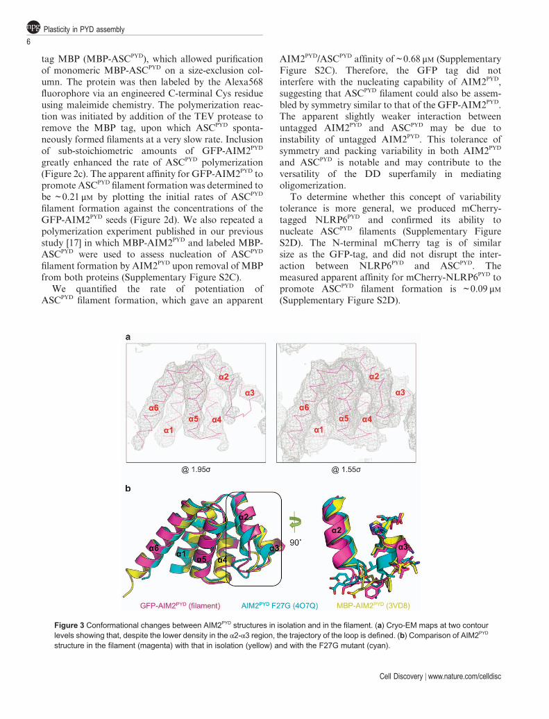
Figure 3 Conformational changes between AIM2PYD structures in isolation and in the filament. (a) Cryo-EM maps at two contourlevels showing that, despite the lower density in the α2-α3 region, the trajectory of the loop is defined. (b) Comparison of AIM2PYD
structure in the filament (magenta) with that in isolation (yellow) and with the F27G mutant (cyan).
Plasticity in PYD assembly
6
Cell Discovery | www.nature.com/celldisc
Structural features of the AIM2PYD filamentWe used the structure refinement program Phenix
and electron scattering factors to assess the agreementbetween the atomic model of the filament and thecryo-EM density. The cropped cryo-EM densitycorresponding to 15 AIM2PYD subunits that has beenadjusted in real space was placed into a crystal-lographic unit cell and back transformed to obtain thestructure factors. Upon rigid body refinement at 5.0 Åresolution with fixed B-factors for each subunit, theR and Rfree factors were 0.43 and 0.47, respectively.Almost all the main chain density was observed, withpart of the α2-α3 loop visible at a lower contour level(Figure 3a). Compared with the crystal structureof the monomeric MBP-fused wild-type AIM2PYD,the filament conformation of AIM2PYD shows maindifferences at this region (Figure 3b), suggesting thatAIM2PYD possesses intrinsic flexibility to allow forsubtle conformational changes during oligomerization.The F27Gmutant of AIM2PYD also exhibits differenceswith the wild-type AIM2PYD at this region (Figure 3b).
The AIM2PYDfilament core has an outer diameter of
~ 9 nm and an inner hole with a diameter of ~ 2 nm(Figure 4a), similar to dimensions of the ASCPYD
filament [17]. The filament has an apparent bumpysurface, in contrast to the smoother surface of theASCPYD
filament due to the longer C-terminal helix inAIM2PYD. While the GFP-AIM2PYD
filament has thesymmetry of a right-handed 1-start helix with 138.9°rotation and 6.0 Å axial rise per subunit, the sequentialintrastrand subunits do not make direct molecularcontact (Figure 4b). If we sequentially label theAIM2PYD subunits according to the helical operator,the three types of interactions that dictate helicalassembly in the AIM2PYD
filament will be between thefirst (i) and the fourth (i+3) subunits for the type Icontacts, between the first (i) and the sixth (i+5)subunits for the type II contacts, and between the first(i) and the third (i+2) for the type III contacts(Figure 4b). Orientation and position of the three typesof interactions are conserved between the AIM2PYD andASCPYD structures (Figures 4c and h). Similar toASCPYD, the type I interaction is the most extensive ofthe three types. Notably, type I interfaces in AIM2PYD
are mainly hydrophobic in nature, while that inASCPYD is formed by salt bridges between positivelyand negatively charged residues.
In the AIM2PYDfilament structure, the type I inter-
action is formed by Ser3, Lys6, Leu10, Leu11, Asp15and Ile46 of the α1/α4 helixes of one subunit andArg24, Phe27, Phe28 and Asp31 of the α2-α3 region ofan adjacent subunit (Figure 4c). Gln54 and Asn55 at
the end of the α4 helix interact with Asn73, Tyr74 andLeu76 in between α5-α6 of an adjacent molecule toform the type II contacts (Figure 4d). Type III inter-actions are mediated by Gly38 and Lys39 of α3 and thenearby Asp15, Asn16 and Ile17 residues of anothersubunit (Figure 4e). To determine whether there aresignificant changes in these interfacial contacts if theAIM2PYD
filament assumes the ASCPYDfilament sym-
metry, we superimposed the AIM2PYD subunit structureonto the respective type I, II and III interaction pairs inthe ASCPYD
filament (Figures 4f and g). This exerciseshows that the orientation and position of the threetypes of interactions are highly similar with either theobserved GFP-AIM2PYD
filament symmetry or theASCPYD
filament symmetry (Figures 4f and g). Thereare minimal adjustments of the interfacial contacts,further supporting the helical net analysis (Figure 2b).Like the ASCPYD
filament, the type I interaction in theAIM2PYD
filament is the most extensive of the threetypes of interactions. However, the type I interface inthe AIM2PYD
filament is mainly hydrophobic in nature,while that in the ASCPYD
filament is formed by saltbridges between positively and negatively chargedresidues.
AIM2HIN forms a filamentous complex with dsDNAPrevious studies have shown that full-length IFI16, an
ALR, cooperatively assembles filaments with dsDNAs[26]. IFI16 has an N-terminal PYD followed by twoHIN domains. Cooperative binding and filament for-mation require the PYD, while the individual HINdomains or the two HIN domains together did not showsignificant dsDNA binding at the close to physiologicalsalt concentration used in the assays [26], consistent withthe observed salt-dependence of IFI16 HIN in interac-tion with dsDNAs [13]. We asked whether AIM2 alsoforms filaments with dsDNAs. It has been shown pre-viously that AIM2 activation in cells is dependent ontransfected dsDNA length [10, 13]. Under one assaycondition, in human peripheral blood mononuclear cells(PBMCs), an 80-bp dsDNA, in comparison withdsDNA of 50-bp or less, was a better inducer of celldeath [13]. Under another condition, in mouse bonemarrow-derived macrophages (BMDMs), a 44-bpdsDNA could kill mouse BMDMs when transfectedat high concentrations (≥20 µg) and even longer dsDNA,100–500 bp in lengths, induced progressively more killingwhen transfected [10]. In contrast, shorter dsDNA didnot efficiently induce cell death [10].
As a 44-bp or 80-bp dsDNA is too short to bevisualized reliably using negative staining electronmicroscopy, we used polymerase chain reaction to
Alvin Lu et al.
7
Cell Discovery | www.nature.com/celldisc
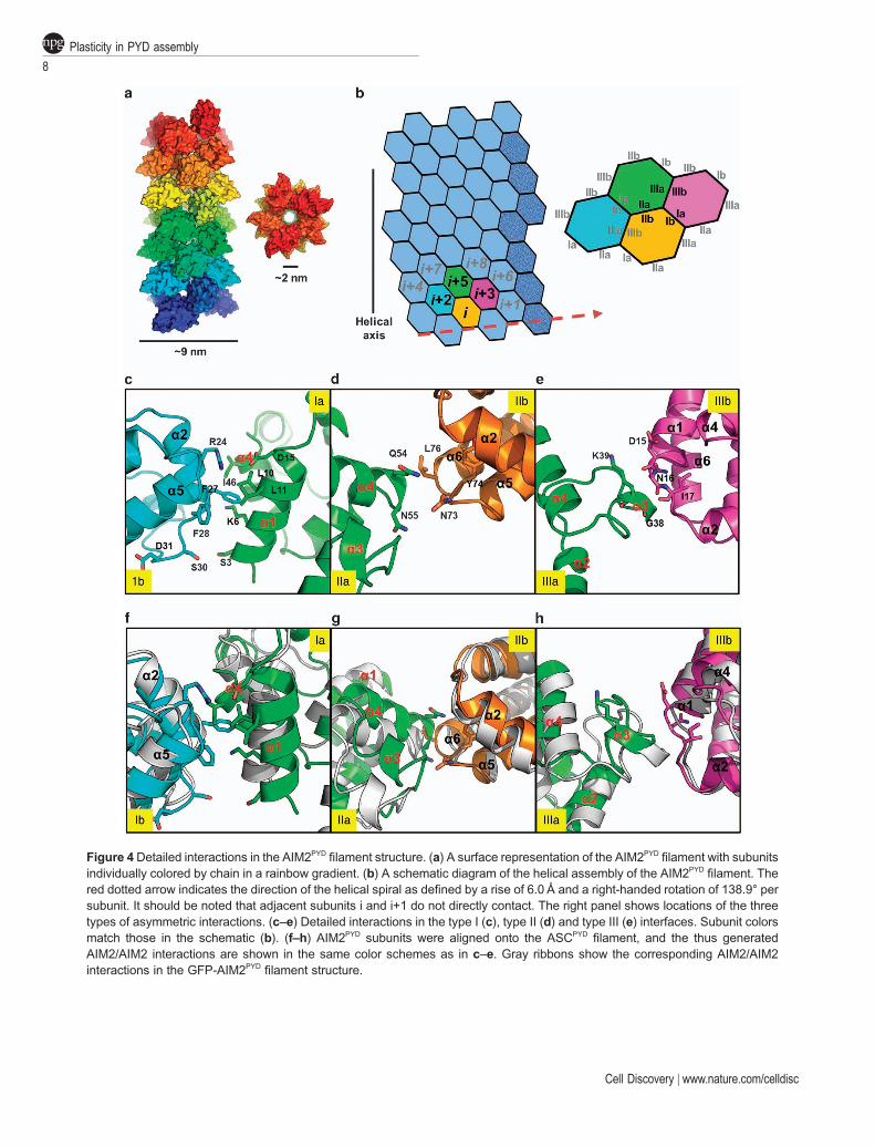
Figure 4 Detailed interactions in the AIM2PYDfilament structure. (a) A surface representation of the AIM2PYD
filament with subunitsindividually colored by chain in a rainbow gradient. (b) A schematic diagram of the helical assembly of the AIM2PYD
filament. Thered dotted arrow indicates the direction of the helical spiral as defined by a rise of 6.0 Å and a right-handed rotation of 138.9° persubunit. It should be noted that adjacent subunits i and i+1 do not directly contact. The right panel shows locations of the threetypes of asymmetric interactions. (c–e) Detailed interactions in the type I (c), type II (d) and type III (e) interfaces. Subunit colorsmatch those in the schematic (b). (f–h) AIM2PYD subunits were aligned onto the ASCPYD
filament, and the thus generatedAIM2/AIM2 interactions are shown in the same color schemes as in c–e. Gray ribbons show the corresponding AIM2/AIM2interactions in the GFP-AIM2PYD
filament structure.
Plasticity in PYD assembly
8
Cell Discovery | www.nature.com/celldisc

generate longer dsDNA fragments. We first triedAIM2HIN, which was expressed as a His-Sumo-fusionwith the tag cleaved off during purification. In contrastto IFI16, we found that AIM2HIN is able to formfilaments with 300 bp, 1 kbp and 2 kbp gel-purifieddsDNA fragments under close to physiological pH andsalt concentrations (Figures 5a and c). The diameter ofthe AIM2HIN/dsDNA filaments is measured to be close
to but o10 nm. The dsDNA alone control showedmuch thinner threads (Figure 5d), confirming thatthe filaments in the presence of AIM2HIN are theAIM2HIN/dsDNA complexes. With increasing dsDNAlengths, the AIM2HIN/dsDNA filaments become curvierand more convoluted. The calculated linear lengths for300 bp, 1 kbp and 2 kbp dsDNA fragments are 108,360 and 720 nm, respectively.
Figure 5 AIM2HIN forms filaments with dsDNA. (a–c) EM images of negatively stained AIM2HIN (0.05 mgml − 1) mixed with 300 bpdsDNA (a), 1 kbp dsDNA (b) and 2 kbp dsDNA (c). The molar ratios of AIM2HIN to dsDNA are 60:1, 200:1 and 400:1, respectively,for 300 bp, 1 kbp and 2 kbp dsDNAs. (d) An EM image of negatively stained 2 kbp dsDNA. (e, f) A model of the AIM2HIN/ dsDNAfilament, shown along (e) and down (f) the helical axis. The dsDNA is illustrated as a surface diagram with the two helical strandscolored in orange and yellow, respectively. AIM2HIN domains are colored in green, cyan and magenta.
Alvin Lu et al.
9
Cell Discovery | www.nature.com/celldisc

Formation of a full-length AIM2/dsDNA complexis hampered by the insolubility of full-length AIM2when overexpressed in E. coli as a His-Sumo-taggedprotein, in contrast to the soluble nature of full-lengthIFI16 [26]. This difference is most likely due to thePYD domain, as the PYD of IFI16 is much lessaggregated than AIM2PYD when expressed alone(Supplementary Figure S3). Unlike AIM2 that wasshown to be auto-inhibited [13, 16], IFI16 appears toexist in an extended conformation in the absence ofdsDNA stimulation [11]. Overexpression of full-lengthAIM2 may relieve this auto-inhibition and thereforeleads to aggregation and insolubility. To overcomethis difficulty, we expressed AIM2 as a fusion tothe large solubility tag His-MBP to inhibit self-oligomerization. Purified monomeric His-MBP-AIM2was then mixed with dsDNA, to which the TEVprotease was added to remove the His-MBP tag. Incontrast to formation of IFI16/dsDNA filaments [26],full-length AIM2 precipitated upon encounteringdsDNA and TEV, suggesting large-scale aggregationupon activation. This apparent higher aggregationtendency of AIM2 than IFI16 may correlate with theneed to sense small amounts of dsDNA in thecytosol.
To model the AIM2HIN/dsDNA filament complex,we used an existing human AIM2HIN/dsDNA complexcrystal structure (PDB ID: 3RN5) [13]. We used 80 bpsas one optimal dsDNA length for AIM2 activation,and first aligned one complex to an ideal 80-bp B-formdsDNA by superimposing the dsDNA molecules. Wereasoned that a most plausible way to model a dsDNAcoated with AIM2HIN is by aligning the same complexto different segments in the 80 bp dsDNA. Weattempted different DNA step sizes. Remarkably, astep size of every 4 bps generated an AIM2HIN/dsDNAfilament complex in which the AIM2HIN molecules arein extensive contact, but with no significant steric clash(Figure 5e). In contrast, a step of 3 or 5 bps generatedAIM2HIN molecules on dsDNA that are either in severeclash or with no contact, suggesting that the 4 bps stepis optimal. Assuming that each HIN domain occupies4 bps, the molar ratio of protein to DNA-binding siteswould have been 0.8:1 for all the EM experiments(Figures 5a and c). In this predicted AIM2HIN/dsDNAfilament model, each HIN molecule is in contact withsix adjacent HINs. The diameter of the model withdsDNA decorated by an outer layer of AIM2HIN
molecules is ~ 7.5 nm (Figure 5f), consistent with theEM observation. The ~ 50-residue linker between thePYD and HIN domains may allow the PYDs to swingaround the dsDNA core to form a short helical
assembly of AIM2PYD molecules for interacting withand nucleating ASCPYD
filaments.
Discussion
AIM2 is a critically important cytoplasmic dsDNAsensor that activates caspase-1 to provide host defenseagainst pathogens. Here we present the cryo-EMstructure of the PYD domain of AIM2 in its acti-vated, filamentous form. The cryo-EM images and thelow-resolution power spectrum are both dominated bythe GFP tag, which is poorly ordered. Despite thisdifficulty, we were able to obtain a map of the centralAIM2PYD
filament at a resolution that is clearlyinterpretable.
We and others have previously identified numerousAIM2PYD mutants that are defective in their abilities toself-aggregate and/or to nucleate ASCPYD
filaments[16, 17, 19] (Supplementary Figure S4A). Strikingly,when these mutation sites are mapped onto theAIM2PYD structure, almost all mutations are exactly atthe observed interfacial contacts in the AIM2PYD
fila-ment structure (Supplementary Figure S4B). The type Iinterface AIM2PYD mutants F27G, F27L, L10A/L11Aand D31K did not self-aggregate [19]. F27G,L10A/L11A and R24E mutations abolished the abilityof AIM2PYD to nucleate ASCPYD
filaments [17]. Thetype II mutant, Y74R, lost the ability to nucleateASCPYD [17]. Located at the edge of both type Ia andtype IIIb interfaces, D15R was also completely defec-tive. Type III mutants, G38E and K39E, appeared tohave partially lost their ability to interact with ASCPYD.The quadruple mutant D19A/E20A/E21A/D23Anear the type Ib interface also showed compromisedability to interact with ASCPYD [16]. These existingmutational data confirmed the validity of theobserved interactions in the AIM2PYD
filament.Human AIM2PYD and IFI16PYD share 31% sequenceidentity. Sequence alignment of IFI16PYD to AIM2PYD
shows that the interfacial residues important forAIM2PYD
filament formation and function are mostlyconserved in IFI16PYD (Supplementary Figure S4C),suggesting that IFI16PYD uses a similar assemblymechanism for initiating inflammasome formation andsignaling.
Our structure and the EM analysis on AIM2HIN/dsDNA interaction provide a conceptual model indsDNA-induced AIM2 activation (Figures 6a and b).First, cytoplasmic dsDNA of sufficient length frominvading pathogens will recruit AIM2 throughHIN/dsDNA interactions. Wrapping of the HINdomain around the dsDNA not only overcomes AIM2
Plasticity in PYD assembly
10
Cell Discovery | www.nature.com/celldisc
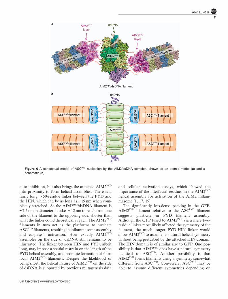
auto-inhibition, but also brings the attached AIM2PYD
into proximity to form helical assemblies. There is afairly long, ~ 50-residue linker between the PYD andthe HIN, which can be as long as ~ 19 nm when com-pletely stretched. As the AIM2HIN/dsDNA filament is~ 7.5 nm in diameter, it takes ~ 12 nm to reach from oneside of the filament to the opposing side, shorter thanwhat the linker could theoretically reach. The AIM2PYD
filaments in turn act as the platforms to nucleateASCPYD
filaments, resulting in inflammasome assemblyand caspase-1 activation. How exactly AIM2PYD
assembles on the side of dsDNA still remains to beillustrated. The linker between HIN and PYD, albeitlong, may impose a spatial restrain on the length of thePYD helical assembly, and promote formation of shortlocal AIM2PYD
filaments. Despite the likelihood ofbeing short, the helical nature of AIM2PYD on the sideof dsDNA is supported by previous mutagenesis data
and cellular activation assays, which showed theimportance of the interfacial residues in the AIM2PYD
helical assembly for activation of the AIM2 inflam-masome [1, 17, 19].
The significantly less-dense packing in the GFP-AIM2PYD
filament relative to the ASCPYDfilament
suggests plasticity in PYD filament assembly.Although the GFP fused to AIM2PYD via a mere two-residue linker most likely affected the symmetry of thefilament, the much longer PYD-HIN linker wouldallow AIM2PYD to assume its natural helical symmetrywithout being perturbed by the attached HIN domain.The HIN domain is of similar size to GFP. One pos-sibility is that AIM2PYD does have a natural symmetryidentical to ASCPYD. Another possibility is thatAIM2PYD forms filaments using a symmetry somewhatdifferent from ASCPYD. Conversely, ASCPYD may beable to assume different symmetries depending on
Figure 6 A conceptual model of ASCPYD nucleation by the AIM2/dsDNA complex, shown as an atomic model (a) and aschematic (b).
Alvin Lu et al.
11
Cell Discovery | www.nature.com/celldisc

which binding partner it interacts with. It is worthnoting that ALRs and most members of the large NLRfamily have a PYD, which use the single adaptor ASCfor inflammasome formation. The minimal changes insubunit interactions in an altered symmetry shouldallow successful PYD/PYD interaction despite thesymmetry difference. Therefore, the observed struc-tural plasticity must be important for the functionalrequirement of ASC in interacting with multiple PYDsin inflammasome formation. In addition, the AIM2PYD
filament structure reported here represents the first casein which adaptability has been observed for the entiresuperfamily of DD-containing proteins.
Many other filamentous systems have also beenknown to possess variability. For example, tubulin canbe polymerized in vitro to form microtubules with 9 to15 or more protofilaments, and transitions betweenthese can be seen in the same microtubule [27]. F-actinpossesses considerably variable and randomized twistbut a relatively constant rise per subunit [28]. The DDfold superfamily represents the most abundant domainin innate immune signaling pathways. These domainsare versatile modules for both homotypic and hetero-typic molecular interactions that afford the scaffoldsfor their respective signaling complexes. Given theconserved mode of interactions in the DD fold super-family, this observed plasticity in PYD assembly maycontribute to the adaptability of the DD fold ingeneral, and likely explain the omnipresence of thesedomains in innate immune pathways.
Materials and Methods
Construct and filament sample preparationTo generate a His-GFP-tagged AIM2PYD, the cDNA of
monomeric GFP (M1-T230, A206K) was cloned into thepET28a backbone using the NdeI and BamHI sites. AIM2PYD
(M1-P100) was inserted in frame with the GFP gene usingBamHI and NotI sites, which introduced a two-residue linker bythe BamHI site. The construct was expressed in E. coli BL21(DE3) cells by overnight induction using IPTG at 18˚C. The cellswere collected and lysed in lysis buffer containing 20mM
HEPES pH 8.0, 200 mM NaCl, 5 mM imidazole, 5 mM β-mer-captoethanol (β-ME) and 10% glycerol. Following sonication,the cell debris was centrifuged at 30 000 g for 30 min. Thesupernatant after centrifugation was incubated with the Ni-NTA affinity resin for 1 h, and washed in lysis buffer containing20mM imidazole. His-GFP-AIM2PYD
filaments were elutedusing lysis buffer with 300 mM imidazole. The eluate was injec-ted onto a Superdex 200 10/300 GL column pre-equilibratedwith gel filtration buffer containing 20mM HEPES pH 8.0,150mM NaCl and 2mM dithiothreitol. The filaments eluted inthe void, with peak at ~ 8 ml. The sample was frozen in smallaliquots for subsequent analysis.
Cryo-EM imaging, indexing and reconstructionThe sample (3 μl) was applied to lacey carbon grids that were
plasma cleaned (Gatan Solarus) and vitrified in a Vitrobot MarkIV (FEI, Hillsboro, OR, USA). Grids were imaged in a TitanKrios at 300 keV, and recorded with a Falcon II direct electrondetector at 1.15 Å per pixel (px). A total of 207 images (each 4 k× 4 k) were selected that were free from drift or astigmatism. Theprogram CTFFIND3 [29] was used for determining the contrasttransfer function and the defocus range used was from 0.6 to5.0 μm. The SPIDER software package [30] was used for mostsubsequent steps. The contrast transfer function was correctedby multiplying each image with the theoretical contrast transferfunction, both reversing phases where they need to be reversedand improving the signal-to-noise ratio. The program e2helix-boxer within EMAN2 [31] was used for boxing long filamentsfrom the micrographs. Overlapping boxes, 384 px long with a 10px shift between adjacent boxes (97% overlap) were extractedfrom these long filaments, yielding 54 973 segments.
Starting with an averaged power spectrum, it was apparentthat many different helical symmetries were possible [22]. Thecorrect helical symmetry was determined by trial-and-error,searching for a solution using the iterative helical real spacereconstruction method [23] that yielded recognizable secondarystructure [24]. The symmetry search was complicated by the factthat out-of-plane tilt cannot be ignored [24], and each symmetrythat was tried needed to allow for such out-of-plane tilt. Thesymmetry that was found, a rise of 6.0 Å and a rotation of 138.9°per subunit, generated a reconstruction where the PYD was seento be largely α-helical, and was thus taken to be correct.
AIM2 model fitting and refinementThe EM density was first transformed to CCP4 format [32].
A small region corresponding to the density of a single AIM2PYD
molecule was cutoff with a mask, and wild-type AIM2PYD
structure (3VDB) was aligned to the density. Helices weremoved slightly by fitting individually in real space and regular-ized in Coot to generate an AIM2PYD structure in the filamentconformation [25]. The modified AIM2PYD structure was fittedindividually into EM density in real space to generate an initialAIM2PYD
filament structure with 15 molecules. The densitycorresponding to the 15-molecule filament was cut using a maskgenerated from the model and put into an arbitrary unit cell inthe P1 space group. Structure factors were calculated usingelectron scattering factors from the resulted map, and the figureof merit and the sigma were added to each reflection uniformly.Structure refinement was carried with PHENIX.refine [33],using rigid body positional refinement and a single B-factor foreach subunit. The refinement led to an R of 0.43, Rfree of 0.47,root mean square deviation in bond lengths of 0.016 Å and rootmean square deviation in bond angles of 0.48°.
Fluorescence polarization assayDetailed protocol could be found in our previous study [17].
In short, monomeric MPB-tagged ASCPYD (M1-Q105) con-taining an engineered Cys at the C-terminus (S106C) wasexpressed in E. coli BL21(DE3) cells and purified by Ni-NTAaffinity pulldown and SEC. The monomers were labeled withMaleimide-Alexa Fluor 568 (Invitrogen, Grand Island, NY,
Plasticity in PYD assembly
12
Cell Discovery | www.nature.com/celldisc

USA) by recommended protocol from the manufacturer. Excessdyes were removed by gel filtration after overnight incubation.Fluorescence polarization measurements were taken with theSpectraMax M5e (Molecular Devices, Sunnyvale, CA, USA)plate reader. For the mCherry-NLRP6PYD/ASCPYD experiment,same procedure was used except Maleimide-Alexa Fluor 488was used to label MBP-ASCPYD.
Negative-stain EM analysis of AIM2HIN/dsDNA filamentsAIM2HIN (residues 138–343) was cloned into pSMT3 vector
using the BamHI and SalI cloning sites and expressed as a Sumo-fusion protein with an additional N-terminal 6× His tag. Therecombinant protein was expressed in E. coli BL21(DE3) cells bygrowing the culture at 37 °C to OD600 of 0.8 and inducing with0.5mM IPTG overnight at 16 °C. The E. coli cells were harvestedand lysed by sonication in lysis buffer containing 25mM Tris-HClpH 8.0, 1.0M NaCl, 5mM imidazole, 5mM β-ME and 5% gly-cerol. The cell lysate was centrifuged at 40 000 g for 40min, and thesupernatant was incubated with pre-equilibrated Ni-NTA resin.After being washed with 20 column-volume lysis buffer containing25mM imidazole, the protein was eluted in buffer A containing25mMTris-HCl pH 8.0, 300mMNaCl, 300mM imidazole, 5mM β-ME and 5% glycerol. To cleave the Sumo-tag, the recombinantprotein was incubated with 1/1 000 (w/w) Ulp1 protease at 4 °Covernight. The cleaved protein was then loaded onto a Heparin SPcolumn and eluted with a gradient of 0.3–1.5M NaCl in buffer A.The peak corresponding to AIM2HIN was further purified usingSuperdex 200 (10/30) SEC in 25mM Tris-HCl pH 8.0, 150mM
NaCl and 2mM DTT. Fractions containing AIM2HIN were col-lected, concentrated and flash frozen in liquid nitrogen for futureuse. To form AIM2HIN/dsDNA filaments, AIM2HIN was diluted inSEC buffer to a final concentration of 0.05mgml−1, and incubatedwith 300 bp, 1 kbp and 2 kbp dsDNA at a molar ratio of 1/60,1/200 and 1/400 of the protein, respectively.
Full-length AIM2 was cloned into pDB.His.MBP vectorusing the NdeI and XhoI cloning sites and expressed as aHis-MBP-fusion protein in E. coli BL21(DE3) cells under thesame condition as that used for AIM2HIN. The protein waspurified with Ni-NTA resin followed by Heparin SP andSuperdex 200 (10/30) columns without removing the MBP-tag.The purified MBP-AIM2 protein was diluted to 0.05 mgml− 1
and mixed with dsDNA of different lengths at indicatedconcentrations, and TEV protease was added to 1/10 (w/w)ratio with the protein to cleave the MBP-tag, allowing forAIM2/dsDNA filament formation.
Modeling the AIM2HIN/dsDNA filamentAn 80-bp ideal B-form DNA (DNA80) is generated in
WinCoot [25]. Superimposing the AIM2HIN/dsDNA structure(PDB ID: 3RN5) on DNA80 generated the first HIN domain onDNA80. Sliding the first HIN domain along DNA80 every fourbase pairs (i → i+4) results in HIN domains closely packed onDNA80 without significant clashes between adjacent molecules.
References
1 Lamkanfi M, Dixit VM. Mechanisms and functions ofinflammasomes. Cell 2014; 157: 1013–1022.
2 Lu A, Wu H. Structural mechanisms of inflammasomeassembly. FEBS J2014; 282: 435–444.
3 Hu Z, Yan C, Liu P, et al. Crystal structure of NLRC4reveals its autoinhibition mechanism. Science 2013; 341:172–175.
4 Ting JP, Lovering RC, Alnemri ES, et al. The NLR genefamily: a standard nomenclature. Immunity 2008; 28:285–287.
5 Kerur N, Veettil MV, Sharma-Walia N, et al. IFI16 acts asa nuclear pathogen sensor to induce the inflammasomein response to Kaposi Sarcoma-associated herpesvirusinfection. Cell Host Microb 2011; 9: 363–375.
6 Rathinam VA, Jiang Z, Waggoner SN, et al. The AIM2inflammasome is essential for host defense againstcytosolic bacteria and DNA viruses.Nat Immunol 2010; 11:395–402.
7 Hornung V, Ablasser A, Charrel-Dennis M, et al. AIM2recognizes cytosolic dsDNA and forms a caspase-1-activating inflammasome with ASC. Nature 2009; 458:514–518.
8 Fernandes-Alnemri T, Yu JW, Datta P, Wu J, Alnemri ES.AIM2 activates the inflammasome and cell death inresponse to cytoplasmic DNA. Nature 2009; 458: 509–513.
9 Burckstummer T, Baumann C, Bluml S, et al. An ortho-gonal proteomic-genomic screen identifies AIM2 as acytoplasmic DNA sensor for the inflammasome. NatImmunol 2009; 10: 266–272.
10 Roberts TL, Idris A, Dunn JA, et al. HIN-200 proteinsregulate caspase activation in response to foreigncytoplasmic DNA. Science 2009; 323: 1057–1060.
11 Liao JC, Lam R, Brazda V, et al. Interferon-InducibleProtein 16: Insight into the Interaction with TumorSuppressor p53. Structure 2011; 19: 418–429.
12 Theobald DL, Mitton-Fry RM, Wuttke DS. Nucleic acidrecognition by OB-fold proteins. Annu Rev Biophys BiomolStruct 2003; 32: 115–133.
13 Jin T, Perry A, Jiang J, et al. Structures of the HIN domain:DNA complexes reveal ligand binding and activationmechanisms of the AIM2 inflammasome and IFI16receptor. Immunity 2012; 36: 561–571.
14 Ru H, Ni X, Zhao L, et al. Structural basis for terminationof AIM2-mediated signaling by p202. Cell Res 2013; 23:855–858.
15 Sung MW, Watts T, Li P. Crystallographic characteriza-tion of mouse AIM2 HIN-200 domain bound to a 15 bpand an 18 bp double-stranded DNA. Acta Crystallogr SectF Struct Biol Cryst Commun 2012; 68: 1081–1084.
16 Jin T, Perry A, Smith P, Jiang J, Xiao TS. Structure of theabsent in melanoma 2 (AIM2) pyrin domain providesinsights into the mechanisms of AIM2 autoinhibitionand inflammasome assembly. J Biol Chem 2013; 288:13225–13235.
17 Lu A, Magupalli VG, Ruan J, et al.Unified polymerizationmechanism for the assembly of ASC-dependent inflamma-somes. Cell 2014; 156: 1193–1206.
18 Yin Q, Sester DP, Tian Y, et al. Molecular mechanism forp202-mediated specific inhibition of AIM2 inflammasomeactivation. Cell Rep 2013; 4: 327–339.
Alvin Lu et al.
13
Cell Discovery | www.nature.com/celldisc

19 Lu A, Kabaleeswaran V, Fu T, Magupalli VG, Wu H.Crystal structure of the F27G AIM2 PYD mutantand similarities of its self-association to DED/DEDinteractions. J Mol Biol 2014; 426: 1420–1427.
20 Cai X, Chen J, Xu H, et al. Prion-like polymerizationunderlies signal transduction in antiviral immune defenseand inflammasome activation. Cell 2014; 156: 1207–1222.
21 Ferrao R, Wu H. Helical assembly in the death domain(DD) superfamily. Curr Opin Struct Biol 2012; 22:241–247.
22 Egelman EH. Reconstruction of helical filaments and tubes.Methods Enzymol 2010; 482: 167–183.
23 Egelman EH. A robust algorithm for the reconstructionof helical filaments using single-particle methods. Ultra-microscopy 2000; 85: 225–234.
24 Egelman EH. Ambiguities in helical reconstruction. eLife2014; 3: 1–9.
25 Emsley P, Cowtan K. Coot: model-building tools formolecular graphics. Acta Crystallogr D Biol Crystallogr2004; 60: 2126–2132.
26 Morrone SR, Wang T, Constantoulakis LM, Hooy RM,Delannoy MJ, Sohn J. Cooperative assembly of IFI16filaments on dsDNA provides insights into host defensestrategy. Proc Natl Acad Sci USA 2014; 111: E62–E71.
27 Chretien D, Metoz F, Verde F, Karsenti E, Wade RH.Lattice defects in microtubules: protofilament numbersvary within individual microtubules. J Cell Biol 1992; 117:1031–1040.
28 Egelman EH, Francis N, DeRosier DJ. F-actin is a helixwith a random variable twist. Nature 1982; 298: 131–135.
29 Mindell JA, Grigorieff N. Accurate determination of localdefocus and specimen tilt in electron microscopy. J StructBiol 2003; 142: 334–347.
30 Frank J, Radermacher M, Penczek P, et al. SPIDER andWEB: processing and visualization of images in 3D electronmicroscopy and related fields. J Struct Biol 1996; 116:190–199.
31 Tang G, Peng L, Baldwin PR, et al. EMAN2: an extensibleimage processing suite for electron microscopy. J StructBiol 2007; 157: 38–46.
32 Collaborative Computational Project N. The CCP4 Suite:Programs for Protein Crystallography. Acta Cryst 1994;D50: 760–763.
33 Adams PD, Afonine PV, Bunkoczi G, et al. PHENIX: acomprehensive Python-based system for macromolecularstructure solution. Acta Crystallogr D Biol Crystallogr2010; 66: 213–221.
(Supplementary Information is linked to the online version of thepaper on the Cell Discovery website.)
This work is licensed under a Creative CommonsAttribution 4.0 International License. The images or
other third party material in this article are included in the article’sCreative Commons license, unless indicated otherwise in thecredit line; if the material is not included under the CreativeCommons license, users will need to obtain permission from thelicense holder to reproduce the material. To view a copy of thislicense, visit http://creativecommons.org/licenses/by/4.0/
Plasticity in PYD assembly
14
Cell Discovery | www.nature.com/celldisc