plasmin in nephrotic urine activates the epithelial sodium channel
TRANSCRIPT

Plasmin in Nephrotic Urine Activates the EpithelialSodium Channel
Per Svenningsen,* Claus Bistrup,*† Ulla G. Friis,* Marko Bertog,‡ Silke Haerteis,‡
Bettina Krueger,‡ Jane Stubbe,* Ole Nørregaard Jensen,§ Helle C. Thiesson,*†
Torben R. Uhrenholt,* Bente Jespersen,*† Boye L. Jensen,* Christoph Korbmacher,‡ andOle Skøtt*
*Physiology and Pharmacology, Institute of Medical Biology, University of Southern Denmark, †Department ofNephrology Y, Odense University Hospital, Odense C, and §Department of Biochemistry and Molecular Biology,University of Southern Denmark, Odense M, Denmark; and ‡Department of Cellular and Molecular Physiology,University of Erlangen-Nuremberg, Erlangen, Germany
ABSTRACTProteinuria and increased renal reabsorption of NaCl characterize the nephrotic syndrome. Here, weshow that protein-rich urine from nephrotic rats and from patients with nephrotic syndrome activatethe epithelial sodium channel (ENaC) in cultured M-1 mouse collecting duct cells and in Xenopuslaevis oocytes heterologously expressing ENaC. The activation depended on urinary serine proteaseactivity. We identified plasmin as a urinary serine protease by matrix-assisted laser desorption/ionization time of-flight mass spectrometry. Purified plasmin activated ENaC currents, and inhibitorsof plasmin abolished urinary protease activity and the ability to activate ENaC. In nephroticsyndrome, tubular urokinase-type plasminogen activator likely converts filtered plasminogen toplasmin. Consistent with this, the combined application of urokinase-type plasminogen activator andplasminogen stimulated amiloride-sensitive transepithelial sodium transport in M-1 cells and in-creased amiloride-sensitive whole-cell currents in Xenopus laevis oocytes heterologously expressingENaC. Activation of ENaC by plasmin involved cleavage and release of an inhibitory peptide fromthe ENaC � subunit ectodomain. These data suggest that a defective glomerular filtration barrierallows passage of proteolytic enzymes that have the ability to activate ENaC.
J Am Soc Nephrol 20: 299 –310, 2009. doi: 10.1681/ASN.2008040364
Nephrotic syndrome is characterized by protein-uria, sodium retention, and edema. Increased renalsodium reabsorption occurs in the cortical collect-ing duct (CCD),1,2 where a rate-limiting step intransepithelial sodium transport is the epithelial so-dium channel (ENaC), which is composed of thethree homologous subunits: �, �, �.3
ENaC activity is regulated by hormones, such asaldosterone and vasopressin (AVP)4,5; however,adrenalectomized rats and AVP-deficient Brattle-boro rats are capable of developing nephrotic syn-drome,1,6 and nephrotic patients do not consis-tently display elevated levels of sodium-retaininghormones,7,8 suggesting that renal sodium hyper-reabsorption is independent of systemic factors.
Consistent with this, sodium retention is confinedto the proteinuric kidney in the unilateral puromy-cin aminonucleoside (PAN) nephrotic model.2,9,10
There is evidence that proteases contribute toENaC activation by cleaving the extracellular loops
Received April 8, 2008. Accepted September 2, 2008.
Published online ahead of print. Publication date available atwww.jasn.org.
P.S. and C.B. contributed equally to this work.
Correspondence: Dr. Claus Bistrup, Department of Nephrology Y,Odense University Hospital, Sdr. Boulevard 29, DK-5000 Odense C,Denmark. Phone: �4565411762; Fax: �4565413452; E-mail:[email protected]
Copyright � 2009 by the American Society of Nephrology
BASIC RESEARCH www.jasn.org
J Am Soc Nephrol 20: 299–310, 2009 ISSN : 1046-6673/2002-299 299

of the �- and �-subunits.11–13 Proteolytic activation of ENaCby extracellular proteases critically involves the cleavage of the� subunit,14 –16 which probably leads to the release of a 43-residue inhibitory peptide from the ectodomain.17 Bothcleaved and noncleaved channels are present in the plasmamembrane,18,19 allowing proteases such as channel activatingprotease 1 (CAP1/prostasin),20 trypsin,20 chymotrypsin,21 andneutrophil elastase22 to activate noncleaved channels from theextracellular side.23,24 We hypothesized that the defective glo-merular filtration barrier in nephrotic syndrome allows thefiltration of ENaC-activating proteins into the tubular fluid,leading to stimulation of ENaC. The hypothesis was tested inthe PAN nephrotic model in rats and with urine from patientswith nephrotic syndrome.
RESULTS
Sodium Retention in PAN Nephrotic SyndromeInvolves a Primary Increase in Renal ENaC ActivityThe first 24 h after PAN injection were characterized by net lossof sodium and water, an increase in hematocrit, and increasedplasma renin (Figure 1, A and B, and Supplemental Figure 1).This was followed by net sodium retention and a positive cu-mulative sodium balance from day 3.6 � 0.8 and onward (Fig-ure 1A) and suppression of plasma renin concentrations inparallel to a fall in hematocrit consistent with expansion ofextracellular fluid volume (Figure 1B, Supplemental Figure 1).Plasma aldosterone concentration was maximal at day 5 andwas suppressed to 1% of this level at day 8 (P � 0.01; Supple-mental Figure 2A). Urinary protein excretion increased fromday 3 and coincided with the transition from negative to pos-itive sodium balance (Figure 1A). There was no significantchange in AVP concentration 8 d after PAN treatment (Sup-plemental Table 1). Sodium accumulation in PAN-treated ratswas resistant to combined treatment with the AT1 receptorantagonist candesartan (1 mg/kg per d) and the mineralocor-ticoid receptor antagonist potassium canrenoate (100 mg/kgper d subcutaneously; n � 8; Supplemental Figure 2B).
Treatment of PAN nephrotic rats with amiloride in astep-up protocol with 100 and 500 �g/d (2074 � 88 �g/kgbody wt) increased daily urinary sodium excretion (Figure1C). Moreover, amiloride had a beneficial effect on ascitic vol-ume, which decreased from 3.5 � 0.5 ml/100 g body wt (n �16) to 1.7 � 0.6 ml/100 g body wt (n � 6; P � 0.05). Theseresults indicate that ENaC activity is enhanced in the PAN-treated animals, whereas ENaC mRNA and protein levels werenot changed (data not shown).
Plasmin in Nephrotic Urine Stimulates ENaC ActivityWe hypothesized that a serine protease may be present innephrotic urine and may directly activate ENaC in theplasma membrane. Whole-cell current did not change insingle M-1 cells when exposed to urine from control rats,whereas urine collected on day 5 after PAN injection yielded
Figure 1. Sodium retention in PAN nephrotic syndrome in rats in-volves a primary increase in renal ENaC activity. (A) Daily sodium bal-ance and urinary protein excretion after PAN injection based on mea-surements of daily sodium intake, fecal sodium output, and urinarysodium output of rats in metabolic cages (Supplemental Figure 1).Nephrotic rats display negative cumulative sodium balance (shadedarea) from days 0 to 3.6 after PAN injection and positive sodium balancethereafter. Nephrotic rats accumulated 2217 � 167 �mol/100 g bodywt sodium (n � 8) from days 0 through 8 compared with 1096 � 70�mol/100 g body wt in controls (n � 11; P � 0.0005). The proteinuriain nephrotic rats was significant from days 2 through 8. Arrow indicatestime of PAN injection. *P � 0.05; ***P � 0.001. (B) Parallel changes inplasma renin concentrations and hematocrit values of nephrotic ratsindicate a shift from volume underfilling to overfilling from days 2through 5. Renin: *P � 0.05 between days 2 and 8 (t test). Hematocrit:*P � 0.05 versus control (Dunnett test). GU, Goldblatt units. (C) Effect ofamiloride treatment on daily urinary sodium output. In PAN nephroticrats, amiloride (2 mg/d per kg body wt) increases daily urinary sodiumexcretion more than in controls (n � 6). Arrow indicates time of PANinjection. *P � 0.05.
BASIC RESEARCH www.jasn.org
300 Journal of the American Society of Nephrology J Am Soc Nephrol 20: 299–310, 2009

a 5.1 � 2.0-fold increase in the observed inward current thatwas prevented in the presence of 2 �M amiloride (controlurine �55 � 12 pA versus nephrotic urine �231 � 53 pA at�150 mV holding potential; n � 4; P � 0.05; Figure 2, A andB). This stimulatory effect was observed within 30 to 60 safter exposure of the M-1 cells to nephrotic urine, and it wasprevented by aprotinin, an inhibitor of serine proteases(Figure 2B). Xenopus laevis oocytes heterologously express-ing ENaC confirmed the stimulatory effect of nephroticurine samples on amiloride-sensitive ENaC whole-cell cur-rents (�Iami; Figure 2C) compared with control oocytes pre-incubated in ND96 bath solution or compared with oocytespreincubated in heat-inactivated nephrotic urine samples.The stimulatory effect of a 30-min exposure to nephroticurine was similar to that of a 5-min exposure to chymotryp-sin.21 These findings suggest that nephrotic urine contains aserine protease, which can activate ENaC.
Zymography of nephrotic urine showed aprotinin-sensitiveprotease activity with a molecular size of approximately 75 kD(Figure 2D). In a serine protease assay with a synthetic sub-strate, nephrotic urine exhibited 10-fold increased activitycompared with control (Figure 2E), whereas no difference inplasma serine protease activity was detected between controland nephrotic rats.
Aprotinin-affinity precipitation of nephrotic urine effec-tively removed serine protease activity and abolished the abil-
ity to activate ENaC current (Figure 3A), whereas total proteinconcentration was not changed (35.8 versus 34.0 mg/ml fornephrotic urine and aprotinin-affinity supernatant, respec-tively). The aprotinin-affinity precipitate was highly enrichedin serine protease activity compared with nephrotic urine(30.80 � 0.60 versus 0.20 � 0.08 �A405 nm/h per mg protein,respectively) and stimulated ENaC activity (Figure 3A). Ionexchange chromatography of the precipitate followed by pro-tease activity screening identified fractions with high activity(Figure 3B). Peak fractions corresponding to elution volume29 to 31 ml were pooled and subjected to repeated aprotinin-affinity precipitation. The precipitate was separated by gel elec-trophoresis, which yielded two distinct bands with a molecularweight of 40 and 80 kD (Supplemental Figure 3A). Matrix-assisted laser desorption/ionization time of-flight (MALDI-TOF) mass spectrometry identified the proteins as plasminfragments and plasmin, respectively (Figure 3C, SupplementalTable 2). Zymography using plasmin but not plasminogen ex-hibited a pattern similar to the proteolytic activity observedwith nephrotic urine (Supplemental Figure 3B). Pure plasminactivated a sodium current in M-1 cells that was inhibited byamiloride and by knockdown of the �ENaC subunit usingsmall interfering RNA (siRNA; Figure 3, D and E). The pro-tease activity of nephrotic urine was inhibited by the serineprotease inhibitor aprotinin, the specific plasmin inhibitorsPefabloc PL, and �2-antiplasmin (Supplemental Figure 4A).
Figure 2. Serine proteases in nephrotic urine stimulatesENaC activity. (A) Traces obtained with whole-cell patch-clamp technique on a single M-1 cell showing baseline cur-rent (black) before addition of nephrotic urine (day 5) in thepresence of 2 �M amiloride (gray). Subsequently, amiloridewas washed away and the same cell was stimulated with thecorresponding urine sample (black). The voltage wasclamped to �60 mV and then stepped to �150 mV for 200ms. The traces shown are recorded approximately 30 to 60 safter addition of the various substances. (B) Mean values fromfour different patch-clamp experiments showing that addi-tion of aprotinin (700 �g/ml) to nephrotic urine samples orthe presence of amiloride (2 �M) prevents the activation ofcurrents in single M-1 cells. *P � 0.05 versus control (n � 4,Dunnett test). (C) Preincubation of ENaC-expressing Xeno-pus laevis oocytes with nephrotic urine samples increasedthe amiloride-sensitive ENaC whole-cell current to a similarextent as preincubation with chymotrypsin. Control oocyteswere preincubated in standard ND96 solution, which wasalso used as bath solution for all other whole-cell currentmeasurements. The stimulatory effect of nephrotic urinesamples was essentially abolished by heat-inactivating theurine samples. The nephrotic urine samples were from threedifferent rats; the non-nephrotic urine sample was from a ratthat was given PAN injection and did not develop nephroticsyndrome probably because of an insufficient injection.Numbers of individual oocytes measured in each group areshown above each column. Columns represent normalizedmean values � SEM from six different batches of oocytes (**P � 0.01; ***P � 0.001). (D) Gelatinase activity of PAN nephrotic urine isabolished when zymograms are developed in the presence of aprotinin (1 mg/ml). (E) Serine protease activity in nephrotic urine is highcompared with urine from control rats. Activity in plasma is low in both. ***P � 0.001.
BASIC RESEARCHwww.jasn.org
J Am Soc Nephrol 20: 299–310, 2009 Plasmin Activates ENaC 301

The selective plasmin inhibitors prevented the ability of ne-phrotic urine to activate ENaC currents in single M-1 cellswhen they were added to the urine samples before exposing thecells to the samples (Figure 4). Thus, plasmin is the dominantprotease in nephrotic urine responsible for ENaC activation.
Plasminogen is Activated by Tubular Urokinase-TypePlasminogen Activator ActivityBefore PAN injection, plasmin or plasminogen was not detectedin urine (Figure 5A). Coincident with the appearance of protein-uria and positive sodium balance at days 3 to 4 (Figure 1A), twobands appear in urine with the expected sizes of plasminogen andactive plasmin (Figure 5A). The antibody recognized pure plas-minogen and plasmin with similar molecular weights as observedin nephrotic urine (data not shown). Plasminogen and plasminwere present in nephrotic urine from days 3 to 4 to 8. Plasmin wasnot detectable in rat plasma by Western blotting (Figure 5A). Plas-minogen may be converted to plasmin by urokinase-type plas-minogen activator (uPA), and immunolabeling of nephrotic ratkidney cryosections for uPA showed immunoreactivity associatedwith the apical surface of CCD (Figure 5B). Western blotting con-firmed expression of uPA in rat kidney cortex (Figure 5B).
To test whether uPA converts plasminogen to plasmin inPAN nephrotic rats, we examined urine from amiloride-treated rats, because amiloride, besides being an inhibitor of
ENaC, potently inhibits uPA activity.25 The high dosage ofamiloride lowered the plasmin/plasminogen ratio in nephroticurine and abolished immunoreactive plasmin in urine (Figure5C, Supplemental Figure 4B). Zymography of urine showed anattenuation of protease activity in nephrotic rats treated withamiloride (Figure 5D). Amiloride, 50 �mol/L, had no directeffect on serine protease activity (26.8 � 1.1 versus 25.1 � 0.7�A405nm/h/ml without and with amiloride). Thus, amiloridedid not inhibit plasmin per se, whereas it inhibited urinaryformation of plasmin from plasminogen.
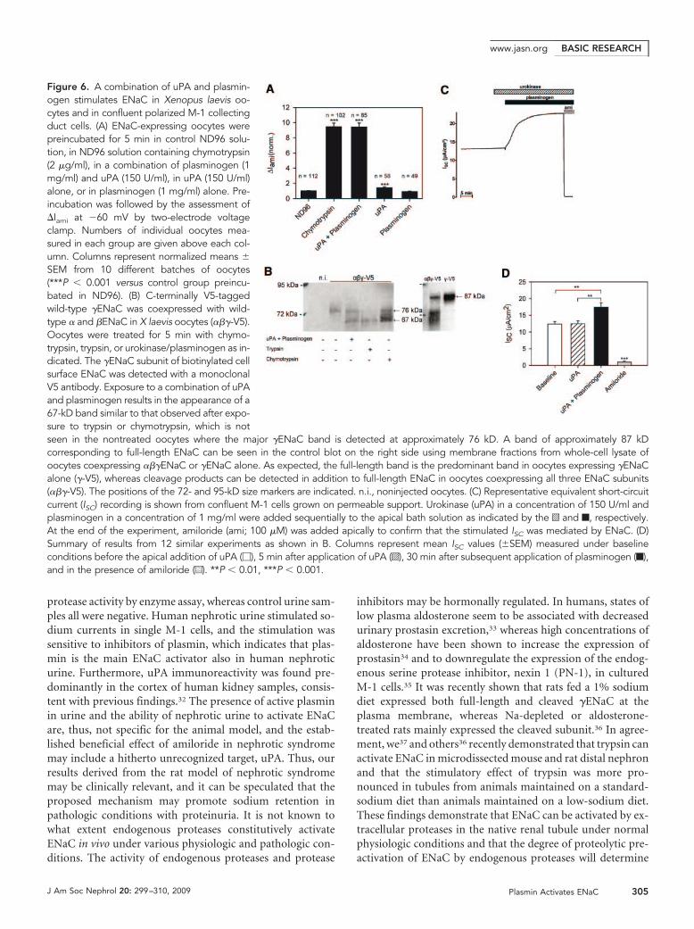
uPA-Mediated Conversion of Plasminogen to PlasminIs Required for ENaC ActivationIn Xenopus laevis oocytes heterologously expressing rat ENaC,preincubation of the oocytes with uPA alone had only a mar-ginal stimulatory effect on the �Iami, and exposure to plasmin-ogen had no significant effect. In contrast, the combination ofplasminogen and uPA resulted in a significant increase in�Iami, similar to the increase observed with chymotrypsin (Fig-ure 6A). This indicates that uPA-mediated generation of plas-min is required for ENaC activation. To investigate whetherENaC activation is associated with �ENaC cleavage at the cellsurface, we used a �ENaC construct with a C-terminal V5 tagto detect biotinylated cell surface �ENaC fragments by West-ern blot. Exposure of the oocytes to a combination of uPA andplasminogen yielded a �ENaC fragment with a molecular
Figure 3. Identification of plasmin as an ENaC-activating protease in nephrotic urine. (A) Afteraprotinin affinity precipitation of nephrotic urine,there is reduced ability of the supernatant tostimulate sodium currents in M-1 cells and serineprotease activity (f), whereas the ability of theprecipitate to stimulate sodium current andserine protease activity is increased (�). *P �0.05 (n � 4). (B) Chromatogram of ion exchangechromatography of the aprotinin-affinity precip-itate and corresponding serine protease activi-ties (solid line, A280nm; dotted line, conductanceof the elution buffer; f, serine protease activity).#Fractions that were pooled, precipitated withaprotinin, and separated by gel electrophoresisto give bands with sizes of 80 and 40 kD (Sup-plemental Figure 3A). (C) MALDI-TOF massspectrometry identified the 80-kD band resultingfrom the procedure in B as plasmin(ogen) (F).Trypsin autolysis peptides (E) were used for in-ternal calibration. (D) Knockdown of �ENaCmRNA and protein in M-1 cells. Cells were trans-fected with �ENaC siRNA, and �ENaC mRNAlevel was assessed the next day using reversetranscriptase-quantitative PCR (upper panel) orafter 2 d at the protein level using immunofluo-rescence (lower panels, n � 4). (E) Stimulation ofwhole-cell inward currents in M-1 cells by native
rat plasmin (10 �g/ml). The stimulatory effect of plasmin was prevented when plasmin was applied in the presence of amiloride(2 �M) or by knockdown of �ENaC by siRNA (n � 4). *P � 0.05 versus control (t test).
BASIC RESEARCH www.jasn.org
302 Journal of the American Society of Nephrology J Am Soc Nephrol 20: 299–310, 2009

weight of approximately 67 kD (Figure 6B). We observed sim-ilar fragments when trypsin or chymotrypsin was used. As pre-viously shown,15,16,26 the 76-kD band corresponds to a cleavageproduct that arises from �ENaC cleavage at its putative furincleavage site when �ENaC is coexpressed with the �- and�-subunits. Full-length �ENaC migrates at approximately 87kD and is readily detectable in whole-cell lysate but not at thecell surface (Figure 6B). Full-length �ENaC is the predominantband detected in oocytes expressing �ENaC in the absence ofthe �- and �-subunits (Figure 6B).
Similar to the findings in the oocyte system, uPA alone had noeffect on the equivalent short circuit current (ISC) from confluentM-1 cells grown on filters (Figure 6, C and D). Subsequent apical
application of plasminogen in the presence of uPA resulted in asignificant increase in ISC. The stimulated ISC was abolished byapical application of amiloride, which confirms that the ISC is me-diated by ENaC expressed in the apical membrane of confluentM-1 cells.27,28 These results confirm that plasmin but not plas-minogen or uPA stimulate ENaC activity. In addition to blockingENaC, amiloride may prevent the conversion of urinary plasmin-ogen to plasmin in vivo.
Plasmin Stimulation Leads to Release of a Peptidefrom the Extracellular Domain of �ENaCProteolytic activation of ENaC is thought to involve dual cleav-age of its �-subunit, which results in the release of a 43-residueinhibitory peptide from its extracellular domain.17 To examinethis in live cells, we inserted a hexahistidine tag in a �ENaCconstruct expression vector between the cleavage sites for furinand prostasin (Figure 7). The tag binds the extracellular flu-orophore NTA-Atto550. M-1 cells transfected with the taggedconstruct display strong fluorescence when exposed to NTA-Atto550, whereas wild-type cells do not (Figure 7). Pretreat-ment of transfected cells with plasmin or nephrotic urine abol-ished the ability of cells to respond to NTA-Atto550 exposure.This indicates effective removal of inhibitory peptide fromectodomain �ENaC by plasmin in live cells (Figure 7).
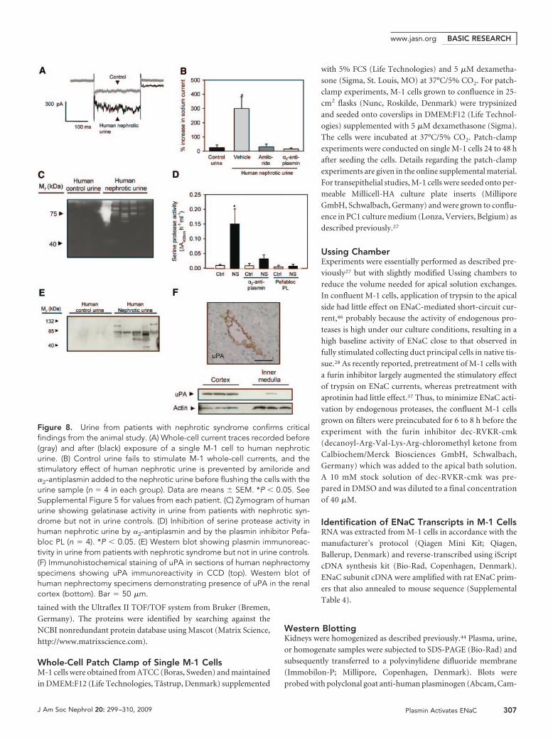
Nephrotic Urine from Human Patients StimulatesENaC ActivityUrine from five patients with nephrotic syndrome (Supple-mental Table 3) augmented sodium currents in M-1 cells (Fig-ure 8, A and B). Addition of �2-antiplasmin (5 �M) or amilo-ride (2 �M) abolished the ability of nephrotic urine tostimulate whole-cell currents in M-1 cells (Figure 8B). Urinezymograms displayed intense proteolytic activity with appar-ent molecular weight of 75 to 80 kD (Figure 8C). Serine pro-tease activity sensitive to plasmin inhibitors was present in pa-tient urine samples but not in urine from four age- and gender-matched control subjects (Figure 8D). Western blots forplasminogen and plasmin displayed several bands in nephroticbut not in control urine (Figure 8E). Immunohistochemicalstaining of human nephrectomy specimens showed uPA pre-dominantly in the CCD (Figure 8F). Western blotting withhuman kidney homogenates confirmed expression of uPA inkidney cortex (Figure 8F).
DISCUSSION
Our findings suggest that induction of nephrotic syndrome inrats leads to filtration of plasminogen into the urine, which isactivated to plasmin by renal tubular uPA activity. Active plas-min in urine stimulates ENaC in vitro by cleavage of the �ENaCsubunit. Urine from patients with nephrotic syndrome con-tains active plasmin, which is the dominant serine protease in
Figure 4. Plasmin is the dominant ENaC-activating protease inPAN nephrotic urine. (A) Whole-cell current traces recorded fromsingle M-1 cells showing baseline current (gray traces) before andafter (black traces) exposure to nephrotic urine containing vehicle,Pefabloc PL (10 �M), or �2-antiplasmin (5 �M). The stimulatoryeffect of nephrotic urine (top traces) on the sodium inward currentwas prevented by the selective plasmin inhibitors Pefabloc PL(middle traces) and by �2-antiplasmin (bottom traces). (B) Meanvalues from similar experiments as shown in A. **P � 0.01 versuscontrol (t test, n � 4).
BASIC RESEARCHwww.jasn.org
J Am Soc Nephrol 20: 299–310, 2009 Plasmin Activates ENaC 303

nephrotic urine able to activate ENaC. On the basis of thetemporal association between appearance of plasmin in urineand primary, renal ENaC-mediated sodium hyper-reabsorp-tion, the findings provide a putative novel mechanistic linkbetween damage to the glomerular filtration barrier, protein-uria, and stimulation of sodium reabsorption in distal tubulesand collecting ducts in nephrotic syndrome.
Simultaneous with the onset of proteinuria at day 3 afterPAN injection, the cumulated sodium balance changed fromnegative to positive, and the renin-angiotensin-aldosteronesystem was suppressed. In this phase, mineralocorticoid recep-tor blockade, adrenalectomy, angiotensin II receptor blockade,or angiotensin converting enzyme blockade had no effect onrenal sodium output or development of ascites.2,29 In agree-ment with previous data,29 treatment of nephrotic rats withamiloride in the period with positive sodium balance increasedsodium excretion and normalized sodium balance, suggestingthat the increased sodium reabsorption occurs through ENaCin the aldosterone-sensitive part of the distal nephron. Consis-tent with this, there is increased expression and activity of theNa,K-ATPase in isolated CCD from rats with nephrotic syn-drome.29
The onset of proteinuria in PAN nephrotic syndrome coin-cided with the shift from a negative to a positive sodium bal-ance. A correlation between proteinuria and an increase insodium reabsorption has also been observed in the HgCl2 ne-phropathy model in rats.1 Taken together with the reports ofextracellular activation of ENaC by proteases,11–13,20 these ob-servations led us to hypothesize that nephrotic urine containsan ENaC-activating factor. We found that PAN nephroticurine stimulated amiloride-sensitive sodium currents depen-dent on serine protease activity that could be heat inactivat-ed.18,20 We identified plasmin in nephrotic urine as the domi-nant serine protease responsible for activation of sodiumcurrents. Only the combination of plasminogen and uPA,
which allows formation of plasmin, stimulates ENaC-medi-ated short circuit current in M-1 cell monolayers and amilo-ride-sensitive whole-cell current in ENaC-expressing Xenopuslaevis oocytes. The large plasmin-induced increase in ENaCcurrents in Xenopus laevis oocytes was similar to that inducedby chymotrypsin, trypsin, or neutrophil elastase.15,21 More-over, the combined application of uPA and plasminogen led tothe appearance of a �ENaC cleavage product at the plasmamembrane, which was of a similar size as the fragment ob-served with trypsin, chymotrypsin, and human neutrophilelastase.16 Consistent with this, plasmin removed a labeled in-hibitory peptide region from the extracellular domain of the�ENaC subunit in live cells. These findings indicate that ENaCactivation by plasmin involves proteolytic cleavage in the ex-tracellular loop of the channel’s �-subunit.
Plasmin and plasminogen were present in nephrotic urine,and the nephrotic urine displayed significantly increasedserine protease activity compared with control urine; however,plasma serine protease activity was not different between con-trol and nephrotic rats, and only plasminogen was detected byWestern blotting in plasma from control rats. This indicatesthat plasmin is unlikely to be filtered in large amounts and thatthe plasminogen-plasmin conversion occurs after glomerularfiltration. A candidate for renal tubular plasminogen activa-tion is uPA.30,31 In agreement with previous findings,30 wefound uPA expressed at the apical surface of CCD. Westernblotting confirmed expression of uPA in rat kidney cortex.Amiloride, in addition to being a potent inhibitor of ENaC, isknown to inhibit uPA effectively,25 and treatment of nephroticrats with amiloride abolished the activation of plasminogen.Tubular uPA is a likely candidate to convert plasminogen toplasmin in nephrotic syndrome.
Human urine samples obtained from patients with ne-phrotic urine displayed plasmin(ogen) immunoreactivity inWestern blots, gelatinase activity in zymograms, and serine
Figure 5. Tubular activation of filteredplasminogen to plasmin by uPA. (A, top)Western blot showing appearance of plas-minogen and plasmin in urine at day 3 afterinduction of PAN nephrosis. (Bottom) West-ern blot on plasma demonstrating presenceof plasminogen but not plasmin in plasmafrom control and nephrotic rats. Plasmino-gen and Plasmin bands are detected atapproximately 90 and 80 kD, respectively.(B, left) Sections of rat kidney demonstrat-ing uPA immunoreactivity in CCD. (Right)Western blots of control and PAN nephroticrat kidney tissue demonstrate presence ofuPA in the renal cortex. Bar � 50 �m. (C)Western blot showing inhibition of conver-sion of plasminogen to plasmin after treat-ment with amiloride. Low dosage: 400�g/d per kg body wt; high dosage: 2 mg/d
per kg body wt. (D) Zymogram of urine from control and nephrotic (NS) rats showing reduced gelatinase activity with high-dosageamiloride.
BASIC RESEARCH www.jasn.org
304 Journal of the American Society of Nephrology J Am Soc Nephrol 20: 299–310, 2009
protease activity by enzyme assay, whereas control urine sam-ples all were negative. Human nephrotic urine stimulated so-dium currents in single M-1 cells, and the stimulation wassensitive to inhibitors of plasmin, which indicates that plas-min is the main ENaC activator also in human nephroticurine. Furthermore, uPA immunoreactivity was found pre-dominantly in the cortex of human kidney samples, consis-tent with previous findings.32 The presence of active plasminin urine and the ability of nephrotic urine to activate ENaCare, thus, not specific for the animal model, and the estab-lished beneficial effect of amiloride in nephrotic syndromemay include a hitherto unrecognized target, uPA. Thus, ourresults derived from the rat model of nephrotic syndromemay be clinically relevant, and it can be speculated that theproposed mechanism may promote sodium retention inpathologic conditions with proteinuria. It is not known towhat extent endogenous proteases constitutively activateENaC in vivo under various physiologic and pathologic con-ditions. The activity of endogenous proteases and protease
inhibitors may be hormonally regulated. In humans, states oflow plasma aldosterone seem to be associated with decreasedurinary prostasin excretion,33 whereas high concentrations ofaldosterone have been shown to increase the expression ofprostasin34 and to downregulate the expression of the endog-enous serine protease inhibitor, nexin 1 (PN-1), in culturedM-1 cells.35 It was recently shown that rats fed a 1% sodiumdiet expressed both full-length and cleaved �ENaC at theplasma membrane, whereas Na-depleted or aldosterone-treated rats mainly expressed the cleaved subunit.36 In agree-ment, we37 and others36 recently demonstrated that trypsin canactivate ENaC in microdissected mouse and rat distal nephronand that the stimulatory effect of trypsin was more pro-nounced in tubules from animals maintained on a standard-sodium diet than animals maintained on a low-sodium diet.These findings demonstrate that ENaC can be activated by ex-tracellular proteases in the native renal tubule under normalphysiologic conditions and that the degree of proteolytic pre-activation of ENaC by endogenous proteases will determine
Figure 6. A combination of uPA and plasmin-ogen stimulates ENaC in Xenopus laevis oo-cytes and in confluent polarized M-1 collectingduct cells. (A) ENaC-expressing oocytes werepreincubated for 5 min in control ND96 solu-tion, in ND96 solution containing chymotrypsin(2 �g/ml), in a combination of plasminogen (1mg/ml) and uPA (150 U/ml), in uPA (150 U/ml)alone, or in plasminogen (1 mg/ml) alone. Pre-incubation was followed by the assessment of�Iami at �60 mV by two-electrode voltageclamp. Numbers of individual oocytes mea-sured in each group are given above each col-umn. Columns represent normalized means �SEM from 10 different batches of oocytes(***P � 0.001 versus control group preincu-bated in ND96). (B) C-terminally V5-taggedwild-type �ENaC was coexpressed with wild-type � and �ENaC in X laevis oocytes (���-V5).Oocytes were treated for 5 min with chymo-trypsin, trypsin, or urokinase/plasminogen as in-dicated. The �ENaC subunit of biotinylated cellsurface ENaC was detected with a monoclonalV5 antibody. Exposure to a combination of uPAand plasminogen results in the appearance of a67-kD band similar to that observed after expo-sure to trypsin or chymotrypsin, which is notseen in the nontreated oocytes where the major �ENaC band is detected at approximately 76 kD. A band of approximately 87 kDcorresponding to full-length ENaC can be seen in the control blot on the right side using membrane fractions from whole-cell lysate ofoocytes coexpressing ���ENaC or �ENaC alone. As expected, the full-length band is the predominant band in oocytes expressing �ENaCalone (�-V5), whereas cleavage products can be detected in addition to full-length ENaC in oocytes coexpressing all three ENaC subunits(���-V5). The positions of the 72- and 95-kD size markers are indicated. n.i., noninjected oocytes. (C) Representative equivalent short-circuitcurrent (ISC) recording is shown from confluent M-1 cells grown on permeable support. Urokinase (uPA) in a concentration of 150 U/ml andplasminogen in a concentration of 1 mg/ml were added sequentially to the apical bath solution as indicated by the o and f, respectively.At the end of the experiment, amiloride (ami; 100 �M) was added apically to confirm that the stimulated ISC was mediated by ENaC. (D)Summary of results from 12 similar experiments as shown in B. Columns represent mean ISC values (�SEM) measured under baselineconditions before the apical addition of uPA (�), 5 min after application of uPA (o), 30 min after subsequent application of plasminogen (f),and in the presence of amiloride (u). **P � 0.01, ***P � 0.001.
BASIC RESEARCHwww.jasn.org
J Am Soc Nephrol 20: 299–310, 2009 Plasmin Activates ENaC 305

the size of the additional stimulatory effect of proteases such asplasmin occurring in the urine under pathophysiologic condi-tions.
In summary, our study introduces the novel concept that aleaky glomerular filtration barrier allows filtration of proteasesor precursors of proteases with the ability to activate ENaC.Plasmin is generated in tubular fluid from filtered plasmino-gen by amiloride-sensitive uPA and is the dominant serineprotease in nephrotic urine. Plasmin activates ENaC in vitro byproteolytic cleavage of the �-subunit. We do not know whetherplasmin-induced cleavage of �ENaC is a direct effect of plas-min on the channel or the result of plasmin-mediated activa-tion of another membrane-anchored protease or protease cas-cade. It may be speculated that ENaC stimulation by plasmincontributes to hitherto unexplained primary renal NaCl reab-sorption in nephrotic syndrome.
CONCISE METHODS
Experimental ProtocolPAN nephrosis in rats was induced as described previously.38 To
evaluate the impact of the renin-angiotensin-aldosterone system
on sodium balance, we treated PAN rats with subcutaneous injec-
tions once daily of antagonists to both angiotensin II receptor and
aldosterone receptor, candesartan approximately 1 mg/kg39,40 and
canrenoate approximately 100 mg/kg,41,42 or vehicle. To compare
ENaC activity in vivo, we treated control and PAN rats with a
blocker of ENaC, amiloride, once daily subcutaneously. Days 1 to
3 were a run-in period without amiloride; on days 4 through 6, a
dosage of 100 �g/rat was given; and on days 7 through 9, the dosage
was increased to 500 �g/rat. These experiments were approved by the
Danish Animal Experiments Inspectorate under the Department of
Justice (171001-096).
Food, Urine, and Fecal AnalysisDetermination of sodium content in food, urine, and feces was as
described previously.38 Daily sodium balance was calculated as intake
minus fecal and urinary output. The magnitude of accumulated so-
dium balance was calculated as area under the curve from days 0
through 8.
Blood AnalysisThe plasma concentration of sodium and urea and the plasma osmo-
lality were determined on the day of decapitation (day 8 after PAN or
vehicle injection). Plasma renin, aldosterone, and AVP concentra-
tions were determined as described previously.38,43,44
Purification of ENaC-Activating ProteinsAprotinin (USB, Cleveland, OH) was coupled to CNBr-activated
Sepharose 4B (Amersham Bioscience, Hillerod, Denmark) according
to the manufacturer’s protocol and added to the nephrotic urine. The
beads were pelleted and washed thoroughly. Bound proteins were
eluted and loaded onto a Resource Q column (Amersham Bioscience)
equilibrated with 20 mM Tris (pH 7.4). The column was eluted with a
linear gradient from 0 to 1 M NaCl. Fractions were collected and
assayed for serine protease activity. Fractions with high serine pro-
tease activity were subjected to aprotinin-affinity precipitation.
Serine Protease Assay and ZymographyWe measured serine protease activity with the chromogenic substrate
S-2222 (Chromogenix, Frederiksberg, Denmark) by spectrophoto-
metrically (VersaMax microplate reader; Molecular Devices, Sunny-
vale, CA) assessing the change in absorbance at 405 nm. Urinary pro-
tease activities were examined by zymography (Invitrogen, Tåstrup,
Denmark) according to the manufacturer’s protocol.
Mass SpectrometrySDS-PAGE, silver staining, and tryptic in-gel digestion were done as
described previously.45 Mass spectra of the tryptic peptides were ob-
Figure 7. Plasmin stimulation leads to release of a peptide from the extracellular domain of �ENaC. (Left) Schematic diagram of the�ENaC subunit showing the putative cleavage sites for furin and prostasin and the localization of the inserted hexahistidine tag, whichcan be visualized with the fluorophore NTA-Atto550. (Right) M-1 cells expressing the hexahistidine-tagged �ENaC subunit are labeledby NTA-Atto550 when treated with vehicle or control urine, whereas cells treated with plasmin or nephrotic urine are unlabeled (n � 4).
BASIC RESEARCH www.jasn.org
306 Journal of the American Society of Nephrology J Am Soc Nephrol 20: 299–310, 2009
tained with the Ultraflex II TOF/TOF system from Bruker (Bremen,
Germany). The proteins were identified by searching against the
NCBI nonredundant protein database using Mascot (Matrix Science,
http://www.matrixscience.com).
Whole-Cell Patch Clamp of Single M-1 CellsM-1 cells were obtained from ATCC (Boras, Sweden) and maintained
in DMEM:F12 (Life Technologies, Tåstrup, Denmark) supplemented
with 5% FCS (Life Technologies) and 5 �M dexametha-
sone (Sigma, St. Louis, MO) at 37°C/5% CO2. For patch-
clamp experiments, M-1 cells grown to confluence in 25-
cm2 flasks (Nunc, Roskilde, Denmark) were trypsinized
and seeded onto coverslips in DMEM:F12 (Life Technol-
ogies) supplemented with 5 �M dexamethasone (Sigma).
The cells were incubated at 37°C/5% CO2. Patch-clamp
experiments were conducted on single M-1 cells 24 to 48 h
after seeding the cells. Details regarding the patch-clamp
experiments are given in the online supplemental material.
For transepithelial studies, M-1 cells were seeded onto per-
meable Millicell-HA culture plate inserts (Millipore
GmbH, Schwalbach, Germany) and were grown to conflu-
ence in PC1 culture medium (Lonza, Verviers, Belgium) as
described previously.27
Ussing ChamberExperiments were essentially performed as described pre-
viously27 but with slightly modified Ussing chambers to
reduce the volume needed for apical solution exchanges.
In confluent M-1 cells, application of trypsin to the apical
side had little effect on ENaC-mediated short-circuit cur-
rent,46 probably because the activity of endogenous pro-
teases is high under our culture conditions, resulting in a
high baseline activity of ENaC close to that observed in
fully stimulated collecting duct principal cells in native tis-
sue.28 As recently reported, pretreatment of M-1 cells with
a furin inhibitor largely augmented the stimulatory effect
of trypsin on ENaC currents, whereas pretreatment with
aprotinin had little effect.37 Thus, to minimize ENaC acti-
vation by endogenous proteases, the confluent M-1 cells
grown on filters were preincubated for 6 to 8 h before the
experiment with the furin inhibitor dec-RVKR-cmk
(decanoyl-Arg-Val-Lys-Arg-chloromethyl ketone from
Calbiochem/Merck Biosciences GmbH, Schwalbach,
Germany) which was added to the apical bath solution.
A 10 mM stock solution of dec-RVKR-cmk was pre-
pared in DMSO and was diluted to a final concentration
of 40 �M.
Identification of ENaC Transcripts in M-1 CellsRNA was extracted from M-1 cells in accordance with the
manufacturer’s protocol (Qiagen Mini Kit; Qiagen,
Ballerup, Denmark) and reverse-transcribed using iScript
cDNA synthesis kit (Bio-Rad, Copenhagen, Denmark).
ENaC subunit cDNA were amplified with rat ENaC prim-
ers that also annealed to mouse sequence (Supplemental
Table 4).
Western BlottingKidneys were homogenized as described previously.44 Plasma, urine,
or homogenate samples were subjected to SDS-PAGE (Bio-Rad) and
subsequently transferred to a polyvinylidene difluoride membrane
(Immobilon-P; Millipore, Copenhagen, Denmark). Blots were
probed with polyclonal goat anti-human plasminogen (Abcam, Cam-
Figure 8. Urine from patients with nephrotic syndrome confirms criticalfindings from the animal study. (A) Whole-cell current traces recorded before(gray) and after (black) exposure of a single M-1 cell to human nephroticurine. (B) Control urine fails to stimulate M-1 whole-cell currents, and thestimulatory effect of human nephrotic urine is prevented by amiloride and�2-antiplasmin added to the nephrotic urine before flushing the cells with theurine sample (n � 4 in each group). Data are means � SEM. *P � 0.05. SeeSupplemental Figure 5 for values from each patient. (C) Zymogram of humanurine showing gelatinase activity in urine from patients with nephrotic syn-drome but not in urine controls. (D) Inhibition of serine protease activity inhuman nephrotic urine by �2-antiplasmin and by the plasmin inhibitor Pefa-bloc PL (n � 4). *P � 0.05. (E) Western blot showing plasmin immunoreac-tivity in urine from patients with nephrotic syndrome but not in urine controls.(F) Immunohistochemical staining of uPA in sections of human nephrectomyspecimens showing uPA immunoreactivity in CCD (top). Western blot ofhuman nephrectomy specimens demonstrating presence of uPA in the renalcortex (bottom). Bar � 50 �m.
BASIC RESEARCHwww.jasn.org
J Am Soc Nephrol 20: 299–310, 2009 Plasmin Activates ENaC 307

bridge, MA), polyclonal rabbit anti-human uPA (Abcam), or poly-
clonal rabbit anti-actin (Abcam). Primary antibodies were detected
with horseradish peroxidase (HRP)-coupled antibodies (Dako,
Glostrup, Denmark) and ECL system (Amersham Biosciences).
Cloning and siRNA ExperimentsFull-length �ENaC subunit cDNA was amplified by PCR using spe-
cific primers (Supplemental Table 4) and cloned into pcDNA6.2 (In-
vitrogen). The hexahistidine tag was inserted between residues 148
and 153 (mouse �ENaC numbering) by using PCR. All constructs
were verified by sequencing (MWG Biotech, Martinsreid, Germany).
Subconfluent M-1 cells were transfected using Lipofectamine 2000
(Invitrogen) according to the manufacturer’s protocol. Transfection
efficiency was 70 to 75% (Supplemental Figure 6). For NTA-Atto550
(Sigma) labeling, cells expressing the hexahistidine-tagged �ENaC
subunit were seeded onto coverslips and incubated at 37°C/5% CO2
in culture medium. Twenty four hours after seeding, cells were stim-
ulated with or without plasmin or urine for 5 min, followed by incu-
bation with 10 �M NTA-550 dissolved in DMEM:F12 for 1 min. Cells
were washed once with DMEM:F12 and fixed with 4% formaldehyde/
PBS. M-1 cells were transfected with siRNA using DharmaFECT1
(Dharmacon, Herlev, Denmark) according to the manufacturer’s
protocol. The �ENaC subunit was knocked down using 75 nM of
sequence specific siRNA (siGENOME, cat. no. D-043105-02 and cat.
no. D-043105-04; Dharmacon). As a negative control, cells were
transfected with Silencer Negative Control (cat. no. AM4611; Am-
bion, Naerum, Denmark). Knockdown was assessed by reverse tran-
scriptase-quantitative PCR 24 h after transfection, at the protein level
by immunofluorescence or functionally after a minimum of 2 d. M-1
cells used for immunofluorescence were transfected with siRNA and
seeded onto coverslips and incubated for 48 h at 37°C/5% CO2 in
culture medium. After fixation in 4% formaldehyde/PBS, the M-1
cells were incubated with rabbit �ENaC antibody (1:200; Sigma) di-
luted in PBS and 0.5% Tween-20. Subsequently, cells were washed
and incubated with Alexa-488 goat anti-rabbit antibody (1:200; Mo-
lecular Probes, Tåstrup, Denmark). Cells were counterstained with
DAPI, and fluorescence was visualized using appropriate filters.
ImmunohistochemistryHuman kidney samples were obtained from randomly selected pa-
tients who underwent unilateral nephrectomy for renal cancer at the
Department of Urology, Odense University Hospital. All patients
gave informed written consent to the use of tissue from the extirpated
kidney. The use of tissue was approved by the public regional ethics
Committee (File 20010035). Human nephrectomy specimens and
cryosections of rat kidneys were fixed in acetone and immunostained
with polyclonal rabbit uPA antibody (Abcam) followed by HRP-cou-
pled antibody (Dako). Sections were treated with 0.01% diaminoben-
zidine (Dako) and 0.02% H2O2 and counterstained with hematoxy-
lin.
Isolation of Oocytes and Two-Electrode Voltage-Clamp ExperimentsIsolation of Xenopus laevis oocytes, injection of cRNA, and two-elec-
trode voltage-clamp experiments were performed essentially as de-
scribed previously.47,48 Details are given in the online supplemental
materials.
Detection of ENaC Cleavage Products at the CellSurfaceBiotinylation experiments were essentially performed as described by
Harris et al.16 using 30 oocytes per group. All biotinylation steps were
performed at 4°C. Oocytes were incubated in the biotinylation buffer
containing 10 mM triethanolamine (pH 9.5), 150 mM NaCl, 2 mM
CaCl2, and 1 mg/ml EZ-link sulfo-NHS-SS-Biotin (Pierce, Rockford,
IL) for 15 min with gentle agitation. The biotinylation reaction was
stopped by washing the oocytes twice with quench buffer containing
192 mM glycine and 25 mM Tris-Cl (pH 7.5). Oocytes were then
washed three times with ND96 solution and lysed by passing them five
times through a 27-G needle in lysis buffer containing 1% Triton
X-100 and 1% Igepal CA-630 (Sigma), 500 mM NaCl, 5 mM EDTA,
and 50 mM Tris-Cl (pH 7.4), supplemented by a protease inhibitor
cocktail (Complete Mini EDTA-Free protease inhibitor cocktail tab-
lets; Roche Diagnostics, Mannheim, Germany). The lysates were in-
cubated for 20 min on ice and centrifuged for 10 min at approximately
13,500 � g. Supernatants were transferred to 1.5-ml Eppendorf tubes
containing 100 �l of Immunopure immobilized Neutravidin beads
(Pierce) equilibrated with lysis buffer. After overnight incubation at
4°C with gentle agitation, the beads were pelleted for 3 min at approx-
imately 13,500 � g. Supernatants were removed, and beads were
washed three times with lysis buffer. A total of 100 �l of 2� SDS-
PAGE sample buffer (Rotiload 1; Roth, Karlsruhe, Germany) was
added to the beads. Samples were boiled for 5 min at 95°C before
loading them on the 10% SDS-PAGE. Monoclonal anti-V5 antibody
was obtained from Invitrogen (Karlsruhe) and used at a dilution of
1:5000. HRP-labeled secondary sheep anti-mouse antibodies were
purchased from Sigma (Taufkirchen, Germany) and used at a dilu-
tion of 1:10,000.
Human Urine Samples and Patient DataFour healthy control subjects and five patients with nephrotic syn-
drome were enrolled in the study. The study was approved by the
public regional ethics committee (file VF 20040231), and oral and
written informed consents were obtained from each participant be-
fore the study. The urine was aspirated from 24-h urine samples. All
analytical procedures were as described for rat urine.
Statistical AnalysisData are presented as means � SEM. Data were compared using the
unpaired t test, and variance was tested by the F test and Welch cor-
rection was used when appropriate. Multiple comparisons were ana-
lyzed by ANOVA and post hoc analysis by Dunnett or Bonferroni
multiple comparison test. The level of significance was P � 0.05.
NOTE ADDED IN PROOF
While the manuscript was in press, a paper by Passero et al. was pub-
lished also showing that plasmin activates ENaC by cleaving the
gamma subunit (J. Biol. Chem. 2008 Nov 3, PMID: 18981180).
BASIC RESEARCH www.jasn.org
308 Journal of the American Society of Nephrology J Am Soc Nephrol 20: 299–310, 2009

ACKNOWLEDGMENTS
This work was supported by the AP Møller Foundation for the Ad-
vancement of Medical Science, the Danish Medical Research Council,
the Danish Kidney Foundation, the Danish Heart Foundation, the
Danish Hypertension Society, the Danish Society of Nephrology, the
Danish Medical Association Research Fund/the Hartelius Family Me-
morial Grant, Institute of Clinical Research (University of Southern
Denmark), King Christian the Xth Foundation, the NOVO Nordisk
Foundation, the Deutsche Forschungsgemeinschaft (SFB423: Kidney
Injury: Pathogenesis and Regenerative Mechanisms; project A12;
C.K.), the Johannes and Frieda Marohn Stiftung (C.K.), an Elitenet-
work Bavaria fellowship (S.H.), and the BioMedTec International
Graduate School “Lead Structures of Cell Function” of the Elitenet-
work Bavaria (S.H.).
The seminal observation that nephrotic urine activates ENaC func-
tion was reported in abstract form at the annual meeting of the American
Society of Nephrology; October 29 through November 1, 2004; St. Louis,
MO (J Am Soc Nephrol 15: 2004, 306A; poster no. SA-PO029).
We thank Mette Svendsen and Dina Dræby for assistance with
animal procedures and Inge Andersen, Gitte Dybmose, Mette Freden-
slund, Bodil Kristensen, Anette Rasmussen, Lis Teusch, Jessica Ott,
and Ralf Rinke for skillful technical assistance. In addition, we thank
Søren Andersen for skilled technical assistance with MALDI-TOF
mass spectrometry. We thank Anthony M. Carter for linguistic ad-
vice.
DISCLOSURESNone.
REFERENCES
1. Deschenes G, Doucet A: Collecting duct (Na�/K�)-ATPase activity iscorrelated with urinary sodium excretion in rat nephrotic syndromes.J Am Soc Nephrol 11: 604–615, 2000
2. Ichikawa I, Rennke HG, Hoyer JR, Badr KF, Schor N, Troy JL, LecheneCP, Brenner BM: Role for intrarenal mechanisms in the impaired saltexcretion of experimental nephrotic syndrome. J Clin Invest 71: 91–103, 1983
3. Canessa CM, Schild L, Buell G, Thorens B, Gautschi I, Horisberger JD,Rossier BC: Amiloride-sensitive epithelial Na� channel is made ofthree homologous subunits. Nature 367: 463–467, 1994
4. Ecelbarger CA, Kim GH, Terris J, Masilamani S, Mitchell C, Reyes I,Verbalis JG, Knepper MA: Vasopressin-mediated regulation of epithe-lial sodium channel abundance in rat kidney. Am J Physiol RenalPhysiol 279: F46–F53, 2000
5. Masilamani S, Kim GH, Mitchell C, Wade JB, Knepper MA: Aldoste-rone-mediated regulation of ENaC alpha, beta, and gamma subunitproteins in rat kidney. J Clin Invest 104: R19–R23, 1999
6. Vogt B, Favre H: Na�,K(�)-ATPase activity and hormones in singlenephron segments from nephrotic rats. Clin Sci (Lond) 80: 599–604,1991
7. Usberti M, Federico S, Meccariello S, Cianciaruso B, Balletta M, Pec-oraro C, Sacca L, Ungaro B, Pisanti N, Andreucci VE: Role of plasmavasopressin in the impairment of water excretion in nephrotic syn-drome. Kidney Int 25: 422–429, 1984
8. Vande Walle JG, Donckerwolcke RA, van Isselt JW, Derkx FH, Joles
JA, Koomans HA: Volume regulation in children with early relapse ofminimal-change nephrosis with or without hypovolaemic symptoms.Lancet 346: 148–152, 1995
9. Chandra M, Hoyer JR, Lewy JE: Renal function in rats with unilateralproteinuria produced by renal perfusion with aminonucleoside. Pedi-atr Res 15: 340–344, 1981
10. Yu Z, Schumacher M, Frey BM, Frey FJ, Vogt B: Regulation of epithe-lial sodium channel in puromycin aminonucleoside-induced unilateralexperimental nephrotic syndrome in normal and analbuminemic Na-gase rats. Nephron Physiol 101: 51–62, 2005
11. Kleyman TR, Myerburg MM, Hughey RP: Regulation of ENaCs byproteases: An increasingly complex story. Kidney Int 70: 1391–1392,2006
12. Planes C, Caughey GH: Regulation of the epithelial Na� channel bypeptidases. Curr Top Dev Biol 78: 23–46, 2007
13. Rossier BC: The epithelial sodium channel: Activation by membrane-bound serine proteases. Proc Am Thorac Soc 1: 4–9, 2004
14. Carattino MD, Hughey RP, Kleyman TR: Proteolytic processing of theepithelial sodium channel gamma subunit has a dominant role inchannel activation. J Biol Chem 283, 25290–25295, 2008
15. Diakov A, Bera K, Mokrushina M, Krueger B, Korbmacher C: Cleavagein the gamma-subunit of the epithelial sodium channel (ENaC) playsan important role in the proteolytic activation of near-silent channels.J Physiol 586, 4587–4608, 2008
16. Harris M, Firsov D, Vuagniaux G, Stutts MJ, Rossier BC: A novelneutrophil elastase inhibitor prevents elastase activation and surfacecleavage of the epithelial sodium channel expressed in Xenopus laevisoocytes. J Biol Chem 282: 58–64, 2006
17. Bruns JB, Carattino MD, Sheng S, Maarouf AB, Weisz OA, Pilewski JM,Hughey RP, Kleyman TR: Epithelial Na� channels are fully activated byfurin- and prostasin-dependent release of an inhibitory peptide fromthe gamma subunit. J Biol Chem 282: 6153–6160, 2007
18. Caldwell RA, Boucher RC, Stutts MJ: Serine protease activation ofnear-silent epithelial Na� channels. Am J Physiol Cell Physiol 286:C190–C194, 2004
19. Hughey RP, Bruns JB, Kinlough CL, Kleyman TR: Distinct pools ofepithelial sodium channels are expressed at the plasma membrane.J Biol Chem 279: 48491–48494, 2004
20. Vallet V, Chraibi A, Gaeggeler HP, Horisberger JD, Rossier BC: Anepithelial serine protease activates the amiloride-sensitive sodiumchannel. Nature 389: 607–610, 1997
21. Chraibi A, Vallet V, Firsov D, Hess SK, Horisberger JD: Proteasemodulation of the activity of the epithelial sodium channel expressedin Xenopus oocytes. J Gen Physiol 111: 127–138, 1998
22. Caldwell RA, Boucher RC, Stutts MJ: Neutrophil elastase activatesnear-silent epithelial Na� channels and increases airway epithelialNa� transport. Am J Physiol Lung Cell Mol Physiol 288: L813–L819,2005
23. Adachi M, Kitamura K, Miyoshi T, Narikiyo T, Iwashita K, Shiraishi N,Nonoguchi H, Tomita K: Activation of epithelial sodium channels byprostasin in Xenopus oocytes. J Am Soc Nephrol 12: 1114–1121,2001
24. Vallet V, Horisberger JD, Rossier BC: Epithelial sodium channel reg-ulatory proteins identified by functional expression cloning. Kidney IntSuppl 67: S109–S114, 1998
25. Vassalli JD, Belin D: Amiloride selectively inhibits the urokinase-typeplasminogen activator. FEBS Lett 214: 187–191, 1987
26. Harris M, Garcia-Caballero A, Stutts MJ, Firsov D, Rossier BC: Prefer-ential assembly of epithelial sodium channel (ENaC) subunits in Xe-nopus oocytes: Role of furin-mediated endogenous proteolysis. J BiolChem 283: 7455–7463, 2008
27. Bertog M, Letz B, Kong W, Steinhoff M, Higgins MA, Bielfeld-Acker-mann A, Fromter E, Bunnett NW, Korbmacher C: Basolateral protein-ase-activated receptor (PAR-2) induces chloride secretion in M-1mouse renal cortical collecting duct cells. J Physiol (Lond) 521: 3–17,1999
BASIC RESEARCHwww.jasn.org
J Am Soc Nephrol 20: 299–310, 2009 Plasmin Activates ENaC 309

28. Letz B, Ackermann A, Canessa CM, Rossier BC, Korbmacher C: Amilo-ride-sensitive sodium channels in confluent M-1 mouse cortical col-lecting duct cells. J Membr Biol 148: 127–141, 1995
29. Deschenes G, Wittner M, Stefano A, Jounier S, Doucet A: Collectingduct is a site of sodium retention in PAN nephrosis: A rationale foramiloride therapy. J Am Soc Nephrol 12: 598–601, 2001
30. Piedagnel R, Tiger Y, Lelongt B, Ronco PM: Urokinase (u-PA) is pro-duced by collecting duct principal cells and is post-transcriptionallyregulated by SV40 large-T, arginine vasopressin, and epidermalgrowth factor. J Cell Physiol 206: 394–401, 2006
31. Wagner SN, Atkinson MJ, Wagner C, Hofler H, Schmitt M, Wilhelm O:Sites of urokinase-type plasminogen activator expression and distri-bution of its receptor in the normal human kidney. Histochem Cell Biol105: 53–60, 1996
32. Xu Y, Hagege J, Mougenot B, Sraer JD, Ronne E, Rondeau E: Differentexpression of the plasminogen activation system in renal thromboticmicroangiopathy and the normal human kidney. Kidney Int 50: 2011–2019, 1996
33. Olivieri O, Castagna A, Guarini P, Chiecchi L, Sabaini G, Pizzolo F,Corrocher R, Righetti PG: Urinary prostasin: A candidate marker ofepithelial sodium channel activation in humans. Hypertension 46:683–688, 2005
34. Narikiyo T, Kitamura K, Adachi M, Miyoshi T, Iwashita K, Shiraishi N,Nonoguchi H, Chen LM, Chai KX, Chao J, Tomita K: Regulation ofprostasin by aldosterone in the kidney. J Clin Invest 109: 401–408,2002
35. Wakida N, Kitamura K, Tuyen DG, Maekawa A, Miyoshi T, AdachiM, Shiraishi N, Ko T, Ha V, Nonoguchi H, Tomita K: Inhibition ofprostasin-induced ENaC activities by PN-1 and regulation of PN-1expression by TGF-[beta]1 and aldosterone. Kidney Int 70: 1432–1438, 2006
36. Frindt G, Ergonul Z, Palmer LG: Surface expression of epithelial Nachannel protein in rat kidney. J Gen Physiol 131: 617–627, 2008
37. Nesterov V, Dahlmann A, Bertog M, Korbmacher C: Trypsin canactivate the epithelial sodium channel (ENaC) in microdissectedmouse distal nephron. Am J Physiol Renal Physiol July 23, 2008 [epubahead of print]
38. Bistrup C, Thiesson HC, Jensen BL, Skott O: Reduced activity of11beta-hydroxysteroid dehydrogenase type 2 is not responsible forsodium retention in nephrotic rats. Acta Physiol Scand 184: 161–169,2005
39. Cervenka L, Wang CT, Navar LG: Effects of acute AT1 receptor block-
ade by candesartan on arterial pressure and renal function in rats. AmJ Physiol 274: F940–F945, 1998
40. Cervenka L, Navar LG: Renal responses of the nonclipped kidney oftwo-kidney/one-clip Goldblatt hypertensive rats to type 1 angiotensinII receptor blockade with candesartan. J Am Soc Nephrol 10[Suppl11]: S197–S201, 1999
41. Thiesson HC, Jensen BL, Bistrup C, Ottosen PD, McNeilly AD, AndrewR, Seckl J, Skott O: Renal sodium retention in cirrhotic rats depends onglucocorticoid-mediated activation of mineralocorticoid receptor dueto decreased renal 11beta-HSD-2 activity. Am J Physiol Regul IntegrComp Physiol 292: R625–R636, 2007
42. Jonassen TE, Promeneur D, Christensen S, Petersen JS, Nielsen S:Decreased vasopressin-mediated renal water reabsorption in rats withchronic aldosterone-receptor blockade. Am J Physiol Renal Physiol278: F246–F256, 2000
43. Bie P, Sandgaard NC: Determinants of the natriuresis after acute, slowsodium loading in conscious dogs. Am J Physiol Regul Integr CompPhysiol 278: R1–R10, 2000
44. Norregaard R, Uhrenholt TR, Bistrup C, Skott O, Jensen BL: Stimula-tion of 11-beta-hydroxysteroid dehydrogenase type 2 in rat colon butnot in kidney by low dietary NaCl intake. Am J Physiol Renal Physiol285: F348–F358, 2003
45. Shevchenko A, Wilm M, Vorm O, Mann M: Mass spectrometric se-quencing of proteins silver-stained polyacrylamide gels. Anal Chem68: 850–858, 1996
46. Bertog M, Foglein A, Korbmacher C: Proteolytic regulation of theepithelial sodium channel (ENaC) in the M-1 mouse collecting ductcell line. Acta Physiol 189: O07–O04, 2007
47. Yang LM, Rinke R, Korbmacher C: Stimulation of the epithelial sodiumchannel (ENaC) by cAMP involves putative ERK phosphorylation sitesin the C termini of the channel’s beta- and gamma-subunit. J BiolChem 281: 9859–9868, 2006
48. Konstans AA, Mavrelos D, Korbmacher C: Conservation of pH sensi-tivity in the epithelial sodium channel (ENaC) with Liddle’s syndromemutation. Pflugers Arch 441: 341–350, 2000
Supplemental information for this article is available online at http://www.jasn.org/.
See related editorial, “Plasmin and Sodium Retention in Nephrotic Syndrome,”on pages 233–234.
BASIC RESEARCH www.jasn.org
310 Journal of the American Society of Nephrology J Am Soc Nephrol 20: 299–310, 2009