pigments and materials across sardinia’s walls...
TRANSCRIPT

An International Journal ofMINERALOGY, CRYSTALLOGRAPHY, GEOCHEMISTRY,ORE DEPOSITS, PETROLOGY, VOLCANOLOGYand applied topics on Environment, Archaeometry and Cultural Heritage
PERIODICO di MINERALOGIAestablished in 1930
Periodico di Mineralogia (2015), 84, 3A (Special Issue), 453-464 DOI: 10.2451/2015PM0025
Pigments and materials across Sardinia’s walls: contribution on the Tomba dei Pesci e delle Spighe in Cagliari
Laura Solla1,*, Paola Meloni1, Ulrico Sanna1, Gianfranco Cargangiu2 and Ombretta Cocco1
1 Università degli Studi di Cagliari, DIMCM, Via Marengo 2, 09123 Cagliari, Italy2 Istituto di Scienze dell’Atmosfera e del Clima (ISAC-CNR), UOS di Cagliari, c/o Università degli
Studi di Cagliari, Dipartimento di Fisica, S.P. Monserrato-Sestu Km 0,70, 09042 Cagliari, Italy* Corresponding author: [email protected]
Abstract
Sardinia is an ancient island where it is possible to find signs and symbols, decorative engraved and colored traces of men and history.
In 1997 an ancient tomb in Cagliari has been discovered: it is located in the necropolis of Tuvixeddu, an old and spread necropolis dating from Punic to Roman times now located in the urban texture. The tomb, consisting of only one chamber, is dated by archaeologists a few centuries AD The name, Tomba dei Pesci e delle Spighe, is related to the painted decorations that were clearly visible on the walls and roof of the burial chamber, not so easily distinguishable anymore because of natural decay processes. Wall paintings appear severely damaged owing to the darkening of some areas, the detachments of others and a widespread presence of superficial concretions on the roof surfaces. This contribution aims to characterize pigments and the painting technique used in this burial chamber and compare them to the roman tradition. The site is confined, with evident problems of conservation because of the critical microclimatic conditions. High humidity and degradation are compromising the legibility of the decorative schemes. Samples of constituting materials and concretions have been collected and analyzed using x-rays diffraction (XRD) and IR spectroscopy (µFTIR-ATR). Ground layers are constituted by calcite and silicates. Diffractograms on degradation products showed the presence of calcite and gypsum. IR spectroscopy (µFTIR) and scanning electron microscopy coupled with an energy dispersive system (SEM-EDS) were used to investigate the nature of pigments. Samples of blue, red and yellow pigments have been investigated. Egyptian blue, cinnabar and earth pigments have been detected. Proteinaceous materials have been detected.
Key words: Roman wall paintings; pigments; ATR-FTIR; SEM-EDS; XRD.

Periodico di Mineralogia (2015), 84, 3A (Special Issue), 453-464 L. Solla et al.454
Introduction
Sardinia is an ancient island inhabited since ancient times. It became land of conquest because of its strategic position in the Mediterranean sea. Signs of these dominations across the island are nowadays still visible as pale and vivid symbols, decorative engraved and colored traces.
In 227 BC Sardinia became roman province until 534 AD when the island passed under the byzantine domination. Colors can guide us through the memories of the ancestors, especially in their funeral architectures. Kalaris, the nowadays Cagliari, was a prospering commercial city on the south coast of the island, surrounded by hills constituted by carbonatic stone of upper Miocene (Cagliari Limestone Succession). This is the principal reason that drove Carthaginians at first and the Romans later to build their necropolis carving out the stone of Tuvixeddu hills in order to obtain funeral chambers. During the centuries, this necropolis has been completely incorporated by the modern city and now it is located approximately in the middle of the urban texture of Cagliari (Figure 1a). Nowadays the passage of our ancestors is still partially visible looking at the hill. At the beginning of the 20th century the urban municipality of Cagliari partially preserved the necropolis from urbanization, but in some areas private citizens were authorized to build their own houses (Salvi, 2000). During this time a lot of constructions were built in the shelter of Tuvixeddu hill closing the tombs or incorporating them in their own foundations: the “Tomba dei Pesci e delle Spighe” was involved too (Salvi, 2000).
In 1996 a roman funeral chamber was rediscovered in the oriental side of Tuvixeddu area. It has been recognized as the so-called “Tomba dei Pesci e delle Spighe” (Angius, 1833; Elena, 1838; Spano, 1861). In 1861 Giovanni Spano wrote about the island and its monuments
and he described a tomb with ten arcades, in a low site, decorated with fish, ears and other ornaments. The tomb, consisting of only one chamber, has been dated by archaeologists to the first centuries AD Documentary evidences show it was open and known until the beginning of the 20th century (Valdés, 1902), when the city expanded its urban area. The name of the tomb is related to the decoration that was clearly visible on the roof of the burial chamber (Spano, 1861), hardly distinguishable today because of the natural decay processes into the chamber. Nowadays only traces of coloured plasters on walls and roof, and some polychrome fragments remain.
This paper concerns a multi-analytical approach used to characterize the technique of wall paintings and the materials used in this burial chamber: ground layers, pigments and binders have been analyzed in order to compare them with the ones known from literature in the roman artefacts.
Recently numerous studies have been carried out on Roman wall paintings across Europe (Edreira, 2001; Mazzocchin, 2003; Mazzocchin, 2004; Mazzocchin et al., 2008; Aliatis et al., 2010; Weber, 2009). In ancient times Pliny the Elder and Vitruvius provided information on the preparation and application of lime, mortars and pigments, and the fresco paint technique. The identification of the pigments and binders used in the Tomba dei Pesci e delle Spighe can provide specific guidelines to conservators and restorers for its conservation.
Materials and methods
The tomb: state of conservation and samplingThe site is confined, with evident problems
of conservation due to the environmental and microclimatic conditions. The burial chamber, today locked by means of a brick wall, was annexed during the 20th century to a store at first and later to a repair shop through a small

Periodico di Mineralogia (2015), 84, 3A (Special Issue), 453-464 455Pigments and materials across Sardinia’s …
opening (Salvi, 2000). In ancient times it was just open: during the rediscovering and photographic documentation the chamber appeared clean, there were just some objects perched (Salvi, 2000). Nevertheless at the moment the environment has a very scarce air exchange. Located just above the chamber there is a garden. Thus the water leaching, high humidity and ongoing degradation have strongly compromised the legibility of the decorative apparatus. Situation seemed to be alarming when the tomb was officially opened by the cultural heritage institutions in 1996, and it seems there will finally be a conservation project on the site.
Degradation products during the sampling campaign were widespread as not compacted excrescences on the walls of the chamber and they have been collected too. Samples have been collected from the wall paintings through the use of a little scalpel and tweezers (Figure 1b, Table 1).
Collected samples coming from the wall paintings have been observed under stereomicroscope and embedded in a KBr and resin pellet in order to obtain the pictorial cross
sections, following the KBr/resin procedure. Micro fragments were placed in a pellet die where a previous KBr pellet-bed (2 t for 1 min) was prepared, covered with additional KBr and pressed (3 t for 2 min). The pellet was reduced in the external part and submitted to the polyester resin embedding procedure. Dry polishing was then performed with the support of a polishing sample holder using silica abrasive papers with
N. Name Color1 TPC1 Red2 TPC3 Blue3 TPC4 Red4 TPC5 Blue5 TPC7 Blue6 TPC10 Red7 TPC13 Red + yellow8 TPC18 Red9 TPC18 Red
10 TPC21 Blue
Table 1. Samples collected in the burial chamber of the Tomba dei Pesci e delle Spighe, Cagliari.
Figure 1. a) Localization of the tomb in the urban area of Cagliari, Sardinia; b) TPC3 sampling point.
a b

Periodico di Mineralogia (2015), 84, 3A (Special Issue), 453-464 L. Solla et al.456
grit from 1000 to 12,000, in order to obtain a high-quality surface in terms of planarity (Prati, 2013).
Dark field observations were performed with an Olympus BX51M optical microscope equipped with fixed oculars (magnification 10×) and objectives with different magnifications (5, 10, 20, 50 and 100×) and an Olympus DP70 digital scanner camera. Visible and ultraviolet light were respectively provided by a 100 W halogen projection lamp and an Ushio Electric USH102D lamp. Cross sections microphotographs were recorded as visible and fluorescent images in order to observe the microstratigraphy and identify the constituting layers of the wall painting, their number, thickness of colors and ground layers, size and features of the components.
Analytical methodsThe characterization of the pictorial and
substrate substances was performed using various techniques.
In order to identify the molecular nature of the layers, spectroscopic analyses were performed through Fourier Transform Infrared Spectroscopy (FTIR). A Thermo-Nicolet iN10 IR microscope was used to collect the FTIR spectra in Attenuated Total Reflectance (µATR-FTIR). Measurements were performed on cross sections using a slide-on ATR objective, equipped with a conical germanium crystal, in the range 4000-675 cm-1, at a spectral resolution of 4 cm-1.
In order to analyze samples in grains with FTIR microscope and reduce the signal of calcite, selected grains have been attacked with a drop of hydrochloric acid (HCl, 3M) so that carbonates could be dissolved maintaining unaffected the others materials constituting the wall painting. FTIR analyses on grains were carried out in transmission mode through the use of a diamond cell. Analyses were performed in the range 4000-400 cm-1, at a spectral resolution
of 4 cm-1. Spectra have been processed with the dedicated software OMNIC Picta™ (Thermo Fisher Scientific, Waltham, MA, USA).
The presence and distribution of the elemental components of the samples were performed by a Zeiss Evo LS15 scanning electron microscope equipped with an energy dispersive analysis system of X-ray spectrometer accessory (INCA EDS) apparatus operating with an accelerating voltage of 15 keV for the electron beam (LaB6 as electron source), beam current 20 µA, probe current 140 pA, map dwell 300 µs. Variable microscope magnification and data collection times have been used.
Degradation products were analyzed with diffractometric analyses performed with a Rigaku Miniflex II X-ray powder diffractometer, operating with a CuKα monochromatic radiation at 15 kV-30 mA, start angle 3°, stop angle 90°, step-size 0.02° 2Th., scan speed 0.2°/min.
Results and discussion
Samples can be divided in two different categories: stratigraphic and bulk.
Deep ground preparation layers can be macro observed looking at the chamber surface. Because of some detachments in the surface it is possible to look at the stratigraphy of the chamber itself.
The wall stratigraphy of the tomb is constituted by a calcareous stone as support material, a pozzolanic layer as base for a lime layer, the decorated surface overlayed by degradation materials.
Pictorial stratigraphy sampling involved the surfaces from the outer layer until the superficial part of the lime layer. Concerning pigments the main colors are red and blue; a yellow ochre color was found in a little area of a pillar.
Whitish excrescences were extensively spread all over the surface of the chamber: some of these have been collected and analyzed through X-ray diffraction in order to assess the nature of

Periodico di Mineralogia (2015), 84, 3A (Special Issue), 453-464 457Pigments and materials across Sardinia’s …
the concretions. Diffractograms pointed out the presence of gypsum and calcite. No pigments were detected in the samples concerning degradation products. Presence of pigments under 3% in volume could not be detected because of the instrument detection limit.
The hypothesis developed about the causes of the excrescences is related to the garden above and water percolation in the porous limestone material where the tomb is engraved. Water leaching and high humidity are the causes of a partial solubilization of the ground materials and their recrystallization as calcite and secondary gypsum in the outer surfaces of the chamber. Sulfur involved in the crystallization of secondary gypsum comes probably from the most common sources of it: total atmospheric deposition, sea water, soil (Sanjurjo Sanchez, 2009), but it is also feasible to suppose as source tools used close by when the repair shop was active. Moreover, Cagliari is a city with spread problems of sulphating processes on cultural heritage.
Blue samplesThe observation under stereomicroscope of the
untreated and stratigraphic samples, observed as grain as collected, allowed to note that they have a ground layer, a dark layer covered by blue crystals overlayed with whitish degradation products. Cross section observation under visible and UV light confirmed the stratigraphy: a ground layer, with a UV yellowish fluorescence, a pigmented layer where on top blue crystals are immersed in a dark greyish layer (Figure 2).
FTIR spectra of the ground layers showed in all the samples the presence of calcite with its characteristic peaks at 1398, 875 and 715 cm-1, as the main component in all the layers of the samples studied but also together with the pigments and aggregates materials. SEM-EDS analyses confirmed the relevant presence of calcite. Silicates could also be used as added materials: Fe, Mg and Si peaks were recognized in small and red particles while the dark ones are composed by Mg, Al and Si.
TPC5 TPC7 TPC21
Figure 2. Cross sections of blue pigments observed under optical microscope in visible and UV light.

Periodico di Mineralogia (2015), 84, 3A (Special Issue), 453-464 L. Solla et al.458
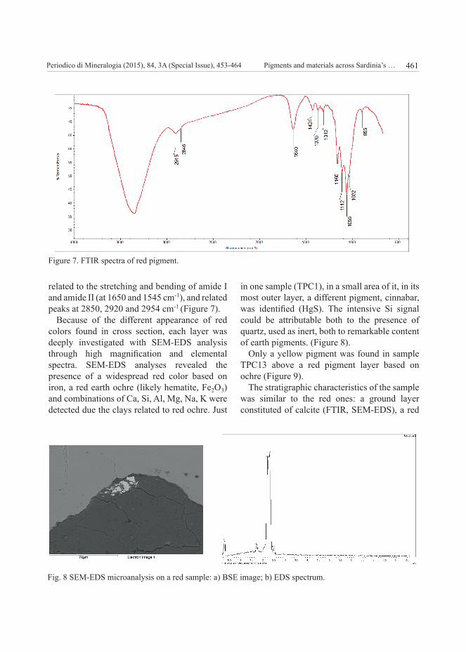
Spectra collected on the samples, in the ground and in the pigmented layers showed characteristics peaks of organic compounds, more precisely of proteinaceous material because of the bands related to amide I and amide II (1650 stretching ν C O amide I, and 1545 cm-1 bending δ N-H amide II) and their related peaks at 2850, 2920 and 2954 cm-1. Some spectra also showed the carbonyl ester band around 1738 cm-1 (ν C O stretching). The bands were sometimes badly resolved in cross section due to the strong presence of calcite and its strong signals and to their low amount in volume percentage. The comparison with references indicates the presence of egg (Figure 3).
In fresco painting technique, pigments were mixed with water in order to make a suspension, then applied on damp surfaces made of hydrated limes and aggregates so that the carbonation process involves calcite as binder. But in roman wall painting tempera is a quite common artistic technique employed (Botticelli, 2008).
FTIR spectra of the greyish layer showed the presence of calcite (1404 and 875 cm-1) and silicates. SEM-EDS microanalysis showed the presence of a phase composed of Ca, Mg, Si
and K. Because of the relative intensity of the peaks, a magnesium silicates, mixed with the calcium carbonate binder, seems to be involved in this dark layer. It can be attributable to a grain mineral used in order to create a tonal base for the blue pigment overall. Light elements as phosphorus were not identified, while carbon is related to the coating used to prepare the sample to the SEM-EDS analysis and The signal fraction of carbon possibly present in the sample can not easily distinguished (Figure 4).
FTIR spectra and SEM-EDS analysis of the blue crystals identified them as grains of Egyptian Blue. Egyptian blue particles were also distinguishable in BSE images because of their different appearance inside the pictorial layer. Phases with high atomic number, which appear brighter in BSE images, were composed of a Cu and Ca silicate. Egyptian blue is constituted by cuprorivaite, a calcium copper tetrasilicate (CaCuSi4O10) whose characteristics peaks are 1160, 1050, 1000 and 753 cm-1 (Figure 5) due to the stretching and bending of the Si-O-Si (Bevilacqua, 2010; Mirti, 1995; Kendrick, 2007; Mazzocchin, 2004).
This pigment was manufactured by mixing
Figure 3. FTIR spectra of the ground layer on sample TPC1.
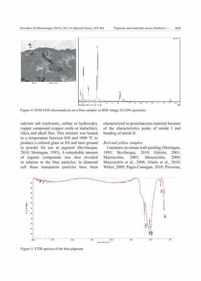
Periodico di Mineralogia (2015), 84, 3A (Special Issue), 453-464 459Pigments and materials across Sardinia’s …
calcium salt (carbonate, sulfate or hydroxide), copper compound (copper oxide or malachite), silica and alkali flux. This mixture was heated to a temperature between 850 and 1000 °C to produce a colored glass or frit and later ground to powder for use as pigment (Bevilacqua, 2010; Montagna, 1993). A remarkable amount of organic compounds was also revealed in relation to the blue particles: in diamond cell these transparent particles have been
characterized as proteinaceous material because of the characteristics peaks of amide I and bending of amide II.
Red and yellow samplesLiterature on roman wall painting (Montagna,
1993; Bevilacqua, 2010; Edreira, 2001; Mazzocchin, 2003; Mazzocchin, 2004; Mazzocchin et al., 2008; Aliatis et al., 2010; Weber, 2009; Pagès-Camagna, 2010; Piovesan,
Figure 4. SEM-EDS microanalysis on a blue sample: a) BSE image; b) EDS spectrum.
Figure 5. FTIR spectra of the blue pigment.

Periodico di Mineralogia (2015), 84, 3A (Special Issue), 453-464 L. Solla et al.460
2011; Amadori, 2015) reports that commonly cinnabar and red ochre were the most widespread red pigments. Stereomicroscopic observations showed that the untreated red samples are constituted of a ground layer, one or more red layers and, in some samples, are overlayed with whitish degradation products. Cross-section observation under visible and UV light confirmed the stratigraphy: a ground layer, with a UV yellowish fluorescence, one or more red pigmented layer with different color appearance. Red layers seem to be hardly fluorescent under UV light. Observation of the pigment grains in diamond cell showed a certain amount of particles with a bluish fluorescence under UV light (Figure 6).
Ground layers FTIR spectra show the presence of calcite (1404 and 875 cm-1) confirmed by SEM-EDS analysis (Gracia, 2001; Duran, 2011; Mazzocchin, 2004; Hradil, 2003; Edreira, 2001; Genestar, 2005).
Strictly concerning the red pigments and FTIR spectra it is important to remember that the most
commonly used red pigments in roman times were oxides and sulfides, transparent to FTIR analysis in the MIR region. SEM-EDS is one of the complementary analysis able to assess the elemental sample composition.
FTIR spectra of red layers detected the presence of silica (1170 and 1080 cm-1) and silicates (1030 and 1090 cm-1), usually associated with red ochre for the presence of clays. The presence of hematite (red ochre) and goethite (yellow ochre) was not directly recognizable by FTIR because of their oxides nature transparent in MIR spectral range, but SEM-EDS analyses showed in all the red cross sections a widespread presence of iron (TPC1, TPC8, TPC10, TPC15) attributable to their presence.
Peaks attributable to calcium oxalates were also visible (1370, 1312, 1112, and a shoulder around 790 cm-1) (Belfiore, 2012).
FTIR spectra also showed in the red samples the characteristic peaks of organic compounds, proteinaceous material because of the bands
Figure 6. Cross sections of red pigments observed under optical microscope in visible and UV light.
Periodico di Mineralogia (2015), 84, 3A (Special Issue), 453-464 461Pigments and materials across Sardinia’s …
related to the stretching and bending of amide I and amide II (at 1650 and 1545 cm-1), and related peaks at 2850, 2920 and 2954 cm-1 (Figure 7).
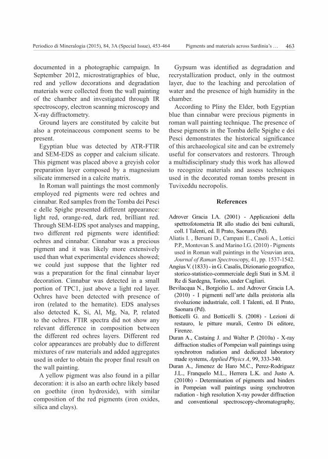
Because of the different appearance of red colors found in cross section, each layer was deeply investigated with SEM-EDS analysis through high magnification and elemental spectra. SEM-EDS analyses revealed the presence of a widespread red color based on iron, a red earth ochre (likely hematite, Fe2O3) and combinations of Ca, Si, Al, Mg, Na, K were detected due the clays related to red ochre. Just
in one sample (TPC1), in a small area of it, in its most outer layer, a different pigment, cinnabar, was identified (HgS). The intensive Si signal could be attributable both to the presence of quartz, used as inert, both to remarkable content of earth pigments. (Figure 8).
Only a yellow pigment was found in sample TPC13 above a red pigment layer based on ochre (Figure 9).
The stratigraphic characteristics of the sample was similar to the red ones: a ground layer constituted of calcite (FTIR, SEM-EDS), a red
Figure 7. FTIR spectra of red pigment.
Fig. 8 SEM-EDS microanalysis on a red sample: a) BSE image; b) EDS spectrum.

Periodico di Mineralogia (2015), 84, 3A (Special Issue), 453-464 L. Solla et al.462
layer constituted by red ochre and a yellow layer constituted of yellow ochre. BSE image showed a sample constituted by compounds with similar atomic number because of the similar gray-scale. SEM-EDS analysis revealed the presence of Fe (likely goethite, FeO(OH)) and combinations of (Figure 10) related to the clay of the ochre (Amadori, 2015).
In general the variety of pigments detected in the Tomba dei Pesci e delle Spighe coincides with those described in literature as commonly used in Roman wall paintings. Something that seemed particularly interesting was indeed related to the painting technique used. Romans extensively used lime as binder in wall paintings,
but they also used tempera to lay the pigments. In the samples investigated proteinaceous material was widely found in the pictorial layer as well as in the ground layer. It is well-known that the Romans used animal fats in order to slow down the carbonation of lime surfaces and obtain good adhesion of the upper tempera layers. In the Tomba dei Pesci e delle Spighe the recognized proteinaceous material was probably used with the same function.
Conclusions
In 1996 the roman Tomba dei Pesci e delle Spighe was discovered and its decorations
Figure 9. Cross section of the yellow pigment observed under optical microscope in visible and UV light on the sample TPC13.
Figure 10. SEM-EDS microanalysis on a yellow sample. a) BSE image; b) EDS microanalysis.
Periodico di Mineralogia (2015), 84, 3A (Special Issue), 453-464 463Pigments and materials across Sardinia’s …
documented in a photographic campaign. In September 2012, microstratigraphies of blue, red and yellow decorations and degradation materials were collected from the wall painting of the chamber and investigated through IR spectroscopy, electron scanning microscopy and X-ray diffractometry.
Ground layers are constituted by calcite but also a proteinaceous component seems to be present.
Egyptian blue was detected by ATR-FTIR and SEM-EDS as copper and calcium silicate. This pigment was placed above a greyish color preparation layer composed by a magnesium silicate immersed in a calcite matrix.
In Roman wall paintings the most commonly employed red pigments were red ochres and cinnabar. Red samples from the Tomba dei Pesci e delle Spighe presented different appearance: light red, orange-red, dark red, brilliant red. Through SEM-EDS spot analyses and mapping, two different red pigments were identified: ochres and cinnabar. Cinnabar was a precious pigment and it was likely more extensively used than what experimental evidences showed; we could just suppose that the lighter red was a preparation for the final cinnabar layer decoration. Cinnabar was detected in a small portion of TPC1, just above a light red layer. Ochres have been detected with presence of iron (related to the hematite). EDS analyses also detected K, Si, Al, Mg, Na, P, related to the ochres. FTIR spectra did not show any relevant difference in composition between the different red ochres layers. Different red color appearances are probably due to different mixtures of raw materials and added aggregates used in order to obtain the proper final result on the wall painting.
A yellow pigment was also found in a pillar decoration: it is also an earth ochre likely based on goethite (iron hydroxide), with similar composition of the red pigments (iron oxides, silica and clays).
Gypsum was identified as degradation and recrystallization product, only in the outmost layer, due to the leaching and percolation of water and the presence of high humidity in the chamber.
According to Pliny the Elder, both Egyptian blue than cinnabar were precious pigments in roman wall painting technique. The presence of these pigments in the Tomba delle Spighe e dei Pesci demonstrates the historical significance of this archaeological site and can be extremely useful for conservators and restorers. Through a multidisciplinary study this work has allowed to recognize materials and assess techniques used in the decorated roman tombs present in Tuvixeddu necropolis.
References
Adrover Gracia I.A. (2001) - Applicazioni della spettrofotometria IR allo studio dei beni culturali, coll. I Talenti, ed. Il Prato, Saonara (Pd).
Aliatis I. , Bersani D., Campani E., Casoli A., Lottici P.P., Montovan S. and Marino I.G. (2010) - Pigments used in Roman wall paintings in the Vesuvian area, Journal of Raman Spectroscopy, 41, pp. 1537-1542.
Angius V. (1833) - in G. Casalis, Dizionario geografico, storico-statistico-commerciale degli Stati in S.M. il Re di Sardegna, Torino, under Cagliari.
Bevilacqua N., Borgiolio L. and Adrover Gracia I.A. (2010) - I pigmenti nell’arte dalla preistoria alla rivoluzione industriale, coll. I Talenti, ed. Il Prato, Saonara (Pd).
Botticelli G. and Botticelli S. (2008) - Lezioni di restauro, le pitture murali, Centro Di editore, Firenze.
Duran A., Castaing J. and Walter P. (2010a) - X-ray diffraction studies of Pompeian wall paintings using synchrotron radiation and dedicated laboratory made systems, Applied Phyics A, 99, 333-340.
Duran A., Jimenez de Haro M.C., Perez-Rodriguez J.L., Franquelo M.L., Herrera L.K. and Justo A. (2010b) - Determination of pigments and binders in Pompeian wall paintings using synchrotron radiation - high resolution X-ray powder diffraction and conventional spectroscopy-chromatography,

Periodico di Mineralogia (2015), 84, 3A (Special Issue), 453-464 L. Solla et al.464
Archaeometry, 52 (2), 286-307.Duran A., Perez-Rodriguez J.L., Jimenez de Harob
M.C., Franquelob M.L. and Robadorc M.D. (2011) - Analytical study of Roman and Arabic wall paintings in the Patio De Banderas of Reales Alcazares’ Palace using non-destructive XRD/XRF and complementary techniques, Journal of Archaeological Science, 38 (9), 2366-2377.
Edreira M.C, Feliu M.J., Fernández-Lorenzo C. and Martín J. (2001) - Roman wall paintings characterization from Cripta del Museo and Alcazaba in Mérida (Spain), Talanta, 59 (6), 131-1139.
Elena F. (1868) - Scavi nella necropoli occidentale di Cagliari, Cagliari, pp. 261.
Genestar C. and Pons C. (2005) - Earth pigments in painting: characterization and differentiation by means of FTIR spectroscopy and SEM-EDS microanalysis, Analytical and Bioanalytical Chemistry, 382, 269-274.
Hradil D., Grygar T., Hradilova J. and Bezdicka P. (2003) - Clay and iron oxide pigments in the history of painting, Applied Clay Science, 22 (5), 223-236.
Kendrick E., Kirk C.J. and Dann S.E. (2007) - Structure and colour properties in the Egyptian Blue Family, M1−xM′xCuSi4O10, as a function of M, M′ where M, M′ = Ca, Sr and Ba, Dyes and Pigments, 73 (1), 13-18.
Mazzocchin G.A., Agnoli F., Mazzocchin S. and Colpo I. (2003) - Analysis of pigments from Roman wall paintings found in Vicenza, Talanta, 61 pp. 565-572
Mazzocchin G.A., Rudello D., Bragato C. and Agnoli F. (2004a) - A short note on Egyptian blue. Journal of Cultural Heritage, 5 (1), 129-133.
Mazzocchin G..A, Agnoli F. and Salvadori M. (2004b) - Analysis of Roman age wall paintings found in Pordenone, Trieste and Montegrotto, Talanta, Volume 64 (3), 73-741.
Montagna G. (1993) - I pigmenti: prontuario per l’arte e il restauro. Nardini editore, Firenze.
Pagès-Camagna S., Laval E., Vigears D. and Duran A. (2010) - Non-destructive and in situ analysis of Egyptian wall paintings by X-ray diffraction and X-ray fluorescence portable systems, Applied Physics A, 100, 671-681.
Piovesan R., Siddall R., Mazzoli C. and Nodaric L (2011) - The Temple of Venus (Pompeii): a study of the pigments and painting techniques, Journal od Archaelogical Science, 38 (10), 2633-2643.
Salvi D. (2000) - Tuvixeddu: vicende di una necropoli”, in Tuvixeddu: la necropoli occidentale di Karales: atti della Tavola rotonda internazionale La necropoli antica di Karales nell’ambito mediterraneo: Cagliari, 30 novembre - 1 dicembre 1996, a cura dell’Associazione culturale Filippo Nissardi. Cagliari, 139-177.
Salvi D. (1996) - Una tomba con i pesci, spighe ed altri fregi nella necropoli cagliaritana di Tuvixeddu: notizia preliminare, in: Quaderni, Ministero per i beni culturali e ambientali, Soprintendenza archeologica per le province di Cagliari e Oristano, 211-215.
Valdés (1902) - Guida pratica di Cagliari, Premiata Tipografia Editrice Pietro Valdés, Cagliari, p.261.
Weber J., Prochaska W. and Zimmermann N. (2009) - Microscopic techniques to study Roman renders and mural paintings from various sites, Materials Characterization, 60, 586-593.
Submitted, February 2015 - Accepted, September 2015