piezoelectric materials as stimulatory biomedical ... · 1 piezoelectric materials as stimulatory...
TRANSCRIPT

The University of Manchester Research
Piezoelectric materials as stimulatory biomedical materialsand scaffolds for bone repairDOI:10.1016/j.actbio.2018.04.026
Document VersionAccepted author manuscript
Link to publication record in Manchester Research Explorer
Citation for published version (APA):Tandon, B., Blaker, J. J., & Cartmell, S. H. (2018). Piezoelectric materials as stimulatory biomedical materials andscaffolds for bone repair. Acta Biomaterialia, 73, 1-20. https://doi.org/10.1016/j.actbio.2018.04.026
Published in:Acta Biomaterialia
Citing this paperPlease note that where the full-text provided on Manchester Research Explorer is the Author Accepted Manuscriptor Proof version this may differ from the final Published version. If citing, it is advised that you check and use thepublisher's definitive version.
General rightsCopyright and moral rights for the publications made accessible in the Research Explorer are retained by theauthors and/or other copyright owners and it is a condition of accessing publications that users recognise andabide by the legal requirements associated with these rights.
Takedown policyIf you believe that this document breaches copyright please refer to the University of Manchester’s TakedownProcedures [http://man.ac.uk/04Y6Bo] or contact [email protected] providingrelevant details, so we can investigate your claim.
Download date:29. Aug. 2019

Accepted Manuscript
Review article
Piezoelectric materials as stimulatory biomedical materials and scaffolds forbone repair
Biranche Tandon, Jonny J. Blaker, Sarah H. Cartmell
PII: S1742-7061(18)30229-0DOI: https://doi.org/10.1016/j.actbio.2018.04.026Reference: ACTBIO 5426
To appear in: Acta Biomaterialia
Received Date: 22 January 2018Revised Date: 19 March 2018Accepted Date: 15 April 2018
Please cite this article as: Tandon, B., Blaker, J.J., Cartmell, S.H., Piezoelectric materials as stimulatory biomedicalmaterials and scaffolds for bone repair, Acta Biomaterialia (2018), doi: https://doi.org/10.1016/j.actbio.2018.04.026
This is a PDF file of an unedited manuscript that has been accepted for publication. As a service to our customerswe are providing this early version of the manuscript. The manuscript will undergo copyediting, typesetting, andreview of the resulting proof before it is published in its final form. Please note that during the production processerrors may be discovered which could affect the content, and all legal disclaimers that apply to the journal pertain.

1
Piezoelectric materials as stimulatory biomedical materials and scaffolds
for bone repair
Biranche Tandon1,2
, Jonny J. Blaker*2
, Sarah H. Cartmell*1
1School of Materials, MSS Tower, The University of Manchester, Manchester, M13 9PL, UK
2Bio-Active Materials Group, School of Materials, MSS Tower, The University of Manchester,
Manchester, M13 9PL, UK
Corresponding Authors: *[email protected], +44 (0) 161 306 3567, and
*[email protected], +44 (0) 161 306 3587
Abstract
The process of bone repair and regeneration requires multiple physiological cues including
biochemical, electrical and mechanical - that act together to ensure functional recovery.
Myriad materials have been explored as bioactive scaffolds to deliver these cues locally to the
damage site, amongst these piezoelectric materials have demonstrated significant potential
for tissue engineering and regeneration, especially for bone repair. Piezoelectric materials
have been widely explored for power generation and harvesting, structural health monitoring,
and use in biomedical devices. They have the ability to deform with physiological
movements and consequently deliver electrical stimulation to cells or damaged tissue without
the need of an external power source. Bone itself is piezoelectric and the charges/potentials it
generates in response to mechanical activity are capable of enhancing bone growth.
Piezoelectric materials are capable of stimulating the physiological electrical
microenvironment, and can play a vital role to stimulate regeneration and repair.

2
This review gives an overview of the association of piezoelectric effect with bone repair, and
focuses on state-of-the-art piezoelectric materials (polymers, ceramics and their composites),
the fabrication routes to produce piezoelectric scaffolds, and their application in bone repair.
Important characteristics of these materials from the perspective of bone tissue engineering
are highlighted. Promising upcoming strategies and new piezoelectric materials for this
application are highlighted.
Statement of significance
Electrical stimulation/electrical microenvironment are known effect the process of bone
regeneration by altering the cellular response and are crucial in maintaining tissue
functionality. Piezoelectric materials, owing to their capability of generating
charges/potentials in response to mechanical deformations, have displayed great potential for
fabricating smart stimulatory scaffolds for bone tissue engineering. The growing interest of
the scientific community and compelling results of the published research articles has been
the motivation of this review article. This article summarizes the significant progress in the
field with a focus on the fabrication aspects of piezoelectric materials. The review of both
material and cellular aspects on this topic ensures that this paper appeals to both material
scientists and tissue engineers.
Keywords
Bioactive; Bone Tissue Engineering; Scaffold fabrication; Characterization; Electrical
stimulation; Nanofibres; 3D printing
Graphical abstract

3
1. Introduction
The biological performance of a material depends on the extent it is capable of mimicking the
microenvironment and delivering cues to stimulate cellular response, a property demonstrated
by some piezoelectric materials [1–4]. Bioelectrical signals [5,6], endogenous electrical fields
[6,7] and external electrical stimulation [8–10] play crucial roles in modulating cellular
behaviour and contribute to bone repair. Piezoelectric materials are capable of delivering
these electrical cues without the need of an external stimulation device and able to enhance
the physiological electric environment to stimulate repair [11–13]. These materials also show
electromechanical behaviour (converse piezoelectric effect) and can be driven by
physiological electrical changes to give rise to mechanical cues. There has been a rapid
increase in the number of publications on the use of piezoelectric materials for bone tissue
4
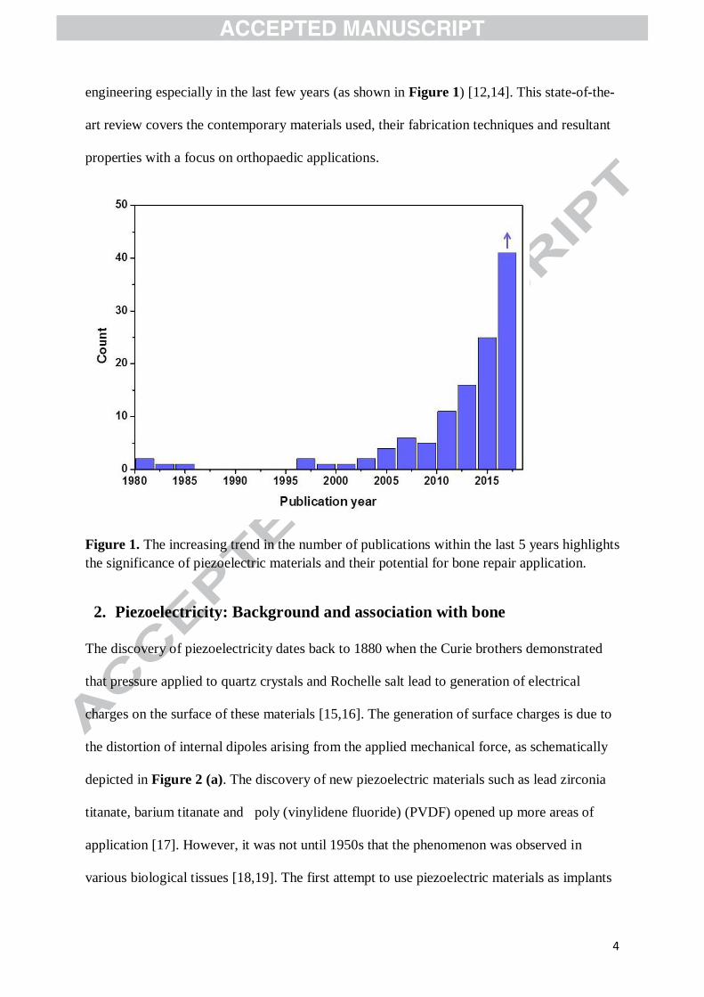
engineering especially in the last few years (as shown in Figure 1) [12,14]. This state-of-the-
art review covers the contemporary materials used, their fabrication techniques and resultant
properties with a focus on orthopaedic applications.
Figure 1. The increasing trend in the number of publications within the last 5 years highlights
the significance of piezoelectric materials and their potential for bone repair application.
2. Piezoelectricity: Background and association with bone
The discovery of piezoelectricity dates back to 1880 when the Curie brothers demonstrated
that pressure applied to quartz crystals and Rochelle salt lead to generation of electrical
charges on the surface of these materials [15,16]. The generation of surface charges is due to
the distortion of internal dipoles arising from the applied mechanical force, as schematically
depicted in Figure 2 (a). The discovery of new piezoelectric materials such as lead zirconia
titanate, barium titanate and poly (vinylidene fluoride) (PVDF) opened up more areas of
application [17]. However, it was not until 1950s that the phenomenon was observed in
various biological tissues [18,19]. The first attempt to use piezoelectric materials as implants

5
for bone was made in the 1980s [20]. Since the discovery of piezoelectric effects in bone by
Fukada and Yasuda in 1957 [21], there have been extensive studies on electromechanical
effects in bone and their role in modulating cellular behaviour to control growth and the
remodelling processes [22–26]. Several theories suggested for the origin of stress generated
potentials (electromechanical effect) in bone have been reviewed by Rajabi et al [14].

6
Figure 2 (a) Schematic of direct and converse piezoelectric effect. (b) types of poling
processes used to align dipoles and improve the piezoelectric characteristics of the material,

7
(c) dependence of functional mode of operation on direction of implantation. Piezoelectric
constants (d31, d32 and d33) are different in magnitude and lead to different magnitudes of
electrical charges or potentials under the influence of similar mechanical stimuli.
A schematic of direct and converse piezoelectric effects, defined as voltage generation on
mechanical deformation, and mechanical deformation on application of voltage, respectively
is shown in Figure 2 (a) [27]. These effect are observed in materials where a mechanical
deformation causes the formation of a net dipole moment and subsequent polarization of the
material [17]. The phenomenon of piezoelectricity can also be observed after randomly
arranged dipoles throughout the material are poled and/or aligned under the influence of high
electric field at a specific temperature [28]. A schematic of the types of poling processes used
for inducing or improving the piezoelectric behaviour in various materials is given in Figure
2 (b). Corona poling is preferred over thermal (oil/fluid) poling to avoid sample
contamination, especially for use in biological and cellular studies. Corona poling and
thermal poling in air have both been applied to pole dense and porous materials, and can be
used for inducing piezoelectric characteristics in complex scaffold structures [11,29,30]. The
magnitudes of temperature and voltage used for poling depend on type of piezoelectric
material and can be controlled to achieve quick, efficient and successful poling.
The piezoelectric charge constant (‘dij’ constant) is an expression of the amount of charge
that the material generates in response to stress applied or alternatively, and represents the
strain experienced by the material per unit electric field applied. The first subscript ‘i’
denotes the direction in which the polarization is generated (or applied electric field) and the
second subscript ‘j’ denotes the direction of applied stress (or induced strain). The subscript
values 1, 2 and 3 represent the orthogonal axes and 4, 5 and 6 denote rotation around their
respective axes (as shown in Figure 2 (c)). The piezoelectric constants of materials used in
the field of bone tissue engineering are summarized in Table 1.

8
Some piezoelectric materials are initially non polar (uncharged), with charges only generated
when stressed; others are permanently polar, i.e. they carry a net dipole moment without the
application of any force. The latter forms a sub-class of piezoelectric materials, termed
ferroelectrics [31]. Ferroelectricity can alter the surface charge (zeta-potential (ζ-potential))
of materials in the wet environment by altering the interactions of ions and salts present in the
fluid around it, particularly relevant for the physiological environment. This can also affect
the adhesion of proteins on material surfaces, influencing the behaviour of cells, and
consequently the process of tissue regeneration.
Table 1 Piezoelectric materials for bone regeneration, and their piezoelectric constants [*d32
and d31 may be same or different depending on whether the films are bi axially or mono
axially stretched; **
piezoelectric materials which have gathered significant interest in the last
decade; ±the piezoelectric constants reported are significantly smaller than bulk ceramics but
are comparable to bone; † [32] can be referred to for details on negative sign of constants]
Material Piezoelectric constant
(pico Coulombs/N)
Ref.
Polymers Poly(L-lactide) (PLLA) d14 = -9.82 [14,33]
Poly(vinylidene fluoride) (PVDF) d33 = -32† , d31 = 6.7
* [34–37]
Poly(vinylidene fluoride-trifluro
ethylene) (PVDF-TrFE)
d33 = -25.2 to -38 δ [38–40]
Poly hydroxy butyrate (PHB) d14 = 1-2 [12,41]
Ceramics Hydroxyapatite (HA) d33 = 1.5 - 2.4 [42]
Barium titanate (BT) d33 = 191 [14,43]
Lithium sodium potassium niobate
(LNKN)
d33 = 98 [44]
Lithium niobate (LN) d33 = 23 [45]
Boron nitride nano tubes (BNNT)**
d33 = 0.3 [14,46]
Potassium sodium niobate (KNN) d33 = 93 [47]
Natural materials Diphenylalanine (FF)**
d33 = 18, d31 = 4 [48]
Collagen**
d15 ~ 2 [14,18,
49]
Bone d15 = 0.1-0.7 [50,51]
Silk**
d14 = -1.5 [52]
Composites HA/BT d33 = 0.6 to 2.8±
[30,53]
LNKN/HA d33 = 2±
[54]

9
3. Characteristics of piezoelectric materials for biomedical use
3.1. Surface character
It is well known that the surface morphology/topography and chemistry modulate protein
adhesion and consequently affect cellular behaviour [55,56]. These characteristics also affect
the wettability and surface charge (ζ-potential) of the scaffolds governing interactions at
material-cell interface [57,58]. Surface properties can alter the protein adhesion and give rise
to varying cell response accordingly. Different fabrication techniques provide control over
morphology of the scaffolds and further functionalization of the materials can be applied to
immobilize biochemical agents [59,60]. The piezoelectric constants and ferroelectricity of
the piezoelectric materials can be optimized and the topographical characteristics tailored
according to requirement.
Control over morphology of scaffolds is important to correctly and independently assess the
contribution of surface charge towards cellular behaviour. A study on hydroxyapatite/barium
titanate (HA/BT) composites involved polishing of composite materials to obtain similar
topography and roughness [61]. This post processing ensured that the observed differences in
cellular behaviour were correctly correlated with the polarization of piezoelectric samples.
Interestingly, it was observed that the polarization of scaffolds had little effect on cellular
activities at day 7 [61].
Aside polarization, the morphology and roughness of piezoelectric scaffolds affects cellular
behaviour. PVDF films and fibrous scaffolds have been shown to alter cell proliferation and
alignment, suggesting that cellular behaviour can be tailored accordingly [57,62]. It has been
reported that the roughness of coated polymer (PVDF films in this case, [35]) contributes to
changes in cellular behaviour along with surface charge. Nano/micro roughness of the surface
10
has been known to cause changes to surface energy and wettability which has a direct effect
on cellular behaviour [35,56,63].
3.2. Ferroelectricity
Recently, the charge distribution around the surface of certain ferroelectric materials
immersed in fluid have been characterized by ζ-potential measurements [11,45]. These
generated potentials of -30 to -80 mV have been associated with improved cellular response
on these materials [11,45]. It has been suggested that enhanced interfacial polarization in
ferroelectric composites is the main contributing factor towards increasing ζ-potential [11].
However, due to the limited number of studies where ζ-potential of bulk ferroelectric
materials has been measured, the correlation between the polarization and ζ-potential of
ferroelectric materials cannot be generalized.
The association of the nature of surface charge (positive or negative) of electroactive
materials to cellular responses is hard to establish and ζ-potential measurements provide
better insights to the correlation [45,64]. Surface charges of piezoelectric materials are altered
in response to different mechanical deformations, and recording these minute ζ-potential
variations is important to control cellular response. A small variation in surface potentials can
lead to a change in local electro kinetics and directly affect cellular behaviour. In case of HA,
it was observed that positive or negative surfaces had little or no difference in contact angle
[65], while significant differences were noted in case polarized PVDF films [66]. The values
observed for negative and positive HA surfaces differed by 2° while in case of PVDF, this
difference was noted to be 20° which could be due to the altered surface chemistry as a result
of corona poling [65,66]. Two different studies by Dubey et al demonstrated that
polarizability and nature of surface charge of potassium sodium niobate (KNN) and its
laminated composites play a key role modulating cellular behaviour and bioactivity of the

11
composites, respectively [54,67]. They also suggested that conductivity of KNN might also
be responsible for increased proliferation of human osteoblast-like Saos2 cells [67].
Kelvin probe force microscopy (KPFM) has been utilized for nanoscale imaging of the
surface potential of different types of materials including metals [68], semi-conductors
[68,69], ferroelectrics [70,71], piezoelectrics [70], chemically functionalized, cell compatible
scaffold surfaces [72,73] and biological materials [70,72,74–78]. However, there are
conflicting reports correlating KPFM observations with surface potential; Rohm et al
conducted KPFM measurements on perovskite thin films of methyl ammonium lead iodide
and observed no correlation between ferroelectric domains and surface potential while results
obtained in time resolved KPFM experiments performed by Strelcov et al on lithium niobate
surfaces confirmed that ferroelectric domains had an influence on surface potential [79,80].
In another study, PVDF-TrFE thin films displayed a correlation in ferroelectric, piezoelectric
and surface potential characteristics [70]. However, due to the presence of screening charges
(charges/adsorbates which compensate the surface electrical properties), it is difficult to map
the surface potentials using this technique and establish a correlation between ferroelectric
domains and surface potentials [81,82].
3.3. Magnitude of voltage/charge generated
The most commonly used constant to describe the piezoelectricity of materials used for bone
tissue engineering is the piezoelectric strain constant. Piezoelectric constant has been
measured and shown to be comparable or greater than the values observed for bone.
Piezoelectric constant of up to 0.7 pC/N has been observed for bone in the shear mode [51].
Values reported for scaffolds are significantly higher in many instances and the charges
generated can be higher than bone when dynamic mechanical stimulus is applied. However,
if these charges are not found to be high enough to elicit a beneficial cellular response, there
will be a need to improve piezoelectric characteristics of the scaffolds. In a study published in
12
2017, Wang et al fabricated core-shell structured composite sub-micron fibres of PVDF and
graphene oxide and reported an increase in piezoelectric constant of 426% in comparison to
neat PVDF fibres [83]. Incorporation of different fillers can help in tuning the piezoelectric
behaviour for bone repair applications [84–86]. The electrical potential output generated by
the materials can also be enhanced by physical structural modification post-fabrication [87].
It is also important to note that piezoelectric property of a scaffold has a role to play only
under controlled dynamic conditions. Piezoelectric scaffolds must be exposed to similar
levels of mechanical deformations as experienced by the cells in vivo to analyse the effect of
charge generated as a consequence of piezoelectric effect; higher values of strain can lead to
cell death [88]. These deformations are in the range 0-0.4% under normal physiological
conditions, whereas on fracture, values up to 15% have been hypothesized [89–91]. A
vertical vibrating plate was used to mechanically stimulate the materials in two different
studies involving dynamic stimulation. However, the amount of mechanical deformation and
consequently, the amount of charge experienced by the cells and the deformation induced in
the scaffolds was not quantified [35,92].
The mode in which a piezoelectric scaffold is stimulated, and the value of piezoelectric
constant are important and interlinked. The importance of directionality (or functional mode)
was highlighted by Feng et al in a study using HA/BT composites in vivo [93]. The
functional mode of operation of the material might differ depending on the direction of
implantation of the piezoelectric scaffold in vivo, as depicted in Figure 2 (c).

13
Figure 3 PFM is a useful tool to study nanoscale interactions by mapping piezoelectric
response of different materials. a) Surface topography, b) PFM phase and c) PFM amplitude
of a PVDF/GO nanofibre; d) AFM topography e) PFM amplitude and f) PFM phase images
of FF nanotubes, and g) a vector PFM map representing the local electromechanical response,
where the colour indicates the molecular orientation direction and the intensity indicates the
magnitude of electromechanical response. Printed with permission from [83,94,95].
3D porous structures can be poled using the existing methods such as corona poling, however
the challenge is to characterize the piezoelectric properties of these structures. Porous
ceramic composites have been characterized using a piezo constant meter. A d33 or d31

14
meter which measures the amount of charge generated in the direction of poling when the
force is applied in a parallel direction (d33 mode) or perpendicular (d31) to it. Piezoresponse
force microscopy (PFM) is a useful AFM-based tool to study the piezoelectric properties of a
variety of synthetic and biological materials at the nano scale, as reviewed by Bystrov et al
and Rodriguez et al [96,97]. PFM has been applied to polymeric fibres and self-assembling
peptide nanotubes (summarized in Figure 3), and can provide insight to understand the
piezoelectric property distribution and its effect on cellular interactions. However, the
limitations of this technique should be considered to ensure that artefacts are not
misinterpreted as true signals [98–100]. A comparison of the piezoelectric properties is only
possible if both polymeric and ceramic materials are characterized similarly, facilitating
understanding of the biological response of these materials.
3.4. Mechanical and physical characteristics
The physical and mechanical properties of scaffolds, including porosity, degradation and
Young’s modulus have key roles in governing the biological performance of piezoelectric
materials. The porosity of the scaffolds need to be controlled to ensure cellular migration,
response and optimize mechanical and piezoelectric properties [30,101]. Piezoelectric
properties of different materials are influenced differentially by porous structure. The study
of porous HA/BT composite scaffolds and their piezoelectric properties displayed an inverse
relation between porosity and piezoelectric constant [30]. The enhancement of piezoelectric
property with increase in density of scaffolds was attributed to the better inter-connectivity of
the active piezoelectric phase of the ceramics [30]. In a different study of porous arrays of
PVDF, higher piezoelectric potential and current values than bulk PVDF films were
demonstrated [101]. These studies suggest towards the possibility of fabrication porous
piezoelectric materials with tuneable porosity and piezoelectric characteristics. The
mechanical properties of piezoelectric scaffolds are important to restore the functionality of

15
the substituted tissue in vivo, in particular material stiffness has a key role to play in cell-
material interactions [102,103]. Boyan et al demonstrated that substrate stiffness of
polymeric surface can modulate the response of mesenchymal stem cells [103]. Further, it is
important to assess the stability of electrical properties such as ferroelectricity, the nature of
the surface charge and piezoelectric constants to ensure that these properties are stimulatory
over the time needed for regeneration, and that any shielding of their properties is tuned.
Shielding can occur due to the cells/tissue growing around the scaffold and render the
scaffold inactive. A major limitation of most studied piezoelectric materials, such as PVDF
and BT, is their non-degradability. Polyhydroxyalkanoates, semi-crystalline poly(α-hydroxy
acids) such as poly(L-lactide) (PLLA) and natural polymers such as silk are gaining traction
as degradable matrices for piezoelectric scaffolds [104–106].
4. Synthesis and processing of piezoelectric materials
4.1. Piezoelectric polymers
The origin of piezoelectricity in polymers is attributed to the inherent crystal or chemical
structure of the polymer material, which induces a net dipole/charge on mechanical
deformation. Piezoelectric polymers have mainly been fabricated in three different
morphologies; films, rods or tubes and fibres [12]. The main piezoelectric polymers
investigated for their osteogenic capacity are PVDF, its copolymers (poly (vinylidene
fluoride-trifluro ethylene)(PVDF-TrFE) and poly (vinylidene fluoride-
hexafluoropropylene)(PVDF-HFP), and PLLA [14]. A direct comparison of the ‘d’ constant
of PLLA and PVDF (shown in Table 1) is hard to establish as they are observed in different
activation modes (subscripts ‘i’ and ‘j’ denote activation modes). The ferroelectric behaviour
of PLLA has only be observed at high temperatures and is known to decay fairly quickly at
room temperature, whereas for PVDF it is stable and observable at room temperature

16
[33,107–109]. The principal methods used for fabricating piezoelectric polymers for bone
tissue engineering are solvent casting, compression moulding (most commonly for films) and
fibre spinning (e.g. electrospinning).
4.1.1. PVDF and its copolymers
PVDF is a semi crystalline polymer known to present five different crystalline phases: α
(alternating trans-gauche conformation (TGTG’)), β (all trans conformation (TTTT)), γ
(alternating (triple trans)-gauche (T3GT3G’)), δ (alternating trans-gauche (TGTG’)) and ε
(alternating triple trans-gauche (T3GT3G’)) [110–114]. The different conformations
determine the polar/non-polar nature of the polymer chain as shown Figure 4. The polar β, γ
and δ phases are formed when the dipoles are arranged parallel to each other and
perpendicular to the polymer chain, while the non-polar α and ε phases are formed when the
net dipole moment cancels due to anti parallel arrangement [115,116]. The polar β- phase is
of interest as it presents high piezoelectric characteristics to the material [115]. Poling of β-
PVDF further improves its piezoelectric characteristics. PVDF-TrFE exists in all trans
conformation (β- phase) and possesses a high electromechanical and piezoelectric coefficient
for specific monomer concentrations [117–120]. PVDF-TrFE does not require special
processing techniques to obtain β- phase crystalline structure and can easily be formed into
complex/porous structures desirable for tissue engineering applications. PVDF-HFP has a
lower crystallinity when compared with PVDF, yet presents good ferroelectric and
piezoelectric behaviour [116,121].

17
Figure 4 Chain confirmations of different phases of PVDF and PVDF-TrFE. Dipole moment
cancels out in α- phase which is not the case for piezoelectric in PVDF-TrFE and β- phase
and γ- phase of PVDF.
Various ways by which PVDF and its copolymers can be obtained in β- phase and
subsequently be made piezoelectric by poling are shown in Figure 5. These methods have
been reviewed in detail by Lanceros-Mendez et al [116]. Two recent works by H. Horibe et
al have highlighted the control of phases using different solvents and anti-solvents during
synthesis procedure [122,123]. It was observed that a solvent with higher dipole moment
resulted in formation of polar β- phase which was also supported by slower solvent
evaporation rate [122,123].
The techniques that involve stretching, such as electrospinning, can result in higher β- phase
than α- phase [35,66,92,124,125]. In a series of studies by Ribeiro et al, PVDF films were
produced with desired piezoelectric properties and tested for their osteogenic capability
[35,66,92,125]. The films were solvent cast into rectangular samples, annealed and
mechanically stretched and corona poled to maximize the piezoelectric performance.

18
Stretching only induces the formation of β- phase and the films need to be poled to align and
lock the dipoles to observe increased piezoelectric response [126,127]. The temperature for
mechanical stretching and the draw ratio was optimized in previous studies by the same
group [34,128]. Protein adsorption, proliferation of pre-osteoblastic cells and the
differentiation of human adipose derived stem cells into osteogenic lineage were found to be
positively affected by these piezoelectric substrates [35,66,92,125]. Piezoelectric PVDF films
and fibres have also been assessed in vivo and their suitability well demonstrated as bone
substitutes [129].
Figure 5 Fabrication methods to obtain PVDF and its copolymers (PVDF-TrFE, PVDF-HFP)
in β- phase.
Piezoelectric polymers have also been utilized as suitable coatings for existing implant
materials for tissue engineering applications [130,131]. In a recent study, PVDF films were

19
deposited on titanium sheets and poled using corona discharge to enhance the bioactivity of
the surface of Ti sheets [131]. A solution of PVDF in N, N-dimethyl formamide (DMF) and
acetone was prepared and cast over a Ti surface. Poling of the samples was carried out using
a corona discharge at 100°C and the effect of polarization on bone marrow mesenchymal
stem cell behaviour assessed. Polarized samples exhibited enhanced cell proliferation (at days
4 and 7) and osteogenic differentiation (higher alkaline phosphatase (ALP) activity at day 21)
of the cells as compared to non-polarized ones.
The converse piezoelectric effect has also been explored and mechanical stimuli delivered
through PVDF actuators has been shown to be effective in stimulating bone growth
[132,133]. This aspect is important to study as patients are immobilized in the early stages
following an injury/fracture. Immobilization leads to an absence of mechanical stimulation
(through physiological movements) to piezoelectric materials for electrical cue generation.
The converse effect can therefore be used in the early stages to improve the biological
performance of the scaffolds. This effect may be driven by external electrical stimulation
devices, implanted batteries or varying physiological electrical environment.
Electrospinning of randomly aligned PVDF fibres/fibre mats have been shown to eliminate
the need of stretching and poling the polymer constructs [134–136]. PVDF copolymer
nanofibre mats obtained by electrospinning are also reported to be piezoelectric and can have
possible application in tissue engineering [137,138]. Electrospinning requires optimization of
various solution and processing parameters to control the diameter and morphology of the
fibres [139]. Arinzeh et al used electrospinning for the fabrication of fibrous PVDF scaffolds
at different voltages applied during the spinning and investigated the effects on β- phase
formation and differentiation of human mesenchymal stem cells in osteogenic lineage [124].
The formation of electroactive phase was found to be effected by the magnitude of voltages
used. The study demonstrated that electrospun scaffolds showed higher ALP activity and

20
matrix mineralization suggesting promoted differentiation of cells into osteogenic lineage. In
a recent study, electrospun scaffolds of PVDF-TrFE were dynamically stimulated to guide
human mesenchymal stem cells into osteogenic lineage [140]. It was found that streaming
potentials of ~61.1μV were generated on dynamic stimulation of annealed scaffolds. These
scaffolds were observed to show increased mineralization and osteogenic gene expression at
day 28 compared to as spun PVDF-TrFE and PCL scaffolds [140].
Films and electro spun mats of PVDF-TrFE have been observed to have different levels of
crystallinity and β- phase content which can play a key role in modulating cellular behaviour
under static and dynamic conditions [62,141]. Porous films of PVDF-TrFE and PVDF-HFP
have also been used and compared with PVDF for suitability in bone tissue engineering
[142]. This study shows that PVDF-TrFE and PVDF-HFP present lower amount of β- phase
than PVDF under similar processing conditions, however, it has been suggested that PVDF-
TrFE is not suitable for bone tissue engineering applications as these scaffolds promoted an
elongated cellular morphology [142]. PVDF microspheres have also been fabricated using the
electrospray technique and their suitability for bone tissue engineering shown using
MC3T3E1 cells and human mesenchymal stem cells [143,144]. However, the role of
piezoelectricity was not detailed in these studies.
4.1.2. PLLA
Semi-crystalline PLLA does not require any additional poling treatment to induce
piezoelectric effect. This has been attributed to the displacement of the C=O bond in PLLA in
response to mechanical stress leading to generation of a net dipole moment and charge [145–
147]. PLLA has been used to fabricate films, fibres and rods with piezoelectric behaviour
[148–151] and it has been observed that crystallinity and polymer orientation play key role in
piezoelectric characteristic [152]. It presents an additional advantage of fabricating
degradable scaffolds [153–155]. Biocompatible PLLA microspheres have displayed good

21
potential for tissue engineering applications [156–158], however, the piezoelectric properties
of those microspheres synthesized have not been studied to the author’s knowledge. Different
morphologies in which piezoelectric materials have been synthesized are shown in Figure 6
(a).
PLLA-based scaffolds have been synthesized in various forms and their suitability as bone
substitutes evaluated extensively; however, the role of piezoelectric characteristics in
controlling cellular behaviour requires further attention. Fabrication techniques used for
manufacturing PLLA based scaffolds have been reviewed by Laurencin et al [159]. In one of
the first attempts on use of PLLA as bone substitute, Ikada et al fabricated PLLA films and
rods which were drawn to fabricate piezoelectric scaffolds to be implanted in cat tibiae [160].
Callus formation around implants was reported to be dependent on the draw ratio of these
piezoelectric scaffolds [160]. Such behaviour was attributed to the currents generated by
strains in piezoelectric scaffolds due to the movements generated by animal while walking.
A selection of pertinent studies in which piezoelectric polymers have been used to test their
feasibility for orthopaedic applications is given in Table 2. It can be seen that piezoelectric
polymers have been used to manufacture 3D scaffolds with only one study exploring the role
of piezoelectricity under dynamic stimulation. Further to this, the effect of protein adhesion
on piezoelectric properties remains to be explored. The existing techniques in combination to
the emerging approaches discussed in section 7 have great potential for tissue engineering
applications and should be used to fabricate 3D constructs to leverage control over cellular
behaviour.
Table 2 Fabrication techniques of piezoelectric polymers and their assessment using different
cell lines for orthopaedic application
Material Fabrication/ processing technique
Cell type Loading regime Key findings Ref.

22
PVDF
Films coated on titanium sheets via solvent casting and poled using corona discharge
Bone marrow mesenchymal stem cells
No mechanical loading (Static)
Increased proliferation, ALP activity and osteogenic gene expression
[131]
Mechanically drawn and electrically poled solvent cast (Drying at 120°C) films
Human adipose stem cells
Dynamic analysis was carried out using a vertical vibration module
Increase in ALP activity in piezoelectric samples under dynamic stimulation
[92]
Static. Comparison of poled and non-poled samples
No significant difference observed in osteogenic differentiation of cells
[66]
MC3T3-E1 murine pre- osteoblasts
Dynamic Increased proliferation on piezoelectric samples under dynamic stimuli
[35]
Static. Comparison between poled β, unpoled β and α- phase samples
Higher cell viability and similar cell density in all samples at day 7
[125]
Electrospun fibres
Human mesenchymal stem cells
Static. Comparison of fibres spun at different voltages
Increased ALP activity Increased mineralization
[124]
PVDF, PVDF-HFP and PVDF-TrFE
Porous solvent cast (Room temperature drying) membranes
MC3T3-E1 murine pre- osteoblasts
Static
Cell proliferation and morphology were influenced by porosity and microstructure of membrane surface
[142]
PVDF-TrFE
3D electrospun scaffolds
Human
mesenchymal
stem cells
Dynamic analysis
using sinusoidal
compression
Enhanced osteogenic differentiation on stimulated scaffolds
[140]
PLLA Piezoelectric films and rods
In vivo analysis Increased callus formation around piezoelectric implants
[160]
4.1.3. Polymer matrix composites and their application as bone substitutes
Polymer matrix composites (PMCs) allow for combination of manufacturing flexibility and
the processing of otherwise brittle ceramics with higher piezoelectric constants to complex
forms. PLLA based composites have extensively been studied for application in the field of
bone tissue engineering [161,162], as reviewed in [159]. Though piezoelectricity has been
observed in electrospun fibre mats based on PLLA [148,163–165], the focus has been to
improve the suitability of the scaffolds for desired application under static environment

23
without the mention of the contribution of piezoelectric properties of fillers or PLLA itself
[166,167].
Table 3 Filler loading percentages used in different studies (number in bracket represents
filler % used for biological studies).
Base material Filler type (particles) Loading percentages Refs
PVDF-TrFE BT
0-5% v/v (5%) [11]
10% v/v [168]
BNNTs 1% w/w [169]
HA BT
0-80% w/w (40%) [53]
90% v/v [61]
40% [170]
20-80% v/v (80%) [171]
50-90% v/v [30]
90% v/v [172]
80% and 90% w/w [173]
Polymer matrix composites can be produced, taking advantage of the flexibility of polymer
processing techniques, and impart piezo-electrical and bioactive behaviours [174].
Membranes of PVDF-TrFE/BT have been reported to support bone formation in several
studies [11,168,175,176]. A recent study published in 2016, Chen et al used poled solvent
cast membranes of PVDF-TrFE/BT to explore the suitability of membranes for repairing
bone defects [11]. BT nano particles were surface modified with polydopamine (PDA) to
improve their dispersion in the polymer matrix, and reduce their tendency to agglomerate or
sediment during processing [11]. Interestingly, instead of mentioning the piezoelectric
properties, the authors highlighted that ferroelectric property of these membranes have a
crucial role to play and help in mimicking the physiological electrical micro-environment
(steady state bioelectric potential). The surface potential of the samples was found to be
dependent on the increased polarization of the samples due to the presence of nano sized BT
[11]. The study shows that the composite membranes have promising potential for clinical
application owing to the improved osteogenic capability demonstrated in vitro and in vivo.
Improved osteogenic behaviour of bone marrow derived mesenchymal stem cells was

24
observed in vitro and enhanced healing of a parietal bone defect in rats was observed in in
vivo experiments [11]. PMCs used for orthopaedic applications, along with the results
obtained are listed in Table 4.
Table 4 Summary of key findings of studies involving use of PMCs. (*Static represents the
assumption that treating defects in skull did not initiate a piezoelectric response.)
Material Fabrication/ processing technique
Cell type Loading regime
Key findings Ref.
PVDF-TrFE-BT
Solvent casting followed by poling
Bone marrow Mesenchymal stem cells and In vivo
Static*
Improved cellular activity and favourable osteogenic differentiation. Rapid bone regeneration in vivo
[11]
In vivo Static*
Higher gene expression of key osteoblast markers was observed around composite membranes
[168]
Solvent extraction using water followed by hot pressing
In vivo Static*
Similar or better morphometric parameters than Polytetrafluoroethylene (PTFE) membranes. A mixed profile of gene expression was observed
[175]
Unlike this study which involved no external mechanical stimulation, Genchi et al used
ultrasound to mechanically stimulate piezoelectric PVDF-TrFE/BNNT composite films and
evaluated the osteogenic differentiation capacity of Saos-2 osteoblast-like cells [169]. Cell
culture on films was performed in presence and absence of ultrasound to analyze the
contribution of piezoelectric behaviour of the films. Composite films were manufactured by
dispersing BNNTs into the solvent and mixing them with the polymer solution. It is
important to note that methylethylketone (MEK) was used as the solvent in this study as
solvents like DMF and DMAc. It was found through simulation that the electrical potentials
generated as a consequence of piezoelectric effect were in the range suitable for cellular
stimulation [169]. Solvent free routes such as dry powder mixing and compression moulding
and addition of bioactive fillers have also been utilized to fabricate piezoelectric scaffolds
[177]. The composites prepared displayed enhanced bioactivity through increased apatite

25
formation over sample surfaces. The percentages of fillers used in selected piezoelectric
composite materials for bone repair are summarized in Table 3.
4.2. Ceramics
Barium titanate (BT), lithium niobate (LN), potassium sodium niobate (KNN),
hydroxyapatite (HA) lithium sodium potassium niobate (LNKN) and zinc oxide (ZO) are
lead-free piezoelectric ceramics that provide a better alternative to lead-based systems which
present toxicological risks [14,178–181]. Park et al conducted studies in which specialized
cylindrical samples of BT were fabricated and implanted in dog femora in an attempt to
augment the process of bone repair [20,182]. The piezoelectric samples were fabricated by
slip casting, firing and subsequent poling of BT powder. The poling of samples was carried
out in a fluid filled chamber with a constant electric field. The results of the study were
appealing in terms of compatibility and interfacial strength of the material with surrounding
tissue. However, no significant differences were observed for piezoelectric and non-
piezoelectric samples.

26
Figure 6 (a) Different morphologies of piezoelectric polymers or polymer matrix composites
explored for bone repair application, (b) Ceramics have been used in a variety of scaffold
morphologies which have been found suitable for bone tissue engineering application
[30,54,183–186] (c) Fabrication techniques for piezoelectric ceramic (or ceramic matrix
composite) based scaffolds. Images adopted from [30,54].
Cold isotactic pressing was used by Wang et al to fabricate porous LNKN scaffolds and
evaluated their suitability as bone substitutes [187]. Poly vinyl alcohol (PVA) was used as a
binder and ammonium oxalate monohydrate (AOM) as the porogen. PVA, AOM and LNKN
were ball milled, die pressed into desired shape and sintered to obtain the samples. Samples
were poled in air by applying a direct current (dc) electric field. Cytotoxicity analysis of the
samples revealed that osteoblasts cells were more active on negatively polarized (negatively
and positively polarized surfaces are defined depending on the type of electrode (during

27
poling) the surface of the material is exposed to) surfaces than on non-polarized samples. LN
was used in two different studies which confirmed the compatibility of charged LN surfaces
with two different osteoblast cell lines [45,188]. However, these studies did not focus on
fabrication of scaffolds, and ceramic samples in the form of wafers or plates were purchased
and processed accordingly for cellular studies. The results confirmed that charged LN
surfaces showed better attachment and proliferation of cells. These recent studies suggest a
new way of characterising piezoelectric scaffolds highlighting the fact that ferroelectricity,
surface charge and surface potential might have a crucial role to play in modulating cellular
behaviour.
HA-based ceramics have gathered significant interest of the scientific community for
designing scaffolds for bone substitutes owing to their bioactivity and osteogenic capability
[189–191]. Polarization of sintered HA samples has been reported to lead to surface charges
generation, playing a crucial role in modulating cellular behaviour [190,192]. The
piezoelectric effect in HA samples was not observed until a study by Gandhi et al in 2014.
HA pellets were fabricated by sintering of HA powders using spark plasma sintering
technique which displayed piezoelectric properties [42,193]. Spark plasma sintering process
is a modification of the traditional sintering process that ensures that the process of sintering
can be completed at lower temperatures and in smaller time durations [194]. Sintered dense
tablets of HA were polarized in a heated furnace using a dc electric field the piezoelectric
characteristics were measured using a piezo constant meter [42].
Polarization of HA samples has been reported to increase the wettability and surface energy;
and the nature of polarization (positive or negatively charged surface) is linked to variable
cellular behaviour [65]. A heated furnace with platinum electrodes on samples was utilized to
perform polarization and mineralization and proliferation of human foetal osteoblast cells
were significantly higher on negatively charged surfaces [65]. It was suggested that the

28
negatively charged surface allowed absorption of calcium ions which facilitated the process
of mineralization on these surfaces and adsorption of the cell adhesive proteins improved the
cellular response [65]. The proposed mechanism suggests piezoelectric materials that are
capable of generating negative charges on the surface have great potential for tissue
engineering application.
Use of modified sintering environments for HA based scaffolds improve the performance of
scaffolds and presents an enhanced cellular response. Kumar et al fabricated HA scaffolds
using different sintering conditions (dry air or water vapour) and analyzed the response of
MC3T3 bone cells on the polarized samples [64]. Sintering in water vapour atmosphere
ensures that dehydration of hydroxide ions, which are believed to be the carrier ions
participating in the process of polarization, does not occur and the samples consequently
present an increased surface energy which contributes to improved cellular behaviour
[65,190,195]. The results of this study have been corroborated by a more recent study by
Nakamura et al which stated that fabrication of HA ceramics by sintering in saturated water
vapour atmosphere increased the polarization capacity of the samples [195]. Subsequently,
these samples had better wettability and showed improved cell adhesion and spreading. Poled
HA ceramics have been found to play a key role in controlling in vivo and in vitro cellular
response, however, different fabrication techniques are used across studies which makes
comparison challenging [29,196–199]. The effect of different sintering atmospheres on the
piezoelectric characteristics has not been discussed in any of the studies above.
The different approaches used to fabricate piezoelectric scaffolds using ceramics or ceramic-
based composites are given in Figure 6 (c). The key findings of different studies concerning
piezoelectric ceramics used for orthopaedic applications have been summarized in Table 5.
29
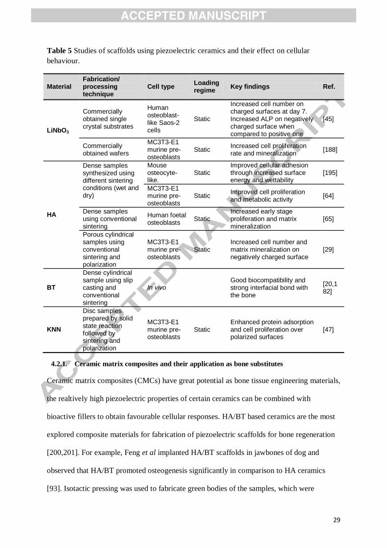
Table 5 Studies of scaffolds using piezoelectric ceramics and their effect on cellular
behaviour.
Material Fabrication/ processing technique
Cell type Loading regime
Key findings Ref.
LiNbO3
Commercially obtained single crystal substrates
Human osteoblast-like Saos-2 cells
Static
Increased cell number on charged surfaces at day 7. Increased ALP on negatively charged surface when compared to positive one
[45]
Commercially obtained wafers
MC3T3-E1 murine pre- osteoblasts
Static Increased cell proliferation rate and mineralization
[188]
HA
Dense samples synthesized using different sintering conditions (wet and dry)
Mouse osteocyte-like.
Static Improved cellular adhesion through increased surface energy and wettability
[195]
MC3T3-E1 murine pre- osteoblasts
Static Improved cell proliferation and metabolic activity
[64]
Dense samples using conventional sintering
Human foetal osteoblasts
Static Increased early stage proliferation and matrix mineralization
[65]
Porous cylindrical samples using conventional sintering and polarization
MC3T3-E1 murine pre- osteoblasts
Static Increased cell number and matrix mineralization on negatively charged surface
[29]
BT
Dense cylindrical sample using slip casting and conventional sintering
In vivo Good biocompatibility and strong interfacial bond with the bone
[20,182]
KNN
Disc samples prepared by solid state reaction followed by sintering and polarization
MC3T3-E1 murine pre- osteoblasts
Static Enhanced protein adsorption and cell proliferation over polarized surfaces
[47]
4.2.1. Ceramic matrix composites and their application as bone substitutes
Ceramic matrix composites (CMCs) have great potential as bone tissue engineering materials,
the realtively high piezoelectric properties of certain ceramics can be combined with
bioactive fillers to obtain favourable cellular responses. HA/BT based ceramics are the most
explored composite materials for fabrication of piezoelectric scaffolds for bone regeneration
[200,201]. For example, Feng et al implanted HA/BT scaffolds in jawbones of dog and
observed that HA/BT promoted osteogenesis significantly in comparison to HA ceramics
[93]. Isotactic pressing was used to fabricate green bodies of the samples, which were

30
subsequently sintered and poled. In their work, it was noted that the osteogenic response was
direction dependent suggesting that piezoelectric effects have play a role. Several other
studies reported fabrication of piezoelectric composite scaffolds of HA/BT with low porosity
using spark plasma sintering or conventional sintering processes [53,61,170,171]. HA/BT
rod-like nanocomposites have recently been fabricated using a hydrothermal process [202].
The piezoelectric charge constant of these composites were reported (1.54 pC/N), close to
that of bone, however, no cell studies were conducted [202].
Unlike these studies, in a more osteomimetic approach, Zhang et al fabricated HA/BT
scaffolds with aligned porous structures and observed significantly higher cell densities and
ALP activity in comparison to dense scaffolds [30,172]. Green bodies of the composite
scaffolds synthesized via ice templating were sintered and poled to make piezoelectric
samples [30,172]. The piezoelectric coefficient observed for these samples was reported to be
higher than that of the bone. However, no difference was noted among polarized and non-
polarized groups of samples due to the absence of dynamic loading. A recent study by Yang
et al (2017) used an in vitro dynamic loading device to apply cyclic loading to HA/BT
piezoelectric composites [173]. It was observed that biocompatibility and bone-inducing
activity of the composite samples was greater than that of HA under cyclic loading. The force
used for cyclic loading of the samples was fixed, however, the amount of strain being
transferred to the composites was not quantified. The indenter used to press the samples did
not cover the whole surface of the sample which might have led to non-homogenous
distribution force through the sample [173]. In a recent study, Shokrollahi et al fabricated
porous freeze cast composites of BT/akermanite (Ca2MgSi2O7) with piezoelectric
characteristics higher than that of bone [203]. Cytotoxic evaluations with human bone
marrow mesenchymal stem cells confirmed that the composited were biocompatible, yet
osteogenic capacity and the role of piezoelectricity ware not studied. Different structures,

31
such as porous scaffolds [30,172], nanoparticles [183–186], layered structures [54] and dense
discs, that have fabricated and used for tissue engineering application are shown
schematically in Figure 6 (b). Different studies in which PMCs and CMCs have been
assessed for their osteogenic capability have been summarized in Table 6.
Table 6 Summary of key findings of studies involving use of CMCs. (#Dynamic represents
that piezoelectric composites were stimulated electrically.)
Material Fabrication/ processing technique
Cell type Loading regime
Key findings Ref.
HA-BT
Dense disc shaped samples using conventional sintering
Osteoblast cells In vitro
Dynamic
Higher number of cells were observed on 10/90(HA/BT) composites under dynamic loading
[173]
Porous disc samples were obtained by ice templating followed by sintering and polarization
MG63 cells Static
Increased proliferation, differentiation and adhesion of cells. No difference between piezoelectric and non-piezoelectric samples
[30,172]
Dense disc shaped samples obtained by SPS followed by poling
Human osteoblast like cell line
Dynamic#
Increase in cell growth and proliferation
[53,170]
LNKN-HA
Laminated composite materials using tape casting followed by sintering and poling
- Static
Higher in vitro bioactivity was observed for composite samples (in comparison with poled HA)
[54]
NKN-HA
Layered composites were formed by compaction, followed by SPS and poling
Human osteoblast like Saos2 cells
Static Increased cellular proliferation with culture duration.
[67]
5. Biological performance of piezoelectric materials
Piezoelectric materials are capable of modulating cellular behaviour through surface charges
generated in response to deformation occurring through cellular interaction, vibration

32
stimulus or both. Steady state bioelectric potentials, which appear in the bone in a non-
stressed state, and injury potentials of the bone arising due to a fracture play a key role in
modulation of bone growth, remodelling and regeneration [204–210]. These electrical signals
have an effect on cellular processes which can also be controlled through external stimulation
devices [205,211–213]. Endogenous potentials up to 100 mV have been observed in non-
stressed bone while, under stress, streaming potential values in the range of 0.5 to 3 μV/με
were observed depending on the frequency of load applied [204,214]. PVDF and its
copolymers have been displayed to mimic these potentials and show an improved osteogenic
response both in-vitro and in-vivo [11,140]. The electric potentials in fractured bone have
been shown to be ~200-250 mV which suggests that cellular response can be further
improved if piezoelectric scaffolds can generate similar potentials under physiological levels
of deformations [208].
Piezoelectricity of the scaffolds is only activated under dynamic conditions. Different studies
in which the effect of mechanical stimulation on cell lines for osteogenic application have
been studied are reviewed in [215,216]. A frequency of 0-5 Hz and deformation of up to 25%
has been studied and shown to effect proliferation and differentiation capacities of different
cell types [215,216]. Several mechanisms have been associated with mechanical stimulation
of cells depending on the cell type and the type of regime used [215–217], however, a direct
comparison is hard to establish due to variability in experimental parameters. It has recently
been shown that cellular behaviour is also modulated by the activation of piezo receptors on
mechanical deformation [218,219].
The use of piezoelectric and electrically conductive materials for delivering electrical
stimulation to desired regions ensure controlled and efficient delivery of these important cues
[220–222]. The use of conductive polymers for this purpose requires an external stimulatory
device, which is not the case with piezoelectric materials. Electrical stimulation to control

33
cellular behaviour also requires optimization of various parameters including the frequency,
amplitude, duration and nature (alternating or direct) of the signal [8,223,224]. To the
author’s knowledge, there has been no mention in previous studies as to how or if these
parameters can be controlled through the mechanical stimulation of piezoelectric materials.
Direct electrical stimulation has been shown to be affect cells by regulating the voltage-gated
calcium channels [8,222]. A similar mechanism has been suggested for piezoelectric
scaffolds which promoted osteogenic differentiation of mesenchymal stem cells [140].
Figure 7 a) and b) show images of piezoelectric scaffolds used in this study and
immunohistochemical staining for collagen type I after 28 days of osteogenic differentiation
in dynamic conditions respectively [140]. Additionally, piezoelectric materials can alter the
adhesion of proteins which has a direct effect on cellular response [66,92,140]. The cellular
behaviour can be altered due to the varying charge distribution (electrical double layer)
around the surface of scaffolds, or due to electric fields and currents which are set up in a
localized manner [45]. It has also been shown that in case of neuronal, cells calcium ionic
pathways play key role in controlling the differentiation of cells exposed to piezoelectric
stimulation [225]. To gain a better understanding of the interactions and provide more
quantitative assessment of the processes involved, it has also been highlighted that modelling
and simulation of piezoelectric materials can be a helpful tool [226].

34
Figure 7 a) Image of piezoelectric scaffolds, approximately 3 mm thick. Scale bar = 6 mm ( i
). Scanning electron microscope (SEM) images of as-spun PVDF-TrFE ( ii ), annealed
PVDF-TrFE ( iii ) and PCL ( iv ) at 2000× magnification; b) Representative gross images
and histological evaluation of scaffolds after 28 days undergoing osteogenic differentiation in
dynamic conditions. Images of as-spun PVDF-TrFE ( i ), annealed PVDF-TrFE ( ii ) and
PCL ( iii ) scaffolds. Immunohistochemical staining for collagen type I of as-spun PVDF-
TrFE ( vi ), annealed PVDF-TrFE ( v ) and PCL ( vi ) scaffolds. (Scale bars: A-C, 6 mm; D-
F, 100 μm); c) 3D printed piezoelectric microstructures including a dot array ( i ), square
arrays ( ii and iii ) and honeycomb array ( iv ). Printed with permission from [140,227]
. Using piezoelectric materials with controlled deformation offers another advantage as the
cells are not only under the influence of an electrical stimulus but also experience different
levels of mechanical strain the scaffold is subjected to and therefore co-stimulation of cells
can be achieved and optimized for synergistic performance. However, this research is still in
an early phase and there is a need to conduct deeper investigation to understand the
mechanisms associated with biological performance of these materials.
6. Emerging piezoelectric materials and strategies
Piezoelectric materials have emerged as a class of smart materials which have a myriad of
applications ranging from tissue engineering, drug delivery to sensors [218]. The emergence
of these materials and their wide usage has motivated researchers to adopt new and

35
innovative approaches such as solution blow spinning (SBS), and 3D printing to make
suitable structures with desired properties. These have been discussed in the sections below.
The magnetoelectric effect has recently gained interest of the tissue engineering community
as it follows the same principle as piezoelectricity, the stimulating force being a magnetic
field [228]. The magnetic field induces deformation which leads to generation of electric
polarization which has been shown to effect cellular proliferation of MC3T3E1 cells [228].
These materials have been shown to be effective in different applications such as drug
delivery, tissue engineering and targeted cell manipulation [228–236].
6.1. 3D printing of piezoelectric materials
3D printed BT [237–239], modified KNN [240], PVDF [241,242] and PLLA [243] have
recently been fabricated for piezoelectric sensors and functional scaffolds which have
limitedly been researched for tissue engineering applications. There are however challenges
in processing and formulation of the raw material for printing and inducing piezoelectricity.
Use of binders and flow additives is essential to optimize the rheological properties for
printing. These parameters greatly depend on the type of material combinations desired and
control over shape and size of scaffolds is difficult to establish. Schult et al fabricated 3D
printed cylindrical green parts (11.7 mm diameter and 3.51 mm height) for making BT
scaffolds [238]. It was observed that the green bodies formed were unstable and sintering
lead to substantial shrinkage. In another study concerning piezoelectric ceramics, Yayun Li
fabricated KNN based 3D ceramic structures using direct ink writing method and observed
piezoelectric and ferroelectric behaviour in the complex structured samples [240]. The solid
content of the ink was adjusted to optimize viscosity for processing. The method involved
preparation of a suitable ink/suspension used with a direct ink writing equipment to fabricate
structures that were subsequently sintered and poled using thermal poling (in silicone oil)
[240]. This method was suggested as an alternative to using dies or lithographic masks to

36
make complex structures. Lithography can be used to fabricate dies, masks and masters with
complex structures over which the desired material can be deposited and moulded [244]. In
an approach based on soft lithography, micro structured PVDF scaffolds were fabricated and
tested for cytocompatibility using human osteosarcoma cells [244]. Preliminary examination
of piezoelectric effects showed their potential as mechanically active tissue engineering
scaffolds and bio sensors [244]. Two photon lithography has also been utilized to fabricate
3D nanocomposite scaffolds which promoted differentiation of human osteoblast like cell
line [245]. The piezoelectric charge constant was measured using PFM to a value of ~0.57
pm/V which is similar to that of bone [245]. Although there are challenges in formulation and
processing there is huge scope for 3D printed piezoelectric materials and patient specific
scaffolds. Piezoelectric microstructures printed using modified BT particles are shown in
Figure 7 b) [227].
6.2. Piezoelectric nanofibre fabrication
Nano fibrous materials have gained interest of the tissue engineering community as they
provide a 3D network with high surface area for cells to adhere and interact with. SBS has
recently emerged as a technique to fabricate fibrous scaffolds for this application [246]. SBS
does not require electric fields and therefore electrically active materials can be formed
without risk of shorting, it also requires simple equipment and is amenable to scaling
[247,248]. In a recent study, Bolbasov et al highlighted the importance of controlling the
amount of piezoelectric β- phase in PVDF-TeFE in order to obtain a better cellular response
when compared to electrospun scaffolds [248]. Electrospinning (ES) and SBS have been
used to fabricate ceramic or composite fibre using piezoelectric materials [249–253]. Isakov
et al fabricated aligned BT nanofibres using ES and characterized their ferroelectric and
piezoelectric properties [253]. A sol- gel precursor solution was the precursor used in the ES

37
setup and the fibres obtained were calcined. Raman spectroscopy and PFM was used to
identify the presence of ferroelectric phases [253]. In another study, SBS was used by Cena et
al to fabricate lithium niobate (LN) microfibres from sol-gel precursors [251]. The solution
used for spinning microfibres was a mixture of poly (vinyl pyrrolidone) (PVP), isopropyl
alcohol, water and LN solution (prepared by dissolving lithium carbonate and ammonium
niobium oxalate in deionized water) [251]. Different electrical measurements such as
impedance, modulus and conductivity were carried out after making pellets from fibres.
However, no piezoelectric or ferroelectric measurements were carried out in this study [251].
A recent study by Holopainen et al discusses the fabrication HA fibres using electroblowing
which involves spinning of fibres by gas assisted ES, a combination of SBS and ES, which
can enable smaller diameter fibres to be produced than SBS at faster rates than ES [254]. The
fibres soaked in simulated body fluid displayed rapid and homogenous formation of apatite
layer suggesting high bioactivity of the fibres synthesized [254].
Apart from the materials discussed above, piezoelectric materials such as diphenylalanine
based peptide nanotubes (FFPNTs), liquid crystal elastomers (LCEs) and boron nitride
nanotubes (BNNTs) have been explored little for tissue engineering applications [95,255–
261]. Silk [52,262,263], PHB [41] and its copolymers have also been manufactured to display
piezoelectric properties, yet their piezoelectric behaviour has not been explored in context of
bone tissue engineering to the author’s knowledge [264]. BNNTS have already shown
promising results for application in the field of bone tissue engineering. Danti et al
incorporated BNNTs in primary human osteoblast cells and explored the effect of
piezoelectricity by stimulating the samples using low frequency ultrasound [265]. It was
observed that mineralization and expression TGF-β1 became significant in the cells with
BNNTs that were stimulated by ultrasound. The mechanism responsible was assumed to be
the electrical cues that were generated by BNNTs due to the piezoelectric effect. Xia li et al

38
culture rat bone marrow derived mesenchymal stem cells on BNNTs layer to assess the
compatibility and osteogenic differentiation capacity of the material [256]. The experiments
were conducted in static environment and the effect of piezoelectricity was not discussed.
BNNTs were shown to enhance proliferation and alkaline phosphatase activity of the cells.
The results suggest that BNNTs might prove as useful fillers for fabricating piezoelectric
scaffolds for bone regeneration. Rodriguez et al fabricated self–assembling peptide hydrogels
by dissolving monomer of diphenylalanine (FF) modified fluorenyl-methoxycarbonyl (Fmoc)
(Fmoc-FF) in dimethyl sulfoxide (DMSO), these self-assembled 3D nano structures were
shown to have piezoelectric properties at nano scale [95]. PFM was used to characterize the
piezoelectric response of the hydrogels [95]. The results concerning piezoelectricity of these
structures are promising as piezoelectric hydrogels can find application in the field of tissue
engineering but the accumulation of degradation products of these hydrogels can cause toxic
effects [48,259,266].
Liquid crystal elastomers are an emerging group of materials that are of interest in
responsive, morphing and piezoelectric systems; however, they can involve complex
synthesis and the materials required for the same are currently expensive [260].
Furthermore, composites based on the piezoelectric component of bone, collagen (fibrilar
bovine Coll type I), have also been used to fabricate materials for bone substitute [267,268].
Zanfir et al demonstrated that use of BT in combination of collagen and hydroxyapatite
ensured that the composite scaffolds exhibit osteoconductive properties [268]. The composite
scaffolds were fabricated by mixing collagen gel with mineral phase (HA/HA+BT) followed
by chemically crosslinking, freezing and freeze-drying. The improved osteoconductivity was
attributed to the incorporation BT nanopowder in the composites [268]. However, the study
did not include any polarization treatment of scaffolds to induce piezoelectricity, which might
further add to the functionality of the scaffolds and improve cellular response.

39
A major limitation of the existing studies is the absence of controlled mechanical stimulation
applied to piezoelectric scaffolds. Different systems such as four point bending, flexcell (a
device used for applying mechanical deformation to materials in a controlled manner),
ultrasound have been utilized over the years to study the effect of mechanical stimulation on
different cell types [216,269–271]. These systems can be adopted to stimulate cells on
piezoelectric substrates to ensure that mechanical as well as electrical cues are delivered to
the cells. However, various parameters associated with the system, such as magnitude, nature
(e.g. cyclic loading, sine wave), and rate of strain might have to be controlled to obtain a
synergistic response. An extensive analysis is still required to test the feasibility of
piezoelectric materials as ideal substitutes for clinical application. Piezoelectric materials
promise great future potential for tissue engineering applications.
7. Concluding remarks
Piezoelectric materials are gaining significant interest in the tissue engineering community
owing to their capability to mimic in vivo micro-environment under static as well as dynamic
conditions. The 3D printing and fibre spinning of piezoelectric materials/composites and their
scaffolds are promising areas that have been little explored for bone tissue engineering
applications. Promising results obtained with ceramics are far from a clinical application
because of the absence of compelling explanations to the variation in responses observed. It
is of utmost importance to apply consistent characterization tools/techniques to these
materials.
The existing research in the field focusses on fabricating piezoelectric scaffolds with
optimized value of piezoelectric ‘d’ constant and assessing cellular responses on polarized
samples. It is very important to analyse the effect of the transfer of mechanical deformations
of the bone to the scaffolds and subsequent generation of electrical cues on different cell

40
types. Piezoelectric films, fibres and rods have shown promising results, however, their
clinical potential is yet to be exploited, a major limitation being the non-degradability of most
of the piezoelectric materials explored to date. The use of emerging strategies like 3D
printing of piezoelectric materials to build complex structures can prove very useful to design
piezoelectric bioactive implants with clinical applicability. Piezoelectric characteristics
should be quantified using PFM followed by cellular studies on mechanically stimulated
scaffolds. Use of PFM will ensure that the mechanism associated with cell material
interactions can be better understood at the nanoscale. The study of piezoelectricity in
(protein based hydrogel)/hydrogel based systems has opened a new and promising area of
research. Another aspect considering clinical application of these materials is the absence of
mechanical deformation in the early stages following implantation of the scaffolds. The
results of in vitro and in vivo studies are appealing and clearly suggest that piezoelectric
materials have the potential for the fabrication of new and exciting smart scaffolds for bone
tissue engineering, as well as other tissues.
Competing interests and funding sources
The authors declare no potential conflict of interests with respect to the content, authorship
and/or the publication of this article.
This work was supported/funded by the Commonwealth Scholarship Commission of the
United Kingdom and The University of Manchester.
References
[1] R.A. Perez, S.J. Seo, J.E. Won, E.J. Lee, J.H. Jang, J.C. Knowles, H.W. Kim,
Therapeutically relevant aspects in bone repair and regeneration, Mater. Today. 18
(2015) 573–589. doi:10.1016/j.mattod.2015.06.011.

41
[2] J.E. Ramirez-Vick, Biophysical stimulation for bone regeneration., JSM Biotechnol.
Biomed. Eng. 1 (2013) 1014.
http://www.pubmedcentral.nih.gov/articlerender.fcgi?artid=4011495&tool=pmcentrez
&rendertype=abstract.
[3] J. Voog, D.L. Jones, Stem cells and the niche: a dynamic duo, Cell Stem Cell. 6 (2010)
103–15. doi:10.1016/j.stem.2010.01.011.
[4] L. Polo-Corrales, M. Latorre-Esteves, J.E. Ramirez-Vick, Scaffold design for bone
regeneration, J. Nanosci. Nanotechnol. 14 (2014) 15–56. doi:10.1166/jnn.2014.9127.
[5] C.D. McCaig, M. Zhao, Physiological electrical fields modify cell behaviour,
BioEssays. 19 (1997) 819–826. doi:10.1002/bies.950190912.
[6] B. Reid, M. Zhao, The electrical response to injury: Molecular mechanisms and wound
healing, Adv. Wound Care. 3 (2014) 184–201. doi:10.1089/wound.2013.0442.
[7] R.H.W. Funk, Endogenous electric fields as guiding cue for cell migration, Front.
Physiol. 6 (2015) 143. doi:10.3389/fphys.2015.00143.
[8] R. Balint, N.J. Cassidy, S.H. Cartmell, Electrical stimulation: A novel tool for tissue
engineering, Tissue Eng. Part B-Reviews. 19 (2013) 48–57.
doi:10.1089/ten.TEB.2012.0183.
[9] C.D. McCaig, A.M. Rajnicek, B. Song, M. Zhao, Controlling cell behavior
electrically: current views and future potential., Physiol. Rev. 85 (2005) 943–978.
doi:10.1152/physrev.00020.2004.
[10] K.S. Kang, J.M. Hong, Y.H. Jeong, Y. Seol, W. Yong, J. Rhie, D. Cho, Combined
effect of three types of biophysical stimuli for bone regeneration, Tissue Eng. Part A.

42
20 (2014) 1767–1777. doi:10.1089/ten.tea.2013.0157.
[11] X. Zhang, C. Zhang, Y. Lin, P. Hu, Y. Shen, K. Wang, S. Meng, Y. Chai, X. Dai, X.
Liu, Y. Liu, X. Mo, C. Cao, S. Li, X. Deng, L. Chen, Nanocomposite membranes
enhance bone regeneration through restoring physiological electric microenvironment,
ACS Nano. 10 (2016) 7279–7286. doi:10.1021/acsnano.6b02247.
[12] C. Ribeiro, V. Sencadas, D.M. Correia, S. Lanceros-Mendez, Piezoelectric polymers
as biomaterials for tissue engineering applications, Colloids Surf B Biointerfaces. 136
(2015) 46–55. doi:10.1016/j.colsurfb.2015.08.043.
[13] A. Wang, Z. Liu, M. Hu, C. Wang, X. Zhang, B. Shi, Y. Fan, Y. Cui, Z. Li, K. Ren,
Piezoelectric nanofibrous scaffolds as in vivo energy harvesters for modifying
fibroblast alignment and proliferation in wound healing, Nano Energy. 43 (2018) 63–
71. doi:10.1016/j.nanoen.2017.11.023.
[14] A.H. Rajabi, M. Jaffe, T.L. Arinzeh, Piezoelectric materials for tissue regeneration: A
review, Acta Biomater. 24 (2015) 12–23. doi:10.1016/j.actbio.2015.07.010.
[15] P.K. Panda, Review: Environmental friendly lead-free piezoelectric materials, J.
Mater. Sci. 44 (2009) 5049–5062. doi:10.1007/s10853-009-3643-0.
[16] J. Curie, P. Curie, Development by pressure of polar electricity in hemihedral crystals
with inclined faces, Bull. Soc. Min. Fr. 3 (1880) 90.
[17] J. Tichý, J. Erhart, E. Kittinger, J. Přívratská, Fundamentals of piezoelectric sensorics:
Mechanical, dielectric, and thermodynamical properties of piezoelectric materials,
Fundam. Piezoelectric Sensorics Mech. Dielectr. Thermodyn. Prop. Piezoelectric
Mater. (2010) 1–207. doi:10.1007/978-3-540-68427-5.

43
[18] E. Fukada, I. Yasuda, Piezoelectric Effects in Collagen, Japanese J. Appl. Physics, Part
1 Regul. Pap. Short Notes Rev. Pap. 3 (1964) 502B. doi:10.1143/JJAP.3.502B.
[19] M.H. SHAMOS, L.S. LAVINE, Piezoelectricity as a Fundamental Property of
Biological Tissues, Nature. 213 (1967) 267–269. doi:10.1038/213267a0.
[20] J.B. Park, B.J. Kelly, G.H. Kenner, A.F. von Recum, M.F. Grether, W.W. Coffeen,
Piezoelectric ceramic implants: In vivo results, J. Biomed. Mater. Res. 15 (1981) 103–
110. doi:10.1002/jbm.820150114.
[21] E. Fukada, I. Yasuda, On the piezoelectric effect of bone, J. Phys. Soc. Japan. 12
(1957) 1158–1162. doi:10.1143/JPSJ.12.1158.
[22] A. Gjelsvik, Bone remodeling and piezoelectricity—I, J. Biomech. 6 (1973) 69–77.
http://www.sciencedirect.com/science/article/pii/0021929073900390.
[23] C.A. Bassett, Electrical effects in bone, Sci. Am. 213 (1965) 18–25.
http://www.ncbi.nlm.nih.gov/pubmed/5825731.
[24] C.A. BASSETT, R.O. BECKER, Generation of electric potentials by bone in response
to mechanical stress, Science. 137 (1962) 1063–4.
http://www.ncbi.nlm.nih.gov/pubmed/13865637.
[25] M.H. Shamos, L.S. Lavine, Physical bases for bioelectric effects in mineralized
tissues., Clin. Orthop. Relat. Res. 35 (1964) 177–88.
[26] F. Vasquez-Sancho, A. Abdollahi, D. Damjanovic, G. Catalan, Flexoelectricity in
Bones, Adv. Mater. 1705316 (2018) 1–5. doi:10.1002/adma.201705316.
[27] Z.L. Wang, Nanopiezotronics, Adv. Mater. 19 (2007) 889–892.
doi:10.1002/adma.200602918.

44
[28] H. a. Sodano, D.J. Inman, G. Park, A review of power harvesting from vibration using
piezoelectric materials, Shock Vib. Dig. 36 (2004) 197–205.
doi:10.1177/0583102404043275.
[29] S.H. Cartmell, S. Thurstan, J.P. Gittings, S. Griffiths, C.R. Bowen, I.G. Turner,
Polarization of porous hydroxyapatite scaffolds: Influence on osteoblast cell
proliferation and extracellular matrix production, J. Biomed. Mater. Res. - Part A. 102
(2014) 1047–1052. doi:10.1002/jbm.a.34790.
[30] Y. Zhang, L. Chen, J. Zeng, K. Zhou, D. Zhang, Aligned porous barium
titanate/hydroxyapatite composites with high piezoelectric coefficients for bone tissue
engineering, Mater. Sci. Eng. C. 39 (2014) 143–149. doi:10.1016/j.msec.2014.02.022.
[31] A.J. Lovinger, Ferroelectric Polymers, Science (80-. ). 220 (1983) 1115–1121.
doi:10.1126/science.220.4602.1115.
[32] I. Katsouras, K. Asadi, M. Li, T.B. Van Driel, K.S. Kjær, D. Zhao, T. Lenz, Y. Gu,
P.W.M. Blom, D. Damjanovic, M.M. Nielsen, D.M. De Leeuw, The negative
piezoelectric effect of the ferroelectric polymer poly(vinylidene fluoride), Nat. Mater.
15 (2016) 78–84. doi:10.1038/nmat4423.
[33] T. Ochiai, E. Fukada, Electromechanical properties of Poly-L-Lactic Acid, Jpn. J.
Appl. Phys. 37 (1998) 3374–3376. doi:10.1143/JJAP.37.3374.
[34] J. Gomes, J. Serrado Nunes, V. Sencadas, S. Lanceros-Mendez, Influence of the β-
phase content and degree of crystallinity on the piezo- and ferroelectric properties of
poly(vinylidene fluoride), Smart Mater. Struct. 19 (2010) 065010. doi:10.1088/0964-
1726/19/6/065010.
[35] C. Ribeiro, S. Moreira, V. Correia, V. Sencadas, J.G. Rocha, F.M. Gama, J.L. Gómez

45
Ribelles, S. Lanceros-Méndez, Enhanced proliferation of pre-osteoblastic cells by
dynamic piezoelectric stimulation, RSC Adv. 2 (2012) 11504.
doi:10.1039/c2ra21841k.
[36] N. Murayama, K. Nakamura, H. Obara, M. Segawa, The strong piezoelectricity in
polyvinylidene fluroide (PVDF), Ultrasonics. 14 (1976) 15–23. doi:10.1016/0041-
624X(76)90067-6.
[37] E. Fukada, T. Sakurai, Piezoelectricity in Polarized Poly(vinylidene fluoride) Films,
Polym. J. 2 (1970) 656–662. doi:10.1295/polymj.2.656.
[38] K. Omote, H. Ohigashi, K. Koga, [18] Temperature dependence of elastic, dielectric,
and piezoelectric properties of “single crystalline’’ films of vinylidene fluoride
trifluoroethylene copolymer, J. Appl. Phys. 81 (1997) 2760. doi:10.1063/1.364300.
[39] P. Sharma, D. Wu, S. Poddar, T.J. Reece, S. Ducharme, A. Gruverman, Orientational
imaging in polar polymers by piezoresponse force microscopy, J. Appl. Phys. 110
(2011). doi:10.1063/1.3623765.
[40] J.L. Lutkenhaus, K. McEnnis, A. Serghei, T.P. Russell, Confinement effects on
crystallization and curie transitions of poly(vinylidene fluoride-co-trifluoroethylene),
Macromolecules. 43 (2010) 3844–3850. doi:10.1021/ma100166a.
[41] E. Fukada, Y. Ando, Piezoelectric properties of poly-β-hydroxybutyrate and
copolymers of β-hydroxybutyrate and β-hydroxyvalerate, Int. J. Biol. Macromol. 8
(1986) 361–366. doi:10.1016/0141-8130(86)90056-5.
[42] A.A. Gandhi, M. Wojtas, S.B. Lang, A.L. Kholkin, S.A.M. Tofail, Piezoelectricity in
poled hydroxyapatite ceramics, J. Am. Ceram. Soc. 97 (2014) 2867–2872.
doi:10.1111/jace.13045.

46
[43] D. Berlincourt, H. Jaffe, Elastic and Piezoelectric Coefficients of Single-Crystal
Barium Titanate, Phys. Rev. 111 (1958) 143–148. doi:10.1103/PhysRev.111.143.
[44] S.W. Yu, S.T. Kuo, W.H. Tuan, Y.Y. Tsai, S.F. Wang, Cytotoxicity and degradation
behavior of potassium sodium niobate piezoelectric ceramics, Ceram. Int. 38 (2012)
2845–2850. doi:10.1016/j.ceramint.2011.11.056.
[45] P. Vaněk, Z. Kolská, T. Luxbacher, J.A.L. García, M. Lehocký, M. Vandrovcová, L.
Bačáková, J. Petzelt, Electrical activity of ferroelectric biomaterials and its effects on
the adhesion, growth and enzymatic activity of human osteoblast-like cells, J. Phys. D.
Appl. Phys. 49 (2016) 175403. doi:10.1088/0022-3727/49/17/175403.
[46] Y. Chen, J. Williams, Synthesis of Boron Nitride Nanotubes Using a Ball-Milling and
Annealing Method, in: Nanoeng. Struct. Funct. Smart Mater., CRC Press, 2005.
doi:doi:10.1201/9780203491966.ch7.
[47] W. Chen, Z. Yu, J. Pang, P. Yu, G. Tan, C. Ning, Fabrication of biocompatible
potassium sodium niobate piezoelectric ceramic as an electroactive implant, Materials
(Basel). 10 (2017) 18–21. doi:10.3390/ma10040345.
[48] S. Vasilev, P. Zelenovskiy, D. Vasileva, A. Nuraeva, V.Y. Shur, A.L. Kholkin,
Piezoelectric properties of diphenylalanine microtubes prepared from the solution, J.
Phys. Chem. Solids. 93 (2016) 68–72. doi:10.1016/j.jpcs.2016.02.002.
[49] M. Minary-Jolandan, M.F. Yu, Uncovering nanoscale electromechanical heterogeneity
in the subfibrillar structure of collagen fibrils responsible for the piezoelectricity of
bone, ACS Nano. 3 (2009) 1859–1863. doi:10.1021/nn900472n.
[50] M. Minary-Jolandan, M.F. Yu, Shear piezoelectricity in bone at the nanoscale, Appl.
Phys. Lett. 97 (2010). doi:10.1063/1.3503965.

47
[51] J.A. Bur, Measurements of dynamic piezoelectric properties of bone as a function of
temperature and humidity, J. Biomech. 9 (1976) 495–507.
[52] T. Yucel, P. Cebe, D.L. Kaplan, Structural origins of Silk piezoelectricity, Adv. Funct.
Mater. 21 (2011) 779–785. doi:10.1002/adfm.201002077.
[53] A.K. Dubey, A. Ea, K. Balani, B. Basu, Multifunctional properties of multistage spark
plasma sintered HA-BaTiO3-based piezobiocomposites for bone replacement
applications, J. Am. Ceram. Soc. 96 (2013) 3753–3759. doi:10.1111/jace.12566.
[54] A.K. Dubey, R. Kinoshita, K. Kakimoto, Piezoelectric sodium potassium niobate
mediated improved polarization and in vitro bioactivity of hydroxyapatite, Rsc Adv. 5
(2015) 19638–19646. doi:10.1039/C5RA00771B.
[55] M. Bruschi, D. Steinmüller-Nethl, W. Goriwoda, M. Rasse, Composition and
modifications of dental implant surfaces, J. Oral Implant. 2015 (2015) 1–14.
doi:10.1155/2015/527426.
[56] F. Zhao, J. Wang, H. Guo, S. Liu, W. He, The effects of surface properties of
nanostructured bone repair materials on their performances, J. Nanomater. 2015 (2015)
1–11. doi:10.1155/2015/893545.
[57] P.M. Martins, S. Ribeiro, C. Ribeiro, V. Sencadas, a C. Gomes, F.M. Gama, S.
Lanceros-Mendez, Effect of poling state and morphology of piezoelectric
poly(vinylidene fluoride) membranes for skeletal muscle tissue engineering, RSC Adv.
3 (2013) 17938–17944. doi:10.1039/C3RA43499K.
[58] K. Cai, M. Frant, J. Bossert, G. Hildebrand, K. Liefeith, K.D. Jandt, Surface
functionalized titanium thin films: Zeta-potential, protein adsorption and cell
proliferation, Colloids Surfaces B Biointerfaces. 50 (2006) 1–8.

48
doi:10.1016/j.colsurfb.2006.03.016.
[59] K.H. Chan, S. Zhuo, M. Ni, Priming the surface of orthopedic implants for osteoblast
attachment in bone tissue engineering, Int. J. Med. Sci. 12 (2015) 701–707.
doi:10.7150/ijms.12658.
[60] S. Huang, N. Liang, Y. Hu, X. Zhou, N. Abidi, Polydopamine-Assisted Surface
Modification for Bone Biosubstitutes, Biomed Res. Int. 2016 (2016).
doi:10.1155/2016/2389895.
[61] F.R. Baxter, I.G. Turner, C.R. Bowen, J.P. Gittings, J.B. Chaudhuri, An in vitro study
of electrically active hydroxyapatite-barium titanate ceramics using Saos-2 cells, J.
Mater. Sci. Mater. Med. 20 (2009) 1697–1708. doi:10.1007/s10856-009-3734-0.
[62] C. Ribeiro, D.M. Correia, S. Ribeiro, V. Sencadas, G. Botelho, S. Lanceros-Méndez,
Piezoelectric poly(vinylidene fluoride) microstructure and poling state in active tissue
engineering, Eng. Life Sci. 15 (2015) 351–356. doi:10.1002/elsc.201400144.
[63] R.A. Gittens, R. Olivares-Navarrete, A. Cheng, D.M. Anderson, T. McLachlan, I.
Stephan, J. Geis-Gerstorfer, K.H. Sandhage, A.G. Fedorov, F. Rupp, B.D. Boyan, R.
Tannenbaum, Z. Schwartz, The roles of titanium surface micro/nanotopography and
wettability on the differential response of human osteoblast lineage cells, Acta
Biomater. 9 (2013) 6268–6277. doi:10.1016/j.actbio.2012.12.002.
[64] D. Kumar, J.P. Gittings, I.G. Turner, C.R. Bowen, A. Bastida-Hidalgo, S.H. Cartmell,
Polarization of hydroxyapatite: Influence on osteoblast cell proliferation, Acta
Biomater. 6 (2010) 1549–1554. doi:10.1016/j.actbio.2009.11.008.
[65] S. Bodhak, S. Bose, A. Bandyopadhyay, Role of surface charge and wettability on
early stage mineralization and bone cell-materials interactions of polarized

49
hydroxyapatite, Acta Biomater. 5 (2009) 2178–2188.
doi:10.1016/j.actbio.2009.02.023.
[66] J. Parssinen, H. Hammarén, R. Rahikainen, V. Sencadas, C. Ribeiro, S. Vanhatupa, S.
Miettinen, S. Lanceros-Méndez, V.P. Hytönen, Enhancement of adhesion and
promotion of osteogenic differentiation of human adipose stem cells by poled
electroactive poly(vinylidene fluoride), J. Biomed. Mater. Res. - Part A. 103 (2015)
919–928. doi:10.1002/jbm.a.35234.
[67] A.K. Dubey, K. Kakimoto, A. Obata, T. Kasuga, Enhanced polarization of
hydroxyapatite using the design concept of functionally graded materials with sodium
potassium niobate, RSC Adv. 4 (2014) 24601–24611. doi:10.1039/c4ra02329c.
[68] W. Melitz, J. Shen, A.C. Kummel, S. Lee, Kelvin probe force microscopy and its
application, Surf. Sci. Rep. 66 (2011) 1–27. doi:10.1016/j.surfrep.2010.10.001.
[69] J. Chang, Y. Liu, K. Heo, B.Y. Lee, S.W. Lee, L. Lin, Direct-write complementary
graphene field effect transistors and junctions via near-field electrospinning, Small. 10
(2014) 1920–1925. doi:10.1002/smll.201302965.
[70] J.H. Yang, T. Ryu, Y. Lansac, Y.H. Jang, B.H. Lee, Shear stress-induced enhancement
of the piezoelectric properties of PVDF-TrFE thin films, Org. Electron. Physics,
Mater. Appl. 28 (2016) 67–72. doi:10.1016/j.orgel.2015.10.018.
[71] S.B. Lang, Review of ferroelectric hydroxyapatite and its application to biomedicine *,
Phase Transitions. 89 (2016) 678–694. doi:10.1080/01411594.2016.1182166.
[72] J.M. Pelto, S.P. Haimi, A.S. Siljander, S.S. Miettinen, K.M. Tappura, M.J. Higgins,
G.G. Wallace, Surface Properties and Interaction Forces of Biopolymer-Doped
Conductive Polypyrrole Surfaces by Atomic Force Microscopy, Langmuir. 29 (2013)

50
6099–6108. doi:10.1021/la4009366.
[73] W. Zhang, N. Liu, H. Shi, J. Liu, L. Shi, B. Zhang, H. Wang, J. Ji, P.K. Chu,
Upregulation of BMSCs Osteogenesis by Positively-Charged Tertiary Amines on
Polymeric Implants via Charge/iNOS Signaling Pathway, Sci. Rep. 5 (2015) 9369.
doi:10.1038/srep09369.
[74] L. Collins, S. Jesse, J.I. Kilpatrick, A. Tselev, M.B. Okatan, S. V. Kalinin, B.J.
Rodriguez, Kelvin probe force microscopy in liquid using electrochemical force
microscopy, Beilstein J. Nanotechnol. 6 (2015) 201–214. doi:10.3762/bjnano.6.19.
[75] B. Moores, F. Hane, L. Eng, Z. Leonenko, Kelvin probe force microscopy in
application to biomolecular films: Frequency modulation, amplitude modulation, and
lift mode, Ultramicroscopy. 110 (2010) 708–711. doi:10.1016/j.ultramic.2010.02.036.
[76] H. Lee, W. Lee, J.H. Lee, D.S. Yoon, Surface potential analysis of nanoscale
biomaterials and devices using Kelvin probe force microscopy, J. Nanomater. 2016
(2016). doi:10.1155/2016/4209130.
[77] J. Liu, R. Gaikwad, A. Hande, S. Das, T. Thundat, Mapping and Quantifying Surface
Charges on Clay Nanoparticles, Langmuir. 31 (2015) 10469–10476.
doi:10.1021/acs.langmuir.5b02859.
[78] E. Birkenhauer, S. Neethirajan, Characterization of electrical surface properties of
mono- and co-cultures of Pseudomonas aeruginosa and methicillin-resistant
Staphylococcus aureus using Kelvin probe force microscopy, RSC Adv. 4 (2014)
42432–42440. doi:10.1039/C4RA07446G.
[79] H. Röhm, T. Leonhard, M.J. Hoffmann, A. Colsmann, X.-B. Shi, Z.-K. Wang, L.-S.
Liao, S. Picozzi, A. Pandey, T.N.G. Row, D.D. Sarma, B.J. Rodriguez, S.A.L. Weber,

51
S.M. Morris, H.J. Snaith, M.K. Riede, Ferroelectric domains in methylammonium lead
iodide perovskite thin-films, Energy Environ. Sci. 10 (2017) 950–955.
doi:10.1039/C7EE00420F.
[80] E. Strelcov, A. V. Ievlev, S. Jesse, I.I. Kravchenko, V.Y. Shur, S. V. Kalinin, Direct
probing of charge injection and polarization-controlled ionic mobility on ferroelectric
LiNbo3 surfaces, Adv. Mater. 26 (2014) 958–963. doi:10.1002/adma.201304002.
[81] S. Tong, I.W. Jung, Y.Y. Choi, S. Hong, A. Roelofs, Imaging ferroelectric domains
and domain walls using Charge Gradient Microscopy: Role of screening charges, ACS
Nano. 10 (2016) 2568–2574. doi:10.1021/acsnano.5b07551.
[82] N. Balke, P. Maksymovych, S. Jesse, A. Herklotz, A. Tselev, C. Eom, I.I. Kravchenko,
P. Yu, S. V Kalinin, Differentiating ferroelectric and nonferroelectric
electromechanical effects with Scanning Probe Microscopy, ACS Nano. 9 (2015)
6484–6492. doi:10.1021/acsnano.5b02227.
[83] X. Liu, J. Ma, X. Wu, L. Lin, X. Wang, Polymeric Nanofibers with Ultrahigh
Piezoelectricity via Self-Orientation of Nanocrystals, ACS Nano. 11 (2017) 1901–
1910. doi:10.1021/acsnano.6b07961.
[84] M. Sharma, V. Srinivas, G. Madras, S. Bose, Outstanding dielectric constant and
piezoelectric coefficient in electrospun nanofiber mats of PVDF containing silver
decorated multiwall carbon nanotubes: assessing through piezoresponse force
microscopy, RSC Adv. 6 (2016) 6251. doi:10.1039/c5ra25671b.
[85] L. Yang, H. Ji, K. Zhu, J. Wang, J. Qiu, Dramatically improved piezoelectric
properties of poly ( vinylidene fl uoride ) composites by incorporating aligned TiO 2
@ MWCNTs, Compos. Sci. Technol. 123 (2016) 259–267.
52
doi:10.1016/j.compscitech.2015.11.032.
[86] C. Mota, M. Labardi, L. Trombi, L. Astolfi, M. D’Acunto, D. Puppi, G. Gallone, F.
Chiellini, S. Berrettini, L. Bruschini, S. Danti, Design, fabrication and characterization
of composite piezoelectric ultrafine fibers for cochlear stimulation, Mater. Des. 122
(2017) 206–219. doi:10.1016/j.matdes.2017.03.013.
[87] W.S. Jung, M.J. Lee, S.H. Baek, I.K. Jung, S.J. Yoon, C.Y. Kang, Structural
approaches for enhancing output power of piezoelectric polyvinylidene fluoride
generator, Nano Energy. 22 (2016) 514–523. doi:10.1016/j.nanoen.2016.02.043.
[88] J.S. Lewis, N. V. Dolgova, T.J. Chancellor, A.P. Acharya, J. V. Karpiak, T.P. Lele,
B.G. Keselowsky, The effect of cyclic mechanical strain on activation of dendritic
cells cultured on adhesive substrates, Biomaterials. 34 (2013) 9063–9070.
doi:10.1016/j.biomaterials.2013.08.021.
[89] R. Al Nazer, J. Lanovaz, C. Kawalilak, J.D. Johnston, S. Kontulainen, Direct in vivo
strain measurements in human bone-A systematic literature review, J. Biomech. 45
(2012) 27–40. doi:10.1016/j.jbiomech.2011.08.004.
[90] M.S. Ghiasi, J. Chen, A. Vaziri, E.K. Rodriguez, A. Nazarian, Bone fracture healing in
mechanobiological modeling: A review of principles and methods, Bone Reports. 6
(2017) 87–100. doi:10.1016/j.bonr.2017.03.002.
[91] L.E. Claes, C.A. Heigele, Magnitudes of local stress and strain along bony surfaces
predict the course and type of fracture healing, J. Biomech. 32 (1999) 255–266.
doi:10.1016/S0021-9290(98)00153-5.
[92] C. Ribeiro, J. Pärssinen, V. Sencadas, V. Correia, S. Miettinen, V.P. Hytönen, S.
Lanceros-Méndez, Dynamic piezoelectric stimulation enhances osteogenic

53
differentiation of human adipose stem cells, J. Biomed. Mater. Res. - Part A. 103
(2015) 2172–2175. doi:10.1002/jbm.a.35368.
[93] F. Jianqing, Y. Huipin, Z. Xingdong, Promotion of osteogenesis by a piezoelectric
biological ceramic, Biomaterials. 18 (1997) 1531–1534. doi:10.1016/S0142-
9612(97)80004-X.
[94] S. V. Kalinin, B.J. Rodriguez, J. Shin, S. Jesse, V. Grichko, T. Thundat, A.P. Baddorf,
A. Gruverman, Bioelectromechanical imaging by scanning probe microscopy:
Galvani’s experiment at the nanoscale, Ultramicroscopy. 106 (2006) 334–340.
doi:10.1016/j.ultramic.2005.10.005.
[95] K. Ryan, J. Beirne, G. Redmond, J.I. Kilpatrick, J. Guyonnet, N.V. Buchete, A.L.
Kholkin, B.J. Rodriguez, Nanoscale Piezoelectric Properties of Self-Assembled Fmoc-
FF Peptide Fibrous Networks, ACS Appl. Mater. Interfaces. 7 (2015) 12702–12707.
doi:10.1021/acsami.5b01251.
[96] D. Denning, J. Guyonnet, B.J. Rodriguez, Applications of piezoresponse force
microscopy in materials research: from inorganic ferroelectrics to biopiezoelectrics
and beyond, Int. Mater. Rev. 61 (2016) 46–70.
doi:10.1179/1743280415Y.0000000013.
[97] V.S. Bystrov, E. Seyedhosseini, S. Kopyl, I.K. Bdikin, A.L. Kholkin, Piezoelectricity
and ferroelectricity in biomaterials: Molecular modeling and piezoresponse force
microscopy measurements, J. Appl. Phys. 116 (2014). doi:10.1063/1.4891443.
[98] L.F. Henrichs, J. Bennett, A.J. Bell, Choice of tip, signal stability, and practical aspects
of piezoresponse-force-microscopy, Rev. Sci. Instrum. 86 (2015).
doi:10.1063/1.4929572.

54
[99] N. Balke, P. Maksymovych, S. Jesse, I.I. Kravchenko, Q. Li, S. V. Kalinin, Exploring
local electrostatic effects with scanning probe microscopy: Implications for
piezoresponse force microscopy and triboelectricity, ACS Nano. 8 (2014) 10229–
10236. doi:10.1021/nn505176a.
[100] R. Proksch, In-situ piezoresponse force microscopy cantilever mode shape profiling, J.
Appl. Phys. 118 (2015). doi:10.1063/1.4927809.
[101] S. Cha, S.M. Kim, H. Kim, J. Ku, J.I. Sohn, Y.J. Park, B.G. Song, M.H. Jung, E.K.
Lee, B.L. Choi, J.J. Park, Z.L. Wang, J.M. Kim, K. Kim, Porous PVDF as effective
sonic wave driven nanogenerators, Nano Lett. 11 (2011) 5142–5147.
doi:10.1021/nl202208n.
[102] B.P. Hung, D.L. Hutton, W.L. Grayson, Mechanical control of tissue-engineered bone,
Stem Cell Res. Ther. 4 (2013) 10. doi:10.1186/scrt158.
[103] R. Olivares-Navarrete, E.M. Lee, K. Smith, S.L. Hyzy, M. Doroudi, J.K. Williams, K.
Gall, B.D. Boyan, Z. Schwartz, Substrate stiffness controls osteoblastic and
chondrocytic differentiation of Mesenchymal Stem Cells without exogenous stimuli,
PLoS One. 12 (2017) e0170312. doi:10.1371/journal.pone.0170312.
[104] D. Thomas, J.-J. Cabibihan, S. Kumar, S.K. Khadheer Pasha, D. Mandal, M. Laad,
B.C. Yadav, S.I. Patil, A. Ghule, P. Mazumdar, S. Rattan, K.K. Sadasivuni,
Biodegradable Nanocomposites for Energy Harvesting, Self-healing, and Shape
Memory, in: D. Ponnamma, K.K. Sadasivuni, J.-J. Cabibihan, M.A.-A. Al-Maadeed
(Eds.), Smart Polym. Nanocomposites Energy Harvest. Self-Healing Shape Mem.
Appl., Springer International Publishing, Cham, 2017: pp. 377–397. doi:10.1007/978-
3-319-50424-7_14.

55
[105] N. Goonoo, A. Bhaw-Luximon, P. Passanha, S.R. Esteves, D. Jhurry, Third generation
poly(hydroxyacid) composite scaffolds for tissue engineering, J. Biomed. Mater. Res. -
Part B Appl. Biomater. 105 (2017) 1667–1684. doi:10.1002/jbm.b.33674.
[106] S.N. Gorodzha, A.R. Muslimov, D.S. Syromotina, A.S. Timin, N.Y. Tcvetkov, K. V.
Lepik, A. V. Petrova, M.A. Surmeneva, D.A. Gorin, G.B. Sukhorukov, R.A.
Surmenev, A comparison study between electrospun polycaprolactone and
piezoelectric poly(3-hydroxybutyrate-co-3-hydroxyvalerate) scaffolds for bone tissue
engineering, Colloids Surfaces B Biointerfaces. 160 (2017) 48–59.
doi:10.1016/j.colsurfb.2017.09.004.
[107] Q.Y. Pan Tasaka, Shigeru, Inagaki, Norihiro, Ferroelectric behavior in Poly-L-lactic
acid, Jpn. J. Appl. Phys. 35 (1996) L1442–L1445.
[108] Prateek, V.K. Thakur, R.K. Gupta, Recent Progress on Ferroelectric Polymer-Based
Nanocomposites for High Energy Density Capacitors: Synthesis, Dielectric Properties,
and Future Aspects, Chem. Rev. 116 (2016) 4260–4317.
doi:10.1021/acs.chemrev.5b00495.
[109] N. Barroca, P.M. Vilarinho, M.H. V. Fernandes, P. Sharma, A. Gruverman, Stability
of electrically induced-polarization in poly (L-lactic) acid for bone regeneration, Appl.
Phys. Lett. 101 (2012) 1–5. doi:10.1063/1.4729619.
[110] S. Weinhold, M.H. Litt, B. Lando, The crystal structure of the gamma phase of
poly(vinylidene fluoride), Am. Chem. Soc. 13 (1980) 1178–1183.
[111] M. Bachmann, W.L. Gordon, S. Weinhold, J.B. Lando, The crystal structure of phase
IV of poly(vinylidene fluoride), J. Appl. Phys. 51 (1980) 5095–5099.
doi:10.1063/1.327425.

56
[112] R. Hasegawa, Y. Takahashi, Y. Chatani, H. Tadokoro, Crystal structures of three
crystalline forms of Poly(vinylidene fluoride), Polym. J. 3 (1972) 600–610.
doi:10.1295/polymj.3.600.
[113] A. Lovinger, Annealing of poly (vinylidene fluoride) and formation of a fifth phase,
Macromolecules. 44 (1982) 40–44. doi:0024-9297/82/2215-0040$01.25/0.
[114] E. Fukada, T. Furukawa, Piezoelectricity and ferroelectricity in polyvinylidene
fluoride, Ultrasonics. 19 (1981) 31–39. doi:10.1016/0041-624X(81)90030-5.
[115] A. Salimi, A.A. Yousefi, FTIR studies of β-phase crystal formation in stretched PVDF
films, Polym. Test. 22 (2003) 699–704. doi:10.1016/S0142-9418(03)00003-5.
[116] P. Martins, A.C. Lopes, S. Lanceros-Mendez, Electroactive phases of poly(vinylidene
fluoride): Determination, processing and applications, Prog. Polym. Sci. 39 (2014)
683–706. doi:10.1016/j.progpolymsci.2013.07.006.
[117] T. Yamada, Piezoelectricity of a vinylidene fluoride-trifluoroethylene copolymer, J.
Appl. Phys. 53 (1982) 6335–6339. doi:10.1063/1.331502.
[118] K. Koga, H. Ohigashi, Piezoelectricity and related properties of vinylidene fluoride
and trifluoroethylene copolymers, J. Appl. Phys. 59 (1986) 2142–2150.
doi:10.1063/1.336351.
[119] J. Xun Wen, Piezoelectric Properties in Uniaxially Drawn Copolymers of Vinylidene
Fluoride and Trifluoroethylene, Jpn. J. Appl. Phys. 23 (1984) 1434–1439.
doi:10.1143/JJAP.23.1434.
[120] H. Ohigashi, K. Koga, Ferroelectric Copolymers of Vinylidenefluoride and
Trifluoroethylene with a Large Electromechanical Coupling Factor, Jpn. J. Appl. Phys.

57
21 (1982) L455. doi:10.1143/JJAP.21.L455.
[121] B. Neese, Y. Wang, B. Chu, K. Ren, S. Liu, Q.M. Zhang, C. Huang, J. West,
Piezoelectric responses in poly(vinylidene fluoride/hexafluoropropylene) copolymers,
Appl. Phys. Lett. 90 (2007). doi:10.1063/1.2748076.
[122] T. Nishiyama, T. Sumihara, E. Sato, H. Horibe, Effect of solvents on the crystal
formation of poly(vinylidene fluoride) film prepared by a spin-coating process, Polym.
J. (2016) 1–7. doi:10.1038/pj.2016.116.
[123] T. Nishiyama, T. Sumihara, Y. Sasaki, E. Sato, M. Yamato, H. Horibe, Crystalline
structure control of poly(vinylidene fluoride) films with the antisolvent addition
method, Polym. J. 48 (2016) 1–4. doi:10.1038/pj.2016.62.
[124] S.M. Damaraju, S. Wu, M. Jaffe, T.L. Arinzeh, Structural changes in PVDF fibers due
to electrospinning and its effect on biological function, Biomed. Mater. 8 (2013)
045007. doi:10.1088/1748-6041/8/4/045007.
[125] C. Ribeiro, J. a Panadero, V. Sencadas, S. Lanceros-Méndez, M.N. Tamaño, D.
Moratal, M. Salmerón-Sánchez, J.L. Gómez Ribelles, Fibronectin adsorption and cell
response on electroactive poly(vinylidene fluoride) films, Biomed. Mater. 7 (2012)
035004. doi:10.1088/1748-6041/7/3/035004.
[126] Y. Jiang, Y. Ye, J. Yu, Z. Wu, W. Li, J. Xu, G. Xie, Study of thermally poled and
corona charged poly(vinylidene fluoride) films, Polym. Eng. Sci. 47 (2007) 1344–
1350. doi:10.1002/pen.20817.
[127] J.M. Kenney, S.C. Roth, Room temperature poling of poly(vinylidene fluoride) with
deposited metal electrodes, J. Res. Natl. Bur. Stand. (1934). 84 (1979) 447.
doi:10.6028/jres.084.022.

58
[128] V. Sencadas, R. Gregorio, S. Lanceros-Mendez, α to β phase transformation and
microestructural changes of PVDF films induced by uniaxial stretch, J. Macromol. Sci.
Part B. 48 (2009) 514–525. doi:10.1080/00222340902837527.
[129] C. Ribeiro, D.M. Correia, I. Rodrigues, L. Guardão, S. Guimarães, R. Soares, S.
Lanceros-Méndez, In-vivo demonstration of the suitability of piezoelectric stimuli for
bone reparation, Mater. Lett. 209 (2017) 118–121. doi:10.1016/j.matlet.2017.07.099.
[130] P.J. Gouveia, S. Rosa, L. Ricotti, B. Abecasis, H. V. Almeida, L. Monteiro, J. Nunes,
F.S. Carvalho, M. Serra, S. Luchkin, A.L. Kholkin, P.M. Alves, P.J. Oliveira, R.
Carvalho, A. Menciassi, R.P. das Neves, L.S. Ferreira, Flexible nanofilms coated with
aligned piezoelectric microfibers preserve the contractility of cardiomyocytes,
Biomaterials. 139 (2017) 213–228. doi:10.1016/j.biomaterials.2017.05.048.
[131] Z. Zhou, W. Li, T. He, L. Qian, G. Tan, C. Ning, Polarization of an electroactive
functional film on titanium for inducing osteogenic differentiation, Sci. Rep. 6 (2016)
35512. doi:10.1038/srep35512.
[132] J. Reis, C. Frias, C. Canto E Castro, M.L. Botelho, A.T. Marques, J.A.O. Simões, F.
Capela E Silva, J. Potes, A new piezoelectric actuator induces bone formation in vivo:
A preliminary study, J. Biomed. Biotechnol. 2012 (2012). doi:10.1155/2012/613403.
[133] C. Frias, J. Reis, F. Capela e Silva, J. Potes, J. Simões, A.T. Marques, Piezoelectric
actuator: Searching inspiration in nature for osteoblast stimulation, Compos. Sci.
Technol. 70 (2010) 1920–1925. doi:10.1016/j.compscitech.2010.06.011.
[134] F. Mokhtari, M. Latifi, M. Shamshirsaz, Electrospinning/electrospray of
polyvinylidene fluoride (PVDF): piezoelectric nanofibers, J. Text. Inst. 5000 (2016) 0.
doi:10.1080/00405000.2015.1083300.

59
[135] Y.R. Wang, J.M. Zheng, G.Y. Ren, P.H. Zhang, C. Xu, A flexible piezoelectric force
sensor based on PVDF fabrics, Smart Mater. Struct. 20 (2011) 045009.
doi:10.1088/0964-1726/20/4/045009.
[136] C. Chang, V.H. Tran, J. Wang, Y.K. Fuh, L. Lin, Direct-write piezoelectric polymeric
nanogenerator with high energy conversion efficiency, Nano Lett. 10 (2010) 726–731.
doi:10.1021/nl9040719.
[137] G. Ico, A. Showalter, W. Bosze, S.C. Gott, B.S. Kim, M.P. Rao, N. V. Myung, J. Nam,
Size-dependent piezoelectric and mechanical properties of electrospun P(VDF-TrFE)
nanofibers for enhanced energy harvesting, J. Mater. Chem. A. 4 (2016) 2293–2304.
doi:10.1039/C5TA10423H.
[138] D. Mandal, S. Yoon, K.J. Kim, Origin of piezoelectricity in an electrospun
poly(vinylidene fluoride-trifluoroethylene) nanofiber web-based nanogenerator and
nano-pressure sensor, Macromol. Rapid Commun. 32 (2011) 831–837.
doi:10.1002/marc.201100040.
[139] Z. Rezvani, J.R. Venugopal, A.M. Urbanska, D.K. Mills, S. Ramakrishna, M.
Mozafari, A bird’s eye view on the use of electrospun nanofibrous scaffolds for bone
tissue engineering: Current state-of-the-art, emerging directions and future trends,
Nanomedicine Nanotechnology, Biol. Med. 12 (2016) 2181–2200.
doi:10.1016/j.nano.2016.05.014.
[140] S.M. Damaraju, Y. Shen, E. Elele, B. Khusid, A. Eshghinejad, J. Li, M. Jaffe, T.L.
Arinzeh, Three-dimensional piezoelectric fibrous scaffolds selectively promote
mesenchymal stem cell differentiation, Biomaterials. 149 (2017) 51–62.
doi:10.1016/j.biomaterials.2017.09.024.

60
[141] L. Persano, C. Dagdeviren, Y. Su, Y. Zhang, S. Girardo, D. Pisignano, Y. Huang, J. a
Rogers, High performance piezoelectric devices based on aligned arrays of nanofibers
of poly(vinylidenefluoride-co-trifluoroethylene), Nat. Commun. 4 (2013) 1633.
doi:10.1038/ncomms2639.
[142] J. Nunes-Pereira, S. Ribeiro, C. Ribeiro, C.J. Gombek, F.M. Gama, A.C. Gomes, D.A.
Patterson, S. Lanceros-Méndez, Poly(vinylidene fluoride) and copolymers as porous
membranes for tissue engineering applications, Polym. Test. 44 (2015) 234–241.
doi:10.1016/j.polymertesting.2015.05.001.
[143] D.M. Correia, R. Gonçalves, C. Ribeiro, V. Sencadas, G. Botelho, J.L.G. Ribelles, S.
Lanceros-Méndez, Electrosprayed poly(vinylidene fluoride) microparticles for tissue
engineering applications, RSC Adv. 4 (2014) 33013–33021.
doi:10.1039/C4RA04581E.
[144] R. Sobreiro-Almeida, M. Tamaño-Machiavello, E. Carvalho, L. Cordón, S. Doria, L.
Senent, D. Correia, C. Ribeiro, S. Lanceros-Méndez, R. Sabater i Serra, J. Gomez
Ribelles, A. Sempere, Human Mesenchymal Stem Cells Growth and Osteogenic
Differentiation on Piezoelectric Poly(vinylidene fluoride) Microsphere Substrates, Int.
J. Mol. Sci. 18 (2017) 2391. doi:10.3390/ijms18112391.
[145] Y. Tajitsu, Fundamental Study on Improvement of Piezoelectricity of Poly ( l -Lactic
Acid ) and Its Application to Film Actuators, IEEE Trans. Ultrason. Ferroelectr. Freq.
Control. 60 (2013) 1625–1629.
[146] M. Sawano, K. Tahara, Y. Orita, M. Nakayama, Y. Tajitsu, New design of actuator
using shear piezoelectricity of a chiral polymer, and prototype device, Polym. Int. 59
(2010) 365–370. doi:10.1002/pi.2758.

61
[147] E. Fukada, Recent developments of polar piezoelectric polymers, IEEE Trans.
Dielectr. Electr. Insul. 13 (2006) 1110–1119. doi:10.1109/TDEI.2006.247839.
[148] S.J. Lee, A.P. Arun, K.J. Kim, Piezoelectric properties of electrospun poly(l-lactic
acid) nanofiber web, Mater. Lett. 148 (2015) 58–62. doi:10.1016/j.matlet.2015.02.038.
[149] M. Yoshida, T. Onogi, K. Onishi, T. Inagaki, Y. Tajitsu, High piezoelectric
performance of poly(lactic acid) film manufactured by solid-state extrusion, Jpn. J.
Appl. Phys. 53 (2014). doi:10.7567/JJAP.53.09PC02.
[150] Y. Harada, K. Kadono, T. Terao, M. Suzuki, Y. Ikada, N. Tomita, Helical
Conformation Endows Poly-l-Lactic Acid Fibers with a Piezoelectric Charge under
Tensile Stress, J. Vet. Med. Sci. 75 (2013) 1187–1192. doi:10.1292/jvms.12-0323.
[151] V. Sencadas, C. Ribeiro, A. Heredia, I.K. Bdikin, A.L. Kholkin, S. Lanceros-Mendez,
Local piezoelectric activity of single poly(L-lactic acid) (PLLA) microfibers, Appl.
Phys. A Mater. Sci. Process. 109 (2012) 51–55. doi:10.1007/s00339-012-7095-z.
[152] C.S. Lovell, J.M. Fitz-Gerald, C. Park, Decoupling the effects of crystallinity and
orientation on the shear piezoelectricity of polylactic acid, J. Polym. Sci. Part B Polym.
Phys. 49 (2011) 1555–1562. doi:10.1002/polb.22345.
[153] E. Castro-Aguirre, F. Iñiguez-Franco, H. Samsudin, X. Fang, R. Auras, Poly(lactic
acid)-Mass production, processing, industrial applications, and end of life, Adv. Drug
Deliv. Rev. 107 (2016) 333–366. doi:10.1016/j.addr.2016.03.010.
[154] M. Santoro, S.R. Shah, J.L. Walker, A.G. Mikos, Poly(lactic acid) nanofibrous
scaffolds for tissue engineering, Adv. Drug Deliv. Rev. 107 (2016) 206–212.
doi:10.1016/j.addr.2016.04.019.

62
[155] S.T. Lovald, T. Khraishi, J. Wagner, B. Baack, Mechanical design optimization of
bioabsorbable fixation devices for bone fractures, J. Craniofac. Surg. 20 (2009) 389–
398. doi:10.1097/SCS.0b013e31819b96fb.
[156] Y. Hu, S. Ma, Z. Yang, W. Zhou, Z. Du, J. Huang, H. Yi, C. Wang, Facile fabrication
of poly(L-lactic acid) microsphere-incorporated calcium alginate/hydroxyapatite
porous scaffolds based on Pickering emulsion templates, Colloids Surfaces B
Biointerfaces. 140 (2016) 382–291. doi:10.1016/j.colsurfb.2016.01.005.
[157] M. Iafisco, B. Palazzo, T. Ito, M. Otsuka, M. Senna, J.M. Delgado-Lopez, J. Gomez-
Morales, A. Tampieri, M. Prat, L. Rimondini, Preparation of core-shell poly(L-lactic)
acid-nanocrystalline apatite hollow microspheres for bone repairing applications, J.
Mater. Sci. Mater. Med. 23 (2012) 2659–2669. doi:10.1007/s10856-012-4732-1.
[158] D.C. Coraca-Huber, E.A. de R. Duek, M. Etchebehere, L.A. Magna, E.M.I.
Amstalden, The use of vancomycin-loaded poly-l-lactic acid and poly-ethylene oxide
microspheres for bone repair: an in vivo study., Clin. (S o Paulo, Brazil). 67 (2012)
793–8. doi:10.6061/clinics/2012(07)15.
[159] G. Narayanan, V.N. Vernekar, E.L. Kuyinu, C.T. Laurencin, Poly (lactic acid)-based
biomaterials for orthopaedic regenerative engineering, Adv. Drug Deliv. Rev. 107
(2016) 247–276. doi:10.1016/j.addr.2016.04.015.
[160] Y. Ikada, Y. Shikinami, Y. Hara, M. Tagawa, E. Fukada, Enhancement of bone
formation by drawn poly(L-lactide), J. Biomed. Mater. Res. 30 (1996) 553–558.
doi:10.1002/(SICI)1097-4636(199604)30:4<553::AID-JBM14>3.0.CO;2-I.
[161] J.S. Fernandes, P. Gentile, M. Martins, N.M. Neves, C. Miller, A. Crawford, R.A.
Pires, P. Hatton, R.L. Reis, Reinforcement of poly-L-lactic acid electrospun

63
membranes with strontium borosilicate bioactive glasses for bone tissue engineering,
Acta Biomater. 44 (2016) 168–177. doi:10.1016/j.actbio.2016.08.042.
[162] S. Tajbakhsh, F. Hajiali, A comprehensive study on the fabrication and properties of
biocomposites of poly(lactic acid)/ceramics for bone tissue engineering, Mater. Sci.
Eng. C. 70 (2016) 897–912. doi:10.1016/j.msec.2016.09.008.
[163] M. Varga, J. Morvan, N. Diorio, E. Buyuktanir, J. Harden, J.L. West, A. Jákli, Direct
piezoelectric responses of soft composite fiber mats, Appl. Phys. Lett. 102 (2013).
doi:10.1063/1.4802593.
[164] J. Morvan, E. Buyuktanir, J.L. West, A. Jákli, Highly piezoelectric biocompatible and
soft composite fibers, Appl. Phys. Lett. 100 (2012). doi:10.1063/1.3683482.
[165] G. Zhao, B. Huang, J. Zhang, A. Wang, K. Ren, Z.L. Wang, Electrospun Poly(L-
Lactic Acid) nanofibers for nanogenerator and diagnostic sensor applications,
Macromol. Mater. Eng. 302 (2017) 1600476. doi:10.1002/mame.201600476.
[166] H. Ma, W. Su, Z. Tai, D. Sun, X. Yan, B. Liu, Q. Xue, Preparation and
cytocompatibility of polylactic acid/hydroxyapatite/graphene oxide nanocomposite
fibrous membrane, Chinese Sci. Bull. 57 (2012) 3051–3058. doi:10.1007/s11434-012-
5336-3.
[167] C. Liu, H.M. Wong, K.W.K. Yeung, S.C. Tjong, Novel electrospun polylactic acid
nanocomposite fiber mats with hybrid graphene oxide and nanohydroxyapatite
reinforcements having enhanced biocompatibility, Polymers (Basel). 8 (2016).
doi:10.3390/polym8080287.
[168] H.B. Lopes, T. de S. Santos, F.S. de Oliveira, G.P. Freitas, A.L. de Almeida, R.
Gimenes, A.L. Rosa, M.M. Beloti, Poly(vinylidene-trifluoroethylene)/barium titanate
64
composite for in vivo support of bone formation, J. Biomater. Appl. 29 (2013) 104–
112. doi:10.1177/0885328213515735.
[169] G.G. Genchi, E. Sinibaldi, L. Ceseracciu, M. Labardi, A. Marino, S. Marras, G. De
Simoni, V. Mattoli, G. Ciofani, Ultrasound-activated piezoelectric P(VDF-
TrFE)/boron nitride nanotube composite films promote differentiation of human
SaOS-2 osteoblast-like cells, Nanomedicine Nanotechnology, Biol. Med. (2017) 1–12.
doi:10.1016/j.nano.2017.05.006.
[170] A.K. Dubey, B. Basu, Pulsed electrical stimulation and surface charge induced cell
growth on multistage spark plasma sintered hydroxyapatite-barium titanate
piezobiocomposite, J. Am. Ceram. Soc. 97 (2014) 481–489. doi:10.1111/jace.12647.
[171] F.J. Vouilloz, M.S. Castro, G.E. Vargas, A. Gorustovich, M.A. Fanovich, Reactivity of
BaTiO3-Ca10(PO4)6(OH)2 phases in composite materials for biomedical applications,
Ceram. Int. 43 (2017) 4212–4221. doi:10.1016/j.ceramint.2016.12.053.
[172] B. Liu, L. Chen, C. Shao, F. Zhang, K. Zhou, J. Cao, D. Zhang, Improved osteoblasts
growth on osteomimetic hydroxyapatite/BaTiO3 composites with aligned lamellar
porous structure, Mater. Sci. Eng. C. 61 (2016) 8–14. doi:10.1016/j.msec.2015.12.009.
[173] Y. Tang, C. Wu, Z. Wu, L. Hu, W. Zhang, K. Zhao, Fabrication and in vitro biological
properties of piezoelectric bioceramics for bone regeneration, Sci. Rep. 7 (2017)
43360. doi:10.1038/srep43360.
[174] A. Bagchi, S.R.K. Meka, B.N. Rao, K. Chatterjee, Perovskite ceramic nanoparticles in
polymer composites for augmenting bone tissue regeneration, Nanotechnology. 25
(2014). doi:10.1088/0957-4484/25/48/485101.
[175] P.H. Scalize, K.F. Bombonato-Prado, L.G. de Sousa, A.L. Rosa, M.M. Beloti, M.

65
Semprini, R. Gimenes, A.L.G. de Almeida, F.S. de Oliveira, S.C. Hallak Regalo, S.
Siessere, Poly(Vinylidene Fluoride-Trifluorethylene)/barium titanate membrane
promotes de novo bone formation and may modulate gene expression in osteoporotic
rat model, J. Mater. Sci. Mater. Med. 27 (2016). doi:10.1007/s10856-016-5799-x.
[176] R. Gimenes, M. Ap, M. Bertolini, J.A. Varela, L.O. Coelho, N.F. Silva, Composites
PVDF-TrFE / BT used as bioactive membranes for enhancing bone regeneration, Proc.
SPIE. 5385 (2004) 539–547. doi:10.1117/12.548647.
[177] T.G.M. Bonadio, V.F. Freitas, T.T. Tominaga, R.Y. Miyahara, J.M. Rosso, L.F.
Cótica, M.L. Baesso, W.R. Weinand, I.A. Santos, R. Guo, A.S. Bhalla, Polyvinylidene
fluoride/hydroxyapatite/β-tricalcium phosphate multifunctional biocomposite:
Potentialities for bone tissue engineering, Curr. Appl. Phys. 17 (2017) 767–773.
doi:10.1016/j.cap.2017.02.022.
[178] C. Opoku, A.S. Dahiya, C. Oshman, F. Cayrel, G. Poulin-Vittrant, D. Alquier, N.
Camara, Fabrication of ZnO nanowire based piezoelectric generators and related
structures, Phys. Procedia. 70 (2015) 858–862. doi:10.1016/j.phpro.2015.08.176.
[179] M.D. Maeder, D. Damjanovic, N. Setter, Lead free piezoelectric materials, J.
Electroceramics. 13 (2004) 385–392. doi:10.1007/s10832-004-5130-y.
[180] P.K. Panda, B. Sahoo, PZT to lead free piezo ceramics: a review, Ferroelectrics. 474
(2015) 128–143. doi:10.1080/00150193.2015.997146.
[181] P. Yu, C. Ning, Y. Zhang, G. Tan, Z. Lin, S. Liu, X. Wang, H. Yang, K. Li, X. Yi, Y.
Zhu, C. Mao, Bone-Inspired Spatially Specific Piezoelectricity Induces Bone
Regeneration, Theranostics. 7 (2017) 3387–3397. doi:10.7150/thno.19748.
[182] J.B. Park, A.F. von Recum, G.H. Kenner, B.J. Kelly, W.W. Coffeen, M.F. Grether,

66
Piezoelectric ceramic implants: A feasibility study, J. Biomed. Mater. Res. 14 (1980)
269–277. doi:10.1002/jbm.820140308.
[183] J. Niskanen, I. Zhang, Y. Xue, D. Golberg, D. Maysinger, F.M. Winnik, Boron nitride
nanotubes as vehicles for intracellular delivery of fluorescent drugs and probes,
Nanomedicine. 11 (2016) 447–463. doi:10.2217/nnm.15.214.
[184] A. Marino, G.G. Genchi, V. Mattoli, G. Ciofani, Piezoelectric nanotransducers: The
future of neural stimulation, Nano Today. 14 (2016) 9–12.
doi:10.1016/j.nantod.2016.12.005.
[185] A. Marino, S. Arai, Y. Hou, E. Sinibaldi, M. Pellegrino, Y. Chang, B. Mazzolai, V.
Mattoli, M. Suzuki, G. Ciofani, Piezoelectric Nanoparticle-Assisted Wireless Neuronal
Stimulation, ACS Nano. 9 (2015) 7678–7689. doi:10.1021/acsnano.5b03162.
[186] G. Ciofani, A. Menciassi, eds., Piezoelectric Nanomaterials for Biomedical
Applications, Springer Berlin Heidelberg, Berlin, Heidelberg, 2012. doi:10.1007/978-
3-642-28044-3.
[187] Q. Wang, J. Yang, W. Zhang, R. Khoie, Y.-M. Li, J.-G. Zhu, Z.-Q. Chen, Manufacture
and cytotoxicity of a lead-free piezoelectric ceramic as a bone substitute-consolidation
of porous lithium sodium potassium niobate by cold isostatic pressing., Int. J. Oral Sci.
1 (2009) 99–104. doi:10.4248/ijos.09005.
[188] N.C. Carville, L. Collins, M. Manzo, K. Gallo, B.I. Lukasz, K.K. McKayed, J.C.
Simpson, B.J. Rodriguez, Biocompatibility of ferroelectric lithium niobate and the
influence of polarization charge on osteoblast proliferation and function, J. Biomed.
Mater. Res. - Part A. 103 (2015) 2540–2548. doi:10.1002/jbm.a.35390.
[189] K.A. Hing, S.M. Best, K.E. Tanner, W. Bonfield, P.A. Revell, Mediation of bone

67
ingrowth in porous hydroxyapatite bone graft substitutes, J. Biomed. Mater. Res. A. 68
(2004) 187–200. doi:10.1002/jbm.a.10050.
[190] F.R. Baxter, C.R. Bowen, I.G. Turner, A.C.E. Dent, Electrically active bioceramics: A
review of interfacial responses, Ann. Biomed. Eng. 38 (2010) 2079–2092.
doi:10.1007/s10439-010-9977-6.
[191] S.B. Lang, Review of ferroelectric hydroxyapatite and its application to biomedicine,
Phase Transitions. 89 (2016) 678–694. doi:10.1080/01411594.2016.1182166.
[192] J.P. Gittings, C.R. Bowen, I.G. Turner, F.R. Baxter, J.B. Chaudhuri, Polarisation
behaviour of calcium phosphate based ceramics, Mater. Sci. Forum. 587-588 (2008)
91–95. doi:10.4028/www.scientific.net/MSF.587-588.91.
[193] A.A. Gandhi, R.D. Gunning, K.M. Ryan, S.A.M. Tofail, The role of texturing and
densification on optical transmittance of hydroxyapatite ceramics, J. Am. Ceram. Soc.
93 (2010) 3773–3777. doi:10.1111/j.1551-2916.2010.03925.x.
[194] Y.W. Gu, N.H. Loh, K.A. Kho, S.B. Tor, P. Cheang, Spark plasma sintering of
hydroxyapatite powders, Biomaterials. 23 (2002) 37–43.
http://www.ncbi.nlm.nih.gov/pubmed/11762852.
[195] M. Nakamura, N. Hori, H. Ando, S. Namba, T. Toyama, N. Nishimiya, K. Yamashita,
Surface free energy predominates in cell adhesion to hydroxyapatite through
wettability, Mater. Sci. Eng. C. 62 (2016) 283–292. doi:10.1016/j.msec.2016.01.037.
[196] S. Itoh, S. Nakamura, T. Kobayashi, K. Shinomiya, K. Yamashita, S. Itoh, Effect of
electrical polarization of hydroxyapatite ceramics on new bone formation, Calcif.
Tissue Int. 78 (2006) 133–142. doi:10.1007/s00223-005-0213-6.

68
[197] S. Itoh, S. Nakamura, M. Nakamura, K. Shinomiya, K. Yamashita, Enhanced bone
regeneration by electrical polarization of hydroxyapatite, Artif. Organs. 30 (2006)
863–869. doi:10.1111/j.1525-1594.2006.00313.x.
[198] T. Kizuki, M. Ohgaki, M. Katsura, S. Nakamura, K. Hashimoto, Y. Toda, S. Udagawa,
K. Yamashita, Effect of bone-like layer growth from culture medium on adherence of
osteoblast-like cells, Biomaterials. 24 (2003) 941–947. doi:10.1016/S0142-
9612(02)00430-1.
[199] M. Ohgaki, T. Kizuki, M. Katsura, K. Yamashita, Manipulation of selective cell
adhesion and growth by surface charges of electrically polarized hydroxyapatite., J.
Biomed. Mater. Res. 57 (2001) 366–73. doi:10.1002/1097-4636.
[200] H. Jiao, K. Zhao, L. Ma, Y. Tang, X. Liu, T. Bian, Preparation and characterization of
BaTiO3/HA nanocomposite materials by hydrothermal synthesis, J. Alloys Compd.
693 (2017) 221–225. doi:10.1016/j.jallcom.2016.09.175.
[201] A. Ehterami, M. Kazemi, B. Nazari, P. Saraeian, M. Azami, Fabrication and
characterization of highly porous barium titanate based scaffold coated by Gel/HA
nanocomposite with high piezoelectric coefficient for bone tissue engineering
applications, J. Mech. Behav. Biomed. Mater. 79 (2018) 195–202.
doi:10.1016/j.jmbbm.2017.12.034.
[202] H. Jiao, K. Zhao, L. Ma, T. Bian, Y. Ma, Y. Tang, Preparation, electrical property and
periodic DFT calculation of barium titanate/hydroxyapatite rod-like nanocomposite
materials, Mater. Sci. Eng. B Solid-State Mater. Adv. Technol. 229 (2018) 184–192.
doi:10.1016/j.mseb.2017.12.035.
[203] H. Shokrollahi, F. Salimi, A. Doostmohammadi, The fabrication and characterization

69
of barium titanate/akermanite nano-bio-ceramic with a suitable piezoelectric
coefficient for bone defect recovery, J. Mech. Behav. Biomed. Mater. 74 (2017) 365–
370. doi:10.1016/j.jmbbm.2017.06.024.
[204] Z.B. Friedenberg, C.T. Brighton, Bioelectric potentials in bone, J. Bone Joint Surg.
Am. 48 (1966) 915–23. http://www.ncbi.nlm.nih.gov/pubmed/5942807.
[205] G. Victoria, B. Petrisor, B. Drew, D. Dick, Bone stimulation for fracture healing:
What’s all the fuss?, Indian J. Orthop. 43 (2009) 117–20. doi:10.4103/0019-
5413.50844.
[206] R.A. Gittens, R. Olivares-Navarrete, R. Tannenbaum, B.D. Boyan, Z. Schwartz,
Electrical implications of corrosion for osseointegration of titanium implants, J. Dent.
Res. 90 (2011) 1389–97. doi:10.1177/0022034511408428.
[207] T.A. Einhorn, Enhancement of fracture-healing, J. Bone Joint Surg. Am. 77 (1995)
940–56. http://www.ncbi.nlm.nih.gov/pubmed/7782368.
[208] T. Zigman, S. Davila, I. Dobric, T. Antoljak, G. Augustin, D. Rajacic, T. Kovac, T.
Ehrenfreund, Intraoperative measurement of bone electrical potential: A piece in the
puzzle of understanding fracture healing, Injury. 44 (2013) S16–S19.
doi:10.1016/S0020-1383(13)70191-8.
[209] M. Zhao, Electrical fields in wound healing-An overriding signal that directs cell
migration, Semin. Cell Dev. Biol. 20 (2009) 674–682.
doi:10.1016/j.semcdb.2008.12.009.
[210] E.T. Wang, M. Zhao, Regulation of tissue repair and regeneration by electric fields,
Chinese J. Traumatol. - English Ed. 13 (2010) 55–61. doi:10.3760/cma.j.issn.1008-
1275.2010.01.011.

70
[211] R. Balint, N.J. Cassidy, S.H. Cartmell, Electrical Stimulation: A Novel Tool for Tissue
Engineering, Tissue Eng. Part B-Reviews. 19 (2013) 48–57.
doi:10.1089/ten.TEB.2012.0183.
[212] M. Griffin, A. Bayat, Electrical stimulation in bone healing: critical analysis by
evaluating levels of evidence, Eplasty. 11 (2011) e34.
http://www.pubmedcentral.nih.gov/articlerender.fcgi?artid=3145421&tool=pmcentrez
&rendertype=abstract.
[213] B. Mollon, V. da Silva, J.W. Busse, T.A. Einhorn, M. Bhandari, Electrical stimulation
for long-bone fracture-healing: A meta-analysis of randomized controlled trials, J.
Bone Jt. Surgery-American Vol. 90 (2008) 2322–2330. doi:10.2106/JBJS.H.00111.
[214] M.W. Otter, V.R. Palmieri, D.D. Wu, K.G. Seiz, L.A. MacGinitie, G.V.B. Cochran, A
comparative analysis of streaming potentials in vivo and in vitro, J. Orthop. Res. 10
(1992) 710–719. doi:10.1002/jor.1100100513.
[215] H.-S. Yu, J.-J. Kim, H.-W. Kim, M.P. Lewis, I. Wall, Impact of mechanical stretch on
the cell behaviors of bone and surrounding tissues, J. Tissue Eng. 7 (2016)
204173141561834. doi:10.1177/2041731415618342.
[216] A. Trumbull, G. Subramanian, E. Yildirim-Ayan, Mechanoresponsive musculoskeletal
tissue differentiation of adipose-derived stem cells, Biomed. Eng. Online. 15 (2016)
43. doi:10.1186/s12938-016-0150-9.
[217] and L.C. Astrid Liedert, Daniela Kaspar, Peter Augat, Anita Ignatius,
Mechanobiology of Bone Tissue and Bone Cells, Academia, Moscow, 2005.
https://www.ncbi.nlm.nih.gov/books/NBK7494/.
[218] B. Tandon, A. Magaz, R. Balint, J.J. Blaker, S.H. Cartmell, Electroactive biomaterials:

71
Vehicles for controlled delivery of therapeutic agents for drug delivery and tissue
regeneration, Adv. Drug Deliv. Rev. (2017). doi:10.1016/j.addr.2017.12.012.
[219] B.M. Gaub, D.J. Müller, Mechanical Stimulation of Piezo1 Receptors Depends on
Extracellular Matrix Proteins and Directionality of Force, Nano Lett. 17 (2017) 2064–
2072. doi:10.1021/acs.nanolett.7b00177.
[220] R. Balint, N.J. Cassidy, S.H. Cartmell, Conductive polymers: Towards a smart
biomaterial for tissue engineering, Acta Biomater. 10 (2014) 2341–2353.
doi:10.1016/j.actbio.2014.02.015.
[221] H. Cui, Y. Wang, L. Cui, P. Zhang, X. Wang, Y. Wei, X. Chen, In vitro studies on
regulation of osteogenic activities by electrical stimulus on biodegradable electroactive
polyelectrolyte multilayers, Biomacromolecules. 15 (2014) 3146–3157.
doi:10.1021/bm5007695.
[222] J. Zhang, M. Li, E.-T. Kang, K.G. Neoh, Electrical stimulation of adipose-derived
mesenchymal stem cells in conductive scaffolds and the roles of voltage-gated ion
channels, Acta Biomater. 32 (2015) 46–56. doi:10.1016/j.actbio.2015.12.024.
[223] A.K. Dubey, S.D. Gupta, B. Basu, Optimization of electrical stimulation parameters
for enhanced cell proliferation on biomaterial surfaces, J. Biomed. Mater. Res. - Part B
Appl. Biomater. 98 B (2011) 18–29. doi:10.1002/jbm.b.31827.
[224] P.C. Grunert, A. Jonitz-Heincke, Y. Su, R. Souffrant, D. Hansmann, H. Ewald, A.
Krüger, W. Mittelmeier, R. Bader, Establishment of a novel In Vitro test setup for
electric and magnetic stimulation of human osteoblasts, Cell Biochem. Biophys. 70
(2014) 805–817. doi:10.1007/s12013-014-9984-6.
[225] M. Hoop, X.-Z. Chen, A. Ferrari, F. Mushtaq, G. Ghazaryan, T. Tervoort, D.
72
Poulikakos, B. Nelson, S. Pan?, Ultrasound-mediated piezoelectric differentiation of
neuron-like PC12 cells on PVDF membranes, Sci. Rep. 7 (2017) 4028.
doi:10.1038/s41598-017-03992-3.
[226] A. Marino, G.G. Genchi, E. Sinibaldi, G. Ciofani, Piezoelectric Effects of Materials on
Bio-Interfaces, ACS Appl. Mater. Interfaces. 9 (2017) 17663–17680.
doi:10.1021/acsami.7b04323.
[227] K. Kim, W. Zhu, X. Qu, C. Aaronson, W.R. McCall, S. Chen, D.J. Sirbuly, 3D optical
printing of piezoelectric nanoparticle-polymer composite materials, ACS Nano. 8
(2014) 9799–9806. doi:10.1021/nn503268f.
[228] C. Ribeiro, V. Correia, P. Martins, F.M. Gama, S. Lanceros-Mendez, Proving the
suitability of magnetoelectric stimuli for tissue engineering applications, Colloids
Surfaces B Biointerfaces. 140 (2016) 430–436. doi:10.1016/j.colsurfb.2015.12.055.
[229] T. Zheng, Z. Yue, G.G. Wallace, Y. Du, P. Martins, S. Lanceros-Mendez, M.J.
Higgins, Local probing of magnetoelectric properties of PVDF/Fe 3 O 4 electrospun
nanofibers by piezoresponse force microscopy, Nanotechnology. 28 (2017) 065707.
doi:10.1088/1361-6528/aa5217.
[230] R. Guduru, S. Khizroev, Magnetic field-controlled release of Paclitaxel drug from
functionalized magnetoelectric nanoparticles, Part. Part. Syst. Charact. 31 (2014) 605–
611. doi:10.1002/ppsc.201300238.
[231] R. Brito-Pereira, C. Ribeiro, S. Lanceros-Mendez, P. Martins, Magnetoelectric
response on Terfenol-D/ P(VDF-TrFE) two-phase composites, Compos. Part B Eng.
120 (2017) 97–102. doi:10.1016/j.compositesb.2017.03.055.
[232] R. Gonçalves, P. Martins, D.M. Correia, V. Sencadas, J.L. Vilas, L.M. León, G.

73
Botelho, S. Lanceros-Méndez, Development of magnetoelectric CoFe 2 O 4
/poly(vinylidene fluoride) microspheres, RSC Adv. 5 (2015) 35852–35857.
doi:10.1039/C5RA04409J.
[233] S. Betal, A.K. Saha, E. Ortega, M. Dutta, A.K. Ramasubramanian, A.S. Bhalla, R.
Guo, Core-shell magnetoelectric nanorobot - A remotely controlled probe for targeted
cell manipulation, Sci. Rep. 8 (2018) 1–9. doi:10.1038/s41598-018-20191-w.
[234] A. Rodzinski, R. Guduru, P. Liang, A. Hadjikhani, T. Stewart, E. Stimphil, C.
Runowicz, R. Cote, N. Altman, R. Datar, S. Khizroev, Targeted and controlled
anticancer drug delivery and release with magnetoelectric nanoparticles, Sci. Rep. 6
(2016) 1–14. doi:10.1038/srep20867.
[235] R. Brito-Pereira, D.M. Correia, C. Ribeiro, A. Francesko, I. Etxebarria, L. Pérez-
Álvarez, J.L. Vilas, P. Martins, S. Lanceros-Mendez, Silk fibroin-magnetic hybrid
composite electrospun fibers for tissue engineering applications, Compos. Part B Eng.
141 (2018) 70–75. doi:10.1016/j.compositesb.2017.12.046.
[236] P. Martins, S. Lanceros-Méndez, Polymer-based magnetoelectric materials, Adv.
Funct. Mater. 23 (2013) 3371–3385. doi:10.1002/adfm.201202780.
[237] K. Kim, W. Zhu, X. Qu, C. Aaronson, W.R. McCall, S.C. Chen, D.J. Sirbuly, 3D
optical printing of piezoelectric nanoparticle - polymer composite materials, ACS
Nano. 8 (2014) 9799–9806. doi:10.1021/nn503268f.
[238] M. Schult, E. Buckow, H. Seitz, Experimental studies on 3D printing of barium
titanate ceramics for medical applications, Curr. Dir. Biomed. Eng. 2 (2016) 95–99.
doi:10.1515/cdbme-2016-0024.
[239] S. Bodkhe, G. Turcot, F.P. Gosselin, D. Therriault, One-Step Solvent Evaporation-

74
Assisted 3D Printing of Piezoelectric PVDF Nanocomposite Structures, ACS Appl.
Mater. Interfaces. 9 (2017) 20833–20842. doi:10.1021/acsami.7b04095.
[240] Y. Li, L. Li, B. Li, Direct Ink Writing of three-Dimensional (K, Na)NbO3-based
piezoelectric ceramics, Materials (Basel). 8 (2015) 1729–1737.
doi:10.3390/ma8041729.
[241] E. Suaste-Gómez, G. Rodríguez-Roldán, H. Reyes-Cruz, O. Terán-Jiménez,
Developing an ear prosthesis fabricated in polyvinylidene fluoride by a 3D printer with
sensory intrinsic properties of pressure and temperature, Sensors (Switzerland). 16
(2016). doi:10.3390/s16030332.
[242] M.B. Kirkpatrick, J.A. Tarbutton, T. Le, C. Lee, Characterization of 3D printed
piezoelectric sensors: Determiniation of d33 piezoelectric coefficient for 3D printed
polyvinylidene fluoride sensors, in: 2016 IEEE SENSORS, IEEE, 2016: pp. 1–3.
doi:10.1109/ICSENS.2016.7808876.
[243] S. Hayashi, Y. Kamimura, N. Tsukamoto, K. Imoto, H. Sugitani, Piezoelectric
characteristics of three-dimensional solid object of poly ( l-lactide ) fabricated by
three-dimensional printing, Jpn. J. Appl. Phys. 54 (2015) 10NF01–1.
doi:10.7567/JJAP.54.10NF01.
[244] D. Gallego, N.J. Ferrell, D.J. Hansford, Fabrication of piezoelectric Polyvinylidene
Fluoride (PVDF) microstructures by Soft Lithography for Tissue Engineering and Cell
Biology applications, MRS Proc. 1002 (2007) 1002–N04–05. doi:10.1557/PROC-
1002-N04-05.
[245] A. Marino, J. Barsotti, G. De Vito, C. Filippeschi, B. Mazzolai, V. Piazza, M. Labardi,
V. Mattoli, G. Ciofani, Two-Photon Lithography of 3D Nanocomposite Piezoelectric
75
Scaffolds for Cell Stimulation, ACS Appl. Mater. Interfaces. 7 (2015) 25574–25579.
doi:10.1021/acsami.5b08764.
[246] J.L. Daristotle, A.M. Behrens, A.D. Sandler, P. Kofinas, A review of the fundamental
principles and applications of solution blow spinning, (2016).
doi:10.1021/acsami.6b12994.
[247] E.N. Bolbasov, Y.G. Anissimov, A. V. Pustovoytov, I.A. Khlusov, A.A. Zaitsev, K. V.
Zaitsev, I.N. Lapin, S.I. Tverdokhlebov, Ferroelectric polymer scaffolds based on a
copolymer of tetrafluoroethylene with vinylidene fluoride: Fabrication and properties,
Mater. Sci. Eng. C. 40 (2014) 32–41. doi:10.1016/j.msec.2014.03.038.
[248] E.N. Bolbasov, K.S. Stankevich, E.A. Sudarev, V.M. Bouznik, V.L. Kudryavtseva, L.
V. Antonova, V.G. Matveeva, Y.G. Anissimov, S.I. Tverdokhlebov, The investigation
of the production method influence on the structure and properties of the ferroelectric
nonwoven materials based on vinylidene fluoride - tetrafluoroethylene copolymer,
Mater. Chem. Phys. 182 (2016) 338–346. doi:10.1016/j.matchemphys.2016.07.041.
[249] Q. Yang, D. Wang, M. Zhang, T. Gao, H. Xue, Z. Wang, Z. Xiong, Lead-free
(Na0.83K0.17)0.5Bi0.5TiO3 nanofibers for wearable piezoelectric nanogenerators, J.
Alloys Compd. 688 (2016) 1066–1071. doi:10.1016/j.jallcom.2016.07.131.
[250] B. Cheng, X. Tao, L. Shi, G. Yan, X. Zhuang, Fabrication of ZrO2 ceramic fiber mats
by solution blowing process, Ceram. Int. 40 (2014) 15013–15018.
doi:10.1016/j.ceramint.2014.06.104.
[251] C.R. Cena, A.K. Behera, B. Behera, Structural, dielectric, and electrical properties of
lithium niobate microfibers, J. Adv. Ceram. 5 (2016) 84–92. doi:10.1007/s40145-015-
0176-7.

76
[252] J. Nunes-Pereira, V. Sencadas, V. Correia, J.G. Rocha, S. Lanceros-Méndez, Energy
harvesting performance of piezoelectric electrospun polymer fibers and
polymer/ceramic composites, Sensors Actuators, A Phys. 196 (2013) 55–62.
doi:10.1016/j.sna.2013.03.023.
[253] P. Sá, J. Barbosa, I. Bdikin, B. Almeida, A.G. Rolo, E.D.M. Gomes, M. Belsley, A.L.
Kholkin, D. Isakov, Ferroelectric characterization of aligned barium titanate
nanofibres, J. Phys. D. Appl. Phys. 46 (2013) 105304. doi:10.1088/0022-
3727/46/10/105304.
[254] J. Holopainen, M. Ritala, Rapid production of bioactive hydroxyapatite fibers via
electroblowing, J. Eur. Ceram. Soc. 36 (2016) 3219–3224.
doi:10.1016/j.jeurceramsoc.2016.05.011.
[255] C. Lee, S. Bhandari, B. Tiwari, N. Yapici, D. Zhang, Y. Yap, Boron Nitride
Nanotubes: Recent advances in their synthesis, functionalization and applications,
Molecules. 21 (2016) 922. doi:10.3390/molecules21070922.
[256] X. Li, X. Wang, X. Jiang, M. Yamaguchi, A. Ito, Y. Bando, D. Golberg, Boron nitride
nanotube-enhanced osteogenic differentiation of mesenchymal stem cells, J. Biomed.
Mater. Res. Part B Appl. Biomater. 104 (2016) 323–329. doi:10.1002/jbm.b.33391.
[257] P. Papadopoulos, P. Heinze, H. Finkelmann, F. Kremer, Electromechanical properties
of smectic C* liquid crystal elastomers under shear, Macromolecules. 43 (2010) 6666–
6670. doi:10.1021/ma1005028.
[258] A. Kholkin, N. Amdursky, I. Bdikin, E. Gazit, G. Rosenman, Strong piezoelectricity in
bioinspired peptide nanotubes, ACS Nano. 4 (2010) 610–614. doi:10.1021/nn901327v.
[259] V. Nguyen, R. Zhu, K. Jenkins, R. Yang, Self-assembly of diphenylalanine peptide

77
with controlled polarization for power generation., Nat. Commun. 7 (2016) 13566.
doi:10.1038/ncomms13566.
[260] T. Wang, M. Farajollahi, Y.S. Choi, I. Lin, J.E. Marshall, N.M. Thompson, S. Kar-
narayan, J.D.W. Madden, S.K. Smoukov, L. I-t, M. Je, T. Nm, M. Jdw, S. Sk,
Electroactive polymers for sensing, Interface Focus. 6 (2016) 1–19.
doi:10.1098/rsfs.2016.0026.
[261] V.S. Bystrov, I. Bdikin, A. Heredia, R.C. Pullar, E. Mishina, A.S. Sigov, A.L.
Kholkin, Piezoelectricity and Ferroelectricity in Biomaterials : From Proteins to Self-
assembled Peptide Nanotubes, Web Source. (2012) 187–211. doi:10.1007/978-3-642-
28044-3_7.
[262] K.N. Kim, J. Chun, S.A. Chae, C.W. Ahn, I.W. Kim, S.W. Kim, Z.L. Wang, J.M.
Baik, Silk fibroin-based biodegradable piezoelectric composite nanogenerators using
lead-free ferroelectric nanoparticles, Nano Energy. 14 (2014) 87–94.
doi:10.1016/j.nanoen.2015.01.004.
[263] J. Melke, S. Midha, S. Ghosh, K. Ito, S. Hofmann, Silk fibroin as biomaterial for bone
tissue engineering, Acta Biomater. 31 (2016) 1–16. doi:10.1016/j.actbio.2015.09.005.
[264] E.I. Paşcu, J. Stokes, G.B. McGuinness, Electrospun composites of PHBV, silk fibroin
and nano-hydroxyapatite for bone tissue engineering, Mater. Sci. Eng. C. 33 (2013)
4905–4916. doi:10.1016/j.msec.2013.08.012.
[265] S. Danti, G. Ciofani, S. Moscato, D. D’Alessandro, E. Ciabatti, C. Nesti, R. Brescia,
G. Bertoni, A. Pietrabissa, M. Lisanti, M. Petrini, V. Mattoli, S. Berrettini, Boron
nitride nanotubes and primary human osteoblasts: in vitro compatibility and biological
interactions under low frequency ultrasound stimulation, Nanotechnology. 24 (2013)

78
465102. doi:10.1088/0957-4484/24/46/465102.
[266] P.T. W. T. Truong, Y. Su, D. Gloria, F. Braet, W.T. Truong, Y. Su, D. Gloria, F.
Braet, P. Thordarson, Dissolution and degradation of Fmoc-diphenylalanine self-
assembled gels results in necrosis at high concentrations in vitro, Biomater. Sci. 3
(2015) 298–307. doi:10.1039/C4BM00244J.
[267] Z. Zhou, D. Qian, M. Minary-Jolandan, Molecular Mechanism of Polarization and
Piezoelectric Effect in Super-Twisted Collagen, ACS Biomater. Sci. Eng. (2016)
acsbiomaterials.6b00021. doi:10.1021/acsbiomaterials.6b00021.
[268] A.V. Zanfir, G. Voicu, C. Busuioc, S.I. Jinga, M.G. Albu, F. Iordache, New Coll-
HA/BT composite materials for hard tissue engineering, Mater. Sci. Eng. C. 62 (2016)
795–805. doi:10.1016/j.msec.2016.02.041.
[269] A. Katiyar, R.L. Duncan, K. Sarkar, Ultrasound stimulation increases proliferation of
MC3T3-E1 preosteoblast-like cells, J. Ther. Ultrasound. 2 (2014) 1. doi:10.1186/2050-
5736-2-1.
[270] Y. Yang, J. Magnay, L. Cooling, J.J. Cooper, A.J. El Haj, Effects of substrate
characteristics on bone cell response to the mechanical environment, Med. Biol. Eng.
Comput. 42 (2004) 22–29. doi:10.1007/BF02351007.
[271] X. Zhou, N.J. Castro, W. Zhu, H. Cui, M. Aliabouzar, K. Sarkar, L.G. Zhang,
Improved Human Bone Marrow Mesenchymal Stem Cell Osteogenesis in 3D
Bioprinted Tissue Scaffolds with Low Intensity Pulsed Ultrasound Stimulation, Sci.
Rep. 6 (2016) 32876. doi:10.1038/srep32876.