phytoalexins from crucifers: structures,...
TRANSCRIPT

PHYTOALEXINS FROM CRUCIFERS:
STRUCTURES, SYNTHESES AND
BIOSYNTHESES
A thesis submitted to the
College of Graduate Studies and Research
in partial fulfillment of the requirements
for the degree of
Doctor of Philosophy
in the
Department of Chemistry
University of Saskatchewan
Saskatoon
By
Denis Paskal Okinyo Owiti
©Copyright Denis Paskal Okinyo Owiti, March 2008. All rights reserved.

i
Permission to use
In presenting this thesis in partial fulfillment of the requirements for a Postgraduate
degree from the University of Saskatchewan, I agree that the Libraries of this University
may make it freely available for inspection. I further agree that permission for copying
of this thesis in any manner, in whole or in part, for scholarly purposes may be granted
by the professor who supervised this thesis work, or in her absence, by the Head of the
Department of Chemistry, or the Dean of the College of Graduate Studies and Research.
It is understood that any copying, publication, or use of this thesis or parts thereof for
financial gain shall not be allowed without my written permission. It is also understood
that due recognition shall be given to me and to the University of Saskatchewan in any
scholarly use which may be made of any material in my thesis.
Requests for permission to copy or to make other use of material in this thesis in
whole or part should be addressed to:
The Head Department of Chemistry University of Saskatchewan Saskatoon, Saskatchewan, S7N 5C9. CANADA.

ii
Abstract
The search for antifungal secondary metabolites from cruciferous plants exhibiting
resistance to pathogenic fungi led to the investigation of Eruca sativa (rocket). Chemical
analysis of extracts showed arvelexin (51) as the only inducible component. Bioassay
guided isolation (FCC, PTLC) and characterization (NMR, MS) led to the identification
of two phytoanticipins, 4-methylthiobutyl isothiocyanate (166) and bis(4-
isothiocyanatobutyl)disulfide (167). Compounds 166 and 167 inhibited the germination
of spores of Cladosporium cucumerinum in TLC biodetection assays.
Next, isotopically labeled compounds containing 2H and 34S at specific sites were
synthesized for use in studying of the biosynthetic pathway of crucifer phytoalexins and
indolyl glucosinolates. Among the synthesized precursors, [4',5',6',7'-2H4]indolyl-3-
[34S]acetothiohydroxamic acid (174a), the first sulfur-34 containing indolyl derivative
was synthesized. In addition, non-isotopically labeled compounds (containing 1-methyl,
1-boc and 1-acetyl groups), that is, substrates used for precursor-directed biosynthesis,
were also prepared.
With the precursors in hand, the biosynthetic pathway(s) and biogenetic
relationship between phytoalexins was investigated using the tuberous crucifers,
Brassica napus L. ssp rapifera (rutabaga) and B. rapa (turnip), and detached leaves of
Erucastrum gallicum (dog mustard). The biosynthetic relationship between indolyl
glucosinolates and phytoalexins was investigated in rutabaga and turnip. The indolyl
moiety of the phytoalexins cyclobrassinin (28), rutalexin (33), spirobrassinin (34),
brassicanate A (43), and rapalexin A (53), as well as indolyl glucosinolates
glucobrassicin (70), 4-methoxyglucobrassicin (156), and neoglucobrassicin (199) was
confirmed to derive from L-tryptophan (78). The 1-methoxy-containing phytoalexins,
erucalexin (38) and 1-methoxyspirobrassinin (35) were shown to derive from indolyl-3-
acetaldoxime (112) through 1-methoxyindolyl-3-acetaldoxime (116). The 1-methoxy
substituent of neoglucobrassicin was also shown to derive from 1-methoxyindolyl-3-
acetaldoxime (116).
The incorporation of indolyl-3-acetothiohydroxamic acid (174) into the
phytoalexins cyclobrassinin, rutalexin, brassicanate A, rapalexin A, and spirobrassinin,
and into the glucosinolate glucobrassicin is reported for the first time. On the other hand,

iii
incorporation of 174 into 4-methoxyglucobrassicin and neoglucobrassicin was not
detected under current experimental conditions. Cyclobrassinin was incorporated into
spirobrassinin among the NH-containing phytoalexins, whereas sinalbin B (31)
[biosynthesized from 1-methoxybrassinin (18)] was incorporated into erucalexin and 1-
methoxyspirobrassinin. The efficient metabolism of [SC2H3]brassicanal A into
[SC2H3]brassicanate A suggested a biogenetic relationship between these two
phytoalexins, whereas absence of incorporation of indolyl-3-acetonitrile (49) into
rutabaga phytoalexins or indolyl glucosinolates indicated that 49 is not a precursor of
these secondary metabolites under the current experimental conditions.
The rutabaga and turnip tubers separately metabolized 1-methylindolyl-3-
acetaldoxime (170) and 1-methylindolyl-3-acetothiohydroxamic acid (178) into 1-
methylglucobrassicin (201); however, no 1-methyl-containing phytoalexins were
detected in the extracts. Rutabaga tissues metabolized 1-(tert-butoxycarbonyl)indolyl-3-
methylisothiocyanate (180) into 1-(tert-butoxycarbonyl)brassinin (181) and 1-(tert-
butoxycarbonyl)spirobrassinin (196), whereas 1-acetylbrassinin (184) was the only
detectable metabolic product of 1-acetylindolyl-3-methylisothiocyanate (183) in both
rutabaga and turnip root tissues.
In conclusion, indolyl-3-acetothiohydroxamic acid (174) seems to be the branching
point between brassinin and glucobrassicin. The biosynthetic pathway of NH-containing
crucifer phytoalexins was mapped and follows the sequence L-tryptophan, indolyl-3-
acetaldoxime, indolyl-3-acetothiohydroxamic acid, brassinin (possibly through indolyl-
3-methylisothiocyanate), and other phytoalexins. The biosynthetic pathway of 1-
methoxy-containing phytoalexins follows a similar sequence through 1-methoxyindolyl-
3-acetaldoxime (biosynthesized from indolyl-3-acetaldoxime).

iv
Acknowledgements
I would like to express my sincere appreciation to my supervisor, Prof M. Soledade
C. Pedras, Thorvaldson Professor, Department of Chemistry, University of
Saskatchewan, for giving me a chance to work in her laboratory and for her excellent
supervision of this thesis work. Her persistent guidance, encouragement, support and
vast knowledge in the biosynthesis of natural products has significantly contributed to
the successful completion of the present thesis work.
I am also grateful to the members of my Advisory Committee: Prof. D. E. Ward
and Prof. J. Müller, Department of Chemistry, University of Saskatchewan; and Prof. H.
Wang, Department of Biochemistry, University of Saskatchewan. Their valuable advice
and help during my PhD. work is greatly acknowledged. I also thank my external
examiner, Prof. R. L. White, Department of Chemistry, Dalhousie University, for his
review of my thesis, suggestions and advice.
I would like to acknowledge the support and encouragement from all past and
present members of Prof. Pedras group: Dr. A. Adio, Dr. P. W. K. Ahiahonu, Dr. P. B.
Chumala, Dr. R. Gadagi, Dr. M. Hossain, Dr. A. Jha, Dr. M. Jha, Dr. O. Oladapo, Dr. Z.
Minic, Dr. S. Montaut, Dr. M. Suchy, Dr. Q-A. Zheng, S. Hossain, S. Islam, W. Jin, I.
Khalaff, J. Liu, V. K. Sarma-Mamillapalle, Md. G. Sarwar, R. B. Snitynsky, and Y. Yu.
I also greatly acknowledge the support and encouragement from my friends Dr. E. Bagu,
J. M. Chitanda, D. Covelli, J. O. Maina, and H. Mohammed, with special thanks to C.
M. Bulogosi.
I also wish to extend my warmest thanks to K. Thoms, Dr. K. Brown, and Dr. G.
Schatte for their technical assistance. I wish to acknowledge the Department of
Chemistry and the College of Graduate Studies and Research, University of
Saskatchewan for financial support.

v
Dedication
To my parents,
Mr. Jacob Joseph Owiti and Mrs. Elizabeth Atieno Owiti
And to my siblings,
Emmanuel Odhiambo, Fredrick Obondi, and Maurine
Awuor

vi
Table of contents
Permission to use...........................................................................................................i
Abstract ....................................................................................................................ii
Acknowledgements......................................................................................................iv
Dedication ....................................................................................................................v
Table of contents..........................................................................................................vi
List of figures ...............................................................................................................xi
List of tables..............................................................................................................xvii
List of abbreviations............................................................................................... xviii
CHAPTER 1 .....................................................................................................................1
1. Introduction ..............................................................................................1
1.1 General objectives ......................................................................................1
1.2 Plant secondary metabolites .......................................................................2
1.2.1 Plant chemical defenses .............................................................................3
1.3 Cruciferous chemical defenses...................................................................9
1.3.1 Phytoalexins .............................................................................................10
1.3.2 Phytoanticipins.........................................................................................14
1.4 Biosynthetic studies in crucifers ..............................................................22
1.4.1 Syntheses of precursors............................................................................22
1.4.1.1 L-Tryptophan and tryptamine..................................................................22
1.4.1.2 Thiohydroxamic acids and [4',5',6',7'-2H4]glucobrassicin (70a).............23
1.4.1.3 Synthesis of indolyl-3-acetaldoximes.......................................................29
1.4.1.4 Synthesis of labeled brassinins, cyclobrassinins, and (±)-[3,3,3-2H3]dioxibrassinin (25b) ..........................................................................31
1.4.2 Biosynthesis .............................................................................................34
1.4.2.1 Phytoalexins.............................................................................................34
1.4.4.2 Glucosinolates..........................................................................................48
1.5 Conclusion................................................................................................58

vii
CHAPTER 2 ...................................................................................................................60
2. Results .....................................................................................................60
2.1 Analysis of antifungal secondary metabolites from Eruca sativa (rocket)..
..................................................................................................................60
2.1.1 Time-course analysis................................................................................60
2.1.2 Isolation and identification of antifungal metabolites..............................62
2.2 Biosynthesis of phytoalexins and glucosinolates.....................................64
2.2.1 Syntheses of compounds ..........................................................................64
2.2.1.1 Synthesis of [4',5',6',7'-2H4]indolyl-3-acetaldoxime (112a) and derivatives
..............................................................................................................65
2.2.1.2 Synthesis of [4',5',6',7'-2H4]indolyl-3-[34S]acetothiohydroxamic acid
(174a) and 1-methyl derivative................................................................69
2.2.1.3 Synthesis of N-protected-indolyl derivatives of indolyl-3-
methylisothiocyanate................................................................................72
2.2.1.4 Synthesis of [4',5',6',7'-2H4]brassinin (16a), [4',5',6',7'-2H4]cyclobrassinin
(28a), [4',5',6',7'-2H4]1-methoxyspirobrassinin (35a) and derivatives....74
2.2.1.5 Synthesis of [SC2H3]brassicanal A (39b).................................................81
2.2.2 Incorporation experiments using synthetic compounds...........................82
2.2.2.1 Brassica napus L. ssp. rapifera (rutabaga) and B. rapa (turnip)............82
Incorporation of L-[2',4',5',6',7'-2H5]tryptophan (78b) .......................................83
Incorporation of [1",1",1"-2H3]1-methoxyindolyl-3-acetaldoxime (116a)..........89
Incorporation of 1-methylindolyl-3-acetaldoxime (170)......................................89
Incorporation of [4',5',6',7'-2H4]indolyl-3-[34S]acetothiohydroxamic acid (174a) .
..............................................................................................................90
Incorporation of 1-methylindolyl-3-acetothiohydroxamic acid (178) .................94
Incorporation of [4',5',6',7'-2H4]indolyl-3-acetonitrile (49a) ..............................95
1-(tert-Butoxycarbonyl)indolyl-3-methylisothiocyanate (180) and 1-(tert-
butoxycarbonyl)brassinin (181) as potential intermediates.....................97
1-Acetylindolyl-3-methylisothiocyanate (183) as a potential intermediate.........99
Incorporation of [4',5',6',7'-2H4]cyclobrassinin (28a) .......................................100
Incorporation of [SC2H3]brassicanal A (39b) ...................................................101

viii
2.2.2.2 Erucastrum gallicum (dog mustard)......................................................102
Time course analyses and feeding experiments.................................................102
Incorporation of [4',5',6',7'-2H4]indolyl-3-acetaldoxime (112a) .......................105
Incorporation of 1-methoxyindolyl-3-acetaldoximes.........................................107
Incorporation of 1-methoxybrassinins...............................................................111
Incorporation of sinalbins B..............................................................................114
CHAPTER 3 .................................................................................................................117
3. Discussion..............................................................................................117
3.1 Secondary metabolites from Eruca sativa (rocket)................................117
3.2 Syntheses of compounds ........................................................................118
3.3 Biosyntheses of phytoalexins and glucosinolates..................................124
3.4 Conclusion..............................................................................................138
CHAPTER 4 .................................................................................................................142
4. Experimental.........................................................................................142
4.1 General ...................................................................................................142
4.2 Analysis of antifungal secondary metabolites from rocket....................145
4.2.1 Time-course analysis..............................................................................145
4.2.2 TLC biodetection....................................................................................145
4.2.3 Isolation of secondary metabolites from rocket .....................................146
4.2.4 Chemical characterization of metabolites from rocket ..........................146
4.2.4.1 4-Methylthiobutyl isothiocyanate (166) .................................................146
4.2.4.2 Bis(4-isothiocyanatobutyl)disulfide (167)..............................................147
4.3 Syntheses of compounds ........................................................................147
4.3.1 [4',5',6',7'-2H4]Indolyl-3-acetaldoxime (112a) .......................................147
4.3.2 Indolyl-3-acetaldoxime (112).................................................................148
4.3.3 [4',5',6',7'-2H4]1-Methoxyindolyl-3-acetaldoxime (116b) .....................149
4.3.4 [1",1",1",4',5',6',7'-2H7]1-Methoxyindolyl-3-acetaldoxime (116c) ........151
4.3.5 [1",1",1"-2H3]1-Methoxyindolyl-3-acetaldoxime (116a).......................152
4.3.6 1-Methoxyindolyl-3-acetaldoxime (116) ...............................................154
4.3.7 1-Methylindolyl-3-acetonitrile (169) .....................................................155

ix
4.3.8 1-Methylindolyl-3-acetaldoxime (170) ..................................................155
4.3.9 34S-Hexamethyldisilathiane (171a) and hexamethyldisilathiane (171) .156
4.3.10 [4',5',6',7'-2H4]Indolyl-3-[34S]acetothiohydroxamic acid (174a)...........157
4.3.11 [4',5',6',7'-2H4]Indolyl-3-acetothiohydroxamic acid (174b)..................159
4.3.12 Indolyl-3-acetothiohydroxamic acid (174)............................................160
4.3.13 1-Methylindolyl-3-acetothiohyrodroxamic acid (178)..........................161
4.3.14 1-(tert-Butoxycarbonyl)indol-3-ylmethylisothiocyanate (180) ............162
4.3.15 1-Acetylindol-3-ylmethylisothiocyanate (183) .....................................164
4.3.16 [4',5',6',7'-2H4]Brassinin (16a)...............................................................165
4.3.17 Brassinin (16) ........................................................................................167
4.3.18 [3,3,3-2H3]1-Methoxybrassinin (18a) ...................................................168
4.3.19 [4',5',6',7'-2H4]1-Methoxybrassinin (18b) .............................................170
4.3.20 [3,3,3,4',5',6',7'-2H7]1-Methoxybrassinin (18c).....................................172
4.3.21 1-Methoxybrassinin (18) .......................................................................172
4.3.22 1-Methylbrassinin (192)........................................................................173
4.3.23 1-(tert-Butoxycarbonyl)brassinin (181) ................................................174
4.3.24 1-Acetylbrassinin (184).........................................................................175
4.3.25 [4',5',6',7'-2H4]Indolyl-3-acetonitrile (49a)............................................176
4.3.26 [4,5,6,7-2H4]Indole (105a) ....................................................................177
4.3.27 [4',5',6',7'-2H4]Cyclobrassinin (28a)......................................................178
4.3.28 Cyclobrassinin (28) ...............................................................................178
4.3.29 1-Methylcyclobrassinin (193) ...............................................................179
4.3.30 [4',5',6',7'-2H4]Sinalbin B (31a).............................................................180
4.3.31 [3,3,3-2H3]Sinalbin B (31b) ..................................................................180
4.3.32 Sinalbin B (31) ......................................................................................181
4.3.33 [4',5',6',7'-2H4]1-Methoxyspirobrassinin (35a)......................................182
4.3.34 1-Methoxyspirobrassinin (35) ...............................................................182
4.3.35 1-Methylspirobrassinin (194)................................................................183
4.3.36 1-Acetylspirobrassinin (195).................................................................184
4.3.37 1-(tert-Butoxycarbonyl)spirobrassinin (196) ........................................185
4.3.38 [SC2H3]Brassicanal A (39b)..................................................................185

x
4.3.39 Brassicanal A (39).................................................................................187
4.4 Administration of precursors to rutabaga and turnip .............................187
4.4.1 Extraction of phytoalexins .....................................................................187
4.4.2 Extraction of glucosinolates...................................................................188
4.5 Administration of precursors to E. gallicum (dog mustard)...................188
4.5.1 Time course experiments .......................................................................188
4.5.2 Administration of precursors..................................................................189
CHAPTER 5 : References ...........................................................................................190
Appendix ................................................................................................................207
A.1 Quantification of [4',5',6',7'-2H4]1-methoxyspirobassinin (35a) using LC-MS-ESI
and LC-HRMS-ESI............................................................................................207
A.1.1 Deuterated component is 10 - 50% of the mixture (Standard mixtures A).
................................................................................................................209
A.1.2 Deuterated component is less than 10% of the mixture (Standard
mixtures B).............................................................................................................211
A.1.3 Conclusion.............................................................................................212

xi
List of figures
Figure 1.1 Chemical structures of (+)-pisatin (14) and (-)-phaseollin (15).................8 Figure 1.2 Structure of cruciferous phytoalexins: brassinin (16), brassitin (17), 1-
methoxybrassinin (18), 4-methoxybrassinin (19), 1-methoxybrassitin (20), 1-methoxybrassenin A (21), 1-methoxybrassenin B (22), wasalexin A (23), wasalexin B (24), dioxibrassinin (25), brassilexin (26), sinalexin (27), cyclobrassinin (28), cyclobrassinin sulfoxide (29), sinalbin A (30), sinalbin B (31), dehydro-4-methoxycyclobrassinin (32), rutalexin (33), (S)-spirobrassinin (34), (R)-1-methoxyspirobrassinin (35), 1-methoxyspirobrassinol (36), (2R,3R)-1-methoxyspirobrassinol methyl ether (37), erucalexin (38), brassicanal A (39), brassicanal B (40), brassicanal C (41), caulilexin A (42), brassicanate A (43), camalexin (44), 6-methoxycamalexin (45), 1-methylcamalexin (46), methyl 1-methoxyindole-3-carboxylate (47), caulilexin B (48), indolyl-3-acetonitrile (49), caulilexin C (50), arvelexin (51), isalexin (52), rapalexin A (53), rapalexin B (54), brussalexin A (55) (Pedras et al., 2007b; 2007c)...................................................................................................................14
Figure 1.3 Chemical structures of sinigrin (56), sinalbin (57), and glucosinolates
(58). ..........................................................................................................15 Figure 1.4 Products of degradation and rearrangement of glucosinolates (58) (Bones
and Rossiter, 2006; Halkier and Gershenzon, 2006; Grubb and Abel, 2006).........................................................................................................16
Figure 1.5 Thiohydroxamic acids (80) and thiohydroximic acids (81): R = alkyl,
aryl; R1 = R2 = R3 = H (Chimiak et al., 2002)..........................................24 Figure 1.6 Map of biosynthetic pathways of crucifer phytoalexins and indolyl
glucosinolates 70 and 199. Dashed arrows represent proposed steps. Enzymes involved in the biosynthetic pathway: i) CYP79B2 and CYP79B3; ii) CYP71A13, and CYP79B15 (Pedras et al., 2007b)..........35
Figure 1.7 Biosynthetic pathway of L-tryptophan (78) and indolyl-3-acetaldoxime
(112) from anthranilic acid (120). ............................................................36 Figure 1.8 Biosynthesis of cruciferous phytoalexins: the occurrence of molecular
rearrangement, and biogenetic relationship between brassinin (16) and other phytoalexins. ...................................................................................38
Figure 1.9 Plausible biogenetic relationship between brassinin (16) and
glucobrassicin (70). ..................................................................................39

xii
Figure 1.10 Biosynthesis of benzylbrassinin (126) (Monde et al., 1994)....................39 Figure 1.11 Biosynthesis of cruciferous phytoalexins: lack of incorporation of
[4',5',6',7'-2H4]glucobrassicin (70a), and incorporation of [4',5',6',7'-2H4]indolyl-3-acetaldoxime (112a) (Pedras et al., 2003b).......................40
Figure 1.12 Probing for the common intermediate in the biosynthesis of
cyclobrassinin (28) and spirobrassinin (34) from brassinin (16) (Monde et al., 1994)...................................................................................................41
Figure 1.13 Biosynthetic sequence of brassilexin (26a), rutalexin (33a), brassicanal A
(39a), and brassicanate A (43a) from indolyl-3-acetaldoxime (112a) (Pedras et al., 2002; 2004)........................................................................42
Figure 1.14 Biosynthetic intermediates of camalexin (44). Enzymes involved in the
biosynthetic pathway: i) CYP71A13; ii) CYP71B15 (PAD3) (Pedras et al., 2007b; Schuhegger et al., 2006).........................................................44
Figure 1.15 Putative biosynthetic pathway of brassicanal A (39). i) Biological
methylation of 2-mercaptoindole-3-carboxaldehyde (135) yields brassicanal A; ii) Incorporation of cyclobrassinin (28) into brassicanal A (39) was not detected in rutabaga root slices. Compounds in bracket are proposed intermediates (Monde et al., 1996; Pedras et al., 2003b). ........45
Figure 1.16 Biosynthesis of brassinin (16a) and cyclobrassinin (28a) from indolyl-3-
acetaldoxime (112a), and 1-methoxybrassinin (18b) from 1-methoxyindolyl-3-acetaldoxime (116a) (Pedras and Montaut, 2004; Pedras et al., 2004). ..................................................................................47
Figure 1.17 Proposed biosynthetic precursors of rapalexin A (53) and B (54) (Pedras
et al., 2007b).............................................................................................48 Figure 1.18 Proposed biosynthetic precursors 56 and 68 of brussalexin A (55) (Pedras
et al., 2007c). ............................................................................................48 Figure 1.19 Side chain elongation: i) BRANCHED-CHAIN
AMINOTRANSFERASE4 (BCAT4); ii) 2-(ω-Methylthioalkyl)malate synthase (MAMS) or Methylthioalkylmalate synthase 1 (MAM1) or Methylthioalkylmalate synthase 3 (MAM3); iii) Isomerization iv) Oxidative decarboxylation; v) Transamination (Graser et al., 2000; Halkier and Gershenzon, 2006)................................................................49
Figure 1.20 Condensation of acetyl CoA with a 2-oxo acid (144)..............................50

xiii
Figure 1.21 Proposed biosynthetic pathway of glucosinolates: i) CYP79F1, CYP79F2 (methionine), CYP79A2 (phenylalanine), CYP79B1, CYP79B3, CYP79B3 (tryptophan); ii) CYP83A1 (methionine), CYP83B1 (phenlylalanine, tryptophan); iii) GST?; iv) C-S lyase; v) UGT74B1 v) AtST5a (phenylalanine, tryptophan), AtST5b, AtST5c (methionine). Structures in bracket are proposed intermediates (Halkier and Gershenzon, 2006). ..................................................................................52
Figure 1.22 Cyclization of cysteine conjugate (152) to thiazoline-4-carboxylic acid
(157), and SUPERROOT1 (SUR1) cleavage of C-S bond (Hansen et al., 2001b).......................................................................................................54
Figure 1.23 UDPG:thiohydroximate glucosyltransferase glycosylation of sodium 3-
phenylpropanothiohydroximate (100) to phenylethyl desulfoglucosinolate (158) (Reed et al., 1993)...........................................................................55
Figure 1.24 Side chain modifications of methionine-derived glucosinolates with
varying side chain lengths. APO2 and APO3 are putative enzymes catalyzing the reactions in Arabidopsis (Halkier and Gershenzon, 2006; Kliebenstein et al., 2001)..........................................................................56
Figure 2.1 HPLC-HRMS-ESI chromatograms of rutabaga phytoalexins:
cyclobrassinin (28), rutalexin (33), spirobrassinin (34), brassicanate A (43), and rapalexin A (53). Chromatogram 1 = fraction A, positive ion mode; Chromatogram 2 = fraction A negative ion mode; Chromatogram 3 = fraction B, positive ion mode................................................................84
Figure 2.2 Incorporation of [2H5]-L-tryptophan (78b) into rutabaga phytoalexins:
[2H4]cyclobrassinin (28a, 7%), [2H4]rutalexin (33a, 32%), [2H4]spirobrassinin (34b, 7%), [2H4]brassicanate A (43a, 4%), and [2H4]rapalexin A (53a, 12%)....................................................................85
Figure 2.3 HPLC-HRMS-ESI chromatogram of rutabaga glucosinolates:
glucobrassicin (70), methoxyglucobrassicin (156), and neoglucobrassicin (199). ........................................................................................................86
Figure 2.4 Incorporation of [2H5]-L-tryptophan (78b) into rutabaga glucosinolates:
[2H5]glucobrassicin (70b, 17 ±±±± 1%), [2H4]-4-methoxyglucobrassicin (156a, 7 ±±±± 2 %) and [2H5]neoglucobrassicin (199a, 12 ±±±± 1 %)................87
Figure 2.5 Administration of 1-methyl-L-tryptophan (200) to rutabaga tubers. .......88 Figure 2.6 Incorporation of [2H3]1-methoxyindolyl-3-acetaldoxime (116b) into
[2H3]neoglucobrassicin (199b, 6%)..........................................................89 Figure 2.7 Incorporation of 1-methylindolyl-3-acetaldoxime (170) into 1-
methylglucobrassicin (201). .....................................................................90

xiv
Figure 2.8 Incorporation of [2H4]indolyl-3-[34S]acetothiohydroxamic acid (174a) into rutabaga phytoalexins: 34S-[2H4]cyclobrassinin (28c, 5%), [2H4]rutalexin (33a, 11 ±±±± 2 %), 34S-[2H4]spirobrassinin (34c, 8%), [2H4]brassicanate A (43a, 3%), [2H3]rapalexin A (53b, 14 ±±±± 9 %), 34S-[2H3]rapalexin A (53c, 4 ±±±± 3 %), and [34S]rapalexin A (53d, 57 ±±±± 10 %)...................................................................................................................92
Figure 2.9 Incorporation of [2H4]indolyl-3-[34S]acetothiohydroxamic acid (174a)
into 34S-[2H4]glucobrassicin (70c, 2 ±±±± 1 %). ............................................93 Figure 2.10 Incorporation of 1-methylindolyl-3-acetothiohydroxamic acid (178) into
1-methylglucobrassicin (201)...................................................................95 Figure 2.11 Incorporation of [2H4]indolyl-3-acetonitrile (49a) into [4',5',6',7'-
2H4]indolyl-3-acetic acid (202a), [H4]indole-3-carboxylic acid (203a) and methyl [2H4]indole-3-carboxylate (121a).................................................97
Figure 2.12 Incorporation of 1-(tert-butoxycarbonyl)indol-3-ylmethylisothiocyanate
(180) into 1-(tert-butoxycarbonyl)brassinin (181) and 1-(tert-butoxycarbonyl)spirobrassinin (196). ......................................................98
Figure 2.13 Incorporation of 1-acetylindol-3-ylmethylisothiocyanate (183) into 1-
acetylbrassinin (184). ...............................................................................99 Figure 2.14 Incorporation of [2H4]cyclobrassinin (28a) into [2H4]spirobrassinin (34b).
................................................................................................................101 Figure 2.15 Incorporation of [SC2H3]brassicanal A (39b) into [SC2H3]brassicanate A
(43b ≥≥≥≥ 99%). ..........................................................................................102 Figure 2.16 Incorporation of [2H4]indolyl-3-acetaldoxime (112a) into [2H4]erucalexin
(38a, 15 ±±±± 3 %) and [2H4]1-methoxyspirobrassinin (35a, 46 ±±±± 6 %).....106 Figure 2.17 Incorporation of [2H4]indolyl-3-acetaldoxime (112a) into [2H4]indolyl-3-
acetonitrile (49a, 89%) and [2H3]arvelexin (51a, 41%). ........................107 Figure 2.18 Incorporation of [2H3]1-methoxyindolyl-3-acetaldoxime (116a) into 1-
methoxyspirobrassinin (35b, 7 ±±±± 3 %) and erucalexin (38b, 5 ±±±± 3 %). .108 Figure 2.19 Incorporation of [2H4]1-methoxyindolyl-3-acetaldoxime (116b) into
[2H4]1-methoxyspirobrassinin (35a, 7 ±±±± 2 %) and [2H4]erucalexin (38a, 6 ±±±± 4 %). ....................................................................................................109
Figure 2.20 Incorporation of [2H7]1-methoxyindolyl-3-acetaldoxime (116c) into
[2H7]1-methoxyspirobrassinin (35c, 4 ±±±± 3 %) and [2H7]erucalexin (38c, 3 ±±±± 2 %). ....................................................................................................110

xv
Figure 2.21 Incorporation of [2H3]1-methoxybrassinin (18a) into [2H3]1-methoxyspirobrassinin (35d, 53 ±±±± 2 %) and [2H3]erucalexin (38d, 7 ±±±± 3 %) (Pedras and Okinyo, 2006b). ............................................................111
Figure 2.22 Incorporation of [2H7]1-methoxybrassinin (18c) into 1-
methoxyspirobrassinin (35e, 64 ±±±± 11 %) and erucalexin (38e, 4 ±±±± 2 %) (Pedras and Okinyo, 2006b)...................................................................112
Figure 2.23 Incorporation of [2H7]1-methoxybrassinin (18c) into [2H7]1-
methoxybrassitin (20a, 99.6%) and [2H4]caulilexin B (48a, ≥≥≥≥ 99.9%)..113 Figure 2.24 Incorporation of [2H3]sinalbin B (31b) into [2H3]1-methoxyspirobrassinin
(35d, 10 ±±±± 1 %) and [2H3]erucalexin (38d, 2 ±±±± 0.2 %) (Pedras and Okinyo, 2006b).......................................................................................115
Figure 2.25 Incorporation of [2H4]sinalbin B (31a) into [2H4]1-methoxyspirobrassinin
(35a, 19 ±±±± 7 %) and [2H4]erucalexin (38a, 5 ±±±± 0.1 %) (Pedras and Okinyo, 2006b).......................................................................................116
Figure 3.1 Antifungal metabolites from rocket (Eruca sativa). ..............................117 Figure 3.2 Incorporation of L-[2H5]tryptophan (78b) into rutabaga phytoalexins and
glucosinolates, and [2H4]cyclobrassinin (28a) into [2H4]spirobrassinin (34b). ......................................................................................................126
Figure 3.3 Lack of metabolism of 1-methyl-L-tryptophan (200) into rutabaga
phytoalexins and glucosinolates.............................................................127 Figure 3.4 Incorporation of [2H4]indolyl-3-acetaldoxime (112a) into [2H4]arvelexin
(51a) and [2H4]indolyl-3-acetonitrile (49a)............................................128 Figure 3.5 Incorporation of [2H4]indolyl-3-acetaldoxime (112a) and [2H4]1-
methoxyindolyl-3-acetaldoxime (116b) into [2H4]1-methoxyspiro-brassinin (35a) and [2H4]erucalexin (38a). ............................................129
Figure 3.6 Metabolism of 1-methylindolyl-3-acetaldoxime (170) and [2H3]1-
methoxyindolyl-3-acetaldoxime (116) in rutabaga root slices...............130 Figure 3.7 Incorporation of [2H4]indolyl-3-[34S]acetothiohydroxamic acid (174a)
into rutabaga phytoalexins and into glucobrassicin (70c). .....................132 Figure 3.8 Metabolism of 1-methylindolyl-3-acetothiohydroxamic acid (178) in
rutabaga and turnip root slices. ..............................................................133 Figure 3.9 Metabolism of [2H4]indolyl-3-acetonitrile (49a) in rutabaga root slices.
................................................................................................................134

xvi
Figure 3.10 Metabolism of 1-(tert-butoxycarbonyl)indolyl-3-methylisothiocyanate (181) and 1-acetylindolyl-3-methylisothiocyanate (183) in rutabaga and turnip root tissues. ..................................................................................135
Figure 3.11 Incorporation of [2H3]1-methoxybrassinin (18a) and [2H3]sinalbin B
(31b) into 1-methoxyspirobrassinin (35d) and erucalexin (38d)...........136 Figure 3.12 Incorporation of [2H7]1-methoxybrassinin (18c) into [2H7]1-
methoxybrassitin (20a) and [2H4]caulilexin B (48a). Dashed arrow shows proposed biosynthetic pathway. .............................................................137
Figure 3.13 Map of biosynthetic pathway of crucifer phytoalexins and glucosinolates
70, 156, 199, and 201. ............................................................................141

xvii
List of tables
Table 2.1 Time-course HPLC analyses of production of arvelexin in rocket (Eruca sativa) .......................................................................................................61
Table 2.2 Incorporation of L-[2',4',5',6',7'-2H5]tryptophan (78b) into rutabaga
phytoalexins and glucosinolates...............................................................87 Table 2.3 Incorporation of [4,5,6,7-2H4]indolyl-3-[34S]acetothiohydroxamic acid
(174a) into rutabaga phytoalexins and glucobrassicin (70c) ...................94 Table 2.4 Biosynthetic incorporation of [4',5',6',7'-2H4]cyclobrassinin (28a) into
[4',5',6',7'-2H4]spirobrassinin (34b) in rutabaga and turnip tissues ........101 Table 2.5 Production of 1-methoxyspirobrassinin (35) and erucalexin (38) in 28-
day-old detached leaves of dog mustard (Pedras and Okinyo, 2006b) ..104 Table 2.6 Incorporation of [4',5',6',7'-2H4]indolyl-3-acetaldoxime (112a) into dog
mustard phytoalexins..............................................................................107 Table 2.7 Incorporation of 1-methoxyindolyl-3-acetaldoximes (116a, 116b, and
116c) into erucalexin and 1-methoxyspirobrassinin ..............................110 Table 2.8 Incorporation of 1-methoxybrassinins into erucalexin and 1-
methoxyspirobrassinin ...........................................................................114 Table 2.9 Incorporation of [3,3,3,4',5',6',7'-2H7]1-methoxybrassinin (18c) into 1-
methoxybrassitin (20a) and caulilexin B (48a)......................................114 Table 2.10 Incorporation of sinalbins B (31a and 31b) into erucalexin and 1-
methoxyspirobrassinin ...........................................................................116

xviii
List of abbreviations
Ac Acetyl
Ac2O Acetic anhydride
AcOH Acetic acid
APCI Atmospheric pressure chemical ionization
BCAT4 BRANCHED-CHAIN AMINOTRANSFERASE4
Boc tert-Butoxycarbonyl
Boc2O Di-tert-butyl dicarbonate
B. Brassica
br Broad 13C NMR Carbon-13 nuclear magnetic resonance
calcd. Calculated
DBU 1,8-Diazabicyclo[5.4.0]undec-7-ene
DIBAH Diisobutylaluminium hydride
DMAP 4-(Dimethylamino)pyridine
DMF Dimethylformamide
DMSO Dimethylsulfoxide
LC-MS-ESI Liquid chromatography-mass spectrometry-electrospray
ionization
Et2O Diethyl ether
EtOAc Ethyl acetate
EtOH Ethanol
FCC Flash column chromatography
FTIR Fourier transformed infrared
GC Gas chromatography
GC-MS Gas chromatography-mass spectrometry
β-D-Glc β-D-Glucose
GST Glutathione-S-transferase
h Hour(s) 1H NMR Proton nuclear magnetic resonance

xix
HPLC High performance liquid chromatography
HR-EIMS High-resolution-electron ionization mass spectrometry
Hz Hertz
i-PrOH Isopropanol
J Coupling constant
L-DOPA (S)-3,4-Dihydroxyphenylalanine
MAM1 Methylthioalkylmalate synthase 1
MAM3 Methylthioalkylmalate synthase 3
MAMS 2-(ω-Methylthioalkyl)malate synthases
MHz Megahertz
min Minute(s)
m/z Mass/charge ratio
NBS N-Bromosuccinimide
PAD3 Phytoalexin deficient 3
PCC Pyridinium chlorochromate
PDA Potato dextrose broth
ppm Parts per million
PTLC Preparative thin layer chromatography
quant. Quantitative
RP-FCC Reversed phase flash column chromatography
rpm Revolutions per minute
r.t. Room temperature
STD Standard deviation
SUR1 SUPERROOT1
THF Tetrahydrofuran
TLC Thin layer chromatography
tR Retention time
v volume
UV ultraviolet

1
CHAPTER 1
1. Introduction
1.1 General objectives
Plants belonging to the family Brassicaceae biosynthesize de novo, indolyl
phytoalexins of diverse chemical structures in response to various forms of stress
(Pedras et al., 2007b). This family is also known to produce phytoanticipins including
indolyl glucosinolates (Fahey et al., 2001), a class of secondary metabolites that is a part
of the complex chemical defense system that these plants use to protect themselves
against various forms of stress including microbial attack (Halkier and Gershenzon,
2006). Many crucifers are susceptible to pathogenic fungal diseases such as blackspot,
blackleg, clubroot, stem rot, and root rot among others, leading to massive yield losses
and low food production for the growing world population. Some crucifers (wild and
cultivated) that are resistant to devastation by some of the fungal diseases have been
identified (Conn et al., 1988; Gugel et al., 1990; Lefol et al., 1997; Pedras and Zaharia,
2000). Some of the resistant crucifers produce phytoalexins with unique structural
characteristics, and possessing remarkable antifungal activity against a variety of
pathogens (Pedras et al., 2007b; 2007c). Such resistant species may provide a good
genetic source for controlling the devastating effect of the various fungal pathogens. The
identification and structural characterization of the various phytoalexins provides an
opportunity for the synthesis of precursors to study their biosynthetic pathway(s). An
understanding of the biosynthetic pathway(s) of secondary metabolites produced by
resistant crucifers may facilitate the transfer of such pathway(s) to the susceptible
species, allowing them to produce similar secondary metabolites that may confer
resistance to pathogens (Dixon, 2005). Due to environmental impact of synthetic

2
chemical compounds, such genetic manipulations provide an alternative to protecting
crop plants against various pathogens, in addition to design and synthesis of safe crop
protecting agents (Pedras et al., 2007f; Pedras and Hossain, 2006).
Objectives of my research were:
(1) Investigation of the antifungal secondary metabolites of Eruca sativa
(rocket).
(2) Investigation of the biosynthetic pathways of phytoalexins produced in
Erucastrum gallicum (dog mustard).
(3) Investigation of the biosynthetic pathway(s) of phytoalexins and indolyl
glucosinolates produced in Brassica napus L. ssp rapifera (rutabaga), and
B. rapa (turnip).
The research above included:
(1) Syntheses of isotopically labeled (2H and 34S) precursors for the study of
biosynthetic pathway(s) and biogenetic relationship of crucifer
phytoalexins and indolyl glucosinolates.
(2) Syntheses of non-natural indolyl-based precursors as probes for the
biosynthetic pathway(s) of crucifer phytoalexins and indolyl
glucosinolates.
1.2 Plant secondary metabolites
Plants, like all other living organisms, possess primary metabolic pathways by
which they synthesize and utilize essential compounds such as sugars, amino acids,
common fatty acids, and nucleotides for their normal growth, development and
reproduction. These chemical compounds are generally referred to as primary
metabolites. Plants also have secondary metabolic pathways by which they produce
compounds (secondary metabolites) that are not essential for their basic metabolic
processes, but are necessary for their survival ability. Secondary metabolite production
may be activated during particular stages of growth and development, their importance is
of an ecological nature as they can mediate interactions such as plant-plant, plant-insect,

3
plant-microbial and plant-animal. There are three principal building blocks for secondary
metabolites: (i) shikimic acid, the precursor of many aromatic compounds including
aromatic amino acids and various polyphenols, (ii) amino acids giving rise to alkaloids
and peptide antibiotics, (iii) acetate, precursor of polyacetylenes, polyphenols, and
isoprenoids (Mann, 1994). Some of these precursors are also utilized in the biosynthesis
of certain primary metabolites such as proteins and fatty acids. Secondary metabolic
pathways are products of the genetic make-up of the producing organisms (Mann, 1994).
Their production and distribution is limited within a group of closely related species,
thus making them important chemotaxonomic markers. For example, the plant genus
Potentilla with more than 300 species in the family Rosaceae was characterized by the
occurrence of certain polyphenolic and triterpenoid compounds. The common
occurrence of ellagic acid and its derivatives in Potentilla is thought to be of taxonomic
importance in this genus (Tomczyk, 2006; Xue et al., 2005). The plant genus Ligularia
in the family Compositae comprises of about 100 species. The species within Ligularia
were chemotaxonomically characterized by the presence of typical pyrrolizidine
alkaloids and sesquiterpenoids. Moreover, many genera within Compositae are known to
contain bisaboline sesquiterpenoids (Tan et al., 2007). Classification of the genera
Stemona and Croomia within the plant family Stemonaceae was not complete because
some specialists thought that they should be separated (Jiang et al., 2006).
Chemotaxonomic investigation of the two genera, Stemona and Croomia revealed that
they both produce stemona alkaloids that are only found in the family Stemonaceae in
the plant kingdom. It was therefore proposed that the two genera belong to the same
family as they had been placed and should not be separated (Jiang et al., 2006). These
examples reveal the importance of secondary metabolites in the chemotaxonomy of plant
kingdom.
1.2.1 Plant chemical defenses
Plants are continuously challenged by biotic and abiotic factors in their
environment, both above and below the ground. Plants must therefore adapt to their
environment for their survival. Part of this adaptation involves the evolutionary
production of a diverse array of secondary metabolites to counter the various forms of

4
challenges. The term allelopathy has been used to refer to chemical interaction between
plants and their environment. This term is derived from the Latin words allelon ‘of each
other’ and pathos ‘to suffer’ (Weir et al. 2004). Use of the term allelopathy has been
extended from plant-plant interaction to microbe-microbe, plant-microbe, and plant-
insect or plant-herbivore chemical communication (Weir et al. 2004). Allelochemicals
occur in aerial and sub aerial parts of the plant; they are delivered into the rhizosphere
through volatile emissions, root exudation, foliar leaching and decomposing residue.
These compounds can affect the growth and development of other neighboring plants,
they act as feeding deterrents against herbivores, and they have antimicrobial activities.
Due to the environment impact and human health concerns about synthetic pesticides
and fungicides, the naturally occuring allelochemicals are receiving special attention for
their applicability in agronomy (Dayan et al., 2000).
One of the strongest known allelochemicals is (S)-3,4-dihydroxyphenylalanine (L-
DOPA, 1) (Nishihara et al., 2004), a non-protein amino acid and a precursor of alkaloids,
phenylpropanoids, flavonoids, lignin, and melanin (Mann, 1994). L-DOPA is exuded by
the roots of Mucuna pruriens (velvet beans), a tropical leguminous plant that has found
among other uses, control of weeds (Nishihara et al, 2005). Velvet beans were found to
be allelopathic to Lactuta sativa (lettuce). Chemical analysis showed that L-DOPA
exuding from velvet bean roots inhibited lettuce radicle growth (Hachinohe and
Matsumoto, 2005; Nishihara et al, 2005), and soybean root growth (Soares et al., 2007).
L-DOPA was also shown to suppress carrot cell growth by 60% (Hachinohe and
Matsumoto, 2005). Studies on the absorption, translocation, and metabolism of L-DOPA
using susceptible (lettuce) and tolerant (Echinochloa crus-galli, barnyardgrass) plants
suggested that L-DOPA is itself the active species-selective toxin and not its metabolic
products (Hachinohe et al., 2004).
CO2HHO
HONH2
1

5
Some invasive weeds use allelochemicals to displace other competing plant
species. Acroptilon repens (Russian knapweed) and Centaurea maculosa (spotted
knapweed) are two examples of successful exotic plants in North America that replace
native plant species (Bais et al., 2003; Stermitz et al., 2003). Chemical analysis of the
root exudates of Russian knapweed yielded 7,8-benzoflavone (2) as the allelopathic
compound (Stermitz et al., 2003). 7,8-Benzoflavone was isolated from soil samples
collected from areas infested with Russian knapweed; the concentration of 2 from the
soil samples supported the previous claims that it is the active allelopathic compound
that these plants use to invade the native plant species (Alford et al., 2007). In bioassays,
7,8-benzoflavone caused mortality in plant species such as Gaillardia aristata, Linaria
dalmatica, and Arobidopsis thaliana (Stermitz et al., 2003). The spotted knapweed,
another successful invasive weed of North American plant species exudes from its roots
a mixture of (-)-catechin (3) and (+)-catechin (4) (Bais et al., 2002; 2003; Perry et al.,
2005). (-)-Catechin exhibited allelopathic activity by inhibiting growth and germination
of other native plant species, whereas (+)-catechin was inactive (Bais et al., 2002). The
allelopathic activity of (-)-catechin was confirmed by its isolation together with (+)-
catechin from soil collected from spotted knapweed-invaded fields (Bais et al., 2002;
2003; Perry et al., 2005). Interestingly, (+)-catechin (4) exhibited antibacterial activity
against root-infesting pathogens, a property that (-)-catechin did not have. These
observations suggested the biological significance of root exudation of the racemic
mixture of catechins (Bais et al., 2002). One other example of successful invasive weed
is Mikania micrantha (bittervine), a native to South and Central America that has
become a serious problem in Southeast Asia and Pacific regions (Shao et al., 2005).
Bittervine suppresses the growth of crops and trees in its neighborhood leading to their
eventual death. Chemical analysis of aerial extracts of bittervine led to the isolation and
identification of three compounds, dihydromikanolide (5), deoxymikanolide (6), and 2,3-
epoxy-1-hydroxy-4,9-germcradiene-12,8:15,6-diolide (7). All the three compounds were
found to inhibit seed germination and radicle elongation of other plant species. The three
compounds were therefore thought to be responsible for the allelopathic activity of
bittervine (Shao et al., 2005).

6
O
O
O
OO
O
O
OO
OO
O
O
O
OO
HO
O
O
HO O
OH
OH
OHOH
HO O
OH
OH
OHOH
Many Oryza sativa (rice) varieties inhibited growth of several plant species
including weeds when they were grown together under field and/or laboratory
conditions. These observations have suggested that probably rice produces and releases
allelochemicals into the neighboring environment causing the inhibitory effect on other
plant species (Kato-Noguchi, 2004; Kato-Noguchi and Ino, 2004; Kong et al., 2006).
Chemical investigation of rice root exudates led to the isolation of momilactone B (8) as
the possible allelopathic candidate. Momilactone B was previously isolated from rice
leaves, husks and straw as growth inhibitor, Kato-Noguchi and co-workers (2002) later
isolated momilactone B from rice root exudates for the first time. Bioassays revealed
that momilactone B was an active allelopathic compound that inhibited the growth of
lettuce, Lepidium sativum (cress), and Medicago sativa (alfalfa) (Kato-Noguchi et al.,
2002; 2004; Kato-Noguchi and Ino, 2004).
H
HOO
HO
O
2 3 4
7 6 5
8

7
Plant secondary metabolites may also act as feeding and oviposition deterrents
against specialized or generalized herbivores (Anaya et al., 2003; Harvey et al., 2005).
One such plant species is the Callicarpa accuminata (alahualte), a shrub or small tree
that was suspected to have allelopathic activity. Chemical investigation of alahualte leaf
extracts led to the isolation and characterization of a number of secondary metabolites
(Anaya et al., 2003). Laboratory testing of some of the secondary metabolites showed
that methyl isopimaric acid (9), akhdarenol (10), and α-amyrin (11) had strong insect
anti-feedant activity against Leptinotarsa decemlineata (Colorado potato beetle). These
tests suggested that these compounds protect alahualte against herbivory by these beetles
(Anaya et al., 2003). The fresh water plant Micranthemum umbrosum (baby tears)
exhibited feeding deterrence to crayfish, a property that was thought to be due to
allelopathic compounds produced by this plant species. Chemical analysis of whole plant
extract led to the isolation of two compounds, elemicin (12), and β-apopicropodophyllin
(13). In anti-feedant bioassays, the two compounds, 12 and 13, exhibited remarkable
activity against crayfish, suggesting that they are the allelopathic components of baby
tears (Lane and Kubanek, 2006).
CO2CH3OH
HO
OCH3
OCH3
H3CO
OO
O
OCH3
OCH3
H3CO
O
A number of plants also respond to various forms of stress including microbial
attack by producing de novo secondary metabolites that are generally referred to as
11 10 9
12 13

8
phytoalexins. The term phytoalexin was proposed to refer to defensive substances that
plants produce during infection (Bailey and Mansfield, 1982). The term was coined from
Greek words meaning, “warding-off agents in plants” (Bailey and Mansfield, 1982).
Müller and co-workers established that infecting potato (Solanum tuberosum) tuber
tissues with an incompatible race of Phytophthora infestans induces resistance to
subsequent infection by inoculation with a compatible race of P. infestans or a tuber-
infecting Fusarium (Hammerschmidt, 1999). Based on these observations, it was
hypothesized that the tuber tissues responded to the incompatible interaction by
producing compounds (phytoalexins) that inhibited further growth of the pathogen as
well as protecting the tissues against later infection (Hammerschmidt, 1999). Following
the proposal of phytoalexin concept in 1940, the first antifungal compound that fit the
definition of a phytoalexin, (+)-pisatin (14) was isolated a few years later from Pisum
sativum (pea) pods inoculated with fungal spores of Moniliania fructiocola. Soon after, a
closely related compound, (-)-phaseollin (15) was isolated from fungus-inoculated pods
of Phaseolus vulgaris (beans) (Bailey and Mansfield, 1982). It was further established
that phytoalexin production was not only elicited by biotic factors, but also abiotic
factors such as mercuric or copper chloride spray (Bailey and Mansfield, 1982).
O
O
O
O
OHH3CO
O
O
HO
O CH3
CH3
H
H
H
Figure 1.1 Chemical structures of (+)-pisatin (14) and (-)-phaseollin (15).
Since isolation of the first two phytoalexins, many others have been isolated and
identified from different plant families (Bailey and Mansfield, 1982). With the
progresses in isolation, characterization and understanding of phytoalexins, a working
definition for the term phytoalexin has been changed to “low molecular weight
antimicrobial compounds produced by plants in response to infection or stress” (Kuć,
14 15

9
1995). Besides microbial attack, phytoalexin accumulation may result from response to
stress by elicitors such as inorganic salts, oligoglucans, ethylene, fatty acids, chitosan
oligomers, and polypeptides (Kuć, 1995). The localization of phytoalexin production at
the site of infection suggests the significant role played by these compounds in response
to pathogen invasion (Dixon et al., 1994).
1.3 Cruciferous chemical defenses
Brassicaceae (syn. Cruciferae) is a plant family containing a large number of
economically important crops generally referred to as crucifers. The cruciferous crops
comprise cabbages (Brassica oleraceae var. capitata), a variety of vegetables such as
broccoli (B. oleraceae var. botrytis), and kales (B. oleraceae var. acephala) among
others. The crucifer family also comprises a number of economically important oilseeds
canola and rapeseed (B. napus and B. rapa), and condiments such as mustards (B.
juncea, B. carinata, Sinapis alba) and wasabi (Wasabiae japonica) that are cultivated
and consumed worldwide (Pedras et al., 1998; 2000). Most of the cultivated crucifers are
susceptible to a number of fungal diseases leading to massive yield losses. The disease
resistance of cruciferous plants (like in many other plant species) can be attributed to
complex metabolic processes involving both induced (phytoalexins) and constitutive
(phytoanticipins) chemical defenses (Pedras and Sorensen, 1998; Pedras et al., 2000).
A number of wild crucifers have been reported to exhibit resistance to a variety of
crucifer pathogens. For example, Camelina sativa (false flax), Capsella bursa-pastoris
(Shepherd’s purse) and Eruca sativa (rocket) were reported to exhibit resistance against
Alternaria brassicae (Conn et al., 1988). Erucastrum gallicum (dog mustard) was
reported to exhibit resistance against Sclerotinia sclerotiorum, a fungal pathogen that
causes stem rot disease of crucifers and many other plant families (Lefol et al., 1997).
Resistance of Thlaspi arvense (stinkweed/pennycress) against blackleg disease has also
been reported (Gugel et al., 1990). The blackleg disease of crucifers is caused by the
fungus Leptosphaeria maculans (Desm.) Ces et de Not. [asexual stage Phoma lingam
(Tode ex Fr.) Desm.]. Some cultivated crucifers have been reported to exhibit resistance
to the devastating effect of fungal pathogens. For example, Sinapis alba (white mustard)
exhibited resistance to economically important diseases of crucifers such as Alternaria

10
blackspot and Phoma blackleg that are caused by the fungal pathogens A. brassicae and
L. maculans/P. lingam, respectively (Pedras and Zaharia, 2000).
1.3.1 Phytoalexins
Crucifer phytoalexins were the first sulfur-containing phytoalexins reported
(Takasugi et al., 1986). Brassinin (16), 1-methoxybrassinin (18), and cyclobrassinin (28)
were isolated from Chinese cabbage (B. campestris L. ssp. pekinensis) following
inoculation with the bacterium Pseudomonas cichorri (Takasugi et al., 1986) (Figure
1.2). Since this first isolation, close to 40 crucifer phytoalexins have been reported
(Figures 1.2). Several of these phytoalexins have been isolated and characterized from
more than one plant species, and their elicitation by diverse pathogens and/or abiotic
factors has been demonstrated (Pedras et al., 2007b). Considering the early development
and wide use of dithiocarbamate fungicides and pesticides, it is interesting to note that
crucifers are the only plants reported to produce compounds containing as toxophore a
dithiocarbamate or thiocarbamate group (16, 17, 18, 19, 20) (Pedras et al., 2007b). A
general structural characteristic among the crucifer phytoalexins includes the presence of
an indole ring or derivative with substitution at C-3 and additional nitrogen or sulfur
atoms (Figure 1.2). The only non-sulfur containing crucifer phytoalexins are methyl 1-
methoxyindole-3-carboxylate (47), caulexin B (48), indolyl-3-acetonitrile (49), caulexin
C (50), arvelexin (51), and isalexin (52) (Figure 1.2). The synthesis and biosynthesis of
crucifer phytoalexins was recently reviewed (Pedras et al., 2007b). Of the 40 crucifer
phytoalexins isolated, erucalexin (38, isolated from dog mustard) is the first phytoalexin
to have a carbon substituent at C-2, whereas all other phytoalexins have carbon
substituents at C-3 (Pedras et al., 2006a; 2007b). In mycelial growth inhibition assays,
erucalexin displayed remarkable potency against Rhizoctonia solani whereas its
regioisomer 1-methoxyspirobrassinin (35) displayed higher inhibitory effect on S.
sclerotiorum (Pedras et al., 2006a; Pedras et al., 2007b). Caulilexins A (42), B (48) and
C (50) were isolated from cauliflower florets and displayed antifungal activity against
Leptosphaeria maculans, R. solani and S. sclerotiorum in mycelial growth inhibition
assays (Pedras et al., 2006b; 2007b). Caulilexin A (42) is the first known phytoalexin to
possess a disulfide bridge and to date it is the most potent phytoalexin against S.

11
sclerotiorum (Pedras et al., 2006b; 2007b). Rapalexins A (53) and B (54) isolated from
B. rapa (canola) represent the first known naturally occurring aromatic isothiocyanates
(Pedras et al., 2007a; 2007b). Both 53 and 54 inhibited the germination of Albugo
candida zoospores on impregnated cellulose membranes, rapalexin A was more potent
than B (Pedras et al., 2007a). Wasalexins A (23) and B (24) were first isolated from
wasabi (Pedras et al., 1999; 2007b), they were recently isolated from Thellungiella
halophila (Pedras and Adio, 2007), whereas wasalexin A was also isolated from
stinkweed (Pedras et al., 2003a). Wasalexin A displayed antifungal activity against
isolates of L. maculans in mycelial growth inhibition assays (Pedras et al., 2003a;
2007b), as well as inhibition of L. maculans spore germination (Pedras et al., 1999;
2007b). Arvelexin (51), first isolated from stinkweed, displayed higher antifungal
activity against R. solani than against L. maculans and S. sclerotiorum in mycelial
growth inhibition assays (Pedras et al., 2003a; 2006b; 2007b). Sinalbins A (30) and B
(31) were first isolated from white mustard (Pedras and Zaharia, 2000; 2007b). Sinalbin
A displayed higher activity than sinalbin B in L maculans spore germination inhibition
assays (Pedras and Zaharia, 2000; 2007b). The phytoalexins rutalexin (33), brassicanate
A (43) and isalexin (52) were isolated from rutabaga tubers for the first time (Pedras et
al., 2004; 2007b). At the same concentration, brassicanate A completely inhibited
mycelial growth of L. maculans, R. solani and S. sclerotiorum, whereas isalexin only
displayed moderate antifungal activity against L. maculans (Pedras et al., 2004; 2007b).
Brassicanals A (39) and C (41) displayed high antifungal activity against L. maculans,
but moderate activity against R. solani and S. sclerotiorum in mycelial growth inhibition
assays (Pedras et al., 2006b; 2007b). The two compounds also inhibited conidial
germination of Bipolaris leersiae (Monde et al., 1991a; Pedras et al., 2007b). Other
phytoalexins that inhibited B. leersiae conidial germination include, brassicanal B (40)
(Monde et al., 1990a; Pedras et al., 2007b), brassitin (17), 1-methoxyspirobrassinol (36),
1-methoxyspirobrassinol methyl ether (37) (Monde et al., 1995b; Pedras et al., 2007b),
dehydro-4-methoxycylobrassinin (32) (Monde et al., 1994b; Pedras et al., 2007b),
dioxybrassinin (25) (Monde et al., 1991a; Pedras et al., 2007b), 1-methoxybrassenins A
(21) and B (22) (Monde et al., 1991b; Pedras et al., 2007b), 1-methoxybrassinin (18), 1-
methoxybrassitin (20) (Takasugi et al., 1988; Pedras et al., 2007b), and 4-

12
methoxybrassinin (19) (Monde et al., 1990b; Pedras et al., 2007b). The antifungal and
antibacterial properties of camalexin (44) against many fungi and bacteria have been
investigated (Pedras et al., 2007b). For example, camalexin displayed mycelial growth
and spore germination inhibition of fungi L. maculans, and A. brassicae, as well as
mycelial growth inhibition of phytopathogenic bacteria Pseudomona syringae, P.
cichorii, Erwinia carotovora, and Xanthomonas campestris (Pedras et al., 1998b). The
antifungal and antibacterial activity of 6-methoxycamalexin (45) was investigated with a
similar range of phytopathogens as that of camalexin (44) (Pedras et al., 2007b), whereas
1-methxylcamalexin (46) was found to inhibit germination of C. cucumerinum spores
(Jimenez et al., 1997; Pedras et al., 2007b). Methyl 1-methoxyindole-3-carboxylate (47)
inhibited L. maculans and Phoma wasabiae mycelial growth; it also inhibited spore
germination of C. cucumerinum on TLC plates (Pedras et al., 2007b; Pedras and
Sorensen, 1998). Cyclobrassinin (28) and spirobrassinin (34) displayed strong inhibitory
effects on mycelial growth of A. brassicae and spore germination of C. cucumerinum,
but the two phytoalexins exhibited moderate antifungal activity against L. maculans, R.
solani, S. sclerotiorum, Botrytis cinerea, Fusarium nivale, and Pithium ultimum (Dahyia
and Rimmer, 1988). The antifungal activity of cyclobrassinin (28) and spirobrassinin
(34) against other fungal species has also been investigated (Pedras et al., 2007b). The
structural difference between brassilexin (26) and sinalexin (27) is the presence of 1-
methoxy group in 27. The two phytoalexins displayed complete inhibition of mycelial
growth in R. solani and S. sclerotiorum, but moderate antifungal activity against A.
brassicae and L. maculans at same concentrations (Pedras and Zaharia, 2001). Sinalexin
(27) also inhibited C. cucumerinum spore germination in TLC biodetection assays
(Pedras and Smith, 1997). Production of 1-methoxy-containing phytoalexins in elicited
stinkweed and white mustard indicates the role played by these phytoalexins as a part of
defense response to pathogen invasion. Since the last review (Pedras et al., 2007b) one
new phytoalexin, brussalexin A (55) was isolated from B. oleracea var. gemmifera
(Brussels sprouts) (Pedras et al., 2007c). Brussalexin A is the first phytoalexin known to
contain an allyl thiolcarbamate group, a skeletal structure that does not seem to fit with
the presently known biosynthetic pathway of crucifer phytoalexins isolated to date
(Figure 1.2) (Pedras et al., 2007b). In mycelial growth inhibition assays, brussalexin A

13
displayed higher inhibitory effect against S. sclerotiorum than against other pathogens
Alternaria brassicicola, L. maculans, and R. solani (Pedras et al., 2007c).
NR
NHSCH3
R1
R2
NOCH3
NSCH3
SCH3
R
NOCH3
O
NH3CS
H3CS
NOCH3
O
N
SCH3
SCH3
NH
O
HN SCH3
S
HO
NR
SN
NR
S
NR1
NH
S
NO
CH3O
NS
NSCH3
OCH3
NH
O
NS
NOCH3
OS
NSCH3SCH3
NOCH3
S
N SCH3
OH
NOCH3
S
N SCH3
OCH3 NH
SCH3
CHON
CHO
S
CH3HO
NH
S
CHO
OSCH3
O NH
CHO
SSCH3 N
H
SCH3
COOCH3
NR
NS
R1
NOCH3
S
NO
SCH3
NOCH3
COOCH3
NR
CNR1
NH
OOCH3
O
NOCH3
NHH
O
NH
NCSOCH3
R
NH
S
HN
O
16 R = H; R1 = S; R2 = H 17 R = H; R1 = O; R2 = H 18 R = OCH3; R1 = S; R2 = H 19 R = H; R1 = S; R2 = OCH3 20 R = OCH3; R1 = O; R2 = H
21 R = H2 22 R = O
23 24
25
33
32
34 35
36
37 39
40
41 42 43
38
26 R = H 27 R = OCH3
44 R = R1 = H2 45 R = H; R1 = OCH3 46 R = CH3; R1 = H
28 R = H; R1 = SCH3 29 R = H; R1 = S(O)CH3 30 R = OCH3; R1 = S(O)CH3 31 R = OCH3; R1 = SCH3
49 R = R1 = H2 50 R = OCH3; R1 = H 51 R = H; R1 = OCH3
52
55
48
53 R = H 54 R = OH
47

14
Figure 1.2 Structure of cruciferous phytoalexins: brassinin (16), brassitin (17), 1-methoxybrassinin (18), 4-methoxybrassinin (19), 1-methoxybrassitin (20), 1-methoxybrassenin A (21), 1-methoxybrassenin B (22), wasalexin A (23), wasalexin B (24), dioxibrassinin (25), brassilexin (26), sinalexin (27), cyclobrassinin (28), cyclobrassinin sulfoxide (29), sinalbin A (30), sinalbin B (31), dehydro-4-methoxycyclobrassinin (32), rutalexin (33), (S)-spirobrassinin (34), (R)-1-methoxyspirobrassinin (35), 1-methoxyspirobrassinol (36), (2R,3R)-1-methoxyspirobrassinol methyl ether (37), erucalexin (38), brassicanal A (39), brassicanal B (40), brassicanal C (41), caulilexin A (42), brassicanate A (43), camalexin (44), 6-methoxycamalexin (45), 1-methylcamalexin (46), methyl 1-methoxyindole-3-carboxylate (47), caulilexin B (48), indolyl-3-acetonitrile (49), caulilexin C (50), arvelexin (51), isalexin (52), rapalexin A (53), rapalexin B (54), brussalexin A (55) (Pedras et al., 2007b; 2007c).
1.3.2 Phytoanticipins
Constitutive chemical defenses exist in healthy plants in their biologically active
form, or as inactive precursors that are activated in response to tissue damage or
pathogen attack. The activation of these chemical defenses involves plant enzymes that
breakdown the preformed compounds to release biologically active products (Osbourne,
1996). The preformed compounds have been referred to as phytoanticipins in order to
distinguish them from phytoalexins (biosynthesized de novo), the name phytoanticipins
was coined by Mansfield (VanEtten et al., 1994). Phytoanticipins were thus defined as
“low molecular weight antimicrobial compounds that are present in plants before
challenge by microorganism or are produced after infection solely from preexisting
constituents” (VanEtten et al., 1994). A large number of phytoanticipins exhibit
antifungal activity and these include phenols and phenolic glycosides, unsaturated
lactones, sulfur compounds, saponins, cyanogenic glycosides, and glucosinolates
(Osbourne, 1996).
Since the isolation of the first two glucosinolates sinigrin (56) and sinalbin (57)
(Figure 1.3) from black (B. nigra) and white (S. alba) mustard seeds, respectively, in the
1980s, about 120 glucosinolates have been isolated and characterized (Fahey et al.,
2001; Halkier and Gershenzon, 2006). The importance of these nitrogen- and sulfur-
containing compounds has increased following their discovery as potential cancer-

15
preventing agents, crop-protection compounds, and biofumigants in agriculture (Halkier
and Gershenzon, 2006; Sarwar et al., 1998). Although glucosinolates have been reported
almost exclusively from the order Capparales, which contain 15 families including
Brassicaceae, some glucosinolates have been isolated from the genus Drypetes from the
family Euphorbiaceae that is completely unrelated to other glucosinolate containing
families (Halkier and Gershenzon, 2006). Structurally glucosinolates are β-D-
thioglucoside N-hydroxysulfates also know as (Z)-N-hydroximinosulfate esters or S-
glucopyranosyl thiohydroximates with a side chain R (58) (Figure 1.3, Fahey et al.,
2001; Halkier and Gershenzon, 2006).
S β
NOSO3 K
D Glc S β
NOSO3 K
D Glc
HO
R S β
NOSO3 K
D Glc
Figure 1.3 Chemical structures of sinigrin (56), sinalbin (57), and glucosinolates (58).
Glucosinolates are derived from one of the eight amino acids and are classified by
their precursor amino acids and types of modification to the side chain (Halkier and
Gershenzon, 2006). Those derived from alanine, leucine, isoleucine, methionine, or
valine are called aliphatic glucosinolates, those derived from phenylalanine and tyrosine
are called aromatic glucosinolates, and those derived from tryptophan are called indolyl
glucosinolates (Halkier and Gershenzon, 2006). All the three classes of glucosinolates,
i.e. aliphatic, aromatic and indolyl glucosinolates have been isolated from Brassicaceae
family (Fahey et al., 2001). Glucosinolates have been used as taxonomic markers to
support evolution based classification schemes (Fahey et al., 2001; Mithen et al., 1987).
Methyl glucosinolate, for example, does not occur in Brassicaceae, but is found in the
closely related Capparaceae. Glucosinolates having glycosylated R-groups appear to be
majorly produced by the families Resedaceae and Moringaceae (Fahey et al., 2001).
Glucosinolate producing plants also possess thioglucosidases known as
myrosinases that occur in a cell compartment (myrosin cells) different from the
56 57 58

16
glucosinolates (vacuole) (Bones and Rossiter, 2006; Grubb and Abel, 2006; Rask et al.,
2000). Upon tissue damage, myrosinases are released and they hydrolyze the glucose
moiety of the respective glucosinolates located in the vacuoles to yield glucose and an
unstable aglycone (59) that can rearrange to form isothiocyanates (60), thiocyanates
(61), nitriles (62), oxazolidine-2-thione (63), and other products that possess biological
(including antifungal) activity (Figure 1.4, Bones and Rossiter, 2006; Halkier and
Gershenzon, 2006; Rask et al., 2000). Some of these degradation products are also
responsible for distinctive flavor of the members of Brassicaceae family (Rask et al.,
2000).
R S β
NOSO3
D Glc
D-Glc
R SH
NOSO3Myrosinase
R N C S
R S C N
R C N
NHO
S
Figure 1.4 Products of degradation and rearrangement of glucosinolates (58) (Bones and Rossiter, 2006; Halkier and Gershenzon, 2006; Grubb and Abel, 2006).
A number of studies have been carried out to determine whether glucosinolates
and/or their degradation products play a significant role in plant defense. Studies
correlating glucosinolate contents of different plant genotypes to their disease resistance
levels have been performed. It was reported that the pathogen resistance of various
Brassica species does not correlate with the glucosinolate levels (Doughty et al., 1991;
59
60
61
62
63
58

17
Giamoustaris and Mithen, 1992; Mithen and Magrath, 1992). On the other hand, Li and
co-workers (1999) noted a correlation between pathogen-induced production of indole
glucosinolates on oilseed rape and its resistance to S. sclerotiorum. These observations
suggested that glucosinolates have a role in microbial disease resistance (Li et al., 1999).
Similarly, the level of indolyl glucosinolates increased in the model plant A. thaliana
upon inoculation with the plant pathogen Erwinia carotovora; the increase was
attributed to the induction of indolyl glucosinolate biosynthetic pathway by this
pathogen (Brader et al., 2001). Since the concentration of the phytoalexin camalexin (44)
from A. thaliana did not increase significantly when compared to the accumulation of
indolyl glucosinolates, these results suggested the importance of glucosinolates in
defense against the plant bacterium E. carotovora (Brader et al., 2001). The
antimicrobial activities of some glucosinolate degradation products against various plant
pathogens (inhibition of pathogen growth or production of spores) have been
investigated and compared with those of parent glucosinolates (Brader et al., 2001;
Manici et al., 1997; 2000; Mayton et al., 1996; Olivier et al., 1999; Mithen et al., 1986).
From a comparative study of the antifungal activity of several isothiocyanate breakdown
products, it was shown that aromatic isothiocyanates are more potent than aliphatic
isothiocyanates against various fungal pathogens. A general trend was noted in which
the fungal toxicity of aliphatic isothiocyanates decreased with increasing side chain
length (Manici et al., 1997; Mithen et al., 1986; Sarwar et al., 1998). Although these
studies showed that the various breakdown products of glucosinolates had antimicrobial
activity, additional evidence for their significant role in protecting plants against various
pathogens was shown in the model plant A. thaliana (Tierens et al., 2001). In their
investigation, Tierens and co-workers found that 4-methylsulphinylbutyl isothiocyanate
(64), a degradation product of 4-methylsulphinylbutyl glucosinolate (65), accumulated in
A. thaliana wild type leaves but was absent in glucosinolate-deficient gsm1-1 mutant. 4-
Methylsulphinylbutyl isothiocyanate displayed antimicrobial activity against a number
of fungi and bacteria in growth inhibition bioassays (Tierens et al., 2001). Manici and
co-workers established that 3-methylsulfinylpropyl isothiocyanate (66) could completely
inhibit the growth of the plant pathogens Fusarium culmorum, R. solani, S. sclerotiorum,
Diaporthe phaseolorum, and Phythium irregulare, whereas the parent compound

18
glucoiberin (3-methylsulfinylpropyl glucosinolate, 67) was inactive towards these fungi
(Manici et al., 1997; 2000).
S β
NOSO3 K
D GlcSO
NCSSO
S β
NOSO3 K
D GlcSNCSS
OO
Volatiles from macerated leaves of genotypes of the crucifers, black mustard
(Brasssica nigra) and Indian mustard (B. juncea), were shown to inhibit fungal growth
and sporulation in two plant pathogens, Helminthosporium solani and Verticillium
dahliae (Mayton et al., 1996; Olivier et al., 1999). Whereas cultivars of black and Indian
mustard predominantly produce allyl glucosinolate, sinigrin (56) (Daxenbichler et al.,
1991, Olivier et al., 1999), GC analyses of their macerated leaf volatiles showed that
allyl isothiocyanate (68) (enzymatic hydrolysis product of 56) was the major volatile
component and that its production significantly varied in different genotypes (Olivier et
al., 1999). The fungicidal activity of the genotypes investigated was reported to correlate
with the production of allyl isothiocyanate (68); for example, genotypes producing
higher amounts of allyl isothiocyanate also exhibited higher fungitoxicity (Olivier et al.,
1999). Allyl isothiocyanate obtained from commercial sources or derived from B. juncea
was shown to exhibit fungitoxicity against Pennicilium expansum strains that were
resistant to the synthetic fungicide thiabendazole (69) (Mari et al, 2002). It was shown
that allyl isothiocyanate could reduce the incidence of blue mould caused by P.
expansum by as much as 90% (Mari et al, 2002), thus making it a potential biofungicide
that can be used to control the devastating effects of this pathogen. Allyl isothiocyanate
also showed remarkable fungitoxity towards L. maculans, an economically important
pathogen of crucifers (Mithen et al., 1986).
67 66
64 65

19
NCS
HN
N N
S
Mithen and co-workers (1986) investigated the antifungal activity of
glucobrassicin (70) and its degradation products against L. maculans. From these
investigations, it was reported that a mixture of glucobrassicin and myrosinase
significantly inhibited fungal growth in the cultures compared to glucobrassicin alone
(Mithen et al., 1986). Antifungal bioassays were also conducted with indole-3-methanol
(71), indolyl-3-acetonitrile (49) and 3,3'-diindolylmethane (72) (Mithen et al., 1986), the
degradation products of glucobrassinin (70) (Hanley et al., 1990). The three compounds
49, 71 and 72 reportedly showed stronger antifungal activity against L. maculans than
the parent glucobrassicin (70), and indole-3-methanol (71) had the greatest activity
(Mithen et al., 1986). 2-Phenylethyl glucosinolate (73) is the dominant glucosinolate of
canola roots (Gardiner et al. 1999). In antimicrobial bioassays using phenylethyl
isothiocyanate (74), a degradation product of phenylethyl glucosinolate (73), 74
inhibited the growth of a range of fungi, oomycetes and bacteria (Smith and Kirkegaard,
2002). The results obtained supported the possible application of these compounds in
biofumigation to control soil pathogens.
NH
OH
NH
CN
S β
NOSO3 K
D GlcNCS
NH
N OSO3 K
S β D Glc
NH
NH
69 68
71 49
73 74
70
72

20
Besides antimicrobial activity, several studies have shown that glucosinolates and
their degradation products play a significant role in plant-insect interaction. These
activities include stimulation of insect feeding and oviposition, direct plant defense by
acting as feeding deterrents (including mammals, birds), and indirect defense by
providing cues to natural enemies of the herbivores (Baker et al., 2006; Halkier and
Gershenzon, 2006; Rask et al., 2000). Glucosinolates and their volatile degradation
products that protect plants against some herbivores may also attract specialized
herbivores or serve as oviposition stimulants (Gabrys and Tjallingii, 2002; Mewis et al.,
2002). Sinigrin (56) for example acted as a feeding detterent to Acyrthosiphon pisum
(pea aphid), but attracted Brevicoryne brassicae (cabbage aphids) to a non-host plant
(Gabrys and Tjallingii, 2002). Non-host plants treated with extracts from host plants
stimulated oviposition behavior of cabbage webworm, Hellula hundalis (Mewis et al.,
2002). Chemical analysis of the host plant (white mustard) extracts revealed that it
predominantly contained aromatic glucosinolates, sinalbin (57) and benzyl glucosinolate
(75), unlike the less preferred plants that contained higher amounts of aliphatic and
indolyl glucosinolates (Mewis et al., 2002). Stimulation of oviposition in cabbage root
fly (Delia radicum) was attributed to indolyl glucosinolate, glucobrassicin (70)
(Roessingh et al., 1992), whereas both aliphatic alkenyl glucosinolates 3-butenyl- (76)
and 4-pentenyl glucosinolates (77) and glucobrassicin (70) stimulated oviposition
behavior in turnip root fly (D. floralis) (Simmonds et al., 1994). Both flies possessed
chemoreceptors that responded to different glucosinolates, the response was correlated to
the number of eggs laid (Roessing et al., 1992; Simmonds et al., 1994). Sinigrin (56),
sinalbin (57), and allyl isothiocyanate (68) triggered oviposition-probing behavior of
turnip sawfly (Athalia rosae), these findings indicated that both glucosinolates and their
degradation products play a significant role in host-finding and oviposition among
insects (Baker et al., 2006). The volatile isothiocyanates derived from short chain
aliphatic glucosinolates may serve to attract insects from a distance and help them to
locate the host plant, whereas glucosinolates within leaf surface waxes (e.g. indolyl
glucosinolates) may serve as stimulants for egg laying (Giamoustaris and Mithen, 1995;
Renwick, 2002).

21
S β
NOSO3 K
D Glc
S β
NOSO3 K
D GlcN OSO3 K
S β D Glc
Various studies have demonstrated that plants respond to insect herbivory by
accumulating higher levels of glucosinolates presumably to increase the plants resistance
to subsequent attacks (Agrawal et al., 2002; Mewis et al., 2005; Traw, 2002; van Dam et
al., 2005). The levels of sinigrin (56) and glucobrassicin (70) were found to increase by
19 and 16%, respectively, in black mustard when fed on by white butterfly (Traw, 2002).
Some plants use glucosinolate degradation products to protect themselves against
specialist herbivores. For example, allyl isothiocyanate (68), but not allyl glucosinolate
sinigrin (56) reduced the growth and survival of the crucifer specialist, Pieris rapae
(white butterfly) (Agrawal and Kurashige, 2003). Although glucosinolates and their
degradation products may be advantageous to specialist herbivores (e.g. host-recognition
and feeding stimulation), they are deleterious to generalists. Sinigrin and its allyl
isothiocyanate degradation product were found to be lethally toxic to the crucifer
generalist Spodoptera eridania (southern armyworm), whereas the specialist Plutella
xylostella (cabbage moth) was only affected by allyl isothiocyanate (Li et al., 2000).
Developing plant lines with increased levels of glucosinolates resulted in greater damage
by the specialists, while reducing grazing levels by the generalists (Giamoustaris and
Mithen, 1995). The Brassicaceae specialists seemed to respond to particular
glucosinolates, suggesting that the plant glucosinolate profile is more important for
plant-insect interaction rather than the total glucosinolate concentration (Giamoustaris
and Mithen, 1995). The specialists adapted to particular crucifers have developed
mechanisms for overcoming the toxic effect of glucosinolates, including rapid excretion,
inhibition of hydrolysis, or sequestering the glucosinolates (Barker et al., 2006; Halkier
75 76
77

22
and Gershenzon, 2006; Renwick, 2002). The insects that sequester glucosinolates use
them for their own protection by dettering predators such as birds, lizards and ants
(Halkier and Gershenzon, 2006).
1.4 Biosynthetic studies in crucifers
Cruciferous plants produce phytoalexins and phytoanticipins of diverse chemical
structures. These secondary metabolites play a role in protecting plants against various
pathogens and have other biological activities, including cytotoxicity and oviposition
stimulation, as described in previous sections (Halkier and Gershenzon, 2006; Pedras et
al., 2007b). Understanding of the biosynthetic pathways of these secondary metabolites
is of vital importance, as this will facilitate the transfer of genes from a resistant to a
susceptible species (Dixon, 2005). Such genetic manipulations will allow the susceptible
species to produce secondary metabolites that may confer resistance to pathogens
(Dixon, 2005).
1.4.1 Syntheses of precursors
Biosynthetic investigations can involve administration of precursors (labeled or
non-labeled) to enzyme preparations or in vivo feeding experiments employing cell
cultures or plant stems, roots, or leaves. Labeling experiments using stable isotopes,
radioisotopes, or non-natural substrates allows for the quantification of incorporation and
mapping of biosynthetic pathways.
1.4.1.1 L-Tryptophan and tryptamine
This section covers the synthesis of L-[4'-2H]tryptophan (78a) and [2',4',5',6',7'-2H5]tryptamine (79a).
[4'-2H]Tryptophan (79a)
The L-[4'-2H]tryptophan (78a) was synthesized from L-tryptophan (78) by
photochemical hydrogen-deuterium exchange reaction (Scheme 1.1, Monde et al., 1994;
Saito et al, 1984; Shizuka et al., 1988). Thus, irradiation of a solution of L-tryptophan in

23
2H2O resulted in incorporation of 2H into the indole nucleus to give L-[4'-2H]tryptophan
(Saito et al, 1984; Shizuka et al., 1988).
NH
2HNH2
CO2H
NH
NH2
CO2Hi
2'
4'
7'
Scheme 1.1 Reagents and conditions: i) hν, 2H2O, p2H 5.5 (Saito et al., 1984; Shizuka et al., 1988).
[2',4',5',6',7'-2H4]Tryptamine (79a)
[2',4',5',6',7'-2H4]Tryptamine (79a) was prepared from [2',4',5',6',7'-2H5]tryptophan
(78b) in one step using catalytic amount of 2-cyclohexene-1-one in cyclohexanol
(Scheme 1.2, Pedras et al., 2004).
NH
2H2H
2H2H
2H
NH2
CO2H
NH
2H2H
2H2H
2H
NH2i
Scheme 1.2 Reagents and conditions: i) C6H12O, C6H8O (Pedras et al., 2004).
1.4.1.2 Thiohydroxamic acids and [4',5',6',7'-2H4]glucobrassicin
(70a)
Thiohydroximic acids are proposed intermediates in the biosynthesis of
glucosinolates (Halkier and Gershenzon, 2006; Grubb and Abel, 2006). Thiohydroxamic
acids (80) contain different atoms: C, S, N, and O and can coordinate with metal ions to
form colored complexes that have been utilized in detection and quantification of
78b 79a
78 78a

24
different metals (Chimiak et al., 2002). Thiohydroxamic acids also play a significant role
in bacterial ion-transport systems (Hwu and Tsay, 1990; Mizukami and Nagata, 1966).
Thiohydroxamic acids (80) exist in equilibrium with thiohydroximic acids (81) (Figure
1.5). Whereas the thione (80) exist predominantly in the solid state, both thione (80) and
thiol (81) tautomers exist in solution, although the thiol isomer does not occur in
significant concentrations (Chimiak et al., 2002; Mizukami and Nagata, 1966).
RN OR2
SR
N OR3
SH
R1
Figure 1.5 Thiohydroxamic acids (80) and thiohydroximic acids (81): R = alkyl, aryl; R1 = R2 = R3 = H (Chimiak et al., 2002).
Unsubstituted thiohydroxamic acids (R1 = R2 = H) decompose by extrusion of
sulfur to yield nitriles and water (Walter and Schaumann, 1971) whereas N-substituted
derivatives (R = aryl; R1 = aryl; R2 = H) are reportedly stable (Brydan and Ryan, 1966).
A number of methods have been developed for synthesis of thiohydroxamic acids based
on thioacylation of hydroxylamine and its N- or O-alkyl derivatives, for example,
thioacylation of hydroxylamines with dithiocarboxylic acids, dithiocarboxylic acid
esters, thionocarboxylic acid esters, or thioacyl chlorides, and also by reaction of
hydroximic acid chlorides or nitrile oxides with hydrogen sulfide (Chimiak et al., 2002;
Walter and Schaumann, 1971).
Dithiocarboxylates (83) obtained by reaction of Grignard reagents (82) with carbon
disulfide are treated with hydroxylamine to give the respective thiohydroxamic acids
(80) (Scheme 1.3). This method was used to prepare benzohydroxamic acid (Walter and
Schaumann, 1971). Reaction of hydroxylamine with dithiocarboxylates (83) gives low
yields of thiohydroxamic acids, but N- and O-derivatives have been prepared in fairly
good yields (Chimiak et al., 2002). This reaction has been used in preparation of alkyl
and aromatic thiohydroxamic acids (Walter and Schaumann, 1971). Thiohydroxamic
acids can be prepared in good yields from S-thioacylthioglycolic acids (86) (Walter and
Schaumann, 1971) (Scheme 1.3). The S-thioacylthioglycolic acids (86) can be prepared
80 81

25
by reaction of the dithiocarboxylates (83) with chloroacetic acid (Jensen and Pedersen,
1961; Ramadas et al., 1983) (Scheme 1.3). S-Thioacylthioglycolic acids (86) have been
prepared from dithioacids (85) which are obtained from reaction of aldehydes (84) with
ammonium polysulfide (Jensen and Pedersen, 1961; Ramadas et al., 1983). Treatment of
dithioacids (85) with chloroacetic acid yields S-thioacylthioglycolic acids (86) (Scheme
1.3). Reaction of the S-thioacylthioglycolic acids (86) with hydroxylamines or their N-
and O-derivatives gives the respective thiohydroxamic acids in good yields of 70-80%
(Chimiak et al., 2002; Jensen and Pedersen, 1961; Ramada et al., 1983; Walter and
Schaumann, 1971).
R MgX RS
SR
HN OH
SCS2 NH2OH
RSCH2COOH
S NH2OHR
HN OH
S
MgX
R CHO(NH4)2Sx
RSH
S 1) ClCH2CO2 Na
2) H
RS
S
MgX
1) ClCH2CO2 Na
2) H
Scheme 1.3 Thioacylation of hydroxylamine with dithiocarboxylate (83) and S-thioacylthioglycolic acid (86); R = alkyl or aryl (Chimiak et al., 2002; Jensen and Pedersen, 1961; Ramada et al., 1983).
Thioacylation of hydroxylamine with thionocarboxylic acid esters (87) give
thiohydroxamic acids (80) in low yields compared to dithiocarboxylic acid esters (88)
(Chimiak et al., 2002) (Scheme 1.4). Thiocarboxylic acid chlorides (89) have not found
wide application in preparation of thiohydroxamic acids because they are labile
compounds and are not easily accessible (Chimiak et al., 2002) (Scheme 1.4).
82 80
80 86
83
83
84 85

26
RX
S
RHN OH
SNH2OH
Scheme 1.4 Thioacylation of hydroxylamine with thiocarboxylic acid esters (87), dithiocarboxylic acid esters (88), and thiocarboxylic acid chlorides (89): R = alkyl or aryl (Chimiak et al., 2002; Walter and Schaumann, 1971).
Nucleophilic attack of hydroximic acid chlorides (90) by hydrosulfide ion yields
thiohydroxamic acids in low yields. Elimination of hydrogen chloride by hydroximic
acid chlorides gives nitrile oxides (91) that can react with sodium hydrogen sulfide to
yield the respective thiohydroxamic acids in good yields (Chimiak et al., 2002; Walter
and Schaumann, 1971) (Scheme 1.5).
RN OH
Cl NaSHR
N OH
SH
RHN OH
S
R C N O
R C N O
NaSHR
N O
SH
H-HCl
Scheme 1.5 Preparation of thiohydroxamic acids (80) from hydroximic acid chlorides (90) and nitrile oxides (91); R = alkyl or aryl (Chimiak et al., 2002; Walter and Schaumann, 1971).
Direct thionation of hydroxamic acids with Lawesson’s reagent have been carried
out on N-alkyl derivatives (92 R2 = alkyl) to yield N-alkylthiohydroxamic acids (94)
87 X = OMe, OEt 88 X = S-alkyl or S-aryl 89 X = Cl
80
90 80
91

27
(Przychodzen, 2006; Przychodzen et al., 2005) (Scheme 1.6). Lawesson’s reagent
thionation of unsubstituted hydroxamic acids (93 R2 = H) does not appear in literature
reports, perhaps due to instability of the resulting thiohydroxamic acids (80) under these
conditions. The most recently developed synthesis of thiohydroxamic acids uses the
concept of counterattack reagent. In these preparations, primary nitro compounds (95)
are treated with potassium hydride, followed by hexamethyldisilathiane
[(CH3)3SiSSi(CH3)3] to give thiohydroxamic acids (80) in 56-92% (Scheme 1.6). This
synthesis is a “one-flask” reaction and can be performed in the presence of additional
functional groups such as esters, acetals, arenes, and sulfides that are stable under the
reaction conditions (Hwu et al., 2005; Hwu and Tsay, 1990; Chimiak et al., 2002).
R3 CH2NO2
KH
(CH3)3SiSSi(CH3)3
R3
HN OH
S
R1
N
S
OH
R2R1
N
O
OH
R2 L.R.
L.R. (Lawesson's Reagent) =S
PS
P OCH3H3CO
S
S
Scheme 1.6 Preparation of thiohydroxamic acids (80) and N-alkyl thiohydroxamic acids (94); R1 = alkyl, phenyl, benzyl, R2 = alkyl, R3 = alkyl, benzyl, CH2CH2CO2Et, (Chimiak et al., 2002; Hwu and Tsay, 1990).
The sodium salts of 2-(3-indolyl)acetothiohydroximate (99) and 3-
phenylpropanothiohydroximate (100) have been prepared from their respective nitriles
(96) in unreported yields (Scheme 1.7, Reed et al., 1993). The nitriles (96) are converted
to imidate hydrochlorides (97) using methanolic hydrochloric acid. Reaction of 97 with
hydrogen sulfide gives the thioates (98) (Marvel et al., 1955), which are treated with
92 R2 = alkyl 93 R2 = H
94 R2 = alkyl
80 95

28
hydroxylamine hydrochloride in the presence of sodium methoxide to yield the desired
thiohydroximate salts (Mizukami and Nagata, 1966; Reed et al., 1993).
C NOCH3
NH2Cl
OCH3
SH2S
Pyridine N OH
SNH2OHHCl
CH3OHR R R R
Na
Scheme 1.7 Synthesis of sodium 2-(3-indolyl)acetothiohydroximate (99) and sodium 3-phenylpropanothiohydroximate (100) from their respective nitriles (Mizukami and Nagata, 1966; Reed et al., 1993).
[4',5',6',7'-2H4]Glucobrassicin (70a) was prepared from [4,5,6,7-2H4]indole (105a)
as shown in Scheme 1.8 (Pedras et al., 2002). Firstly, [4,5,6,7-2H4]indole was
synthesized starting from [2H8]toluene (101a) (Pedras et al., 1998). Treatment of nitric
acid with [2H8]toluene in acetic anhydride gave [3,4,5,6-2H4]2-nitrotoluene (102a) which
was converted to nitrobenzylbromide 103a with N-bromosuccinimide. The
nitrobenzylbromide 103a was allowed to react with potassium cyanide to give [3,4,5,6-2H4]2-nitrophenylacetonitrile (104a). Catalytic reduction of [3,4,5,6-2H4]2-
nitrophenylacetonitrile yielded [4,5,6,7-2H4]indole (105a). Next, [4,5,6,7-2H4]indole was
transformed to [4,5,6,7-2H4]indole-3-carboxaldehyde (106a) which was allowed to react
with nitromethane to give [4,5,6,7-2H4]-3-(2'-nitrovinyl)indole (107a). The [4,5,6,7-2H4]-
3-(2'-nitrovinyl)indole was transformed to 1-acetyl-[4,5,6,7-2H4]-3-(2'-nitrovinyl)indole
(108a) which was reacted with titanium (IV) chloride and triethylsilane, the resulting
hyroxymoyl intermediate was then treated with 1-thio-β-D-glucose tetraacetate to give
glycosyl thiohydroximate 109a. The O-sulfation of 109a followed by removal of acetate
groups from 110a gave [4',5',6',7'-2H4]glucobrassicin (70a) (Scheme 1.8, Pedras et al.,
2002).
99 R = Indole 100 R = Benzyl
98 97 96 R = Indole or R = Benzyl

29
2H2H
2H
2H 2H
C2H3
2H2H
2H
2H NO2
C2H2CN
NH
2H2H
2H
2HNH
2H2H
2H
2H
CHO
NR1
2H2H
2H
2H
NO2
NR1
2H2H
2H
2H
S
N OR2
O OR3OR3
R3O
R3O
2H2H
2H
2H NO2
C2H3
2H2H
2H
2H NO2
C2H2Bri ii iii
iv
vvi, vii
viii, ix, x, xi
Scheme 1.8 Reagents and conditions: i) HNO3, Ac2O; ii) NBS; iii) KCN; iv) H2, 10% Pd/C; v) DMF/POCl3; vi) CH3NO2, NH4OAc; vii) Ac2O, DMAP; viii) TiCl4, Et3SiH; ix) 1-thio-β-D-glucose tetraacetate, Et3N; x) HSO3Cl, pyridine, KHCO3; xi) CH3OK, CH3OH (Pedras et al., 2002).
1.4.1.3 Synthesis of indolyl-3-acetaldoximes
This section covers the synthesis of [4',5',6',7'-2H4]indolyl-3-acetaldoxime (112a)
and [1",1",1"-2H3]1-methoxyindolyl-3-acetaldoxime (116a).
[4',5',6',7'-2H4]Indolyl-3-acetaldoxime (112a)
[4',5',6',7'-2H4]Indolyl-3-acetaldoxime (112a) was prepared from [4,5,6,7-2H4]indole-3-carboxaldehyde (106a) as shown in Scheme 1.9 (Pedras et al., 2002).
Firstly [4,5,6,7-2H4]indole-3-carboxaldehyde was transformed to [4',5',6',7'-2H4]indolyl-
3-methoxyethylene (111a) which was hydrolyzed to give [4',5',6',7'-2H4]indolyl-3-
acetaldehyde. Treatment of [4',5',6',7'-2H4]indolyl-3-acetaldehyde with hydroxylamine
107a R1 = H 108a R1 = Ac
109a R1 = Ac, R2 = H, R3 = Ac 110a R1 = Ac, R2 = SO3
-K+, R3 = Ac 70a R1 = H, R2 = SO3
-K+, R3 = H
106a 105a
101a 102a 104a 103a

30
hydrochloride and sodium acetate yielded [4',5',6',7'-2H4]indolyl-3-acetaldoxime (112a,
Scheme 1.9) (Pedras et al., 2002).
NH
2H2H
2H
2H
CHO
NH
2H2H
2H
2H
OCH3
NH
2H2H
2H
2H
N OHiii, iii
Scheme 1.9 Reagents and conditions: i) BuLi, Ph3P(Cl)CH2OCH3; ii) HCl, (CH3)2CO; iii) HONH2·HCl, NaOAc (Pedras et al., 2002).
[1",1",1"- 2H3]1-Methoxyindolyl-3-acetaldoxime (116a)
[1",1",1"-2H3]1-Methoxyindolyl-3-acetaldoxime (116a) was synthesized in 6 steps
beginning from tryptamine (Scheme 1.10) (Pedras and Montaut, 2004). The Nb amino
group of tryptamine was acetylated with acetic anhydride in pyridine to provide Nb-
acetyltryptamine (113) that was reduced with sodium cyanoborohydride in glacial acetic
acid to yield Nb-acetyl-2,3-dihyroindolyl-3-ethylamine (114). Sodium tungstate
catalyzed oxidation of 114 with hydrogen peroxide followed by methylation of the 1-
hydroxyindolyl intermediate with hexadeuterated dimethylsulphate (Somei and
Kawasaki, 1989) yielded [1",1",1"-2H3]Nb-acetyl-1-methoxytryptamine (115a). The Nb-
acetyl group was hydrolyzed with sodium hydroxide to provide [1",1",1"-2H3]1-
methoxytryptamine which was oxidized with hydrogen peroxide (Burckard et al., 1965)
to yield [1",1",1"-2H3]1-methoxyindolyl-3-acetaldoxime (116a) (Scheme 1.10).
106a 111a 112a
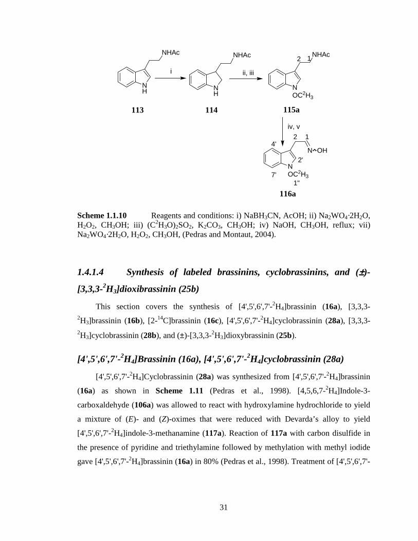
31
NOC2H3
NHAc12
i
NH
NHAc
ii, iii
NOC2H3
N OH
12
2'
4'
7'
iv, v
NH
NHAc
1"
Scheme 1.1.10 Reagents and conditions: i) NaBH3CN, AcOH; ii) Na2WO4·2H2O, H2O2, CH3OH; iii) (C2H3O)2SO2, K2CO3, CH3OH; iv) NaOH, CH3OH, reflux; vii) Na2WO4·2H2O, H2O2, CH3OH, (Pedras and Montaut, 2004).
1.4.1.4 Synthesis of labeled brassinins, cyclobrassinins, and (±±±±)-
[3,3,3-2H3]dioxibrassinin (25b)
This section covers the synthesis of [4',5',6',7'-2H4]brassinin (16a), [3,3,3-2H3]brassinin (16b), [2-14C]brassinin (16c), [4',5',6',7'-2H4]cyclobrassinin (28a), [3,3,3-2H3]cyclobrassinin (28b), and (±)-[3,3,3-2H3]dioxybrassinin (25b).
[4',5',6',7'-2H4]Brassinin (16a), [4',5',6',7'-2H4]cyclobrassinin (28a)
[4',5',6',7'-2H4]Cyclobrassinin (28a) was synthesized from [4',5',6',7'-2H4]brassinin
(16a) as shown in Scheme 1.11 (Pedras et al., 1998). [4,5,6,7-2H4]Indole-3-
carboxaldehyde (106a) was allowed to react with hydroxylamine hydrochloride to yield
a mixture of (E)- and (Z)-oximes that were reduced with Devarda’s alloy to yield
[4',5',6',7'-2H4]indole-3-methanamine (117a). Reaction of 117a with carbon disulfide in
the presence of pyridine and triethylamine followed by methylation with methyl iodide
gave [4',5',6',7'-2H4]brassinin (16a) in 80% (Pedras et al., 1998). Treatment of [4',5',6',7'-
113 114 115a
116a

32
2H4]brassinin with pyridinium bromide perbromide followed by triethylamine gave
[4',5',6',7'-2H4]cyclobrassinin in 40% (Scheme 1.11).
NH
2H2H
2H
2H NH
2H2H
2H
2H
NH2
NH
2H2H
2H
2H
NHSCH3
S1'
3'4'
7'
1
2
3i, ii
CHO
NH
2H2H
2H
2H
S
NSCH34'
7'
1'
3'
v
iii, iv
Scheme 1.11 Reagents and conditions: i) HONH2·HCl, EtOH; ii) NaOH, Devarda’s alloy, CH3OH, 70%; iii) Et3N, CS2; iv) ICH3, 80%; v) pyridine/HBr/Br2 40% (Pedras et al., 1998).
[3,3,3-2H3]Brassinin (16b) and [3,3,3-2H3]cyclobrassinin (28b)
[3,3,3-2H3]Brassinin (16b) and [3,3,3-2H3]cyclobrassinin (28b) were synthesized as
shown on Scheme 1.12 (Monde et al., 1994). [3,3,3-2H3]Brassinin (16b) was synthesized
as described for [4',5',6',7'-2H4]brassinin (16a), but starting from indole-3-
carboxaldehyde (106) (Scheme 1.12). [3,3,3-2H3]Cyclobrassinin (28b) was prepared in
25% yield from [3,3,3-2H3]brassinin (Scheme 1.12) (Monde et al., 1994).
106a 117a
28a
16a

33
NH N
H
NH2
NH
NHSC2H3
S
i,ii
CHO
NH
S
NSC2H3
v
iii, iv
Scheme 1.12 Reagents and conditions: i) HONH2·HCl, EtOH; ii) NaOH, Devarda’s alloy, CH3OH; iii) Py, Et3N, CS2; iv) IC2H3, rt, 18 h 34%; v) pyridine/HBr/Br2, 25% (Monde et al., 1994).
[2-14C]Brassinin (16c)
[2-14C]Brassinin (16c) was synthesized from [2-14C]indole (105b) as shown in
Scheme 1.13 (Pedras et al., 2002). Formylation of [2-14C]indole gave [2-14C]indole-3-
carboxaldehyde (106b) which was used to prepare [2-14C]brassinin (16c) as described
for 16a (Pedras et al., 2002).
NH N
H
NH2
NH
NHSCH3
S
NH
CHO
i ii, iii
iv, v
= 14C*
* * *
*
Scheme 1.13 Reagents and conditions: i) POCl3, DMF; ii) HONH2·HCl, NaOAc; iii) NaBH3CN, TiCl3, NH4OAc; iii) Pyridine, Et3N, CS2; iv) ICH3 (Pedras et al., 2002).
106 117 16b
28b
106b 105b
16c
117b

34
(±±±±)-[3,3,3-2H3]Dioxibrassinin (25a)
(±)-[3,3,3-2H3]Dioxibrassin (25a) was synthesized starting from isatin (118)
(Scheme 1.14). Addition of nitromethane to isatin followed by hydrogenation yielded 3-
hydroxyindolyl-3-methanamine (119). Reaction of amine 119 with carbon disulfide in
the presence of pyridine and triethylamine, followed by deuterated methyl iodide
(IC2H3) yielded [3,3,3-2H3]dioxybrassinin (25a) in 78% (Monde et al., 1994).
NH
O
ONH
O
OHNH2 HCl.
NH
O
HO.
HN SC2H3
Si, ii, iii iv
Scheme 1.14 Reagents and conditions; i) CH3NO2; ii) H2, Pd/AcOH; iii) HCl; iv) Pyridine, Et3N, CS2, IC
2H3, 78% (Monde et al., 1994; Pedras et al., 2003b).
1.4.2 Biosynthesis
The biosynthetic pathways of crucifer phytoalexins and glucosinolates have been
mapped via feeding experiments employing synthetic precursors labeled with stable and
radio-isotopes, as well as non-natural compounds (Halkier and Gershenzon, 2006;
Pedras et al., 2007b).
1.4.2.1 Phytoalexins
The biosynthesis of phytoalexins was previously reviewed by Pedras and co-
workers (2003b and 2007b, Figure 1.6).
25a 119 118

35
NR
N OH
NR
NHSCH3
S
NOCH3
N OSO3 K
S β D Glc
NH
N OSO3 K
S β D Glc
NOCH3
CN
NH
CN
NH
SN
i
ii
R = OCH3
R = HR = OCH3
R = H
R = H
NH
CHO
SCH3
NH
COOCH3
SCH3
NR
S
NSCH3
NH
S
N SCH3
O
NOCH3
S
N SCH3
O
NH
HN
SCH3
O S
HO
NOCH3
S
NO
SCH3
NH
S
NCH3O
O
NH
SN
R = OCH3
R = Hor R = OCH3
R = H
R = H
R = H
R = Hor R = OCH3
NH2
COOH
NH
NH3
O2C
H
Figure 1.6 Map of biosynthetic pathways of crucifer phytoalexins and indolyl glucosinolates 70 and 199. Dashed arrows represent proposed steps. Enzymes involved in the biosynthetic pathway: i) CYP79B2 and CYP79B3; ii) CYP71A13, and CYP79B15 (Pedras et al., 2007b).
112 R = H 116 R = OCH3
16 R = H 18 R = OCH3
49
44
50
78 199
120
70
25 39
43
35
38
34
26
33
28 R = H 31 R = OCH3

36
L-Tryptophan (78), a precursor of majority of crucifer phytoalexins is
biosynthesized from anthranilic acid (120) via the shikimate pathway (Dewick, 1998). In
plants, anthranilic acid (120) is transformed to indole (105) which is conjugated with L-
serine (122) to produce L-tryptophan (Dewick, 1998; Shraudolf, 1966). Biosynthesis of
phytoalexins from L-tryptophan proceeds through indolyl-3-acetaldoxime (112) that
appears to be the metabolic branching point in a few secondary pathways such as indole
phytoalexins and indole glucosinolates (Pedras et al, 2007b) (Figures 1.6 and 1.7).
NH2
COOH
NH
NH3
O2C
H
NH
NH3
O2C
H
HO
NH
N OH
Figure 1.7 Biosynthetic pathway of L-tryptophan (78) and indolyl-3-acetaldoxime (112) from anthranilic acid (120).
Cytochrome P450 enzymes from the CYP79 family that catalyze conversion of L-
tryptophan to indolyl-3-acetaldoxime have been isolated from different plant sources
such as white mustard and the model plant A. thaliana (Hansen and Halkier, 2005; Hull
et al., 2000; Mikkelsen et al., 2000; Naur et al., 2003b). The enzymes CYP79B1
(isolated from white mustard), CYP79B2, and CYP79B3 (isolated from A. thaliana)
have been proposed to catalyze N-hydroxylation of L-tryptophan to N-hydroxy-L-
tryptophan that decarboxylates to give indolyl-3-acetaldoxime (Hansen and Halkier,
2005; Hull et al., 2000; Mikkelsen et al., 2000; Naur et al., 2003b). These
transformations have been extensively studied in the biosynthetic pathways of
glucosinolates and are described in the next section (Hansen and Halkier, 2005; Hull et
al., 2000; Mikkelsen et al., 2000; Naur et al., 2003b).
The biosynthesis of the crucifer phytoalexins, brassinin (16), cyclobrassinin (28)
and spirobrassinin (34) was investigated in turnip roots using isotopically labeled (13C
and 2H) precursors. The origin of indole group was confirmed through deuterium
120
122
105 78 112

37
labeling experiments using L-[4'-2H]tryptophan (Monde et al., 1994) (Figure 1.8). The
occurrence of a molecular rearrangement in the biosynthetic pathway from L-tryptophan
(78) to brassinin (16) was investigated by administration of DL-[13C-2]tryptophan to
turnip root tissues. The 13C NMR of isolated spirobrassinin revealed a 4-fold
enhancement of the imino carbon NMR signal. These results suggested that a molecular
rearrangement of L-tryptophan leads to indolyl-3-methylisothiocyanate (123) which is an
intermediate in the biosynthetic pathway of brassinin (16), and that the thiocarbonyl
carbon of brassinin originates from C-2 of L-tryptophan (Figure 1.8) (Monde et al.,
1994). Administration of L-[3,3,3-2H3]methionine indicated that the methyl groups of
brassinin, cyclobrassinin and spirobrassinin derived from L-methionine. Additional
experiments employing a mixture of L-[3,3,3-2H3]methionine and L-[35S]methionine
revealed intact incorporation of L-methionine into brassinin. The origin of the
thiocarbonyl sulfur from L-cysteine was confirmed after incubation of turnip tissues with
L-[35S]cysteine; high radioactivities of the isolated brassinin and spirobrassinin
supported the conclusion that the thiocarbonyl sulfur atom originates from L-cysteine
(Monde et al., 1994) (Figure 1.8). The biogenetic relationship between the phytoalexins
brassinin (16), dioxibrassinin (25), cyclobrassinin (28), and spirobrassinin (34) in turnip
root tissues was investigated with [3,3,3-2H3]brassinin (16b), [3,3,3-2H3]dioxibrassinin
(25a), and [3,3,3-2H3]cyclobrassinin (28b) (Figure 1.8). Labeled brassinin was
effectively incorporated into cyclobrassinin and spirobrassinin, but neither
cyclobrassinin nor dioxibrassinin were incorporated in spirobrassinin. Incubation of
Japanese radish roots (Raphanus sativus var. hortensis) with [3,3,3-2H3]brassinin further
confirmed the intermediacy of brassinin in the biosynthetic pathway of cyclobrassinin
(Monde et al., 1995). These experiments therefore revealed that brassinin (16) is an
advanced precursor in the biosynthetic pathway of many other crucifer phytoalexins
(Monde et al., 1994; 1995; Pedras et al., 2004).

38
NH
NH3
O2C
H
NH
NCS
NH3
CO2H
HS
NH3
CO2H
H3CS
NH
NHSCH3
S
NH
S
NSCH3
NH
S
N SCH3
O
NH
HN
SCH3
O S
HO
XX
= 13C from tryptophan
= 35S from cysteine
= 35S from methionine
= C2H3 from methionine
Figure 1.8 Biosynthesis of cruciferous phytoalexins: the occurrence of molecular rearrangement, and biogenetic relationship between brassinin (16) and other phytoalexins.
The biogenetic relationship between the phytoalexin brassinin (16) and the indole
glucosinolate glucobrassicin (70) was subject of discussion. Glucobrassicin (70)
appeared to be biogenetically related to brassinin (16) (Figure 1.9) since both are
biosynthesized from L-tryptophan (Goetz and Schraudolf, 1983; Kutáček et al., 1962;
Kutáček and Králová, 1972). Glucobrassicin (70) is also known to undergo enzymatic
hydrolysis by myrosinases, Lossen-type rearrangement of the resulting intermediate
yields indolyl-3-methylisothiocyanate (123, Agerbik et al., 1998; Hanley et al., 1990;
Hanley and Parsley, 1990; Latxague et al., 1991), a proposed intermediate between
tryptophan and brassinin (Monde et al., 1994).
123 78 16
25
34 28

39
NH
N OSO3 K
S β D Glc
NH
NCS
NH
NHSCH3
S
??
Figure 1.9 Plausible biogenetic relationship between brassinin (16) and glucobrassicin (70).
The preparation of indolyl-3-methylisothiocyanate (123) was unsuccessful
(Kutschy et al., 1998) due to its instability (Hanley et al., 1990; Hanley and Parsley,
1990). Monde and co-workers (1994), however, attempted to trap the putative
isothiocyanate 123 in the biosynthesis of brassinin. Monde and co-workers (1994)
reported that homogenizing turnip roots in the presence of sodium methanethiolate
yielded brassinin (16) and a compound that was identified as PhCH2-CH2-NH-CS-SCH3,
which was named phenylethylbrassinin (124). These metabolites were not isolated in the
absence of sodium mathanethiolate (Monde et al., 1994). The putative role of
isothiocyanate in the biosynthesis of brassinin was further demonstrated with benzyl
isothiocyanate (125) (Figure 1.10). Administration of 125 to turnip tubers in the absence
of sodium methanethiolate yielded a new metabolite that was named benzylbrassinin
(126) (Monde et al., 1994).
NCSHN SCH3
S
Figure 1.10 Biosynthesis of benzylbrassinin (126) (Monde et al., 1994).
125 126
16
70 123

40
The potential intermediacy of glucobrassicin (70) in the biosynthesis of brassinin
(16) was investigated with [4',5',6',7'-2H4]glucobrassicin (70a) using brown mustard (B.
juncea) leaves elicited with L. maculans, and with UV-irradiated turnip root slices.
[4',5',6',7'-2H4]Glucobrassicin (70a) was not incorporated into any of the phytoalexins
produced by either plant tissue, but was rather metabolized to indole-3-carboxaldehyde
(106a) and methyl indole-3-carboxylate (121a) by turnip root slices (Pedras et al., 2001;
2003b) (Figure 1.11). On the other hand, [4',5',6',7'-2H4]indolyl-3-acetaldoxime (112a)
was efficiently incorporated into brasslexin (26a), cyclobrassinin (28a) and
spirobrassinin (34b) in turnip root slices (Figure 1.11). These results suggested that the
biosynthetic pathway of brassinin possibly branches before formation of glucobrassicin,
and that indolyl-3-acetaldoxime or other related intermediate is the common biosynthetic
precursor between phytoalexins and glucosinolates (Pedras et al., 2003b).
NH
N OH
NH
N OSO3 K
S β D Glc
NH
S
NSCH3
NH
SN
NH
S
N SCH3
O+ +
XNH
R
Figure 1.11 Biosynthesis of cruciferous phytoalexins: lack of incorporation of [4',5',6',7'-2H4]glucobrassicin (70a), and incorporation of [4',5',6',7'-2H4]indolyl-3-acetaldoxime (112a) (Pedras et al., 2003b).
[3,3,3-2H3]Dioxibrassinin (25a) and [3,3,3-2H3]cyclobrassinin (28b) were not
incorporated into [3,3,3-2H3]spirobrassinin (34a) suggesting that cyclobrassinin and
spirobrassinin derive from brassinin (16) via a common intermediate (Monde et al.,
1994). The contribution of such an intermediate was probed with a non-natural
precursor, 2-methylbrassinin (127, Figure 1.12). In feeding experiments, turnip tissues
70
112 28 26 34
106 R = CHO 121 R = COOCH3

41
metabolized 2-methylbrassinin (127) to two metabolites, 129 and 130, that were
suggested to form through a pinacol-type rearrangement of the diol intermediate 128.
These results suggested that a similar diol intermediate could be involved in the
biosynthesis of cyclobrassinin (28) and spirobrassinin (34) from brassinin (16) (Monde
et al., 1994).
NH
CH3
HN SCH3
S
NH
CH3
HN SCH3
S
NH
O
CH3
OH
HO
NH
SCH3
O
N
S
N SCH3
CH3
+
Figure 1.12 Probing for the common intermediate in the biosynthesis of cyclobrassinin (28) and spirobrassinin (34) from brassinin (16) (Monde et al., 1994).
In other biosynthetic investigations [4',5',6',7'-2H4]brassinin (16a) was incorporated
into rutalexin (33a), brassicanal A (39a), and brassicanate A (43a) in rutabaga root slices
(Pedras et al. 2004, Figure 1.13). The occurrence of brassicanal A and brassicanate A in
rutabaga root tubers suggested a possible biogenetic relationship between these two
metabolites. Interestingly, the percent incorporation of brassinin (16a) into brassicanate
A (43a) was much higher than that for brassicanal A (39a) (Pedras et al., 2004).
Additional deuterium incorporation experiments revealed cyclobrassinin (28a) to be an
advanced biogenetic precursor of brassilexin (26a) (Pedras et al., 1998; 2007b) and
rutalexin (33a) (Pedras et al., 2004; 2007b). Rutalexin (33) was isolated from rutabaga
for the first time and its characterization was a revision of the structure of a compound
that was previously misassigned as cyclobrassinone (131) (Pedras et al., 2004).
127 128
130
129

42
NH
S
NO
OCH3
The biosynthesis of brassilexin (26a) from indolyl-3-acetaldoxime (112a),
brassinin (16a), and cyclobrassinin (28a) was also established in feeding experiments
with turnip root slices and in leaves of B. carinata and B. juncea (Pedras et al., 1998;
2001, 2007b) (Figure 1.13). The sequence of the biosynthetic pathway of brassilexin
(26), rutalexin (33), brassicanal A (39), and brassicanate A (43) was therefore
established (Pedras et al., 1998; 2001; 2007b) (Figure 1.13).
NH
N OH
NH
NHSCH3
S
NH
S
NSCH3
NH
SN
NH
S
NCH3O
ONH
CHO
SCH3NH
COOCH3
SCH3
NH
CN
Figure 1.13 Biosynthetic sequence of brassilexin (26a), rutalexin (33a), brassicanal A (39a), and brassicanate A (43a) from indolyl-3-acetaldoxime (112a) (Pedras et al., 2002; 2004).
Efficient metabolism of [4',5',6',7'-2H4]indolyl-3-acetaldoxime (112a) to indolyl-3-
acetonitrile (49a) (Figure 1.13) by B. juncea leaves suggested that under these
experimental conditions 112, but not glucobrassicin (70), is the key intermediate in the
biosynthesis of 49 (Pedras et al., 2002).
The biosynthesis of camalexin (44), the principle phytoalexin of the model plant A.
thaliana is shown in Figure 1.14. Camalexin was first proposed to be biosynthesized
131
112 16 28 26
33 39 43 49

43
through condensation of indole-3-carboxaldehyde (106) with cysteine followed by
cyclization and decarboxylation (Browne et al., 1991). The thiazole ring of camalexin
was established to originate from cysteine based on a more efficient incorporation of
[35S]cysteine than [35S]methionine, thus supporting the initial hypothesis by Browne and
co-workers (1991). Additional experiments showing incorporation of cysteine labeled
with 2H, 14C, or 13C and 15N into camalexin supported the previous conclusions (Zook
and Hammerschmidt, 1997). The efficient incorporation of radioactivity from
[14C]anthranilate than [3H]tryptophan showed that anthranilic acid (120) but not L-
tryptophan (78) was the precursor of camalexin (Tsuji et al., 1993; Zook and
Hammerschmidt, 1997). Further biosynthetic experiments using A. thaliana cell cultures
showed that the biosynthetic pathway of camalexin possibly diverges from that of
tryptophan at the indole step. The efficiency of incorporation of [14C]indole into
camalexin was twice that of [14C]anthranilate, suggesting that indole is a closer precursor
to camalexin than anthranilic acid, whereas tryptophan is not a precursor (Zook, 1998).
Contrary to these reports, in vivo labeling experiments showed that [14C-2]indolyl-3-
acetaldoxime was incorporated into camalexin in A. thaliana (Glawischnig et al., 2004;
Pedras et al., 2007b, Figure 1.14). Additional experiments using A. thaliana leaves
showed incorporation of 6-fluoroindolyl-3-acetaldoxime, 6-fluorotryptophan and 14C-
labeled tryptophan into camalexin and derivatives (Glawischnig et al., 2004). The
fluorinated or 14C-labeled tryptophan was incorporated albeit with low efficiency
(Glawischnig et al., 2004), hence the previous assumption that tryptophan is not a
precursor of camalexin (Tsuji et al., 1993; Zook and Hammerschmidt, 1997) did not
hold. It was also established that the enzymes CYP79B2 and CYP79B3 catalyze the
conversion of L-tryptophan (78) to indolyl-3-acetaldoxime (112) in the biosynthetic
pathway of camalexin in A. thaliana (Glawischnig et al., 2004). Dihydrocamalexic acid
(132) was shown to accumulate in infected root cultures of phytoalexin deficient A.
thaliana mutant pad3 (Bednarek et al., 2005). In other biosynthetic studies, (S)-
dihydrocamalexic acid (132) was converted to camalexin (44) on incubation with
CYP71B15 (PAD3) (Schuhegger et al., 2006). These results suggested that CYP71B15
catalyzes the oxidative decarboxylation of dihydrocamalexic acid (132), the last step in
the biosynthetic pathway of camalexin (Pedras et al., 2007b; Schuhegger et al., 2006). In

44
the most recent report, involvement of CYP71A13 enzyme in camalexin biosynthesis
was demonstrated. It was established that CYP71A13 is required for camalexin (44)
production. Incubation of CYP71A13 with indolyl-3-acetaldoxime (112) indicated that
CYP71A13 catalyzes the dehydration of 112 to indolyl-3-acetonitrile (49) in the
biosynthesis of camalexin (Nafisi et al., 2007). These results rather suggested that
indolyl-3-acetonitrile (49) is an intermediate in the biosynthesis of camalexin (44)
(Nafisi et al., 2007). The conversion of indolyl-3-acetonitrile (49) to dihydrocamalexic
acid (132) is neither understood nor established.
NH
N OH
NH
CN
NH
SN
CO2H
NH
SN
i ii
L-Cysteine
Figure 1.14 Biosynthetic intermediates of camalexin (44). Enzymes involved in the biosynthetic pathway: i) CYP71A13; ii) CYP71B15 (PAD3) (Pedras et al., 2007b; Schuhegger et al., 2006).
The phytoalexins brassicanal A (39) and brassicanate A (43) are examples of
phytoalexins that have only one sulfur atom. Previously, a trapping experiment was
performed to establish the potential intermediates in the biosynthetic pathway of
brassicanal A (39) (Figure 1.15). Addition of aniline to UV-irradiated turnip root slices
resulted in production of a new compound 136 (Figure 1.15). Compound 136 was
suggested to be the aniline trapped biosynthetic intermediate 2-mercaptoindole-3-
carboxaldehyde (135) since its biological methylation leads to brassicanal A (Monde et
al., 1996). The proposed intermediate 135 was suggested to derive from hydrolysis of
dehydrocyclobrassinin (133), which in turn would be formed from enzymatic
dehydrogenation of cyclobrassinin (28). Dehydrocyclobrassinin (133) has not been
isolated, although the 4-methoxy-dervative (32) is known (Monde et al., 1996).
Biosynthetic investigations employing rutabaga root slices revealed [4',5',6',7'-2H4]brassinin incorporation into brassicanal A (39a) and brassicanate A (43a), whereas
112 49 132 44

45
incorporation of [4',5',6',7'-2H4]cyclobrassinin was not detected into either brassicanal A
or brassicanate A (Pedras et al., 2004). Brassicanal A (39) is a putative precursor of
brassicanate A (43) (Pedras et al., 2004), and luck of incorporation of cyclobrassinin
under the experimental conditions suggested that possibly the trapped intermediate
(Monde et al., 1996) was derived from brassinin (16) and not cyclobrassinin (28). Origin
of the thiomethyl group at C-2 of indole therefore remains to be established, but could
perhaps be deriving from methionine.
NH
S
NSCH3
NS
NSCH3
NH
S
NSCH3
HO
H2O
NH
CHO
SHNH
CHO
SCH3
NH
S
NHPh
PhNH2
i
ii X
Figure 1.15 Putative biosynthetic pathway of brassicanal A (39). i) Biological methylation of 2-mercaptoindole-3-carboxaldehyde (135) yields brassicanal A; ii) Incorporation of cyclobrassinin (28) into brassicanal A (39) was not detected in rutabaga root slices. Compounds in bracket are proposed intermediates (Monde et al., 1996; Pedras et al., 2003b).
The occurrence of brassinin (16), 1-methoxybrassinin (18) and cyclobrassinin (28)
in Pseudomonas cichorii-inoculated Chinese cabbage and Japanese radish roots
136
135
133
39
134 28

46
suggested a possible N-hydroxylation and subsequent methylation of brassinin to give 1-
methoxybrassinin, and that oxidative cyclization of 1-methoxybrassinin would lead to
cyclobrassinin (Monde et al., 1995; Pedras et al., 2003b). [3,3,3-2H3]Brassinin (16b)
however, was efficiently incorporated into cyclobrassinin (28b) and not into 1-
methoxybrassinin (18a) (Monde et al., 1995b) (Figure 1.16). These findings suggested
that 28 is biosynthesized directly from brassinin (16) and not through 1-
methoxybrassinin (18). These conclusions were further supported by absence of
incorporation of [3,3,3-2H3]1-methoxybrassinin (18a) into cyclobrassinin (Monde et al.,
1995b). These metabolic studies indicated that perhaps the biosynthetic pathway of 1-
methoxy-containing phytoalexins diverges from the NH-containing phytoalexins before
formation of brassinin (16), and that 1-methoxybrassinin (18) is the biosynthetic
precursor of 1-methoxy-containing phytoalexins. Additional investigation employing
rutabaga root slices showed equal incorporation of [4',5',6',7'-2H4]indolyl-3-acetaldoxime
(112a) into [4',5',6',7'-2H4]brassinin (16a) and [4',5',6',7'-2H4]1-methoxybrassinin (18b)
(Pedras et al., 2004), suggesting two parallel biosynthetic pathways leading to 16 and 18
(Figure 1.16) (Pedras et al., 2003b). To establish the plausible intermediate between
indolyl-3-acetaldoxime (112) and 1-methoxybrassinin (18), [1",1",1"-2H3]1-
methoxyindolyl-3-acetaldoxime (116a) was synthesized for the first time and was
incubated with rutabaga and kohlrabi root slices. Mass spectrometry analysis of the
isolated 1-methoxybrassinin (18a) revealed deuterium incorporation from [1",1",1"-2H3]1-methoxyindolyl-3-acetaldoxime (116a) in both rutabaga and kohlrabi root slices.
Additional feeding experiments revealed incorporation of [4',5',6',7'-2H4]indolyl-3-
acetaldoxime into brassinin (16a), 1-methoxybrassinin (18b), and spirobrassinin (34b)
(Pedras et al., 2004). These metabolic studies were the first to demonstrate that
biosynthetic pathway of NH-containing and 1-methoxy-containing phytoalexins divert at
the aldoxime step. It was therefore surmised that indolyl-3-acetaldoxime (112) is
transformed into brassinin and then to other NH-containing phytoalexins, whereas N-
oxydation and methylation of 112 yields 1-methoxyindolyl-3-acetaldoxime (116), the
precursor of 1-methoxybrassinin (18) which is the plausible precursor of other 1-
methoxy-containing phytoalexins (Pedras and Montaut, 2004). Biosynthetic pathways of
erucalexin (38) and 1-methoxyspirobrassinin (35) have been studied (Pedras et al.,

47
2007b; Pedras and Okinyo, 2006b) and are part of this thesis, as reported in the Results
and Discussion sections.
NOCH3
N OH
NH
N OH
NH
NHSCH3
S
NOCH3
NHSCH3
S
NH
S
NSCH3
X
Figure 1.16 Biosynthesis of brassinin (16a) and cyclobrassinin (28a) from indolyl-3-acetaldoxime (112a), and 1-methoxybrassinin (18b) from 1-methoxyindolyl-3-acetaldoxime (116a) (Pedras and Montaut, 2004; Pedras et al., 2004).
The biosynthesis of many other 1-methoxy-containing phytoalexins such as 1-
methoxybrassenins A (21) and B (22), wasalexins A (23) and B (24), and sinalbins A
(30) and B (31), among others remain to be investigated. It is therefore necessary to
carry out biosynthetic studies to unravel the pathways leading to such phytoalexins as
this may facilitate transfer of these pathways to other cruciferous crops.
The three most recently reported phytoalexins, rapalexins A (53) and B (54), and
brussalexin A (55) have introduced yet new structures among the crucifer phytoalexins
(Pedras et al., 2007b; 2007c). The rapalexins A and B were proposed to derive from
putative indolyl glucosinolates or from thiohydroximic acids such 137 and 138 (Figure
1.17). Lossen-type rearrangement to intermediate 139 would then yield 53 and 54
(Figure 1.17). Brussalexin A (55) represents a unique chemical structure that does not
seem to fit with the presently known biosynthetic pathway of crucifer phytoalexins
isolated to date (Pedras et al., 2007b). It was therefore proposed that brussalexin A could
biosynthetically derive from addition of indolyl-3-methane thiol (142) to allyl
isothiocyanate 68 (produced from myrosinase hydrolysis of sinigrin, 56, Figure 1.18)
(Pedras et al., 2007c).
112 16 28
116 18

48
NH
NCSOCH3
R
NH
OCH3
R N
SXNH
OCH3
R S X
NOY
Figure 1.17 Proposed biosynthetic precursors of rapalexin A (53) and B (54) (Pedras et al., 2007b).
NO3SO
Sβ
K
NCS
NH
S
HN
O
NH
SH
DGlc
Figure 1.18 Proposed biosynthetic precursors 56 and 68 of brussalexin A (55) (Pedras et al., 2007c).
1.4.4.2 Glucosinolates
The biosynthesis of glucosinolates can be divided into three different stages: (i)
chain elongation of certain aliphatic and aromatic amino acids via sequential addition of
methylene groups, (ii) construction of the core structure of glucosinolates, and (iii)
secondary modification of the side chain (Grubb and Abel, 2006; Halkier and
Gershenzon, 2006).
Side chain elongation
Chain elongated aliphatic glucosinolates are the most abundant glucosinolates
found in crucifers including the model plant A. thaliana and many other Brassicaceae
species (Halkier and Gershenzon, 2006). The extent of side chain elongation and type of
137 R = H 138 R = OH 139
53 R = H 54 R = OH
56 68
142
55

49
modifications are important for the biological activity of the glucosinolate breakdown
product (Manici et al., 1997; Mithen et al., 1986; Sarwar et al., 1998). The side chain-
elongation reactions have been investigated using in vivo labeling studies, isolation of
the participating intermediates, and characterization of the enzymes catalyzing various
reactions of the biosynthetic pathway. Side chain elongation reactions are shown in
Figure 1.19.
RCOOH
NH2
RCOOH
O
Acetyl-CoA
RCOOH
COOH
OH
RCOOH
COOHHO
COOH
O
RCOOH
NH2
R
i ii
iii
ivv
Figure 1.19 Side chain elongation: i) BRANCHED-CHAIN AMINOTRANSFERASE4 (BCAT4); ii) 2-(ω-Methylthioalkyl)malate synthase (MAMS) or Methylthioalkylmalate synthase 1 (MAM1) or Methylthioalkylmalate synthase 3 (MAM3); iii) Isomerization; iv) Oxidative decarboxylation; v) Transamination (Graser et al., 2000; Halkier and Gershenzon, 2006).
Side chain-elongation begins with oxidative deamination of the parent amino acid
(143) to yield the corresponding 2-oxo acid (144). One of the enzymes catalyzing
oxidative deamination of amino acids, BRANCHED-CHAIN AMINOTRANSFERASE4
(BCAT4) in the biosynthetic pathway of methionine-derived glucosinolates was
characterized from A. thaliana (Schuster et al., 2006). The 2-oxo acid (144) enters a
three step-cycle in which it (i) condenses with acetyl CoA to form a substituted 2-malate
derivative (145) which (ii) isomerizes by a 1,2-shift of the hydroxyl group to form a 3-
malate derivative (146) that is (iii) oxidatively decarboxylated to yield a new 2-oxo acid
143 144
146
145
147 148

50
with one additional methylene group (147) (Graser et al., 2000; Halkier and Gershenzon,
2006; Falk et al., 2004; Textor et al., 2004; 2007). The 2-oxo acid 147 can undergo
transamination to regenerate the amino acid (148) which enters the biosynthetic pathway
of short chain aliphatic or aromatic glucosinolates, or it may undergo further cycles of
chain-elongation to form longer homologs (Textor et al., 2004; 2007). Chain elongation
products of up to nine cycles are known to occur in plants (Fahey et al, 2001), but only
six cycles are known in the model plant A. thaliana (Textor et al., 2004; 2007). All
chain-elongated 2-oxo acids are converted back to the corresponding amino acids that
participate in the biosynthetic pathway of glucosinolates of varying side-chain lengths
(Graser et al., 2000).
An extensive study on the sequence of chain-elongation reactions was conducted
by carrying out in vivo feeding experiments using stable and radiolabeled isotopes
(Graser et al., 2000). These experiments revealed that only the acetate methyl group is
incorporated to form the methylene group in chain-elongated products, thus suggesting
that the carboxyl group of the first acetate residue is lost as carbon dioxide during the
second round of chain-elongation, and during aldoxime formation (Graser et al., 2000).
RCOOH
O
CH2CSCoAO
H
RCOOH
COOH
OH
CoASH
COOH
NH2
R
Figure 1.20 Condensation of acetyl CoA with a 2-oxo acid (144).
It was established that the nitrogen atom of the parent amino acid is incorporated
into the core glucosinolate structure arising from the chain-elongated amino acid. It was
therefore concluded that the transfer of amino group in the first step initiates the
formation of 2-oxo acid, and re-incorporation into the biosynthetic pathway terminates
the chain elongation reaction to regenerate the amino acid that is required to take part in
the biosynthetic pathway of chain-elongated glucosinolates (Graser et al., 2000).
145 148 144

51
Detection of intermediates and isolation of chain-elongated homologs have further
supported the chain elongation reactions (Halkier and Gershenzon, 2006). The enzyme
catalyzing the first step of chain-elongation reaction, 2-(ω-methylthioalkyl)malate
synthases (MAMS), was partially purified from rocket (Falk et al., 2004). MAMS
however could not catalyze the second cycle of chain-elongation indicating that it was
specific for the first round only (Falk et al., 2004). Another methylthioalkylmalate
synthase, methylthioalkylmalate synthase 1 (MAM1) from A. thaliana was capable of
catalyzing both first and second rounds of chain-elongation cycles (Kryomann et al.,
2001; Textor et al., 2004). MAM1 was reported to exist together with MAM3 (formerly
known as MAM-L for MAM-like) in A. thaliana (Textor et al., 2007). Unlike MAM1,
MAM3 is capable of catalyzing all six condensation reactions of methionine chain-
elongation that occur in Arabidopsis, it also exhibits a broad substrate range of other
non-methionine derived 2-oxo acids (Textor et al., 2007).
Biosynthesis of the glucosinolate core structure
The biosynthesis of aliphatic, aromatic, and indolyl core glucosinolate structure
involves common intermediates (N-hydroxy amino acid, aldoximes, aci-nitro or nitrile
oxides, S-alkyl thiohydroximates, thiohydroxamic acids, and desulfoglucosinolates)
(Figure 1.21).

52
RCOOH
NH2
[O]
RCOOH
NHOH
CO2
H2
RN OH
[O]
RN
OHO
Cys
RN
OH
S
NH3
H
CO2
RN
OH
SH
UDP-Glc
RN
OH
Sβ D Glc PAPS
RN
OSO3
Sβ D Glc
i
ii
iiiiv
vi
v
Figure 1.21 Proposed biosynthetic pathway of glucosinolates: i) CYP79F1, CYP79F2 (methionine), CYP79A2 (phenylalanine), CYP79B1, CYP79B3, CYP79B3 (tryptophan); ii) CYP83A1 (methionine), CYP83B1 (phenylalanine, tryptophan); iii) GST?; iv) C-S lyase; v) UGT74B1 v) AtST5a (phenylalanine, tryptophan), AtST5b, AtST5c (methionine). Structures in brackets are proposed intermediates (Halkier and Gershenzon, 2006).
The first common step in glucosinolate and cyanogenic glucoside biosynthesis is
the conversion of amino acids (148) to their respective aldoximes (150) (Bennet et al.,
1993; 1997; Dawson et al. 1993; Dewick, 1984; Du et al., 1995; Wittstock and Halkier,
2000). The aldoxime forming enzyme was proposed to catalyze N-hydroxylation of
amino acids to the corresponding N-hydroxyamino acids (149) that decarboxylate to
produce the aldoxime (150). Cytochrome P450 family type of enzymes were shown to
catalyze such transformations (Halkier and Møller, 1991; Koch et al., 1992). The
148 149 150
151 153
155 154
152

53
enzymes CYP79F1 and CYP79F2 metabolize chain elongated derivatives of methionine
to their respective aldoximes in the biosynthesis of aliphatic glucosinolates, but not
tyrosine, tryptophan, or their derivatives (Chen et al., 2003; Hansen et al., 2001a;
Reintanz et al., 2001). CYP79A2 on the other hand catalyzes the metabolism
phenylalanine to phenylacetaldoxime in the biosynthetic pathway of aromatic
glucosinolates (Wittstock and Halkier, 2000). The enzymes CYP79B1, CYP79B2 and
CYP79B3 catalyze the conversion of L-tryptophan (78) to indolyl-3-acetaldoxime (112)
in the biosynthesis of indolyl glucosinolates (Hansen and Halkier, 2005; Hull et al.,
2000; Mikkelsen et al., 2000; Naur et al., 2003b). Overexpression of CYP79B2 in
Arabidopsis resulted in significant increase in levels of indolyl glucosinolates
glucobrassicin (70) and 4-methoxyglucobrassicin (156), but not of aliphatic or aromatic
glucosinolates (Mikkelsen et al., 2000).
Following its formation, the aldoxime (150) is conjugated with a suitable thiol
donor to yield an S-alkyl-thiohydroximate (152). Both cysteine and methionine are
known thiol donors in the biosynthetic pathway of many secondary metabolites, the
former is however a more efficient donor than the latter (Mikkelsen et al., 2002). The
aldoxime metabolizing enzymes in the biosynthesis of glucosinolates (in A. thaliana)
have been characterized. The enzyme CYP83A1 metabolizes aliphatic aldoximes much
more efficiently than CYP83B1 (Bak and Feyereisen, 2001; Hemm et al., 2003; Naur et
al., 2003a). The enzyme CYP83B1 catalyzes the metabolism of aromatic aldoximes
derived from tyrosine and phenylalanine (Hansen et al., 2001b), as well as indolyl-3-
acetaldoxime (Bak and Feyereisen, 2001). CYP83A1 enzyme is capable of metabolizing
indolyl-3-acetaldoxime albeit with low efficiency (Bak and Feyereisen, 2001).
Conjugation of the aldoxime (150) with the thiol donor (e.g. L-cysteine) requires
conversion of 150 into a reactive intermediate that can be attacked by the nucleophilic
thiol donor. The CYP83 enzymes have been proposed to catalyze the oxidation of
respective aldoximes to highly reactive 1-aci-nitro compounds (151) or nitrile oxides
(Bak et al., 2001; Bak and Feyereisen, 2001; Hansen et al., 2001b). Proper conjugation
of the sulfur donor (L-cysteine) with the reactive intermediate (aci-nitro compound or
nitrile oxide) is thought to be catalyzed by a glutathione-S-transferase type enzyme
(Mikkelsen et al., 2004). The C-S bond of S-(thiohydroximoyl)-L-cysteine (152) is

54
hydrolyzed by C-S lyase to form thiohydroximic acid (153) which is the next
intermediate in the biosynthetic pathway of glucosinolates. The L-cysteine conjugate
(152) does not accumulate in plant tissues and has therefore not been isolated. In vitro,
152 rapidly cyclizes to 2-substituted thiazoline-4-carboxylic acid (157) (Figure 1.22),
leading to a suggestion that the C-S lyases form tightly coupled complexes with CYP83
enzymes to prevent cylization of the C-S lyase substrates (Hansen et al., 2001b; Halkier
and Gershenzon, 2006). SUPERROOT1 (SUR1) was characterized as a C-S lyase
enzyme in the biosynthetic pathway of A. thaliana glucosinolates (Figures 1.21 and
1.22) (Mikkelsen et al., 2004).
RN
OH
S
NH2
H
CO2H
RN
S
CO2H
H
NH2OH
RN
OH
SH SUR1
Figure 1.22 Cyclization of cysteine conjugate (152) to thiazoline-4-carboxylic acid (157), and SUPERROOT1 (SUR1) cleavage of C-S bond (Hansen et al., 2001b).
The thiohydroximic acid (153, Figure 1.22) formed is highly reactive and has not
been isolated, but is subsequently glycosylated by a sugar nucleotide to produce
desulfoglucosinolate (154, Figure 1.21). Uridine diphosphate glucose (UDPG) was
found to be the most efficient glucose donor (Mitsuo and Underhill, 1971). A putative
UDPG:thiohydroximate glucosyltransferase in the biosynthetic pathway of benzyl
glucosinolate was isolated from Arabidopsis and was characterized as UGT74B1 (Grubb
et al., 2004). Loss of UGT74B1 gene function only caused partial reduction of
glucosinolate accumulation, suggesting that additional enzymes catalyzing glucosylation
of thiohydroximates are present (Grubb et al., 2004). Enzyme preparations of
UDPG:thiohydroximate glucosyltransferase purified from Brassica napus seedlings
were reported to glycosylate sodium 3-phenylpropanothiohydroximate (100) to
phenylethyl desulfoglucosinolate (158) (Figure 1.23, Reed et al., 1993). The use of
similar enzyme preparations with sodium 2-(3-indolyl)acetothiohydroximate (99) were
152 157 153

55
reported to give only detectable amounts of desulfoglucobrassicin (159) since 99 was not
stable in the buffer used (pH 6) (Figure 1.23, Reed et al., 1993).
S β
N OH
D Glc
NH
N OH
S β D Glc
S
N OH
NH
N OH
S Na
Na
UDP-[U-14C]glucose ?
UDP-[U-14C]glucose
*
*
Glc = [U-14C]glucose*
Figure 1.23 UDPG:thiohydroximate glucosyltransferase glycosylation of sodium 3-phenylpropanothiohydroximate (100) to phenylethyl desulfoglucosinolate (158) (Reed et al., 1993).
The last common step in building glucosinolate core structure (155) (for aliphatic,
aromatic and indolyl glucosinolates) is the introduction of an active sulfate group from
3'-phosphoadenosine-5'-phosphosulfate (PAPS) to desulfoglucosinolate (154). Sulfation
of desulfoglucosinolates is catalyzed by sulfotransferases (Glending and Poulton, 1988;
1990; Jain et al., 1989;1990; Klein et al., 2006; Piotrowski et al., 2004). Three
sulfotransferases AtST5a, AtST5b, and AtST5c exhibiting broad substrate range have
been characterized from A. thaliana (Piotrowski et al., 2004). Whereas AtST5b and
AtST5c had higher affinity for aliphatic glucosinolates, AtST5a preferred tryptophan-
and phenylalanine-derived desulfoglucosinolates (Piotrowski et al., 2004).
Side chain modification
Glucosinolate side chain modifications play a significant role in determining the
biological activity of the hydrolysis product (Halkier and Gershenzon, 2006). The side
chain modifications have been noted to occur in glucosinolates derived from methionine
100 158
159 99

56
and its homologs (160), the transformations may involve sequential oxidations,
eliminations, alkylations, and esterifications (Figure 1.24) (Halkier and Gershenzon,
2006; Grubb and Abel, 2006). The sulfur can be successively oxidized to
methylsulfinylalkyl (161) and methylsulfonylalkyl (162) moieties (Halkier and
Gershenzon, 2006). Further modifications may involve oxidative cleavage of the
methylsulfinylalkyl moiety to yield alkenyl (163) or hyroxyalkyl (164) side chains for
which controlling genetic loci are known in Arabidopsis and Brassica species (Halkier
and Gershenzon, 2006).
S β
NOSO3 K
D GlcSO
n
S β
NOSO3 K
D GlcS n
S β
NOSO3 K
D GlcSO
n
O
S β
NOSO3 K
D Glcn
S β
NOSO3 K
D GlcHO n
S β
NOSO3 K
D GlcO n
O
AOP2
AOP3
Figure 1.24 Side chain modifications of methionine-derived glucosinolates with varying side chain lengths. APO2 and APO3 are putative enzymes catalyzing the reactions in Arabidopsis (Halkier and Gershenzon, 2006; Kliebenstein et al., 2001).
The AOP2 gene product catalyzes the conversion of methylsulfinylalkyl
glucosinolates (161) to alkenyl glucosinolates (163), whereas the AOP3 gene product
controls the formation of hydroxyalkyl glucosinolates (164) from 161 (Kliebenstein et
al., 2001). Although biosynthesis of glucosinolate esters (165) have been investigated
160
161
162
163
164
165

57
through administration of isotopically labeled precursors and intermediates, no studies
have been carried out to determine the enzymes involved in their biosynthetic pathways
(Graser et al., 2001).

58
1.5 Conclusion
Phytoalexins and glucosinolates are only two components of the complex
mechanism by which plants respond to various forms of stress including overcoming
microbial attack. The increased rate of accumulation of glucosinolates in infected or
stressed plants is an indication of the role that they play in protecting plants against
subsequent attacks (Agrawal et al., 2002; Mewis et al., 2005; Traw, 2002; van Dam et
al., 2005). On the other hand, the timing, rate of accumulation and relative amounts of
phytoalexins have shown their significant role in plant defense against pathogen invasion
(Dixon, 1986; Dixon and Harrison, 1994; Kuć, 1995; Smith, 1996). Phytoalexins are
often isolated in insufficient quantities that limit investigating their antimicrobial
activity. In order to get sufficient amounts, synthetic strategies have been developed and
the antimicrobial activities of the various phytoalexins have been established against a
range of plant pathogens (Pedras et al., 2003b; 2007b; 2007c). Although there has been
tremendous effort to unravel the biosynthetic pathway of glucosinolates, especially with
the sequencing of Arabidopsis genome (Grubb and Abel, 2006; Halkier and Gershenzon,
2006), very little has been done in phytoalexin biosynthetic pathway in terms of genetic
analysis. An increasing number of wild and cultivated plants exhibiting resistance to a
variety of pathogens are being found and identified (Conn et al., 1988; Gugel et al.,
1990; Lefol et al., 1997; Pedras and Zaharia, 2000). Such resistant species may provide
good genetic sources to control the devastating effect of various pathogens, and yet most
remain uninvestigated. By isolation and characterization of phytoalexins, their synthesis
allows for further investigation of their interaction with various plant pathogens (Pedras
et al., 2007b). For example, erucalexin (38) isolated as a phytoalexin of Erucastrum
gallicum (dog mustard), a wild crucifer known to be resistant to Sclerotinia
sclerotiorum, exhibited remarkable antifungal activity against this pathogen (Pedras et
al., 2006a). To date, the phytoalexin caulilexin A (42) has exhibited the strongest
antifungal activity against S. sclerotiorum (Pedras et al., 2006b), plants producing
caulilexin A such as cauliflower are therefore good genetic sources for controlling S.
sclerotiorum. Many more phytoalexins with unique skeletal structures such as rapalexins
A (53) and B (54) (Pedras et al., 2007a), and brussalexin (55) (Pedras et al., 2007c) have

59
been isolated, synthesized and their antifungal activities against a range of pathogens
established. Identification of phytoalexins from the resistant species also provides an
opportunity for the synthesis of various biosynthetic intermediates that can be used to
investigate their biosynthetic pathways (Pedras et al., 2003b; 2007b). An understanding
of the biosynthetic pathways will allow for genetic manipulation of the susceptible
species, to make them produce metabolites similar to those of resistant plants, thus
enhancing their disease resistance with a result in improved crop production.

60
CHAPTER 2
2. Results
2.1 Analysis of antifungal secondary metabolites from
Eruca sativa (rocket)
The search for phytoalexins from cruciferous plants resistant to economically
important fungal diseases led to the investigation of rocket (Eruca sativa), a wild
crucifer reported to exhibit resistance against Alternaria brassicae (Conn et al., 1988).
Although a number of glucosinolates and their degradation products (isothiocyanates)
have been isolated from rocket (Bennett et al., 2002; Cataldy et al., 2007; Cerny et al.,
1996; Kim and Ishii, 2006), no phytoalexins have been reported to date. Previous
investigations involving administration of destruxin B (a host-selective toxin produced
by the pathogen A. brassicae) to petiolated leaves of rocket did not induce detectable
amounts of phytoalexins (Pedras et al., 2003c). Since cruciferous plants produce
different secondary metabolites depending on the type of stress (Pedras et al., 2007b;
2007e), the current investigation has undertaken abiotic elicitation of rocket with
copper(II) chloride, followed by HPLC-DAD analyses and TLC biodetection of
bioactive compounds employing spores of Cladosporium cucumerinum.
2.1.1 Time-course analysis
In preliminary experiments, time course analyses were conducted by spraying 14-
and 21-day-old plants with copper(II) chloride to the point of run-off. After incubation
for 24 hours, leaves were excised, extracted and subjected to HPLC-DAD analyses.
Additional leaves were harvested every 24 hours up to 168 hours; control leaves were
sprayed with water, were extracted and analyzed similarly. Comparison of the

61
metabolite profiles of elicited and control leaf extracts indicated that a peak in the
chromatograms of elicited extracts was absent in those of control extracts. This peak
with tR =14.2 min was identified as arvelexin (51) by comparison of its retention time
and UV spectra with those of an authentic sample. The amount of arvelexin in leaf
extracts was determined by HPLC-DAD using calibration curves (Table 2.1). Amounts
of arvelexin were generally higher in 21-day-old plants than in 14-day-old plants. The
highest amount of arvelexin was detected in 21-day-old leaf extracts obtained after a 24-
hour incubation period. Gradual decrease of arvelexin amounts was noted from 48 to
168-hour incubation periods (Table 2.1).
NH
CNOCH3
Table 2.1 Time-course HPLC analyses of production of arvelexin in rocket (Eruca sativa)
Phytoalexin Incubation time
(h)
Total amount of phytoalexin
(µmole/100 g of fresh tissue ±
STD)a
Arvelexin (51) 24 6.7 ± 1.2
48 6.7 ± 2.3
72 2.6 ± 0.8
96 1.9 ± 1.0
120 1.4 ± 0.3
144 1.0 ± 0.6
168 0.9 ± 0.8 aAmounts of arvelexin (51) determined by HPLC-DAD using calibration curves. Total amount ± standard deviation (STD); average of three independent experiments conducted in triplicate.
51

62
Since arvelexin (51) was the only induced metabolite of rocket detected by HPLC-
DAD, an alternative method for detection of antifungal compounds (TLC biodetection)
was used to supplement HPLC-DAD analyses. TLC biodetection was conducted using
spores of Cladosporium cucumerinum to detect additional antifungal compounds present
in the extracts (Pedras and Sorensen, 1998). Extracts from elicited and control leaves
were spotted on TLC plates strips (developed with EtOAc-hexane, 60:40, followed by
air drying). The dried plates were sprayed with a suspension of C. cucumerinum spores
in double strength PDB and incubated for 36 hours in darkness; control plates were
similarly developed and sprayed with a spore suspension. The TLC plates containing
elicited or control extracts indicated three spots that appeared white on a gray
background indicating the presence of antifungal compounds. The white spots were due
to inhibition of spore germination on a gray background of fungal mycelia. Since HPLC-
DAD analyses of these extracts indicated that arvelexin (51) was the only induced
compound, the antifungal spots observed were likely due to constitutive secondary
metabolites.
2.1.2 Isolation and identification of antifungal metabolites
In order to carry out isolation and identification of antifungal metabolites, 21-day-
old plants were elicited and were extracted following experimental procedures similar to
those of time course analyses. Extraction was carried out sequentially with n-hexane,
ethyl acetate and methanol. The extracts were analyzed by HPLC-DAD and antifungal
metabolites were detected by spore germination inhibition assays employing
Cladosporium cucumerinum spores. Multiple chromatographic separations using FCC
and PTLC were conducted; fractions obtained were subjected to HPLC-DAD analysis
and TLC-biodetection. Two compounds with tR = 22.5 (166) and 34.7 (167) min (HPLC-
DAD) were purified from elicited and control leaf extracts. These two compounds
inhibited spore germination in TLC-biodetection assays, suggesting that they are
constitutive antifungal components of rocket. The third bioactive spot did not yield
sufficient amount of compound for bioassays or for characterization. The two
metabolites (166 and 167) were characterized by 1H NMR, 13C NMR, and HRMS-EI, as
follows.

63
The HRMS-EI of compound 166 with tR = 22.5 min indicated a molecular ion at
m/z 161.0333 that was consistent with the molecular formula C6H11NS2. The 1H NMR
spectrum showed 11 protons (four methylene protons at δH 1.77, 1.84, 2.56, 3.58, and a
singlet at δH 2.12 indicating presence of a SCH3 group) as suggested by HRMS. The 13C
NMR spectroscopic data corroborated the structural assignment based on 1H NMR.
From these data, the structure of compound 166 was assigned as 4-methylthiobutyl
isothiocyanate. Compound 166 was previously isolated and identified from rocket
extracts (Bennett et al., 2002); the spectroscopic data obtained were consistent with that
reported (Vermeulen et al., 2003).
SNCS
The HRMS-EI of compound 167 with tR = 34.7 min (HPLC-DAD) indicated a
molecular ion at m/z 292.0196 which was consistent with the molecular formula
C10H16N2S4, and a fragment ion at m/z 146.0095 which was consistent with the
molecular formula C5H8NS2. 1H NMR spectrum showed 8 protons (four methylene
groups at δH 1.85, 2.74, and 3.59) whereas the 13C NMR spectrum indicated the presence
of 5 carbons (four methylene carbons at δC 45.1, 38.3, 29.0, 26.4, and a broad singlet at
130.9) thus suggesting a symmetrical structure. The structure of compound 167 was
therefore assigned as bis(4-isothiocyanatobutyl)disulfide. Compound 167 was previously
isolated and identified from rocket extracts; the spectroscopic data obtained were
consistent with that reported (Cerny et al., 1996).
SCNS S
NCS
166
167

64
2.2 Biosynthesis of phytoalexins and glucosinolates
Phytoalexins and glucosinolates play a vital role in plant defense systems.
Cultivated crucifers are susceptible to various fungal diseases such as blackleg,
blackspot, and Sclerotinia stem rot among others. A few wild species have been reported
to be resistant to some of these fungal diseases. Production of crops with enhanced
disease resistance can be achieved through genetic manipulation by, for example,
making them capable of producing metabolites involved in defense mechanisms (Dixon,
2005). The transfer of such positive traits will require an understanding of the
biosynthetic pathways of these metabolites. The study of biosynthetic pathways requires
synthesis of potential biosynthetic precursors containing isotopes that allow
measurements of their incorporation.
2.2.1 Syntheses of compounds
The use of perdeuterated compounds in biosynthetic studies is invaluable as
extremely small amounts of deuterated metabolites can be detected unambiguously by
mass spectrometry, e.g. ESI-HRMS or APCI-HRMS (Pedras and Okinyo, 2006a). The
indole derivatives of immediate interest needed to contain at least three deuterium atoms,
as this number can allow the unambiguous detection and assignment of labeled
phytoalexins using HRMS analysis of m/z [M + 3]+/─ and corresponding fragment ions
m/z [M ─ X + 3]+/−. Potential precursors containing different amounts of deuterium were
synthesized: [4',5',6',7'-2H4]indolyl-3-acetaldoxime (112a), [4',5',6',7'-2H4]1-
methoxyindolyl-3-acetaldoxime (116b), [1",1",1",4',5',6',7'-2H7]1-methoxyindolyl-3-
acetaldoxime (116c), [4',5',6',7'-2H4]indolyl-3-[34S]acetothiohydroxamic acid (174a),
[4',5',6',7'-2H4]1-methoxybrassinin (18b), [3,3,3,4',5',6',7'-2H7]1-methoxybrassinin (18c),
[4',5',6',7'-2H4]sinalbin B (31a), [4',5',6',7'-2H4]cyclobrassinin (28a), and
[SC2H3]brassicanal A (39b).
In addition compounds (non-isotopically labeled) that would lead to non-natural
products if incorporated, i.e. substrates used for precursor-directed biosynthesis
(Glaswischnig et al., 2004; Xu et al., 2007), were prepared: 1-methylindolyl-3-
acetaldoxime (170), 1-methylindolyl-3-acetothiohydroxamic acid (178), 1-(t-
butoxycarbonyl)indol-3-ylmethyl-isothiocyanate (180) and 1-acetylindol-3-

65
ylmethylisothiocyanate (183). Incorporation of such precursors can be determined by
comparison of HPLC-DAD chromatograms of fed and control extracts. Synthesis of
non-natural phytoalexin derivatives followed by HPLC-DAD and HPLC-MS-ESI
analyses can be employed to unambiguously identify the phytoalexins incorporating the
non-natural precursors.
2.2.1.1 Synthesis of [4',5',6',7'-2H4]indolyl-3-acetaldoxime (112a)
and derivatives
This section covers the synthesis of [4',5',6',7'-2H4]indolyl-3-acetaldoxime (112a),
[4',5',6',7'-2H4]1-methoxyindolyl-3-acetaldoxime (116b), [1",1",1",4',5',6',7'-2H7]1-
methoxyindolyl-3-acetaldoxime (116c), and 1-methylindolyl-3-acetaldoxime (170)
[4',5',6',7'-2H4]Indolyl-3-acetaldoxime (112a)
NO22H
2H2H
2H
2H
NH
2H2H
2H
2H NH
2H2H
2H
2H
CN
NH
2H2H
2H
2H
N OH
i, ii iii
iv, v
12
2'
3'4'
7'
Scheme 2.1 Reagents and conditions: i) ClCH2CN, NaOH, DMSO, 40%; ii) 10% Pd/C, H2, EtOAc, 61%; iii) Mg, ICH3, BrCH2CN, Et2O, 57%; iv) DIBAH; v) HONH2·HCl, NaOAc, EtOH, 33% from 49a (Pedras and Okinyo, 2006a).
[3,4,5,6-2H4]2-Nitrophenylacetonitrile was prepared from [2,3,4,5,6-2H5]nitrobenzene (168a) through vicarious nucleophilic substitution of hydrogen with
168a 105a 49a
112a
66
chloroacetonitrile (Makosza and Winiarski, 1984). This procedure provided a mixture of
[3,4,5,6-2H4]2-nitrophenylacetonitrile and [2,3,5,6-2H4]4-nitrophenylacetonitrile in a
10:1 ratio at 40% yield in one step (Pedras and Okinyo, 2006a). Hydrogenation of the
mixture under balloon pressure at room temperature yielded [4,5,6,7-2H4]indole (105a)
and [2,3,5,6-2H4]4-aminophenylacetonitrile; [2,3,5,6-2H4]4-aminophenylacetonitrile was
separated from the reaction mixture as a hydrochloride salt upon treatment of the
reaction mixture with HCl (1 M), yielding [4,5,6,7-2H4]indole in 61% (Pedras and
Okinyo, 2006a). [4',5',6',7'-2H4]Indolyl-3-acetonitrile (49a) was prepared by reacting
indolylmagnesium iodide (prepared from [4,5,6,7-2H4]indole, and Mg/methyl iodide)
with bromoacetonitrile (Pedras et al., 2003a). Reduction of [4',5',6',7'-2H4]indolyl-3-
acetonitrile to the unstable [4',5',6',7'-2H4]indolyl-3-acetaldehyde with
diisobutylaluminium hydride (DIBAH), followed by hydrolysis with HCl (2 M,
Miyashita et al., 1997) and immediate treatment with hydroxylamine hydrochloride and
sodium acetate yielded [4',5',6',7'-2H4]indolyl-3-acetaldoxime (112a, Scheme 2.1) in
33% over two steps (Pedras and Okinyo, 2006a).

67
[4',5',6',7'-2H4]1-Methoxyindolyl-3-acetaldoxime (116b) and
[1",1",1",4',5',6',7'- 2H7]1-methoxyindolyl-3-acetaldoxime (116c)
NOC2H3
2H2H
2H
2H
NHAc
NOC2H3
2H2H
2H
2H
N OH
12
2'
3'4'
7'
12
NH
2H2H
2H
2H
CN
NOCH3
2H2H
2H
2H
NHAc12
i, ii
NH
2H2H
2H
2H
NHAc
iii, iv
NOCH3
2H2H
2H
2H
N OH
12
2'
3'4'
7'
iii, v vi, vii
vi, viii
Scheme 2.2 Reagents and conditions: i) 10% Pd/C, H2, Ac2O; ii) NaBH3CN, AcOH; iii) Na2WO4·2H2O, H2O2, CH3OH; iv) (CH3O)2SO2, K2CO3; v) (C2H3O)2SO2, K2CO3, CH3OH; vi) NaOH, CH3OH, reflux; vii) Na2WO4·2H2O, H2O2, CH3OH, 5% from 49a (Pedras and Okinyo, 2006a); viii) Na2WO4·2H2O, H2O2, CH3OH, 4% from 49a (Pedras and Okinyo, 2006a).
[4',5',6',7'-2H4]1-Methoxyindolyl-3-acetaldoxime and [1",1",1",4',5',6',7'-2H7]1-
methoxyindolyl-3-acetaldoxime were synthesized as shown in Scheme 2.2. [4',5',6',7'-2H4]Nb-Acetyltryptamine was prepared in one step by hydrogenation of [4',5',6',7'-2H4]indolyl-3-acetonitrile in acetic anhydride at room temperature. Reduction of
[4',5',6',7'-2H4]Nb-acetyltryptamine with sodium cyanoborohydride in glacial acetic acid
yielded [4',5',6',7'-2H4]Nb-acetyl-2,3-dihyroindolyl-3-ethylamine (114a). Oxidation of
114a with hydrogen peroxide followed by methylation of the 1-hydroxyindolyl
intermediate with dimethylsulphate or hexadeuterated dimethylsulphate (Somei and
Kawasaki, 1989) yielded [4',5',6',7'-2H4]Nb-acetyl-1-methoxytryptamine (115b) or
[1",1",1",4',5',6',7'-2H7]Nb-acetyl-1-methoxytryptamine (115c), respectively. Sodium
hydroxide hydrolysis of the Nb-acetyl groups of 115b and 115c yielded [4',5',6',7'-2H4]1-
49a 114a 115b
116b 115c 116c

68
methoxytryptamine and [1",1",1",4',5',6',7'-2H7]1-methoxytryptamine, respectively.
Hydrogen peroxide oxidation of [4',5',6',7'-2H4]1-methoxytryptamine in the presence of
sodium tungstate (Burckard et al., 1965) yielded [4',5',6',7'-2H4]1-methoxyindolyl-3-
acetaldoxime (116b) in 5% over five steps. [1",1",1",4',5',6',7'-2H7]1-Methoxytryptamine
was similarly oxidized to yield [1",1",1",4',5',6',7'-2H7]1-methoxyindolyl-3-acetaldoxime
(116c) in 4% over five steps (Scheme 2.2, Pedras and Okinyo, 2006a). [1",1",1"-2H3]1-
Methoxyindolyl-3-acetaldoxime (116a) and 1-methoxyindolyl-3-acetaldoxime (116)
were similarly prepared.
1-Methylindolyl-3-acetaldoxime (170)
NCH3
N OH
NCH3
CN
NH
CN
i ii, iii
Scheme 2.3 Reagents and conditions: i) NaH, ICH3, 96%; ii) DIBAH; iii) HONH2·HCl, NaOAc, EtOH, 66%.
1-Methylindolyl-3-acetonitrile (169) was obtained in 96% yield via methylation of
indolyl-3-acetonitrile (49) with methyl iodide in the presence of sodium hydride.
Reduction of 1-methylindolyl-3-acetonitrile with diisobutylaluminium hydride followed
by hydrolysis with 5 % HCl (Pedras and Okinyo 2006a) provided 1-methylindolyl-3-
acetaldehyde. Reacting 1-methylindole-3-acetaldehyde with hydroxylamine
hydrochloride and sodium acetate yielded 1-methylindolyl-3-acetaldoxime (170) in 66%
(Scheme 2.3).
49 169 170

69
2.2.1.2 Synthesis of [4',5',6',7'-2H4]indolyl-3-[34S]acetothio-
hydroxamic acid (174a) and 1-methyl derivative
This section covers the synthesis of 34S-hexamethyldisilathiane (171a), [4',5',6',7'-2H4]indolyl-3-[34S]acetothiohydroxamic acid (174a), and 1-methylindolyl-3-
acetothiohydroxamic acid (178).
[4',5',6',7'-2H4]indolyl-3-[34S]acetothiohydroxamic acid (174a)
i ii 34S8 + 2 Na [Na2
34S] (CH3)3Si34SSi(CH3)318
Scheme 2.4 Reagents and conditions: i) Naphthalene, THF, reflux; ii) (CH3)3SiCl, r.t., 60% based on 34S.
Preparation of [4',5',6',7'-2H4]indolyl-3-[34S]acetothiohydroxamic acid (174a,
Scheme 2.5) required an 34S-labeled reagent to introduce the labeled sulfur atom. 34S-
Hexamethyldisilathiane (171a) was therefore synthesized by a modification of the
procedure by So and Boudjouk (1989; 1992) as shown in Scheme 2.4. Thus, sodium was
cut into small pieces under n-hexane and was introduced into a flask charged with sulfur
and naphthalene in dry tetrahydrofuran under atmosphere of argon. After 2 hours of
sonication and 2 hours of refluxing, the reaction flask was cooled in an ice-bath followed
by drop wise addition of chlorotrimethylsilane. The reaction mixture was stirred for 10
minutes in ice-bath and 3 hours at room temperature after which excess tetrahydrofuran
and chlorotrimethylsilane were distilled off at atmospheric pressure, followed by
vacuum distillation to yield 34S-hexamethyldisilathiane (171a, b.p. 68-71 ºC at 510 torr)
as a vile smelling colorless liquid in 60% yield based on 34S. The 34S-
hexamethyldisilathiane (171a) was analyzed by GC and GC-MS-EI. Non-labeled
hexamethyldisilathiane (171) was similarly prepared and analyzed by GC-MS-EI which
was consistent with that reported by So and Boudjouk (1989; 1992).
171a

70
NH
2H2H
2H
2H NH
2H2H
2H
2H
CHO
NH
2H2H
2H
2H
NO2
NH
2H2H
2H
2H
NO2
NH
2H2H
2H
2H
NHOH
34S
i ii
iii
iv
Scheme 2.5 Reagents and conditions: i) POCl3, DMF, 87%; ii) NH4OAc, CH3NO2, reflux 128-130 ºC, 99%; iii) NaBH4, i-PrOH, SiO2, CHCl3, 42%; iv) KH, (Me3Si)2
34S (171a), THF, 76%.
Vilsmeier-Haack formylation of [4,5,6,7-2H4]indole (105a) provided [4,5,6,7-2H4]indole-3-carboxaldehyde (106a) in good purity to be used in the next step without
further purification (Pedras et al., 1998). Knöevenagel type condensation between
[4,5,6,7-2H4]indole-3-carboxaldehyde with excess nitromethane in presence of catalytic
amount of ammonium acetate under reflux conditions (Canoira et al., 1989) yielded
[4,5,6,7-2H4]-3-(2'-nitrovinyl)indole (172a) in near quantitative yield. [4,5,6,7-2H4]-3-
(2'-Nitroethyl)indole (173a) was prepared in 42% yield via sodium borohydride
reduction of 172a in the presence of silica gel and using chloroform and isopropanol as
solvent (Sinhababu and Borchardt, 1983). In order to prepare thiohydroxamic acid 174a,
a solution of [4,5,6,7-2H4]-3-(2'-nitroethyl)indole in dry tetrahydrofuran was added drop
wise to potassium hydride powder at 0 ºC under argon atmosphere, the reaction mixture
was allowed to warm up to room temperature. Addition of 34S-hexamethyldisilathiane
(171a) to the resulting nitronate (Hwu and Tsay, 1990) yielded [4,5,6,7-2H4]indolyl-3-
[34S]acetothiohydroxamic acid (174a) in 76% (Scheme 2.5).
105a 106a 172a
173a 174a

71
1-Methylindolyl-3-acetothiohyrodroxamic acid (178)
NH
CHO
NCH3
CHO
NCH3
NO2
NCH3
NO2
NCH3
NHOH
S
i ii
iii
iv
Scheme 2.6 Reagents and conditions: i) NaH, ICH3, 98%; (ii) NH4OAc, CH3NO2, reflux 128-130 ºC, 98%; (iii) NaBH4, i-PrOH, SiO2, CHCl3, 63%; (iv) KH, (Me3Si)2S (171), THF, 67%.
Preparation of 1-methylindolyl-3-acetothiohydroxamic acid (178, Scheme 2.6)
began with methylation of indole-3-carboxaldehyde (106) using methyl iodide in the
presence of sodium hydride. Knoevangel type condensation of the resulting 1-
methylindole-3-carboxaldehyde (175) with excess nitromethane in the presence of
catalytic amount of ammonium acetate (Canoira et al., 1989) provided 1-methyl-3-(2-
nitrovinyl)indole (176) in 98% yield. Sodium borohydride reduction of 176 in the
presence of silica gel (Sinhababu and Borchardt, 1983) gave 1-methyl-3-(2-
nitroethyl)indole (177) in 63% yield. Reaction of 177 with potassium hydride followed
by treatment of the resulting nitronate with hexamethyldisilathiane (171, Hwu and Tsay,
1990) afforded 1-methylindolyl-3-acetothiohydroxamic acid (178) in 67 % yield.
106 175 176
177 178

72
2.2.1.3 Synthesis of N-protected-indolyl derivatives of indolyl-3-
methylisothiocyanate
This section covers the synthesis of (tert-butoxycarbonyl)indolyl-3-methyl-
isothiocyanate (180), and 1-acetylindolyl-3-methylisothiocyanate (183) as probes for the
role of indolyl-3-methylisothiocyanate (123) in the biosynthetic pathway of crucifer
phytoalexins.
1-(tert-Butoxycarbonyl)indolyl-3-methyl isothiocyanate (180)
NH
CHO
i, ii, iii iv
N
NHSCH3
S
O OH3C
H3CCH3
N
O OH3C
H3CCH3
NH2
N
NCS
O OH3C
H3CCH3
v
Scheme 2.7 Reagents and conditions: i) Boc2O, DMAP, THF, quant.; ii) HONH2·HCl, Na2CO3, EtOH, 48-50 ºC, quant.; iii) NiCl2·6H2O, NaBH4, CH3OH; iv) Cl2CS, CaCO3, CH2Cl2-H2O, 56 % from oxime (Kutschy et al., 1997; 1998); v) Pyridine, Et3N, CS2, ICH3, 72% from oxime.
Indole-3-carboxaldehyde (106) was treated with di-tert-butyl dicarbonate (Boc2O)
and 4-(dimethylamino)pyridine (DMAP) in tetrahydrofuran to yield 1-(tert-
butoxycarbonyl)indole-3-carboxaldehyde in quantitative yield. Standard oximation
procedure employing hydroxylamine hydrochloride and sodium carbonate afforded 1-
106
181
179 180

73
(tert-butoxycarbonyl)indolyl-3-carboxaldehyde oxime that after sodium borohydride and
nickel(II) chloride reduction yielded amine 179. Reaction of amine 179 with
thiophosgene in the presence of calcium carbonate yielded 1-(tert-
butoxycarbonyl)indolyl-3-methyl isothiocyanate (180) in 56% (Scheme 2.7, Kutschy et
al., 1997; 1998).
1-Acetylindolyl-3-methyl isothiocyanate (183)
NH
CHO
N
NHSCH3
S
O
N
NCS
O
N
NH2
O
i, ii, iii iv
v
Scheme 2.8 Reagents and conditions: i) Ac2O, pyridine, 96%; ii) HONH2·HCl, NaOAc, EtOH, 45-50 ºC, 88%; iii) NiCl2·6H2O, NaBH4, CH3OH; iv) Cl2CS, CaCO3, CH2Cl2-H2O, 31% from oxime; v) Et3N, CS2, then ICH3, pyridine, 41 % from oxime.
1-Acetylindole-3-carboxaldehyde was obtained in 96% yield from indole-3-
carboxaldehyde (106) and acetic anhydride in pyridine. Heating 1-acetylindole-3-
carboxaldehyde with sodium carbonate and hydroxylamine hydrochloride at
temperatures above 50 ºC resulted in loss of acetyl group with formation of indolyl-3-
carboxaldehyde oxime. Thus, 1-acetylindolyl-3-carboxaldehyde oxime was prepared in
84% yield by heating 1-acetylindole-3-carboxaldehyde with hydroxylamine
hydrochloride and sodium acetate at 50 ºC for 10 minutes. Sodium borohydride and
nickel(II) chloride reduction (Kutschy et al., 1997; 1998) of 1-acetylindolyl-3-
carboxaldehyde oxime at 0 °C provided amine 182, which was reacted with
106 182 183
184

74
thiophosgene in the presence of calcium carbonate (Kutschy et al., 1997; 1998) to give
1-acetylindolyl-3-methyl isothiocyanate (183) in 31% yield (Scheme 2.8).
2.2.1.4 Synthesis of [4',5',6',7'-2H4]brassinin (16a), [4',5',6',7'-2H4]cyclobrassinin (28a), [4',5',6',7'-2H4]1-methoxyspirobrassinin (35a)
and derivatives
This section covers the synthesis of [4',5',6',7'-2H4]brassinin (16a), [4',5',6',7'-2H4]1-methoxybrassinin (18b), [3,3,3,4',5',6',7'-2H7]1-methoxybrassinin (18c), 1-
methylbrassinin (192), 1-(t-butoxycarbonyl)brassinin (181), 1-acetylbrassinin (184),
[4',5',6',7'-2H4]cyclobrassinin (28a), 1-methylcyclobrassinin (193), [4',5',6',7'-2H4]sinalbin B (31a), [4',5',6',7'-2H4]1-methoxyspirobrassinin (35a), 1-
methylspirobrassinin (194), 1-acetylspirobrassinin (195), and 1-(t-
butoxycarbonyl)spirobrassinin (196).
[4',5',6',7'-2H4]Brassinin (16a) and [4',5',6',7'-2H4]cyclobrassinin (28a)
NH
2H2H
2H
2H NH
2H2H
2H
2H
N OH
NH
2H2H
2H
2H
NH2
NH
2H2H
2H
2H
NHSCH3
S1'
3'4'
7'
1
2
3
i
CHO
NH
2H2H
2H
2H
S
NSCH34'
7'
1'
3'v, vi
ii
iii, iv
Scheme 2.9 Reagents and conditions: i) HONH2·HCl, Na2CO3, EtOH, reflux, 87%; ii) NaOH, Devarda’s alloy, CH3OH; iii) Pyridine, Et3N, CS2, 60 min, 0 ºC; iv) ICH3, 50%; v) pyridine/HBr/Br2, THF; vi) DBU, 58% (Pedras et al., 1998).
106a 185a 117a
28a 16a

75
[4',5',6',7'-2H4]Cyclobrassinin (28a) was synthesized following previously
established procedure (Pedras et al., 1998). [4,5,6,7-2H4]Indole-3-carboxaldehyde (106a)
was transformed to the corresponding oximes 185a upon treatment with hydroxylamine
hydrochloride and sodium carbonate. Reduction of 185a yielded [4',5',6',7'-2H4]indole-3-
methanamine (117a), which was treated with triethylamine and carbon disulfide in
pyridine, followed by addition of methyl iodide to yield [4',5',6',7'-2H4]brassinin (16a) in
50%. [4',5',6',7'-2H4]Cyclobrassinin (28a), was prepared by treating 16a with pyridinium
bromide perbromide, followed by alkalynation with 1,8-diazabicyclo[5.4.0]undec-7-ene
(DBU) to yield [4',5',6',7'-2H4]cyclobrassinin in 58% (Scheme 2.9).

76
[4',5',6',7'-2H4]1-Methoxybrassinin (18b) and [4',5',6',7'-2H4]sinalbin B
(31a)
NH
2H2H
2H
2H NOCH3
2H2H
2H
2H
NOCH3
2H2H
2H
2H
NHSC2H3
S
NOCH3
2H2H
2H
2H
N OH
NOCH3
2H2H
2H
2H
NH2
NOCH3
2H2H
2H
2H
NHSCH3
SNOCH3
2H2H
2H
2H
S
NSCH3
1
1'
3' 4'
7'
1'
1'
3'
3'
4'
4'
7'
7'
1
1
2
3
2
3
NH
2H2H
2H
2H
i ii,iii
iv, v
vi
vii, viii
ix, x
xi, xiix
NOCH3
2H2H
2H2H
OS
N SCH3
Scheme 2.10 Reagents and conditions: i) NaBH3CN, AcOH, 82%; ii) Na2WO4·H2O, H2O2; iii) (CH3O)2SO2, K2CO3, CH3OH, 52%; iv) POCl3, DMF, 93%; v) HONH2·HCl, Na2CO3, EtOH, reflux, 99%; vi) NaBH3CN, TiCl3, NH4OAc, CH3OH; vii) Pyridine, Et3N, CS2, 60 min, 0 ºC; viii) ICH3, 67%; ix) Pyridine, Et3N, CS2, 60 min, 0 ºC; x) IC2H3, 68% (Pedras and Okinyo, 2006a); xi) NBS, CH2Cl2; xii) Et3N, 47% (adapted from Pedras and Zaharia, 2000); xiii) PCC, CH2Cl2, 41% (adapted from Pedras et al., 2006a).
[4',5',6',7'-2H4]1-Methoxybrassinin (18b) was obtained from [4,5,6,7-2H4]indole
(Scheme 2.10). First [4,5,6,7-2H4]indole was reduced with sodium cyanoborohydride in
acetic acid to provide [4,5,6,7-2H4]indoline (186a) which was obtained in 82%.
Hydrogen peroxide oxidation of [4,5,6,7-2H4]indoline to 1-hydroxyindole intermediate
followed by dimethylsulphate methylation (Somei and Kawasaki, 1989) yielded [4,5,6,7-
105a 186a 187a
188a 189a
18b
18c
31a 35a

77
2H4]1-methoxyindole (187a) in 52%. Standard Vilsmeier-Haack formylation of 187a
yielded [4,5,6,7-2H4]1-methoxyindole-3-carboxaldehyde, which was transformed to
[4',5',6',7'-2H4]1-methoxyindolyl-3-carboxaldehyde oxime (188a) on treatment with
hydroxylamine hydrochloride and sodium carbonate. Reduction of the oxime 188a
employing sodium cyanoborohydride and titanium (III) chloride in presence of
ammonium acetate (Pedras and Zaharia, 2000) yielded [4',5',6',7'-2H4]1-methoxyindolyl-
3-methanamine (189a) which was used in the next step without further purification.
Triethylamine and carbon disulfide treatment of a solution of amine 189a in pyridine
yielded dithiocarbamate intermediate in situ, which was reacted with methyl iodide to
afford [4',5',6',7'-2H4]1-methoxybrassinin in 67% yield over two steps from the oxime
188a (Pedras and Okinyo, 2006a). By adapting experimental procedure for preparation
of sinalbin B (31, Pedras and Zaharia, 2000), [4',5',6',7'-2H4]1-methoxybrassinin was
reacted with N-bromosuccinimide (NBS) followed by triethylamine to yield [4',5',6',7'-2H4]sinalbin B (31a, Scheme 2.10) in 47%.
[3,3,3,4',5',6',7'-2H7]1-Methoxybrassinin (18c)
[3,3,3,4',5',6',7'-2H7]1-Methoxybrassinin (18c) was prepared following similar
experimental procedure as that for [4',5',6',7'-2H4]1-methoxybrassinin (Scheme 2.10).
Methylation of the dithiocarbamate intermediate was conducted with deuterated methyl
iodide to provide [3,3,3,4',5',6',7'-2H7]1-methoxybrassinin in 68% yield over two steps
from oxime 188a (Pedras and Okinyo, 2006a).

78
1-Methylbrassinin (192)
NCH3
CHO
NCH3
N OH
NCH3
NH2
NCH3
NHSCH3
S
i ii
iii, iv
Scheme 2.11 Reagents and conditions: i) HONH2·HCl, Na2CO3, EtOH, reflux, 93%; ii) NaOH, Devarda’s alloy, CH3OH; iii) Pyridine, Et3N, CS2, 60 min, 0 ºC; iv) ICH3, 52%.
Reaction of 1-methylindole-3-carboxaldehyde (175) with hydroxylamine
hydrochloride in the presence of sodium carbonate provided 1-methylindolyl-3-
carboxaldehyde oxime (190) in 93% yield. Devarda’s alloy reduction of oxime 190
provided 1-methylindolyl-3-methanamine (191) which was transformed to 1-
methylbrassinin (192) in 52% yield following established experimental procedure for
synthesis of brassinin (Pedras et al., 2000; 2003b).
1-(tert-Butoxycarbonyl)brassinin (181)
1-(tert-Butoxycarbonyl)brassinin (181) was previously prepared in 97% yield from
1-(tert-butoxycarbonyl)indolyl-3-methylisothiocyanate and sodium methane thiolate
(Kutschy et al., 1997; 1998); however, in my work, this route provided 181 in only 40%
yield. Thus, the traditional route for brassinin synthesis was used (Pedras et al., 2003b).
Freshly prepared amine 179 was treated with triethylamine and carbon disulfide to
afford a dithiocarbamate intermediate in situ, methylation of this intermediate with
methyl iodide yielded 1-(tert-butoxycarbonyl)brassinin in 72% (Scheme 2.7).
175 190 191
192

79
1-Acetylbrassinin (184)
1-Acetylbrassinin (184) was synthesized from the freshly prepared amine 182
(Scheme 2.8) by slight modification of procedure used for preparation of brassinin.
Addition of triethylamine and carbon disulfide to a solution of freshly prepared amine
182 at room temperature with stirring for 90 minutes provided intermediate 1-
acetylindolyl-3-methyl dithiocarbamate in situ, which was reacted with methyl iodide to
give 1-acetylbrassinin (184, Scheme 2.8) in 41% yield.
1-Methylcyclobrassinin (193)
NH
S
NSCH3
NCH3
S
NSCH3
i
Scheme 2.12 Reagents and conditions: i) NaH, ICH3, THF, 0 ºC, 82%.
1-Methylcyclobrassinin (193) was obtained from a reaction of cyclobrassinin (28)
with methyl iodide at 0 ºC (Scheme 2.12). 1-Methylcyclobrassinin (193) was previously
prepared from 1-methylbrassinin (192) (Kutschy et al., 1998).
[4',5',6',7'-2H4]1-Methoxyspirobrassinin (35a)
[4',5',6',7'-2H4]1-Methoxyspirobrassinin (35a) was prepared from [4',5',6',7'-2H4]1-
methoxybrassinin ((18b, Scheme 2.10) following previously set route (Pedras et al.,
2006a). Thus, a solution of [4',5',6',7'-2H4]1-methoxybrassinin in dichloromethane was
treated with pyridinium chlorochromate to provide [4',5',6',7'-2H4]1-
methoxyspirobrassinin in 41% yield.
28 193

80
1-Methylspirobrassinin (194)
NCH3
S
N SCH3
ONH
S
N SCH3
O i
Scheme 2.13 Reagents and conditions: i) NaH, ICH3, THF, 0 ºC, 91% (Pedras and
Hossain, 2006).
1-Methylspirobrassinin (194) was obtained in 91% yield via methylation of
spirobrassinin (34) with methyl iodide and sodium hydride at 0 °C (Pedras and Hossain,
2006, Scheme 2.13).
1-Acetylspirobrassinin (195)
NH
S
N SCH3
O i
N
O
S
N SCH3
O
Scheme 2.14 Reagents and conditions: i) Pyridine, Ac2O, 80%.
1-Acetylspirobrassinin (195) was obtained in 80% yield from reaction of
spirobrassinin (34) with acetic anhydride in pyridine.
34 194
34 195

81
1-(tert-Butoxycarbonyl)spirobrassinin (196)
NH
S
N SCH3
Oi
N
O OH3C
H3CCH3
S
N SCH3
O
Scheme 2.15 Reagents and conditions: i) Boc2O, DMAP, THF, Quant.
1-(tert-Butoxycarbonyl)spirobrassinin (196) was prepared in 99% yield from
spirobrassinin (34) and Boc2O in the presence of DMAP (Scheme 2.15).
2.2.1.5 Synthesis of [SC2H3]brassicanal A (39b)
NH
ONH
SNH
CHO
SH
NH
CHO
SC2H3
i ii
iii
Scheme 2.16 Reagents and conditions: i) NaHCO3, P4S10, THF, 72%; ii) NaH, HCO2Et (Pedras and Okanga, 1999); iii) Et3N, IC2H3, Et2O, 56%.
2-Mercaptoindole-3-carboxaldehyde (135) was synthesized following previously
established procedure (Pedras and Okanga, 1999). Reaction of 135 with deuterated
197 198 135
39b
34
196

82
methyl iodide in the presence of triethylamine afforded [SC2H3]brassicanal A (39b) in
56 % isolated yield.
2.2.2 Incorporation experiments using synthetic compounds
The present biosynthetic studies involved administration of perdeuterated
precursors and 34S containing indolyl-3-acetothiohydroxamic acid to different plant
tissues that were known to produce a variety of phytoalexins and glucosinolates. Non-
natural compounds containing methyl, acetyl and Boc groups at the indole nitrogen were
also administered to these plant tissues. The N-substituents served as non-isotopic labels
to probe biosynthetic pathway(s) of phytoalexins and glucosinolates. The main plant
tissues used were Brassica napus (rutabaga), B. rapa (turnip), and Erucastrum gallicum
(dog mustard).
2.2.2.1 Brassica napus L. ssp. rapifera (rutabaga) and B. rapa
(turnip)
Rutabaga and turnip are two examples of tuberous brassicas that produce a variety
of indole phytoalexins and indole glucosinolates (Monde et al., 1994; Pedras et al., 2004;
Pedras and Montaut, 2004). The incorporation of phytoalexin precursors was established
to be more efficient in tubers than in leaves or stems of brassicas (Pedras and Montaut,
2004). For this reason, these plant tissues were used to investigate biosynthetic
pathway(s) of crucifer phytoalexins and glucosinolates.
In feeding experiments, rutabaga and turnip tubers were sliced horizontally after
which cylindrical holes were made on one surface. Following overnight incubation at 20
°C in darkness, the slices were irradiated with UV on the surface with holes for 20
minutes, and further incubated for 24 hours. The slices were then incubated separately
with (i) labeled precursors (5 × 10⁻⁴ M), (ii) non-labeled precursors (5 × 10⁻⁴ M), and (iii)
carrier solution. The aqueous phase was collected (24, 48, 72 or 96 hours after
administration), was extracted and was subjected to preliminary clean up to yield
fractions containing phytoalexins (established by HPLC-DAD and HPLC-MS-ESI). The
percentage of isotope (2H, and/or 34S) incorporation was established from analysis of

83
data obtained by HPLC-HRMS-ESI (positive or negative ion modes) according to the
equation:
% of (2H, and/or 34S) incorporation = {[M + n]+/−/([M] +/− + [M + n]+/−)} × 100 (n =
3, 4, 5), n is the number of deuterium atoms, and M is the peak area or relative
intensity of the molecular ion peak.
The HPLC-HRMS-ESI data indicated that the ions [M + n]+/− were not present in
natural abundance samples.
Extracts containing indolyl glucosinolates were obtained by cutting tissue around
the holes, soaking in methanol, and grinding (mortar and pestle), followed by overnight
extraction with aqueous methanol solution. The tissue extracts were subjected to RP-
FCC to yield a fraction that contained indolyl glucosinolates (established by HPLC-
DAD and HPLC-MS-ESI). The percentage of isotope (2H, and 34S) incorporation was
established from HPLC-HRMS-ESI (negative ion mode) data similar to phytoalexins.
Incorporation of L-[2',4',5',6',7'-2H5]tryptophan (78b)
Rutabaga slices were prepared as previously described. Commercially available L-
[2',4',5',6',7'-2H5]tryptophan (78b) was administered to UV-elicited slices followed by
72-hour incubation. The aqueous solution was collected, was extracted, and was
subjected to preliminary clean up to yield fraction A that contained the phytoalexins
cyclobrassinin (28), rutalexin (33), brassicanate A (43), rapalexin A (53), and fraction B
that contained spirobrassinin (34) (established by HPLC-DAD and HPLC-MS-ESI)
(Figure 2.1). Control slices were incubated with L-tryptophan (78) or with carrier
solution and were treated similarly. HPLC-HRMS-ESI analyses (positive ion mode) of
the phytoalexin fraction resulting from feeding L-[2',4',5',6',7'-2H5]tryptophan displayed
the cyclobrassinin peak at tR = 30.5 min containing two ions at m/z 235.0358 and
239.0609, that were consistent with the molecular formulae C11H11N2S2 and
C11H72H4N2S2, respectively. The ion at m/z 239.0609 was not detected in control
samples incubated with non-labeled L-tryptophan. Based on the peak area of each ion,

84
the percentage of deuterium incorporation was found to be 7% {[1/(14 + 1)] × 100}
(Table 2.2).
0 4 8 12 16 20 24 28 32 36 min
2.0e4
4.0e4
6.0e4
8.0e4
2.0e4
4.0e4
6.0e4
8.0e4
8.0e4
6.0e4
4.0e4
2.0e4
Inte
nsity
Inte
nsity
Inte
nsity
0 4 8 12 16 20 24 28 32 36 min
2.0e4
4.0e4
6.0e4
8.0e4
2.0e4
4.0e4
6.0e4
8.0e4
8.0e4
6.0e4
4.0e4
2.0e4
Inte
nsity
Inte
nsity
Inte
nsity
Figure 2.1 HPLC-HRMS-ESI chromatograms of rutabaga phytoalexins: cyclobrassinin (28), rutalexin (33), spirobrassinin (34), brassicanate A (43), and rapalexin A (53). Chromatogram 1 = fraction A, positive ion mode; Chromatogram 2 = fraction A negative ion mode; Chromatogram 3 = fraction B, positive ion mode.
The rutalexin peak at tR = 15.9 min (negative ion mode) indicated two ions at m/z
231.0233 and 235.0484 that were consistent with the molecular formulae C11H7N2O2S
and C11H32H4N2O2S, respectively. Based on the peak area of each ion, the percentage of
deuterium incorporation was found to be 32% {[47/(99 + 47)] × 100} (Table 2.2). The
brassicanate A peak at tR = 17.5 min (negative ion mode) indicated two ions at m/z
220.0437 and 224.0688 that were consistent with the molecular formulae C11H10NO2S
and C11H62H4NO2S, respectively. The percentage of deuterium incorporation was
calculated based on the peak area of each ion and was found to be 4% {[0.5/(11 + 0.5)] ×
28
43
53
33
34
1
2
3

85
100} (Table 2.2). The rapalexin A peak at tR = 25.7 min (negative ion mode) indicated
two ions at m/z 203.0284 and 207.0535 that were consistent with the molecular formulae
C10H7N2OS and C10H32H4N2OS, respectively. Based on the peak area of each ion, the
percentage of deuterium incorporation was found to be 12% {[12/(85 + 12)] × 100}. The
spirobrassinin peak at tR = 14.7 min (positive ion mode) indicated two molecular ions at
m/z 251.0307 and 255.0558 that were consistent with the molecular formulae
C11H11N2OS2 and C11H72H4N2OS2, respectively. The percentage of deuterium
incorporation was calculated based on the peak area of each ion and was found to be
17% {[19/(90 + 19)] × 100}(Table 2.2, Figure 2.2).
NH
S
N2H
2H
2H2H
CH3O
O
NH
2H2H
2H2H
SCH3
NH
OCH3
2H
2H2H
2H
NCS
NH
S
N2H
2H
2H2H
SCH3
NH
2H2H
2H2H
2H
NH3
CO2
COOCH3
NH
2H2H
2H2H
OS
N SCH3
Figure 2.2 Incorporation of L-[2H5]tryptophan (78b) into rutabaga phytoalexins: [2H4]cyclobrassinin (28a, 7%), [2H4]rutalexin (33a, 32%), [2H4]spirobrassinin (34b, 17%), [2H4]brassicanate A (43a, 4%), and [2H4]rapalexin A (53a, 12%).
Incorporation of L-[2',4',5',6',7'-2H5]tryptophan (78b) into indole glucosinolates
was investigated in rutabaga tissues as well. After cutting tissue around the holes from
slices incubated with L-[2',4',5',6',7'-2H5]tryptophan, grinding, and extracting, RP-FCC
yielded a fraction that contained glucosinolates glucobrassicin (70), 4-
methoxyglucobrassicin (156), and neoglucobrassicin (199) (established by HPLC-DAD
78b
33a 28a
34b
43a 53a

86
and HPLC-MS-ESI) (Figure 2.3). Control tissues incubated with non-labeled L-
tryptophan or with carrier solution were treated similarly.
0 4 8 12 16 20 24 28 min
2.0e4
4.0e4
6.0e4
8.0e4
Inte
nsi
ty
0 4 8 12 16 20 24 28 min
2.0e4
4.0e4
6.0e4
8.0e4
Inte
nsi
ty
Figure 2.3 HPLC-HRMS-ESI chromatogram of rutabaga glucosinolates: glucobrassicin (70), methoxyglucobrassicin (156), and neoglucobrassicin (199).
HPLC-HRMS-ESI analyses (negative ion mode) of the glucosinolate fraction
resulting from feeding of L-[2',4',5',6',7'-2H5]tryptophan displayed the glucobrassicin
peak at tR = 12.0 min to contain two ions at m/z 447.0537 and 452.0851 that were
consistent with the molecular formulae C16H19N2O9S2 and C16H142H5N2O9S2,
respectively. The ion at m/z 452.0851 was not detected in control samples incubated with
non-labeled L-tryptophan. The percentage of deuterium incorporation was calculated
based on the peak area of each ion and was found to be 17 ± 1% (Table 2.2). The 4-
methoxyglucobrassicin peak at tR = 14.5 min indicated two ions at m/z 477.0643 and
481.0894 that were consistent with molecular formulae C17H21N2O10S2 and
C17H172H4N2O10S2, respectively. Based on the peak area of each ion, the percentage of
deuterium incorporation was found to be 7 ± 2 %. The neoglucobrassicin peak at tR =
16.1 min indicated two ions at m/z 477.0643 and 482.0956 that were consistent with
molecular formulae C17H21N2O10S2 and C17H162H5N2O10S2, respectively. The percentage
of deuterium incorporation was calculated from the peak area of each ion and was found
to be 12 ± 1 % (Table 2.2).
199
156 70

87
NH
2H2H
2H2H
2H
NH3
CO2
NH
N OSO3-K+
S β D Glc
2H
2H
2H
2H
2H
NH
N OSO3-K+
S β D Glc
2H
2H
2HOCH3
2H
NOCH3
N OSO3-K+
S β D Glc
2H
2H
2H
2H
2H
Figure 2.4 Incorporation of L-[2H5]tryptophan (78b) into rutabaga glucosinolates: [2H5]glucobrassicin (70b, 17 ± 1%), [2H4]-4-methoxyglucobrassicin (156a, 7 ± 2 %) and [2H5]neoglucobrassicin (199a, 12 ± 1 %).
Table 2.2 Incorporation of L-[2',4',5',6',7'-2H5]tryptophan (78b) into rutabaga phytoalexins and glucosinolates
Compound % Incorporation
[4',5',6',7'-2H4]Cyclobrassinin (28a) a,c7
[4',5',6',7'-2H4]Rutalexin (33a) a,d32
[4',5',6',7'-2H4]Spirobrassinin (34a) a,c17
[4',5',6',7'-2H4]Brassicanate A (43a) a,d4
[2',5',6',7'-2H4]Rapalexin A (53a) a,d12
[2',4',5',6',7'-2H5]Glucobrassicin (70b) b,d17 ± 1
[2',5',6',7'-2H4]4-Methoxyglucobrassicin (156a) b,d7 ± 2
[2',4',5',6',7'-2H5]Neoglucobrassicin (199a) b,d12 ± 1
% of 2H incorporation = {[M + n]+/−/([M] +/− + [M + n]+/−)} × 100 (n = 5). aIncorporation determined from a single feeding experiment. bIncorporation determined from average of two independent experiments ± standard deviation (STD). cPositive ion mode. dNegative ion mode.
78b
70b
156a
199a

88
The biosynthetic pathways of 1-substituted rutabaga metabolites 1-
methoxybrassinin (18) and neoglucobrassicin (199) were probed with 1-methyl-L-
tryptophan (200), a non-natural compound. Rutabaga root slices were prepared as
previously described. After incubation with 1-methyl-L-tryptophan (24, 48, 72, and 96
hours), the aqueous phase was collected, was extracted, and was subjected to preliminary
clean up to yield fraction A that contained the phytoalexins cyclobrassinin (28), rutalexin
(33), brassicanate A (43), rapalexin A (53), and fraction B that contained spirobrassinin
(34) (established by HPLC-DAD and HPLC-MS-ESI). Tissues around the holes were
cut, were ground (mortar and pestle), and were extracted. The extracts were subjected to
RP-FCC to yield a fraction that contained the glucosinolates glucobrassicin (70), 4-
methoxyglucobrassicin (156), and neoglucobrassicin (199) (established by HPLC-DAD
and HPLC-MS-ESI). Control slices were incubated with carrier solution and were
similarly treated. From HPLC-DAD and HPLC-MS-ESI analyses, no 1-methyl
containing phytoalexins or indole glucosinolates were detected under various feeding
conditions and incubation periods (Figure 2.5). It was also surprising that no 1-methyl-
L-tryptophan was recovered, neither was any of its metabolic products detected in the
extracts.
NCH3
NH3
CO2
IndolePhytoalexinsX
X
Indole glucosinolates
Figure 2.5 Administration of 1-methyl-L-tryptophan (200) to rutabaga tubers.
200

89
Incorporation of [1",1",1"- 2H3]1-methoxyindolyl-3-acetaldoxime (116a)
The biogenesis of neoglucobrassicin (199) from 1-methoxyindolyl-3-acetaldoxime
(116) was investigated. Rutabaga root slices were prepared as previously described and
were incubated with [1",1",1"-2H3]1-methoxyindolyl-3-acetaldoxime (116a) for 96
hours; control slices were incubated separately with 1-methoxyindolyl-3-acetaldoxime
(116) and with carrier solution. Following incubation and extraction, RP-FCC of the
extracts yielded a fraction that contained the glucosinolates glucobrassicin (70), 4-
methoxyglucobrassicin (156), and neoglucobrassicin (199) (established by HPLC-DAD
and HPLC-MS-ESI). HPLC-HRMS-ESI analyses (negative ion mode) of the
glucosinolate fraction resulting from feeding of [1",1",1"-2H3]1-methoxyindolyl-3-
acetaldoxime displayed the neoglucobrassicin peak at tR = 13.8 min containing two ions
at m/z 477.0643 and 480.0831 that were consistent with molecular formulae
C17H21N2O10S2 and C17H182H3N2O10S2, respectively. The ion at m/z 480.0831 was absent
in control samples incubated with 1-methoxyindolyl-3-acetaldoxime. Based on the peak
area of each ion, the percentage of deuterium incorporation was found to be 6% {[3/(48
+ 3)] × 100} (Figure 2.6).
NOC2H3
N OSO3-K+
S β D Glc
NOC2H3
N OH
Figure 2.6 Incorporation of [2H3]1-methoxyindolyl-3-acetaldoxime (116b) into [2H3]neoglucobrassicin (199b, 6%).
Incorporation of 1-methylindolyl-3-acetaldoxime (170)
The biosynthetic pathway(s) of 1-substituted phytoalexins and glucosinolates was
further probed with 1-methylindolyl-3-acetaldoxime (170). No 1-methyl containing
metabolite has been identified from rutabaga or turnip extracts, incorporation of 1-
methylindolyl-3-acetaldoxime into phytoalexins or glucosinolates can therefore be
116a 199b

90
determined by comparing HPLC-DAD chromatograms of fed and control extracts,
followed by HPLC-HRMS-ESI analyses to confirm the compound structure. In the
present studies, 1-methylindolyl-3-acetaldoxime was administered separately to UV-
irradiated rutabaga and turnip root slices. After incubation and extraction, preliminary
clean up of the extracts yielded fractions containing phytoalexins and glucosinolates
(established by HPLC-DAD and HPLC-MS-ESI); similar experiments were conducted
for root slices incubated with carrier solution. Surprisingly, no 1-methyl containing
phytoalexin was detected from phytoalexin fractions, but chromatograms of polar
fractions indicated a new peak that was absent in those of control fractions. HPLC-
HRMS-ESI analyses (negative ion mode) of the new metabolite peak at tR = 13.5 min
indicated an ion at m/z 461.0693 that was consistent with molecular formula
C17H21N2O9S2. The new metabolite structure was deduced to be 1-methylglucobrassicin
(Figure 2.7). Both rutabaga and turnip root slices metabolized 1-methylindolyl-3-
acetaldoxime (170) to 1-methylglucobrassicin (201).
NCH3
N OSO3-K+
S β D Glc
NCH3
N OH
Figure 2.7 Incorporation of 1-methylindolyl-3-acetaldoxime (170) into 1-methylglucobrassicin (201).
Incorporation of [4',5',6',7'-2H4]indolyl-3-[34S]acetothiohydroxamic acid
(174a)
The potential involvement of indolyl-3-acetothiohydroxamic acid in the
biosynthetic pathway of indole phytoalexins and glucosinolates was investigated in
rutabaga root tubers. Thus, [4',5',6',7'-2H4]indolyl-3-[34S]acetothiohydroxamic acid
(174a) was administered to UV-irradiated rutabaga root slices followed by 72-hour
incubation. Control experiments were conducted similarly by incubating rutabaga root
170 201

91
slices with non-labeled indolyl-3-acetothiohydroxamic acid or with carrier solution.
Extraction of the aqueous phase and preliminary clean up of the extracts yielded fraction
A that contained the phytoalexins cyclobrassinin (28), rutalexin (33), brassicanate A
(43), rapalexin A (53), and fraction B that contained spirobrassinin (34) (established by
HPLC-DAD, HPLC-MS-ESI, and HPLC-HRMS-ESI) (Figure 2.8). HPLC-HRMS-ESI
analyses (positive ion mode) of the phytoalexin fraction resulting from feeding of
[4',5',6',7'-2H4]indolyl-3-[34S]acetothiohydroxamic acid displayed the cyclobrassinin
peak at tR = 30.1 min containing two ions at m/z 235.0358 and 241.0567 that were
consistent with the molecular formulae C11H11N2S2 and C11H72H4N2S
34S, respectively.
Based on the peak area of each ion, the percentage of 34S and deuterium incorporation
was found to be 5% {[1/(19 + 1)] × 100}. The rutalexin peak at tR = 15.9 min (negative
ion mode) indicated two ions at m/z 231.0233 and 235.0484 that were consistent with the
molecular formulae C11H7N2O2S and C11H32H4N2O2S, respectively. The percentage of
deuterium incorporation was calculated based on the peak areas of each ion and was
found to be 11 ± 2 % (Table 2.3). The brassicanate A peak at tR = 17.5 min (negative ion
mode) indicated two ions at m/z 220.0437 and 224.0688 that were consistent with the
molecular formulae C11H10NO2S and C11H62H4NO2S, respectively. Based on the peak
area of each ion, the percentage of deuterium incorporation was found to be 3%
{[0.3/(10 + 0.3)] × 100} (Table 2.3). The rapalexin A peak at tR = 25.8 min (negative
ion mode) indicated four ions at m/z 203.0284, 205.0242, 206.0472 and 208.0446 that
were consistent with the molecular formulae C10H7N2OS, C10H7N2O34S, C10H4
2H3N2OS,
and C10H42H3N2O
34S, respectively. The ion at m/z 205.0242 with formula C10H7N2O34S
suggested incorporation of 34S into rapalexin A (53d) which was calculated based on
peak area of each ion at m/z 203.0284 and 205.0242 and was found to be 57 ± 10 %
(Table 2.3). The ion at m/z 206.0472 consistent with the molecular formula
C10H42H3N2OS indicated incorporation of deuterium from indole ring of [4',5',6',7'-
2H4]indolyl-3-[34S]acetothiohydroxamic acid into rapalexin A (53b). Based on the peak
area of each ion at m/z 203.0284 and 206.0472 the percentage of deuterium
incorporation was found to be 14 ± 9 % (Table 2.3). The ion at m/z 208.0446 consistent
with the molecular formula C10H42H3N2O
34S suggested incorporation of 34S and
deuterium into rapalexin A (53c), the percentage of incorporation was calculated based

92
on peak area of each ion at m/z 203.0284 and 208.0446 and was found to be 4 ± 3 %
(Table 2.3). The spirobrassinin peak at tR = 14.6 min (positive ion mode) contained two
ions at m/z 251.0307 and 257.0516 that were consistent with the molecular formulae
C11H11N2OS2 and C11H72H4N2OS34S, respectively. The percentage of 34S and deuterium
incorporation was calculated based on the peak area of each ion and was found to be 8%
{[5/(59 + 5)] × 100} (Table 2.3).
NH
S
N2H
2H
2H2H
CH3O
O
NH
2H2H
2H2H
SCH3
COOCH3
NH
2H2H
2H2H
O
NH
S
N2H
2H
2H2H
SCH3
S
N SCH3
NH
OCH3
2H
2H2H
NCS
NH
OCH3 NCS
NH
OCH3
2H
2H2H
NCS
NH
NHOH
S
2H2H
2H2H
S = 34S
Figure 2.8 Incorporation of [2H4]indolyl-3-[34S]acetothiohydroxamic acid (174a) into rutabaga phytoalexins: 34S-[2H4]cyclobrassinin (28c, 5%), [2H4]rutalexin (33a, 11 ± 2 %), 34S-[2H4]spirobrassinin (34c, 8%), [2H4]brassicanate A (43a, 3%), [2H3]rapalexin A (53b, 14 ± 9 %), 34S-[2H3]rapalexin A (53c, 4 ± 3 %), and [34S]rapalexin A (53d, 57 ± 10 %).
The incorporation of [4',5',6',7'-2H4]indolyl-3-[34S]acetothiohydroxamic acid
(174a) into rutabaga glucosinolates was investigated in the polar extracts as well. Thus,
tissue around the holes was cut and was extracted. Preliminary clean up of the extract
yielded a fraction that contained the glucosinolates glucobrassicin (70), 4-
174a
28c
34c 43a
53b 53c 53d
33a

93
methoxyglucobrassicin (156), and neoglucobrassicin (199) (established by HPLC-DAD
and HPLC-MS-ESI). Control slices were treated similarly. HPLC-HRMS-ESI analyses
(negative ion mode) of the glucosinolate fraction resulting from feeding [4',5',6',7'-2H4]indolyl-3-[34S]acetothiohydroxamic acid displayed the glucobrassicin peak at tR =
12.0 min containing two ions at m/z 447.0537 and 453.0746 that were consistent with the
molecular formulae C16H19N2O9S2 and C16H152H4N2O9S
34S, respectively. These results
suggested intact incorporation of [4',5',6',7'-2H4]indolyl-3-[34S]acetothiohydroxamic acid
into glucobrassicin (70c). The percentage of 34S and deuterium incorporation was
calculated based on the peak area of each ion and was found to be 2 ± 1 % (Figure 2.9,
Table 2.3). HPLC-HRMS-ESI analyses of 4-methoxyglucobrassicin peak at tR = 14.5
min indicated an ion at m/z 477.0643 that was consistent with molecular formulae
C17H21N2O10S2, whereas neoglucobrassicin peak at tR = 16.1 min indicated an ion at m/z
477.0643 that was consistent with the molecular formulae C17H21N2O10S2. The absence
of incorporation of [4',5',6',7'-2H4]indolyl-3-[34S]acetothiohydroxamic acid into 4-
methoxyglucobrassicin and neoglucobrassicin suggested that indolyl-3-
acetothiohydroxamic acid is not a biogenetic precursor in the biosynthetic pathway of
these two glucosinolates.
NH
NHOH
34S
NH
N OSO3-K+
34S β D Glc2H
2H
2H2H 2H
2H
2H
2H
Figure 2.9 Incorporation of [2H4]indolyl-3-[34S]acetothiohydroxamic acid (174a) into 34S-[2H4]glucobrassicin (70c, 2 ± 1 %).
174a 70c

94
Table 2.3 Incorporation of [4',5',6',7'-2H4]indolyl-3-[34S]acetothiohydroxamic acid (174a) into rutabaga phytoalexins and glucobrassicin (70c)
Compound % Incorporation 34S-[4',5',6',7'-2H4]Cyclobrassinin (28c) a,c5
[4',5',6',7'-2H4]Rutalexin (33a) b,d11 ± 2 34S-[4',5',6',7'-2H4]Spirobrassinin (34c) a,c8
[4',5',6',7'-2H4]Brassicanate A (43a) a,d3
[5',6',7'-2H4]Rapalexin A (53b) b,d14 ± 9 34S-[5',6',7'-2H4]Rapalexin A (53c) b,d4 ± 3 34S-Rapalexin A (53d) b,d57 ± 10 34S-[2',4',5',6',7'-2H5]Glucobrassicin (70c) b,d2 ± 1
% of 2H and 34S incorporation = {[M + n]+/−/([M] +/− + [M + n]+/−)} × 100 (n = 4). aIncorporation determined from a single feeding experiment. bIncorporation determined from average of two independent experiments ± standard deviation (STD). cPositive ion mode. dNegative ion mode.
Incorporation of 1-methylindolyl-3-acetothiohydroxamic acid (178)
The incorporation of 1-methylindolyl-3-acetothiohydroxamic acid (178) into
indole phytoalexins and glucosinolates was investigated. 1-Methylindolyl-3-
acetothiohydroxamic acid was separately administered to UV-irradiated rutabaga and
turnip slices followed by 48- and 72-hour incubations. The aqueous phase was collected,
was extracted, and the resulting extract was subjected to preliminary clean up to yield
fraction A that contained the phytoalexins cyclobrassinin (28), rutalexin (33),
brassicanate A (43), rapalexin A (53), and fraction B that contained spirobrassinin (34)
(established by HPLC-DAD and HPLC-MS-ESI). Control slices were incubated with
carrier solution and were treated similarly. After cutting tissue around the holes,
grinding, and extracting, preliminary clean up of the extracts yielded a fraction that
contained the glucosinolates glucobrassicin (70), 4-methoxyglucobrassicin (156), and
neoglucobrassicin (199) (established by HPLC-DAD and HPLC-MS-ESI). The HPLC-
DAD and HPLC-MS-ESI analyses of phytoalexin fractions did not indicate the presence
of 1-methyl containing phytoalexins, whereas a new metabolite peak was detected in

95
glucosinolate extracts. HPLC-HRMS-ESI analyses (negative ion mode) of the new peak
at tR = 13.5 min indicated an ion at m/z 461.0693 that was consistent with molecular
formula C17H21N2O9S2, similar to that obtained from administration of 1-methylindolyl-
3-acetaldoxime. The structure of 1-methylglucobrassicin (201) was deduced (Figure
2.10).
NCH3
N OSO3-K+
S β D Glc
NCH3
NHOH
S
Figure 2.10 Incorporation of 1-methylindolyl-3-acetothiohydroxamic acid (178) into 1-methylglucobrassicin (201).
Incorporation of [4',5',6',7'-2H4]indolyl-3-acetonitrile (49a)
[4',5',6',7'-2H4]Indolyl-3-[34S]acetothiohydroxamic acid (174a) decomposes to
[4',5',6',7'-2H4]indolyl-3-acetonitrile (49a) under feeding conditions (in carrier solution).
Therefore it was important to determine the metabolic products of [4',5',6',7'-2H4]indolyl-
3-acetonitrile and their potential relationship with other rutabaga phytoalexins. A time-
course feeding experiment was conducted to determine the metabolism of 49a in
rutabaga tissues (Figure 2.11). Firstly, non-isotopically labeled indolyl-3-acetonitrile
(49) was administered to rutabaga slices that were prepared as previously described.
Following incubation for 4, 8, 16, and 24 hours the aqueous phase was collected, was
extracted and was subjected to preliminary clean up to yield non-polar fractions. HPLC-
DAD analyses of the non-polar fractions after 4-hour incubation indicated two new
peaks at tR = 4.8 and 5.5 min that showed increasing amounts between 4- and 16-hour
incubations, then gradual decrease after 16-hour incubation. Between 8- and 16-hour
incubation a new metabolite peak with increasing intensity was detected at tR = 12.3
min. After 24-hour incubation indolyl-3-acetonitrile was completely metabolized, the
intensity of the peaks at tR = 4.8 and 5.5 min decreased significantly whereas that at tR =
178 201

96
12.3 min increased. The three new peaks were absent in extracts incubated with carrier
solution only, suggesting that they were metabolic products of indolyl-3-acetonitrile
(49). The three metabolites were identified by comparison of their UV spectra and
retention times with those of compounds in the HPLC-DAD library. Thus, the
metabolites at tR = 4.8, 5.5, and 12.3 min were identified as indole-3-acetic acid (202),
indole-3-carboxylic acid (203), and methyl indole-3-carboxylate (121), respectively. A
similar experiment was conducted with [4',5',6',7'-2H4]indolyl-3-acetonitrile (49a). The
aqueous phase was extracted after 16-hour incubation to allow for the detection of the
three new peaks. No deuterium labeling was detected in any of the phytoalexins
cyclobrassinin (28), rutalexin (33), brassicanate A (43), rapalexin A (53), and
spirobrassinin (34) (established HPLC-MS-ESI). The HPLC-HRMS-ESI analyses
(negative ion mode) of the three new compounds with peaks at tR = 4.8, 5.5, and 12.3
min indicated the masses at m/z 164.0655, 178.0811, and 178.0811 that were consistent
with the molecular formulae C9H22H4NO2, C10H4
2H4NO2 and C10H42H4NO2,
respectively. The peaks at tR = 4.8, 5.5, and 12.3 min were consistent with [4',5',6',7'-2H4]indolyl-3-acetic acid (202a), [4,5,6,7-2H4]indole-3-carboxylic acid (203a), and
methyl [4,5,6,7-2H4]indole-3-carboxilate (121a), respectively (Figure 2.11). HPLC-MS-
ESI analyses (negative ion mode) of fractions from tissue extracts did not show other
deuterium labeled metabolic products. It was therefore concluded that indolyl-3-
acetonitrile in not a precursor of phytoalexins or glucosinolates that are produced in
rutabaga tissues.

97
NH
CN2H
2H
2H2H
NH
CO2H2H
2H
2H2H
NH
CO2H2H
2H
2H2H
NH
COOCH32H
2H
2H2H
+
Figure 2.11 Incorporation of [2H4]indolyl-3-acetonitrile (49a) into [2H4]indolyl-3-acetic acid (202a), [2H4]indole-3-carboxylic acid (203a) and methyl [4,5,6,7-2H4]indole-3-carboxylate (121a).
1-(tert-Butoxycarbonyl)indolyl-3-methylisothiocyanate (180) and 1-(tert-
butoxycarbonyl)brassinin (181) as potential intermediates
Potential involvement of indolyl-3-methylisothiocyanate (123) in the biosynthetic
pathway of indole phytoalexins was investigated using 1-(tert-butoxycarbonyl)indolyl-3-
methyl-isothiocyanate (180). Due to instability of indolyl-3-methylisothiocyanate
(Hanley et al., 1990; Hanley and Parsley, 1990), the stable 1-(tert-
butoxycarbonyl)indolyl-3-methyl-isothiocyanate (Kutchy et al., 1997; 1998) was used in
the present investigations with 1-Boc group serving as the label. Rutabaga slices were
prepared as previously described and were incubated with 180; control experiments were
conducted by incubating slices with carrier solution. After 72-hour incubation the
aqueous phase was collected, was extracted and was subjected to preliminary clean up to
yield fraction A that contained phytoalexins cyclobrassinin (28), rutalexin (33),
brassicanate A (43), rapalexin A (53), and fraction B that contained spirobrassinin (34)
(established by HPLC-DAD and HPLC-MS-ESI). Two new metabolite peaks were
detected at tR = 30.1 and 34.1 min that were absent in chromatogram of control extracts.
HPLC-MS-ESI analyses (positive ion mode) of the metabolite with peak at tR = 34.1 min
49a 202a 203a
121a

98
contained an ion at m/z 337 and fragment ions at m/z 230, 174 and 130. The metabolite
with peak at tR = 30.1 min contained an ion at m/z 351 and a fragment ion at m/z 251.
The retention times, UV spectra, and fragmentation patterns of the metabolites at tR =
30.1 and tR = 34.1 min were comparable with those of synthetic standards, 1-(tert-
butoxycarbonyl)spirobrassinin (196) and 1-(tert-butoxycarbonyl)brassinin (181),
respectively, (Figure 2.12). These results were consistent with the expected involvement
of indolyl-3-methylisothiocyanate in the biosynthetic pathway of indole phytoalexins.
Additional experiments were conducted by incubating rutabaga root slices with 1-(tert-
butoxycarbonyl)brassinin (181). After 72-hour incubation the aqueous phase was
collected, was extracted and was subjected to preliminary clean up to yield a fraction
that contained phytoalexins; control experiments were prepared similarly by incubating
slices with carrier solution. HPLC-DAD analysis of the phytoalexin fraction displayed a
new peak at tR = 30.1 min that was absent in chromatogram of control extract. The new
metabolite was identified as 1-(tert-butoxycarbonyl)spirobrassinin (196) by comparison
of retention time, UV spectra, and fragmentation pattern (established by HPLC-DAD
and HPLC-MS-ESI) (Figure 2.12).
N
NHSCH3
S
O OH3C
H3CCH3
N
NCS
O OH3C
H3CCH3
N
O OH3C
H3CCH3
S
N SCH3
O
Figure 2.12 Incorporation of 1-(tert-butoxycarbonyl)indol-3-ylmethylisothiocyanate (180) into 1-(tert-butoxycarbonyl)brassinin (181) and 1-(tert-butoxycarbonyl)spirobrassinin (196).
180 181 196

99
1-Acetylindolyl-3-methylisothiocyanate (183) as a potential intermediate
Boc is a large group that does not occur in natural products whereas the acetyl
group is comparatively smaller and occurs in some natural products. 1-Acetylindolyl-3-
methylisothiocyanate (183) was therefore used to establish the involvement of
isothiocyanates in the biosynthetic pathway of indole phytoalexins. Rutabaga and turnip
root slices (prepared as previously described) were incubated with 183; control
experiments were conducted similarly by incubating slices with carrier solution.
Following 48- and 72-hour incubations, the aqueous phase was extracted and was
fractionated to yield fraction A that contained the phytoalexins cyclobrassinin (28),
rutalexin (33), brassicanate A (43), rapalexin A (53), and fraction B that contained
spirobrassinin (34) (established by HPLC-DAD and HPLC-MS-ESI). A new peak was
detected at tR = 22.2 min that was absent in the chromatogram of control extracts.
HPLC-MS-ESI analyses (positive ion mode) of the phytoalexin fractions indicated ions
at m/z 172 and 130. The new metabolite was identified as 1-acetylbrassinin (184) by
comparison of retention time, UV spectra, and fragmentation pattern with those of
authentic sample (Figure 2.13). The phytoalexin fractions, however, did not yield 1-
acetylspirobrassinin (195) or any 1-acetyl containing phytoalexins. Incubation of turnip
and rutabaga root slices separately with 1-acetylbrassinin (183) did not yield any 1-
acetly-containing metabolites following 96 hours of incubation.
N
NHSCH3
S
O
N
NCS
O
Figure 2.13 Incorporation of 1-acetylindol-3-ylmethylisothiocyanate (183) into 1-acetylbrassinin (184).
183 184

100
Incorporation of [4',5',6',7'-2H4]cyclobrassinin (28a)
The biogenetic relationship between cyclobrassinin (28) and spirobrassinin (34)
was investigated in rutabaga and turnip root slices that produce both metabolites. Thus,
[4',5',6',7'-2H4]cyclobrassinin (28a) was administered to UV-irradiated rutabaga and
turnip slices (prepared as previously described); control experiments were prepared
similarly and incubated with cyclobrassinin (28) or with carrier solution only. Following
incubation for 24, 48, and 72 hours, the aqueous phase was collected, was extracted, and
was subjected to preliminary clean up to yield fraction A that contained phytoalexins
cyclobrassinin (28), rutalexin (33), brassicanate A (43), rapalexin A (53), and fraction B
that contained spirobrassinin (34) (established by HPLC-DAD and HPLC-MS-ESI).
Rutabaga root slices did not produce detectable amounts of spirobrassinin after 24 hours
of incubation, but both tissues showed increasing amounts of spirobrassinin from 48- to
72-hour incubations. HPLC-MS-ESI analyses (positive ion mode) of the phytoalexin
fractions resulting from feeding [4',5',6',7'-2H4]cyclobrassinin displayed the
spirobrassinin peak at tR = 13.2 min to contain two ions at m/z 251 and 255. The ion at
m/z 255 was absent in control samples incubated with non-labeled cyclobrassinin. Based
on the relative intensity of each molecular ion, deuterium incorporation was found to be
10% {[2/(18 + 2)] × 100} after 24-hour incubation, but 15% {[4/(22 + 4)] × 100} and
6% {[1/(16 + 1)] × 100} after 48- and 72-hour incubations, respectively in turnip root
slices (Figure 2.14). In rutabaga tissues, deuterium incorporations were 7% {[6/(77 + 6)]
× 100} and 4% {[0.9/(24 + 0.9)] × 100} for 48- and 72-hour incubations, respectively
(Table 2.4). The decrease in incorporations with longer incubation period could result
from pool dilution with increasing amount of natural abundant spirobrassinin.

101
NH
2H2H
2H
2H
S
NSCH3
NH
2H2H
2H2H
OS
N SCH3
Figure 2.14 Incorporation of [2H4]cyclobrassinin (28a) into [2H4]spirobrassinin (34b).
Table 2.4 Biosynthetic incorporation of [4',5',6',7'-2H4]cyclobrassinin (28a) into [4',5',6',7'-2H4]spirobrassinin (34b) in rutabaga and turnip tissues
% Deuterium incorporation into spirobrassinina,b
Incubation (h) Rutabaga Turnip
24 Nd 10
48 7 15
72 4 6
Nd = Not detected. % of 2H incorporation = {[M + n]+/−/([M] +/− + [M + n]+/−)} × 100 (n = 4). aIncorporation determined from a single feeding experiment. bPositive ion mode.
Incorporation of [SC2H3]brassicanal A (39b)
The biogenetic relationship between brassicanal A (39) and brassicanate A (43)
was investigated in rutabaga tissues that produce both metabolites. UV-irradiated root
slices (prepared as previously described) were incubated with [SC2H3]brassicanal A
(39b). Following 72-hour incubation, the aqueous phase was extracted and was
subjected to preliminary clean up to yield fraction A that contained phytoalexins
cyclobrassinin (28), rutalexin (33), brassicanate A (43), rapalexin A (53), and fraction B
that contained spirobrassinin (34) (established by HPLC-DAD and HPLC-MS-ESI).;
control experiments were prepared similarly and incubated with brassicanal A and with
carrier solution. HPLC-MS-ESI analyses (negative ion mode) of the phytoalexin fraction
resulting from feeding [SC2H3]brassicanal A displayed the brassicanate A peak at tR =
15.2 min to contain two ions at m/z 220 and 223. The ion at m/z 223 was not detected in
28a 34b

102
control samples incubated with non-labeled brassicanal A. Based on the relative
intensity of the molecular ion the percentage of deuterium incorporation was found to be
≥ 99%, suggesting that brassicanal A could be the last biogenetic intermediate in the
biosynthetic pathway of brassicanate A.
NH
CHO
SC2H3NH
COOCH3
SC2H3
Figure 2.15 Incorporation of [SC2H3]brassicanal A (39b) into [SC2H3]brassicanate A (43b ≥ 99%).
2.2.2.2 Erucastrum gallicum (dog mustard)
Dog mustard is a wild crucifer that was reported to exhibit resistance against the
plant pathogen, S. sclerotiorum (Lefol et. al, 1997). Biotic (S. sclerotiorum) and abiotic
[copper(II) chloride] elicitation of dog mustard leaves led to isolation and identification
of four phytoalexins: indolyl-3-acetonitrile (49), arvelexin (51), erucalexin (38), and 1-
methoxyspirobrassinin (35) (Pedras and Ahiahonu 2004; Pedras et al., 2006a). The
occurrence of erucalexin and its regioisomer 1-methoxyspirobrassinin in elicited leaves
of dog mustard suggested a possible biogenetic relationship between these two
metabolites. Toward this end, the biosynthetic pathway of erucalexin and 1-
methoxyspirobrassinin, and other dog mustard phytoalexins were investigated.
Time course analyses and feeding experiments
In order to investigate biosynthesis of erucalexin (38) and 1-methoxyspirobrassinin
(35), time course analyses were conducted with detached dog mustard leaves to establish
suitable plant age and incubation period for feeding experiments. Plants of different
ages, 21, 28 and 35 days were sprayed with a solution of copper(II) chloride (2 × 10⁻³ M)
to induce phytoalexin production. The petiolated leaves were incubated for 24 to 120
hours under continuous light followed by extraction, preliminary clean up and HPLC-
39b 43b

103
DAD analyses to determine production of phytoalexins; control experiments were
prepared similarly from dog mustard leaves sprayed with water. The amounts of
erucalexin and 1-methoxyspirobrassinin in leaf extracts were determined by HPLC-DAD
using calibration curves (Table 2.5). From these analyses, it was established that leaves
from 21-day-old plants exhibited a slow uptake of carrier solution, and most of the
detached leaves wilted before the end of the experiment period. Plant leaves older than
35 days exhibited faster uptake of carrier solution, but elicitation levels were not as
efficient as was observed with 21- and 28-day-old plants. The 28-day-old plants
therefore proved more suitable for feeding experiments since their leaf extracts
contained higher amounts of erucalexin and 1-methoxyspirobrassinin than the 21-day-
old plants, and exhibited efficient uptake of carrier solution. The 28-day-old plant
extracts were used to establish a suitable incubation period for feeding experiments.
These analyses revealed that the amounts of erucalexin increased from 24-hour
incubation to a maximum at 48-hour incubation, after which a decrease was noted from
72- to 120-hour incubations (Table 2.5). The amounts of 1-methoxyspirobrassinin were
not significantly different between 24- and 48-hour incubations; a significant decrease
was noted from 72- to 120-hour incubations. It was also noted that the amounts of
erucalexin were consistently higher than those of 1-methoxyspirobrassinin through out
the time course analyses. Since maximum production of erucalexin and 1-
methoxyspirobrassinin was at 48-hour incubation, this time was used to conduct feeding
experiments.

104
Table 2.5 Production of 1-methoxyspirobrassinin (35) and erucalexin (38) in 28-day-old detached leaves of dog mustard (Pedras and Okinyo, 2006b)
Phytoalexin Incubation time
(h)
Total amount of phytoalexin
(µmole/100 g of fresh tissue ±
STD)a
Erucalexin 24 5.9 ± 1.2
48 14.9 ± 2.4
72 5.8 ± 2.7
96 5.4 ± 0.1
120 2.7 ± 1.2
1-Methoxyspirobrassinin 24 3.7 ± 0.2
48 3.4 ± 0.5
72 1.1 ± 0.6
96 2.0 ± 0.9
120 0.7 ± 0.01 aAmounts of erucalexin and 1-methoxyspirobrassinin ± standard deviation (STD) were determined from duplicate experiments. In each experiment, three leaves were extracted and subjected to preliminary clean up, followed by HPLC-DAD analysis.
In feeding experiments, 28-day-old dog mustard plants were sprayed with
copper(II) chloride (2 × 10⁻³ M) to the point of run off. After 24-hour incubation the
leaves were excised at the petiole and were incubated with perdeuterated compounds (5
× 10-4 M) followed by a second copper(II) chloride spray. Following 48 hours of
incubation, the leaves were extracted. The extracts were subjected to preliminary clean
up to yield a fraction that contained the phytoalexins 1-methoxyspirobrassinin (35),
erucalexin (38), indolyl-3-acetonitrile (49), and arvelexin (51) (established by HPLC-
DAD and HPLC-MS-ESI). Control experiments were conducted using: (i) doubly-
elicited leaves of dog mustard incubated with non-labeled compounds (5 × 10⁻⁴ M), (ii)
doubly-elicited leaves incubated with carrier solution, (iii) non-elicited leaves incubated
with carrier solution.

105
The percentage of deuterium (2H) incorporation was established according to the
equation:
% of 2H incorporation = [M + n]+/([M] + + [M + n]+) × 100 (n = 3, 4, 7), n is the
number of deuterium atoms, and M is the peak area or relative intensity of each
molecular ion.
The HPLC-HRMS-ESI data indicated that [M + n]+ was not present in natural
abundance samples.
Incorporation of [4',5',6',7'-2H4]indolyl-3-acetaldoxime (112a)
The origin of indole group in erucalexin and 1-methoxyspirobrassinin was
established through administration of [4',5',6',7'-2H4]indolyl-3-acetaldoxime (112a) to
doubly elicited detached dog mustard leaves. After 48 hours of incubation the leaves
were extracted and extracts subjected to preliminary clean up to yield a fraction that
contained the phytoalexins 1-methoxyspirobrassinin (35), erucalexin (38), indolyl-3-
acetonitrile (49), and arvelexin (51) (established by HPLC-DAD and HPLC-MS-ESI);
control experiments were conducted similarly by incubating doubly elicited leaves with
non-labeled indolyl-3-acetaldoxime and with carrier solution. HPLC-HRMS-ESI
analyses (positive ion mode) of the phytoalexin fraction resulting from feeding
[4',5',6',7'-2H4]indolyl-3-acetaldoxime displayed the erucalexin and 1-
methoxyspirobrassinin peaks at tR = 14.5 and 18.2 min, respectively, to contain two ions
at m/z 281.0412 and 285.0664 corresponding to the molecular formulae C12H13N2O2S2
and C12H92H4N2O2S2, respectively. The ion at m/z 285.0664 was not detected in control
samples incubated with non-labeled indolyl-3-acetaldoxime (112). Based on the peak
area of each ion, the percentage of deuterium incorporation was found to be 15 ± 3 % for
erucalexin and 46 ± 6 % for 1-methoxyspirobrassinin (Figure 2.16, Table 2.6).

106
NOCH3
2H2H
2H2H
OS
N SCH3
NOCH3
2H2H
2H2H
S
NO
SCH3NH
2H2H
2H
2H
N OH
+
Figure 2.16 Incorporation of [2H4]indolyl-3-acetaldoxime (112a) into [2H4]erucalexin (38a, 15 ± 3 %) and [2H4]1-methoxyspirobrassinin (35a, 46 ± 6 %).
Biogenetic relationship between indolyl-3-acetaldoxime (112) with other dog
mustard phytoalexins, indolyl-3-acetonitrile (49) and arvelexin (51) was investigated
(Figure 2.17). These phytoalexins are not detectable by HPLC-MS-ESI and HPLC-
HRMS-ESI, but they can be detected by HRMS-EI. In order to carry out this study, dog
mustard extracts [(i) incubated with [4',5',6',7'-2H4]indolyl-3-acetaldoxime (112a); (ii)
incubated with indolyl-3-acetaldoxime (112); (iii) incubated with carrier solution] were
subjected to preparative HPLC purification. HRMS-EI analysis of a fraction resulting
from feeding [4',5',6',7'-2H4]indolyl-3-acetaldoxime displayed the indolyl-3-acetonitrile
peak to contain two molecular ions at m/z 156.0687 and 160.0938 which were consistent
with molecular formulae C10H8N2 and C10H42H4N2, respectively (Figure 2.17). The
molecular ion at m/z 160.0938 was not detected in indolyl-3-acetonitrile fractions from
control extract incubated with non-labeled indolyl-3-acetaldoxime. Based on the relative
intensity of each molecular ion, the percentage of deuterium incorporation was found to
be 89 % {[93/(11 + 93)] × 100}. HRMS-EI analyses of arvelexin (51) fraction from
feeding [4',5',6',7'-2H4]indolyl-3-acetaldoxime displayed two molecular ions at m/z
186.0793 and 189.0981 that were consistent with molecular formulae C11H10N2O and
C11H72H3N2O, respectively (Figure 2.17). The molecular ion at m/z 189.0981 was not
detected in arvelexin fractions from control extract incubated with non-labeled indolyl-
3-acetaldoxime. Percentage of deuterium incorporation was calculated from the relative
intensity of each molecular ion and was found to be 41% {[69/(100 + 69)] × 100}
(Figure 2.17, Table 2.6).
112a 38a 35a

107
NH
2H2H
2H
2H
CN
NH
OCH32H
2H
2H
CN
NH
2H2H
2H
2H
N OH
+
Figure 2.17 Incorporation of [2H4]indolyl-3-acetaldoxime (112a) into [2H4]indolyl-3-acetonitrile (49a, 89%) and [2H3]arvelexin (51a, 41%).
Table 2.6 Incorporation of [4',5',6',7'-2H4]indolyl-3-acetaldoxime (112a) into dog mustard phytoalexins
Compound % Incorporation
[4',5',6',7'-2H4]1-Methoxyspirobrassinin (35a) a,c46 ± 6
[4',5',6',7'-2H4]Erucalexin (38a) a,c15 ± 3
[4',5',6',7'-2H4]Indolyl-3-acetonitrile (49a) b89
[4',5',6',7'-2H4]Arvelexin (51a) b41
% of 2H incorporation = {[M + n]+/−/([M] +/− + [M + n]+/−)} × 100 (n = 4). aIncorporation determined from average of two independent experiments ± standard deviation (STD). bIncorporation determined from a single feeding experiment. cPositive ion mode.
Incorporation of 1-methoxyindolyl-3-acetaldoximes
Biogenetic origin of 1-methoxy group in erucalexin and 1-methoxyspirobrassinin
was investigated using perdeuterated 1-methoxyindolyl-3-acetaldoximes: [1",1",1"-2H3]1-methoxyindolyl-3-acetaldoxime (116a), [4',5',6',7'-2H4]1-methoxyindolyl-3-
acetaldoxime (116b), and [1",1",1",4',5',6',7'-2H7]1-methoxyindolyl-3-acetaldoxime
(116c).
First, doubly elicited dog mustard leaves were incubated with [1",1",1"-2H3]1-
methoxyindolyl-3-acetaldoxime (116a) for 48 hours. The leaves were extracted and
subjected to preliminary clean up to yield a fraction that contained the phytoalexins 1-
methoxyspirobrassinin (35), erucalexin (38), indolyl-3-acetonitrile (49), and arvelexin
(51) (established by HPLC-DAD and HPLC-MS-ESI); control experiments were
prepared similarly by incubating doubly elicited leaves with 1-methoxyindolyl-3-
112a 49a 51a

108
acetaldoxime (116) and with carrier solution. HPLC-HRMS-ESI analyses (positive ion
mode) of the phytoalexin fraction from feeding [1",1",1"-2H3]1-methoxyindolyl-3-
acetaldoxime displayed the erucalexin and 1-methoxyspirobrassinin peaks at tR = 14.5
and 18.2 min, respectively, to contain two ions at m/z 281.0412 and 284.061
corresponding to the molecular formulae C12H13N2O2S2 and C12H102H3N2O2S2,
respectively (Figure 2.18, Table 2.7). The ion at m/z 284.061 was not detected in
control fractions incubated with non-labeled 1-methoxyindolyl-3-acetaldoxime.
Deuterium incorporation was calculated based on the peak area of each molecular ion
and was found to be 5 ± 3 % for erucalexin and 7 ± 3 % for 1-methoxyspirobrassinin
(Figure 2.18, Table 2.7).
NOC2H3
OS
N SCH3
NOC2H3
S
NO
SCH3NOC2H3
N OH
+
Figure 2.18 Incorporation of [2H3]1-methoxyindolyl-3-acetaldoxime (116a) into 1-methoxyspirobrassinin (35b, 7 ± 3 %) and erucalexin (38b, 5 ± 3 %).
Next, doubly elicited dog mustard leaves were incubated with [4',5',6',7'-2H4]1-
methoxyindolyl-3-acetaldoxime (116b) for 48 hours. The leaves were extracted and
subjected to preliminary clean up to yield a fraction that contained the phytoalexins 1-
methoxyspirobrassinin (35), erucalexin (38), indolyl-3-acetonitrile (49), and arvelexin
(51) (established by HPLC-DAD and HPLC-MS-ESI); control experiments were
prepared similarly by incubating doubly elicited leaves with 1-methoxyindolyl-3-
acetaldoxime (116) and with carrier solution. HPLC-HRMS-ESI analyses (positive ion
mode) of the phytoalexin fraction from feeding [4',5',6',7'-2H4]1-methoxyindolyl-3-
acetaldoxime displayed the erucalexin and 1-methoxyspirobrassinin peaks at tR = 14.5
and 18.2 min, respectively, to contain two ions at m/z 281.0412 and 285.0664 that were
consistent with the molecular formulae C12H13N2O2S2 and C12H92H4N2O2S2, respectively
(Figure 2.19). The ion at m/z 285.0664 was not detected in control fractions that were
116a 38b 35b

109
incubated with 1-methoxyindolyl-3-acetaldoxime. Based on the peak area of each ion,
deuterium incorporation was found to be 6 ± 4 % for erucalexin and 7 ± 2 % for 1-
methoxyspirobrassinin (Figure 2.19, Table 2.7).
NOCH3
2H2H
2H
2H
N OH
NOCH3
2H2H
2H2H
OS
N SCH3
NOCH3
2H2H
2H2H
S
NO
SCH3+
Figure 2.19 Incorporation of [2H4]1-methoxyindolyl-3-acetaldoxime (116b) into [2H4]1-methoxyspirobrassinin (35a, 7 ± 2 %) and [2H4]erucalexin (38a, 6 ± 4 %).
Lastly, [1",1",1",4',5',6',7'-2H7]1-methoxyindolyl-3-acetaldoxime (116c) was
administered to doubly elicited dog mustard leaves. Following 48-hour incubation and
extraction, preliminary clean up yielded a fraction that contained the phytoalexins 1-
methoxyspirobrassinin (35), erucalexin (38), indolyl-3-acetonitrile (49), and arvelexin
(51) (established by HPLC-DAD and HPLC-MS-ESI); control experiments were
prepared similarly by incubating doubly elicited leaves with 1-methoxyindolyl-3-
acetaldoxime (116) and with carrier solution. HPLC-HRMS-ESI analyses (positive ion
mode) of the phytoalexin fraction resulting from feeding [1",1",1",4',5',6',7'-2H7]1-
methoxyindolyl-3-acetaldoxime displayed the erucalexin and 1-methoxyspirobrassinin
peaks at tR = 14.5 and 18.2 min, respectively, to contain two ions at m/z 281.0412 and
288.0852 that were consistent with the molecular formulae C12H13N2O2S2 and
C12H62H7N2O2S2, respectively (Figure 2.20). The ion at m/z 288.0852 was absent in
control extracts incubated with non-labeled 1-methoxyindolyl-3-acetaldoxime. The
percentage of deuterium incorporation was calculated based on the peak area of each ion
and was found to be 3 ± 2 % for erucalexin and 4 ± 3 % for 1-methoxyspirobrassinin
(Figure 2.20, Table 2.7).
116b 38a 35a

110
NOC2H3
2H2H
2H2H
OS
N SCH3
NOC2H3
2H2H
2H2H
S
NO
SCH3NOC2H3
2H2H
2H
2H
N OH
+
Figure 2.20 Incorporation of [2H7]1-methoxyindolyl-3-acetaldoxime (116c) into [2H7]1-methoxyspirobrassinin (35c, 4 ± 3 %) and [2H7]erucalexin (38c, 3 ± 2 %).
Deuterium labeling of erucalexin and 1-methoxyspirobrassinin indicated only [M +
7]+ suggesting intact incorporation of [2H7]1-methoxyindolyl-3-acetaldoxime without
loss of 1-methoxy group (Figure 2.20).
Table 2.7 Incorporation of 1-methoxyindolyl-3-acetaldoximes (116a, 116b, and 116c) into erucalexin and 1-methoxyspirobrassinin
% Incorporation
Precursor Erucalexin 1-Methoxyspirobrassinin
[1",1",1"-2H3]1-methoxyindolyl-3-
acetaldoxime (116a)
a,c5 ± 3 a,c7 ± 3
[4',5',6',7'-2H4]1-methoxyindolyl-3-
acetaldoxime (116b)
b,c6 ± 4 b,c7 ± 2
[1",1",1",4',5',6',7'-2H7]1-methoxyindolyl-
3-acetaldoxime (116c)
b,c3 ± 2 b,c4 ± 3
% of 2H incorporation = {[M + n]+/−/([M] +/− + [M + n]+/−)} × 100 (n = 3, 4, 7). aIncorporation determined from average of five independent experiments ± standard deviation (STD). bIncorporation determined from average of three independent experiments ± standard deviation (STD). cPositive ion mode.
116c 38c 35c

111
Incorporation of 1-methoxybrassinins
Biosynthetic pathway of erucalexin and 1-methoxyspirobrassinin was studied with
perdeuterated 1-methoxybrassinins; [3,3,3-2H3]1-methoxybrassinin (18a), and
[3,3,3,4',5',6',7'-2H7]1-methoxybrassinin (18c) which are advanced precursors in the
biosynthetic pathway. Thus, doubly elicited dog mustard leaves were incubated with
[3,3,3-2H3]1-methoxybrassinin (18a) for 48 hours followed by extraction. Preliminary
clean up of the extract yielded a fraction that contained the phytoalexins 1-
methoxyspirobrassinin (35), erucalexin (38), indolyl-3-acetonitrile (49), and arvelexin
(51) (established by HPLC-DAD and HPLC-MS-ESI); control experiments were
prepared similarly by incubating doubly elicited leaves with 1-methoxybrassinin (18)
and with carrier solution only. HPLC-HRMS-ESI analyses (positive ion mode) of the
phytoalexin fraction resulting from feeding [3,3,3-2H3]1-methoxybrassinin displayed the
erucalexin and 1-methoxyspirobrassinin peaks at tR = 14.5 and 18.2 min, respectively, to
contain two ions at m/z 281.0412 and 284.0601 which were consistent with molecular
formulae C12H13N2O2S2 and C12H102H3N2O2S2, respectively (Figure 2.21). The ion at
m/z 284.0601 was not detected in control fractions incubated with non-labeled 1-
methoxybrassinin. Based on the peak area of each molecular ion, deuterium
incorporation was found to be 7 ± 3 % for erucalexin and 53 ± 2 % for 1-
methoxyspirobrassinin (Figure 2.21, Table 2.8).
NOCH3
OS
N SC2H3
NOCH3
S
NO
SC2H3NOCH3
NHSC2H3
S +
Figure 2.21 Incorporation of [2H3]1-methoxybrassinin (18a) into [2H3]1-methoxyspirobrassinin (35d, 53 ± 2 %) and [2H3]erucalexin (38d, 7 ± 3 %) (Pedras and Okinyo, 2006b).
Next, doubly elicited dog mustard leaves were incubated with [3,3,3,4',5',6',7'-2H7]1-methoxybrassinin (18c). After 48 hours the leaves were extracted followed by
preliminary clean up to yield a fraction that contained the phytoalexins 1-
18a 38d 35d

112
methoxyspirobrassinin (35), erucalexin (38), indolyl-3-acetonitrile (49), and arvelexin
(51) (established by HPLC-DAD and HPLC-MS-ESI); control experiments were
prepared similarly by incubating doubly elicited leaves with 1-methoxybrassinin (18)
and with carrier solution. HPLC-HRMS-ESI analyses (positive ion mode) of the
phytoalexin fraction resulting from feeding [3,3,3,4',5',6',7'-2H7]1-methoxybrassinin
displayed the erucalexin and 1-methoxyspirobrassinin peaks at tR = 14.5 and 18.2 min,
respectively, to contain two ions at m/z 281.0412 and 288.0852 that were consistent with
the molecular formulae C12H13N2O2S2 and C12H62H7N2O2S2, respectively (Figure 2.22).
The ion at m/z 288.0852 was not detected in control extracts incubated with non-labeled
1-methoxybrassinin. Based on the peak area of each ion, deuterium incorporation was
found to be 4 ± 2 % for erucalexin and 64 ± 11 % for 1-methoxyspirobrassinin (Figure
2.22, Table 2.8).
NOCH3
2H2H
2H2H
OS
N SC2H3
NOCH3
2H2H
2H2H
S
NO
SC2H3NOCH3
2H2H
2H
2H
NHSC2H3
S +
Figure 2.22 Incorporation of [2H7]1-methoxybrassinin (18c) into 1-methoxyspirobrassinin (35e, 64 ± 11 %) and erucalexin (38e, 4 ± 2 %) (Pedras and Okinyo, 2006b).
HPLC-DAD chromatograms of leaf extracts incubated with 1-methoxybrassinins
(18, 18a, 18c) indicated a new metabolite peak at tR = 8.9 min that was absent in leaf
extracts incubated with carrier solution only. The new metabolite was identified as
caulilexin B (48) by comparison of retention time and UV spectra with those of an
authentic sample. In order to determine deuterium incorporation, fed and control leaf
extracts were separated by preparative HPLC to yield caulilexin B at tR = 8.9 min and 1-
methoxybrassitin at 18.1 min in less than 0.1 mg each.
HPLC-HRMS-ESI analysis of caulilexin B (purified from leaf extracts incubated
with [3,3,3,4',5',6',7'-2H7]1-methoxybrassinin) indicated a fragment ion at m/z 164.1007
that was consistent with the molecular formula C10H62H4NO, suggesting a 1-
18c 35e 38e

113
methoxyindole-3-methine fragment. The fragmentation pattern is characteristic of 1-
methoxy containing phytoalexins such as 1-methoxybrassinin (18), 1-methoxybrassitin
(20), and wasalexins A (23) and B (24) (Pedras et al., 2006c). HRMS-EI analysis of
caulilexin B resulting from extracts fed [3,3,3,4',5',6',7'-2H7]1-methoxybrassinin
displayed an ion at m/z 208.1149 that was consistent with the molecular formula
C11H82H4N2O2. These data suggested deuterium incorporation into [4',5',6',7'-
2H4]caulilexin B (48a) to be ≥ 99.9% (Figure 2.23, Table 2.9).
The amount of 1-methoxybrassitin (20) increased 5 fold in extracts incubated with
1-methoxybrassinin compared to those incubated with carrier solution only. The HRMS-
EI analysis of 1-methoxybrassitin fraction obtained from leaf extracts incubated with
[3,3,3,4',5',6',7'-2H7]1-methoxybrassinin displayed two ions at m/z 250.0776 and
257.1215 that were consistent with the molecular formulae C12H14N2O2S and
C12H72H7N2O2S, respectively (Figure 2.23). Based on the relative intensity of each
molecular ion, percentage of deuterium incorporation was found to be 99.6% {[100/(0.4
+ 100)] × 100} (Figure 2.23, Table 2.9), suggesting that 1-methoxybrassinin is
immediate precursor of 1-methoxybrassitin.
NOCH3
2H2H
2H
2H
NHSC2H3
SNOCH3
2H2H
2H
2H
NHSC2H3
ONOCH3
2H2H
2H
2H
NHH
O+
Figure 2.23 Incorporation of [2H7]1-methoxybrassinin (18c) into [2H7]1-methoxybrassitin (20a, 99.6%) and [2H4]caulilexin B (48a, ≥ 99.9%).
18c 20a 48a

114
Table 2.8 Incorporation of 1-methoxybrassinins into erucalexin and 1-methoxyspirobrassinin
% Incorporation a,b
Precursor Erucalexin 1-Methoxyspirobrassinin
[3,3,3-2H3]1-Methoxybrassinin (18a) 7 ± 3 53 ± 2
[3,3,3,4',5',6',7'-2H7]1-Methoxybrassinin
(18c)
4 ± 2 64 ± 11
% of 2H incorporation = {[M + n]+/−/([M] +/− + [M + n]+/−)} × 100 (n = 3, 7). aIncorporation determined from average of three independent experiments ± standard deviation (STD). bPositive ion mode.
Table 2.9 Incorporation of [3,3,3,4',5',6',7'-2H7]1-methoxybrassinin (18c) into 1-methoxybrassitin (20a) and caulilexin B (48a)
Compound % Incorporation a
[1",1",1",4',5',6',7'-2H7]1-Methoxybrassitin (20a) 99.6
[4',5',6',7'-2H4]Caulilexin B (48a) ≥99.9
% of 2H incorporation = {[M + n]+/([M] + + [M + n]+)} × 100 (n = 4, 7). aIncorporation determined from one set of experiments.
Incorporation of sinalbins B
Sinalbin B (31) was detected in stem extracts of white mustard (Sinapis alba)
elicited with L. maculans and was suggested to be a biosynthetic precursor of sinalbin A
(30). Since sinalbin B contains a 1-methoxy group, its biogenetic relationship with
erucalexin was investigated. Thus, doubly elicited dog mustard leaves were incubated
with [3,3,3-2H3]sinalbin B (31b) for 48 hours. Following extraction, preliminary clean
up yielded a fraction that contained the phytoalexins 1-methoxyspirobrassinin (35),
erucalexin (38), indolyl-3-acetonitrile (49), and arvelexin (51) (established by HPLC-
DAD and HPLC-MS-ESI); control experiments were prepared similarly by incubating
doubly elicited leaves with sinalbin B (31) and with carrier solution. HPLC-HRMS-ESI
analyses (positive ion mode) of the phytoalexin fraction resulting from feeding [3,3,3-2H3]sinalbin B displayed the erucalexin (38d) and 1-methoxyspirobrassinin (35d) peaks
at tR = 14.5 and 18.2 min, respectively, to contain two ions at m/z 281.0412 and 284.061

115
that were consistent with molecular formulae C12H13N2O2S2 and C12H102H3N2O2S2,
respectively. The ion at m/z 284.061 was not detected in control extracts incubated with
non-labeled sinalbin B. The percentage of deuterium incorporation was calculated from
the peak area of each ion and was found to be 2 ± 0.2 % for erucalexin and 10 ± 1 % for
1-methoxyspirobrassinin (Figure 2.24, Table 2.10).
NOCH3
S
NSC2H3
NOCH3
OS
N SC2H3
NOCH3
S
NO
SC2H3+
Figure 2.24 Incorporation of [2H3]sinalbin B (31b) into [2H3]1-methoxyspirobrassinin (35d, 10 ± 1 %) and [2H3]erucalexin (38d, 2 ± 0.2 %) (Pedras and Okinyo, 2006b).
Next, doubly elicited dog mustard leaves were incubated with [4',5',6',7'-2H4]sinalbin B (31a). After 48 hours, the leaves were extracted followed by preliminary
clean up to yield a fraction that contained phytoalexins 1-methoxyspirobrassinin (35),
erucalexin (38), indolyl-3-acetonitrile (49), and arvelexin (51) (established by HPLC-
DAD and HPLC-MS-ESI); control experiments were prepared similarly by incubating
doubly elicited leaves with sinalbin B (31) and with carrier solution. HPLC-HRMS-ESI
analyses (positive ion mode) of the phytoalexin fraction resulting from feeding of
[4',5',6',7'-2H4]sinalbin B displayed the erucalexin and 1-methoxyspirobrassinin peaks at
tR = 14.5 and 18.2 min, respectively, to contain two ions at m/z 281.0412 and 285.0664
that were consistent with the molecular formulae C12H13N2O2S2 and C12H92H4N2O2S2,
respectively (Figure 2.25). The ion at m/z 285.0664 was not detected in control fractions
incubated with non-labeled sinalbin B. The percentage of deuterium incorporation was
calculated from the peak area of each ion and was found to be 5 ± 0.1 % for erucalexin
and 19 ± 7 % for 1-methoxyspirobrassinin (Figure 2.25, Table 2.10). These results were
consistent with those obtained from incorporation of [3,3,3-2H3]sinalbin B (31b).
31b 38d 35d

116
NOCH3
2H2H
2H
2H
S
NSCH3
NOCH3
2H2H
2H2H
OS
N SCH3
NOCH3
2H2H
2H2H
S
NO
SCH3+
Figure 2.25 Incorporation of [2H4]sinalbin B (31a) into [2H4]1-methoxyspirobrassinin (35a, 19 ± 7 %) and [2H4]erucalexin (38a, 5 ± 0.1 %) (Pedras and Okinyo, 2006b).
Table 2.10 Incorporation of sinalbins B (31a and 31b) into erucalexin and 1-methoxyspirobrassinin
% Incorporation
Precursor Erucalexin 1-Methoxyspirobrassinin
[4',5',6',7'-2H4]sinalbin B (31a) a,c5 ± 0.1 b,c19 ± 7
[3,3,3-2H3]sinalbin B (31b) a,c2 ± 0.2 b,c10 ± 1
% of 2H incorporation = {[M + n]+/−/([M] +/− + [M + n]+/−)} × 100 (n = 3,4). aIncorporation determined from average of two independent experiments ± standard deviation (STD). bIncorporation determined from average of three independent experiments ± standard deviation (STD). cPositive ion mode.
31a 38a 35a

117
CHAPTER 3
3. Discussion
3.1 Secondary metabolites from Eruca sativa (rocket)
Rocket (Eruca sativa) was previously reported to exhibit resistance against the
phytopathogenic fungus Alternaria brassicae (Conn et al., 1988). Considering the role of
phytoalexins in plant disease resistance, investigation of phytoalexin production in
rocket was carried out. Phytoalexin elicitation followed by analysis of leaf extracts
showed arvelexin (51) (Pedras et al., 2003a) as the only elicited compound. Bioassay
guided isolation using spores of Cladosporium cucumerinum, however, led to the
isolation of two antifungal metabolites, 4-methylthiobutyl isothiocyanate (166) and
bis(4-isothiocyanatobutyl)disulfide (167). Compounds, 166 and 167 inhibited
germination of C. cucumerinum spores in TLC biodetection assays. Both
metabolites 166 and 167 were previously isolated from rocket and suggested to derive
from their respective glucosinolates (Bennett et al., 2002; Cerney et al., 1996). The
bis(4-isothiocyanatobutyl)disulfide was proposed to be formed via oxidation of the
unstable 4-(mercaptobutyl) isothiocyanate (Cerney et al., 1996), whereas 4-
methylthiobutyl isothiocyanate derived from myrosinase hydrolysis of 4-methylthiobutyl
glucosinolate (Bennett et al., 2002).
SNCS SCN
S SNCS
NH
CNOCH3
Figure 3.1 Antifungal metabolites from rocket (Eruca sativa).
51 166 167

118
In general, crucifers produce a mixture of phytoalexins, whose composition and
amounts depend on the type of stress (Pedras et al., 2007b). It was therefore surprising to
find that rocket seems to produce only one phytoalexin upon elicitation with copper(II)
chloride. Perhaps it would be of interest to analyze tissues infected with different
pathogens. In fact, recent results have shown that canola and rapeseed produced
phytoalexin mixtures related to the virulence of the pathogen (Pedras et al., 2007e).
3.2 Syntheses of compounds
Preparation of perdeuterated compounds such as [4',5',6',7'-2H4]indolyl-3-
acetaldoxime (112a), [4',5',6',7'-2H4]brassinin (16a), [4',5',6',7'-2H4]cyclobrassinin (28a),
and [4',5',6',7'-2H4]sinalbin B (31a) required the starting material, [4,5,6,7-2H4]indole
(105a), to be at least 99% deuterated. Because the commercially available indoles did
not offer this percentage of deuteration at specific sites or were very expensive, a shorter
procedure was used to prepare [4,5,6,7-2H4]indole (105a) in two steps and 24% overall
yield starting from the readily available NMR solvent [2,3,4,5,6-2H5]nitrobenzene (168a,
99%, 2H5) and 2-chloroacetonitrile (Pedras and Okinyo, 2006a) (Scheme 3.1). Indole
was previously prepared from 2-nitrophenylacetonitrile which was obtained from
toluene in three steps, requiring a chromatographic separation for each step (Van den
Berg et al., 1988). Makosza and Winiarski (1984), however, developed a one step
reaction in the preparation of 2-nitrophenylacetonitrile and derivatives by employing
vicarious nucleophilic substitution of stabilized R-chlorocarbanions with nitrobenzene.
This method (Makosza and Winiarski, 1984) was therefore adapted to the preparation of
[4,5,6,7-2H4]indole (105a) as shown in Scheme 3.1. Next [4',5',6',7'-2H4]indolyl-3-
acetonitrile (49a) was prepared from [4,5,6,7-2H4]indolylmagnesium iodide and
bromoacetonitrile (Pedras et al., 2003; Pedras and Okinyo, 2006a). Subsequent reduction
of [4',5',6',7'-2H4]indolyl-3-acetonitrile (49a) with DIBAH followed by hydrolysis and
treatment with HONH2·HCl and NaOAc yielded [4',5',6',7'-2H4]indolyl-3-acetaldoxime
(112a) (Pedras and Okinyo, 2006) (Scheme 3.1). Non-isotopically labeled indolyl-3-
acetaldoxime (112), and 1-methylindolyl-3-acetaldoxime (170) were similarly prepared.

119
NO22H
2H2H
2H
2H
NH
2H2H
2H
2H NH
2H2H
2H
2H
CN
NH
2H2H
2H
2H
N OHi, ii iii
12
2'
4'
7'
NOC2H3
2H2H
2H
2H
NHAc
NOC2H3
2H2H
2H
2H
N OH
12
2'
4'
7'
12
NOCH3
2H2H
2H
2H
NHAc12
NH
2H2H
2H
2H
NHAc
NOCH3
2H2H
2H
2H
N OH
12
2'
4'
7'
x, xi
iv, v
vi, vii
viii, ix
xii, xiii
xiv, xv
Scheme 3.1 Reagents and conditions: i) ClCH2CN, NaOH, DMSO, 40%; ii) 10% Pd/C, H2, EtOAc, 61%; iii) Mg, ICH3, BrCH2CN, Et2O, 57%; iv) DIBAH; v) HONH2·HCl, NaOAc, EtOH, 33%; vi) 10% Pd/C, H2, Ac2O; vii) NaBH3CN, AcOH; viii) Na2WO4·2H2O, H2O2, CH3OH; ix) (C2H3O)2SO2, K2CO3, CH3OH; x) NaOH, CH3OH, reflux; xi) Na2WO4·2H2O, H2O2, CH3OH, 4% from 49a; xii) Na2WO4·2H2O, H2O2, CH3OH; xiii) (CH3O)2SO2, K2CO3; xiv) NaOH, CH3OH, reflux; xv) Na2WO4·2H2O, H2O2, CH3OH, 5% from 49a (Pedras and Okinyo, 2006a).
Preparation of perdeuterated 1-methoxyindolyl-3-acetaldoximes 116b and 116c
(Scheme 3.1) needed [4',5',6',7'-2H4]tryptamine as the starting material. Attempts to
synthesize [4',5',6',7'-2H4]tryptamine via hydrogenation of [4',5',6',7'-2H4]indolyl-3-
acetonitrile (49a) in ethyl acetate or ethanol were unsuccessful. Hydrogenation of 49a in
acetic anhydride thus provided [4',5',6',7'-2H4]Nb-acetyltryptamine that was reduced with
NaBH3CN to yield [2H4]Nb-acetyl-2,3-dihyroindolyl-3-ethylamine (114a) (Pedras and
Okinyo, 2006a). Oxidation of 114a with Na2WO4/H2O2 (Somei and Kawasaki, 1989)
and subsequent methylation with (C2H3O)2SO2 or (CH3O)2SO2 yielded
[1",1",1",4',5',6',7'-2H7]Nb-acetyl-1-methoxytryptamine (115c) and [4',5',6',7'-2H4]Nb-
acetyl-1-methoxytryptamine (115b), respectively. The oxidation of the amines proved
difficult mostly due to decomposition of the products under the reaction conditions used
168a 105a 49a 112a
115b
115c 116c 114a
116b

120
(Pedras and Okinyo, 2006a) but were accomplished in 4 – 5% yield (Scheme 3.1).
Simple and efficient methods of oxidation of aliphatic amines to oximes are however not
available. For example, CH3ReO3/H2O2 catalyzed oxidation of primary alkylamines
possessing the α-C-H bond was found to yield mixtures of oximes, nitroso dimers, and
azoxy compounds (Yamazaki, 1997).
A number of synthetic strategies have been developed for the preparation of free
thiohydroxamic acids as described in the Introduction section (Chimiak et al., 2002;
Walter and Schaumann, 1971). The simplest and most efficient preparation involves
conversion of primary nitro compounds (alkyl or benzyl) to their respective
thiohydroxamic acids using hexamethyldisilathiane (171) (Hwu and Tsay, 1990).
Indolyl-3-acetothiohydroxamic acid (174) or its derivatives were however not reported.
In addition, the biosynthetic investigations employing indolyl-3-acetothiohydroxamic
acid required labeling of the sulfur and the indolyl ring. Sulfur 34 and deuterium were
chosen for labeling experiments since metabolites incorporating them could
unambiguously be detected by mass spectrometry (HRMS-ESI). In this work, a simple
three step preparation was developed starting from indole-3-carboxaldehyde (106)
(Scheme 3.2). 1-Methylindolyl-3-acetothiohydroxamic acid (178) was similarly
prepared starting from 1-methylindole-3-carboxaldehyde (175). The use of sodium
hydride instead of potassium hydride to prepare 174 provided only indolyl-3-acetonitrile
(49) and indolyl-3-acetaldoxime (112) in a 1:1 ratio (Scheme 3.2). The 13C NMR data
indicated that indolyl-3-acetothiohydroxamic acid (174), and not indolyl-3-
acetothiohydroximic acid (204), is the most likely tautomer present in solution since the
C-1 is 189.0 ppm. In general the C-1 of thiohydroximates has lower value (ca 162-172
ppm) (Pedras and Okinyo, 2008).

121
NH
CHO
NH
NO2
NH
NHOH
S
NH
N OH
NH
CN
+
NH
NOH
SH
1 12 2
i, ii iii
iv
Scheme 3.2 Synthesis of indolyl-3-acetothiohydroxamic acid (174). Reagents and conditions: i) CH3NO2, NH4OAc, 128-130 °C, 98%; ii) NaBH4, i-PrOH, SiO2, CHCl3, 71%; iii) KH, (Me3Si)2S (171), THF, 74%; . iv) NaH, (Me3Si)2S, THF (Pedras and Okinyo, 2008).
Once the synthesis of indolyl-3-acetothiohydroxamic acid (174) was established
(Scheme 3.2), the [4',5',6',7'-2H4]indolyl-3-[34S]acetothiohydroxamic acid (174a) was
synthesized for the first time following steps in Scheme 3.2 and using [4,5,6,7-2H4]indole-3-carboxaldehyde (106a) as the starting material. The 34S-
hexamethyldisilathiane (171a) was not commercially available, and was therefore
prepared (from sulfur 34, sodium, and chlorotrimethylsilane) as described in the Result
and Experimental sections. With 171a in hand, [4',5',6',7'-2H4]indolyl-3-
[34S]acetothiohydroxamic acid (174a) was prepared following steps in Scheme 3.2
(Pedras and Okinyo, 2008).
NH
NHOH
34S
2H2H
2H2H
12
1' 2'
4'
7'
172 106 174 204
49 112
174a

122
The indolyl-3-methylisothiocyante (123) was proposed to be an intermediate in the
biosynthetic pathway of brassinin (16) (Pedras et al., 2003b; Pedras and Okinyo, 2008).
Attempts to synthesize 123 were unsuccessful (Kutchy et al., 1997; 1998) due to its
instability (Hanley et al., 1990; Hanley and Parsley, 1990). The synthesis of the stable 1-
(t-butoxycarbonyl)indolyl-3-methylisothiocyanate (180) was reported (Kutchy et al.,
1997; 1998). In this thesis, 1-acetylindolyl-3-methylisothiocyanate (183) was prepared
for the first time (Scheme 3.3). The 1-acetylbrassinin (184) was obtained from freshly
prepared amine 182 following previously established procedures for the preparation of
brassinin (16) (Scheme 3.3, Pedras et al., 2003b) and is described in Result and
Experimental sections.
NH
CHO
NAc
NCS
NAc
NH2
i, ii, iii iv
NAc
NHSCH3
S
Scheme 3.3 Reagents and conditions: i) Ac2O, pyridine, 96%; ii) HONH2·HCl, NaOAc, EtOH, 45-50 ºC, 88%; iii) NiCl2·6H2O, NaBH4, CH3OH; iv) Cl2CS, CaCO3, CH2Cl2-H2O, 31% from oxime; v) Et3N, CS2, ICH3, pyridine, 41 % from oxime.
[4',5',6',7'-2H4]Cyclobrassinin (28a) was prepared from [4,5,6,7-2H4]indole (105a)
following previously established procedure (Pedras et al., 1998) (Scheme 3.4).
Perdeuterated 1-methoxybrassinins 18b and 18c were obtained from [4,5,6,7-2H4]indole
(105a) as shown in Scheme 3.4 (Pedras and Okinyo, 2006a). [4',5',6',7'-2H4]Sinalbin B
(31a) was prepared from [4',5',6',7'-2H4]1-methoxybrassinin (18b) following the
established synthesis for sinalbin B (31) (Pedras and Zaharia, 2000), whereas [4',5',6',7'-
183 182 106
184

123
2H4]1-methoxyspirobrassinin (35a) was prepared from 18b following the established
synthesis for 1-methoxyspirobrassinin (35) (Scheme 3.4, Pedras et al., 2006a).
NH
2H2H
2H
2H NH
2H2H
2H
2H
NHSCH3
S1'
3'4'
7'
1
2
3CHO
NH
2H2H
2H
2H
S
NSCH34'
7'
1'
3'vi, vii
ii, iii, iv, v
NH
2H2H
2H
2H NOCH3
2H2H
2H
2H
NOCH3
2H2H
2H
2H
NHSC2H3
S
NOCH3
2H2H
2H
2H
N OH
NOCH3
2H2H
2H
2H
NH2
NOCH3
2H2H
2H
2H
NHSCH3
S
NOCH3
2H2H
2H
2H
S
NSCH3
1
1'
3'
4'
7'
1'1'
3'3' 4'
4'
7'
7'
1
1
2
3
2
3
NH
2H2H
2H
2H
i
ix, x xi, xii
xiii
xiv, xv
xvi, xviixviii, xix
1 2
34
5
6 7
NOCH3
2H2H
2H2H
OS
N SCH3
xx
viii
Scheme 3.4 Reagents and conditions: i) POCl3, DMF, 87%; ii) HONH2·HCl, Na2CO3, EtOH, reflux, 87%; iii) NaOH, Devarda’s alloy, CH3OH; iv) Pyridine, Et3N, CS2, 60 min, 0 ºC; v) ICH3, 50%; vi) pyridine/HBr/Br2, THF, vii) DBU, 58% (Pedras et al., 1998); viii) NaBH3CN, AcOH, 82%; ix) Na2WO4·2H2O, H2O2; x) (CH3O)2SO2, K2CO3, CH3OH, 52%; xi) POCl3, DMF, 93%; xii) HONH2·HCl, Na2CO3, EtOH, reflux, 99%; xiii) NaBH3CN, TiCl3, NH4OAc, CH3OH; xiv) Pyridine, Et3N, CS2, 60 min, 0 ºC; xv) ICH3, 67%; xvi) Pyridine, Et3N, CS2, 60 min, 0 ºC; xvii) IC2H3, 68% (Pedras and Okinyo, 2006a); xviii) NBS, CH2Cl2; xix) Et3N, 47% (adapted from Pedras and Zaharia, 2000); xx) PCC, CH2Cl2, 41% (adapted from Pedras et al., 2006a).
106a 16a 28a
105a 186a 187a 188a
189a 18b 35a
31a 18c

124
2-Mercaptoindole-3-carboxaldehyde (135) was prepared following the established
synthetic procedures (Pedras and Okanga, 1999). The 135 was then used to synthesize
[SC2H3]brassicanal A (39b) (Scheme 3.5).
NH
CHO
SHNH
CHO
SC2H3i
Scheme 3.5 Synthesis of [SC2H3]brassicanal A (39b). Reagents and conditions: i) Et3N, IC2H3, Et2O, 56%.
3.3 Biosyntheses of phytoalexins and glucosinolates
Most phytoalexins (Pedras et al., 2003b; 2007b) and glucosinolates were shown to
derive from L-tryptophan (78) (Josefsson, 1972; Kutáček and Králová, 1972; Mikkelsen
et al., 2002; Rausch et al., 1983). A number of new phytoalexins with diverse chemical
structures have been recently isolated from crucifers (Pedras et al., 2007b; 2007c).
Rutabaga tubers, for example, were shown to produce a variety of previously unknown
phytoalexins (Pedras et al., 2004), as well as three indolyl glucosinolates, glucobrassicin
(70), 4-methoxyglucobrassicin (156) and neoglucobrassicin (199) (Pedras and Montaut.,
2004). Rutabaga tubers therefore, provided a good tissue for biosynthetic studies of
phytoalexins and glucosinolates, and to determine the biosynthetic precursors common
to these metabolites. To date, dog mustard (Erucastrum gallicum) is the only plant
known to produce erucalexin (38), the first and only phytoalexin known so far to possess
a carbon substitution at C-2 of the indolyl group (Pedras et al., 2006a). Deuterium
labeling of potential precursors was chosen since small amounts of deuterated
metabolites can be detected unambiguously by mass spectrometry (ESI-HRMS or APCI-
HRMS or EI). The majority of crucifer phytoalexins can be detected by HPLC-MS-ESI
in crude extracts without sample purification (Pedras et al., 2006c). HPLC-HRMS-ESI
and EI analyses were therefore carried out to determine the levels of isotope (2H and 34S)
incorporation into various phytoalexins and glucosinolates. In addition, non-isotopically
39b 135

125
labeled compounds containing non-natural groups such as methyl, acetyl, and Boc
groups were also used in biosynthetic investigations. The resulting metabolites (from
feeding non-isotopically labeled compounds) were identified by comparing their UV
spectra, retention time and fragmentation patterns from HPLC-MS-ESI analyses with
those of authentic standards.
The phytoalexins produced by rutabaga, cyclobrassinin (28a), rutalexin (33a),
spirobrassinin (34b), brassicanate A (43a), and rapalexin A (53a) incorporated L-
[2',4',5',6',7'-2H5]tryptophan (78b) in different amounts (established by HPLC-HRMS-
ESI). Rutalexin (33), previously shown to derive from cyclobrassinin (28) in rutabaga
root tissues showed the most efficient incorporation of 78b. Other biosynthetic
experiment showed a 99% incorporation of [SC2H3]brassicanal A (39b) into brassicanate
A (43b), suggesting that 39 could be the last precursor in the biosynthetic pathway of 43.
Of the three indolyl glucosinolates, glucobrassicin (70b) showed the most efficient
incorporation of L-[2',4',5',6',7'-2H5]tryptophan (78b), followed by neoglucobrassicin
(199a), and 4-methoxyglucobrassicin (156a). The difference in incorporation may result
from additional intermediates (steps involve oxidation and methylation at C-4 and N-1)
leading to 4-methoxyglucobrassicin and neoglucobrassicin, respectively.

126
NH
S
N2H
2H
2H2H
CH3O
O
NH
2H2H
2H2H
SCH3NH
OCH3
2H
2H2H
2H
NCS
NH
S
N2H
2H
2H2H
SCH3
NH
2H2H
2H2H
2H
NH3
CO2
COOCH3
NH
2H2H
2H2H
OS
N SCH3
NR
N OSO3-K+
S β D Glc
2H
2H
2HR1
2H
Figure 3.2 Incorporation of L-[2H5]tryptophan (78b) into rutabaga phytoalexins and glucosinolates, and cyclobrassinin (28a) into spirobrassinin (34b).
Previously, absence of incorporation of cyclobrassinin (28) into spirobrassinin (34)
led to a conclusion that 28 is not a biosynthetic precursor of 34 (Monde et al., 1994). On
the contrary, the present biosynthetic experiments showed incorporation of
cyclobrassinin (28) into spirobrassinin (34) on incubation with either rutabaga or turnip
root slices. Spirobrassinin was not detected in rutabaga after 24 hour incubation, but
deuterium incorporation after 48- and 72-hour incubation was 7 and 4%, respectively. In
turnip root slices, cyclobrassinin incorporation for 24-, 48- and 72-hour incubation was
10, 15, and 6%, respectively. The incorporation levels in both tissues decreased after 72-
hour incubation, perhaps due to dilution with natural abundant spirobrassinin. These
results suggested that cyclobrassinin is an intermediate in the biosynthetic pathway of
33a
53a
70b R = R1 = 2H 156a R = H, R1 = OCH3 199a R = OCH3, R
1 = 2H
28a
34b
78b
43a

127
spirobrassinin, albeit with low incorporation levels compared to brassinin (Pedras et al.,
2004). The low incorporation levels of cyclobrassinin may imply that this biosynthetic
pathway operates in parallel with direct oxidation of brassinin to spirobrassinin, which is
the most favored pathway. Rutabaga and turnip tubers metabolize cyclobrassinin to
unidentified products. This metabolism possibly depletes the cyclobrassinin available for
the spirobrassinin biosynthesis.
Incubation of rutabaga root slices with 1-methyl-L-tryptophan (200) under various
feeding conditions did not produce detectable amounts of 1-methylated metabolites;
neither was 200 recovered from the extracts. Perhaps 1-methyl-L-tryptophan (200) is
metabolized into several polar metabolites.
NCH3
NH3
CO2
NCH3
N OHX
Figure 3.3 Lack of metabolism of 1-methyl-L-tryptophan (200) into rutabaga phytoalexins and glucosinolates
Although indolyl-3-acetaldoxime (112) and 1-methoxyindolyl-3-acetaldoxime
(116) show incorporation into indolyl phytoalexins and glucosinolates (Pedras et al.,
2007b), 112 and 116 have not been detected in any plant tissues, and 1-methoxyindolyl-
3-acetaldoxime is not as stable as indolyl-3-acetaldoxime (Pedras and Montaut, 2004). In
the biosynthetic experiments using dog mustard (Erucastrum gallicum), the
incorporation of [4',5',6',7'-2H4]indolyl-3-acetaldoxime (112a) into arvelexin (51a,
Figure 3.4) suggested oxidation and methylation at C-4 of 112 to give 4-
methoxyindolyl-3-acetaldoxime. These results suggest that possibly 4-methoxy-
containing phytoalexins are derived from 4-methoxyindolyl-3-acetaldoxime. The natural
occurrence of 4-methoxyindolyl-3-acetaldoxime is not known; it has neither been
isolated nor identified from any plant tissue. In this thesis, the biosynthesis of a 4-
methoxy-containing phytoalexin from indoyl-3-acetaldoxime is being reported for the
200 170

128
first time. The efficient incorporation of [4',5',6',7'-2H4]indolyl-3-acetaldoxime (112a)
into [4',5',6',7'-2H4]indolyl-3-acetonitrile (49a, 89%) suggested that 112 is an immediate
precursor in the biosynthetic pathway of 49.
NH
CN2H
2H
2H2H
NH
CNOCH32H
2H2H
NH
2H2H
2H
2H
N OH
Figure 3.4 Incorporation of [2H4]indolyl-3-acetaldoxime (112a) into [2H3]arvelexin (51a) and [2H4]indolyl-3-acetonitrile (49a).
Other biosynthetic experiments using dog mustard showed that the indolyl and 1-
methoxy groups of erucalexin (38) and 1-methoxyspirobrassinin (35) are derived from
indolyl-3-acetaldoxime (112) and 1-methoxyindolyl-3-acetaldoxime (116). The HPLC-
HRMS-ESI data indicated incorporation of [4',5',6',7'-2H4]indolyl-3-acetaldoxime (112a)
into 1-methoxyspirobrassinin (35a, 46 ± 6 %) and erucalexin (38a, 15 ± 3 %) (Figure
3.5). 1-Methoxyindolyl-3-acetaldoxime (116) is a more advanced precursor than indolyl-
3-acetaldoxime (112) and was therefore expected to exhibit higher incorporation levels
since it is closer to erucalexin and 1-methoxyspirobrassinin in the biosynthetic pathway.
Contrary to these expectations, [1",1",1"-2H3]1-methoxyindolyl-3-acetaldoxime (116a)
incorporation into 1-methoxyspirobrassinin (35b) and erucalexin (38b) were 7 ± 3 %
and 5 ± 3, respectively. Additional experiments employing [4',5',6',7'-2H4]1-
methoxyindolyl-3-acetaldoxime (116b) indicated 7 ± 2 and 6 ± 4 % incorporations into
1-methoxyspirobrassinin (35a) and erucalexin (38a), respectively (Figure 3.5). These
incorporation levels were still lower that those of [4',5',6',7'-2H4]indolyl-3-acetaldoxime
(112a). In order to confirm if the low incorporation of 116 into erucalexin and 1-
methoxyspirobrassinin was due to loss of the 1-methoxy groups from 116a and 116b,
[1",1",1",4',5',6',7'-2H7]1-methoxyindolyl-3-acetaldoxime (116c) was used in the next
biosynthetic experiments. It was expected that loss of 1-methoxy group from 116c would
result in metabolites containing four and seven deuterium. HPLC-HRMS-ESI data
49a 112a 51a

129
however indicated intact incorporation of [1",1",1",4',5',6',7'-2H7]1-methoxyindolyl-3-
acetaldoxime into 1-methoxyspirobrassinin (35c, 4 ± 3 %) and erucalexin (38c, 3 ± 2 %)
since only [M + 7]+/- peak was detected. These results suggested that the low levels of
incorporation of 1-methoxyindolyl-3-acetaldoxime were not due to loss of 1-methoxy
group, but likely due to its decomposition since it is not as nearly as stable as indolyl-3-
acetaldoxime. Even though low incorporation levels were recorded, 1-methoxyindolyl-3-
acetaldoxime appears to be an intermediate in the biosynthetic pathway of both
erucalexin and 1-methoxyspirobrassinin. Thus, oxidation and biological methylation of
indolyl-3-acetaldoxime gives 1-methoxyindolyl-3-acetaldoxime, which is the
biosynthetic precursor of 1-methoxy-containing metabolites (Figure 3.5, Pedras et al.,
2007b).
NOCH3
2H2H
2H2H
OS
N SCH3
NOCH3
2H2H
2H2H
S
NO
SCH3
NH
2H2H
2H
2H
N OH
NOCH3
2H2H
2H
2H
N OH
Figure 3.5 Incorporation of [2H4]indolyl-3-acetaldoxime (112a) and [2H4]1-methoxyindolyl-3-acetaldoxime (116b) into [2H4]1-methoxyspirobrassinin (35a) and [2H4]erucalexin (38a).
In rutabaga and turnip tissues, 1-methylindolyl-3-acetaldoxime (170) was not
incorporated into phytoalexins, but was metabolized into 1-methylglucobrassicin (201,
Figure 3.6). In other biosynthetic experiments using rutabaga tissues, [1",1",1"-2H3]1-
methoxyindolyl-3-acetaldoxime (116a) was incorporated into neoglucobrassicin (199b,
Figure 3.6).
35a
38a
116b 112a

130
NR
N OH
NR
N OSO3-K+
S β D Glc
Figure 3.6 Metabolism of 1-methylindolyl-3-acetaldoxime (170) and [2H3]1-methoxyindolyl-3-acetaldoxime (116) in rutabaga root slices.
Indolyl-3-acetothioihydroxamic acid (174) is one of the plausible biosynthetic
precursors between indolyl-3-acetaldoxime (112) and brassinin (16). [4',5',6',7'-2H4]Indolyl-3-[34S]acetothiohydroxamic acid (174a) was synthesized for the first time
and was incubated with rutabaga root slices. The HPLC-HRMS-ESI data indicated that
the phytoalexins cyclobrassinin (28c, 5%) and spirobrassinin (34c, 8%), as well as the
glucosinolate glucobrassicin (70c, 2 ± 1 %), incorporated 174a intact since only [M +
6]+/- peak was detected (Figure 3.7). The other phytoalexins rutalexin (33a, 11 ± 2 %)
and brassicanate A (43a, 3%) incorporated only four deuterium from the indolyl group
since only the [M + 4]+/- peak was detected (Figure 3.7). Since rutalexin (33) is
biosynthesized from cyclobrassinin (28), lack of intact incorporation of 174a suggested
that rutalexin does not derive directly from cyclobrassinin, but through an unidentified
intermediate, hence the resulting exchange of sulfur-34. The HPLC-HRMS-ESI analyses
of rapalexin A showed incorporation of [M + 2]+/- (53d, 57 ± 10 %), [M + 3]+/- (53b, 14
± 9 %), and [M + 5]+/- (53c, 4 ± 3 %) ions. The peaks [M + 2]+/-, [M + 3]+/-, and [M +
5]+/- indicated incorporation of sulfur-34, three deuterium, and sulfur-34 and three
deuterium, respectively (Figure 3.7). The sulfur-34 incorporated into 53c and 53d could
possibly be resulting from enzyme-catalyzed exchange of rapalexin A (53) S with 34S
extruded from decomposed [4',5',6',7'-2H4]indolyl-3-[34S]acetothiohydroxamic acid.
Since rapalexin A consists of 4-methoxyindole that is directly attached to an
isothiocyanate, the incorporation of [4',5',6',7'-2H4]indolyl-3-[34S]acetothiohydroxamic
acid (174a) into 53b and 53c (Figure 3.7) suggested a degradation of the
170 R = CH3 116 R = OCH3
201 R = CH3 199 R = OCH3

131
acetothiohydroxamic side chain of 174a to possibly yield [4',5',6',7'-2H4]indole.
Oxidation and methylation at C-4 gives 4-methoxyindole, the plausible biosynthetic
precursor of rapalexin A (53b and 53c, Figure 3.7). In the case of glucosinolates, the
intact incorporation of [4',5',6',7'-2H4]indolyl-3-[34S]acetothiohydroxamic acid (174a)
into glucobrassicin (70c, 2 ± 1%), and its lack of incorporation into 4-
methoxyglucobrassicin (156) and neoglucobrassicin (199) suggested that 174 is not a
biosynthetic precursor of 156 and 199. Overall, these results suggested that the N-1 and
C-4 methoxy groups are introduced before formation of indolyl-3-acetothiohydroxamic
acid. Previous biosynthetic studies showed 1-methoxyindolyl-3-acetaldoxime (116) to be
the first 1-methoxy-containing intermediate between indolyl-3-acetaldoxime (112) and
1-methoxybrassinin (18). In the present biosynthetic studies, indolyl-3-acetaldoxime
(112) was incorporated into arvelexin (51) whereas 1-methoxyindolyl-3-acetaldoxime
(116) was incorporated into neoglucobrassicin (199). These results showed that
oxidation and methylation (N-1 and C-4) occurred at the indolyl-3-acetaldoxime stage,
and that 1-methoxyindolyl-3-acetaldoxime and 4-methoxyindolyl-3-acetaldoxime are the
first methoxy-containing intermediates in the biosynthesis of 1- or 4-methoxy-containing
phytoalexins and glucosinolate.

132
NH
S
N2H
2H
2H2H
CH3O
O
NH
2H2H
2H2H
SCH3
COOCH3
NH
2H2H
2H2H
O
NH
S
N2H
2H
2H2H
SCH3
S
N SCH3
NH
NHOH
S
2H2H
2H2H
NH
OCH3
2H
2H2H
NCS
NH
OCH3 NCS
NH
OCH3
2H
2H2H
NCS
S = 34S
+ +NH
N OSO3-K+
S β D Glc
2H
2H
2H
2H
Figure 3.7 Incorporation of [2H4]indolyl-3-[34S]acetothiohydroxamic acid (174a) into rutabaga phytoalexins and into glucobrassicin (70c).
The metabolism of 1-methylindolyl-3-acetothiohydroxamic acid (178) into 1-
methylglucobrassicin (201, Figure 3.8) in rutabaga root slices indicated that 1-methoxy
containing metabolites such as neoglucobrassicin (199) must then be deriving from 1-
methoxyindolyl-3-acetothiohydroxamic acid. The metabolism of 1-methylindolyl-3-
acetaldoxime (170) (Figure 3.6) and 1-methylindolyl-3-acetothiohydroxamic acid (178)
(Figure 3.12) into 1-methylglucobrassicin (201) and not into phytoalexins produced by
turnip and rutabaga tissues suggested that the post-aldoxime enzymes in the biosynthetic
pathway of indolyl glucosinolates are not substrate-specific compared to those in the
biosynthetic pathway of phytoalexins. As described in the Introduction section, many
174a
34c 43a
28c 33a
70c 53c 53d 53b

133
post-aldoxime enzymes in the biosynthetic pathway of glucosinolates recognize the
functional groups but not the side chain (aliphatic, aromatic, indolyl) (Grubb and Abel,
2006; Halkier and Gershenzon, 2006), hence the results obtained with 170 (Figure 3.6)
and 178 (Figure 3.8).
NCH3
N OSO3-K+
S β D Glc
NCH3
NHOH
S
Figure 3.8 Metabolism of 1-methylindolyl-3-acetothiohydroxamic acid (178) in rutabaga and turnip root slices.
On standing in the carrier solution, indolyl-3-acetothiohydroxamic acid (174)
decomposes to indolyl-3-acetonitrile (49) with extrusion of sulfur. Since phytoalexins
such as rutalexin (33) and brassicanate A (43) did not incorporate 34S from 174a, the
biogenetic relationship between indolyl-3-acetonitrile with rutabaga phytoalexins was
investigated. Incubation of rutabaga root slices with [4',5',6',7'-2H4]indolyl-3-acetonitrile
(49a) yielded as metabolic products, [4,5,6,7-2H4]indolyl-3-acetic acid (202a), [4,5,6,7-2H4]indole-3-carboxylic acid (203a), and [4',5',6',7'-2H4]methyl indole-3-carboxylate
(121a) (Figure 3.9). Since the phytoalexins or indole glucosinolates did not show
deuterium incorporation from [4',5',6',7'-2H4]indolyl-3-acetonitrile, the previous detected
deuterium incorporations were therefore from [4',5',6',7'-2H4]indolyl-3-
[34S]acetothiohydroxamic acid (174a) or a derivative.
201
178

134
NH
CN2H
2H
2H2H
NH
CO2H2H
2H
2H2H
NH
CO2H2H
2H
2H2H
NH
COOCH32H
2H
2H2H
Figure 3.9 Metabolism of [2H4]indolyl-3-acetonitrile (49a) in rutabaga root slices.
The metabolism of 1-(tert-butoxycarbonyl)indolyl-3-methylisothiocyanate (180)
(by rutabaga root slices) to 1-(tert-butoxycarbonyl)brassinin (181) and 1-(tert-
butoxycarbonyl)spirobrassinin (196), and additional experiments showing metabolism of
1-(tert-butoxycarbonyl)brassinin (181) to 1-(tert-butoxycarbonyl)spirobrassinin (196)
were consistent with the proposed biosynthetic pathway of indole phytoalexins (Pedras
et al., 2003b) (Figure 3.10). Similar to 180, 1-acetylindolyl-3-methylisothiocyanate
(183) was metabolized to 1-acetylbrassinin (184), and not into 1-acetylspirobrassinin
(195) (Figure 3.10). Incubation of either rutabaga or turnip root slices with 1-
acetylbrassinin (184) for 96 hours did not yield detectable amount of 1-
acetylspirobrassinin (195) or any 1-acetyl-contanining phytoalexin. Perhaps 1-
acetylspirobrassinin (195) is readily hydrolyzed in the plant tissue unlike 1-(tert-
butoxycarbonyl)spirobrassinin (196).
49a 202a 203a
121a

135
NR
NHSCH3
SNR
NCS
NBoc
S
N SCH3
O
Figure 3.10 Metabolism of 1-(tert-butoxycarbonyl)indolyl-3-methylisothiocyanate (181) and 1-acetylindolyl-3-methylisothiocyanate (183) in rutabaga and turnip root tissues.
The biosynthetic pathways of 1-methoxyspirobrassinin (35) and erucalexin (38)
were studied using deuterium labeled 1-methoxybrassinin (18) and sinalbin B (31)
(Figure 3.11). The HPLC-HRMS-ESI data indicated incorporation of [3,3,3-2H3]1-
methoxybrassinin (18a) into 1-methoxyspirobrassinin (35d, 53 ± 2 %) and erucalexin
(38d, 7 ± 3 %) in detached dog mustard leaves (Pedras et al., 2007b; Pedras and Okinyo,
2006b). Incorporation of [3,3,3-2H3]1-methoxybrassinin (18a) into erucalexin suggested
a possible C-3 to C-2 side chain migration in 1-methoxybrassinin to yield 1-
methoxyisobrassinin that undergoes spirocyclization to give erucalexin. Additional
experiments employing [3,3,3,4',5',6',7'-2H7]1-methoxybrassinin (18c) indicated intact
incorporation into 1-methoxyspirobrassinin (35e, 64 ± 11 %) and erucalexin (38e, 4 ± 2
%) since only [M + 7]+/- peaks were detected from HPLC-HRMS-ESI analyses. These
observations supported the proposed side chain migration in the biosynthesis of
erucalexin, and also showed that the rearrangement is intramolecular. The occurrence of
intramolecular rearrangement was further demonstrated by incorporation of [3,3,3-2H3]sinalbin B (31b) into both 1-methoxyspirobrassinin (35d, 10 ± 1 %) and erucalexin
(38d, 2 ± 0.2 %). Additional biosynthetic experiments with [4',5',6',7'-2H4]sinalbin B
(31a) indicated incorporation of four deuterium into both 1-methoxyspirobrassinin (35a,
19 ± 7 %) and erucalexin (38a, 5 ± 0.1 %) (Pedras et al., 2007b; Pedras and Okinyo,
2006b). Sinalbin B derives from 1-methoxybrassinin and is therefore a closer precursor
to erucalexin and 1-methoxyspirobrassinin in the biosynthetic pathway. The
incorporation levels of sinalbin B into erucalexin were however closer to those obtained
from 1-methoxybrassinin, but much lower in 1-methoxyspirobrassinin. An efficient
180 R = Boc 183 R = Acetyl
181 R = Boc 184 R = Acetyl
196

136
incorporation of sinalbin B was expected since it is an advanced precursor in the
biosynthetic pathway. The low levels of incorporation of sinalbin B into 1-
methoxyspirobrassinin (compared to 1-methoxybrassinin) possibly indicates that
sinalbin B is not a direct precursor, but serves as additional pathway to 1-
methoxyspirobrassinin, or perhaps sinalbin B is not efficiently taken up by the plant
tissues. In chemical stability studies, sinalbin B (31) was shown to decompose to
undetermined products (Pedras and Zaharia, 2000). In the present biosynthetic studies,
the instability of 31 in the precursor solution may have contributed to the low
incorporation levels observed in erucalexin (38) and 1-methoxyspirobrassinin (35).
NOCH3
OS
N SC2H3
NOCH3
S
NO
SC2H3
NOCH3
NHSC2H3
SNOCH3
S
NSC2H3
Figure 3.11 Incorporation of [2H3]1-methoxybrassinin (18a) and [2H3]sinalbin B (31b) into 1-methoxyspirobrassinin (35d) and erucalexin (38d).
Erucalexin yields undetermined products upon standing at room temperature (in
CH3CN) due to its instability. In general, the incorporation levels of most of the
precursors administered were higher in 1-methoxyspirobrassinin than in erucalexin. The
observed difference could in part be attributed to different rates of transportation of these
precursors to different compartments where these metabolites are biosynthesized. On the
other hand, biosynthesis of erucalexin involves a molecular rearrangement with
concomitant spirocyclization, whereas that of 1-methoxyspirobrassinin goes through a
18a 31b
38d
35d

137
one step oxidative spirocyclization. Possibly the additional step(s) in the biosynthetic
pathway(s) of erucalexin may be contributing to pool dilution with the natural
abundance. More so, production of erucalexin is about 4-5 times that of 1-
methoxyspirobrassinin, this further suggests pool dilution in the biosynthetic pathway of
erucalexin with a resultant low level of incorporation.
[3,3,3,4',5',6',7'-2H7]1-Methoxybrassinin (18c) was efficiently incorporated into
[3,3,3,4',5',6',7'-2H7]1-methoxybrassitin (20a, 99.6%) suggesting that 18 could be the
immediate precursor (Figure 3.12). Caulilexin B (48) was detected in extracts of dog
mustard leaves incubated with 1-methoxybrassinin. The EI data indicated deuterium
composition of [4',5',6',7'-2H4]caulilexin B (48a) to be ≥ 99.9% suggesting that the
indolyl ring was solely derived from [3,3,3,4',5',6',7'-2H7]1-methoxybrassinin (18c,
Figure 3.12). It, however, remains to be established whether caulilexin B (48) is directly
derived from 1-methoxybrassinin (18), or through 1-methoxybrassitin (20). Although
caulilexin B was not detected in extracts not fed 1-methoxybrassinin, it appears that the
enzyme(s) involved in its biosynthesis are expressed in the presence of this precursor.
NOCH3
2H2H
2H
2H
NHSC2H3
S
NOCH3
2H2H
2H
2H
NHSC2H3
O
NOCH3
2H2H
2H
2H
NHH
O
Figure 3.12 Incorporation of [2H7]1-methoxybrassinin (18c) into [2H7]1-methoxybrassitin (20a) and [2H4]caulilexin B (48a). Dashed arrow shows proposed biosynthetic pathway.
20a
18c
48a

138
3.4 Conclusion
The incorporation of L-[2',4',5',6',7'-2H5]tryptophan (78b) into the phytoalexins
cyclobrassinin (28a), rutalexin (33a), spirobrassinin (34b), brassicanate A (43a), and
rapalexin A (53a), as well as glucosinolates, glucobrassicin (70b), 4-
methoxyglucobrassicin (156a) and neoglucobrassicin (199a) show that the indolyl
moiety of these metabolites derives from L-tryptophan (78) (Pedras et al., 2007b).
The incorporation of [4',5',6',7'-2H4]indolyl-3-acetaldoxime (112a) and
perdeuterated 1-methoxyindolyl-3-acetaldoximes into 1-methoxyspirobrassin (35) and
erucalexin (38) was consistent with previous studies (Pedras et al., 2004). These
observations suggested oxidation and methylation of indolyl-3-acetaldoxime to gives 1-
methoxyindolyl-3-acetaldoximes in the biosynthesis of 1-methoxybrassinin (Pedras et
al., 2004), the advanced precursor of 1-methoxy-containing phytoalexins such as 1-
methoxyspirobrassin (35) and erucalexin (38) (Pedras and Okinyo, 2006b). 1-
Methoxyindolyl-3-acetaldoxime (116) is a biosynthetic precursor of 1-methoxy-
containing phytoalexins (Pedras et al., 2007b) as well as indolyl glucosinolate
neoglucobrassicin (199). The metabolism of 1-methylindolyl-3-acetaldoxime (170) and
1-methylindolyl-3-acetothiohydroxamic acid (178) into 1-methylglucobrassicin (201)
and not into indolyl phytoalexins suggested that the enzymes in the biosynthetic pathway
of indolyl glucosinolates are specific to the functional groups, but not the indolyl side
chain, unlike enzymes in the biosynthetic pathway of phytoalexins. Incorporation of
[4',5',6',7'-2H4]indolyl-3-acetaldoxime (112a) into arvelexin (51) indicated oxidation and
methylation of C-4 of indolyl-3-acetaldoxime, and that 4-methoxy-containing
phytoalexins and 4-methoxyglucobrassicin could be derived from 4-methoxyindolyl-3-
acetaldoxime. Indolyl-3-acetaldoxime (112) is a biogenetic precursor of indolyl-3-
acetonitrile (49), but 49 is not a precursor of phytoalexins produced by rutabaga.
The intact incorporation of [4',5',6',7'-2H4]indolyl-3-[34S]acetothiohydroxamic acid
(174a) into phytoalexins cyclobrassinin (28c) and spirobrassinin (34c), and glucosinolate
glucobrassicin (70c) suggested that 174a could be the last common intermediate between
brassinin and glucobrassicin (Pedras and Okinyo, 2008). Lack of incorporation of 174a
into 4-methoxyglucobrassicin (156) and neoglucobrassicin (199) indicated that oxidation
and methylation occurs before the formation of indolyl-3-acetothiohydroxamic acid.

139
The metabolism of 1-(tert-butoxycarbonyl)indolyl-3-methylisothiocyanate (180)
by rutabaga root slices into 1-(tert-butoxycarbonyl)brassinin (181) and 1-(tert-
butoxycarbonyl)spirobrassinin (196) was consistent with the previously proposed
biosynthetic pathway (Pedras et al., 2003b). The metabolism of 1-acetylindolyl-3-
methylisothiocyanate (183) into 1-acetylbrassinin (184) in both rutabaga and turnip root
slices supported the previous results from metabolism of 180.
The incorporation of cyclobrassinin (28a) into spirobrassinin (34b) was shown in
rutabaga and turnip root slices. Although the levels of incorporation were much lower
than those of brassinin (16) (Monde et al., 1994; Pedras et al., 2004), this pathway
appears to contribute to the biosynthesis of spirobrassinin contrary to earlier reports
(Monde et al., 1994). In addition, similar to the biogenetic relationship of NH-containing
phytoalexins (cyclobrassinin and spirobrassinin), sinalbin B (31) incorporation into 1-
methoxyspirobrassinin (35) was much lower than that of 1-methoxybrassinin (18)
(Pedras and Okinyo, 2006b). Intact incorporation of [3,3,3,4',5',6',7'-2H7]1-
methoxybrassinin (18c) into erucalexin (38e) suggested an intramolecular
rearrangement. Caulilexin B (48) appeared to be derived from 1-methoxybrassinin (18),
although its biosynthesis from 1-methoxybrassitin (20) is a likely pathway.
A map of the biosynthetic pathways studied in this thesis is shown in Figure 3.13.
L-tryptophan (78) is transformed to indolyl-3-acetaldoxime (112). Dehydration of 112
gives indolyl-3-acetonitrile (49), whereas oxidation and methylation at C-4 followed by
dehydration gives arvelexin (51). Oxidation of indolyl-3-acetaldoxime (112),
conjugation with cysteine, followed by C-S lyase catalyzed hydrolysis of the C-S bond
yields indolyl-3-acetothiohydroxamic acid (174) (Halkier and Gershenzon, 2006; Pedras
and Okinyo, 2008), possibly the last common intermediate between brassinin (16) and
glucobrassicin (70) (Pedras and Okinyo, 2008). Lossen-type rearrangement of 174
would give indolyl-3-methylisothiocyanate (123) that is thiomethylated to yield
brassinin (16) (Pedras and Okinyo, 2008), the precursor of many other NH-containing
indolyl phytoalexins (Pedras et al., 2007b). In the present biosynthetic studies, indolyl-3-
acetothiohydroxamic acid (174) was incorporated into the phytoalexins cyclobrassinin
(28), spirobrassinin (34), rutalexin (33) and brassicanate A (43) (Pedras and Okinyo,

140
2008), all previously shown to derive from brassinin (16) (Pedras et al., 2004), as well as
the new phytoalexin rapalexin A (53).
The 1-methoxy-containing phytoalexins 20, 31, 35, 38, and 48 are derived from 1-
methoxybrassinin (18), their biosynthesis from 1-methoxyindolyl-3-acetaldoxime (116)
possibly follows a similar pathway as that of NH-containing phytoalexins. 1-
Methoxyspirobrassinin (35) and erucalexin (38) were also shown to be biosynthesized
from sinalbin B (31) (Pedras and Okinyo, 2006b).

141
NH
NH3
O2C
H
NR
N OH
NH
CN
NH
CN OCH3
R = H
NH
CHO
SCH3
NH
COOCH3
SCH3
NOCH3
NHSCH3
O
NOCH3
NHH
O
NH
S
N SCH3
O NH
S
NCH3O
O
R = H
R = H
R = H
R = OCH3
NR
NHSCH3
S
NR
NHOH
S
R = Hor R = OCH3
NR
S
NSCH3
NOCH3
S
N SCH3
O
NOCH3
S
NO
SCH3
R = OCH3R = OCH3
R = Hor R = OCH3
R = Hor R = OCH3
NH
OCH3 NCS
R = H
R = H
NR
N OSO3-K+
S β D GlcR1
R = Hor R = CH3
+
Figure 3.13 Map of biosynthetic pathway of crucifer phytoalexins and glucosinolates 70, 156, 199, and 201.
199 49 51 112 R = H 116 R = OCH3
43
39
53
20
48
34
35 38
33
28 R = H 31 R = OCH3
16 R = H 18 R = OCH3
174 R = H 178 R = CH3
70 R = R1 = H 156 R = H, R1 = OCH3
199 R = OCH3, R1 = H
201 R = CH3, R1 = H

142
CHAPTER 4
4. Experimental
4.1 General
All chemicals were purchased from Sigma-Aldrich Canada Ltd., Oakville, ON,
except for isotope-labeled compounds that were purchased from Cambridge Isotope
Laboratories Inc., Andover, MA, and C/D/N Isotopes Inc., Pointe-Claire, Quebec. All
solvents were HPLC grade and were used as such, except chloroform and
dichloromethane that were redistilled. The solvents used in the syntheses were dried and
freshly distilled before use. The following drying agents were used: DMF over
molecular sieves, Et2O and THF over Na/benzophenone, and CH2Cl2 over CaH2.
Analytical TLC was performed on precoated silica gel TLC plates (Merck,
Kieselgel 60 F254, 5 × 2 cm × 0.2 mm layer thickness) aluminum sheets. The compounds
were eluted with suitable solvent systems, then visualized under UV light (254/366 nm),
and by dipping the plates in a 5% (w/v) aqueous phosphomolybdic acid solution
containing 1% (w/v) cerium(IV) sulfate and 4% (v/v) H2SO4, followed by heating at 200
ºC.
PTLC was performed on precoated silica gel, (Merck, Kieselgel 60 F254, 20 × 20
cm × 0.25 mm thickness). The compounds were eluted with suitable solvent systems
(v/v), then visualized under UV light (254/366 nm).
FCC was performed on silica gel, Merck grade 60, mesh size 230-400, 60 Å or on
J. T. Baker reversed phase C-18 silica gel, 40 µm, 275 Å.
HPLC analysis was carried out with an Agilent 1100 series or Hewlett Packard
HPLC systems equipped with quaternary pump, autosampler and diode array detector
(wavelength range 190-600 nm), degasser and a Hypersil octadecylsilane (ODS) column

143
(5 µm particle size silica, 200 × 4.6 mm I.D.), equipped with an in-line filter. The mobile
phase consisted; Method A: linear gradient of water-acetonitrile (75:25 to 25:75 in 35
min, to 0:100 in 5 min) and a flow rate of 1 mL/min; Method B: linear gradient of water-
acetonitrile (100:0 to 50:50 in 25 min, to 100:0 in 5 min) and a flow rate of 1 mL/min.
Samples were dissolved in acetonitrile for Method A, and in methanol-water (50:50) for
Method B.
HPLC-MS-ESI analysis was carried out with an Agilent 1100 series HPLC system
equipped with an autosampler, binary pump, degasser, and a diode array detector
connected directly to a mass detector (Agilent G2440A MSD-Trap-XCT ion trap mass
spectrometer) with an ESI source. Chromatographic separation was carried out at room
temperature using an Eclipse XSB C-18 column (5 µm particle size silica, 150 × 4.6 mm
I.D.). The mobile phase consisted of a linear gradient of; Method A: 0.2% formic acid in
water and 0.2% formic acid in acetonitrile (75:25 to 25:75 in 35 min, to 0:100 in 5 min)
and a flow rate of 1 mL/min; Method B: 0.2% formic acid in water and 0.2% formic acid
in acetonitrile (90:10 to 50:50 in 25 min, to 90:10 in 5 min) and a flow rate of 1 mL/min.
Data acquisition was carried out in positive and negative polarity modes in a single LC
run. Data processing was carried out by Agilent Chemstation Software. Samples were
dissolved in acetonitrile for Method A, and in methanol-water (50:50) for Method B.
HPLC-HRMS-ESI was performed on an Agilent HPLC 1100 series directly
connected to QSTAR XL Systems Mass Spectrometer (Hybrid Quadrupole-TOF
LC/MS/MS) with turbo spray ESI source. Chromatographic separation was carried out at
room temperature using a Hypersil ODS C-18 column (5 µm particle size silica, 200 ×
2.1 mm I.D.) or a Hypersil ODS C-18 column (5 µm particle size silica, 100 × 2.1 mm
I.D.). The mobile phase consisted of a linear gradient of; Method A: 0.1% formic acid in
water and 0.1% formic acid in acetonitrile (75:25 to 25:75 in 35 min, to 0:100 in 5 min)
and a flow rate of 0.25 mL/min; Method B: 0.1% formic acid in water and 0.1% formic
acid in acetonitrile (97:3 to 50:50 in 25 min, to 97:3 in 10 min) and a flow rate of 0.25
mL/min. Data acquisition was carried out in either positive or negative polarity mode per
LC run. Data processing was carried out by Analyst QS Software. Samples were
dissolved in acetonitrile for Method A and in methanol-water (50:50) for Method B.

144
GC analysis was performed on a Varian CP-3800 GC equipped with a split (30:1)
capillary autosampler, 30 m × 0.25 mm (I.D.) VF-5ms (front) and VF-17ms (back) (0.25
µm film thickness) capillary columns and FID. The carrier gas was helium at a flow rate
of 1 mL/min. The temperature program comprised of an initial temperature of 80 ºC (2
min) to 300 ºC at 10 ºC/min and a hold at this temperature for 5 min.
GC-MS analysis was performed on a Fisons GC 8000 series model 8060 equipped
with a split (30:1) capillary injector directly connected to a VG 70 SE Mass
Spectrometer. The MS was operated in the EI mode at 70 eV and a trap current of 100
µA. The source temperature was held at 200 ºC and the multiplier voltage at 250 V. The
mass range was set at 25 – 350. The GC-MS column used was DB-5ms, 30 m × 0.20
mm (I.D.) and 0.25 µm film thickness. The carrier gas was helium at a flow rate of 1
mL/min. The temperature program involved initial temperature of 40 ºC (5 min) to 200
ºC at 10 ºC/min and a hold at this temperature for 15 min.
NMR spectra were recorded on 500 MHZ Bruker Avance spectrometer. For 1H
NMR (500 MHz), the chemical shifts (δ) values are reported in parts per million (ppm)
relative to the internal standard TMS. The δ values are referenced to CHCl3 in C2HCl3 at
7.27 ppm or referenced to CH2H2CN in C2H3CN at 1.94 ppm. Multiplicities are
indicated by the following symbols: s = singlet, d = doublet, dd = doublet of doublets, m
= multiplet and br = broad. For 13C NMR (125.8 MHz) the δ values are referenced to
C2HCl3 (77.23 ppm) or C2H3CN (118.69 ppm).
MS [high resolution (HR), electron ionization (EI)] were obtained on a VG 70 SE
mass spectrometer employing a solids probe.
Seeds of rocket (E. sativa) were purchased from a commercial seed company. Dog
mustard (E. gallicum) seeds were obtained from Plant Gene Resources, Agriculture and
Agric-Food Canada Research Station, Saskatoon, Sk. The seeds were sown in
commercial potting soil mixture, and plants were grown in a growth chamber under
controlled environmental conditions (22ºC/18ºC, with 16/8 day/night cycle) for 2-5
weeks. Rutabaga and turnip roots were purchased from Sobeys, Preston Avenue,
Saskatoon, Sk.

145
4.2 Analysis of antifungal secondary metabolites from
rocket
4.2.1 Time-course analysis
Time course analysis was carried out to determine the production of antifungal
metabolites by rocket. Three-week old plants were sprayed with CuCl2 (2 × 10⁻³ M)
solution to the point of run-off. Leaves were excised at 24-hour intervals up to 168 hours
(each experiment was carried out in triplicate). Fresh leaf weights were measured and
the leaves were frozen in liquid nitrogen, were crushed with a glass rod, and then
extracted in EtOAc (30 mL) overnight. The EtOAc extract was filtered off and the
solvent was removed under reduced pressure. Non-elicited leaves (control experiment)
were similarly treated. The HPLC-UV profiles of extracts from both elicited and control
leaves were recorded. Antifungal compounds were visualized by carrying out TLC
biodetection with Cladosporium cucumerinum.
4.2.2 TLC biodetection
A solution of double strength potato dextrose broth (PDB) was prepared by
autoclaving a suspension of PDB (4.8 g) in H2O (100 mL), and spores of C.
cucumerinum were added to make a concentration of 1 × 10⁶ spores/mL. Bioassays were
conducted following previously established method (Pedras and Sorensen, 1998).
Samples were spotted on 2 × 20 cm TLC plate strips (aluminum backing), which were
developed using solvent system, EtOAc-hexane (60:40). The developed plates were air
dried (60 min), were sprayed with the spore suspension prepared above, and were
incubated under high humidity in the dark for 36 h. Control plates were similarly
prepared but without spotting leaf extracts. From these experiments, the antifungal areas
appeared white (due to inhibition of spore germination) against a dark gray background
due to fungal growth. The white spots were not observed in control plates.

146
4.2.3 Isolation of secondary metabolites from rocket
In order to isolate the antifungal compounds, a large scale experiment was carried
out. Elicited rocket leaves from 40 pots (358 g of fresh leaf weight) were divided into
three Erlenmeyer flasks, then frozen in liquid nitrogen, crushed with a glass rod, and
extracted with hexane (250 mL per flask) overnight. The hexane extract was filtered off
and the remaining residue was sequentially extracted with EtOAc and CH3OH (250 mL
per flask), respectively. From TLC biodetection experiments, all the three extracts had
similar inhibition spots and were thus combined for purification purposes. The combined
crude extracts were purified by FCC (hexane-EtOAc, 80:20 – 0:100, then CH2Cl2-
CH3OH, 95:5 v/v). The resulting fractions were analyzed by HPLC-DAD and TLC
biodetection. A fraction showing a peak at tR = 34.7 min on HPLC chromatogram
exhibited inhibition of spore germination on TLC plate and was further purified by FCC
(hexane-EtOAc, 100:0 - 80:20; then CH2Cl2-CH3OH, 100:0 to 99:1, v/v) to yield bis(4-
isothiocyanatobutyl)disulfide (56.9 mg, 167).
The next fraction that exhibited antifungal activity showed a peak at a tR = 22.5
min on HPLC-DAD analysis. This fraction was further purified by a combination of
FCC (hexane-EtOAc, 100:0 to 90:10, v/v) and PTLC (hexane-EtOAc, 80:20, v/v
multiple development) to yield 4-methylthiobutyl isothiocyanate (4.9 mg, 166).
4.2.4 Chemical characterization of metabolites from rocket
4.2.4.1 4-Methylthiobutyl isothiocyanate (166)
SNCS
Colorless oil.
HPLC-DAD Method A, tR = 22.5 min. 1H NMR (C2HCl3) δ 3.58 (dd, J = 6, 6 Hz, 2H), 2.56 (dd, J = 7, 7 Hz, 2H), 1.87 (s, 3H),
1.85 (m, 2H), 1.76 (m, 2H). 13C NMR (C2HCl3) δ 131.0, 45.1, 33.7, 29.2, 26.2, 15.8.
166

147
HR-EIMS m/z measured: 161.0331 (161.0333 calcd. for C6H11NS2).
EIMS m/z (% relative abundance); 161 [M]+ (100), 146 (52).
4.2.4.2 Bis(4-isothiocyanatobutyl)disulfide (167)
SCNS S
NCS
Yellowish oil.
HPLC-DAD Method A, tR = 34.7 min. 1H NMR (C2HCl3) δ 3.59 (dd, J = 6, 6 Hz, 2H), 2.74 (dd, J = 6.5, 6.5 Hz, 2H), 1.85 (m,
4H). 13C NMR (C2HCl3) δ 130.9, 45.1, 38.3, 29.0, 26.4.
HR-EIMS m/z measured: 292.0196 (292.0196 calcd. for C10H16N2S4).
EIMS m/z (% relative abundance); 292 [M]+ (10), 114 (100), 87 (14), 71 (69), 55 (52).
4.3 Syntheses of compounds
4.3.1 [4',5',6',7'-2H4]Indolyl-3-acetaldoxime (112a)
NH
2H2H
2H
2H
N OH
A solution of DIBAH (850 µL, 1.28 mmol) in toluene was added drop wise to a
solution of [4',5',6',7'-2H4]indolyl-3-acetonitrile (49a, 100 mg, 0.63 mmol) in dry toluene
(8 mL) cooled to -78 °C under an atmosphere of argon. The reaction mixture was
allowed to stir at -78 °C for 10 min, was diluted with ice-cold HCl (10 mL, 2 M) and
immediately extracted in EtOAc (20 mL × 3) (Miyashita et al., 1997). The combined
112a
167

148
organic extract was washed with H2O (10 mL × 2), was dried over Na2SO4, and was
concentrated under reduced pressure to yield crude [4',5',6',7'-2H4]indolyl-3-
acetaldehyde, which was used for the next step without purification. A solution of
HONH2·HCl (111 mg, 1.60 mmol) and CH3COONa (131 mg, 1.60 mmol) in H2O (1
mL) was added to a cooled solution (1 °C) of crude [4',5',6',7'-2H4]indolyl-3-
acetaldehyde in EtOH (8.5 mL). The reaction mixture was allowed to stir at 1 °C for 10
min, and then at r.t. for 15 min. The reaction mixture was concentrated, the residue was
taken in H2O (15 mL), was extracted with EtOAc (15 mL × 4), the combined organic
extract was dried over Na2SO4 and was concentrated under reduced pressure. Separation
by FCC (CH2Cl2-CH3OH, 97:3, v/v) yielded [4',5',6',7'-2H4]indolyl-3-acetaldoxime
(112a, 36.8 mg, 33% yield from 49a). Integration of 1H NMR signals indicated the
presence of syn and anti isomers (1.1:0.9) (Pedras and Okinyo, 2006a).
HPLC-DAD Method A, two peaks at tR = 11.8 and 12.9 min.
Major isomer: 1H NMR (500 MHz C2H3CN) δ 9.17 (br s, NH), 8.37 (s, OH), 7.49 (dd, J
= 6, 6 Hz, 1H), 7.12 (d, J = 2 Hz, 1H), 3.60 (d, J = 6 Hz, 2H).
Minor isomer: 1H NMR (500 MHz C2H3CN) δ 9.17 (br s, NH), 8.85 (s, OH), 7.15 (d, J =
1 Hz, 1H), 6.83 (dd, J = 5, 5 Hz, 1H), 3.78 (d, J = 5 Hz, 2H).
HR-EIMS m/z measured 178.1044 (178.1044 calcd for C10H62H4N2O).
EIMS m/z (relative abundance) 178 [M]+ (76), 134 (100).
4.3.2 Indolyl-3-acetaldoxime (112)
NH
N OH
A solution of DIBAH (850 µL, 1.28 mmol) in toluene was added drop wise to a
solution of indolyl-3-acetonitrile (49, 100 mg, 0.64 mmol) in dry toluene (8 mL) cooled
to -78 °C under an atmosphere of argon. The reaction mixture was allowed to stir at -78
112

149
°C for 10 min, was diluted with ice-cold HCl (10 mL, 2 M) and immediately extracted in
EtOAc (20 mL × 3) (Miyashita et al., 1997). The combined organic extract was washed
with H2O (10 mL × 2), was dried over Na2SO4, and was concentrated under reduced
pressure to yield crude indolyl-3-acetaldehyde. The crude indolyl-3-acetaldehyde was
treated as reported above for [4',5',6',7'-2H4]indolyl-3-acetaldehyde to yield indolyl-3-
acetaldoxime (112, 35.7 mg, 32% yield from 49, Pedras and Okinyo, 2006a).
HPLC-DAD Method A, two peaks at tR = 11.8 and 12.9 min.
Major isomer: 1H NMR (500 MHz C2H3CN) δ 9.16 (br s, NH), 8.85 (s, OH), 7.57 (d, J =
8 Hz, 1H), 7.43 (d, J = 8 Hz, 1H), 7.17 (dd, J = 8 Hz, 1H), 7.15 (s, 1H), 7.08 (dd, J =
7.5, 7.5 Hz, 1H), 6.84 (dd, J = 5.5 Hz, 1H), 3.78 (d, J = 5.5 Hz, 2H).
Minor isomer: 1H NMR (500 MHz C2H3CN) δ 9.16 (br s, NH), 8.37 (s, OH), 7.57 (d, J =
8 Hz, 1H), 7.50 (dd, J = 6 Hz, 1H), 7.43 (d, J = 8 Hz, 1H), 7.17 (dd, J = 8, 8 Hz, 1H),
7.12 (s, 1H), 7.08 (dd, J = 7.5, 7.5 Hz, 1H), 3.61 (d, J = 6 Hz, 2H).
HR-EIMS m/z measured 174.0793 (174.0793 calcd for C10H10N2O).
EIMS m/z (relative abundance) 174 [M]+ (57), 157 (20), 130 (100), 117 (14), 103 (12).
4.3.3 [4',5',6',7'-2H4]1-Methoxyindolyl-3-acetaldoxime (116b)
NOCH3
2H2H
2H
2H
N OH
Ten percent Pd/C (80 mg) was added to a solution of [4',5',6',7'-2H4]indolyl-3-
acetonitrile (49a, 80 mg, 0.50 mmol) in Ac2O (4 mL), and the mixture was stirred at r.t.
under H2 atmosphere (balloon pressure). After 14 h the catalyst was filtered off, the
filtrate was concentrated under reduced pressure, the residue was dissolved in CH2Cl2
(15 mL), was washed with a 10% solution of NaHCO3 (8 mL), H2O (8 mL × 2), and was
116b

150
dried over Na2SO4. After removal of the solvent under reduced pressure, crude
[4',5',6',7'-2H4]Nb-acetyltryptamine was obtained (87.9 mg, 85%) in sufficient purity to
use directly in the next step. To a solution of [4',5',6',7'-2H4]Nb-acetyltryptamine (100
mg, 0.495 mmol) in glacial acetic acid (2 mL) at r.t., NaBH3CN (47 mg, 0.74 mmol) was
added in portions. The reaction mixture was allowed to stir for 3 h at room temperature,
was diluted with H2O (5 mL), was basified with NaOH, and was extracted with Et2O (15
mL × 3). The combined organic extract was dried over Na2SO4 and concentrated under
reduced pressure to yield crude [4',5',6',7'-2H4]Nb-acetyl-2,3-dihydrotryptamine (114a,
117 mg, 99%), which was used in the next step without purification. A solution of
Na2WO4·2H2O (17.2 mg, 0.0943 mmol) in H2O (199 µL) was added to a solution of
[4',5',6',7'-2H4]Nb-acetyl-2,3-dihydrotryptamine (112.8 mg, 0.542 mmol) in CH3OH (2.2
mL) cooled to -20 ºC under stirring, followed by drop wise addition of a solution of
H2O2 (520 µL, 5.42 mmol) in CH3OH (540 µL). After being stirred at r.t. for 10 min,
K2CO3 (601 mg, 4.44 mmol) and (CH3O)2SO2 (78 µL, 0.87 mmol) were added under
vigorous stirring at r.t. (Somei and Kawasaki, 1989). After 60 min, the reaction mixture
was diluted with H2O (12 mL), was extracted with Et2O (20 mL × 3), the combined
organic extract was dried over Na2SO4 and was concentrated under reduced pressure to
yield crude [4',5',6',7'-2H4]Nb-acetyl-1-methoxytryptamine (115b, 79.4 mg, 62%). A
solution of the crude [4',5',6',7'-2H4]Nb-acetyl-1-methoxytryptamine (79.4 mg) in 15%
methanolic solution of NaOH (20 mL) was allowed to reflux for 24 h. After removing
the solvent under reduced pressure, the residue was taken in H2O, was extracted with
CH2Cl2-CH3OH (95:5, v/v), was dried over Na2SO4 and was concentrated under reduced
pressure to yield crude [4',5',6',7'-2H4]1-methoxytryptamine (41 mg, 63%). An aqueous
solution of Na2WO4·2H2O (1.4 mg, 0.0042 mmol) in H2O (60 µL) was added to the
solution of [4',5',6',7'-2H4]1-methoxytryptamine (41 mg, 0.21 mmol) in CH3OH (400
µL), the mixture was cooled to -15 ºC and H2O2 (48 µL, 0.5 mmol) was added under
stirring (Burckard et al., 1965). After being stirred for 60 min at r.t., the mixture was
diluted with H2O (5 mL), was basified with 20% NaOH, was extracted with CH2Cl2 (20
mL × 3), the combined organic extract was dried over Na2SO4 and the solvent was
removed under reduced pressure to afford a crude residue (24.3 mg). The crude residue
was separated by preparative TLC (CH2Cl2-CH3OH, 95:5, v/v) to yield [4',5',6',7'-2H4]1-

151
methoxyindolyl-3-acetaldoxime (116b, 4.8 mg, 5% yield over 5 steps). Integration of 1H
NMR signals indicated the presence of syn and anti isomers (1.1:0.9) (Pedras and
Okinyo, 2006a).
HPLC-DAD Method A, two peaks at tR = 14.5 and 15.4 min.
Major isomer: 1H NMR (500 MHz C2HCl3) δ 7.60 (dd, J = 6, 6 Hz, 1H), 7.15 (s, 1H),
4.08 (s, 3H), 3.64 (d, J = 6 Hz, 2H).
Minor isomer: 1H NMR (500 MHz C2HCl3) δ 7.17 (s, 1H), 6.93 (dd, J = 5, 5 Hz, 1H),
4.09 (s, 3H), 3.83 (d, J = 5 Hz, 2H).
HR-EIMS m/z measured 208.1142 (208.1149 calcd for C11H82H4N2O2).
EIMS m/z (relative abundance) 208 [M]+ (100), 190 (25), 164 (45), 159 (54), 132 (70).
4.3.4 [1",1",1",4',5',6',7'- 2H7]1-Methoxyindolyl-3-acetaldoxime (116c)
NOC2H3
2H2H
2H
2H
N OH
A solution of Na2WO4·2H2O (32.3 mg, 0.098 mmol) in H2O (206 µL) was added to
a solution of [4',5',6',7'-2H4]Nb-acetyl-2,3-dihydrotryptamine (114a, 117 mg, 0.563
mmol) in CH3OH (2.3 mL) under stirring. The mixture was cooled to -20 ºC and a
solution of H2O2 (541 µL, 5.63 mmol) in CH3OH (562 µL) was added drop wise. After
being stirred for 10 min at r.t., K2CO3 (623 mg, 4.50 mmol) and (C2H3O)2SO2 (85 µL,
0.90 mmol) were added to the reaction mixture under vigorous stirring (Somei and
Kawasaki, 1989). After being stirred for 60 min at r.t., the reaction mixture was diluted
with H2O (12 mL), was extracted with Et2O (20 mL × 3), the combined organic extract
was dried over Na2SO4 and was concentrated under reduced pressure to yield crude
[1",1",1",4',5',6',7'-2H7]Nb-acetyl-1-methoxytryptamine (115c, 84.8 mg, 63%). The crude
[1",1",1",4',5',6',7'-2H7]Nb-acetyl-1-methoxytryptamine (84.8 mg) was treated as reported
116c

152
above for [4',5',6',7'-2H4]Nb-acetyl-1-methoxytryptamine (116b) to yield
[1",1",1",4',5',6',7'-2H7]1-methoxyindolyl-3-acetaldoxime (116c, 4.2 mg, 4% yield over 5
steps). Integration of 1H NMR signals indicated the presence of syn and anti isomers
(1.1:0.9) (Pedras and Okinyo, 2006a).
HPLC-DAD Method A, two peaks at tR = 14.5 and 15.4 min.
Major isomer: 1H NMR (500 MHz C2HCl3) δ 7.60 (dd, J = 6, 6 Hz, 1H), 7.15 (s, 1H),
3.64 (d, J = 6 Hz, 2H).
Minor isomer: 1H NMR (500 MHz C2HCl3): δ 7.17 (s, 1H), 6.93 (dd, J = 5, 5 Hz, 1H),
3.83 (d, J = 5 Hz, 2H).
HR-EIMS m/z measured 211.1335 (211.1338 calcd for C11H52H7N2O2).
EIMS m/z (relative abundance) 211 [M]+ (100), 193 (29), 167 (48), 159 (51), 132 (61).
4.3.5 [1",1",1"- 2H3]1-Methoxyindolyl-3-acetaldoxime (116a)
NOC2H3
N OH
Py (0.5 mL) was added to a solution of tryptamine (1 g) in Ac2O (1.5 mL) under
stirring. After 60 min the reaction mixture was dissolved in CH2Cl2 (10 mL), was
washed with a 10% solution of NaHCO3 (8 mL), H2O (8 mL × 2), and was dried over
Na2SO4. The solvent was removed under reduced pressure to yield crude Nb-
acetyltryptamine (quant.) which was used in the next step without purification. To a
solution of Nb-acetyltryptamine (500 mg, 2.48 mmol) in glacial acetic acid (8 mL) was
added NaBH3CN (232 mg, 3.72 mmol) in portions. After 3 h at room temperature, the
reaction mixture was diluted with H2O (5 mL), was basified with 20% NaOH, and was
extracted with Et2O (30 mL × 3). The combined organic extract was dried over Na2SO4,
was concentrated under reduced pressure, and the resulting residue was subjected to
FCC (CH2Cl2-CH3OH, 98:2 and 97:3, v/v) to yield Nb-acetyl-2,3-dihydrotryptamine
116a

153
(114) (398 mg, 79%). A solution of Na2WO4·2H2O (56.4 mg, 0.171 mmol) in H2O (390
µL) was added to a stirred solution of Nb-acetyl-2,3-dihydrotryptamine (200 mg, 0.980
mmol) in CH3OH (3.9 mL) cooled to -20 ºC, followed by drop wise addition of a
solution of H2O2 (875 µL, 9.10 mmol) in CH3OH (980 µL). After being stirred at r.t. for
10 min, K2CO3 (18 g, 7.84 mmol) and (C2H3O)2SO2 (135 µL, 1.57 mmol) were added
under vigorous stirring at r.t. (Somei and Kawasaki, 1989). After 60 min the reaction
mixture was diluted with H2O (20 mL), was extracted with Et2O (30 mL × 3), the
combined organic extract was dried over Na2SO4 and was concentrated under reduced
pressure. The residue was subjected to FCC (CH2Cl2-CH3OH, 98:2 v/v) to yield
[1",1",1"-2H3]Nb-acetyl-1-methoxytryptamine (140.5 mg, 61%). A solution of [1",1",1"-2H3]Nb-acetyl-1-methoxytryptamine (140.5 mg) in 15% methanolic solution of NaOH
(28 mL) was allowed to reflux for 24 h. After removing the solvent under reduced
pressure, the residue was dissolved in water (20 mL), was extracted with CH2Cl2-
CH3OH (95:5, v/v, 40 mL × 3), was dried over Na2SO4 and was concentrated under
reduced pressure to yield crude [1",1",1"-2H3]1-methoxytryptamine (115a, 101.6 mg,
88%). An aqueous solution of Na2WO4·2H2O (2.24 mg, 0.00680 mmol) in H2O (95 µL)
was added to the solution of [1",1",1"-2H3]1-methoxytryptamine (66 mg, 0.34 mmol) in
CH3OH (630 µL). The mixture was cooled to -15 ºC and H2O2 (79 µL, 0.82 mmol) was
added under stirring (Burckard et al., 1965). After being stirred for 60 min at r.t., the
mixture was diluted with H2O (8 mL), was basified with 20 % NaOH, was extracted
with CH2Cl2 (20 mL × 3), the combined organic extract was dried over Na2SO4 and the
solvent was removed under reduced pressure. The residue was subjected to FCC
(CH2Cl2-CH3OH, 100:0, 98:2 and 90:10 v/v), followed by PTLC (CH2Cl2-CH3OH, 98:2
v/v) to yield [1",1",1"-2H3]1-methoxyindolyl-3-acetaldoxime (116a, 28.1 mg, 40% yield,
Pedras and Montaut, 2004).
HPLC-DAD Method A, two peaks at tR = 14.5 and 15.4 min. 1H NMR (500 MHz C2HCl3) δ 7.58-7.61 (m, 2H), 7.43-7.46 (m, 2H), 7.26-7.30 (m, 2H),
7.18 (s, 1H), 7.13-7.17 (m, 2H), 6.95 (dd, J = 5, 5 Hz, 1H), 3.84 (d, J = 5 Hz, 2H), 3.65
(d, J = 6 Hz, 2H).
HR-EIMS m/z measured 207.1082 (207.1087 calcd for C11H92H3N2O2).

154
EIMS m/z (relative abundance) 207 [M]+ (100), 163 (36), 129 (40), 102 (17).
4.3.6 1-Methoxyindolyl-3-acetaldoxime (116)
NOCH3
N OH
A solution of Na2WO4·2H2O (56.4 mg, 0.171 mmol) in H2O (390 µL) was added to
a stirred solution of Nb-acetyl-2,3-dihydrotryptamine (114, 200 mg, 0.980 mmol) in
CH3OH (3.9 mL) cooled to -20 ºC. The mixture was treated with drop wise addition of a
solution of H2O2 (875 µL, 9.10 mmol) in CH3OH (980 µL). After being stirred at r.t. for
10 min, K2CO3 (18 g, 7.84 mmol) and (CH3O)2SO2 (145 µL, 1.60 mmol) were added
under vigorous stirring at r.t. (Somei and Kawasaki, 1989). After 60 min the reaction
mixture was diluted with H2O (20 ml), was extracted with Et2O (30 mL × 3), the
combined organic extract was dried over Na2SO4 and was concentrated under reduced
pressure. The residue was subjected to FCC (CH2Cl2-CH3OH, 98:2 v/v) to yield Nb-
acetyl-1-methoxytryptamine (152.6 mg, 67%). The Nb-acetyl-1-methoxytryptamine (100
mg) was treated as reported above for [1",1",1"-2H3]Nb-acetyl-1-methoxytryptamine
(116a) to yield 1-methoxyindolyl-3-acetaldoxime (116, 23.7, mg, 27% yield, Pedras and
Montaut, 2004).
HPLC-DAD Method A, two peaks at tR = 14.5 and 15.4 min. 1H NMR (500 MHz C2HCl3) δ 7.59-7.63 (m, 2H), 7.44-7.46 (m, 2H), 7.27-7.31 (m, 2H),
7.17 (s, 1H), 7.15-7.16 (m, 2H), 6.95 (dd, J = 5.5, 5.5 Hz, 1H), 4.09 (s, 3H), 4.08 (s, 3H),
3.85 (d, J = 5 Hz, 2H), 3.66 (d, J = 6 Hz, 2H).
HR-EIMS m/z measured 204.0885 (204.0899 calcd for C11H12N2O2).
EIMS m/z (relative abundance) 204 [M]+ (8), 173 (71), 160 (100), 145 (49), 129 (55),
117 (37), 102 (33).
116

155
4.3.7 1-Methylindolyl-3-acetonitrile (169)
NCH3
CN
NaH (66.8 mg, 1.67 mmol) and ICH3 (104 µL, 1.67 mmol) were added to cooled
solution (0 °C) of indolyl-3-acetonitrile (49, 130 mg, 0.833 mmol) in dry THF (10 mL)
under stirring. After 105 min, the reaction mixture was diluted with ice-cold H2O (20
mL) and was extracted with CH2Cl2 (30 mL × 3). The combined organic extract was
dried over Na2SO4, was concentrated under reduced pressure and the residue obtained
was subjected to FCC (CH2Cl2-hexane, 70:30, then 80:20, v/v) to yield 1-methylindolyl-
3-acetonitrile (169, 135.9 mg, 96% yield).
HPLC-DAD Method A, tR = 17.7 min. 1H NMR (500 MHz C2HCl3) δ 7.60 (d, J = 8 Hz, 1H), 7.36 (d, J = 8 Hz, 1H), 7.31 (dd, J
= 7.5, 7.5 Hz, 1H), 7.20 (dd, J = 7.5, 7.5 Hz, 1H), 7.11 (s, 1H), 3.85 (s, 2H), 3.80 (s, 3H).
HR-EIMS m/z measured 170.0843 (170.0844 calcd for C11H10N2).
EIMS m/z (relative abundance) 170 [M]+ (100), 144 (60), 128 (13), 115 (12).
4.3.8 1-Methylindolyl-3-acetaldoxime (170)
NCH3
N OH
A solution of DIBAH (790 µL, 1.18 mmol) in toluene was added drop wise to a
solution of 1-methylindolyl-3-acetonitrile (169, 100 mg, 0.588 mmol) in dry toluene (6.5
mL) cooled to -78 °C under argon atmosphere. The reaction mixture was allowed to stir
169
170

156
at -78 °C for 10 min, was diluted with ice-cold HCl (10 mL, 2 M) and immediately
extracted with EtOAc (30 mL × 2). The combined organic extract was washed with H2O
(10 mL × 2), was dried over Na2SO4 (Pedras and Okinyo 2006a), and was concentrated
under reduced pressure to yield 1-methylindolyl-3-acetaldehyde which was used for the
next step without purification. A solution of HONH2·HCl (104.3 mg, 1.50 mmol) and
CH3COONa (123.1 mg, 1.50 mmol) in H2O (1 mL) was added to a cooled solution (0
°C) of indolyl-3-acetaldehyde in EtOH (8.5 mL) under stirring. The reaction mixture
was treated as reported above for indolyl-3-acetaldoxime (112). Separation by FCC
(CH2Cl2-CH3OH, 98:2, v/v) yielded 1-methylindolyl-3-acetaldoxime (170, 73.3 mg,
66% yield).
HPLC-DAD Method A, two peaks at tR = 12 and 11.3 min. 1H NMR (500 MHz C2H3CN) δ 8.84 (s, OH), 7.56 (d, J = 8 Hz, 1H), 7.37 (d, J = 8 Hz,
1H), 7.22 (dd, J = 7.5, 7.5 Hz, 1H), 7.09 (dd, J = 7.5, 7.5 Hz, 1H), 7.06 (s, 1H), 6.84 (dd,
J = 5.5, 5.5 Hz, 1H), 3.77 (d, J = 7.5 Hz, 2H), 3.76 (s, 3H).
HR-EIMS m/z measured 188.0955 (188.0950 calcd for C11H12N2O).
EIMS m/z (relative abundance) 188 [M]+ (56), 171 (22), 144 (100), 131 (14).
4.3.9 34S-Hexamethyldisilathiane (171a) and hexamethyldisilathiane
(171)
(CH3)3SiSSi(CH3)3(CH3)3Si34SSi(CH3)3
Hexamethyldisilathiane (171) was synthesized in 69% yield (based on S) by a
modification of reported procedure (So and Boudjouk, 1989; 1992). The optimized
synthetic conditions were employed in preparation of 34S-hexamethyldisilathiane (171a).
Finely cut pieces of Na (270.5 mg, 11.8 mmol) were added to a dry two-necked 50 mL
round-bottomed flask charged with sulfur-34 (200 mg, 5.88 mmol) and naphthalene
(70.9 mg, 0.553 mmol) in dry THF (6 mL). The reaction mixture was sonicated for about
2 h during which sodium and sulfur dissolves. The color of the reaction mixture changes
171a 171

157
from yellow to orange to brown to light yellow to green to light green. The reaction
mixture was then refluxed for 2 h, was cooled in ice-bath followed by drop wise addition
of chlorotrimethylsilane (2.2 mL, 17.64 mmol) and the reaction mixture turns purple.
The reaction mixture was stirred in ice-bath for further 10 min, then at r.t. for 3 h. A
short-path distillation apparatus was used to distil off THF and excess
chlorotrimethylsilane at atmospheric pressure. 34S-Hexamethyldisilathiane (171a) was
then distilled off under vacuum (b.p. 68-71 ºC at 510 torr) and was obtained as a
colorless vile smelling liquid in 60% yield based on 34S (633.5 mg). The 34S-
hexamethyldisilathiane was analyzed by GC and GC-MS-EI.
GC tR = 5.15 min.
GC-MS-EI m/z (relative abundance) 180 [M]+ (50), 165 (100).
4.3.10 [4',5',6',7'-2H4]Indolyl-3-[ 34S]acetothiohydroxamic acid
(174a)
NH
NHOH
34S
2H2H
2H
2H
Freshly distilled POCl3 (260 µL, 2.84 mmol) was added drop wise to a solution of
[4,5,6,7-2H4]indole (105a, 313 mg, 2.58 mmol) in DMF (2 mL) and the reaction mixture
was stirred at r.t. After 50 min, a solution of aqueous NH3 (10 mL, 28%) was added and
the reaction mixture was extracted with EtOAc (60 mL × 3), the combined organic
extract was washed with ice-cold H2O (30 mL × 2), was dried over Na2SO4 and was
concentrated under reduced pressure to yield [4,5,6,7-2H4]indole-3-carboxaldehyde
(106a, 332.9 mg, 87%, Pedras et al., 1998). NH4OAc (96.4 mg, 1.25 mmol) was added
to a suspension of [4,5,6,7-2H4]indole-3-carboxaldehyde (332.9 mg, 2.23 mmol) in
CH3NO2 (966 µL, 17.84 mmol). The resulting mixture was refluxed at 128 - 130 °C for
174a

158
2 h (Canoira et al., 1989). The reaction mixture was then diluted with H2O (20 mL), was
extracted with CH2Cl2 (35 mL × 3), and the combined organic extract was washed with
H2O (40 mL), then brine (40 mL), was dried over Na2SO4 and was concentrated under
reduced pressure to yield [4',5',6',7'-2H4]3-(2-nitrovinyl)indole (172a) in 99% (422 mg).
To a mixture of [4',5',6',7'-2H4]3-(2-nitrovinyl)indole (422 mg, 2.20 mmol) and silica gel
(4.40 g) in isopropanol (6.4 mL) and CHCl3 (35 mL) NaBH4 (341 mg, 92 mmol) was
added in portions of ca 85 mg over 15 min (Sinhababu and Borchardt, 1983). The
reaction mixture was stirred for further 60 min, during which the intense yellow color of
the reaction mixture disappeared. The excess NaBH4 was then destroyed with 2 M HCl,
the reaction mixture was filtered off and the silica gel was rinsed with CH2Cl2 (20 mL ×
3). The combined organic extract was washed with H2O (40 mL), was dried over
Na2SO4 and was concentrated under reduced pressure. The reaction mixture was
subjected to FCC (CH2Cl2) to yield [4',5',6',7'-2H4]3-(2-nitroethyl)indole (173a, 178 mg,
42%) as a brown solid. KH (331 mg, 2.47 mmol) in a dry 50 mL round-bottomed flask
was rinsed with n-hexanes under atmosphere of argon to give a white powder. The flask
was kept in ice-bath and a solution of [4',5',6',7'-2H4]3-(2-nitroethyl)indole (60 mg, 0.309
mmol) in dry THF (4 mL) was added drop wise. After stirring for 30 min with cooling in
ice-bath, the reaction mixture was allowed to warm up to r.t. and stirred for an additional
60 min. The reaction flask was wrapped with aluminum foil and 34S-
hexamethyldisilathiane (171a, 310 µL, 1.43 mmol) was added drop wise (Hwu and
Tsay, 1990). After 60 min, the reaction mixture was cooled in an ice-bath, was diluted
with ice-cold H2O (5 mL), was neutralized with 5% H2SO4 (2 mL), was extracted with
CH2Cl2 (30 mL × 2), the combined organic extract was dried over Na2SO4, and the
solvent was removed under reduced pressure. The residue was subjected to FCC
(CH2Cl2-EtOAc, 100:0, then 60:40, v/v) to yield [4',5',6',7'-2H4]indolyl-3-
[34S]acetothiohydroxamic acid (174a, 49.8 mg, 76% yield) as brownish crystals (Pedras
and Okinyo, 2008).
HPLC-HRMS-ESI Method A, tR = 13.1 min. 1H NMR (500 MHz C2HCl3) δ 8.26 (br s, 1H), 7.20 (s, 1H), 4.27 (s, 2H).
HPLC-HRMS-ESI m/z measured 213.0797 (213.0795 calcd for C10H72H4N2O
34S).

159
HPLC-MS-ESI m/z (relative abundance) 213 [M + H]+ (100), 195 (15), 180 (13), 134
(18).
FTIR νmax (KBr)/cm-1; 3410, 3307, 3056, 2904, 1552, 1455, 1419, 1353, 1339, 1227,
1131, 1062, 971, 744.
4.3.11 [4',5',6',7'-2H4]Indolyl-3-acetothiohydroxamic acid (174b)
NH
NHOH
S
2H2H
2H
2H
KH (331 mg, 2.47 mmol) in a dry 50 mL round-bottomed flask was rinsed with n-
hexanes under an atmosphere of argon to give a white powder. The reaction flask was
kept in an ice-bath and a solution of [4',5',6',7'-2H4]3-(2-nitroethyl)indole (60 mg, 0.309
mmol) in dry THF (4 mL) was added drop wise. After stirring for 30 min with cooling in
an ice-bath, the reaction mixture was allowed to warm up to r.t. and stirred for an
additional 60 min. The reaction flask was wrapped with aluminum foil and
hexamethyldisilathiane (171, 258 µL, 1.24 mmol) was added drop wise (Hwu and Tsay,
1990). The reaction mixture was then treated as reported above for [4',5',6',7'-2H4]indolyl-3-[34S]acetothiohydroxamic acid (174a) to yield [4',5',6',7'-2H4]indolyl-3-
acetothiohydroxamic acid (174b, 50.5 mg, 78% yield) as brownish crystals (Pedras and
Okinyo, 2008).
HPLC-HRMS-ESI Method A, tR = 13.1 min. 1H NMR (500 MHz C2HCl3) δ 8.26 (br s, 1H), 7.20 (s, 1H), 4.26 (s, 2H).
HPLC-HRMS-ESI m/z measured 211.0830 (211.0837 calcd for C10H72H4N2OS).
HPLC-MS-ESI m/z (relative abundance) 211 [M + H]+ (100), 193 (16), 178 (14), 134
(17).
174b

160
4.3.12 Indolyl-3-acetothiohydroxamic acid (174)
NH
NHOH
S
NH4OAc (148.8 mg, 1.93 mmol) was added to a suspension of indolyl-3-
carboxaldehyde (106, 500 mg, 3.44 mmol) in CH3NO2 (1.5 mL, 27.5 mmol). The
resulting mixture was refluxed at 128 - 130 °C for 2 h. The reaction mixture was then
diluted with H2O (40 mL), was extracted with CH2Cl2 (60 mL × 3), and the combined
organic extract was washed with H2O (60 mL), then brine (60 mL), was dried over
Na2SO4 and was concentration under reduced pressure to yield 3-(2'-nitrovinyl)indole
(172, 642 mg, 98%) (Canoira et al., 1989). To a mixture of 3-(2'-nitrovinyl)indole (400
mg, 2.13 mmol) and silica gel (4.26 g) in isopropanol (6.3 mL) and CHCl3 (34 mL)
NaBH4 (330 mg, 8.73 mmol) was added in portions of ca 83 mg over 15 min (Sinhababu
and Borchardt, 1983). The reaction mixture was then treated as reported above for
[4',5',6',7'-2H4]-3-(2-nitrovinyl)indole (172a) to yield 3-(2'-nitroethyl)indole (173, 204.7
mg, 51%) as a brown solid. KH (338 mg, 2.53 mmol) was rinsed with n-hexanes under
atmosphere of argon to give a white powder. The reaction flask was kept in an ice-bath
and a solution of 3-(2'-nitroethyl)indole (60 mg, 0.316 mmol) in dry THF (4 mL) was
added drop wise. After stirring for 30 min with cooling in an ice-bath, the reaction
mixture was allowed to warm up to r.t. and stirred for an additional 60 min. The reaction
flask was wrapped with aluminum foil and hexamethyldisilathiane (171, 306 µL, 1.48
mmol) was added drop wise (Hwu and Tsay, 1990). The reaction mixture was then
treated as reported above for [4',5',6',7'-2H4]indolyl-3-[34S]acetothiohydroxamic acid
(174a) to yield indolyl-3-acetothiohydroxamic acid (174, 48.1 mg, 74% yield) (Pedras
and Okinyo, 2008).
HPLC-HRMS-ESI Method A, tR = 13.1 min.
174

161
1H NMR (500 MHz C2HCl3) δ 8.26 (br s, 1H), 7.54 (d, J = 8 Hz, 1H), 7.44 (d, J = 8 Hz,
1H), 7.28 (dd, J = 7.5, 7.5 Hz, 1H), 7.17-7.24 (m, 2H), 4.26 (s, 2H). 13C NMR (125.8 MHz C2HCl3) δ 189.0, 136.7, 126.9, 124.5, 123.5, 121.0, 119.0, 111.9,
107.2, 36.9.
HPLC-HRMS-ESI m/z measured 207.0579 (207.0586 calcd for C10H11N2OS).
HPLC-MS-ESI m/z (relative abundance) 207 [M + H]+ (100), 189 (22), 174 (39), 130
(35).
4.3.13 1-Methylindolyl-3-acetothiohyrodroxamic acid (178)
NCH3
NHOH
S
NaH (110 mg, 2.76 mmol) and ICH3 (172 µL, 2.76 mmol) were added to a cooled
solution (0 °C) of indole-3-carboxaldehyde (106, 200 mg, 1.38 mmol) in dry THF (20
mL) with stirring under argon atmosphere. After 45 min of stirring at 0 °C, the reaction
mixture was poured into ice-cold H2O (40 mL), was extracted with EtOAc (60 mL × 3),
the combined organic extract was washed with brine, was dried over Na2SO4, and was
concentrated under reduced pressure. The crude mixture was subjected to FCC (CH2Cl2-
CH3OH, 100:0-98:2, v/v) to yield 1-methylindole-3-carboxaldehyde (175, 215 mg,
98%). NH4OAc (54.4 mg, 0.706 mmol) was added to a suspension of 1-methylindole-3-
carboxaldehyde (200 mg, 1.26 mmol) in CH3NO2 (547 µL, 10.1 mmol) and the resulting
mixture was refluxed at 128 - 130 °C for 2 h (Canoira et al., 1989). The reaction mixture
was then diluted with H2O (20 mL), was extracted with Cl2Cl2 (30 mL × 3), and the
combined organic extract was washed with H2O (20 mL), then brine (20 mL), was dried
over Na2SO4 and was concentrated under reduced pressure to yield 1-methyl-3-(2'-
nitrovinyl)indole (176, 249 mg, 98%,). To a mixture of 1-methyl-3-(2’-nitrovinyl)indole
(249 mg, 1.23 mmol) and silica gel (2.46 g) in isopropanol (3.7 mL) and CHCl3 (20 mL),
NaBH4 (191 mg, 54 mmol) was added in portions of ca 48 mg over 15 min (Sinhababu
178

162
and Borchardt, 1983). The reaction mixture was then treated as reported above for
[4',5',6',7'-2H4]-3-(2-nitrovinyl)indole (172a). Separation of the crude mixture by FCC
(CH2Cl2-hexane, 80:20, v/v) yielded 1-methyl-3-(2'-nitroethyl)indole (177, 158.1 mg,
63%). KH (262 mg, 1.96 mmol) was rinsed with n-hexanes under an atmosphere of
argon to give a white powder. The reaction flask was kept in an ice-bath and a solution
of 1-methyl-3-(2'-nitroethyl)indole, (100 mg, 0.490 mmol) in dry THF (5.5 mL) was
added drop wise. After stirring for 30 min with cooling in an ice-bath, the reaction
mixture was allowed to warm up to r.t. and stirred for an additional 60 min. The reaction
flask was wrapped with aluminum foil and hexamethyldisilathiane (207 µL, 0.980
mmol) was added drop wise (Hwu and Tsay, 1990). The reaction mixture was then
treated as reported above for [4',5',6',7'-2H4]indolyl-3-[34S]acetothiohydroxamic acid
(174a). The resulting crude mixture was separated by FCC (CH2Cl2-EtOAc, 100:0, then
80:20, v/v) to yield 1-methylindolyl-3-acetothiohydroxamic acid (178, 71.8 mg, 67%
yield) as brownish oil which solidified on cooling to 5 °C.
HPLC-HRMS-ESI Method A, tR = 17.2 min. 1H NMR (500 MHz C2HCl3) δ 7.53 (d, J = 8 Hz, 1H), 7.42 (d, J = 8 Hz, 1H), 7.30 (dd, J
= 7.5, 7.5 Hz, 1H), 7.18-7.25 (m, 2H), 4.25 (s, 2H), 3.82 (s, 3H).
HPLC-HRMS-ESI m/z measured 221.0830 (221.0837 calcd for C11H13N2OS).
HPLC-MS-ESI m/z (relative abundance) 221 [M + H]+ (92), 203 (7), 189 (11), 165 (7),
144 (100).
4.3.14 1-(tert-Butoxycarbonyl)indol-3-ylmethylisothiocyanate
(180)
N
NCS
O OH3C
H3CCH3
180

163
A solution of Boc2O (364.5 mg, 1.67 mmol) in THF (2 mL) was added to a stirred
solution of indole-3-carboxaldehyde (106, 200 mg, 1.38 mmol) in THF (6 mL). The
reaction mixture was cooled to 5 °C and DMAP was added. After 20 min, the reaction
mixture was concentrated under reduced pressure and the resulting residue was subjected
to FCC (hexane-EtOAc, 60:40, v/v) to yield 1-(tert-butoxycarbonyl)indole-3-
carboxaldehyde (345.8 mg, quant.). A solution of HONH2·HCl (133 mg, 1.92 mmol) and
Na2CO3 (93.6 mg, 0.883) in H2O (360 µL) was added to a stirred solution of 1-(tert-
butoxycarbonyl)indole-3-carboxaldehyde (300 mg, 1.22 mmol) in EtOH (2.3 mL). The
resulting mixture was heated for 5 min at 50 °C. the reaction mixture was then
concentrated under reduced pressure and the residue was taken in H2O (6 mL), and was
extracted with Et2O (10 mL × 3). The combined organic extract was dried over Na2SO4
and was concentrated under reduced pressure to yield crude 1-(tert-
butoxycarbonyl)indolyl-3-carboxaldehyde oxime (330 mg, quant.) which was used
without purification. A solution of 1-(tert-butoxycarbonyl)indolyl-3-carboxaldehyde
oxime (120 mg, 0.462 mmol) in CH3OH (4.6 mL) was added to a solution of
NiCl2·6H2O (109.8 mg, 0.462) in CH3OH (7 mL). NaBH4 (112.7 mg, 2.98 mmol) was
added in one portion and the reaction mixture was stirred for 5 min. The black
precipitate that formed was filtered off and the filtrate was concentrated to about a third
of the original volume. The remaining mixture was poured into H2O (23 mL) containing
aqueous NH3 (700 µL, 28%), and was extracted with EtOAc (15 mL × 3). The combined
organic extract was dried over Na2SO4 and was concentrated to yield 1-(tert-
butoxycarbonyl)indol-3-ylmethanamine (179, 106.5 mg, 94%) which was used for the
next step without purification. A solution of 1-(tert-butoxycarbonyl)indolyl-3-
methanamine (106.5 mg, 0.433 mmol) in CH2Cl2 (1.4 mL) was added drop wise (ca 6
min) to a vigorously stirred mixture of Cl2CS (33 µL, 0.43 mmol) and CaCO3 (55.6 mg,
0.550 mmol) in CH2Cl2/H2O (1.4/2.8 mL). After 5 min, the two layers were separated
and the aqueous solution was extracted with CH2Cl2 (10 mL × 3). The combined organic
extract was filtered over charcoal, the solvent was removed under reduced pressure and
the residue was subjected to FCC (benzene) to yield 1-(tert-butoxycarbonyl)indolyl-3-
methyl isothiocyanate (180, 74 mg, 56% yield, Kutschy et al., 1997; 1998).

164
HPLC-DAD Method A, tR = 37.6 min. 1H NMR (500 MHz C2HCl3) δ 8.18 (d, J = 6.5 Hz, 1H), 7.65 (s, 1H), 7.59 (d, J = 8 Hz,
1H), 7.40 (dd, J = 7.5, 7.5 Hz, 1H), 7.33 (dd, J = 7.5, 7.5 Hz, 1H), 4.83 (s, 2H), 1.71 (s,
9H).
HR-EIMS m/z measured 288.0937 (288.0933 calcd for C15H16N2O2S).
EIMS m/z (relative abundance) 288 [M]+ (10), 232 (22), 205 (9), 174 (21), 130 (64), 57
(100).
4.3.15 1-Acetylindol-3-ylmethylisothiocyanate (183)
N
NCS
O
Pyridine (4 mL) was added to a stirred suspension of indole-3-carboxaldehyde
(106, 200 mg, 1.38 mmol) in Ac2O (4 mL). After 90 min, the reaction mixture was
diluted with toluene and concentrated to yield 1-acetylindole-3-carboxaldehyde (253 mg,
98%) which was used in the next step without purification. A solution of HONH2·HCl
(74.4 mg, 17 mmol) and CH3COONa (87.8 mg, 17 mmol) in H2O (2.5 mL) was added to
a solution of 1-acetylindole-3-carboxaldehyde (200 mg, 17 mmol) in EtOH (17 mL), the
resulting mixture was heated for 10 min (47 - 50 °C) with stirring. The reaction mixture
was concentrated under reduced pressure, the residue was taken in H2O (10 mL), was
extracted with Et2O (30 mL × 3), the combined organic extract was dried over Na2SO4,
and was concentrated to yield 1-acetylindole-3-carboxaldehyde oxime (182.1 mg, 84%).
A solution of NiCl2·H2O (141 mg, 0.594 mmol) in CH3OH (6 mL) was added to a
solution of 1-acetylindole-3-carboxaldehyde oxime (120 mg, 0.594 mmol) in CH3OH (9
mL). The reaction mixture was cooled to 0 °C and NaBH4 (144.1 mg, 6.45 mmol) was
added in one portion (Kutschy et al., 1997; 1998). After stirring for 10 min, the black
precipitate that formed was filtered off and the filtrate was concentrated to about a third
of the original volume. The remaining mixture was poured into H2O (20 mL) containing
183

165
aqueous NH3 (1 mL, 28%), and was extracted with EtOAc (30 mL × 2). The combined
organic extract was dried over Na2SO4, was concentrated, and the crude mixture was
subjected to FCC (CH2Cl2-CH3OH-NH4OH 28%, 92:8:1, v/v) to yield 1-acetylindol-3-
ylmethanamine (182, 78 mg). A solution of 182 in CH2Cl2 (1.5 mL) was added drop
wise (ca 8 min) to a vigorously stirred mixture of Cl2CS (31 µL, 0.41 mmol) and CaCO3
(53 mg, 0.52 mmol) in CH2Cl2-H2O (1.5:3 mL, Kutschy et al., 1997; 1998). After being
stirred for 5 min the two layers were separated and the aqueous solution was extracted
with CH2Cl2 (10 mL × 3). The combined organic extract was filtered over charcoal, the
solvent was removed under reduced pressure and the residue was purified by FCC
(CH2Cl2) to yield 1-acetylindolyl-3-methyl isothiocyanate (183, 42 mg, 31% yield from
1-acetylindole-3-carboxaldehyde oxime).
HPLC-DAD Method A, tR = 24.9 min. 1H NMR (500 MHz C2HCl3) δ 8.46 (d, J = 8 Hz, 1H), 7.55 (s, 1H), 7.56 (d, J = 8 Hz,
1H), 7.47 (dd, J = 8, 8 Hz, 1H), 7.36 (dd, J = 7.5, 7.5 Hz, 1H), 4.85 (s, 2H), 2.66 (s, 3H). 13C NMR (125.8 MHz C2HCl3): δ 168.6, 136.4, 134.7, 128.5, 126.4, 124.4, 123.6, 118.9,
117.2, 116.6, 41.1, 24.2.
HR-EIMS m/z measured 230.0520 (230.0514 calcd for C12H10N2OS).
EIMS m/z (relative abundance) 230 [M]+ (18), 172 (32), 130 (100), 102 (7).
4.3.16 [4',5',6',7'-2H4]Brassinin (16a)
NH
2H2H
2H
2H
NHSCH3
S
A solution of HONH2·HCl (102.6 mg, 1.48 mmol) and Na2CO3 (78.2 mg, 0.738
mmol) in H2O (1.5 mL) was added to a solution of [4,5,6,7-2H4]indole-3-carboxaldehyde
(110 mg, 0.738 mmol) in EtOH (3 mL). The resulting mixture was refluxed (68 - 70 ºC)
16a

166
for 45 min. EtOH was evaporated and the residue was washed with ice-cold H2O (3 mL)
to yield [4',5',6',7'-2H4]indolyl-3-carboxaldehyde oxime (185a, 105.5 mg, 87%). A
solution of NaOH (24 mL, 1 M) was added to a solution of [4',5',6',7'-2H4]indolyl-3-
carboxaldehyde oxime (95 mg, 0.638) in CH3OH (4.7 mL) and the resulting solution
was cooled in an ice-bath. Devarda’s alloy (3 g) was added to the cooled mixture with
stirring at r.t. After 50 min the reaction mixture was diluted with H2O (20 mL), and was
filtered under vacuum. The reaction mixture was concentrated under reduced pressure,
and the remaining mixture was extracted with Et2O (60 mL × 3). The combined organic
extract was dried over Na2SO4 and was concentrated under reduced pressure to yield
[4',5',6',7'-2H4]indolyl-3-methanamine (117a, 61.3 mg, 71%) which was used in the next
step without purification. Et3N (57 µL, 0.41 mmol) and CS2 (24 µL, 0.42 mmol) were
added to a cooled solution (0 ºC) of [4',5',6',7'-2H4]indolyl-3-methylamine (61.3 mg, 0.41
mmol) in pyridine (580 µL). After stirring for 60 min at 0 ºC, ICH3 (26 µL, 0.41 mmol)
was added and the reaction mixture was kept at 5 ºC for 90 min. The reaction mixture
was then diluted with H2O (4 mL), was extracted with Et2O (15 mL × 3), the combined
organic extract was dried over Na2SO4 and concentrated under reduced pressure. The
crude product was separated by FCC (hexane-CH2Cl2, 60:40 to 80:20, v/v) to yield
[4',5',6',7'-2H4]brassinin (16a, 69.6 mg, 50% yield from 185a, Pedras et al., 1998).
HPLC-DAD Method A, tR = 20.1 min. 1H NMR (500 MHz C2H3CN) δ 9.25 (br s, 1H), 8.25 (br s, 1H), 7.31 (s, 1H), 5.04 (d, J =
5 Hz, 2H), 2.55 (s, 3H).
HR-EIMS m/z measured 240.0694 (240.0693 calcd for C11H82H4N2S2); EIMS m/z
(relative abundance) 240 [M]+ (36), 166 (12), 134 (100), 106 (13).

167
4.3.17 Brassinin (16)
NH
NHSCH3
S
A solution of HONH2·HCl (190 mg, 2.74 mmol) and Na2CO3 (161 mg, 1.52 mmol)
in H2O (2.7 mL) was added to a solution of indole-3-carboxaldehyde (106, 200 mg, 1.38
mmol) in EtOH (6 mL). The resulting mixture was refluxed (68 - 70 ºC) for 45 min.
EtOH was evaporated and the residue was filtered off and washed with ice-cold H2O (10
mL) to yield indole-3-carboxaldehyde oxime (185, 213 mg, 96%). A solution of NaOH
(10 mL, 1 M) was added to a solution of indole-3-carboxaldehyde oxime (40 mg, 0.25
mmol) in CH3OH (2 mL) and the resulting solution was cooled in an ice-bath. Devarda’s
alloy (1.2 g) was added to the cooled mixture with stirring at r.t. After 50 min the
reaction mixture was diluted with H2O (10 mL), and was filtered under vacuum. The
reaction mixture was then concentrated under reduced pressure, the remaining aqueous
mixture was extracted with Et2O (30 mL × 3). The combined organic extract was dried
over Na2SO4 and was concentrated under reduced pressure to yield indolyl-3-
methanamine (117, 31 mg, 85%) which was treated as reported above for [4',5',6',7'-2H4]brassinin (16a) to yield brassinin (16, 31.1 mg, 53% yield from 117, Pedras et al.,
1998).
HPLC-DAD Method A, tR = 20.1 min. 1H NMR (500 MHz C2H3CN) δ 9.26 (br s, 1H), 8.25 (br s, 1H), 7.63 (d, J = 8 Hz, 1H),
7.42 (d, J = 8 Hz, 1H), 7.30 (s, 1H), 7.16 (dd, J = 8, 8 Hz, 1H), 7.08 (dd, J = 8, 8 Hz,
1H), 5.03 (d, J = 5 Hz, 2H), 2.55 (s, 3H).
HR-EIMS m/z measured 236.0438 (236.0442 calcd for C11H12N2S2).
EIMS m/z (relative abundance) 236 [M]+ (27), 162 (8), 130 (100), 102 (11).
16

168
4.3.18 [3,3,3-2H3]1-Methoxybrassinin (18a)
NOCH3
NHSC2H3
S
A solution of Na2WO4·2H2O (98.3 mg, 0.298 mmol) in H2O (600 µL) was added to
a stirred solution of indoline (186, 199 mg, 1.70 mmol) in CH3OH (5 mL) and the
suspension was then cooled to -20 °C. A solution of H2O2 (1.4 mL, 15 mmol) in CH3OH
(1.7 mL) was added drop wise to the cooled suspension over 10 min, the reaction
mixture was stirred at r.t. for 10 min, after which solid K2CO3 (1.88 g, 13.6 mmol) and
(CH3O)2SO2 (245 µL, 2.72 mmol) were added under vigorous stirring (Somei and
Kawasaki, 1989). After being stirred for 10 min, the reaction mixture was diluted with
H2O (20 mL), was extracted with Et2O (30 mL × 3), the combined organic extract was
dried over Na2SO4 and was concentrated under reduced pressure. The crude material was
separated by FCC (hexane-CH2Cl2, 70:30, v/v) to yield 1-methoxyindole (187, 112 mg,
45%). Freshly distilled POCl3 (69 µL, 0.75 mmol) was added to a solution of 1-
methoxyindole (100 mg, 0.68 mmol) in DMF (600 µL) and the reaction mixture was
stirred at r.t. After 40 min, a solution of aqueous NH3 (4 mL, 28 %) was added to the
reaction mixture followed by extraction with Et2O (10 mL × 3). The combined organic
extract was dried over Na2SO4 and was concentrated under reduced pressure to yield 1-
methoxyindole-3-carboxaldehyde (113 mg, 96 %). A solution of HONH2·HCl (91 mg,
1.2 mmol) and Na2CO3 (61.5 mg, 0.580 mmol) in H2O (870 µL) was added to a solution
of 1-methoxyindole-3-carboxaldehyde (114.7 mg, 0.655 mmol) in EtOH (3 mL). The
resulting reaction mixture was refluxed (83 - 85 ºC) for 60 min, was diluted with H2O (3
mL) and was extracted with Et2O (20 mL × 3). The combined organic extract was
washed with brine (10 mL × 2), was dried over Na2SO4 and was concentrated under
reduced pressure to yield 1-methoxyindole-3-carboxaldehyde oxime (188, 131.4 mg, 99
%). NaBH3CN (182 mg, 2.88 mmol) and NH4OAc (245.5 mg, 3.18 mmol) were added
to a solution of 1-methoxyindole-3-carboxaldehyde oxime (55 mg, 0.29 mmol) in
18a

169
CH3OH (695 µL) cooled to 0 ºC and the resulting mixture was treated with TiCl3 in 2 M
HCl (892 µL, 2.04 mmol) neutralized with NaOH (Pedras and Zaharia, 2000). After
being stirred for 15 min at 0 ºC the reaction mixture was diluted with H2O (2.4 mL), was
basified with 5 M NaOH (mL) and was extracted with CH2Cl2 (15 mL × 3). The
combined organic extract was dried over Na2SO4 and was concentrated under reduced
pressure to yield crude 1-methoxyindolyl-3-methanamine (189, 43 mg) which was used
in the next step without purification (Pedras and Zaharia, 2000). Et3N (38 µL, 0.27
mmol) and CS2 (15 µL, 0.25 mmol) were added to a solution of the amine 189 (43 mg,
0.24 mmol) in pyridine (115 µL) cooled to 0 ºC. After being stirred for 60 min at 0 ºC,
IC2H3 (15 µL, 0.24 mmol) was added and the reaction mixture was kept at 5 ºC for 90
min. The reaction mixture was diluted with H2O (2 mL), was extracted with Et2O (10
mL × 2), the combined organic extract was dried over Na2SO4 and was concentrated
under reduced pressure. The reaction mixture was separated by FCC (hexane-CH2Cl2,
60:40 to 50:50, v/v) to yield [3,3,3-2H3]1-methoxybrassinin (18a, 41 mg, 53% yield from
oxime 188, Pedras and Okinyo, 2006a).
HPLC-DAD Method A, tR = 25.3 min. 1H NMR (500 MHz C2HCl3) δ 7.62 (d, J = 8 Hz, 1H), 7.47 (d, J = 8 Hz, 1H), 7.35 (s,
1H), 7.32 (dd, J = 7, 7 Hz, 1H), 7.18 (dd, J = 7.5, 7.5 Hz, 1H), 7.02 (br s, 1H), 5.04 (d, J
= 4 Hz, 2H), 4.12 (s, 3H).
HR-EIMS m/z measured 269.0732 (269.0736 calcd for C12H112H3N2OS2).
EIMS m/z (relative abundance) 269 [M]+ (7), 238 (100), 187 (14), 160 (81), 145 (19),
129 (37).

170
4.3.19 [4',5',6',7'-2H4]1-Methoxybrassinin (18b)
NOCH3
2H2H
2H
2H
NHSCH3
S
NaBH3CN (195 mg, 1.68 mmol) was added to a stirred solution of [4,5,6,7-2H4]indole (105a, 135 mg, 1.12 mmol) in glacial acetic acid (2 mL) under an argon
atmosphere. The reaction mixture was stirred at r.t. for 60 min, was diluted with H2O (4
mL), was basified with NaOH (mL), was extracted with Et2O (30 mL × 3), the combined
organic extract was dried over Na2SO4 and was concentrated under reduced pressure.
The crude material was separated by FCC (CH2Cl2) to yield [4,5,6,7-2H4]indoline (186a,
113.2 mg, 82%). A solution of Na2WO4·2H2O (53 mg, 0.16 mmol) in H2O (0.35 mL)
was added to the stirred solution of [4,5,6,7-2H4]indoline (113.2 mg, 0.920 mmol) in
CH3OH (3 mL) and the suspension was cooled to -20 °C. A solution of H2O2 (766 µL,
7.96 mmol) in CH3OH (1 mL) was added drop wise to the cooled suspension. The
reaction mixture was stirred at r.t. for 10 min, after which solid K2CO3 (12 g, 7.36
mmol) and (CH3O)2SO2 (133 µL, 1.47 mmol) were added under vigorous stirring (Somei
and Kawasaki, 1989). After being stirred for 10 min, the reaction mixture was diluted
with H2O (20 mL), was extracted with Et2O (30 mL × 3), the combined organic extract
was dried over Na2SO4 and was concentrated under reduced pressure. The crude material
was separated by FCC (hexane-CH2Cl2, 70:30, v/v) to yield [4,5,6,7-2H4]1-
methoxyindole (187a, 73 mg, 52%). Freshly distilled POCl3 (69 µL, 0.75 mmol) was
added to a solution of [4,5,6,7-2H4]1-methoxyindole (102 mg, 0.68 mmol) in DMF (600
µL) and the reaction mixture was stirred at r.t. After 40 min, a solution of aqueous NH3
(4 mL, 28%) was added to the reaction mixture, the reaction mixture was extracted with
Et2O (10 mL × 3), the combined organic extract was dried over Na2SO4 and was
concentrated under reduced pressure to yield [4,5,6,7-2H4]1-methoxyindole-3-
carboxaldehyde (113 mg, 93%). A solution of HONH2·HCl (80.6 mg, 1.16 mmol) and
18b
171
Na2CO3 (61.5 mg, 0.580 mmol) in H2O (810 µL) was added to a solution of [4,5,6,7-2H4]1-methoxyindole-3-carboxaldehyde (103 mg, 0.580 mmol) in EtOH (2.7 mL). The
resulting mixture was refluxed (85 ºC) for 60 min, was diluted with H2O (3 mL) and was
extracted with Et2O (20 mL × 3). The combined organic extract was washed with brine
(10 mL × 2), was dried over Na2SO4 and was concentrated under reduced pressure to
yield [4',5',6',7'-2H4]1-methoxyindolyl-3-carboxaldehyde oxime (188a, 122.1 mg, 99 %).
NaBH3CN (163 mg, 2.58 mmol) and NH4OAc (219 mg, 2.84 mmol) were added to a
solution of 188a (50 mg, 0.26 mmol) in CH3OH (640 µL) cooled to 0 ºC and the
resulting mixture was treated with TiCl3 in 2 M HCl (795 µL, 24 mmol) neutralized with
NaOH (Pedras and Zaharia, 2000). After being stirred for 15 min at 0 ºC, the reaction
mixture was diluted with H2O (2.2 mL), was basified with NaOH (5 M) and was
extracted with CH2Cl2 (15 mL × 3). The combined organic extract was dried over
Na2SO4 and was concentrated under reduced pressure to yield crude [4',5',6',7'-2H4]1-
methoxyindolyl-3-methanamine (189a, 40.33 mg) which was used in the next step
without purification. Et3N (34 µL, 0.25 mmol) and CS2 (13 µL, 0.22 mmol) were added
to a cooled solution (0 ºC) of [4',5',6',7'-2H4]1-methoxyindolyl-3-methanamine (40.3 mg,
0.224 mmol) in pyridine (105 µL). After being stirred for 60 min at 0 ºC, ICH3 (14 µL,
0.22 mmol) was added to the reaction mixture which was kept at 5 ºC for 90 min. The
reaction mixture was diluted with H2O (2 mL), was extracted with Et2O (10 mL × 2), the
combined organic extract was dried over Na2SO4 and was concentrated under reduced
pressure. The resulting crude reaction mixture was separated by FCC (hexane-CH2Cl2,
60:40 to 50:50, v/v) to yield [4',5',6',7'-2H3]1-methoxybrassinin (18b, 46.5 mg, 67%
yield from 188a, Pedras and Okinyo, 2006a).
HPLC-DAD Method A, tR = 25.3 min. 1H NMR (500 MHz C2HCl3) δ 7.35 (s, 1H), 7.04 (br s, NH), 5.04 (d, J = 4.5 Hz, 2H),
4.12 (s, 3H), 2.67 (s, 3H).
HR-EIMS m/z measured 270.0794 (270.0795 calcd for C12H102H4N2OS2).
EIMS m/z (relative abundance) 270 [M]+ (6), 239 (64), 222 (15), 191 (8), 164 (100), 149
(15), 133 (43).

172
4.3.20 [3,3,3,4',5',6',7'-2H7]1-Methoxybrassinin (18c)
NOCH3
2H2H
2H
2H
NHSC2H3
S
Et3N (37 µL, 0.27 mmol) and CS2 (15 µL, 0.24 mmol) were added to a cooled
solution (0 ºC) of freshly prepared amine 189a (44 mg, 0.24 mmol) in pyridine (118 µL).
After being stirred for 60 min at 0 ºC, IC2H3 (16 µL, 0.24 mmol) was added and the
reaction mixture was treated as reported above for [4',5',6',7'-2H3]1-methoxybrassinin
(18b) to yield [3,3,3,4',5',6',7'-2H7]1-methoxybrassinin (18c, 52.7 mg, 68% yield from
oxime 188a, Pedras and Okinyo, 2006a).
HPLC-DAD Method A, tR = 25.3 min. 1H NMR (500 MHz C2HCl3) δ 7.32 (br s, 1H), 7.04 (br s, NH), 5.02 (br s, 2H), 4.11 (s,
3H).
HR-EIMS m/z measured 273.0984 (273.0987 calcd for C12H72H7N2OS2).
EIMS m/z (relative abundance) 273 [M]+ (7), 242 (100), 191 (12), 164 (85), 149 (19),
133 (40).
4.3.21 1-Methoxybrassinin (18)
NOCH3
NHSCH3
S
Et3N (37 µL, 0.27 mmol) and CS2 (14 µL, 0.24 mmol) were added to a solution of
freshly prepared amine 189 (42.1 mg, 0.238 mmol) in pyridine (112 µL) cooled to 0 ºC.
18c
18

173
After being stirred for 60 min at 0 ºC, ICH3 (15 µL, 0.238 mmol) was added and the
reaction mixture was treated as reported above for [4',5',6',7'-2H3]1-methoxybrassinin
(18b) to yield 1-methoxybrassinin (18, 44.6 mg, 57% yield from oxime 188, Pedras and
Okinyo, 2006a).
HPLC-DAD Method A, tR = 25.3 min. 1H NMR (500 MHz C2HCl3) δ 7.62 (d, J = 8 Hz, 1H), 7.47 (d, J = 8 Hz, 1H), 7.35 (s,
1H), 7.32 (dd, J = 8, 8 Hz, 1H), 7.19 (dd, J = 8, 8 Hz, 1H), 7.03 (br s, 1H), 5.04 (d, J = 4
Hz, 2H), 4.12 (s, 3H), 2.67 (s, 3H).
HR-EIMS m/z measured 266.0556 (266.0548 calcd for C12H14N2OS2).
EIMS m/z (relative abundance) 266 [M]+ (5), 235 (89), 187 (14), 160 (100), 145 (29),
129 (61).
4.3.22 1-Methylbrassinin (192)
NCH3
NHSCH3
S
A solution of HONH2·HCl (87.6 mg, 1.26 mmol) and Na2CO3 (66.7 mg, 0.629) in
H2O (826 µL) was added to a stirred solution of 1-methylindole-3-carboxaldehyde (175,
100 mg, 0.629 mmol) in EtOH (3 mL). The resulting mixture was refluxed (80 – 83 °C)
for 30 min. The solvent was evaporated and the residue was taken in H2O (20 mL), and
was extracted with Et2O (20 mL × 3). The combined organic extract was dried over
Na2SO4 and the reaction mixture was concentrated under reduces pressure to yield crude
1-methylindole-3-carboxaldehyde oxime (190, 101.9 mg, 93 %). A solution of NaOH
(18 mL, 1 M) was added to a solution of 190 (80 mg, 0.46 mmol) in CH3OH (4 mL) and
the resulting mixture was cooled in an ice-bath. Devarda’s alloy (2.13 g) was added to
the cooled mixture with stirring at r.t. After 90 min the reaction mixture was diluted with
H2O (20 mL), and was filtered under vacuum. The reaction mixture was concentrated
under reduced pressure, and the remaining aqueous mixture was extracted with Et2O
192

174
(120 mL × 3). The combined organic extract was dried over Na2SO4 and was
concentrated under reduced pressure to yield crude 1-methylindolyl-3-methanamine
(191, 64 mg, 90 %) which was used in the next step without purification. Et3N (61 µL,
0.44 mmol) and CS2 (24 µL, 0.40 mmol) were added to a cooled solution (0 ºC) of the
amine 191 (64 mg, 0.40 mmol) in pyridine (190 µL). After being stirred for 60 min at 0
ºC, ICH3 (25 µL, 0.40 mmol) was added and the reaction mixture was treated as reported
above for [4',5',6',7'-2H3]1-methoxybrassinin (18b) to yield 1-methylbrassinin (192, 60.2
mg, 52% yield from oxime 190).
HPLC-DAD Method A, tR = 24.4 min. 1H NMR (500 MHz C2HCl3) δ 7.63 (d, J = 8 Hz, 1H), 7.36 (d, J = 8 Hz, 1H), 7.30 (dd, J
= 8, 8 Hz, 1H), 7.18 (dd, J = 8, 8 Hz, 1H), 7.10 (s, 1H), 5.05 (d, J = 3.5 Hz, 2H), 3.80 (s,
3H), 2.66 (s, 3H).
HR-EIMS m/z measured 250.0603 (250.0598 calcd for C12H14N2S2).
EIMS m/z (relative abundance) 250 [M]+ (25), 144 (100), 176 (11).
4.3.23 1-(tert-Butoxycarbonyl)brassinin (181)
N
NHSCH3
S
O OH3C
H3CCH3
Et3N (62 µL, 0.45 mmol) and CS2 (24 µL, 0.41 mmol) were added to a cooled
solution of freshly prepared amine 179 (100 mg, 0.407 mmol) in pyridine (600 µL) at 0
ºC. After stirring for 60 min at 0 ºC, ICH3 (26 µL, 0.41 mmol) was added and the
reaction mixture was kept at 5 ºC. After 90 min the reaction mixture was diluted with
H2O (5 mL), was extracted with Et2O (20 mL × 3), the combined organic extract was
dried over Na2SO4 and was concentrated. The crude product was purified by FCC
181

175
(hexane-EtOAc, 70:30, v/v) to yield 1-(tert-butoxycarbonyl)brassinin (181, 112 mg,
72% yield from 1-(tert-butoxycarbonyl)indolyl-3-carboxaldehyde oxime, Kutschy et al.,
1997; 1998).
HPLC-DAD Method A, tR = 34.1 min. 1H NMR (500 MHz C2HCl3) δ 7.68 (d, J = 8 Hz, 1H), 7.56 (d, J = 8 Hz, 1H), 7.31 (dd, J
= 8, 8 Hz, 1H), 7.25 (dd, J = 8, 8 Hz, 1H), 6.90 (s, 1H), 5.06 (d, J = 3.5 Hz, 2H), 2.66 (s,
3H), 1.74 (s, 9H).
HR-EIMS m/z measured 336.0967 (336.0966 calcd for C16H20N2O2S2).
EIMS m/z (relative abundance) 336 [M]+ (5), 288 (10), 232 (22), 174 (22), 130 (64), 57
(100).
4.3.24 1-Acetylbrassinin (184)
N
NHSCH3
S
O
Et3N (49 µL, 0.35 mmol) and CS2 (21 µL, 0.35 mmol) were added to a solution of
freshly prepared amine 182 (33 mg, 0.18 mmol) in CH2Cl2 (2 mL) at r.t. After stirring
for 90 min, ICH3 (13 µL, 0.21 mmol) was added with stirring for further 20 min. The
reaction mixture was concentrated, and the crude product was subjected to FCC
(CH2Cl2) to yield 1-acetylbrassinin (184, 28.4 mg, 41% yield from 1-acetylindole-3-
carboxaldehyde oxime).
HPLC-DAD Method A, tR = 22.2 min. 1H NMR (500 MHz C2HCl3) δ 7.65 (d, J = 8 Hz, 1H), 7.53 (d, J = 8 Hz, 1H), 7.28 (dd, J
= 8, 8 Hz, 1H), 7.22 (dd, J = 8, 8 Hz, 1H), 7.01 (s, 1H), 5.07 (d, J = 3.5 Hz, 2H), 2.66 (s,
3H), 2.64 (s, 3H).
184

176
HR-EIMS m/z measured 278.0550 (278.0548 calcd for C13H14N2OS2).
EIMS m/z (relative abundance) 278 [M]+ (9), 230 (16), 172 (29), 130 (100), 102 (7).
4.3.25 [4',5',6',7'-2H4]Indolyl-3-acetonitrile (49a)
NH
2H2H
2H
2H
CN
ICH3 (400 µL, 2.80 mmol) was added to Mg (turnings, 80 mg, 3.3 mmol) in dry
Et2O (5 mL) with stirring under argon atmosphere. After the Mg was consumed, excess
ICH3 was distilled off and fresh dry Et2O (4 mL) was added. A solution of [4,5,6,7-2H4]indole (105a, 100 mg, 0.830 mmol) in dry Et2O (700 µL) was added drop wise and
the mixture was allowed to stir for 15 min at r.t., followed by drop wise addition of
bromoacetonitrile (160 µL, 2.70 mmol, Pedras et al., 2003a). After being stirred for 20
min, the reaction mixture was concentrated under reduced pressure and the residue was
taken in H2O (40 mL). The resulting mixture was neutralized with HCl (2 mL, 1 M) and
was extracted with CH2Cl2 (30 mL × 3). The combined organic extract was dried over
Na2SO4 and was concentrated to dryness under reduced pressure. Separation by FCC
(hexane-CH2Cl2, 50:50 to 20:80) yielded [4',5',6',7'-2H4]indolyl-3-acetonitrile (49a, 75.7
mg, 57% yield, Pedras and Okinyo, 2006a).
HPLC-DAD Method A, tR = 12.9 min. 1H NMR (500 MHz C2HCl3) δ 8.23 (br s, NH), 7.24 (s, 1H), 3.86 (s, 2H).
HR-EIMS m/z measured 160.0940 (160.0939 calcd for C10H42H4N2).
EIMS m/z (relative abundance) 160 [M]+ (62), 159 (100), 134 (32).
49a

177
4.3.26 [4,5,6,7-2H4]Indole (105a)
NH
2H2H
2H
2H
A solution of [2,3,4,5,6,-2H5]nitrobenzene (168a, 4.2 mL, 39 mmol) and
chloroacetonitrile (250 µL, 3.94 mmol) in DMSO (3.9 mL) was added drop wise over 30
min to a thoroughly stirred suspension of NaOH (1.60 g, 39.4 mmol) in DMSO (3.9 mL)
at 20 ºC (Makosza and Winiarski, 1984). After 60 min, the reaction mixture was poured
into ice-cold HCl (25 mL, 1 M), was diluted with H2O (10 mL), and was extracted with
CHCl3 (50 mL × 3). The combined organic extract was washed with H2O (70 mL), was
dried over Na2SO4 and was concentrated under reduced pressure. Separation by FCC
(hexane-CH2Cl2, 100:0 to 25:75) yielded a mixture of [3,4,5,6,-2H4]2-
nitrophenylacetonitrile and [2,3,5,6,-2H4]4-nitrophenylacetonitrile (in a 10:1 ratio, 40%
yield). Next, 10% Pd/C (220 mg) was added to the solution of [3,4,5,6,-2H4]2- and
[2,3,5,6,-2H4]4-nitrophenylacetonitrile (200 mg) in freshly distilled EtOAc (20 mL) and
the resulting reaction mixture was stirred under H2 atmosphere (balloon pressure) at r.t.
After 40 h, the catalyst was filtered off, and the filtrate was concentrated under reduced
pressure. The residue was taken in CH2Cl2 (15 mL), was washed with HCl (10 mL × 2, 1
M), was dried over Na2SO4 and was concentrated under reduced pressure to yield
[3,4,5,6,-2H4]indole (105a, 97.7 mg, 61% yield, Pedras and Okinyo, 2006a).
HPLC-DAD Method A, tR = 15.1 min. 1H NMR (500 MHz C2HCl3) δ 8.12 (br s, NH), 7.23 (br s, 1H), 6.61 (br s, 1H).
HR-EIMS m/z measured 121.0827 (121.0830 calcd for C8H32H4N).
EIMS m/z (relative abundance) 121 [M]+ (100), 94 (22), 71 (11), 69 (12), 57 (17).
28

178
4.3.27 [4',5',6',7'-2H4]Cyclobrassinin (28a)
NH
2H2H
2H
2H
S
NSCH3
Pyridinium bromide perbromide (41 mg, 0.13 mmol) was added to a stirred
solution of [4',5',6',7'-2H4]brassinin (16a, 30 mg, 0.13 mmol) in dry THF (4 mL). After
40 min, DBU (59 µL, 0.40 mmol) was added and reaction mixture was stirred for further
60 min. The reaction mixture was concentrated under reduced pressure and was
subjected to FCC (CH2Cl2-hexane, 80:20 to 100:0, v/v) to yield [4',5',6',7'-2H4]cyclobrassinin (28a, 17.4 mg, 58% yield, Pedras et al., 1998).
HPLC-DAD Method A, tR = 25.7 min. 1H NMR (500 MHz C2HCl3) δ 7.92 (br s, 1H), 5.10 (s, 2H), 2.57 (s, 3H).
HR-EIMS m/z measured 238.0537 (238.0536 calcd for C11H62H4N2S2).
EIMS m/z (relative abundance) 238 [M]+ (11), 165 (100), 121 (60).
4.3.28 Cyclobrassinin (28)
NH
S
NSCH3
Pyridinium bromide perbromide (27.8 mg, 0.087 mmol) was added to a stirred
solution of brassinin (16, 20 mg, 0.085 mmol) in dry THF (2.5 mL). After stirring for 40
min, DBU (40 µL, 0.27 mmol) was added and the reaction mixture was treated as
28a
28

179
reported above for [4',5',6',7'-2H4]cyclobrassinin (28a) to yield cyclobrassinin (28, 8.7
mg, 44%, yield, Pedras et al., 1998).
HPLC-DAD Method A, tR = 25.7 min. 1H NMR (500 MHz C2HCl3) δ 7.92 (br s, 1H), 7.49 (d, J = 7.5 Hz, 1H), 7.33 (d, J = 8
Hz, 1H), 7.14-7.20 (m, 2H), 5.10 (s, 2H), 2.57 (s, 3H).
HR-EIMS m/z measured 234.0274 (234.0285 calcd for C11H10N2S2).
EIMS m/z (relative abundance) 234 [M]+ (13), 161 (100), 117 (43).
4.3.29 1-Methylcyclobrassinin (193)
NCH3
S
NSCH3
NaH (5.1 mg, 0.13 mmol) and ICH3 (8 µL, 0.13 mmol) were added to a stirred
solution of cyclobrassinin (28, 15 mg, 064 mmol) in dry THF (2 mL) at 0 ºC. After being
stirred for 1 h at 0 ºC, the reaction mixture was treated with ice-cold H2O (10 mL), was
extracted with CH2Cl2 (15 mL × 3), the combined organic extract was dried over Na2SO4
and was concentrated. The residue obtained was subjected to FCC (CH2Cl2-hexane,
60:40 and 100:0, v/v) to yield 1-methylcyclobrassinin (193, 13 mg, 82% yield, Kutschy
et al., 1998).
HPLC-DAD Method A, tR = 32.5 min. 1H NMR (500 MHz C2HCl3) δ 7.50 (d, J = 8 Hz, 1H), 7.31 (d, J = 8 Hz, 1H), 7.21 (dd, J
= 8 Hz, 1H), 7.14 (dd, J = 7.5, 7.5 Hz, 1H), 5.10 (s, 2H), 3.66 (s, 3H), 2.59 (s, 3H).
HR-EIMS m/z measured 248.0440 (248.0442 calcd for C12H12N2S2).
EIMS m/z (relative abundance) 248 [M]+ (14), 175 (100), 130 (15).
193

180
4.3.30 [4',5',6',7'-2H4]Sinalbin B (31a)
NOCH3
2H2H
2H
2H
S
NSCH3
N-Bromosuccinimide (13.2 mg, 0.074 mmol) was added to a solution of [4',5',6',7'-2H4]1-methoxybrassinin (18b, 20 mg, 0.074 mmol) in dry CH2Cl2 (1 mL). After being
stirred for 10 min at r.t., Et3N (21 µL, 0.15 mmol) was added and the reaction mixture
was stirred for further 5 min (Pedras and Zaharia, 2000). Silica gel was immediately
added to the reaction mixture which was concentrated to dryness at r.t. and was
subjected to FCC (hexane-EtO2, 90:10, v/v) to yield [4',5',6',7'-2H4]sinalbin B (31a, 9.3
mg, 47% yield).
HPLC-DAD Method A, tR = 33.9 min. 1H NMR (500 MHz C2H3CN) δ 5.10 (s, 2H), 4.02 (s, 3H), 2.61 (s, 3H).
HR-EIMS m/z measured 268.0648 (268.0642 calcd for C12H82H4N2OS2).
EIMS m/z (relative abundance) 268 [M]+ (19), 236 (46), 195 (68), 165 (100), 121 (58).
4.3.31 [3,3,3-2H3]Sinalbin B (31b)
NOCH3
S
NSC2H3
N-Bromosuccinimide (13.2 mg, 0.074 mmol) was added to a solution of [3,3,3-2H3]1-methoxybrassinin (18a, 20 mg, 0.074 mmol) in dry CH2Cl2 (1 mL). After being
stirred for 10 min at r.t., Et3N (21 µL, 0.15 mmol) was added and the reaction mixture
31a
31b
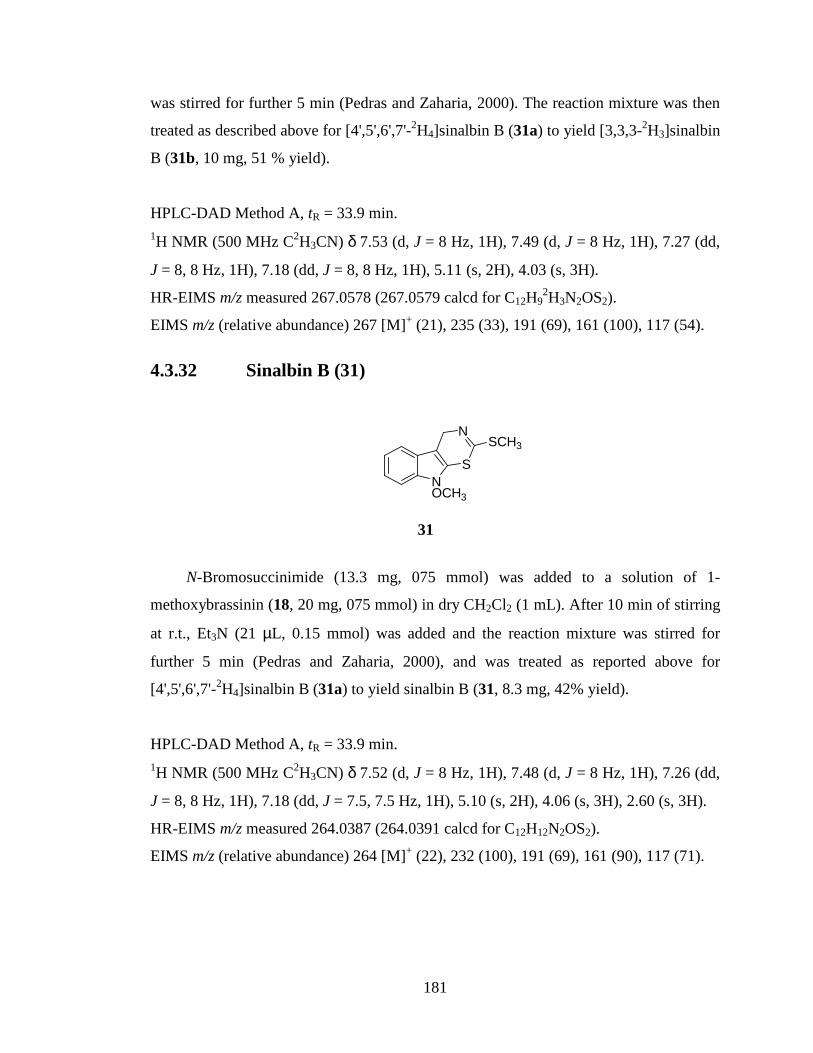
181
was stirred for further 5 min (Pedras and Zaharia, 2000). The reaction mixture was then
treated as described above for [4',5',6',7'-2H4]sinalbin B (31a) to yield [3,3,3-2H3]sinalbin
B (31b, 10 mg, 51 % yield).
HPLC-DAD Method A, tR = 33.9 min. 1H NMR (500 MHz C2H3CN) δ 7.53 (d, J = 8 Hz, 1H), 7.49 (d, J = 8 Hz, 1H), 7.27 (dd,
J = 8, 8 Hz, 1H), 7.18 (dd, J = 8, 8 Hz, 1H), 5.11 (s, 2H), 4.03 (s, 3H).
HR-EIMS m/z measured 267.0578 (267.0579 calcd for C12H92H3N2OS2).
EIMS m/z (relative abundance) 267 [M]+ (21), 235 (33), 191 (69), 161 (100), 117 (54).
4.3.32 Sinalbin B (31)
NOCH3
S
NSCH3
N-Bromosuccinimide (13.3 mg, 075 mmol) was added to a solution of 1-
methoxybrassinin (18, 20 mg, 075 mmol) in dry CH2Cl2 (1 mL). After 10 min of stirring
at r.t., Et3N (21 µL, 0.15 mmol) was added and the reaction mixture was stirred for
further 5 min (Pedras and Zaharia, 2000), and was treated as reported above for
[4',5',6',7'-2H4]sinalbin B (31a) to yield sinalbin B (31, 8.3 mg, 42% yield).
HPLC-DAD Method A, tR = 33.9 min. 1H NMR (500 MHz C2H3CN) δ 7.52 (d, J = 8 Hz, 1H), 7.48 (d, J = 8 Hz, 1H), 7.26 (dd,
J = 8, 8 Hz, 1H), 7.18 (dd, J = 7.5, 7.5 Hz, 1H), 5.10 (s, 2H), 4.06 (s, 3H), 2.60 (s, 3H).
HR-EIMS m/z measured 264.0387 (264.0391 calcd for C12H12N2OS2).
EIMS m/z (relative abundance) 264 [M]+ (22), 232 (100), 191 (69), 161 (90), 117 (71).
31

182
4.3.33 [4',5',6',7'-2H4]1-Methoxyspirobrassinin (35a)
NOCH3
2H2H
2H2H
OS
N SCH3
PCC (111.7 mg, 0.518 mmol) was added in one portion to a stirred solution of
[4',5',6',7'-2H4]1-methoxybrassinin (18b, 20 mg, 0.074 mmol) in CH2Cl2 (1 mL). After
being stirred for 60 min at r.t. (Pedras et al., 2006a), the reaction mixture was diluted
with CH2Cl2, silica gel was added and the mixture was concentration to dryness, and was
subjected to FCC (hexane-EtOAc, 80:20, v/v) to yield [4',5',6',7'-2H4]1-
methoxyspirobrassinin (35a, 8.6 mg, 41% yield).
HPLC-DAD Method A, tR = 18.2 min. 1H NMR (500 MHz C2H3CN) δ 4.60 (d, J = 15.5 Hz, 1H), 4.46 (d, J = 15.5 Hz, 1H),
3.99 (s, 3H), 2.62 (s, 3H).
HR-EIMS m/z measured 284.0588 (284.0591 calcd for C12H82H4N2O2S2).
EIMS m/z (relative abundance) 284 [M]+ (78), 253 (11), 225 (28), 180 (46), 152 (100),
120 (17).
4.3.34 1-Methoxyspirobrassinin (35)
NOCH3
OS
N SCH3
35a
35

183
PCC (113.2 mg, 0.525 mmol) was added in one portion to a stirred solution of 1-
methoxybrassinin (18, 20 mg, 0.075 mmol) in CH2Cl2 (1 mL). After being stirred for 60
min at r.t., the reaction mixture was treated as reported above for [4',5',6',7'-2H4]1-
methoxyspirobrassinin (35a) to yield 1-methoxyspirobrassinin (35, 8.7 mg, 41% yield,
Pedras et al., 2006a).
HPLC-DAD Method A, tR = 18.2 min. 1H NMR (500 MHz C2H3CN) δ 7.39-7.43 (m, 2H), 7.17 (d, J = 7.5 Hz, 1H), 7.06 (d, J =
7.5 Hz, 1H), 4.60 (d, J = 15.5 Hz, 1H), 4.46 (d, J = 15.5 Hz, 1H), 3.99 (s, 3H), 2.62 (s,
3H).
HR-EIMS m/z measured 280.0343 (280.0340 calcd for C12H12N2O2S2).
EIMS m/z (relative abundance) 280 [M]+ (73), 249 (11), 221 (28), 176 (45), 148 (100),
116 (16).
4.3.35 1-Methylspirobrassinin (194)
NCH3
S
N SCH3
O
NaH (4.8 mg, 0.12 mmol) and ICH3 (7.5 µL, 0.12 mmol) were added to a cooled
solution (0 ºC) of spirobrassinin (34, 15 mg, 0.06 mmol) in dry THF (2 mL) under
stirring. After stirring for 60 min at 0 ºC, ice-cold H2O (10 mL) was added. The resulting
mixture was extracted with EtOAc (15 mL × 3), the combined organic extract was dried
over Na2SO4 and was concentrated under reduced pressure. The crude extract was
subjected to FCC (CH2Cl2-CH3OH, 99:1 and 98:2, v/v) to yield 1-methylspirobrassinin
(194, 14.4 mg, 91% yield, Pedras and Hossain, 2006).
HPLC-DAD Method A, tR = 16.9 min.
194

184
1H NMR (500 MHz C2HCl3) δ 7.38 (d, J = 7.5 Hz, 1H), 7.34 (dd, J = 8, 8 Hz, 1H), 7.12
(dd, J = 7.5, 7.5 Hz, 1H), 6.85 (d, J = 8 Hz, 1H), 4.63 (d, J = 15 Hz, 1H), 4.53 (d, J = 15
Hz, 1H), 3.25 (s, 3H), 2.67 (s, 3H).
HR-EIMS m/z measured 264.0397 (264.0391 calcd for C12H12N2OS2).
EIMS m/z (relative abundance) 264 [M]+ (71), 217 (67), 191 (100), 159 (35), 130 (38).
4.3.36 1-Acetylspirobrassinin (195)
N
O
S
N SCH3
O
Pyridine (500 µL) was added to a stirred solution of spirobrassinin (34, 5 mg) in
Ac2O (500 µL). After being stirred for 60 min, the reaction mixture was diluted with
toluene and was concentrated under reduced pressure to yield 1-acetylspirobrassinin
(195, 4.8 mg, 80% yield).
HPLC-DAD Method A, tR = 16.9 min. 1H NMR (500 MHz C2HCl3) δ 7.39 (d, J = 7.5 Hz, 1H), 7.36 (dd, J = 8, 8 Hz, 1H), 7.14
(dd, J = 7.5, 7.5 Hz, 1H), 6.88 (d, J = 8 Hz, 1H), 4.62 (d, J = 15 Hz, 1H), 4.48 (d, J = 15
Hz, 1H), 2.66 (s, 3H), 2.64 (s, 3H).
HR-EIMS m/z measured 292.0339 (292.0340 calcd for C13H12N2O2S2).
EIMS m/z (relative abundance) 292 [M]+ (100), 250 (70), 203 (26), 177 (100), 145 (38),
117 (34).
195

185
4.3.37 1-(tert-Butoxycarbonyl)spirobrassinin (196)
N
O OH3C
H3CCH3
S
N SCH3
O
A solution of Boc2O (21 mg, 0.096 mmol) in THF (200 µL) was added to a stirred
solution of spirobrassinin (DO-075, 20 mg, 0.08 mmol) in THF (600 µL). The reaction
mixture was cooled to 5 ºC and DMAP (0.21 mg, 0.0017 mmol) was added. After being
stirred for 40 min, the reaction mixture was concentrated under reduced pressure and
was subjected to FCC (hexane-EtOAc, 50:50, v/v) to yield 1-(tert-
butoxycarbonyl)spirobrassinin (196, 31.4 mg, quant.).
HPLC-DAD Method A, tR = 30.1 min. 1H NMR (500 MHz C2HCl3) δ 7.40 (d, J = 7.5 Hz, 1H), 7.37 (dd, J = 8, 8 Hz, 1H), 7.16
(dd, J = 7.5, 7.5 Hz, 1H), 6.80 (d, J = 8 Hz, 1H), 4.64 (d, J = 15 Hz, 1H), 4.55 (d, J = 15
Hz, 1H), 2.65 (s, 3H), 1.75 (s, 9H).
HR-EIMS m/z measured 350.0767 (350.0759 calcd for C16H18N2O3S2).
EIMS m/z (relative abundance) 350 [M]+ (7), 250 (61), 203 (15), 177 (31), 149 (19), 116
(11), 57 (100).
4.3.38 [SC2H3]Brassicanal A (39b)
NH
CHO
SC2H3
196
39b

186
NaHCO3 (504 mg, 6 mmol) and P4S10 (800 mg, 1.80 mmol) were added to a
solution of oxindole (197, 400 mg, 3 mmol) in dry THF (8 mL). The reaction mixture
was stirred at r.t. for 2.5 h and then filtered, the filtrate was concentrated to remove THF.
Ice-cold H2O was added to the residue and the flask kept in an ice-bath. The precipitate
that formed was filtered off to yield indoline-2-thione (198, 322.4 mg, 72%). NaH (373
mg, 15.8 mmol) was added to a stirred solution of indoline-2-thione (200 mg, 1.34
mmol) in HCOOEt (3.8 mL, 47.4 mmol). After 5 min, the reaction mixture was diluted
with H2O (10 mL), was acidified with HCl (2.5 M), and the precipitate that formed was
filtered off to yield 2-mercaptoindole-3-carboxaldehyde (113 mg). The remaining
aqueous solution was extracted with EtOAc (100 mL × 3), the combined organic extract
was dried over Na2SO4 and was concentrated to yield 2-mercaptoindole-3-
carboxaldehyde (135, 100 mg). The total yield of 2-mercaptoindole-3-carboxaldehyde
obtained was 213 mg (90%, Pedras and Okanga, 1999). Et3N (35 µL, 0.25 mmol) and
IC2H3 (32 µL, 0.51) were added to a solution of 2-mercaptoindole-3-carboxaldehyde (30
mg, 0.17 mmol) in dry Et2O (2 mL). The reaction mixture was allowed to stir for 20 min
and the precipitate that formed was filtered off and rinsed with Et2O (5 mL × 3). The
filtrate was washed with H2O (10 mL), was dried over Na2SO4 and was concentrated.
The crude reaction mixture was subjected to FCC (CH2Cl2-CH3OH, 98:2, v/v) to yield
[SC2H3]brassicanal A (39b, 18.3 mg, 56% yield).
HPLC-DAD Method A, = 11.3 min. 1H NMR (500 MHz C2H3CN) δ 10.14 (s, 1H), 8.05 (d, J = 8.5 Hz, 1H), 7.45 (d, J = 8.5
Hz, 1H), 7.22-7.24 (m, 2H).
HR-EIMS m/z measured 194.0591 (194.0593 calcd for C10H62H3NOS).
EIMS m/z (relative abundance) 194 [M]+ (100), 176 (25), 160 (63), 148 (22), 121 (11).
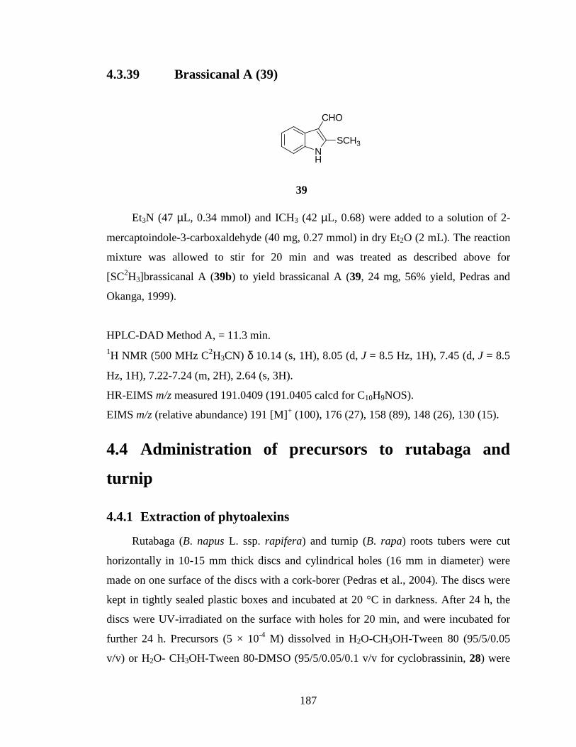
187
4.3.39 Brassicanal A (39)
NH
CHO
SCH3
Et3N (47 µL, 0.34 mmol) and ICH3 (42 µL, 0.68) were added to a solution of 2-
mercaptoindole-3-carboxaldehyde (40 mg, 0.27 mmol) in dry Et2O (2 mL). The reaction
mixture was allowed to stir for 20 min and was treated as described above for
[SC2H3]brassicanal A (39b) to yield brassicanal A (39, 24 mg, 56% yield, Pedras and
Okanga, 1999).
HPLC-DAD Method A, = 11.3 min. 1H NMR (500 MHz C2H3CN) δ 10.14 (s, 1H), 8.05 (d, J = 8.5 Hz, 1H), 7.45 (d, J = 8.5
Hz, 1H), 7.22-7.24 (m, 2H), 2.64 (s, 3H).
HR-EIMS m/z measured 191.0409 (191.0405 calcd for C10H9NOS).
EIMS m/z (relative abundance) 191 [M]+ (100), 176 (27), 158 (89), 148 (26), 130 (15).
4.4 Administration of precursors to rutabaga and
turnip
4.4.1 Extraction of phytoalexins
Rutabaga (B. napus L. ssp. rapifera) and turnip (B. rapa) roots tubers were cut
horizontally in 10-15 mm thick discs and cylindrical holes (16 mm in diameter) were
made on one surface of the discs with a cork-borer (Pedras et al., 2004). The discs were
kept in tightly sealed plastic boxes and incubated at 20 °C in darkness. After 24 h, the
discs were UV-irradiated on the surface with holes for 20 min, and were incubated for
further 24 h. Precursors (5 × 10⁻⁴ M) dissolved in H2O-CH3OH-Tween 80 (95/5/0.05
v/v) or H2O- CH3OH-Tween 80-DMSO (95/5/0.05/0.1 v/v for cyclobrassinin, 28) were
39

188
pipetted into each hole (500 µL per hole) and the discs were further incubated at 20 °C
in darkness. Following adsorption of precursor solution, the holes were filled with water.
The aqueous solution in the holes was withdrawn with a pipette after appropriate
incubation period (24, 48, 72 or 96 h) and was extracted with EtOAc (30 mL × 2). The
combined organic extract was dried over Na2SO4 and solvent was removed under
reduced pressure. The residue obtained was dissolved in CH3CN (400 µL) and was
analyzed by HPLC-DAD. The residue was then subjected to preliminary clean up by
micro-FCC in a Pasteur pipette column packed with silica gel to 35 mm, eluting with 20
mL of CH2Cl2 and then 20 mL of CH2Cl2-EtOAc (40:60, v/v) to yield two fractions. The
phytoalexin containing fractions obtained were analyzed by HPLC-DAD, HPLC-MS-
ESI, and HPLC-HRMS-ESI. Control experiments were similarly prepared by incubating
rutabaga and turnip root tubers with non-labeled precursors or with carrier solution only.
4.4.2 Extraction of glucosinolates
Tissue around the holes was cut, was soaked in CH3OH, was ground (pestle and
mortar) and was extracted with an aqueous CH3OH solution. Extraction was carried out
overnight on a shaker at 170 rpm, after which the tissue was filtered off and the filtrate
was concentrated under reduced pressure. The residue obtained was subjected to RP-
FCC (H2O-CH3CN, 100:0; 90:10, 80:20, v/v) to yield 3 fractions containing a mixture of
indole glucosinolates. The fractions were dissolved in H2O-CH3OH (50:50) and were
analyzed by HPLC-DAD, HPLC-MS-ESI, and HRMS-ESI.
4.5 Administration of precursors to E. gallicum (dog
mustard)
4.5.1 Time course experiments
In order to carry out feeding experiments with dog mustard, the maximum
production period of erucalexin (38) and 1-methoxyspirobrassinin (35) was determined
using detached leaves. Dog mustard plants were grown in growth chambers with a 16-h
photoperiod (22ºC/18ºC) at ambient humidity. The 28-day old plants were sprayed with
CuCl2 (2 × 10⁻³ M) solution in the growth chambers. After 24-h incubation, the leaves

189
were excised at the base of petiole and immediately immersed into distilled H2O (1 mL)
in a vial. The petiolated leaves were sprayed with CuCl2 (2 × 10⁻³ M) solution followed
by incubation at r.t. under continuous light. After 24-h incubation, leaves were frozen in
liquid nitrogen, ground with a glass rod and were extracted in EtOAc (30 mL) overnight.
The extract was filtered and was concentrated under reduced pressure. The residue
obtained was subjected to preliminary clean up through a micro-FCC column employing
a Pasteur pipette column packed with silica gel to 35 mm and eluting with 20 mL of
CH2Cl2 and then 20 mL of CH2Cl2-EtOAc (25:75, v/v). The leaves were similarly
extracted after 48-, 72-, 96- and 120-h incubation periods, each experiment was
performed in duplicate. The fractions obtained were analyzed by HPLC-DAD and the
amounts of erucalexin and 1-methoxyspirobrassinin present was determined from
calibration curves prepared from pure erucalexin and 1-methoxyspirobrassinin. From
these experiments, the maximum production of erucalexin was detected after 48-h
incubation and this incubation period was adapted for feeding experiments. To prove
that the observed metabolites were phytoalexins, similar experiments were carried out
with leaves sprayed with water. HPLC-DAD analysis did not indicate the presence of
erucalexin, 1-methoxyspirobrassinin or any elicited metabolites in these extracts.
4.5.2 Administration of precursors
Plants were grown and elicited as discussed for the time course analyses. The
petiolated leaves from elicited 28-day-old plants were placed in vials containing 1 mL of
precursor (5 × 10⁻⁴ M) dissolved in H2O-CH3OH-Tween 80 (95/5/0.05 v/v). Following
uptake of precursor solution, the vials were filled with H2O and were incubated for 48 h
under continuous light. The leaves were extracted as described for time course analyses,
and the phytoalexin containing fractions were analyzed by HPLC-DAD, HPLC-MS-ESI
and HPLC-HRMS-ESI.

190
CHAPTER 5 : References
Agerbirk, N., Olsen, C. E., Soerensen, H., 1998. Initial and final products, nitriles, and ascorbigens produced in myrosinase-catalyzed hydrolysis of indole glucosinolates. J. Agric. Food. Chem. 46, 1563-1571.
Agrawal, A. A., Conner, J. K., Johnson, M. T. J., Wallsgrove, R., 2002. Ecological
genetics of an induced plant defense against herbivores: additive genetic variance and cost of phynotypic plasticity. Evolution 56, 2206-2213.
Agrawal, A. A., Kurashige, N. S., 2003. A role for iosthiocyanates in plant resistance
against the specialist herbivore Pieris papae. J. Chem. Ecol. 29, 1403-1415. Alford, É. R., Perry, L. G., Qin, B., Vivanco, J. M., Paschke, M. W., 2007. A putative
allelopathic agent of Russian knapweed occurs in invaded soils. Soil Biol. Biochem. 39, 1812-1815.
Anaya, A. L., Mata, R., Sims J. J., González-Coloma, A., Cruz-Ortega, R., Guadano, A.,
Hernández-Bautista, B. E., Midland, S. L., Rios, G., Gómez-Pompa, A., 2003. Allelochemical potential of Callicarpa acuminate. J. Chem. Ecol. 29, 2761-2776.
Bailey, J. A., Mansfield, J. W. Eds., in Phytoalexins. Blackie and Son, Glasgow, U.K.,
1992, pp 1-16. Bais, H. P., Walker, T. S., Kennan, A. J., Stermitz, F. R., Vivanco, J. M., 2003.
Structure-dependent phytotoxicity of catechins and other flavonoids: flavonoid conversions by cell-free protein extracts of Centaurea maculosa (Spotted Knapweed) roots. J. Agric. Food Chem. 51, 897-901.
Bais, H. P., Walker, T. S., Stermitz, F. R., Hufbauer, R. A., Vivanco, J. M., 2002.
Enantiomeric-dependent phytotoxicity and antimicrobial activity of (±)catechins. A rhizosecreted racemic mixture from spotted knapweed. Plant Physiol. 128, 1173-1179.
Bak, S., Feyereisen, R., 2001. The involvement of two P450 enzymes, CYP83B1 and
CYP83A1, auxin homeostasis and glucosinolate biosynthesis. Plant Physiol. 127, 108-118.
Bak, S., Tax, F. E., Feldmann, K. A., Galbraith, D. W., Feyereisen, R., 2001. CYP83B1,
a cytochrome P450 at the metabolic branch point in auxin and indole glucosinolate biosynthesis in Arabidopsis. Plant Cell 13, 101-111.
Baker, A. M., Molotsane, R., Müller, C., Shaffner, U., Städler, E., 2006. Chemosensory
and behavioral responses of the turnip sawfly, Athalia rosae, to glucosinolates and isothiocyanates. Chemoecology 16, 209-218.

191
Bednarek, P., Schneider, B., Svatos, A., Oldham N. J., Hahlbrook, K., 2005. Structural complexity, differential response to infection, and tissue specificity of indolic and phenylpropanoid secondary metabolism in Arabidopsis roots. Plant Physiol. 138, 1058-1070.
Bennett, R. N., Mellon, F. A., Botting N. P., Eagles, J., Rosa, E. A. S., Williamson, G.,
2002. Identification of the major glucosinolates (4-mecaptobutyl glucosinolate) in the leaves of Eruca sativa L. (salad rocket). Phytochemistry 61, 25-30.
Bones, A, M., Rossiter, J. T., 2006. The enzymatic and chemically induced
decomposition of glucosinolates. Phytochemistry 1053-1067. Brader, G., Tas, É, Palva, E. T., 2001. Jasmonate-dependent induction of indole
glucosinolates in Arabidopsis by culture filtrates of the nonspecific pathogen Erwinia carotovora. Plant Physiol. 126, 849-860.
Browne, L. M., Conn, K. L., Ayer, W. A., Tewari, J. P., 1991. The camalexins: new
phytoalexins produced in leaves of Camelina sativa (Cruciferae). Tetrahedron 47, 3909-3914.
Brydan, G. A., Ryan, D. E., 1966. Thiobenzoylphenylhydroxylamine: Potential
analytical reagent. Anal. Chim. Acta 35, 190-194. Burckard, P., Fleury, J-P., Weiss, F. 1965. Contribution à l’étude de l’oxydation des
amines par l’eau oxygénée en présence de pertungstate alcalin. Bull. Soc. Chim. Fr. 10, 2730-2733.
Canoira, L., Rodriguez, J. G., Subirats, J. B., Escario, J-A., Jimenez, I., Martinez-
Fernandez, A. R., 1989. Synthesis, structure and anti-fungal activity of 3-(2'-nitrovinyl)indoles. J. Med. Chem. 24, 39-42.
Cataldi, T. R. I., Rubino, A., Lelario, F., Bufo, S. A., 2007. Naturally occurring
glucosinolates in plant extracts of rocket salad (Eruca sativa L.) identified by liquid chromatography coupled with negative ion electrospray ionization and quadrupole ion-trap mass spectrometry. Rapid. Commun. Mass Spectrom. 21, 2374-2388.
Cerney, M. S., Taube, E., Battaglia R. J., 1996. Identification of bis(4-
isothiocyanatobutyl)disulfide and its precursor from rocket salad (Eruca sativa). Agric. Food Chem. 44, 3835-3839.
Chen, S., Glawischnig, E., Jørgensen, K., Naur, P., Jørgensen, B., Olsen, C-E., Hansen,
C. H., Rasmussen, H., Pickett, J. A., Halkier, B. A., 2003. CYP79F1 and CYP79F2 have distinct functions in the biosynthesis of aliphatic glucosinolates in Arabidopsis. Plant J. 33, 923-937.

192
Chimiak, A., Przychodzen, W., Rachon, J., 2002. The thiohydroximate system. Heteroat. Chem. 13, 169-194.
Conn, K. L., Tewar, J. P., Dahiya, J. S., 1988 Resistance to Alternaria brassicae and
phytoalexin elicitation in rapeseed and other crucifers Plant Sci. 56, 21-25. Dahyia, J. S., Rimmer, S. R., 1988. Phytoalexin accumulation in tissues of Brassica
napus inoculated with Leptosphaeria maculans. Phytochemistry 27, 3105-3107. Dawson, G. W., Hick, A. J., Bennett, R. N., Donald, A., Pickett, J. A., Wallsgrove, R.
M., 1993. Synthesis of glucosinolate precursors and investigations into the biosynthesis of phenylalkyl- and methythioalkylglucosinolates. J. Biol. Chem. 268, 27154-27159.
Daxenbichler, M. E., Spencer, G. F., Carlson, D. G., Rose, G. B., Brinker, A. M.,
Powell, R. G., 1991. Glucosinolate composition of seeds from 297species of wild plants. Phytochemistry 30, 2623-2638.
Dayan, F. E., Romagni, J. G., Duke, S O., 2000. Investigating the mode of action of
natural phytotoxins. J. Chem. Ecol. 26, 2079-2094. Dewick, P. M., 1984. The biosynthesis of cyanogenic glycosides and glucosinolates.
Nat. Prod. Rep. 1, 546-549. Dewick, P. M., 1998. The biosynthesis of shikimate metabolites. Nat. Prod. Rep. 15, 17-
58. Dixon, R. A., 2005. Engineering of plant natural product pathways. Curr. Opin. Plant
Biol. 8, 329-336. Dixon, R. A., Harrison, M. J., Lamb, C. J., 1994. Early events in the activation of plant
defense responses. Annu. Rev. Phytopathol. 32, 479-501. Doughty, K. J., Porter, A. J. R., Morton, A. M., Kiddle, G., Bock, C. H., Wallsgrove, R.,
1991. Variation in the glucosinolate content of oilseed rape (Brassica napus L.) leaves II. Response to infection by Alternaria brassicae (Berk.) Sacc. Ann. Appl. Biol. 118, 469-477.
Du, L., Lykkesfeldt, J., Olsen, E. C., Halkier, B. A., 1995. Involvement of cytochrome
P450 in oxime production in glucosinolate biosynthesis as demonstrated by an in vitro microsomal enzyme system isolated from jasmonic acid-induced seedlings of Sinapis alba L. Proc. Natl. Acad. Scie. USA 92, 12505-12509.
Fahey, J. W., Zalcmann, A. T., Talalay, P., 2001. The chemical diversity and distribution
of glucosinolates and isothiocyanates among plants. Phytochemistry 56, 5-51.

193
Falk, K. L., Vogel, C., Textor, S., Bartram, S., Hick, A., Pickett, J. A., Gershenzon, J., 2004. Glucosinolate biosynthesis: demonstration and characterization of the condensing enzyme of the chain elongation cycle in Eruca sativa. Phytochemistry 65, 1073-1084.
Gabrys, B., Tjallingii, W. F., 2002. The role of sinigrin in host plant recognition by
aphids during initial plant penetration. Entomol. Exp. Appl. 104, 89-93. Gardiner, J. B., Morra, M. J., Eberlein, C. V., Brown, P. D., Borek, V., 1999.
Allelochemicals released in soil following incorporation of rapeseed (Brassica napus) green manures. J. Agric. Food Chem. 47, 3837-3842.
Giamoustaris, A., Mithen, R., 1995. The effect of modifying the glucosinolate content of
leaves of oilseed rape (Brassica napus ssp oleifera) on its interaction with specialist and generalist pests. Ann. Appl. Biol. 126, 347-363.
Glaswischnig, E., Hansen, B. G., Olsen, C. E., Halkier, B. A., 2004. Camalexin is
synthesized from indole-3-acetaldoxime, a key branching point between primary and secondary metabolism in Arabidopsis. Proc. Natl. Acad. Sci. USA 101, 8245-8250.
Glending, T. M., Poulton, J. E., 1988. Glucosinolate biosynthesis. Plant Physiol. 86,
319-321. Glending, T. M., Poulton, J. E., 1990. Partial purification and characterization of a 3'-
phosphoadenosine-5'-phosphosulfate; desulfoglucosinolate sulfotransferase from cress (Lepidium sativum). Plant Physiol. 94, 811-818.
Goetz, J. K., Schraudolf, H., 1983. Two natural indole glucosinolates from Brassicaceae.
Phytochemistry 22, 905-907. Graser, G., Oldham, N. J., Brown, P. D., Temp, U., Gershenzon, J., 2001. The
biosynthesis of benzoic acid glucosinolate esters in Arabidopsis thaliana. Phytochemistry 57, 23-32.
Graser, G., Schneider, B., Oldham, N. J., Gershenzon, J., 2000. The methionine chain
elongation pathway in the biosynthesis of glucosinolates in Ercuca sativa (Brassicaceae). Arch. Biochem. Biophys. 378, 411-419.
Grubb, C. D., Abel, S., 2006. Glucosinolate metabolism and its control. Trends Plant
Sci. 11, 89-100. Grubb, C. D., Zipp, B. J., Ludwig-Müller, J., Masuno, M. N., Molinski, T. F., Abel, S.,
2004. Arabidopsis glucosyltransferase UGT74B1 functions in glucosinolate biosynthesis and auxin homeostasis. Plant J. 40, 893-908.

194
Gugel, R. K., Séguin-Swartz, G., Petrie, A., 1990. Pathogenicity of 3 isolates Leptosphaeria maculans on Brassica species and other crucifers. Can. J. Plant Pathol. 12, 75-82.
Hachinohe, M., Matsumoto, H., 2005. Involvement of reactive oxygen species generated
from melanin synthesis pathway in phytotoxicity of L-DOPA. J. Chem. Ecol. 31, 237-245.
Hachinohe, M., Sunohara, Y., Matsumoto, H., 2004. Absorption, translocation and
metabolism of L-DOPA in barnyardgrass and lettuce: their involvement in species-selective phytotoxic action. Plant Growth Regul. 43, 237-243.
Halkier, B. A., Gershenzon, J., 2006. Biology and biochemistry of glucosinolates. Ann.
Rev. Plant Biol. 57, 303-333. Halkier, B. A., Møller, B. L., 1991. Involvement of cytochrome P-450 in the
biosynthesis of dhurin in Sorghum bicolor (L.) Moench. Plant Physiol. 96, 10-17. Hammerschmidt, R., 1999. Phytoalexins: what have we learned after 60 years? Annu.
Rev. Phytopathol. 37, 285-306. Hanley, B. A., Parsley, K. R., 1990. Identification of 1-methoxyindolyl-3-methyl
isothiocyanate as an indole glucosinolate breakdown product. Phytochemistry 29, 769-771.
Hanley, B. A., Parsley, K. R., Lewis, J. A., Fenwick, G. R., 1990. Chemistry of indole
glucosinolates: intermediacy of indol-3-ylmethyl isothiocyanate in the enzymatic hydrolysis of indole glucosinolates. J. Chem. Soc. Perkin Trans. 1, 2273-2276.
Hansen, B. G., Halkier, B. A., 2005. New insight into the biosynthesis and regulation of
indole compounds in Arabidopsis thaliana. Planta 221, 603-606. Hansen, C. H., Du, L., Naur, P., Olsen, C. E., Axelsen, K. B., Hick, A. J., Pickett, J. A.,
Halkier, B. A., 2001b. CYP83B1 is the oxime-metabolizing enzyme in the glucosinolate pathway of Arabidopsis. J. Biol. Chem. 276, 24790-24796.
Hansen, C. H., Wittstock, U., Olsen, C. E., Hick, A. J., Pickett, J. A., Halkier, B. A.,
2001a. Cytochrome P450 CYP79F1 from Arabidopsis catalyzes the conversion of dihomomethionine and trihomomethionine to the corresponding aldoximes in the biosynthesis of aliphatic glucosinolates. J. Biol. Chem. 276, 11078-11085.
Harvey, J. A., Nouhuys, S. V., Biere, A., 2005. Effects of quantitative variation in
allelochemicals in Plantago lanceolata on development of a generalist and a specialist herbivore and their endoparasitoids. J. Chem. Ecol. 31, 287-302.

195
Hemm, M. R., Ruegger, M. O., Chapple, C., 2003. The Arabidopsis ref2 mutant is defective in gene encoding CYP83A1 and shows both phenylpropanoid and glucosinolate phenotypes. Plant Cell 15, 179-194.
Hull, A. K., Vij, R., Celenza, J. L., 2000. Arabidopsis cytochrome P450s that catalyze
the first step of tryptophan-dependent indole-3-acetic acid biosynthesis. Proc. Natl. Acad. Scie. USA 97, 2379-2384.
Hwu, J. R., Lin, C-F., Tsay, S-C., 2005. Hexamethyldisilathiane in novel chemical
transformations: concept of “counterattack reagents”. Phosphorus, Sulfur, and Silicon 180, 1389-1393.
Hwu, J. R., Tsay, S-C., 1990. Counterattack reagents: thiosilanes in the conversion of
nitro compounds to thiohydroxamic acids and thiohydroximates. Tetrahedron 46, 7413-7428.
Jain, J. C., GrootWassink, J. W. D., Kolenovsky, A. D., Underhill, E. W., 1990.
Purification and properties of 3'-phosphoadenosine-5'-phosphosulphate: desulfoglucosinolate sulphotransferase from Brassica juncea cell cultures. Phytochemistry 29, 1425-1428.
Jain, J. C., Michayluk, M. R., GrootWassink, J. W. D., Underhill, E. W., 1989.
Distributions of enzymes catalyzing glucosylation and sulfation steps of glucosinolate biosynthesis in Brassica juncea seedlings and cultured cells. Plant Sci. 64, 25-29.
Jensen, K. A., Pedersen, C., 1961. Studies of thio acids and their derivatives. II.
Carboxymethyl dithioesters. Acta Chem. Scand. 15, 1087-1096. Jiang, R-W., Hon, P-M., Xu, Y-T., Chan, Y-M., Xu, H-X., Shaw, P-C., But, P. P-H.,
2006. Isolation and chemotaxonomic significance of tuberostemospironine-type alkaloids from Stemona tuberose. Phytochemistry, 67, 52-57.
Jimenez, L. D., Ayer, W. A., Tewari, J. P., 1997. Phytoalexins produced in the leaves of
Capsella bursa-pastoris (shepherd’s purse). Phytoprotection 78, 99-103. Josefsson, E., 1972. Conversion of indole-3-acetaldehyde oxime to 3-indolylmethyl-
glucosinolate in Sinapis alba. Physiol. Plant. 27, 236-239. Kato-Noguchi, H., 2004. Allelopathic substance in rice root exudates: rediscovery of
momilactone B as an allelochemical. J. Plant Physiol. 161, 271-276. Kato-Noguchi, H., Ino, T., 2002. Rice seedlings release allopathic substances. Biol.
Plant. 46, 157-159. Kato-Noguchi, H., Ino, T., 2004. Release level of momilactone B from rice plants. Plant
Prod. Sci. 7, 189-190.

196
Kato-Noguchi, H., Ino, T., Ichii, M., 2003. Changes in release levels of momilactone B into the environment from rice throughout its life cycle. Func. Plant Biol. 30, 995-997.
Kato-Noguchi, H., Ino, T., Sata, N., Yamamura, S., 2002. Isolation and identification of
a potent allelopathic substance in rice root exudates. Physiol. Plant. 115, 401-405.
Kim, S-J., Ishii, G., 2006. Glucosinolate profiles in the seeds, leaves and roots of rocket
salad (Eruca sativa Mill.) and anti-oxidative activities of intact plant powder and purified 4-methoxyglucobrassicin. Soil Sci. Plant Nutr. 52, 394-400.
Klein, M., Reichelt, M., Gershenzon, J., Papenbrock, J., 2006. The three
desulfoglucosinolate sulfotransferase proteins in Arabidopsis have different substrate specificities and are differentially expressed. FEBS J. 273, 122-136.
Koch, B., Nielsen, V. S., Halkier, B. A., Olsen, C. E., Møller, B. L., 1992. The
biosynthesis of cyanogenic glucosides in seedlings of cassava (Manihot esculenta Crantz). Arch. Biochem. Biophys. 292, 141-150
Kong, C. H., Li, H. B., Hu, F., Xu, X. H., Wang, P., 2006. Allelochemicals released by
rice roots and residues in soil. Plant Soil 288, 47-56. Kryomann, J., Textor, S., Tokuhisa, J. G., Falk, K. L., Bartram, S., Gershenzon, J.,
Mitchell-Olds, T., 2001. A gene controlling variation n Arabidopsis glucosinolate composition is part of the methionine chain elongation pathway. Plant Physiol. 127, 1077-1088.
Kuć, J., 1995. Phytoalexins, stress metabolism, and disease resistance in plants. Annu.
Rev. Phytopathol. 33, 275-297. Kutáček, M., Králová, M., 1972. Biosynthesis of the glucobrassicin aglycone from 14C
and 15N labelled L-tryptophan precursors. Biol. Plant. 14, 279-285. Kutáček, M., Procházka, Ž., Vereš, K., 1962. Biogenesis of glucobrassicin, the in vitro
precursor of ascorbigen. Nature 194, 393-394. Kutschy, P., Achbergerová, I., Dzurilla, M., Takasugi, M., 1997. Synthesis of indole
phytoalexins brassinin and cyclobrassinin via [1-(tert-Butoxycarbonyl)indol-3-yl]methyl isothiocyanate as the key biomimetic intermediate. Synlett 289-290.
Kutschy, P., Dzurilla, M., Takasugi, M., Achbergerová, I., Homzová, R., Rácová, M.,
1998. New syntheses of indole phytoalexins and related compounds. Tetrahedron 54, 3549-3566.
Lane, A. L., Kubanek, J., 2006. Structure-activity relationship of chemical defenses from
the freshwater plant Micranthemum umbrosum. Phytochemistry 67, 1224-1231.

197
Latxague, L., Gardrat, C., Coustille, J. L., Viaud, M. C., Rollin, P., 1991. Identification of enzymatic degradation products from synthesized glucobrassicin by gas chromatography-mass spectromentry. J. Chromatogr. 586, 166-170.
Lefol, C., Séguin-Swartz, G., Morrall, R. A. A., 1997. Resistance to Sclerotinia
sclerotiorum in a weed related to canola. Can. J. Plant Pathol. 19, 113 (Abstr.). Li, Q., Eigenbrode, S. D., Stringam, G. R., Thiagarajah, M. R., 2000. Feeding and
growth of Plutella xylostella and Spodotpera eridania on Brassica juncea with varying glucosinolate concentrations and myrosinase activity. J. Chem. Ecol. 26, 2401-2419.
Li, Y., Kiddle, G. A., Bennett, R. N., Wallsgrove, R. M., 1999. Local and systematic
changes in glucosinolates in Chinese and European cultivars of oilseed rape (Brassica napus) after inoculation with Sclerotinia sclerotiorum (stem rot). Annu. Appl. Biol. 134, 45-58.
Makosza, M., Winiarski, J., 1984. Vicarious nucleophilic substitution of hydrogen in
nitroarenes with α-substituted nitriles and esters. Direct α-cyanoalkylation and α-carbalkoxyalkylation of nitroarenes. J. Org. Chem. 49, 1494-1499.
Manici, L. M., Lazzeri, L., Baruzzi, G., Leoni, O., Galletti, S., Palmieri, S., 2000.
Suppressive activity of some glucosinolate enzyme degradation products on Phythium irregulare and Rhizoctonia solani in sterile soil. Pest Manage. Sci. 56, 921-926.
Manici, L. M., Lazzeri, L., Palmieri, S., 1997. In vitro fungitoxic activity of some
glucosinolates and their enzyme-derived products toward plant pathogenic fungi. J. Agric. Food Chem. 45, 2768-2773.
Mann, J., 1994. Chemical aspects of biosynthesis. Oxford University Press, Oxford, UK,
pp. 2-4. Mari, M., Leoni, O., Iori, R., Cembali, T., 2002. Antifungal vapour-phase activity of
allyl isothiocyanate against Penicillium expansum on pears. Plant Pathol. 51, 231-236.
Marvel, C. S., Radzitzky, P., Brader, J. J., 1955. An improved preparation of dithioesters
and some reactions and spectral properties of these compounds. J. Am. Chem. Soc. 77, 5997-6000.
Mayton, H. S., Olivier, C., Vaughn, S. F., Loria, R., 1996. Correlation of fungicidal
activity of Brassica species with allyl isthiocyanate production in macerated leaf tissues. Phytopathology 86, 267-271.

198
Mewis, I., Appel, H. M., Hom, A., Raina, R., Schultz, J. C., 2005. Major signaling pathways modulate Arabidopsis glucosinolate accumulation and response to both phloem-feeding and chewing insects. Plant Physiol. 138, 1149-1162.
Mewis, I. Z., Ulrich, C., Schnitzler, W. H., 2002. The role of glucosinoaltes and their
hydrolysis products in oviposition and host-plant finding by cabbage webworm, Hellula undalis. Entomol. Exp. Appl. 105, 129-139.
Mikkelsen, M. D., Hansen, C. H., Wittstock, U., Halkier, B. A., 2000. Cytochrome P450
CYP79B2 from Arabidopsis catalyses the conversion of tryptophan to indole-3-acetaldoxime, a precursor of indole glucosinolates and indole-3-acetic acid. J. Biol. Chem. 275, 33712-33717.
Mikkelsen, M. D., Naur, P., Halkier, B. A., 2004. Arabidopsis mutants in the C-S lyase
of glucosinolate biosynthesis establish a critical role for indole-3-acetaldoxime in auxin homeostasis. Plant J. 37, 770-777.
Mikkelsen, M. D., Petersen, B. L., Olsen, C. E., Halkier, B. A., 2002. Biosynthesis and
metabolic engineering of glucosinolates. Amino acids 22, 279-295. Mithen, R. F., Lewis, B. G., Fenwick, G. R., 1986. In vitro activity of glucosinolates and
their products against Leptosphaeria maculans. Trans. Br. Mycol. Soc. 87, 433-440.
Mithen, R. F., Lewis, B. G., Heaney, R. K., Fenwick, G. R., 1987. Glucosinolates of
wild and cultivated Brassica species. Phytochemistry 26, 1969-1973. Mithen, R. F., Magrath, R., 1992. Glucosinolates and resistance to Leptosphaeria
maculans in wild and cultivated Brassica species. Plant Breed. 108, 60-68. Mitsuo, M., Undherhill, E. W., 1971. Purification and properties of a UDP glucose:
thiohydroximate glucosyltransferase from higher plants. Phytochemistry 10, 2279-2286.
Miyashita, K., Kondoh, K., Tsuchiya, K., Miyabe, H., Imanishi, T., 1997. Synthesis of
several 3-substituted 2-trifluoromethylindoles via Mannich reaction of 2-trifluoromethylindoles. Chem.Pharm. Bull. 45, 932-935.
Mizukami, S., Nagata, K., 1966. Thiohydroxamic acids and their metal chelates. I.
Synthesis of thiohydroxamic acids and O-methyl-thiohydroxamic acids. Chem. Pharm. Bull. 14, 1249-1255.
Monde, K., Katsui, N., Shirata, A., Takasugi, M., 1990a. Brassicanal A and B, novel
sulfur-containing phytoalexins from the Chinese cabbage Brassica campestris L. ssp. pekinensis. Chem Lett. 209-210.

199
Monde, K., Sasaki, K., Shirata, A., Takasugi, M., 1990b. 4-Methoxybrassinin, a sulphur-containing phytoalexin from Brassica oleracea. Phytochemistry 29, 1499-1500.
Monde, K., Sasaki, K., Shirata, A., Takasugi, M., 1991a. Brassicanal C and two
dioxindoles from cabbage. Phytochemistry 30, 2915-2917. Monde, K., Sasaki, K., Shirata, A., Takasugi, M., 1991b. Methoxybrassenins A and B,
sulphur-containing stress metabolites from Brassica oleracea var. capitata. Phytochemistry 30, 3921-3922.
Monde, K., Takasugi, M., Ohnishi, T., 1994a. Biosynthesis of cruciferous phytoalexins.
J. Am. Chem. Soc. 116, 6650-6657. Monde, K., Takasugi, M., Shirata, K., 1995b. Three sulfur-containing stress metabolites
from Japanese radish. Phytochemistry 39, 581-586. Monde, K., Takasugi, M., Takasugi, M., Kobayashi, K., Somei, M., 1994b. Dehydro-4-
methoxycyclobrassinin, a sulfur containing phytoalexin isolated from turnip Brassica campestris L. ssp. rapa. Heterocycles 38, 263-267.
Monde, K., Tamura, K., Takasugi, M., 1995a. No involvement of methoxybrassinin in
the biosynthesis of cyclobrassinin. Phytochemistry 39, 587-589. Monde, K., Tanaka, A., Takasugi, M., 1996. Trapping experiment with aniline for a
biosynthetic intermediate of sulfur-containing cruciferous phytoalexins. J. Org. Chem. 61, 9053-9054.
Nafisi, M., Goregaoker, S., Botanga, C. J., Glawischnig, E., Olsen, C. E., Halkier, B. A.,
Glazebrook, J., 2007. Arabidopsis cytochrome P450 monooxygenase 71A13 catalyzes the conversion of indole-3-acetaldoxime in camalexin synthesis. Plant Cell 19, 2039-2052.
Naur, P., Hansen, C. H., Bak, S., Hansen, B. G., Jensen, N. B., Nielsen, H. L., Halkier,
B. A., 2003b. CYP79B1 from Sinapis alba converts tryptophan to indole-3-acetaldoxime. Arch. Biochem. Biophys. 409, 235-241.
Naur, P., Petersen, B. L., Mikkelsen, M. D., Bak, S., Rasmussen, H., Olsen, C. E.,
Halkier, B. A., 2003a. CYP83A1 and CYP83B1, two nonredundant cytochrome P450 enzymes metabolizing oximes in the biosynthesis of glucosinolates in Arabidopsis. Plant Physiol. 133, 63-72.
Nishihara, E., Parvez, M. M., Araya, H., Fujii, Y., 2004. Germination growth response
of different plant species to the allelochemical L-3,4-dihydroxyphenylalanine (L-DOPA). Plant Growth Regul. 42, 181-189.

200
Nishihara, E., Parvez, M. M., Araya, H., Kawashima, S., Fujii, Y., 2005. L-3,4-Dihydroxyphenylalanine (L-DOPA), an allelochemical exuded from velvetbean (Mucuna pruriens) roots. Plant Growth Regul. 45, 113-120.
Olivier, C., Vaughn, S. F., Mizubuti, S. G., Loria, R., 1999. Variation in allyl
isothiocyanate production within Brassica species and correlation with fungicidal activity. J. Chem. Ecol. 25, 2687-2701.
Osbourne, A. E., 1996. Preformed antimicrobial compounds and plant defense against
fungal attack. Plant Cell 8, 1821-1831. Pedras, M. S. C., Adio, A. M., 2007. Phytoalexins and phytoanticipins from the wild
crucifers Thellungiella halophila and Arabidopsis thaliana: rapalexin A and wasalexins and camalexin. Phytochemistry, in press.
Pedras, M. S. C., Adio, A. M., Suchy, M., Okinyo D. P. O., Zheng, Q-A., Jha, M.,
Sarwar, M. G., 2006c. Detection, characterization and identification of crucifer phytoalexins using high-performance liquid chromatography with diode array detection and electrospray ionization mass spectrometry. J. Chromatogr. A 1133, 172-183.
Pedras, M. S. C., Ahiahonu, P. W. K., 2004. Phytotoxin production and phytoalexin
elicitation by the phytopathogenic fungus Sclerotinia sclerotiorum. J. Chem. Ecol. 30, 2163-2179.
Pedras, M. S. C., Chumala, P. B., Suchy, M., 2003a. Phytoalexins from Thlaspi arvense,
a wild crucifer resistant to virulent Leptosphaeria maculans: structure, synthesis and antifungal activity. Phytochemistry 64, 949-956.
Pedras, M. S. C., Hossain, M., 2006. Metabolism of crucifer phytoalexins in Sclerotinia
sclerotiorum: detoxification of strongly antifungal compournds involves glucosylation. Org. Biomol. Chem. 4, 2581-2590.
Pedras, M. S. C., Jha, M., Ahiahonu, P. W. K., 2003b. The synthesis and biosynthesis of
phytoalexins produced by cruciferous plants. Curr. Org. Chem. 7, 1635-1647. Pedras, M. S. C., Jha, M., Minic, Z., Okeola, O. G., 2007f. Isosteric probes provide
structural requirements essential for detoxification of the phytoalexin brassinin by the fungal pathogen Leptosphaeria maculans. Bioorg. Med. Chem. 15, 6054-6061.
Pedras, M. S. C., Loukaci, A., Okanga, F. I., 1998. The cruciferous phytoalexins
brassinin and cyclobrassinin are intermediates in the biosynthesis of brassilexin. Bioorg. Med. Chem. Lett. 8, 3037-3038.

201
Pedras, M. S. C., Montaut, S., 2004. The biosynthesis of crucifer phytoalexins: unprecedented incorporation of a 1-methoxyindolyl precursor. Chem. Commun., 452-453.
Pedras, M. S. C., Montaut, S., Suchy, M., 2004. Phytoalexins from the crucifer rutabaga:
structures, syntheses, biosyntheses, and antifungal activity. J. Org. Chem. 69, 4471-4476.
Pedras, M. S. C., Montaut, S., Xu, Y., Khan, A. Q., Loukaci, A., 2001. Assembling the
biosynthetic puzzle of crucifer metabolites: indole-3-acetaldoxime is incorporated efficiently into phytoalexins but glucobrassicin is not. Chem. Commun. 1572-1573.
Pedras, M. S. C., Montaut, S., Zaharia, I. L., Gai, Y., Ward, D. E., 2003c.
Transformation of host-selective toxin destruxin B by wild crucifers: probing a detoxification pathway. Phytochemistry 64, 957-963.
Pedras, M. S. C., Nycholat, C. M., Montaut, S., Xu, Y., Khan, A. Q., 2002. Chemical
defenses of crucifers: elicitation and metabolism of phytoalexins and indole-3-acetonitrile in brown mustard and turnip. Phytochemistry 59, 611-625.
Pedras, M. S. C., Okanga, F. I., 1999. Strategies of cruciferous pathogenic fungi:
detoxification of the phytoalexin cyclobrassinin by mimicry. J. Agric. Food Chem. 47, 1196-1202.
Pedras, M. S. C., Okanga, F. I., Zaharia, I. L., Khan, A. Q., 2000. Phytoalexins from
crucifers: synthesis, biosynthesis, and biotransformation. Phytochemistry 53, 161-176.
Pedras, M. S. C., Okinyo D. P. O., 2006a. Syntheses of perdeuterated indoles and
derivatives as probes for the biosyntheses of crucifer phytoalexins. J. Label. Compd. Radiopharm. 49, 33-45.
Pedras, M. S. C., Okinyo D. P. O., 2006b. En route to erucalexin: a unique
rearrangement in the crucifer phytoalexin biosynthetic pathway. Chem. Commun. 1848-1850.
Pedras, M. S. C., Okinyo D. P. O., 2008. Remarkable incorporation of the first sulphur
containing indole derivative: another piece in the puzzle of crucifer phytoalexins. Org. Biomol. Chem. 6, 51-54.
Pedras, M. S. C., Sarwar, M. G., Suchy, M., Adio, A. M., 2006b. The phytoalexins from
cauliflower, caulilexins A, B and C: Isolation, structure, syntheses and antifungal activity. Phytochemistry 67, 1503-1509.
Pedras, M. S. C., Smith, K. C., 1997. Sinalexin, a phytoalexin from white mustard
elicited by destruxin B and Alternaria brassicae. Phytochemistry 46, 833-837.
202
Pedras, M. S. C., Sorensen, J. L., 1998. Phytoalexin accumulation and antifungal compounds from the crucifer wasabi. Phytochemistry 49, 1959-1965.
Pedras, M. S. C., Sorensen, J. L., Okanga, F. I., Zaharia, I. L., 1999. Wasalexins A and
B, new phytoalexins from wasabi: isolation, synthesis, and antifungal activity. Bioorg. Med. Chem. Lett. 9, 3015-3020.
Pedras, M. S. C., Suchy, M., Ahiahonu, P. W. K., 2006a. Unprecedented chemical
structure and biomimetic synthesis of erucalexin, phytoalexin from the wild crucifer Erucastrum gallicum. Org. Biomol. Chem. 4, 691-701.
Pedras, M. S. C., Zaharia, I. L., 2000. Sinalbin A and B, phytoalexins from Sinapis alba:
elicitation, isolation and synthesis. Phytochemistry 55, 213-216. Pedras, M. S. C., Zaharia, I. L., 2001. Unprecedented Vilsmeier formylation: expedient
syntheses of the cruciferous phytoalexins sinalexin and brassilexin and discovery of a new heteroatomic ring system. Org. Lett. 3, 1213-1216.
Pedras, M. S. C., Zheng, Q-A., Gadagi, R. S., 2007a. The first naturally occurring
aromatic isothiocyanates, rapalexins A and B, are cruciferous phytoalexins. Chem. Commun. 1848-1850.
Pedras, M. S. C., Zheng, Q-A., Gadagi, R. S., Rimmer, S. R. 2007e. Phytoalexins and
polar metabolites from the oilseeds canola and rapeseed: differential metabolic responses to the biotroph Albugo candida and to abiotic stress. Phytochemistry, in press.
Pedras, M. S. C., Zheng, Q-A., Sarma-Mamillapalle, V. K., 2007b. The Phytoalexins
from Brassicacea: Structure, biological activity, synthesis and biosynthesis. Nat. Prod. Commun. 2, 319-330.
Pedras, M. S. C., Zheng, Q-A., Sarwar, G. M., 2007c. Efficient synthesis of brussalexin
A, remarkable phytoalexins from Brussels sprouts. Org. Biomol. Chem. 5, 1167-1169.
Perry, L. G., Thelen, G. C., Ridenour, W. M., Weir, T. L., Callaway, R. M., Paschke, M.
W., Vivanco, J. M., 2005. Dual role for an allelochemical: (±)catechin from Centaurea maculosa root exudates regulates conspecific seedling establishment. J. Ecol. 93, 1126-1135.
Piotrowski, M., Schemenewitz, A., Lopukhina, A., Müller, A., Janowitz, T., Weiler, E.
M., Oecking, C., 2004. Desulfoglucosinolate sulfotransferases from Arabidopsis thaliana catalyze the final step in the biosynthesis of the glucosinolate core structure. J. Biol. Chem. 279, 50717-50725.
Przychodzen, W., 2006. Lawesson’s reagent for direct thionation of hydroxamic acids:
substituents effect on LR reactivity. Heteroat. Chem. 17, 676-684.

203
Przychodzen, W., Doszczak, L., Rachon, J., 2005. Substituent effects on 15N NMR chemical shifts in selected N-alkylthiohydroxamic acids. A comparative study. Magn. Reson. Chem. 43, 27-30.
Ramadas, S. R., Srinivasan, P. S., Ramachandran, J., Sastry, V. V. S. K., 1983. Methods
of synthesis of dithiocarboxylic acids and esters. Synthesis 605-722. Rask, L., Andreasson, E., Ekbom, B., Eriksson, S., Pontoppidan, B., Meijer, J., 2000.
Myrosinase:gene family evolution and herbivore defense in Brassicaceae. Plant Mol. Biol. 42, 93-113.
Rausch, T., Butcher, D. N., Hilgenberg, W., 1983. Indole-3-methylglucosinolate
biosynthesis and metabolism in clubroot diseased plants. Physiol. Plant 58, 93-100.
Reed, D. W., Davin, L., Jain, J. C., Deluca, V., Nelson, L., Underhill, E. W., 1993.
Purification and properties of UDP-Glucose: Thiohydroximate Glucosyltransferase from Brassica napus L. seedlings. Arch. Biochem. Biophys. 305, 526-532.
Reintanz, B., Lehnen, M., Reichelt, M., Gershenzon, J., Kowalczyk, M., Sandberg, G.,
Godde, M., Uhl, R., Palme, K., 2001. bus, a Bushy Arabidopsis CYP79F1 knockout mutant with abolished synthesis of short-chain aliphatic glucosinolates. Plant Cell 13, 351-367.
Renwick, J. A. A., 2002. The chemical world of crucivores: lures, treats and traps.
Entomol. Exp. Appl. 104, 35-42. Roessingh, P., Städler, E., Fenwick, G. R., Lewis, J. A., Nielsen, J. K., Hurter, J., Ramp,
T., 1992. Oviposition and tarsal chemoreceptors of the cabbage root fly are stimulated by glucosinolates and host plant extracts. Entomol. Exp. Appl. 65, 267-282.
Saito, I., Sugiyama, H., Yamamoto, A., Muramatsu, S., Matsuura, T., 1984.
Photochemical hydrogen-deuterium exchange reaction of tryptophan. The role in nonradiative decay of singlet tryptophan. J. Am. Chem. Soc. 106, 4286-4287.
Sarwar, M., Kirkegaard, J. A., Wong P. T. W., Desmarchelier, J. M., 1998.
Biofumigation potential of Brassicas – III. In vitro toxicity of isothiocyanates to soil-borne fungal pathogens. Plant Soil 201, 103-112.
Schuhegger, R., Nafisi, M., Mansourova, M., Petersen, B. L., Olsen, C. E., Svatoš, A.,
Halkier, B. A., Glawischnig, E., 2006. CYP71B15 (PAD3) catalyzes the final step in camalexin biosynthesis. Plant Physiol. 141, 1248-1254.

204
Schuster, J., Knill, T., Reichelt, M., Gershenzon, J., Binder, S., 2006. BRANCHED-CHAIN AMINOTRANSFERASE4 is part of the chain elongation pathway in the biosynthesis of methionine-derived glucosinolates in Arabidopsis. Plant Cell 18, 2664-2679.
Shao, H., Peng, S., Wei, X., Zhang, D., Zhang, C., 2005. Potential allelochemicals from
invasive weed Mikania micrantha H. B. K. J. Chem. Ecol. 31, 1657-1667. Shizuka, H., Serizawa, M., Shimo, T., Saito, I., Matsuura, T., 1988. Fluoresence-
quenching of tryptophan. Remarkable efficient internal proton-induced quenching and charge-transfer quenching. J. Am. Chem. Soc. 110, 1930-1934.
Shraudolf, H., 1966. Der stoffwechsel von indolederivaten in Sinapis alba L. synthese
und umsetzung vol L-tryptophan in etiolierten hyptokotylsementen nach applikation von indole-2-14C. Phytochemistry 5, 83-90.
Simmonds, M. S. J., Blaney, W. M., Mithen, R., Birch, A. N. E., Lewis, J., 1994.
Behavioural and chemosensory responses of the turnip root fly (Delia floralis) to glucosinolates. Entomol. Exp. Appl. 71, 41-57.
Sinhababu, A. K., Borchardt, R. T., 1983. Silica gel-assisted reduction of nitrostyrenes to
2-aryl-1-nitroalkanes with sodium borohydride. Tetrahedron Letters 24, 227-230. Smith, B. J., Kirkegaard, J. A., 2002. In vitro inhibition of soil microorganisms by 2-
phenylethyl isothiocyanate. Plant Pathol. 51, 585-593. So, J-H., Boudjouk, P., 1989. Convenient synthesis of hexamethyldisilathiane and
tetramethyldisilathiane. Synthesis 4, 306-307. So, J-H., Boudjouk, P., 1992. Hexamethyldisilathiane. Inorg. Synth. 29, 30-32. Soares, A. R., Ferrarese, M. L. L., Siqueira, R. C., Böhm, F. N, K, Z, M., Ferrarese-
Filho, O., 2007. L-DOPA increases lignification associated with Glycine max root growth-inhibition. J. Chem. Ecol. 33, 265-275.
Somei, M., Kawasaki, T., 1989. A new and simple synthesis of 1-hydroxyindole
derivatives. Heterocycles 29, 1251-1254. Stermitz, F. R., Bais, H. P., Foderaro, T. A., Vivanco, J. M., 2003. 7,8-Benzoflavone: a
phytotoxin from root exudates of invasive Russian Knapweed. Phytochemistry 64, 493-497.
Takasugi, M., Katsui, N., Shirata, A., 1986. Isolation of three novel sulphur-containing
phytoalexins from the Chinese cabbage Brassica campestris L. ssp. pekinensis. J. Chem. Soc., Chem. Comm. 1077-1078.

205
Takasugi, M., Monde, K., Katsui, N., Shirata, A., 1988. Novel sulphur-containing phytoalexins from the Chinese cabbage Brassica campestris L. ssp. pekinensis (Cruciferae). Bull. Chem. Soc. Japan 61, 285-289.
Tan, A-M., He, H-P., Zhang, M., Wang, Z-T., Hao, X-J., 2007. Two new sesquiterpenes
from Ligularia lankongensis. Helv. Chim. Acta 90, 101-104. Textor, S., Bartram, S., Kryomann, J., Falk, K. L., Hick, A., Pickett, J. A., Gershenzon,
J., 2004. Biosynthesis of methionine-derived glucosinolates in Arabidopsis thaliana: recombinant expression and characterization of methylthioalkylmalate synthase, the condensing enzyme of the chain-elongation cycle. Planta 218, 1026-1035.
Textor, S., Kraker, J-W., Hause, B., Gershenzon, J., Tokuhisa, J. G., 2007. MAM3
catalyzes formation of all aliphatic glucosinolate chain lengths in Arabidopsis. Plant Physiol. 144, 60-71.
Tierens, K. F. M., Thomma, B. P. H., Brouwer, M., Schmidt, J., Kistner, K., Porzel, A.,
Mauch-Mani, B., Cammue, B. P. A., Broekaert, W. F., 2001. Study of the role of antimicrobial glucosinolate-derived isothiocyanates in resistance of Arabidopsis to microbial pathogens. Plant Physiol. 125, 1688-1699.
Tomczyk, M., 2006. Secondary metabolites from Potentilla argentea. Biochem. Syst.
Ecol. 34, 770-773. Traw, M.B., 2002. Is induction response negatively correlated with constitutive
resistance in black mustard? Evolution 56, 2196-2205. Tsuji, J., Zook, M., Somerville, S., Last, R., Hammerschmidt, R., 1993. Evidence that
tryptophan is not a direct a direct biosynthetic intermediate of camalexin in Arabidopsis thaliana. Physiol. Mol. Plant Pathol. 3, 221-229.
Van den Berg, E. M. M., Baldew, A. U., De Goede, A. T. J. W., Raap, J., Lugtenburg, J.,
1988. Synthesis of three isotopomers of L-tryptophan via a combination of organic synthesis and biotechnology. Recl. Trav. Chim. Pays-Bas 107, 73-81.
Van Dam, N. M., Raaijmakers, C. E., van der Putten, W. H., 2005. Root herbivory
reduces growth and survival of the shoot feeding specialist Pieris rapae on Brassica nigra. Entomol. Exp. Appl. 115, 161-170.
VanEtten, H. D., Mansfield, J. W., Bailey, J. A., Farmer, E. E., 1994. Two classes of
plant antibiotics: phytoalexins versus phytoanticipins. Plant Cell 6, 1191-1192. Vermeulen, M., Zwanenburg, B., Chittenden, G. J. F., Verhagen, H., 2003. Synthesis of
isothiocyanate-derived mercapturic acids. Eur. J. Med. Chem. 38, 729-737.

206
Walter, W., Schaumann, E., 1971. The chemistry of thiohydroxamic acids. Synthesis 111-130.
Weir, T. L., Park, S-W., Vivanco J. M., 2004. Biochemical and physiological
mechanisms mediated by allelochemicals. Curr. Opin. Plant Biol. 7, 472-479. Wittstock, U., Halkier, B. A., 2000. Cytochrome P450 CYP79A2 from Arabidopsis
thaliana L. catalyzes the conversion of L-phenylalanine to phenylacetaldoxime in the biosynthesis of benzylglucosinolate. J. Biol. Chem. 275, 14659-14666.
Xu, Y., Zhan, J., Wijeratne, E. M. K., Burns, A. M., Gunatilaka, A. A. L., Molnar, I.,
2007. Cytotoxic and antihaptotactic beauvericin analogues from precursor-directed biosynthesis with the insect pathogen Beauveria bassiana ATCC 7159. J. Nat. Prod. 70, 1467-1471.
Xue, P-F., Luo, G., Zeng, W., Zhao, Y-Y., Liang, H., 2005. Secondary metabolites from
Potentilla multifida L. (Rosaceae). Biochem. Syst. Ecol. 33, 725-728. Yamazaki, S. 1997. Methyltrioxorhenium-catalyzed oxidation of secondary and primary
amines with hydrogen peroxide. Bull. Chem. Soc. Jpn. 70, 877-883. Zook, K., 1998. Biosynthesis of camalexin from tryptophan pathway intermediates in
cell-suspension cultures of Arabidopsis. Plant Physiol. 118, 1389-1393. Zook, K., Hammerschmidt, R., 1997. Origin of the thiazole ring of camalexin, a
phytoalexin from Arabidopsis thaliana. Plant Physiol. 113, 463-468.

207
Appendix
A.1 Quantification of [4',5',6',7'-2H4]1-methoxyspiro-
bassinin (35a) using LC-MS-ESI and LC-HRMS-ESI
NOCH3
2H2H
2H2H
OS
N SCH3
NOCH3
OS
N SCH3
1' 1'
4'4'
7'7'
1 1
3 3
4 42 2
Figure A.1 1-Methoxyspirobrassinin (35) and [4',5',6',7'-2H4]1-methoxy-spirobrassinin (35a).
In this work, incorporation of isotopically (2H, 34S) labeled precursors into
secondary metabolites (phytoalexins and indolyl glucosinolates) were established by LC-
MS-ESI [Ion Trap (IT)], LC-HRMS-ESI [quadrupole-time of flight (Q-ToF)], and
HRMS-EI [double focusing magnetic sector (DFMS)] analyses. The reproducibility and
accuracy of the LC-MS-ESI (IT) and LC-HRMS-ESI (Q-ToF) methods of anlyses were
compared using standard mixtures (Tables A.1 and A.2) of 1-methoxyspirobrassinin
(35) and [4',5',6',7'-2H4]1-methoxyspirobrassinin (35a). The standard mixtures were
prepared to allow comparison of incorporation levels between 1 and 50%. Additional
analytical methods, 1H-NMR and HRMS-EI (DFMS), were used to analyze the standard
mixtures and the results obtained were compared with those of LC-MS-ESI (IT) and LC-
HRMS-ESI (Q-ToF) analyses (Tables A.1 and A.2).
35 35a

208
The percentage amounts of [4',5',6',7'-2H4]1-methoxyspirobrassinin (35a) present
in the mixtures (Tables A.1 and A.2) were determined by the methods:
1) LC-MS-ESI, LC-HRMS-ESI, and HRMS-EI analyses
(Eq.1) % of 35a = {[M + n]+/−/([M] +/− + [M + n]+/−)} × 100 (n = 4)
n is the number of deuterium atoms, and M is the peak area or relative
intensity of the molecular ion peak in the mixtures containing 35 and 35a.
2) 1H NMR analyses
(Eq.2) % of 35a = {([H-4a/b] – [H-7'])/[H-4a/b]} × 100
[H-7'] is the integration of proton peak due to H-7', and [H-4] is the
integration of the proton peak due to 2H-4a/b (δH 4.60 or 4.46) in the
mixtures containing 35 and 35a.
The proton peak due to H-7' (δH 7.06) is well separated and simple, this peak was
integrated and calibtrated as 1, and was used as a standard to intergrate the peaks due to
2H-4a/b (δH 4.60 and 4.46) (Figures A.1 and A.2). The integration values of proton
peaks due to H-4a (δH 4.60) and H-4b (4.46) (Figure A.2) were therefore used to
calculate the percentage amounts of [4',5',6',7'-2H4]1-methoxyspirobrassinin (35a) in the
mixtures according to equation Eq.2 and are summarized in Table A.1.

209
34567 ppm
3.02
3.11
1.01
1.01
1.00
1.01
2.00
34567 ppm
3.68
3.65
1.25
1.25
1.00
1.01
2.01
Figure A.2 1H NMR spectra of 1-methoxyspirobrassinin (35) and [4',5',6',7'-2H4]1-methoxyspirobrassinin (35a). Spectrum 1 = 1-methoxyspirobrassinin; Spectrum 2 = mixture of 1-methoxyspirobrassinin (35) and [4',5',6',7'-2H4]1-methoxyspirobrassinin (35a).
A.1.1 Deuterated component is 10 - 50% of the mixture
(Standard mixtures A)
Stock solutions of 1-methoxyspirobrassinin (35) and [4',5',6',7'-2H4]1-
methoxyspirobrassinin (35a) were prepared at concentrations of 1.76 × 10⁻³ M. From
each stock solution of 35 and 35a, 500.0 µL were withdrawn, combined, and were
evaporated to dryness. The resulting mixture (A-1) was taken in C2H3CN (500.0 µL) and
was analyzed by 1H-NMR. From mixture A-1, 100.0 µL were withdrawn and analyzed
by HPLC-MS-ESI (IT), HPLC-HRMS-ESI (Q-ToF), and HRMS-EI (DFMS) (Table
H-4b
1
2 H-7'
H-4a
H-4a
H-4b
H-7'

210
A.1). To the remaining mixture A-1 was added 500.0 µL from stock solution of 1-
methoxyspirobrassinin (35). The resulting mixture, A-2, was evaporated to dryness and
similarly analyzed as A-1 above. Following 1H-NMR analysis, 100.0 µL of A-2 were
withdrawn and the remaining portion was treated as A-1 above to prepare mixtures A-3
and A-4 respectively (Table A.1). Stock solutions of 1-methoxyspirobrassinin (35) and
[4',5',6',7'-2H4]1-methoxyspirobrassinin (35a) were similarly analyzed by 1H-NMR,
HPLC-MS-ESI, HPLC-HRMS-ESI, and HRMS-EI.
Table A.1 Quantification of the amounts of [4',5',6',7'-2H4]1-methoxyspirobrassinin (35a) present in a mixture with 1-methoxyspirobrassinin (35) using LC-MS-ESI.
LC-MS-ESI a,c,e
(IT)
Mixture
Av (%) STD
LC-HRMS-ESI a,d,e
(Q-ToF)
HRMS-EI a,d
(DFMS)
1H-NMR b,d
A-1 50.0 0.57 50.0 50.0 50.0
A-2 34.0 1.07 33.0 36.0 37.0
A-3 18.0 0.84 17.0 21.0 25.0
A-4 12.0 0.45 10.0 16.0 20.0 aThe percentage amounts of [2H4]1-methoxyspirobrassinin (35a) in the mixtures A-1 to A-4 calculated according to equation Eq.1. bThe percentages amounts of [2H4]1-methoxyspirobrassinin (35a) in the mixtures A-1 to A-4 calculated according to equation Eq.2. c% of 35a calculated from average of three independent determinations ± standard deviation (STD). d% of 35a calculated from one determination. ePositive ion mode.
As shown in Table A.1, LC-MS-ESI (IT) analyses were conducted in triplicate
measurements. There were no significant differences in the percentage amounts
calculated for LC-MS-ESI (IT) and LC-HRMS-ESI (Q-ToF) analyses (Table A.1). The
percentages obtained from HRMS-EI (DFMS) analyses were slightly higher than those
obtained from LC-MS-ESI and LC-HRMS-ESI, but showed similar trend (Table A.1).
Although percentages from 1H-NMR analyses were slightly higher than those obtained
by LC-MS-ESI, LC-HRMS-ESI, and HRMS-EI, these analyses have shown that either

211
or both LC-MS-ESI (IT) and LC-HRMS-ESI (Q-ToF) can be used to determine levels of
incorporation in the study of biosynthetic pathway(s) of crucifer phytoalexins.
A.1.2 Deuterated component is less than 10% of the mixture
(Standard mixtures B)
The accuracy of the of LC-MS-ESI (IT) method in the determination of secondary
metabolites incorporating less than 10% of the precursor was established with standard
mixtures of [4',5',6',7'-2H4]1-methoxyspirobrassinin (35a) and 1-methoxyspirobrassinin
(35). Thus, stock solutions of 35a and 35 were prepared at concentrations of 2.85 × 10⁻⁴
M. Stock solution of 1-methoxyspirobrassinin (90.0 µL) was added to that of [4',5',6',7'-2H4]1-methoxyspirobrassinin (10.0 µL), followed by addition of CH3CN (400.0 µL) to
the resulting solution to give mixture B-1. The prepared mixture B-1 was analyzed by
LC-MS-ESI (IT) and HRMS-EI (DFMS) in triplicate measurements (Table A.2). The
remaining mixtures, B-2 to B-5 were similarly prepared by mixing stock solutions of 1-
methoxyspirobrassinin (92.0, 94.0, 96.0 and 98.0 µL) with that of [4',5',6',7'-2H4]1-
methoxyspirobrassinin (8.0, 6.0, 4.0, and 2.0 µL), respectively, that were treated as B-1
above, followed by LC-MS-ESI and HRMS-EI analyses. The percentage amounts of
[4',5',6',7'-2H4]1-methoxyspirobrassinin in the mixtures were calculated (similar to
standard mixtures A) and are summarized in Table A.2.
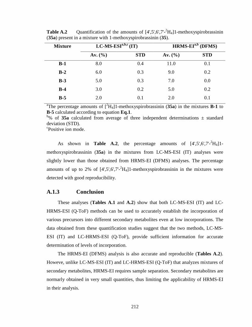
212
Table A.2 Quantification of the amounts of [4',5',6',7'-2H4]1-methoxyspirobrassinin (35a) present in a mixture with 1-methoxyspirobrassinin (35).
LC-MS-ESI a,b,c (IT) HRMS-EI a,b (DFMS) Mixture
Av. (%) STD Av. (%) STD
B-1 8.0 0.4 11.0 0.1
B-2 6.0 0.3 9.0 0.2
B-3 5.0 0.3 7.0 0.0
B-4 3.0 0.2 5.0 0.2
B-5 2.0 0.1 2.0 0.1 aThe percentage amounts of [2H4]1-methoxyspirobrassinin (35a) in the mixtures B-1 to B-5 calculated according to equation Eq.1. b% of 35a calculated from average of three independent determinations ± standard deviation (STD). cPositive ion mode.
As shown in Table A.2, the percentage amounts of [4',5',6',7'-2H4]1-
methoxyspirobrassinin (35a) in the mixtures from LC-MS-ESI (IT) analyses were
slightly lower than those obtained from HRMS-EI (DFMS) analyses. The percentage
amounts of up to 2% of [4',5',6',7'-2H4]1-methoxyspirobrassinin in the mixtures were
detected with good reproducibility.
A.1.3 Conclusion
These analyses (Tables A.1 and A.2) show that both LC-MS-ESI (IT) and LC-
HRMS-ESI (Q-ToF) methods can be used to accurately establish the incorporation of
various precursors into different secondary metabolites even at low incorporations. The
data obtained from these quantification studies suggest that the two methods, LC-MS-
ESI (IT) and LC-HRMS-ESI (Q-ToF), provide sufficient information for accurate
determination of levels of incorporation.
The HRMS-EI (DFMS) analysis is also accurate and reproducible (Tables A.2).
Howeve, unlike LC-MS-ESI (IT) and LC-HRMS-ESI (Q-ToF) that analyzes mixtures of
secondary metabolites, HRMS-EI requires sample separation. Secondary metabolites are
normarly obtained in very small quantities, thus limiting the applicability of HRMS-EI
in their analysis.