physiotherapy and occupational therapy in the hypermobile ... · pdf filephysiotherapy and...
TRANSCRIPT

143
Chapter 9
Physiotherapy and occupational therapyin the hypermobile adultRosemary Keer
Katherine Butler
INTRODUCTION
Joint hypermobility syndrome (JHS) is under-recognized, poorly understood and, sadly, oftenpoorly managed (Beighton 1999). The syndromedoes not necessarily cause problems and for manyindividuals it can be an asset, amply illustrated inthe world of music, dance and sport. For others itcan be a liability, producing significant problemswhich are often ignored by the medical profession(Phipps 2003, HMSA 2006). Early recognition andappropriate management can decrease suffering,reduce the need for unnecessary tests and inva-sive procedures, help to avoid surgery, reducechronic pain and fear (Cherpel & Marks 1999)and prevent the destructive spiral into depressionand physical deconditioning. It is the aim ofthis chapter to discuss the presentation of thesyndrome to aid recognition and diagnosis by pri-mary care practitioners and to explore assessmentand management strategies to decrease sufferingand disability, increase function and enable thehypermobile individual to effectively self-managethe condition.
PRESENTATION
ASSESSMENT
Pain is the most common complaint, often beingwidely distributed, involving several joints andareas of the body, and varying in duration fromacute (15 days) to as long as 45 years (El-Shahaly& El-Sherif 1991). A typical presentation is illu-strated in Keer & Grahame (2003), and Simmonds
& Keer (2007). The onset frequently occurs in child-hood or adolescence, particularly in girls betweenthe ages of 13–19 years, with 75% of hypermobileadolescents developing symptoms by the age of15 (Kirk et al 1967) (Chapters 2, 10 and 11). Anec-dotally, onset is also often associated with trauma,pregnancy, childbirth, a change in physical activ-ity, such as a marked decrease contributing todeconditioning, or an unresolved joint problemwhich leads to compensatory strategies producingother joint problems. In addition, the patient willoften report past episodes of medical or therapyintervention which were unhelpful and may evenhave exacerbated their symptoms. Exploring theirhistory in this respect not only helps to identifypotential generalized hypermobility, but will alsogive valuable information regarding what has nothelped in the past, so that mistakes are notrepeated.
It is unusual for pain to be the only symptom, soasking for details about other symptoms can giveclues to tissues or systems which may be potentialcauses of the problem and may require examina-tion (Table 9.1). Stiffness is a very common com-plaint and may seem at odds with the range ofmovement on testing. This is a subjective feelingof stiffness and although the range of movementmay appear normal it may not be normal for thatparticular patient. Asking ‘have you ever been ableto touch the floor with your palms flat’ (Oliver2000) can be a useful indicator of previous hyper-mobility, a factor that is acknowledged in boththe 5-point questionnaire (Hakim & Grahame2003) and the Brighton Criteria (Grahame et al2000) (Chapter 1). The reports of stiffness mayalso be a reflection of decreasing flexibility with
© 2010 Elsevier Ltd.
DOI: 10.1016/B978-0-7020-3005-5.00013-6

age, or due to injury or muscle stiffness/spasm.Paraesthesiaes are frequently non-dermatomaland there may be an increased disposition todevelopment of a neuropathy (Francis et al 1987,March et al 1988).
Aggravating factors to determine the Severity,Irritability & Nature (SIN) (Maitland 1986) are notclear cut. Latency is frequently a problem, withsymptoms developing several hours or even aday later, making it very difficult to identify theaggravating activity. This makes it difficult tojudge the response to a physical examinationand treatment. Careful questioning about theindividual’s response to physical activity will help,although caution is advised with regard to theamount and vigour of testing, particularly initially.
Hypermobile individuals frequently dislikestatic postures and report difficulty with standingstill, such as when queuing, shopping or attendingexhibitions, and sitting for prolonged periods oftime. Hypermobility of the spine was found toincrease the prevalence of back pain in industrialworkers working in standing/sitting postures(Larsson et al 1995) and hypermobility of the spineand knees produced symptoms in musicians dueto sustained standing postures (Larsson et al 1993).
In addition, hypermobile individuals dislikerepetitive activities, being more likely to developmusculoskeletal lesions (Acasuso-Diaz et al 1993)and reporting an increased frequency of previousepisodes and more recurrent episodes of overusesoft tissue lesions such as bursitis, tendonitis andfasciitis at a single site (Hudson et al 1995, 1998)
as well as fibromyalgia (Goldman 1991, Acasuso-Diaz & Collantes-Estevez 1998) (Chapter 5). It isoften the case that pain or injury has occurred withminimal provocation during everyday activities,such that the patient cannot say what has broughtthe pain on. This can be very frustrating for thetherapist and patient alike, but it is worth viewingthis as a valuable piece of the jigsaw, leading tomore accurate recognition and diagnosis ofgeneralized hypermobility.
Certain aspects of the patient’s history can bevaluable in providing clues to hypermobility. Backand knee pain are reported most commonly asa source of pain in childhood (Biro et al 1983)(Chapter 11). Individuals who no longer demon-strate hypermobility due to age, injury or stiffnesssecondary to pain, may be able to confirm that theywere more flexible when younger, or that there is afamily history of increased flexibility. Studies haveshown that 27–65% of patients with symptomatichypermobility seen in clinic had relatives withhypermobility (Finsterbush & Pogrund 1982, Biroet al 1983, Bridges et al 1992). There may alsobe evidence of involvement of other connective tis-sue, because patients report that they bruise easily(Al-Rawi et al 1985, Kaplinsky et al 1998) and thereis frequently a history of varicose veins, hernia,or prolapses (Wynne-Davies 1971, Al-Rawi et al1985, El-Shahaly & El-Sherif 1991).
Hypermobile individuals frequently recount along history of soft tissue injuries, strains andsprains (Acasuso-Diaz et al 1993), which can be sug-gestive of vulnerable tissues or areas of vulnerabil-ity due to increased tissue laxity, poor control andmuscular weakness. Dislocations and subluxationsare more common (Finsterbush & Pogrund 1982).This should prompt a holistic assessment lookingat the functioning of the whole body, to identifyreasons why a particular area is causing a problem.How well the individual recovered from previousinjury/problems will have a bearing on subsequenttreatment, expectations and prognosis, particularlyas there is anecdotal evidence of poor healing anda slower recovery in this patient group (Russek2000).
In addition, questions relating to gender, age,ethnicity, weight, details of work or daily activities,leisure and exercise or sport activities will help togive a full picture of the impact of JHS on the indi-vidual. It is also important to ask special questionsregarding general health and medical history interms of surgery or illnesses, related or not, such
Table 9.1 Common symptoms reported andpossible tissue or system at fault
SYMPTOM REPORTED SYSTEM TO BEEXAMINED
Stiffness Muscle system
(muscular tension)
‘Feeling like a 90 year old’ Joints
Hormonal
Audible clicking, popping, clunking,
sensations of subluxation, vulnerability,
instability or frank dislocation
Motor control
Stability system
Sensations of parasthesiae, tingling,
numbness, deadness
Neural system
Tiredness, fatigue, faintness, feeling
unwell, flu-like symptoms
Autonomic nervous
system
Physical
deconditioning
144 Section 2 Therapy

as fatigue, chronic fatigue syndrome and fibromy-algia, as well as details of medication, so thatthe physical examination can be tailored to theindividual’s presentation without risking a flare-up. Discussing medication may also highlightthe possibility of JHS as a diagnosis, becausethere is often a poor response to non-steroidalanti-inflammatory drugs (NSAIDs), and localanaesthetics have been reported as less effectivein EDS type III patients (Arendt-Neilson 1990,Hakim et al 2005).
EXAMINATION
Observation of the patient’s mannerisms andpostures during history-taking may give an indica-tion of hypermobility. Contact of the hand to theface may reveal hyperextension of the digits, con-stantly changing position or fidgeting and/or sit-ting in an unusual posture (legs wound roundeach other, slumped to the side or resting on thelateral border of the feet) are familiar sightings(Fig. 9.1).
Skin is a very important feature and helps toconfirm a diagnosis of hypermobility. To the touchit is silky, soft, extensible and thin. Elasticity canbe tested by picking up a section from the back ofthe hand between the thumb and first finger(Chapter 2). Wounds may show poor healing with
a thinner or papery scar. There may be stretchmarks (striae atrophicae) not associated withweight loss or pregnancy, which appear duringthe growth phase in certain areas such as acrossthe lumbar spine and pelvis, around the hips,thighs and shoulders.
The Beighton scale (Beighton et al 1973) gives anindication of hypermobility in certain joints (JHM),and while it is quick and easy to use in the clinic, itis limited in its application as only a few joints aretested. Other scales are discussed in more detail inChapter 1. A diagnosis of JHS can be confirmedusing the Brighton Criteria (Grahame et al 2000)which assesses the effect of weaker connective tis-sue on the body as a whole, but its use is primarilyas a research tool. In an out-patient setting, makinga diagnosis of JHS will depend on a thorough his-tory and physical examination of the whole body.
It may be appropriate or indeed necessary torefer a patient, whom one suspects of beinghypermobile, to a rheumatologist for a definitivediagnosis or further investigations when:
l the patient’s symptoms do not fit theexamination findings (except with regard to therange of movement as mentioned below)
l a clear diagnosis regarding the nature of theconnective tissue disease is required and it isnecessary to exclude other pathology
A B
Fig. 9.1 Typical sitting posture: (a) legs wound round each other, (b) resting on the lateral border of the feet
Chapter 9 Physiotherapy and occupational therapy in the hypermobile adult 145

l a complex presentation involving many bodyareas and systems requires access to other healthcare professionals
l further investigations are required to providemore information regarding pathology, such asin the spine or peripheral joints to identifydegeneration or instability (Grahame 2003).
Pain is also thought to arise because the connec-tive tissue (ligament, tendon, muscle, capsule) inhypermobile individuals is more lax and less resil-ient and suggests a predisposition to the effects oftrauma (acute and chronic) from overuse and mis-use. Microtrauma occurs more frequently and withless provocation (Acasuso-Diaz et al 1993, Hudsonet al 1998), meaning that even everyday activitiescan be the cause of tissue trauma and pain. Forsome, there is the added problem of subluxation,dislocation or instability which can produce, or bethe result of, poor movement control. The hyper-mobile individual has an increased susceptibilityto instability due to increased extensibility in theconnective tissue restraints (capsule, ligament,muscle, tendon), affecting the passive control sys-tem, decreased muscle tone and strength, affectingthe active control system, and deficient propriocep-tive acuity (Mallik et al 1994, Hall et al 1995), affect-ing the neural and feedback control system, whichis further outlined in Panjabi’s model of stability(1992) (Chapter 12.8).
Poor movement control has the potential to leadto further joint problems, setting off a chain reac-tion or ‘domino’ effect whereby a problem in onejoint can develop into widespread pain affectingmany joints over a period of time. Increasedhealing time may lead to more well-developedcompensatory strategies which affect other areas,progressing onto chronic pain and physicaldeconditioning (Fig. 9.2).
It is helpful to think in terms of compensa-tory relative flexibility, a concept proposed bySahrmann (2002). This states that movement occursmore readily at a joint with less stiffness than at ajoint with relatively more stiffness. For example,hamstring muscle tension or stiffness either as aresult of habitually sitting for long periods, or asa result of injury, will inhibit movement at thehip into flexion during forward bending, poten-tially leading to overflexion in the spine. It is com-mon to see decreased hip flexion and increasedspinal flexion in hypermobile individuals and thismay be contributing to low-grade irritation and
strain to lumbar spine tissues, producing pain.Figure 9.3a is an example of this and is in contrastto Figure 9.3b which shows well-balanced fullrange flexion in a hypermobile individual.
There is evidence of neurophysiological defectssuch as pain enhancement and autonomic dysfunc-tion (Gazit et al 2003) (Chapter 6.1), which willhave the effect of increasing disability, leading toless activity and a vicious spiral of chronic painand physical deconditioning (Rose 1985). Psycho-logical distress is common, with concerns anddifficulties occurring around work, home life andraising children, as well as fear of pain, serious ill-ness or permanent disability. Individuals haveoften had to withstand years of having their com-plaints dismissed, being misunderstood or notbeing believed by the medical profession at large(Phipps 2003). This has, understandably in somecases, led to anger, confusion and depression.
Tissue traumaPAIN
Misuse/overuse
Repetitiveactivities
Sustainedpostures
Injury
Muscle spasmShortening/imbalance
Altered movement pattern/movement abnormalities
Domino effectwith more areas affected
Chronic pain statePhysical deconditioning
Poor healing Defectiveproprioception
Fig. 9.2 Basis of symptoms
146 Section 2 Therapy

The physical examination starts with observation.The patient, undressed to their underclothes andstanding in their normal stance, is assessed from allfour directions (front, back and each side) and com-pared with what is considered the ideal skeletalalignment. This is a standard based on sound scien-tific principles which involves minimal stress andstrain on the body tissues and allows maximal effi-ciency (Kendall et al 1993). Details of a thorough pos-tural examination are given in Kendall et al (1993)and should include postural alignment, symmetryand weight distribution from head to toe.
Hypermobile individuals show a tendency torest at the end of joint range and have poor pos-tural alignment. Even if they are initially ableto stand or sit in a good position, it is difficult tosustain the position for long periods. It is easierfor their bodies to take the path of least resistanceand settle into a position which requires decreasedmuscle work, resting on tension in their softtissues. Familiar postures are slumped sitting,standing with hyperextended knees and hips(Fig. 9.4) and hanging on the hip. This leads tostress and strain in the collagenous tissues. If thesepostures are sustained, fluid is forced out and tis-sue nutrition is adversely affected. The tissueundergoes creep, which leads to deformation,
stretching and weakening over time. There is alsoa weakening of surrounding muscles through dis-use and changes in how and where load is trans-ferred through the kinetic chain. Hyperextensionat the knee provides a good illustration (Kendall
A B
Fig. 9.3 Compensatory relative flexibility: (a) forward flexion with decreased hip flexion relative to spinal flexion, (b) forwardflexion with balanced movement throughout the spine and hips
Fig. 9.4 Standing with hyperextending knees
Chapter 9 Physiotherapy and occupational therapy in the hypermobile adult 147
et al 1993), with the patella displaced caudally andcontact of the femoral condyles on the tibial pla-teau shifted more anteriorally. This has thepotential to weaken and damage tissue, disruptnutrition of joint tissue and produce pain. Thereare also effects along the kinetic chain at a distancefrom the knee, with the ankle joint held inplantarflexion, producing changes in the calfmuscles, and also into the hip and lumbar spine.
ACTIVE MOVEMENT
Hypermobile individuals tend to look and movewell, even if they have pain. Thus during activerange of movement testing their reports ofsymptoms (subjective) are often at odds with thephysical examination (objective). It is possible to seehow misunderstandings may arise; the person whocomplains of widespread debilitating pain affectingseveral joints can, when asked, bend forward totouch their toes. This tends to go against medicalwisdom and may lead to the individual being dis-missed or considered a hypochondriac. The explana-tion may be that although the movement looks to befull range, it may not be ‘normal’ or full range forthat particular person. Additionally, it may be thatsome areas are moving into hypermobile range (suchas lumbar spine) and masking reduced range in anadjacent area (such as the hip joint). It is importantto assess the quality of movement and criticallyappraise where, when and how much movement isoccurring throughout the body or kinetic chain,while keeping the biomechanics in mind. Hypermo-bility may be masked when range of movement isdecreased due to fear, pain or stiffness followinginjury or inactivity or in the case of an older personwho may have lost their normal degree of flexibilitywith age. Clinical reasoning will lead to examinationof other areas (skin, other joints, historical flexibility)for confirmation of hypermobility.
It is helpful to examine the effect of moving onearea in the context of the whole body. This maymean examining ease and range of movement inall areas of the spine as well as some peripheraljoints, if symptomatic. For instance, full flexion atthe shoulders may only be possible with lumbarspine extension, because latissimus dorsi is short-ened or overactive (Sahrmann 2002), producingan area of relative stiffness. This has the potentialto lead to overuse injuries at the shoulder throughrepetition of an overhead activity, particularly rele-vant in the sports person. It would therefore be
necessary to assess the shoulder and whole spine.For the less active, it may be as simple as increasedpronation in the midfoot as a result of stiffnessat the ankle into dorsiflexion, either through talo-crural stiffness (from injury) and/or soft tissueshortness/tightness in the calves or higher in thekinetic chain. Poor movement patterns may alsobe due to muscle deconditioning and weakness.A case highlighted in Simmonds and Keer (2008)involved an individual lifting with poor musclecontrol around the wrist which led to the wristbeing pushed into ulnar deviation under load,causing strain to the ligament and tendon on theradial side. Close questioning and detailed exami-nation of everyday habitual activities can highlightsignificant problems, which can be easily rectifiedonce identified.
Functional activities, assessed as relevant to theindividual, need to be examined. These includewalking (over a set distance), climbing stairs, sittingto standing, bending, mini squat to full squat, one-leg stand, heel raise and any specific activity knownto cause problems. In addition to pain, it is also help-ful to look for even weight bearing, balance, co-ordination, symmetry and load transfer (Lee & Lee2007). Pain is not always reproduced on testingunless the problem area is in the acute stage. Thisappears to be due to the tendency for pain to beaggravated by sustained postures or repeatedactivities. If repeating an activity or sustaining a pos-ture does not reproduce pain, the examination mustrely on an analysis of the quality and patterns ofmovement throughout the kinetic chain. Forwardspinal bend is useful for assessing the relative contri-bution of lumbar, thoracic and hip joints into flexionas well as highlighting segmental hinging or tran-slation.Mini squat is helpful in assessing lower limbalignment during a functional activity. One-legstand assesses the ability to transfer load effectivelythrough the pelvis and hip. Heel raise can be helpfulin assessing control around the foot and ankle.
Hypermobiles frequently use a ‘bracing’ patternwith breath holding in an attempt to improve sta-bility and produce more force. As the globalmuscles of the trunk are used, the ribs can becomefixed so that chest expansion, and therefore effi-cient respiration, is affected. The individual eitherbreathes apically or into the abdomen, with the riskof increasing abdominal pressure downwardswhich can have a deleterious effect on the pelvicfloor. It is useful to assess the breathing pattern atrest and with activity.
148 Section 2 Therapy

Many hypermobiles suffer from feelings ofinstability and ‘giving way’, subluxations and dis-locations, and due to the lax connective tissue,joints may become unstable through injury. Treat-ment is therefore focused on control of movementand it is important to assess the local posturalmuscles associated with trunk stability, such astransversus abdominus, deep multifidus and thepelvic floor muscles in terms of timing, atrophy,loss of tonic function, loss of co-ordination andasymmetry. There is also support for the use ofreal time ultrasound imaging to assess perfor-mance of the deep stability muscles, which can beparticularly useful for identifying ‘cues’ which thepatient can use to activate the correct muscle(Fig. 9.5).
Examination continues with detailed inspectionof individual structures that have been identifiedas possible contributors to the patient’s symptoms.
1. Articular movement testing, both passive andaccessory movement, to identify hypermobility,hypomobility and symptom reproduction.
2. Muscle testing, assessing strength, tone, length,timing and tenderness of trigger points.
3. Neural testing, both in terms of function(sensation, reflexes and power) and mobility(Butler 2000). This may involve testing straightleg raise (SLR), upper limb tension tests(ULTTs), slump and thoracic slump, particularlyif involvement of the autonomic nervous systemis suspected.
4. Specific tests to identify pathology such asimpingement (hip and shoulder) and activestraight leg raise (ASLR).
PROPRIOCEPTION
It is known that proprioceptive acuity is dimin-ished in individuals with JHS (Mallik et al 1994,Hall et al 1995) and this can have a significanteffect on the way an individual moves. Theresearch shows that both joint position sense andthreshold to detection of movement are impairedsuch that they have less awareness of approachingthe end of joint range (Chapter 6.4). The reason forthis impairment is not known but may be related toincreased laxity and elasticity in the tissues, whichmay explain why the application of tape has beenfound to enhance proprioception in those with adeficit (Callaghan et al 2002, 2008) and also whysupportive, tight garments have been reported ashelpful in improving proprioceptive feedback(Simmonds & Keer 2007).
In the laboratory, threshold to detection of pas-sive movement has been found to be more reliablethan joint position sense (Juul-Kristensen et al2008), but detection is affected by the position inrange of movement when testing (Ageberg et al2007). In addition, the effect of pain and injuryshould be taken into consideration as Juul-Kristensen et al (2008) found that proprioceptionwas poorer in elbows with epicondylitis than inthe controls’ elbows.
It can be difficult to measure proprioceptionaccurately in the clinic and tests are generallylinked to function. The ability to stand on two legswith eyes closed, or on one leg with eyes openand closed as in the Romberg test, will revealexaggerated swaying and/or loss of balance oncethe eyes are closed. As in the study above, pain
A B
Fig. 9.5 Real-time ultrasound scans of transversus abdominus (TrA): (a) TrA at rest (b) TrA contracting, note slight thickeningand sliding of third layer
Chapter 9 Physiotherapy and occupational therapy in the hypermobile adult 149
may contribute to the increased sway observed dueto poor stability/control around the ankle, knee,hip and/or sacro-iliac joint. Standing on one legand observing the strategies employed in order toremain balanced can highlight poor load transfer-ence through the lower limb, but particularlythrough the pelvis and hip. Hip hitch, or apseudo-Trendelenburg, is frequently observed.
MANAGEMENT
A management plan is formulated following dis-cussion between the patient and the therapist. It isbased on the findings of the assessment in terms ofwhat is causing the problem and where treatmentshould be directed, and an in-depth discussion withthe patient about their expectations. Many patientsare hoping for a miracle cure, others want to helpthemselves but have received conflicting advice,while others are fearful of getting worse and ‘end-ing up in a wheelchair’. Recent research has showngreater benefit and cost-effectiveness in treatmentfor musculoskeletal disorders when patients, whoexpressed a preference, received their preferredtreatment, compared to those who expressed nopreference (Preference Collaborative Review Group2008). In addition, a qualitative study (Cooper et al2008) exploring perceptions of physiotherapy in agroup of chronic low back pain patients highlightedthe importance that patients place on communica-tion. It underpinned the other key themes whichincluded individual care, good explanation regard-ing diagnosis and treatment and involvement indecision making and treatment time. AmandaSperritt (Gawthrop et al 2007) echoes these findingsin an article in the BMJ about her experiences ofEDS. She comments that ‘the most helpful pro-fessionals treat me as a partner in the managementof the condition’ and states that listening and sup-port from the therapist help ‘to make life that biteasier’. Amanda also reports that physiotherapywas valuable because it allowed her to play anactive part in her treatment, as well as teachingher to use her muscles to protect her joints. It is onlyby listening to the patient’s story that a prioritizedproblem list and achievable and appropriate goals,which are the key to successful client care, can beagreed (Simmonds & Keer 2007). Gaining a clearunderstanding of the patient’s problem through acomprehensive assessment helps to ensure appro-priate and effective intervention.
However, there are reports of physical therapybeing unhelpful or even exacerbating symptoms.There may be several reasons for this and mostare underpinned by a failure to recognize that thepatient has JHS and, as such, has more lax, lessresilient connective tissue (Acasuso-Diaz et al1993). Extra care should be taken with manualtherapy. Recovery and healing is slower than inthe non-hypermobile, so if this factor is notappreciated there is a temptation to progress morequickly or with more vigour in order to produce achange.
Functional restoration is the main aim of treat-ment since this enables the individual to self-manage the condition. It is made up of differentcomponents.
1. Restoration of a normal range of movement forthat particular individual, even if it ishypermobile (Keer et al 2003, Maillard &Murray 2003).
2. Restoration of efficient and effective movementpatterns throughout full range of movement,including the hypermobile range. This willinclude correcting and preventing movementdysfunction and regaining joint stability.
3. Education, reassurance, advice and problem-solving (Rose 1985, Russek 1999).
4. Improving general fitness (Simmonds & Keer2007) (Chapter 13).
Three patterns of presentation have beenproposed (Finsterbush & Pogrund 1982), whichrequire different therapeutic approaches.
1. Episodes of acute musculoskeletal pain,dislocation and subluxation, initially respondwell to the usual therapeutic modalities ofelectrotherapy, support, ice, movement andadvice.
2. Recurrent episodes or a series of episodes ofpain at different sites with some physicaldeconditioning characterize the intermediatestage and require therapeutic modalities whichare adapted and modified to provide functionalrestoration.
3. Chronic, long-standing, severe and unremittingpain with profound physical deconditioningmay require a multidisciplinary painmanagement programme using cognitivebehavioural skills (Harding 2003) (Chapter 8).
It is important to try to give treatment that hasmeaning for the patient. This may be a treatment
150 Section 2 Therapy
modality or exercise which instantly takes the painaway, or addresses an area of particular concern.This gives the patient a sense of control or morecomfort and as such promotes improved move-ment patterns and muscle recruitment but alsohas the power to motivate compliance to exercisesor advice.
Acute symptom strategies
In the acute stages the primary aim is relief of pain.To this end the usual therapeutic modalities listedabove are utilized. Rest is usually recommendedfor up to five days while the inflammatory stagetakes place (Mattacola & Dwyer 2002), with move-ment to prevent the deconditioning effects ofimmobilization thereafter. Supporting the injuredpart can be very important because it allows move-ment of the body without strain to the injured tis-sue. Support can vary from a tight garment to aspecific splint or brace. If the skin can tolerate it,tape is extremely useful as it moves more readilywith the body. Support can help rest an area andput less stress on the body as a whole, but also,more importantly, it can allow pain-free move-ment. This has the real potential to prevent devel-opment of chronic pain by helping to reduceafferent stimulation of peripheral nociceptors.Vierck (2006) suggests that if tissue injury healing(particularly injury to deep tissues) and pain donot settle within 3 months there is a risk of devel-oping chronic widespread pain and fibromyalgia.
‘Injury’ in a hypermobile individual may be asmall everyday incident, such as picking up a bag,which provokes instantaneous pain (Simmonds &Keer 2007) or a sustained posture with joints atthe end of their range of movement. In both thesecases, if hypermobile connective tissue is notrecognized it may be hard to analyse what causedthe problem and easy to dismiss the patient’s com-plaints. Inflammation is not necessarily a dominantfeature.
There is an increased risk of deconditioning(Rose 1985) and an increased healing time (Russek2000) in the hypermobile individual, so, whilebeing mindful of the normal healing times, it iswise to be cautious and proceed slowly. Pacingof activities and specific exercises which do notprovoke pain are important to prevent an exacer-bation. Care should be taken during the prolifer-ative stage up to 21 days after injury and it maybe necessary to continue using support. During
the remodelling stage controlled stress needs tobe applied to produce a robust repair, but againcare is needed in this patient group with particularattention to a gradual controlled progression ofstress.
Taping has several uses. It can provide supportfor injured tissue, such as a medial ligament strainat the knee and reduce pain (Macgregor et al 2005,Aminaka and Gribble 2008). There is also evidencethat it can facilitate better muscle activation andpatterning. Macgregor et al (2005) found thatpatella tape which produced a lateral stretch acrossthe patella increased VMO activity in people withpatellofemoral pain. Christou (2004) found similareffects with both medial tape and placebo tapeand suggested the mechanism is via cutaneousstimulation. Interestingly, they also found that tapehad the opposite effect in healthy individuals.There is evidence for tape enhancing propriocep-tion, but only in those whose proprioception ispoor (Callaghan et al 2002, 2008). Individuals withnormal proprioception were either shown to haveno effect or a worsening of joint position sensewhen tape was applied to the knee. This was truewhether the subject had a healthy knee (Callaghan2002) or suffered from patellofemoral pain syn-drome (Callaghan 2008). There is also evidence ofpatella taping having a beneficial effect on dynamicpostural control in patients with patellofemoralpain syndrome compared to healthy individuals(Aminaka & Gribble 2008).
Intermediate symptom strategies
This group may have progressed from the acutestage without good healing or resolution and findthemselves with a ‘domino effect’, whereby oneinitial injury sets off a chain reaction. This may bethe result of resting too much and becomingdeconditioned, or through developing poor com-pensation strategies, or being encouraged to pushthrough the pain and irritating more tissues. Atthis stage it is very important to assess the wholeperson and all areas of symptoms in order to beable to identify what is continuing to ‘drive’ theproblem and thereby apply appropriate treatment.
The treatment is now based on rehabilitationand functional restoration with the aim of retur-ning the individual back to their former self. Oncepain is under control, treatment is concerned withrestoring a full range of movement and ensuringgood motor control throughout the movement,
Chapter 9 Physiotherapy and occupational therapy in the hypermobile adult 151

even if it is hypermobile. Manual therapy is oftenused at this stage to restore normal movementand can be applied to the affected tissue(s) as indi-cated by the assessment.
1. Joints. Gentle, rhythmic, large-amplitudeoscillations (Maitland 1986) have been foundanecdotally to be most effective at reducing painand improving joint range. However, high-velocity thrust techniques (HVT) are generallythought to be contraindicated, particularly in thecervical spine, although they can be successfullyapplied to a stiff thoracic spine in skilled hands.
2. Neural tissue. Restriction to normal neuralmobility can be treated effectively with gentlemobilizing techniques as described by Butler(2000). Sustained end of range mobilization isnot recommended in the hypermobileindividual and overzealous ‘stretching’ canresult in muscle spasm and guarding.
3. Muscle and fascia. Myofascial release, a massagetherapy aimed at releasing tension in musclesand fascia (Gordon & Gueth 2006) uses long,stretching, gentle sustained pressure. It isthought to improve relaxation and circulation,and decrease pain, anxiety and lactic acid buildup. It can be a useful technique to releasemuscles that have developed an overactive anddominant pattern, but for best effect is used withpatient participation and awareness (Lee & Lee2007). This helps to highlight to the patientwhere they may be overworking their musclesand how to release them in the future. It may benecessary to ‘switch off’ and relax an overactivemuscle before an inhibited or ‘switched off’muscle can be voluntarily contracted.
Re-educating goodmovement patterns andmotorcontrol often starts with an appreciation and prac-tice in attaining and maintaining good posturalalignment, firstly in static postures and progre-ssing onto dynamic postures and specific tasks.Maintaining a good standing and sitting posturefor more than a few seconds can be challenging inthe beginning for some hypermobiles, because theirpostural muscles are unused to holding joints (theknee, pelvis and spine in particular) in a moreneutral position, so their endurance capabilitiesneed to be built up slowly and gradually.
It is usually necessary to begin this process innon-weightbearing positions to decrease the loadthrough the joint(s). Effective and efficient joint con-trol begins with gaining control of the pelvis and
trunk before moving to the peripheral joints.Assessment will have identified which of the deepstability muscles (transversus abdominus, pelvicfloor multifidus) need facilitation to recruit effec-tively both in isolation and in a co-ordinated fash-ion. Activation is then practised while maintaininga neutral pelvic/lumbar spine position as well asa relaxed breathing pattern to prevent a ‘bracing’or co-contraction rigidity strategy. This is oftenstarted in lying (supine, prone and side lying) andprogressed to sitting and standing. The emphasisis on a low-level (20–30% of maximal voluntarycontraction (Richardson et al 2004)) isometric holdrepeated several times with relaxation in between.
The ability to maintain good control duringweight-bearing is challenged by standing, initiallyon both legs, and progressed to one leg. All thetime attention is paid to ensuring good joint align-ment and relaxed but accurate and balanced mus-cle activation. Once control with static postures isbeing achieved, movement can be added in theform of knee bends or practising weight transfer-ence in stride stand in preparation for gait training.In addition, proprioception and balance can bechallenged by introducing an unstable base, inthe form of a gym ball, balance board (Fig. 9.6) orfoam roller.
Fig. 9.6 Challenging balance and proprioception throughknee bending on a balance board
152 Section 2 Therapy
There has been little research into effective treat-ment for symptoms as a result of JHS, althoughthere is now evidence in the medical literature thatlow back pain, function and recurrence can besignificantly improved through re-educating spinalstability strategies (Hides et al 1996, O’Sullivan2000, Richardson et al 2004) and it seems logical thatthese recommendations can effectively be appliedto the hypermobile population. Furthermore itseems that it is not only weakness of the stabilitymuscles that is the problem but also delayed activa-tion which allows unprotected displacement of thetrunk to occur with movement of the limbs(Hodges & Richardson 1996, 1998). Specific, low-threshold training of transversus abdominus (TrA)(O’Sullivan 1997) and multifidus (Hides et al 1996)has been shown to be effective in treating chroniclow back pain and preventing recurrences, andmore recently Tsao and Hodges (2007) have shownthis specific training can correct the timing delay ina relatively short period of time. However, there isconflicting evidence on the effectiveness of specificstabilization exercises in chronic back pain, withsome considering them no better than other activephysiotherapeutic interventions (May & Johnson2008). Clinical experience provides anecdotal evi-dence of the ineffectiveness and harm of non-specific exercises, so the decision of what form ofexercises to use will depend on clinical reasoning.
For peripheral joints there is evidence thatutilizing close chained exercises in the lower limbs
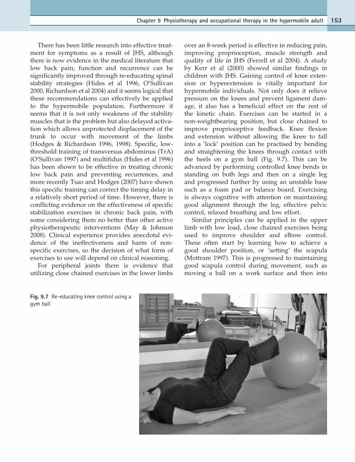
over an 8-week period is effective in reducing pain,improving proprioception, muscle strength andquality of life in JHS (Ferrell et al 2004). A studyby Kerr et al (2000) showed similar findings inchildren with JHS. Gaining control of knee exten-sion or hyperextension is vitally important forhypermobile individuals. Not only does it relievepressure on the knees and prevent ligament dam-age, it also has a beneficial effect on the rest ofthe kinetic chain. Exercises can be started in anon-weightbearing position, but close chained toimprove proprioceptive feedback. Knee flexionand extension without allowing the knee to fallinto a ‘lock’ position can be practised by bendingand straightening the knees through contact withthe heels on a gym ball (Fig. 9.7). This can beadvanced by performing controlled knee bends instanding on both legs and then on a single legand progressed further by using an unstable basesuch as a foam pad or balance board. Exercisingis always cognitive with attention on maintaininggood alignment through the leg, effective pelviccontrol, relaxed breathing and low effort.
Similar principles can be applied in the upperlimb with low load, close chained exercises beingused to improve shoulder and elbow control.These often start by learning how to achieve agood shoulder position, or ‘setting’ the scapula(Mottram 1997). This is progressed to maintaininggood scapula control during movement, such asmoving a ball on a work surface and then into
Fig. 9.7 Re-educating knee control using agym ball
Chapter 9 Physiotherapy and occupational therapy in the hypermobile adult 153

more range on a wall. Further progression can beachieved by increasing the resistance and movinginto weightbearing in four-point kneeling.
Once good effective joint control has beenestablished and exercising has begun to work onmaintaining control throughout movement, atten-tion progresses to improving the endurance andstrength capabilities of the body in order to with-stand high load activities without losing control.Muscle endurance training is thought to be impor-tant, because type I muscle fibres (slow twitch)atrophy at a faster rate than type II fibres(Harrelson 1998). The emphasis is therefore onincreased repetitions and hold time. It is importantto ensure that there is good relaxation betweenmuscle contractions, to delay the onset of fatigue.In addition, eccentric muscle work appears to beparticularly difficult for the hypermobile individ-ual. Patients frequently use gravity to return thelimb to the starting position rather than using slowcontrolled eccentric muscle activity. Right from theearly stages of the rehabilitation programme con-trol through the full range of movement shouldbe emphasized and worked on.
In order to build strength in a muscle, the load isprogressively increased in terms of force and speed.Resistance can be applied using free weights orexercise bands. There is some evidence that musclehypertrophy (through strength training) can leadto increased muscle/joint stiffness (Ocarino et al2008), which may be a useful additional mechanismto gain control of the hypermobile joint.
Ramsay and Riddoch (2001) found that balletdancers were far superior to physiotherapy stu-dents in proprioceptive accuracy and suggestedthat rehabilitation should encourage repetition ofmovement with verbal and visual feedback toimprove sensory motor performances. Biofeedbackin the form of mirrors and photography (stills andvideo) can be used to increase awareness of pos-ture and joint control, both in static positions andduring performance of specific exercises and tasks.More specialized biofeedback, such as surfaceelectromyographic (EMG) biofeedback, can helpdetect the presence of excessive muscular activityduring the performance of a functional task(Polnauer & Marks 1964, Philipson et al 1990)(Chapter 12.1.). Video is particularly useful whenassessing and treating musicians, because thepatient can face a video monitor and play theirinstrument while postural cues are given by thetherapist (Dommerholt & Norris 1997). The video
camera is placed at various angles, allowing themusician to see his or her posture from severalperspectives and the session can be recorded forfurther study and review. This assessment tech-nique can easily be utilized to reviewwriting, sport-ing activities, or performance of daily living tasks.
Chronic symptom strategies
Individuals who fall into this group have often hadsymptoms for a long time, with severe decon-ditioning, kinesiophobia and psychological effects.They may have become dependent on usingaids (collars, braces, sticks, crutches or evenwheelchairs), be unable to work and claiming inca-pacity benefit or registered disabled. Like manychronic pain patients they will very often holdunhelpful beliefs about their pain and disability.But being hypermobile may add additionalconcerns regarding the weakness or vulnerabilityof their tissues, which increases the fear and beliefthat pain equates to damage. Understanding andthen challenging these beliefs through interactiveinformative discussion and paced exercising andactivity can encourage a more positive attitudeand improve confidence.
Pacing is vital to those with hypermobility(Harding 2003) and fibromyalgia. Cycles of over-activity/underactivity are common and result in aflare-up of pain which requires a period of rest torecover. Repetition of this cycle leads to a downwardspiral resulting in deconditioning and a confirmation(in the patient’s mind) of weakness and inadequacy.Teaching pacing skills allows the patient to work attheir own pace but within recognized guidelines(Harding 2003). The emphasis is on maximizing tis-sue health and endurance with a range of exercisesand activities. Variety is key, with frequent changesbetween more active and more sedentary tasks,different postures (flexed, extended), smaller move-ments and larger movements, more intense and lessintense muscle work as well as balancing flexibilitywith strength and endurance (Harding 2003). Ashas been seen with exercising in general, individualswith JHS need to increase activity levels more slowlyto reduce the risk of flare-ups.
SPECIFIC EXERCISE
It is important that the exercises prescribed arespecific and target the intended muscle, movementor task. The form of the exercise or movement is of
154 Section 2 Therapy

paramount importance. Remedial exercise beco-mes as much cognitive as it is physical. For thepatient to learn the skills necessary to gain effectivecontrol of movement it is necessary for them toengage with their body and develop greater bodyawareness. Education regarding anatomy, biome-chanics and how this particular exercise will helptheir problem, along with imagery and biofeed-back, are powerful tools. Mental practice of motorimagery (mental representation of movement with-out any body movement) has been shown toimprove motor and task performance with thegreatest improvement occurring when inter-ventions combine physical and mental practice(Dickstein & Deutsch 2007). In addition, studieshave shown that motor imagery can increase mus-cle strength (Zijdewind et al 2003, Sidaway &Trzaska 2005). This is thought to occur as a resultof neural adaptations such as degree of motor unitactivation, improved coordination and decreasedco-contraction of antagonist muscles (Sidaway &Trzaska 2005). Although the effects of motor imag-ery in JHS have not been studied clinical experi-ence suggests it is a valuable adjunct to treatment.
While exercising they can be taught techniquesto check that their postural alignment remainsgood, that a certain muscle is not dominant, thata weaker or more inhibited muscle is working. Thiscan be achieved through the use of video, mirrors,pressure biofeedback and the therapist’s input.However, if the patient is ultimately to be able tomake use of these skills to prevent and correctproblems in the future, they have to develop theability to correct themselves.
Generic exercises do not work without goodinstruction and practice, because invariably anindividual will ‘take the path of least resistance’.Patients usually take the easy option, or if theyhave been in pain, long-standing compensatorystrategies may inhibit weaker, less dominant or‘forgotten’ muscles. The inhibition of the vastusmedialis obliquus (VMO) following knee surgeryis well known to physiotherapists. Facilitation isneeded to ‘switch’ the correct muscle on, as isensuring the reduction of swelling and pain.It is important that an assessment highlights anindividual’s muscle imbalance and corrects it.It may be appropriate to use manual therapy toinhibit overactive muscles and facilitate under-active ones. This then forms the exercise and ispractised in a simple easy way and graduallymade more challenging. Although hypermobile
individuals do not appear to suffer inflammationin the same way, the principles are similar. WithoutEMG devices (Chapter 12.1) it is often difficult totell which muscle is working, but palpation(therapists or patient) and feel (patient feedback)can be used successfully, as well as being criticalof the form the exercise is taking from the perspec-tive of the whole body.
In the fibromyalgia literature it is thought that‘exercises that are too vigorous might trigger im-mune activation with release of pro-inflammatorycytokines provoking a sickness response’ (Maier &Watkins 1998), which may be a factor in the exacer-bation of symptoms that JHS individuals oftenreport. Exercise should be pain free, although a dis-tinction is made between training pain and anaggravation of symptoms. If an exercise is painfulit is either not being performed correctly or is not‘right’ for that patient at that particular moment intime. In the author’s experience this is particularlytrue for individuals with fibromyalgia and JHS andtime spent modifying an exercise until it is pain-freebut still achieving its aim pays dividends in terms ofpatient confidence and progression.
For those individuals who do not achieve a goodresponse with the treatment approach outlinedabove, a multi-disciplinary pain management pro-gramme (PMP) may be required. There is, how-ever, some evidence to suggest that patients withchronic pain, either caused by hypermobility or inthe presence of hypermobility, do not respond aswell as expected in a PMP. Hakim & Ashton(2005), on analysing the characteristics of thosewho did poorly in a PMP, found hypermobilityto be a common finding. This does not appear tobe an isolated case and a programme modifiedto hypermobile individuals is going some way toaddress this problem (Chapter 8).
STRETCHING
Many hypermobiles like to stretch, but have beenadvised not to by health professionals for fear ofdamaging their tissues or joints through over-stretching. An unpublished audit at Guy’s Hospitalin 1986 reported by Harding (2003) revealed thatpatients with joint hypermobility found stretchinghelpful. This came as a surprise to the audit’sauthors, but has been borne out repeatedly in clin-ical experience. Stiffness is a common complaint,with many hypermobiles saying they ‘feel like a90-year-old’, but it is not clear what produces this
Chapter 9 Physiotherapy and occupational therapy in the hypermobile adult 155

feeling. We know that hypermobile joints, like non-hypermobile joints, stiffen through disuse inresponse to pain, but it is also possible that hyp-ermobile individuals are more likely to use theirglobal muscles for stability and are therefore sub-ject to more increased muscle tension and spasm.Stretching can be a helpful antidote to this, but itis important that the hypermobile individual doesnot overstretch, which is potentially easy to do.
It becomes necessary to differentiate betweenstretching performed in order to regain and main-tain muscle length, relieve muscle tension, orrestore and maintain joint range, and stretching toincrease an already hypermobile range of motion.It is good to stretch, but care is required. Educatingan individual about how they can stretch safelywithout overstretching into their hypermobile ormore vulnerable areas will help develop betterbody awareness, a skill which can be used in thefuture to ensure safe exercising.
EDUCATION AND ADVICE
The power of finally receiving a diagnosis afteryears of searching, suffering and frustration cannot be overemphasized. Knowledge is empo-wering. It allows the patient to move from negativeto more positive emotions and start to focus onways of managing their condition, safe in theknowledge that it is not a life-threatening disorder,and that with the correct input they can make asignificant change, not only to their own life, butalso to the lives of family and friends.
Understanding the implications of the disorderis thought to help an individual cope more effec-tively with pain (Russek 1999). Education aboutthe condition is one of the most important aspectsof medical intervention and forms a major part ofphysiotherapy and occupational therapy input.It will generally consist of advice regarding jointcare, lifestyle modifications, the judicious use ofaids and supports, help with associated problems,exercise and fitness. As therapists we are usuallyable to give our patients more time, to listen totheir individual anxieties or concerns. This helpsto gain a full understanding of how the conditionis impacting on their life and reassures the patientthat someone is finally taking them seriously.Detailed discussion about their daily activitiescan identify specific problems, which, with aproblem-solving approach, can be resolved, bettermanaged and prevented from recurring.
JOINT CARE
Hypermobile individuals tend to rest at the end oftheir range of movements, particularly at theknees, hips and lumbar spine. Educating patientsto avoid unhelpful postures such as ‘W’ sitting(Fig. 9.8), sitting with the leg tucked under the but-tock, sitting cross-legged ‘Indian style’ and kneel-ing on plantarflexed feet or resting on the lateralborders of the feet (Fig. 9.1b) for prolonged periodswill help to prevent soft tissue strain and sub-sequent symptoms. Further advice on joint care isgiven in Chapter 12.2.
Joint care for therapists
Work-related thumb pain is an occupational hazardfor manual therapists around the world (Gloveret al 2005, McMahon et al 2006, Campo et al 2008),and the presence of hypermobility may be a signifi-cant factor in its development (Fig. 9.9). The life-time prevalence of thumb problems in Australianphysiotherapists was 65%with a current prevalenceof 41% in the observational study by McMahon et al(2006). Factors found to be significantly associatedwith thumb problems included thumb joint hyper-mobility or an inability to stabilize the joints ofthe thumb. Hypermobility of the carpometacarpal(CMC) joint (see Chapter 12.2) of the thumb anddecreased thumb strength were significant factors
Fig. 9.8 Unhelpful postures. Sitting in a ‘W’
156 Section 2 Therapy

contributing to thumb pain in another study of Aus-tralian physiotherapists (Snodgrass et al 2003) andgeneralized joint hypermobility (considered with ascore of 3/9 on the Beighton scale) was more preva-lent (although not statistically significant) in thepain group compared to the non-pain group. Inter-estingly, although there was no difference betweenthe prevalence of osteoarthritis (visible onradiographs) in the pain and non-pain groups, theprevalence of osteoarthritis in females in this studywas higher than in a normal population (19% com-pared to 2% respectively). This suggests that femalephysiotherapists are more at risk of developingosteoarthritis at the CMC joint than the normal pop-ulation and that hypermobility (both at the joint andin general) in addition to occupation may be a con-tributing factor.
The type and duration of therapy is also a con-tributing factor. Campo et al (2008) found thatmanual techniques of soft tissue work and jointmobilizations increased the risk of work-relatedmusculoskeletal disorders in the wrist and handby 14 and 8 times respectively in those PhysicalTherapists in the USA who treated ten or morepatients a day. It is clearly important thattherapists engaged in manual therapy are madeaware of the risks and encouraged to take ap-propriate measures. These will include modifyingtechniques, using equipment (massage tools, mobi-lization wedge), reducing hours or patients andwearing protective splints (Chapter 12.2).
Other areas of the body have the potential tocause problems in an occupation as physically
active as physiotherapy. Patient transfers, patientrepositioning and bent or twisted postures havebeen given as factors in the development of backpain (Campo et al 2008) and the hypermobilephysiotherapist would be well advised to take extracare to avoid problems. As yet, hypermobility isnot screened for in the student physiotherapypopulation, but should possibly be consideredin order to highlight those at risk of problems fromperforming or having performed on them repeatedmobilization/manipulation techniques during theirunder- or particularly post-graduate studies.
LIFESTYLE MODIFICATIONS
There can be specific areas of concern regardingwork, home and family life. Helpful advice onactivities of daily living is given in Keer et al(2003) and the issues of hypermobility and work-related musculoskeletal disorders are discussed inMangharam (2003).
Computer use is ubiquitous, often ergonomi-cally unsound, and frequently intense. Optimalergonomic positioning while at the computershould be encouraged for all patients and key-board short cuts should be used wherever possiblein order to decrease mouse usage during a sessionat the computer.
Some general concepts that are readily acceptedwhen using a computer keyboard and mouse are:
l keep wrists neutral
l do not rest wrists while typing
l move the whole arm while keying
l avoid stretching the fingers to reach keys thatare far away
l keep fingers curved and relaxed
l use a light touch
l keep fingernails short (Stegink-Jansen et al 2000)
l avoid double clicking as much as possible whenusing the mouse
l position mouse within easy reach to avoidoverstretching.
Specific advice regarding pregnancy is discussedin Chapter 12.6 and sport and performance inChapter 13.
AIDS AND SUPPORTS
In general, the use of aids and supports isdiscouraged because it can encourage muscleweakness and dependency. However, there is a
Fig. 9.9 Hypermobility in the thumb and fingers of atherapist while performing manual therapy techniqueson the cervical spine
Chapter 9 Physiotherapy and occupational therapy in the hypermobile adult 157

place for supporting an area during the recoveryphase of an injury with a gradual decrease in use,as discussed above. Furthermore, if there is a riskof continued strain to a joint, despite attempts toimprove stability and muscle strength, there is anargument for judicious use of an aid or support.This may involve wearing a collar for travelling,to help prevent a neck strain as a result of suddenstops or humps in the road, or wearing a supporton the knee to be able to take part in a particularsport. Aids and supports can be particularly help-ful in the hand to enable individuals to continueto work and these are described in more detail inChapter 12.2.
ASSOCIATED PROBLEMS
There may be other areas in the body producingsymptoms as a consequence of lax connectivetissue. Flat feet are common and it may be sensi-ble to enlist the help of a podiatrist to perform agait analysis and possibly fit orthotics. Urogenitalproblems, such as prolapse and urinary inconti-nence, may require referral to a specialist physio-therapist for a pelvic examination and adviceregarding pelvic floor exercises (Chapter 12.6).Many hypermobiles suffer symptoms of irritablebowel and in cases advice on diet and reductionin constipation-inducing medication can be suffi-cient while others benefit from more extensiveinvestigations and treatment as described inChapters 6.2 and 6.3. A poor response to localanaesthetics, if present (Arendt-Niesen et al1990, Hakim et al 2005), can be distressing forsome hypermobiles when attending the dentistor obstetrician and liaison and education withother health professionals can reduce anxiety. Thetendency towards low blood pressure and (pre-)syncope may be relieved by an increase in lowerlimb muscle work and taking in a little more waterand salt, but if these measures fail, further investi-gation and treatment of the autonomic nervoussystem may be required (Chapter 6.1).
GENERAL EXERCISE AND FITNESS
Once the patient has regained a good level offunction and has control of their pain, attentioncan move to developing a fitness programme
and helping the patient return to their chosenactivities and sport (Chapter 13). In general,unsupervised sessions in the gym are not advis-able for the hypermobile patient, because theymay over-do the exercises or do them incorrectly,or indeed exercises may take the patient intotheir hypermobile range and thus sessions witha trained therapist are advised initially. The ther-apist can teach, observe and correct the patient toensure they can perform the exercises/tasks in anaccurate and controlled way. We only retain a lit-tle of what we are taught and this lends weightto the argument of a review with the patient tocheck performance at a later date. Some unex-pected adaptations can often be seen oncepatients have been left to exercise on their own.Exercising in the gym is not for everyone, sodiscussion with the patient may identify sports,activities or other exercise which will enablemaintenance of fitness. Particularly goodsuggestions include Pilates, Alexander Techniqueand Tai Chi where the emphasis is on slow, con-trolled movement. Yoga is not contraindicatedbut care should be taken to avoid overstretching.Good instruction and supervision in the initialstages is essential. It is helpful to find an activitywhich is enjoyable and achievable so the patientis motivated to develop a life-long habit of exer-cise (Keer 2003). This subject is discussed in moredetail in Chapter 13.
SUMMARY
Generalized joint hypermobility and JHS are fre-quently overlooked in musculoskeletal con-ditions, but they are not difficult to identify whenlooked for and should be a consideration in allmusculoskeletal pain presentations. JHS requiresa holistic approach in terms of assessment andmanagement, which may involve a multidiscip-linary team.
An individualized, modified, therapeutic pro-gramme is recommended, based on functional res-toration with the emphasis on movement control,joint stability and self-management. Early, accu-rate diagnosis and effective intervention will helpto prevent the downward spiral into chronicpain and deconditioning and thereby reducesuffering.
158 Section 2 Therapy

References
Acasuso-Diaz M, Collantes-
Estevez E, Sanchez Guijo P: Joint
hyperlaxity and musculoskeletal
lesions: study of a population of
homogenous age, sex and physical
exertion, Br J Rheumatol
32:120–122, 1993.
Acasuso-Diaz M, Collantes-
Estevez E: Joint hypermobility
in patients with fibromyalgia,
Arthritis Care Res 11:39–42, 1998.
Ageberg E, Flenhagen J, Ljung J:
Test-retest reliability of knee
kinaesthesia in healthy adults,
Bio Med Central Musculoskeletal
Disorders 8:57, 2007.
Al-Rawi ZS, Al-Aszawi AJ,
Al-Chalabi T: Joint mobility
among university students in Iraq,
Br J Rheumatol 4:326–331, 1985.
Aminaka N, Gribble PA: Patellar
taping, patellofemoral pain
syndrome, lower extremity
kinematics and dynamic postural
control, Journal of Athletic Training
43:21–28, 2008.
Arendt-Nielsen L, Kaaland P,
Bjerring P, et al: Insufficient
effect of local analgesics in
Ehlers-Danlos type III patients
(connective tissue disorder), Acta
Anaesthesiol Scand 34:358–361,
1990.
Beighton PH, Solomon L,
Soskolne CL: Articular mobility in
an African population, Annals of
Rheumatology Disease 32:413–418,
1973.
Beighton P, Grahame R, Bird H:
Hypermobility of Joints, ed 3,
London, 1999, Springer Verlag.
Biro F, Gewanter HL, Baum J:
The hypermobility syndrome,
Pediatrics 72:701–706, 1983.
Bridges AJ, Smith E, Reid J: Joint
hypermobility in adults referred
to rheumatology clinics, Ann
Rheum Dis 51:793–796, 1992.
Butler DS: The Sensitive Nervous
System, Adelaide, Australia, 2000,
Neuro Orthopaedic Institute
(NOI) Publications.
Callaghan MJ, Selfe J, Bagley PJ, et al:
The effects of patellar taping on
knee joint proprioception, Journal
of Athletic Training 37(1):19–24,
2002.
Callaghan MJ, Selfe J, Mc Henry A,
et al: Effects of patellar taping on
knee joint proprioception in
patients with patellofemoral
pain syndrome, Man Ther
13(3):192–199, 2008.
Campo M, Weiser S, Koenig KL, et al:
Work-Related Musculoskeletal
Disorders in Physical Therapists:
A Prospective Cohort Study With
1-Year Follow-up, Phys Ther
88(5):608–619, 2008.
Cherpel A, Marks R: The benign joint
hypermobility syndrome, New
Zealand Journal of Physiotherapy
27:9–22, 1999.
Christou EA: Patellar taping
increases vastus medialis oblique
activity in the presence of
patellofemoral pain, J Electromyogr
Kinesiol 14(4):495–504, 2004.
Cooper K, Smith BH, Hancock E:
Patient-centredness in
physiotherapy from the
perspective of the chronic low
back pain patient, Physiotherapy
94:244–252, 2008.
Dickstein R, Deutsch JE: Motor
imagery in physical therapy
practice, Phys Ther 87(7):942–953,
2007.
Dommerholt J, Norris RN: Physical
therapy management of the
instrumental musician,
Orthopaedic Physical Therapy
Clinics of North America 6:185,
1997.
El-Shahaly HA, El-Sherif AK: Is the
benign hypermobility syndrome
benign? Clin Rheumatol
10:302–307, 1991.
Ferrell WR, Tennant N, Sturrock RD,
et al: Amelioration of symptoms by
enhancement of proprioception in
patients with joint hypermobility
syndrome, Arthritis & Rheumatism
50:3323–3328, 2004.
Finsterbush A, Pogrund H:
The hypermobility syndrome.
Musculoskeletal complaints in 100
consecutive cases of generalised
joint hypermobility, Clin Orthop
168:124–127, 1982.
Francis H, March L, Terenty T, et al:
Benign joint hypermobility with
neuropathy: documentation and
mechanism of tarsal tunnel
syndrome, J Rheumatol
14(3):577–581, 1987.
Gawthrop F, Mould R, Sperritt A,
et al: A patient’s journey: Ehlers-
Danlos syndrome, Br Med J
335:448–450, 2007.
Gazit Y, Nahir AM, Grahame R, et al:
Dysautonomia in the
hypermobility syndrome,
Am J Med 115:33–40, 2003.
Glover W, McGregor A, Sullivan C,
et al: Work-related
musculoskeletal disorders
affecting members of the
Chartered Society of
Physiotherapy, Physiotherapy
91:138–147, 2005.
Goldman JA: Hypermobility and
deconditioning: Important links
to Fibromyalgia/Fibrositis, South
Med J 84:1192–1196, 1991.
Gordon C, Gueth JN: Fibromyalgia
Syndrome: Fact, Fiction, and
Future Remedies, Critical Reviews
in Physical and Rehabilitation
Medicine 18(4):343–369, 2006.
Grahame R, Bird HA, Child A, et al:
The revised (Brighton 1998)
criteria for the diagnosis of benign
joint hypermobility syndrome
(BJHS), J Rheumatol 27:1777–1779,
2000.
Grahame R: Hypermobility and the
heritable disorders of connective
tissue. In Keer R, Grahame R,
editors: Hypermobility Syndrome:
Recognition and Management for
Physiotherapists, Edinburgh, 2003,
Butterworth Heinemann,
pp 23–24.
Hakim A, Grahame R: A simple
questionnaire to detect
Chapter 9 Physiotherapy and occupational therapy in the hypermobile adult 159

hypermobility: an adjunct to the
assessment of patients with
diffuse musculoskeletal pain,
Int J Clin Pract 57:163–166, 2003.
Hakim AJ, Ashton S: Undiagnosed,
Joint Hypermobility Syndrome
patients have poorer outcome
than peers following chronic back
pain rehabilitation, Rheumatology
44(3 Suppl 1):255, 2005.
Hakim AJ, Norris P, Hopper C, et al:
Local Anaesthetic Failure; Does
Joint Hypermobility Provide The
Answer? J R Soc Med 298(2):84,
2005.
Hall MG, Ferrell WR, Sturrock RD,
et al: The effect of the
hypermobility syndrome on knee
joint proprioception, Br J
Rheumatol 34:121–125, 1995.
Harding V: Joint Hypermobility and
chronic pain: possible linking
mechanisms and management
highlighted by a cognitive-
behavioural approach. In Keer,
Grahame, editors: Hypermobility
Syndrome: Recognition and
Management for physiotherapists,
Edinburgh, 2003, Butterworth
Heinemann, pp 147–161.
Harrelson GL: Physiological factors
of rehabilitation. In Andrews JR,
Harrelson GL, Wilk KE, editors:
Physical Rehabilitation of the Injured
Athlete, ed 2, London, 1998,
WB Saunders, pp 13–37.
Hides JA, Richardson CA, Jull GA:
Multifidus muscle recovery is not
automatic after resolution of
acute, first-episode low back pain,
Spine 21:2763–2769, 1996.
HMSA Newsletter, Autumn 2006,
p 6 & 9.
Hodges PW, Richardson CA:
Inefficient muscular stabilization
of the lumbar spine associated
with low back pain. A motor
control evaluation of transverses
abdominus, Spine 21:2640–2650,
1996.
Hodges PW, Richardson CA:
Delayed postural contraction of
transversus abdominis in low
back pain associated with
movement of the lower limb,
J Spinal Disord 11:46–56, 1998.
Hudson N, Starr MR, Esdaile JM,
et al: Diagnostic associations with
hypermobility in rheumatology
patients, Br J Rheumatol
34:1157–1161, 1995.
Hudson N, Fitzcharles M-A,
Cohen M, et al: The association of
soft tissue rheumatism and
hypermobility, Br J Rheumatol
37:382–386, 1998.
Juul-Kristensen B, Lund H,
Hansen K, et al: Poorer elbow
proprioception in patients with
lateral epicondylitis than in
healthy controls: a cross sectional
study, J Shoulder Elbow Surg
17:72S–81S, 2008.
Juul-Kristensen B, Lund H, Hansen K,
et al: Test-retest reliability of joint
position sense and kinaesthetic
sense in the elbow of healthy
subjects, Physiother Theory Pract
24:65–72, 2008.
Kaplinsky C, Kenet G, Seligsohn U,
et al: Association between
hyperflexibility of the
thumb and an unexplained
bleeding tendency: is it a rule of
thumb? Br J Haematol 101:
260–263, 1998.
Keer R, Edwards-Fowler A, Mansi E:
Management of the hypermobile
adult. In Keer R, Grahame R,
editors:Hypermobility Syndrome:
Recognition and Management
for Physiotherapists, Edinburgh,
2003, ButterworthHeinemann, p 89.
Kendall FP, McCreary EK,
Provance PG: Muscles Testing and
Function, ed 4, Baltimore, 1993,
Williams & Wilkins.
Kerr A, Macmillan C, Uttley W,
et al: Physiotherapy for children
with hypermobility syndrome,
Physiotherapy 86:313–316,
2000.
Kirk JA, Ansell BM, Bywaters EGL:
The hypermobility syndrome.
Muscular complaints associated
with generalized joint
hypermobility, Ann Rheum Dis
26:419–425, 1967.
Larsson LG, Baum J, Mudholker GS,
et al: Benefits and disadvantages
of joint hypermobility among
musicians, N Engl J Med
329:1079–1082, 1993.
Larsson LG, Mudholker GS, Baum J,
et al: Benefits and liabilities
of hypermobility in the back
pain disorders of industrial
workers, J Intern Med 5:461–467,
1995.
Lee D, Lee LJ: Course notes 10 day
series: The lumbopelvic hip course,
2007, Twickenham, UK.
Macgregor K, Gerlach S, Mellor R,
et al: Cutaneous stimulation from
patellar tape causes a differential
increase in vasti muscle activity in
people with patellofemoral pain,
Journal of Orthopaedic Research
23(2):351–358, 2005.
Maier SF, Watkins LR: Cytokines for
psychologicals: implications of
bidirectional immune-to-brain
communication for understanding
behaviour, mood, and cognition,
Psychol Rev 105:83–107, 1998.
Maillard S, Murray K: Hypermobility
Syndrome in children. In Keer R,
Grahame R, editors: Hypermobility
Syndrome: Recognition and
Management for Physiotherapists,
Edinburgh, 2003, Butterworth
Heinemann, p 42.
Maitland GD: Vertebral Manipulation,
ed 5, London, 1986, Butterworth
Scientific.
Mallik AK, Ferrell WR,
McDonald AG, et al: Impaired
proprioceptive acuity at the
proximal interphalangeal joint in
patients with the hypermobility
syndrome, Br J Rheumatol
33:631–637, 1994.
Mangharam J: Joint hypermobility
and work-related musculoskeletal
disorders (WRMSD). In Keer R,
Grahame R, editors: Hypermobility
Syndrome: Recognition and
Management for Physiotherapists,
Edinburgh, 2003, Butterworth
Heinemann, pp 127–146.
March LM, Francis H, Webb J: Benign
joint hypermobility with
160 Section 2 Therapy

neuropathies: documentation and
mechanism of median, sciatic,
and common peroneal nerve
compression, Clin Rheumatol
7(1):35–40, 1988.
Mattacola CG, Dwyer MK:
Rehabilitation of the ankle after
acute sprain or chronic instability,
Journal of Athletic Training
37:413–429, 2002.
May S, Johnson R: Stabilisation
exercises for low back pain: a
systematic review, Physiotherapy
94:179–189, 2008.
McMahon M, Stiller K, Trott P: The
prevalence of thumb problems
in Australian physiotherapists
is high: an observational study,
Australian Journal of Physiotherapy
52:287–292, 2006.
Mottram SL: Dynamic stability
of the scapula, Man Ther
2:123–131, 1997.
Ocarino JM, Fonseca ST, Silva PL,
et al: Alterations of stiffness and
resting position of the elbow joint
following flexors resistance
training, Man Ther 13:411–418,
2008.
Oliver J: Hypermobility: Recognition
and Management, In Touch
94:9–12, 2000.
O’Sullivan PB, Phyty GD,
Twomey LT, et al: Evaluation of
specific stabilizing exercise in the
treatment of chronic low back pain
with radiologic diagnosis of
spondylolysis or spondylolisthesis,
Spine 22:2959–2967, 1997.
O’Sullivan PB: Lumbar segmental
‘instability’: clinical presentation
and specific stabilizing exercise
management, Man Ther 5:2–12,
2000.
Panjabi MM: The stabilizing system
of the spine. Part1. Function,
dysfunction, adaptation and
enhancement, J Spinal Disord
5:383–389, 1992.
Philipson L, Sorbye R, Larsson P,
et al: Muscular load levels in
performing musicians as
monitored by electromyography,
Medical Problems of Performing
Artists 5:79–82, 1990.
Phipps C: I still don’t think anybody
believes me, The Guardian 14th
Oct, 2003.
Polnauer FF, Marks M: Senso-motor
study and its application to violin
playing, Urbana, Ill, 1964,
American String Teachers
Association.
Preference Collaborative Review
Group: Patients’ preferences
within randomized trials: systemic
review and patient level meta-
analysis, Br Med J 337:a1864, 2008.
Ramsay J, Riddoch J: Position-
matching in the upper limb:
professional ballet dancers
perform with outstanding
accuracy, Clin Rehabil 15:324–330,
2001.
Richardson C, Hodges PW, Hides J:
Therapeutic Exercise for lumbopelvic
stabilization, ed 2, Edinburgh, 2004,
Churchill Livingstone.
Rose BS: The hypermobility
syndrome. Loose-limbed and
liable, New Zealand Journal of
Physiotherapy 13:18–19, 1985.
Russek LN: Hypermobility syndrome,
Phys Ther 79:591–599, 1999.
Russek LN: Examination and
treatment of a patient with
hypermobility syndrome, Phys
Ther 80:386–398, 2000.
Sahrmann SA: Diagnosis and
Treatment of Movement Impairment
Syndromes, St Louis, 2002, Mosby
Inc.
Sidaway B, Trzaska (Robinson) A:
Can mental practice increase
ankle dorsiflexor torque? Phys
Ther 85(10):1053–1060, 2005.
Simmonds JV, Keer R: Hypermobility
and the hypermobility syndrome,
Man Ther 12:298–309, 2007.
Simmonds JV, Keer R: Hypermobility
and the hypermobility syndrome
Part 2: Assessment and
management of hypermobility
syndrome: illustrated via case
studies, Man Ther 13(2):e1–e11,
2008.
Snodgrass SJ, Rivett DA, Chiarelli P,
et al: Factors related to thumb
pain in physiotherapists, Aust J
Physiother 49:243–250, 2003.
Stegink-Jansen CW, Patterson R,
Viegas SF: Effects of fingernail
length on finger and hand
performance, J Hand Ther
13(3):211–217, 2000.
Tsao H, Hodges PW: Immediate
changes in feedforward postural
adjustments following voluntary
motor training, Exp Brain Res
181:537–546, 2007.
Vierck CJ: Mechanisms underlying
development of spatial distributed
chronic pain (fibromyalgia), Pain
124:242–263, 2006.
Wynne-Davies R: Familial joint
laxity, Proc R Soc Med 64:689–690,
1971.
Zijdewind I, Toering ST, Bessem B,
et al: Effects of motor training on
torque production of ankle plantar
flexor muscles, Muscle Nerve
28(2):168–173, 2003.
Chapter 9 Physiotherapy and occupational therapy in the hypermobile adult 161
