physiology - hls
TRANSCRIPT

Physiology - HLSDone By
Dana Tarawneh & Banan AlBalawneh
Corrected By
Samah Freihat

Physiology LabDr. Tamara Alqudah

The Experiments• Red Blood Cell (RBC) Count• White Blood Cell (WBC) Count• Differential Leukocyte count (DLC)• Reticulocyte count• Packed cell volume (PCV)• Hemoglobin concentration• Erythrocyte Sedimentation rate (ESR)• Blood Type• Bleeding Time• Clotting Time• Osmotic Fragility Test

Packed Cell Volume (PCV) Hematocrit (HCT)
• PCV is the ratio of the volume of packed red cells (without spaces between RBCs) to the total blood volume. It’s an easy & a cheap way to diagnose anemia or polycythemia
• Males: 40%- 54%• Females: 36%- 46% it’s less than males' percentage range due to decreased no. of RBCs in
females
• It decreases in cases of anemia and increases in polycythemia and dehydration. (it helps in the follow up of dehydrated patients )

The procedure
• Blood sample is put inside a blood container, and it enters the capillary tube through capillary attraction. Or we can simply prick the tip of the finger and put the capillary tube on the drop of blood that has formed, and the drop of blood will enter the tube.
• After that, we need to seal and close the tube using some clay. We put the tip of the tube on the clay and a little amount of it will enter and seal the tube.
• Later on, we move the tube into a device known as a microcentrifuge. Note that :
1. Capillary tubes have a specific place built for them inside the device as it’s designed to hold these specific tubes
2. The device rotates very fast and due to that, it will separate the components of the solution based on their density.

• A blood sample is centrifuged in aheparinized capillary tube (red tip) it indicates the coat of heparin (anti-coagulant) found inside the tube. This helps in keeping the blood in its fluid – viscous state.
• The RBCs become packed at the bottomof the tube.• The PCV is then calculated according to
the following formula:• PCV= RBC height x100
Total height
• Beware not to include the buffy coat• PCV low à probably anemia
• PCV high à probably polycythemia or dehydration
The procedure

Hemoglobin Concentration• Hemoglobin is a globular protein made up of four subunits. Each subunit contains a heme
group conjugated to a polypeptide. Heme is an iron-containing porphyrin derivative.• Heme has the ability to bind oxygen reversibly and carry it to tissues.Ø Normal values of hemoglobin• 14-17.5 g/ 100 ml in males or 14-17.5 g/dl
• 12-15 g/ 100 ml of in females or 12-15 g/dl• If the number is below these ranges, the patient has anemia.
Ø Different methods can be used to find the hemoglobin concentration one of themis Sahli’s method.
Ø Sahli’s method is based on the fact that when blood is mixed with HCl, hemoglobin isconverted to acid hematin which is brown in color we use it to know the HB in the blood sample

Sahli’s apparatus1. The space between the brown colored
areas is to insert a tube containing diluted blood sample to compare the colors.
2. the pipette is supposed to be used to put the blood inside the tube after sucking it to a certain height, but nobody uses it anymore, everyone uses the new automatic pipette.
3. The glass rod is used to stir the blood sample with HCl, to make sure all RBCs are broken down faster.


The procedure1. Add HCl into the tube up to 2g% mark2. Mix the EDTAsample gently and fill the pipette with 20 Ul blood.3. Wipe the external surface of the pipette to remove any excess blood.4. Add the blood into the tube containing HCl. Wash out the contents of the hemoglobin pipette
by drawing in and blowing out the acid few times so that the blood is mixed with the acidthoroughly.
5. Allow to stand undisturbed for 10 min. (This is because, maximum conversion of hemoglobin to acid hematin, occurs in the first ten minutes)
6. Place the hemoglobinometer tube in the comparator and add distilled water to the solution dropby drop stirring with the glass rod until it’s colour matches that of the comparator glass.(usually, the color of the sample is darker)
7. Remove the stirrer and take the reading directly
ØHemoglobin concentration is read directly from the graduated scale on the dilution tube.


Erythrocyte Sedimentation Rate (ESR)
• The rate at which RBCs sediment in a period of one hour.
• The ESR is a simple non-specific screening test that indirectly measures the presence ofinflammation in the body.
• It reflects the tendency of red blood cells to settle more rapidly in the presence of somedisease states, usually because of increases in plasma fibrinogen, immunoglobulins, andother acute-phase reaction proteins.
• It’s a cheap & very simple test that helps us to know if there is some inflammatory condition in the blood. The downsides though is that the test is non-specific meaning that it won’t lead us to the place of inflammation – if it was there -.
• Changes in red cell shape or numbers may also affect the ESR.

• In our lab we use the Wintrobe tube which is 100 mm long.
• EDTA anticoagulated blood is drawn into the Wintrobetube till the zero mark
• The tube is placed in its rack in a strictly vertical position for 1 hour at room temperature (exactly 1 hour)
• the RBCs – under the influence of gravity - settle out from the plasma.
• The rate at which they settle is measured as the numberof millimeters of clear plasma present at the top of thecolumn after one hour (mm/hr) (how much of the plasma has become clear of RBCS)
The procedure

At the beginning of the experiment After one hour
• Note that each lines equals 2 degrees
• So here it’s 18 millimeters per hour
• If we left the tube for more than an hour the number will increase ofc.
• RBCs are denser than the plasma, they will settle down.
• If the patient was suffering an inflammaroty disease, ESR will be 20-30 millimeters per hour and even more, depending on the inflammation occurring.

RBCs sedimentation
• The RBCs sediment because their density is greater than that of plasma. The sedimentation increases with stacking of RBCs(rouleaux formation)
• Rouleaux formation is possible because of the discoidshape of RBCs
• Normally, RBCs have negative charges on the outside of thecells, which cause them to repel each other.
• Many plasma proteins have positive charges and can neutralize the negative charges of the RBCs, which allows for theformation of the rouleaux.
• Therefore, an increase in plasma proteins (present ininflammatory conditions) will increase the rouleaux formations,which settle more readily than single red blood cells

•Normal ESR values• Men < 15mm/hr• Women < 20mm/hr
• High ESRØ Inflammation maybe
ØAnemia maybe
ØOld ageØPregnancyØTechnical factors: tilted ESR tube, high room temperature.
• Some interferences which decrease ESR:• Abnormally shaped RBC can’t form the rouleaux formation (sickle cells and spherocytosis) where
the patient has inflammation, but his ESR is still low, so we know there’s an abnormality
• Polycythemia large number of RBCs à more negative charges à harder movement
• Technical factors: low room temperature, delay in test performance (>2 hours), clotted blood sample

RDW (Red Cell Distribution Width)
MPV Mean platelet volume
• here we have some RBCs indices :1. MCV à lower than normal à IDA,
Thalassemia 2. MCV à higher than normal à
Folate or Vit B12 deficiency.3. MCH à lower than normal à IDA 4. MCH à higher than normal à HS5. RDW shows us the variation
between the sizes of RBCs.

• At least 30 commonly occurring antigens and hundreds of other rare antigens composedof glycoproteins and glycolipids are found on the surface of RBCs.
• Each of which can at times cause antigen- antibody reactions leading to immediate ordelayed agglutination and hemolysis of RBCs.
• Most of the antigens are weak.• Two particular types of antigens (agglutinogens) are likely to cause blood transfusion
reactions: the ABO system of antigens and the Rh system.• Based on these two systems we have 8 blood groups:• A +ve, A –ve, B +ve, B –ve, AB +ve, AB –ve, O +ve & O -ve
Blood Groups
ABO Blood Group
• The ABO blood group is based on two glycolipid antigens called A and B.
• Blood plasma usually contains antibodies called agglutinins that react with the A or B antigens. These are the anti-A antibody, which reacts with antigenA, and the anti-B antibody, which reacts with antigen B.
• Agglutinins start to appear in the blood within a few months after birth.• They are formed naturally. Their production is thought to be stimulated
when the immune system encounters the "missing" ABO blood groupantigens in food or in micro-organisms.

If a pt. With A blood type is given type B – his antibodies ( anti- B antibodies) will start attack the transfused RBCs & they’ll cause agglutination of RBCs ( the RBCs will stick together & this will lead to their hemolysis
Blood Type Ag expression on plasma membrane
Plasma Antibodies
A A Ag Anti- B Ag
B B Ag Anti- A Ag
AB BOTH A Ag & B Ag -
O - Both Anti- A & Anti- B Antibodies

Rh blood group• There are six common types of Rh antigens, each of which
is called an Rh factor. These types are designated C, D ( the most
important one) , E, c, d, and e.
• The type D antigen is widely prevalent in the populationand considerably more antigenic than the other Rh antigens.
• Anyone who has this type of antigen is said to be Rh positive (85% of population), whereas a person who doesn’thave type D antigen is said to be Rh negative.
• In contrast to ABO system there is no preformed Anti-D in theRh–ve individual (Normally people don’t have Anti- D antibodies, BUT they’ll start to have this Ag if they receive incompatible blood transfusion)
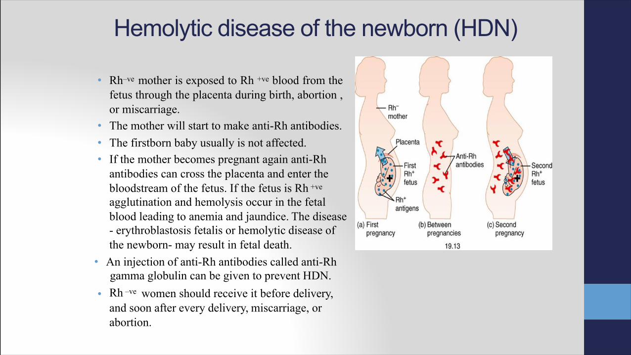
Hemolytic disease of the newborn (HDN)
• Rh–ve mother is exposed to Rh +ve blood from thefetus through the placenta during birth, abortion ,or miscarriage.
• The mother will start to make anti-Rh antibodies.• The firstborn baby usually is not affected.• If the mother becomes pregnant again anti-Rh
antibodies can cross the placenta and enter the bloodstream of the fetus. If the fetus is Rh +ve
agglutination and hemolysis occur in the fetal blood leading to anemia and jaundice. The disease- erythroblastosis fetalis or hemolytic disease of the newborn- may result in fetal death.
• An injection of anti-Rh antibodies called anti-Rhgamma globulin can be given to prevent HDN.
• Rh –ve women should receive it before delivery,and soon after every delivery, miscarriage, or abortion.

Determination of blood type1. Prick the tip of a finger with a lancet and put three separate drops
of blood on a clean microscopic slide.2. Add one drop of Anti-A (blue) to the first drop,Anti-B (yellow) to
the second drop, and Anti-D (transparent) to the third drop.3. Mix well, using separate wooden sticks.4. The results are read directly from the slide.
Ø If agglutination occurs in the first drop the blood type is A , if agglutination occur in the second drop the blood type is B, if itoccurs in both it is AB and if it doesn't occur in any drop it is type O.
Ø If agglutination ( RBCs stick together )occurs in the Rh drop the blood isconsidered as Rh+ve. (This reaction might take some time to develop)
ØThe strength of agglutination reaction is not the same in all people,so in some cases it may be necessary to examine the slide under the microscope to look for agglutination.

*Anti-A : contains antibodies against A Ag*Anti-B : contains antibodies against B Ag*Anti-D : contains antibodies against Rh factor D
* We need to use 3 wooden sticks each 1 for each drop of blood.

Type AB
Type A
Type O
Type B
Type A +ve

What is the type of blood in eachtest presented below?
1.
2.
3.
4.

Hemostasis• Hemostasis is prevention of blood loss from circulatory
system.• Depends on the integrity of blood vessels, platelets (number &
function) and clotting factors.
The hemostatic response to vascular injury is achievedby several mechanisms:
1. Vasoconstriction (to decrease the amount of blood loss & making the other 2 steps easier to happen)
2. Formation of a platelet plug (it can close the injury but it's not strong enough)
3. Formation of a blood clot

Bleeding time• A bleeding time is used to evaluate the second phase of hemostasis, which involves
adherence of the platelets to the injured vessel, platelet activation and aggregation(formation of a plug).
ü The time measures how long it takes for a platelet plug to form.ü Normal range: 3-5 minutes
üIt increases when the platelets count is low (thrombocytopenia), platelet function is abnormal or with the use of aspirin .
• Disadvantages: Insensitive, Invasive & operator dependent.Easy test, very cheap (doesn’t need a lot of instruments)

• Duke method1. Clean the tip of the finger or the ear lobe with
alcohol.2. Puncture the skin with a special lancet. The
wound should be 3–4 mm deep.3. Wipe the blood drop by a filter paper
every 30 seconds4. Repeat until no more blood is absorbed
by the filter paper.5. Multiply the number of blood drops by 30
seconds• Or divide the number of spots of blood by 2 and
that will give you the bleeding time in minutes. 11/2= 5.5 m
Clotting time• It measures the time required for a blood sample to coagulate in vitro
( in the lab). Clotting time depends on the availability ofcoagulation factors.
• Normal value is 6-10 minutes.
• It is prolonged in conditions like hemophilia, vitamin K deficiency,liver diseases, and warfarin overdose (by affecting certain coagulation factors) .

1. Clean the tip of the finger with alcohol then prick it with a lancet.2. Draw blood into non-heparinized (bc. We want the sample to clot) capillary tubes – we
might fill 4-6 tubes.
1. After 2 minutes, start breaking the capillary tubes to see whether a thread (bc. It
takes the shape of the tube) of coagulated blood is formed between the two brokenends ( if the blood wasn’t coagulated, we’ll wait for 30 seconds and break the tube at the other
end or from the middle until the blood is coagulated) .2. It is preferred to calculate the clotting time from the average of two capillary tubes.
Non-heparinized Heparinized

Osmotic fragility• when RBCs reside in an isotonic medium, the intracellular and
extracellular fluids are in osmotic equilibrium across the cell membrane, and there is no net influx or efflux of water.
• When RBCs reside in a hypotonic medium, a net influx of water occurs so the cells swell and the integrity of their membranes is disrupted resulting in hemolysis
• When RBCs reside in a hypertonic media , a net efflux of water occurs so the cells lose their normal biconcave shape, undergoing collapse.
To measures the tendency of RBCs to rupture once they’re put in hypotonic solution.

280mOsm/L
This RBC has an intracellular osmolarity of 280mOsm/L, (A) we put it in a solution with same osmolarity then the RBC will maintain its size & its shape.(B) we put it in a hypertonic solution the net efflux of water is going to be higher than influx, so the RBC is going to shrink>(C) we put it hypotonic solution it will swell, and it might rupture>The rupture depends on:1* the tonicity of the fluid.More hypotonic --> RBCs more likely to rupture,The integrity of the plasma membrane2* in young RBCs the plasma membrane is very flexible as they age they lose this flexibility, or if the person has a certain diseaes.3*the surface area to volume ratio – the RBCs are biconcave disks which makes the suface area large any condtion might decrease this ratio will make the RBCs more susceptible to hemolysis

Osmotic fragility test• A test designed to measures red blood cell’s resistance to hemolysis ( Normal RBCs have
the ability to resist hemolysis ) when exposed to a series of increasingly dilute salinesolutions.
• The susceptibility of RBCs to hemolysis is a function of:ØSurface area to volume ratio.ØCell membrane composition and integrity
• This test is mainly used to diagnose hereditary spherocytosis but it is also usedin some countries to screen for thalassemia.

• The procedure:1. Put labeled centrifuge tubes in a rack.2. Prepare NaCl solutions of different concentrations starting
from 0.9% NaCl till 0.2% NaCl. (each time decrease the conc. 0.5% . Starting from isotonic towards
hypotonic solution))
3. Add 10 ml of each solution to a different tube then add one drop of blood to each tube.4. Shake each tube well and allow them to stand for 20 minutes.
After 20 minutes, the tubes are centrifuged for 10 minutes5. Transfer supernatant fluid from each tube into spectrophotometer
cuvettes6. The absorbance is then measured at 540 nm and used to calculate the
percentage of hemolysis for each solution. (we measure the amount of Hg in the solution, if the RBCs rupture the Hg is goning to be released into the solution, so Hg conc.in the solution is a representation of the dgree of hemolysis that happened)
7. The results are plotted against the NaCl concentrations, this yields an osmoticfragility curve which is then compared to a standard curve.

Hemolysisstarts
CompleteHemolysis
• In this example• From 0.7% to 0.5%there is nohemolysis. RBCs have acumulated in the base of the tube
• At theconcentration of 0.45% hemolysis starts andthe solution becomes red in color, butthere are some settled RBCs inthe tube.
• At theconcentration of 0.30%,thesolution is bright red and there are no settled RBCs(complete hemolysis).
Then we take the fluid from these tubes & put it in the spectrophotometer then we calculate the conc. of Hg in each tube to get Graph For RBC Osmatice Fragility.


- Normalindividual’s RBCscanresistthehemolysisupto0.5% solution.- InherediatyspherocytosistheRBCsisgoningtolosetheirbiconcaveshapeandbecomesphereswithlowsurfacearea:volumeratioandhigherosmoticfragility(lessresistanttohemolysissothehemolysisstatrsearlier)- thecurveisshiftedtotheright.- InsicklecellanemiaandthalassemiacanRBCswithstandmorehypotonicsolution,moreresistanttohemolysis

• Decreased red cell fragility (increased resistance to hemolysis) is seen with thefollowing conditions:
ØThalassemia.ØIron deficiency anemia.ØSickle cell anemia
üThese cells have a high surface area: volume ratio
• Increased red cell fragility (increased susceptibility to hemolysis) is seen in thefollowing conditions:
ØHereditary spherocytosisØAutoimmune hemolytic anemiaØToxic chemicals, poisons, infections, and some drugs.ØSevere burns.
üThese cells have a low surface area: volume ratio