physiological responses and gene co- · pdf filephysiological responses and gene co-expression...
TRANSCRIPT

Physiological Responses and Gene Co-ExpressionNetwork of Mycorrhizal Roots under K+ Deprivation1[OPEN]
Kevin Garcia, Deborah Chasman, Sushmita Roy, and Jean-Michel Ané*
Department of Bacteriology (K.G., J.-M.A.), Department of Computer Sciences (S.R.), and Department ofAgronomy (J.-M.A.), University of Wisconsin, Madison, Wisconsin 53706; Wisconsin Institute for Discovery,University of Wisconsin, Madison, Wisconsin 53715 (D.C., S.R.); and Department of Biostatistics and MedicalInformatics, University of Wisconsin, Madison, Wisconsin 53792 (S.R.)
ORCID IDs: 0000-0003-0821-1024 (K.G.); 0000-0002-7324-0131 (D.C.); 0000-0002-3128-9439 (J.-M.A.).
Arbuscular mycorrhizal (AM) associations enhance the phosphorous and nitrogen nutrition of host plants, but little is knownabout their role in potassium (K+) nutrition. Medicago truncatula plants were cocultured with the AM fungus Rhizophagusirregularis under high and low K+ regimes for 6 weeks. We determined how K+ deprivation affects plant development andmineral acquisition and how these negative effects are tempered by the AM colonization. The transcriptional response of AMroots under K+ deficiency was analyzed by whole-genome RNA sequencing. K+ deprivation decreased root biomass and externalK+ uptake and modulated oxidative stress gene expression in M. truncatula roots. AM colonization induced specifictranscriptional responses to K+ deprivation that seem to temper these negative effects. A gene network analysis revealedputative key regulators of these responses. This study confirmed that AM associations provide some tolerance to K+
deprivation to host plants, revealed that AM symbiosis modulates the expression of specific root genes to cope with thisnutrient stress, and identified putative regulators participating in these tolerance mechanisms.
Potassium (K+) is a macronutrient required by allorganisms for growth and development. In plants, K+
participates in many diverse processes, includingplasma membrane polarization, growth, stomatal ap-erture regulation, and the acquisition of other nutrients(Wang and Wu, 2013). K+ represents 2% to 10% of theplant dry biomass, and its optimal cytoplasmic con-centration for enzyme activities is around 100 to 200mM
(Leigh and Wyn Jones, 1984). Thus, a high cellular K+
concentration is vital for plants to maintain a widerange of physiological processes (Adams and Shin,2014; Benito et al., 2014). Depending on soil type, theconcentration of available K+ varies from 0.1 to 1 mM
(Asher and Ozanne, 1967). Only two fractions of K+ areimmediately accessible to plants: K+ in water solution
and K+ in exchangeable form. These two fractions rep-resent about 2.2% of the total amount of soil K+ (Zörbet al., 2014). This very low availability combined withthe constitutive demand of plants leads to the formationof depletion zones near the root surface (Drew andNye,1969). Plants have developed efficient strategies to dealwith limited K+ availability, including the establish-ment of symbiotic associations with microbes.
Arbuscular mycorrhizal (AM) fungi colonize the rootcortex intercellularly and intracellularly (Brundrett,2004; Harrison, 2005). These AM fungi belong to thefungal subphylum of Glomeromycotina and probablyplayed a critical role in the emergence and evolution ofland plants (Delaux et al., 2015; Spatafora et al., 2016).This AM symbiosis is the oldest mycorrhizal associa-tion and is found in about 65% of extant land plants,including many major crop species (Wang and Qiu,2006). AM associations are characterized by the for-mation of arbuscules in the cortical cells of host plants,which are the site of nutrient exchange between theplant and the fungus (Smith and Read, 2008). The im-provement of plant nutrient acquisition through AMassociations becomes particularly important whenhydromineral resources are scarce. In return for thenutrients provided by AM fungi, host plants provideup to 20% to 25% of their photosynthesis-derived car-bohydrates to their symbionts (López et al., 2008).
The improvement of plant nutrition through AMsymbiosis and the molecular basis of nutrient transferare well characterized for phosphorus, nitrogen, andsulfur (Govindarajulu et al., 2005; Javot et al., 2007; Jinet al., 2012; Casieri et al., 2013; Courty et al., 2016;
1 This work was supported by the National Science Foundation(grant no. NSF-IOS 1331098 to J.-M.A. and CAREER award grant no.NSF-DBI 1350677), by a Sloan Foundation research fellowship, byUniversity of Wisconsin-Madison startup funds to S.R., and by theEnvironmental Protection Agency (grant no. 83573701).
* Address correspondence to [email protected] author responsible for distribution of materials integral to the
findings presented in this article in accordance with the policy de-scribed in the Instructions for Authors (www.plantphysiol.org) is:Jean-Michel Ané ([email protected]).
K.G. and J.-M.A. conceived the research plans; J.-M.A. supervisedthe experiments; K.G. designed and performed most of the experi-ments; K.G. and D.C. analyzed the data; K.G. and D.C. wrote thearticle with contributions of all the authors; J.-M.A. and S.R. super-vised and complemented the writing.
[OPEN] Articles can be viewed without a subscription.www.plantphysiol.org/cgi/doi/10.1104/pp.16.01959
Plant Physiology�, March 2017, Vol. 173, pp. 1811–1823, www.plantphysiol.org � 2017 American Society of Plant Biologists. All Rights Reserved. 1811 www.plantphysiol.orgon May 25, 2018 - Published by Downloaded from
Copyright © 2017 American Society of Plant Biologists. All rights reserved.

Garcia et al., 2016). However, the role of mycorrhizalsymbioses in plant K+ nutrition is still understudiedandmisunderstood (Garcia and Zimmermann, 2014). Asmall number of studies reported AM fungal mediationof plant K+ nutrition. The assessment of K+ distributionin mycorrhizal plants using particle-induced x-rayemission experiments revealed a higher K+ concentra-tion in root sections of Aster tripolium colonized byRhizophagus irregularis than in noninoculated plants(Scheloske et al., 2004). The same trend of K+ enrich-ment for plants mycorrhized by various AM fungi alsowas observed in maize (Zea mays) root steles (Kaldorfet al., 1999), Pelargonium peltatum shoots (Perner et al.,2007), Lactuca sativa leaves (Baslam et al., 2013), andwheat (Triticum aestivum) stems (Oliveira et al., 2016).The expression of three K+ transport systems likely in-volved in phloem loading/unloading (ZmAKT2), xy-lem release (ZmSKOR), and sodium/K+ homeostasis(ZmSOS1) was differentially regulated in AM maizeroots (Estrada et al., 2013). These findings suggestedthat AM symbioses could play a significant role inplant K+ nutrition by the reorganization of molecularresponses.
If K+ amendments are not used frequently, long-termK+ deprivation can result in irreversible damage to thecrop, including poor pest tolerance and significant yieldlosses. Soils from many parts of the world are increas-ingly K+ deficient, reinforcing the need for under-standing how plants can cope with K+ deprivation(Moody and Bell, 2006; Römheld and Kirkby, 2010;Zörb et al., 2014). However, most studies have onlyinvestigated the short-term molecular plant responsesto K+ deprivation, and not in the context of AM sym-biosis. The transcriptional responses of plant roots to K+
deprivation were studied in various species, includingArabidopsis (Arabidopsis thaliana; Armengaud et al.,2004), rice (Oryza sativa; Ma et al., 2012; Shankar et al.,2013), wheat (Ruan et al., 2015), and sugarcane (Sac-charum officinarum; Zeng et al., 2015). However, in allthese studies, K+ was completely absent from the me-dium and the plants were analyzed after a relativelyshort period of time (1–2 weeks). Following thesedrastic and short-term treatments, the up-regulationof high-affinity K+ transporters often was reported.This response is a rapid and transient strategy butprobably not a sustainable one if the stress is pro-longed. The transcriptional response of plant rootscolonized by an AM fungus during K+ deprivation isstill unknown.
To address this question, we examined, to ourknowledge for the first time, the effect of long-term K+
deprivation on the development, nutrition, gene ex-pression, and AM formation using the model legumeMedicago truncatula. To evaluate the impact of AMsymbiosis on plant adaptation to long-term K+ depri-vation, we inoculatedM. truncatula plants with the AMfungus R. irregularis and watered them with high- orlow-K+ solution. Plant biomass and the tissue content ofvarious ions were determined after 6 weeks. The tran-scriptional profile of AM roots under K+ deprivation
was investigated using RNA sequencing (RNA-seq)and revealed mycorrhiza-specific responses under K+
deprivation. Finally, a network-based prioritizationanalysis revealed putative regulators controlling AMsymbiosis and K+ deprivation in M. truncatula.
RESULTS
AM Symbiosis Improves the K+ Acquisitionof M. truncatula
The impact of AM associations on K+ nutrition isunderstudied and, in particular, was never investigatedin legumes. M. truncatula plants were inoculated withspores of R. irregularis (AM) or were kept non-mycorrhized (NM) for 6weeks. All plants werewateredwith either high- or low-K+ solution as described pre-viously, and various physiological parameters weremeasured (Fig. 1). The quantification of fungal coloni-zation revealed that around 30% of plant roots werecolonized under both K+-sufficient and K+-deficientconditions, indicating that K+ availability did not af-fect the establishment of AM symbiosis (Fig. 1A). Theexpression of four AM-specific marker genes identifiedby RNA-seq was determined in plants growing athigh or low K+. Transcripts coding for PHOSPHATETRANSPORTER4 (PT4), REDUCED ARBUSCULARMYCORRHIZA1 (RAM1) and RAM2, and STUNTEDARBUSCULE (STR) were up-regulated only in AMroots at high and low K+ levels, indicating that thesymbiosis is functional (Fig. 1B). Interestingly, theup-regulation of these transcripts was more pronouncedin M. truncatula roots at high K+ than at low K+, sug-gesting a slight alteration of the AM symbiosis under K+
deprivation.Although no significant difference was observed in
shoot dry biomass between AM and NM plants at highand low K+, AM plants displayed a significantly higherroot dry biomass than NM plants under K+ deprivation(Fig. 1C). Sodium and K+ contents in plant roots andshoots were determined by inductively coupled plasmaoptical emission spectrometry (ICP-OES). The impos-sibility of separating the fungus from colonized tissuesresulted in noncomparable ion content determina-tion between AM and NM plant roots. As a conse-quence, only ion contents in the shoot are presented(Fig. 1, D and E). Both AM andNMplants watered withlow-K+ solution displayed a significant lower shootK+ content than those grown under K+-sufficient con-ditions (Fig. 1D). A significant reduction of K+ contentwas observed in NM plants in comparison with AMplants exclusively under K+ limitation (Fig. 1D).
Together, these results indicate that the mycorrhizalstatus helped M. truncatula deal efficiently with long-term K+ deprivation. Interestingly, the sodium con-tent was twice higher in the shoot of NM plants thanAM plants and whatever the external K+ availability,suggesting a buffering role of the fungus in prevent-ing the accumulation of sodium cations into the plant(Fig. 1E).
1812 Plant Physiol. Vol. 173, 2017
Garcia et al.
www.plantphysiol.orgon May 25, 2018 - Published by Downloaded from Copyright © 2017 American Society of Plant Biologists. All rights reserved.

AM Association Reverses Most Transcriptional Responsesto K+ Deprivation and Reduces the Production of ReactiveOxygen Species in M. truncatula Roots
RNA-seq was performed on roots from AM and NMplants grown under a low- or high-K+ regime. RNAsfrom three to four biological replicates composed offour plants each were sequenced, and the reliability ofour data set was analyzed using principal componentanalyses and multidimensional scaling (SupplementalFigs. S1 and S2). K-means clustering also was per-formed to identify transcripts following the same ex-pression profile along each condition: AM or NM plantsat high (AM+K orNM+K) or low (AM2K orNM2K)K+
levels. This analysis revealed groups of genes withcondition-specific responses: for example, two clusters ofgenes that were mainly up-regulated (clusters 1 and 21)and two that were mainly down-regulated (clusters8 and 20) in the AM2K condition (Supplemental Fig. S3;Supplemental Table S4).The first step was to identify transcripts specifi-
cally regulated at a low-K+ level in NM plants. Taking
the NM+K condition as a control, DESeq analysisallowed the identification of 353 and 317 up- and down-regulated genes, respectively, in M. truncatula roots un-der K+ deprivation (Fig. 2A; Supplemental Table S1).Gene Ontology (GO) enrichment analysis identified avery significant overrepresentation of metal-bindingtranscripts, particularly among up-regulated transcripts(Fig. 2B). Of these proteins annotated as metal binding,one-third encoded peroxidases (six and two were up-and down-regulated, respectively) or cytochrome P450enzymes (two and 15 were up- and down-regulated,respectively). These proteins are known to play an im-portant role in oxidative stress responses in plants, inparticular under K+ deprivation (Apel and Hirt, 2004;Hernandez et al., 2012; Wang et al., 2012). Overall,62 transcripts encoding proteins involved in oxidativestress were differentially regulated under K+ deprivation(Supplemental Table S2), and both up- and down-regulated transcript groups were significantly enrichedfor oxidoreductases (Fig. 2B). Also, two-thirds of thedifferentially expressed hydrolasesweredown-regulated
Figure 1. Impact of AM symbiosis on plant fitnessunder K+ deprivation. A and B, The rate of fungalcolonization (A) and the fragments per kilobase oftranscript per million mapped reads (FPKM) of PT4,RAM1, RAM2, and STR (B) were measured onM. truncatula roots grown under high-K+ (+K) or low-K+ (2K) conditions after 6 weeks of coculture usingthe gridline intersection method. C, Shoot and rootdry weight (DW) were measured for AM and NMplants grown under +K and 2K conditions. D and E,K+ (D) and sodium (E) contents were determined byICP-OES analysis in shoots of AM and NM plantsunder +K and 2K conditions. Significant differenceswere obtained using Student’s t test between AM andNM plants (C–E) and between2K and +K conditions(C; *, P , 0.05 and **, P , 0.01). n = 6. Experimentswere replicated three times independently.
Plant Physiol. Vol. 173, 2017 1813
Potassium Deprivation in Mycorrhizal M. truncatula
www.plantphysiol.orgon May 25, 2018 - Published by Downloaded from Copyright © 2017 American Society of Plant Biologists. All rights reserved.

(Fig. 2B; Supplemental Table S3). Overall, our datarevealed thatmany enzymeswere differentially regulatedin M. truncatula roots under K+ deprivation, suggestingthat posttranslational regulations play an important rolein the long-term adaptation to K+ deprivation.
To evaluate how AM symbiosis affects the tran-scriptional response of M. truncatula to K+ deprivation,the expression of transcripts regulated in NM2Kplants was investigated in AM2K plants. Investigat-ing the expression of genes in AM plants at high andlow K+ levels revealed that the expression of two-thirds of them (387 transcripts) was altered in AMplants under K+ deprivation (Fig. 3). As mentionedabove, many of these transcripts code for proteinslikely involved in oxidative stress responses. Thus,the production of reactive oxygen species (ROS) inM. truncatula roots colonized or not by R. irregularis athigh or low K+ was determined using carboxy-2,7-difluorodihydrofluorescein diacetate (carboxy-H2DFFDA;Fig. 4). In NM plants, K+ deprivation significantly en-hanced the production of ROS in roots. However, this ef-fect was reduced significantly and visible in only a fewcells when plants were mycorrhized (Fig. 4).
Altogether, these observations revealed that AMsymbiosis compensates the transcriptional response ofM. truncatula roots and prevents the production of ROSin roots under K+-limiting conditions.
Specific Transcriptional Responses Are Triggered by AMAssociation under K+ Deprivation in M. truncatula Roots
Using NM+K plants as a control, DEGs regulated inAM+K, AM2K, and NM2K plants were identified tohighlight specific and shared expression patterns ineach condition (Supplemental Fig. S4; Supplemental
Table S5). Although 230 transcripts were similarly andsignificantly regulated in both AM+K and AM2Kplants, revealing molecular players that were involvedin AM symbiosis independently of extracellular K+
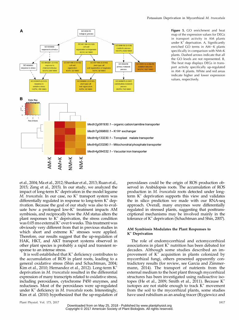
availability, including various transport systems,511 transcripts were regulated specifically in plant rootsunder AM2K conditions (Supplemental Fig. S5;Supplemental Table S5). A GO enrichment analysisrevealed an overrepresentation of genes involved inredox homeostasis, plant cell wall formation, and re-sponses to oxidative stress (Fig. 5A). Interestingly, fivetranscripts encoding for transport systems wereup-regulated in AM plants under K+ deprivation: anortholog of the plasma membrane K+/H+ exchangerAtCHX20 from Arabidopsis (Medtr7g099800.1), a pu-tative major facilitator superfamily (MFS) transporter(Medtr2g081930.1), a mitochondrial phosphate trans-porter (Medtr6g033280.1), and vacuolar malate(Medtr4g133230.1) and ion (Medtr4g094332.1) trans-porters (Fig. 5B; Supplemental Fig. S6). Among the30 most DEGs in AM2K plants (Table I), a clade Atype 2C protein phosphatase (Medtr5g009370.1) wasexpressed and could be involved in the posttranslationalregulation of K+ transport systems. These results suggestthat AM symbiosis activates specific mechanisms to tol-erate long-term K+ deprivation, including transportmechanisms to likely facilitate the acquisition of K+.
Identification of Putative Regulators for the Long-TermAdaptation of M. truncatula to K+ Deprivation in AM andNM Roots
We sought to identify regulatory networks associatedwith the AM and K+ DEG sets described previously(Supplemental Fig. S4) by comparing them with our
Figure 2. Transcriptional profiling ofM. truncatula roots under K+ deprivation.A, Venn diagram showing the number ofM. truncatula root genes up-regulated(yellow) and down-regulated (green) inresponse to K+ deprivation, based on q ,0.05 and a 1.5-fold change cutoff thresh-old. B, Significantly enriched GO molec-ular function terms for up-regulated (yellow)and down-regulated (green) genes analyzedwith the AgriGO Web-based tool (http://bioinfo.cau.edu.cn/agriGO/analysis.php).
1814 Plant Physiol. Vol. 173, 2017
Garcia et al.
www.plantphysiol.orgon May 25, 2018 - Published by Downloaded from Copyright © 2017 American Society of Plant Biologists. All rights reserved.

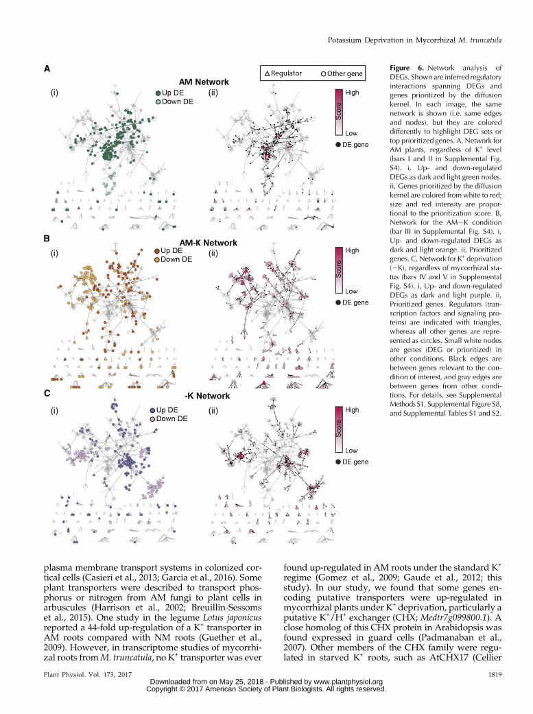
global M. truncatula regulatory network (Marx et al.,2016). This network was inferred using the MERLIN al-gorithm (Roy et al., 2013). We defined five condition-specific gene sets, each corresponding to a bar inSupplemental Figure S4 and Supplemental Table S5. Toassess the connectivity patterns, we overlaid the gene setson the regulatory network (Supplemental Methods S1).Figure 6 shows regulatory network connections involv-ing the DEGs and their prioritized network neighbors,summarized into three major categories: genes differen-tially expressed in AM roots regardless of K+ level (Fig.6Ai), genes specifically differentially expressed in theAM2K condition (Fig. 6Bi), and genes differentiallyexpressed under K+ deprivation regardless of AM status(Fig. 6Ci). Genes within each gene set were statisticallyenriched for direct interactions comparedwith randomlychosen genes, controlling for the degree (Z $ 7.09;Supplemental Table S6). Within each condition, geneswith the same direction of expression change (up- ordown-regulated) represented distinct network compo-nents (Fig. 6, i images) and had significantly shorter net-work distances than pairs of genes with oppositeexpression patterns (Mann-Whitney rank-sum test P ,0.001; Supplemental Fig. S7). This result indicates thatgenes thatwere induced represent distinct components of apathway compared with genes that were repressed. Dis-tinct regulatory systems are likely involved in controllingthe up- and down-regulated components of each DEG set.Two complementary network-based approaches
were used to identify important regulatory genes
associated with responses to AM and low-K+ condi-tions: diffusion kernel-based prioritization and statis-tical enrichment of targets of regulators in DEG sets.While the diffusion kernel approach can prioritize anygene in the network, the gene set enrichment analysisfocuses on regulatory proteins such as transcriptionfactors and signaling proteins. The diffusion kernel-based approach identified several candidate genesfromAM roots putatively involved in the response to K+
deprivation (Fig. 6Bii; Table II). For example, among thetop 25 predicted genes for the AM2KDEG set were ninekinases, including a calcineurin B-like (CBL)-interactingkinase (Medtr2g105010.1) and an ethylene responsefactor (Medtr4g078710.1; Table II). Full prioritizationresults for all gene sets are available in SupplementalTable S7.
Statistical enrichment of regulatory targets in the DEGsets identified 56 putative regulators (SupplementalTable S7; Supplemental Fig. S8; Supplemental MethodsS1) that included both new and known proteins such asMYB, BHLH, and AP2/ERF transcription factors as wellas chromatin-modifying enzymes. For example, targetsfrom a CBL-interacting kinase (Medtr2g105010.1; ninetargets), a Cys desulfurase (Medtr8g093560.1; seven tar-gets), and a GATA transcription factor (Medtr3g109760.1;24 targets) were enriched for up-regulated genes in re-sponse to K+ deprivation in both AM2K and NM2Kconditions, suggesting their involvement in the toleranceto low-K+ stress. Similarly, targets from a dehydration-responsive element-binding protein (Medtr1g019110.1;
Figure 3. Clustering and heat map of the expres-sion values for differentially expressed genes(DEGs) in NM plants under K+ deprivation. Theheat map displays DEGs identified previously inNM plants at low K+ (Fig. 2; Supplemental TableS1). An important part of the transcriptional re-sponses of M. truncatula roots under K+ depriva-tion was altered during mycorrhizal association.White and red areas indicate higher and lowerexpression values, respectively.
Plant Physiol. Vol. 173, 2017 1815
Potassium Deprivation in Mycorrhizal M. truncatula
www.plantphysiol.orgon May 25, 2018 - Published by Downloaded from Copyright © 2017 American Society of Plant Biologists. All rights reserved.

six targets), a C2H2-type zinc finger protein(Medtr3g102980.1; three targets), and a leucine-rich re-peat (LRR) receptor-like kinase (Medtr8g014970.1;10 targets) were specifically enriched for up-regulatedgenes in the AM2K condition. Finally, 29 and 17 targetsfrom a cyclin-dependent kinase (Medtr8g092290.1) anda Tyr kinase (Medtr7g116650.1), respectively, wereenriched for up-regulated genes in AM roots regard-less of extracellular K+ level, suggesting a major role ofthese proteins in the regulation of AM symbiosis.
Interestingly, the regulators identified by bothmethods were consistent with each other. The 56 regu-lators identified by the enrichment analysis receiveddiffusion kernel percentile ranks of 95 or greater com-pared with all ranked transcription factors and signal-ing proteins in the regulatory network. Each methodalso was able to identify some unique prioritized genesthat the other would miss or rank lower. Overall, pri-oritization and regulator enrichment approachesrevealed promising candidates for further functionalanalyses of the regulatory network controlling
responses to changes in AM fungi and K+ availability inM. truncatula.
DISCUSSION
K+ Deprivation Affects Growth, K+ Acquisition, and GeneExpression in M. truncatula Roots
K+ is an essential macronutrient for plants. Its limita-tion contributes to slowing down many vital processes.Plants elicit rapid responses to strong K+ deprivation byinducing plasma membrane hyperpolarization and theup-regulation of high-affinity transporters (Schachtmanand Shin, 2007). Prolonged K+ deficiency has beenreported previously to limit root development(Armengaud et al., 2004; Shin and Schachtman, 2004;Kellermeier et al., 2013), which was observed in our ex-periments (Fig. 1). Transcriptional analysis of plants fac-ing short-term and extreme K+ deprivation revealed anincrease in the expression of K+ transporters and channelsfrom the HAK, HKT, and AKT families (Armengaud
Figure 4. Impact of AM symbiosis and K+ depri-vation on the production of ROS inM. truncatula.A, ROS fluorescence images are shown in AM andNM plants at high (+K) and low (2K) K+ levels.Green pseudocolor indicates ROS production.Bars = 250 mm. B, Quantified data from threerepresentative images taken from six plants percondition. ROS were visualized by staining theroots of plants inoculated and watered with high-or low-K+ solutions for 6 weeks with 20 mM
carboxy-H2DFFDA (DFFDA). **, P , 0.01.
1816 Plant Physiol. Vol. 173, 2017
Garcia et al.
www.plantphysiol.orgon May 25, 2018 - Published by Downloaded from Copyright © 2017 American Society of Plant Biologists. All rights reserved.
et al., 2004;Ma et al., 2012; Shankar et al., 2013; Ruan et al.,2015; Zeng et al., 2015). In our study, we analyzed theimpact of long-term K+ deprivation in the model legumeM. truncatula. In our case, no K+ transport system wasdifferentially regulated in response to long-term K+ dep-rivation. Because the goal of our study was also to eval-uate how a prolonged low-K+ treatment impacts AMsymbiosis, and reciprocally how the AM status alters theplant responses to K+ deprivation, the stress conditionwas 0.05mMexternalK+ over 6weeks. This treatmentwasobviously very different from that in previous studies inwhich short and extreme K+ stresses were applied.Therefore, our results suggest that the up-regulation ofHAK, HKT, and AKT transport systems observed inother plant species is probably a rapid and transient re-sponse to an intense stress.It is well established that K+ deficiency contributes to
the accumulation of ROS in plant roots, leading to ageneral oxidative stress (Shin and Schachtman, 2004;Kim et al., 2010; Hernandez et al., 2012). Long-term K+
deprivation in M. truncatula resulted in the differentialexpression of many transcripts related to oxidative stress,including peroxidases, cytochrome P450 enzymes, andreductases. Most of the peroxidases were up-regulatedunder K+ deficiency in M. truncatula roots. Interestingly,Kim et al. (2010) hypothesized that the up-regulation of
peroxidases could be the origin of ROS production ob-served in Arabidopsis roots. The accumulation of ROSproduction in M. truncatula roots detected under long-term K+ deprivation supports this view and validatesthe in silico prediction we made with our RNA-seqapproach. Overall, many enzymes were differentiallyregulated in stressed plants, suggesting that posttrans-criptional mechanisms may be involved mainly in thetolerance of K+ deprivation (Schachtman and Shin, 2007).
AM Symbiosis Modulates the Plant Responses toK+ Deprivation
The role of endomycorrhizal and ectomycorrhizalassociations in plant K+ nutrition has been debated fordecades. Although some studies described an im-provement of K+ acquisition in plants colonized bymycorrhizal fungi, others presented apparently con-tradictory results (for review, see Garcia and Zimmer-mann, 2014). The transport of nutrients from theexternal medium to the host plant through mycorrhizalstructures has been investigated using radioactive iso-topes (He et al., 2009; Smith et al., 2011). Because K+
isotopes are not stable enough to track K+ movementfrom the soil to the mycorrhizal plants, some studieshave used rubidium as an analog tracer (Rygiewicz and
Figure 5. GO enrichment and heatmap of the expression values for DEGsin transport activity in AM plantsunder K+ deprivation. A, Significantlyenriched GO terms in AM2K plantsspecifically in comparison with NM+Kplants. Dashed arrows indicate that allthe GO levels are not represented. B,The heat map displays DEGs in trans-port activity specifically up-regulatedin AM2K plants. White and red areasindicate higher and lower expressionvalues, respectively.
Plant Physiol. Vol. 173, 2017 1817
Potassium Deprivation in Mycorrhizal M. truncatula
www.plantphysiol.orgon May 25, 2018 - Published by Downloaded from Copyright © 2017 American Society of Plant Biologists. All rights reserved.

Bledsoe, 1984; Hawkes and Casper, 2002). Further ex-perimentswill be needed inM. truncatula using rubidiumto evaluate the actual transport of K+ from the soil to themycorrhizal plants. However, in a previous publicationon ectomycorrhizae, we demonstrated an improvementof maritime pine (Pinus pinaster) K+ nutrition when col-onized by an ectomycorrhizal fungus only under K+
deprivation (Garcia et al., 2014). In this study, the shootK+ content of M. truncatula also was significantly higherin AM plants compared with NM plants but only at lowK+ levels and after 6 weeks of coculture. This effectshould be investigated in other plant species. We canassume that under K+-sufficient conditions, host plantscan acquire external K+ by themselves. However, whenK+ ions in solution have been limiting for a relatively longperiod of time, AM fungi may help their hosts to acquireK+ from the soil. Our analysis revealed thatM. truncatulaplants colonized by R. irregularis displayed increases ofbiomass and K+ ion content as well as specific tran-scriptional responses to the low-K+ regime. These resultsconfirmed that AM symbiosis was able to provide spe-cific adaptation mechanisms to the host plant to toleratelong-term K+ deprivation.
ROS have been detected in legume roots colonized byboth AM fungi and nitrogen-fixing bacteria, particu-larly in colonized cells (Salzer et al., 1999; Fester andHause, 2005; Puppo et al., 2013). Moreover, some
NADPH oxidase-encoding genes were recently found tobe up-regulated in colonized cortical cells ofM. truncatularoots, suggesting their role in arbuscule development(Belmondo et al., 2016a, 2016b). Our results revealed thatthe two-thirds of M. truncatula root transcriptional re-sponses to low K+ were absent in mycorrhizal plants,including many enzymes putatively involved in ROSproduction. The ROS accumulation observed in NMroots was correlated with the external K+ availability butnot with the AM symbiosis, suggesting specific ROSproduction in response to K+ deprivation. This accumu-lation was reduced significantly in AM plants at low K+,indicating that AM symbiosis helped M. truncatula tocope with long-term K+ deprivation.
Also, NM plants accumulated much more sodium inshoots than AM plants. Sodium can be toxic to the cellsat a high level. By preventing its accumulation, AMsymbiosis may protect M. truncatula from salt stress bybuffering the uptake of sodium. Although the role ofAM associations on salt tolerance was suggested inother plants (e.g. basil [Ocimum basilicum; Zuccarini andOkurowska, 2008], olive [Olea europaea; Porras-Sorianoet al., 2009], and maize [Estrada et al., 2013]), furtherexperiments will be needed to unravel the molecularmechanisms of this adaptation in M. truncatula.
The uptake of nutrients from inner symbiotic struc-tures requires the specific expression and regulation of
Table I. List of the top 30 transcripts specifically up-regulated in mycorrhizal plants under K+ deprivation (AM2K condition)
Gene Identifier Locus Name in Mt4.0v1 Mean Read No. Log2 Ratio, AM2K Versus NM2K
Medtr5g0113400.1 Subtilase family protein 11.207 8.4835Medtr1g0907370.1 OPC-8:0 CoA ligase1 4.84641 6.855Medtr1g0907070.1 SKU5 similar3 5.20123 6.31504Medtr2g0635600.1 HSP20-like chaperones superfamily protein 1.07509 2.86477Medtr1g0872000.1 Adenine nucleotide a-hydrolase-like superfamily protein 1.64562 2.64534Medtr4g0112300.1 Late embryogenesis abundant protein (LEA) family protein 5.10893 2.4046Medtr3g4669800.1 AGAMOUS-like80 1.85953 2.27527Medtr8g0429000.1 Root hair specific12 0.816833 2.14694Medtr4g0291900.1 Peroxidase superfamily protein 3.51445 2.10615Medtr3g0888450.1 Thiamine diphosphate-binding fold superfamily protein 0.939511 2.02649Medtr7g1082500.1 Thioredoxin superfamily protein 51.2384 1.97414Medtr6g0883200.1 Xyloglucan endotransglucosylase/hydrolase26 0.766958 1.87461Medtr5g0258000.1 Unknown protein 3.00168 1.75596Medtr7g0920900.1 Unknown protein 1.44917 1.73762Medtr1g0834400.1 Dormancy/auxin-associated family protein 22.4113 1.72544Medtr4g1024500.1 Expansin A7 0.911692 1.69244Medtr1g0237000.1 Cytochrome P450, family 83, subfamily B, polypeptide 1 1.00486 1.68266Medtr3g0850200.1 Protein of unknown function (DUF3339) 5.66561 1.65014Medtr7g1012700.1 Unknown protein 4.36525 1.64898Medtr6g0846400.1 Cold-regulated47 4.58727 1.6426Medtr1g0987600.1 Unknown protein 2.99537 1.6257Medtr4g0990100.1 Plant invertase/pectin methylesterase inhibitor superfamily protein 2.63717 1.61548Medtr7g1099200.1 Galactinol synthase1 0.0000541 1.60318Medtr3g1029700.1 Unknown protein 1.68067 1.56649Medtr1g1108700.1 B-box-type zinc finger protein with CCT domain 0.713362 1.55694Medtr4g0551700.1 HXXXD-type acyltransferase family protein 10.5731 1.55133Medtr4g0639400.1 Protein kinase1B 1.34305 1.54197Medtr2g4377700.1 Peroxidase superfamily protein 3.42251 1.53209Medtr5g0093700.1 Highly ABA-induced PP2C gene2 1.25488 1.5162Medtr2g0702000.1 Glutathione S-transferase TAU25 1.10787 1.4356
1818 Plant Physiol. Vol. 173, 2017
Garcia et al.
www.plantphysiol.orgon May 25, 2018 - Published by Downloaded from Copyright © 2017 American Society of Plant Biologists. All rights reserved.
plasma membrane transport systems in colonized cor-tical cells (Casieri et al., 2013; Garcia et al., 2016). Someplant transporters were described to transport phos-phorus or nitrogen from AM fungi to plant cells inarbuscules (Harrison et al., 2002; Breuillin-Sessomset al., 2015). One study in the legume Lotus japonicusreported a 44-fold up-regulation of a K+ transporter inAM roots compared with NM roots (Guether et al.,2009). However, in transcriptome studies of mycorrhi-zal roots fromM. truncatula, no K+ transporter was ever
found up-regulated in AM roots under the standard K+
regime (Gomez et al., 2009; Gaude et al., 2012; thisstudy). In our study, we found that some genes en-coding putative transporters were up-regulated inmycorrhizal plants under K+ deprivation, particularly aputative K+/H+ exchanger (CHX; Medtr7g099800.1). Aclose homolog of this CHX protein in Arabidopsis wasfound expressed in guard cells (Padmanaban et al.,2007). Other members of the CHX family were regu-lated in starved K+ roots, such as AtCHX17 (Cellier
Figure 6. Network analysis ofDEGs. Shown are inferred regulatoryinteractions spanning DEGs andgenes prioritized by the diffusionkernel. In each image, the samenetwork is shown (i.e. same edgesand nodes), but they are coloreddifferently to highlight DEG sets ortop prioritized genes. A, Network forAM plants, regardless of K+ level(bars I and II in Supplemental Fig.S4). i, Up- and down-regulatedDEGs as dark and light green nodes.ii, Genes prioritized by the diffusionkernel are colored fromwhite to red;size and red intensity are propor-tional to the prioritization score. B,Network for the AM2K condition(bar III in Supplemental Fig. S4). i,Up- and down-regulated DEGs asdark and light orange. ii, Prioritizedgenes. C, Network for K+ deprivation(2K), regardless of mycorrhizal sta-tus (bars IV and V in SupplementalFig. S4). i, Up- and down-regulatedDEGs as dark and light purple. ii,Prioritized genes. Regulators (tran-scription factors and signaling pro-teins) are indicated with triangles,whereas all other genes are repre-sented as circles. Small white nodesare genes (DEG or prioritized) inother conditions. Black edges arebetween genes relevant to the con-dition of interest, and gray edges arebetween genes from other condi-tions. For details, see SupplementalMethods S1, Supplemental Figure S8,and Supplemental Tables S1 and S2.
Plant Physiol. Vol. 173, 2017 1819
Potassium Deprivation in Mycorrhizal M. truncatula
www.plantphysiol.orgon May 25, 2018 - Published by Downloaded from Copyright © 2017 American Society of Plant Biologists. All rights reserved.

et al., 2004). As a consequence, further analyses will berequired in M. truncatula to determine if and how thisCHX can transport K+ during AM symbiosis underlong-term K+ deprivation.
Network Analysis Identified Putative RegulatorsControlling the Tolerance of Mycorrhizal Roots toK+ Deprivation
TheMERLIN algorithm (Roy et al., 2013) was used toinfer anM. truncatula regulatory network from publiclyavailable gene expression data representing a wide ar-ray of conditions and tissues. Such a network analysisallows us to identify regulatory connections betweenDEGs but also to predict which specific regulators(signaling proteins and transcription factors) may con-trol the expression of genes of interest. In our study, thisapproach identified differentially regulated genes andprioritized putative regulators that could be involved incontrolling the tolerance of AM plants to K+ depriva-tion. Among the top 30 up-regulated genes in AM2Kroots, a clade A type 2C protein phosphatase(Medtr5g009370.1) was identified. This gene encodesan ortholog of AtPP2CA in Arabidopsis involved in theregulation of the weak-rectifying K+ channel AKT2(Chérel et al., 2002) as well as of the uptake and effluxShaker K+ channels AKT1 and GORK, respectively(Lee et al., 2007; Lan et al., 2011; Lefoulon et al., 2016).Both prioritization and target enrichment analyses
suggested the importance of a CBL-interacting ki-nase (Medtr2g105010.1) in AM2K plant roots. CBL-interacting kinases are major regulators of nutrienttransporters, abscisic acid responses, and K+ homeo-stasis (D’Angelo et al., 2006; Ho et al., 2009; Hashimotoet al., 2012; Liu et al., 2013; Ragel et al., 2015). This ob-servation suggests a role of Medtr2g105010.1 in theposttranslational regulation of transport systems inM. truncatula roots colonized by AM fungi to promotethe symbiotic acquisition of K+. The targets of regula-tors involved in plant adaptation to oxidative stress alsowere predicted in AM2K roots and included a Cysdesulfurase (Medtr8g093560.1) and a dehydration-responsive element-binding protein (Medtr1g019110).Dehydration-responsive element-binding proteins areinvolved in salt and hydric stresses, which are conse-quences of K+ deprivation in plants, and the over-expression of one of them in Arabidopsis (AtDREBP2C)improved its tolerance to global oxidative stress(Hwang et al., 2012).
Altogether, our study revealed that AM symbiosisallows host plants to cope with long-term K+ depriva-tion. This tolerance mechanism probably involves theregulation of a gene network to protect host plantsagainst oxidative stress but also mechanisms to facili-tate K+ uptake from the soil through symbiotic struc-tures. Further studies will be needed to validate thesepredictions and to fully decipher the role of AM asso-ciations in plant K+ nutrition.
Table II. Top prioritized genes for AM plants in K+ deprivation
Shown are the top 25 prioritized genes for the AM2K DEG set. Prioritization (Rank and Score) columns give the rank and score from the diffusionkernel analysis. Target Enrichment indicates whether a prioritized gene in this list also has target enrichment in a DEG set; nonregulators are des-ignated NA in this column.
Gene Identifier Locus Name in Mt4.0v1 Rank (AM2K) Score (AM2K) Target Enrichment
Medtr3g115620.1 Wuschel-related homeobox protein 1 0.634459 AM2K, downMedtr7g005400.1 Somatic embryogenesis receptor kinase-like protein 2 0.578335 AM2K, downMedtr2g087090.1 Ser/Thr-kinase Nek4 3 0.497066 AM2K, downMedtr2g089440.1 S-locus lectin kinase family protein 4 0.454545Medtr4g078710.1 Ethylene response factor 5 0.396948Medtr4g058015.1 Electron transporter, putative 6 0.389796 AM2K, downMedtr1g080210.1 Light-dependent short-hypocotyl protein 7 0.389508 AM2K, downMedtr1g097580.1 LRR receptor-like kinase 8 0.369039Medtr5g014640.1 bHLH DNA-binding family protein 9 0.352642Medtr4g133938.1 Nuclear transcription factor Y protein 10 0.328559 AM2K, upMedtr2g105010.1 CBL-interacting kinase 11 0.326467 2K, upMedtr1g107460.1 LRR receptor-like kinase family protein 12 0.321412Medtr3g115500.1 Receptor Ser/Thr kinase 12 0.321412Medtr5g012490.1 Eukaryotic aspartyl protease family protein 12 0.321412Medtr4g128990.1 Receptor-like kinase 15 0.304361 AM2K, downMedtr7g084000.1 Light-regulated protein, putative 16 0.298936 AM2K, upMedtr3g097150.1 Aspartic proteinase nepenthesin 17 0.29324 AM2K, downMedtr5g014520.1 bHLH DNA-binding family protein 18 0.291187Medtr3g069590.1 RHO guanyl-nucleotide exchange factor 19 0.28769 AM2K, downMedtr4g117040.1 Cys-rich RLK (receptor-like kinase) protein 20 0.283186Medtr2g087390.1 FAF-like protein 21 0.279036 NAMedtr2g087430.1 DUF3049 family protein 21 0.279036 NAMedtr4g068780.1 CASP-like protein 21 0.279036 NAMedtr8g027040.1 Cytochrome P450 family 78 protein 21 0.279036 NAMedtr8g008820.1 Receptor-like kinase plant 25 0.273366
1820 Plant Physiol. Vol. 173, 2017
Garcia et al.
www.plantphysiol.orgon May 25, 2018 - Published by Downloaded from Copyright © 2017 American Society of Plant Biologists. All rights reserved.

MATERIALS AND METHODS
Plant Growth Conditions, Fungal Inoculation, andLong-Term K+ Deficiency Treatment
Medicago truncatula ‘Jemalong A17’ seeds were acid scarified and surfacesterilized, plated on 1% (w/v) agar supplemented with 1 mg mL21 GA3, ver-nalized at 4°C for 4 d, and allowed to germinate overnight at room temperature.The germinated seedlings were placed for 2 weeks on modified Fahräeus me-dium as described previously (Catoira et al., 2000). The plants were transferredto pots filled with Turface and inoculated with 400 spores of Rhizophagusirregularis. Plants were watered regularly with standard (+K; 3.75 mM K+) LongAshton solution [3.75 mM KNO3, 2 mM Ca(NO3)2$4H2O, 7.5 mM NaH2PO4$H2O,1 mM MgSO4$7H2O, 0.05 mM NaCl, 5 mM MnSO4, 0.5 mM CuSO4, 1 mM ZnSO4,16.5 mM H3BO3, and 0.1 mM Na2MoO4] or low-K+ (2K; 0.05 mM K+) solution[0.05 mM KNO3, 3.85 mM Ca(NO3)2$4H2O, 7.5 mM NaH2PO4$H2O, 1 mM
MgSO4$7H2O, 0.05 mMNaCl, 5 mM MnSO4, 0.5 mM CuSO4, 1 mM ZnSO4, 16.5 mM
H3BO3, and 0.1 mM Na2MoO4]. The plants were harvested after 6 weeks oftreatment.
Mycorrhizal Quantification, Dry Weight Determination,and Ion Content in Plants
Shoots and roots of 6-week-oldM. truncatula plants growing in K+-sufficientor K+-deficient conditions were harvested separately. Mycorrhizal colonizationrates were assessed using the gridline intersection method on roots stainedpreviously with Sheaffer ink (McGonigle et al., 1990). Fresh weight was de-termined, and roots and shoots were dried at 60°C for 1 week for dry weightdetermination. The dried samples were sent to the Soil & Forage AnalysisLaboratory (University of Wisconsin-Madison) to determine the K+, sodium,phosphorus, and calcium contents in plant tissues by ICP-OES.
RNA Isolation and Sequencing
Frozen root tissues were ground in liquid nitrogen, and total RNA wasextracted from the resulting powder using the PureLink RNAMini Kit (ThermoFisher) and treated with TURBO DNase (Thermo Fisher). RNA quality wasassessed using 2100 BioAnalyzer technology (Agilent Technologies). Six mi-crogramsof total RNAwasused to construct poly(A) selection libraries using theIllumina TruSeq RNA Sample Preparation kit; 100-nucleotide single-end readswere obtained on an Illumina HiSeq2000 platform using TruSeq version3 (University of Wisconsin Biotechnology Center DNA Sequencing Facility).
RNA-seq Analysis, Annotations, Cutoff, and Analysis
RNA-seq data were processed to obtain DEGs using the TopHat2-SE, Cuf-flinks2, Cuffmerge2, and Cuffdiff2 applications from the iPlant Collaborativeplatform (http://www.iplantcollaborative.org). The DESeq Bioconductorpackage version 3.3 for R (Anders andHuber, 2010) was used for the analysis ofDEGs. Cuffdiff2 sequencing data were analyzed using the cummeRbundpackage to generate principal component and multidimensional scaling anal-yses on biological and technical replicates. K-means clustering also was per-formed using the cummeRbund package for Rwith K = 25. For further analyses,DEGs were called by DESeq using a 1.5-fold change cutoff threshold and falsediscovery rate-adjusted q , 0.05. DEGs were annotated with the Mt4.0v1 ver-sion of the M. truncatula ‘Jemalong A17’ genome (http://jcvi.org/medicago/index.php). GO enrichment analysis was conducted with the AgriGO Web-based tool (http://bioinfo.cau.edu.cn/agriGO/analysis.php) using theMt4.0v1 version of the M. truncatula genome. The Venn diagram was obtainedusing the Bioinformatics & Evolutionary Genomics Web tool (http://bioinformatics.psb.ugent.be/webtools/Venn/). Finally, clustering and heatmaps were generated on R with scripts based on the heatmap.2 function asavailable in the gplots Bioconductor package. The data discussed in this articlehave been deposited in the National Center for Biotechnology Informa-tion’s Gene Expression Omnibus (Edgar et al., 2002) and are accessiblethrough the Gene Expression Omnibus Series accession number GSE94266(https://www.ncbi.nlm.nih.gov/geo/query/acc.cgi?acc=GSE94266).
ROS Detection and Measurement
To detect ROS, the root samples of AM and NM 6-week-old plants grownunder K+-sufficient or K+-deficient conditions were incubated with 20 mM
carboxy-H2DFFDA (Thermo Fisher) for 20 min in high- or low-K+ solution,respectively. The roots were washed twice with high- or low-K+ solution beforemicroscopy visualization. All fluorescence images were captured using a LeicaDMi8 microscope and a Leica DFC365 FX camera. ROS fluorescence wasquantified and converted into pseudocolor images using the ImageJ softwareprogram. Background noise was subtracted from the fluorescence intensityvalue for quantification.
Regulatory Network Analysis and Visualization
The base network visualized in all parts of Figure 6 was selected from aglobal inferred M. truncatula regulatory network (Marx et al., 2016) byextracting any interaction between a DEG and top n computationally prior-itized genes for each K+ and AM condition, where n is the size of the exper-imentally identified DEG set. For visual simplicity, connected componentsinvolving only one edge were omitted. Network images were created usingCytoscape (Shannon et al., 2003). For full details of the regulatory networkanalyses, including prioritization and regulator gene set enrichment, seeSupplemental Methods S1.
Accession Numbers
Sequence data from this article can be found in the GenBank/EMBL datalibraries under the accession numbers GSE94266.
Supplemental Data
The following supplemental materials are available.
Supplemental Figure S1. Principal component analysis of the changes intranscript abundance in AM and NM M. truncatula roots under high-and low-K+ regimes.
Supplemental Figure S2. Multidimensional scaling plot showing the two-dimensional distribution of the samples.
Supplemental Figure S3. K-means cluster analysis of DEGs ofM. truncatularoots under high- and low-K+ regimes.
Supplemental Figure S4. Impact of AM symbiosis on the transcriptionalprofiling of M. truncatula roots under K+ deprivation.
Supplemental Figure S5. Representative clustering and heat map of ex-pression values for DEG in transport activity specifically regulated byAM symbiosis independently of K+ availability.
Supplemental Figure S6. Phylogenetic analysis of the cation/H+ exchangerMedtr7g099800.1.
Supplemental Figure S7. Network connectivity statistics suggest differen-tial regulation for induced and repressed gene sets.
Supplemental Figure S8. Predicted regulators of mycorrhizal and low-K+
gene sets represent relevant pathways and propose candidates for futurestudy.
Supplemental Table S1. List of up- and down-regulated transcripts in NMM. truncatula roots under K+ deprivation.
Supplemental Table S2. List of differentially expressed oxidativestress-related transcripts in NM M. truncatula roots under K+
deprivation.
Supplemental Table S3. List of differentially expressed hydrolase tran-scripts in NM M. truncatula roots under K+ deprivation.
Supplemental Table S4. List of transcripts categorized in the 25 clusters ofthe K-means cluster analysis (Supplemental Fig. S4).
Supplemental Table S5. List of transcripts differentially regulated inM. truncatula roots in comparison with the NM2K condition.
Supplemental Table S6. Edge density analysis results for input gene sets.
Supplemental Table S7.Genes ranked by diffusion kernel and enrichment-based prioritization analyses.
Supplemental Methods S1. Regulatory network-based interpretation andprioritization of DEGs.
Plant Physiol. Vol. 173, 2017 1821
Potassium Deprivation in Mycorrhizal M. truncatula
www.plantphysiol.orgon May 25, 2018 - Published by Downloaded from Copyright © 2017 American Society of Plant Biologists. All rights reserved.

ACKNOWLEDGMENTS
We thank Dr. Sabine Zimmermann for constructive comments on the articleand the University of Wisconsin Biotechnology Center DNA SequencingFacility for providing RNA-seq facilities and services.
Received December 22, 2016; accepted January 31, 2017; published February 3,2017.
LITERATURE CITED
Adams E, Shin R (2014) Transport, signaling, and homeostasis of potas-sium and sodium in plants. J Integr Plant Biol 56: 231–249
Anders S, Huber W (2010) Differential expression analysis for sequencecount data. Genome Biol 11: R106
Apel K, Hirt H (2004) Reactive oxygen species: metabolism, oxidativestress, and signal transduction. Annu Rev Plant Biol 55: 373–399
Armengaud P, Breitling R, Amtmann A (2004) The potassium-dependenttranscriptome of Arabidopsis reveals a prominent role of jasmonic acidin nutrient signaling. Plant Physiol 136: 2556–2576
Asher C, Ozanne P (1967) Growth and potassium content of plants in so-lution cultures maintained at constant potassium concentrations. Soil Sci103: 155–161
Baslam M, Garmendia I, Goicoechea N (2013) The arbuscular mycorrhizalsymbiosis can overcome reductions in yield and nutritional quality ingreenhouse-lettuces cultivated at inappropriate growing seasons. SciHortic (Amsterdam) 164: 145–154
Belmondo S, Calcagno C, Genre A, Puppo A, Pauly N, Lanfranco L(2016a) The Medicago truncatula MtRbohE gene is activated in arbuscu-lated cells and is involved in root cortex colonization. Planta 243: 251–262
Belmondo S, Calcagno C, Genre A, Puppo A, Pauly N, Lanfranco L(2016b) NADPH oxidases in the arbuscular mycorrhizal symbiosis. PlantSignal Behav 11: e1165379
Benito B, Haro R, Amtmann A, Cuin TA, Dreyer I (2014) The twins K+ andNa+ in plants. J Plant Physiol 171: 723–731
Breuillin-Sessoms F, Floss DS, Gomez SK, Pumplin N, Ding Y, Levesque-Tremblay V, Noar RD, Daniels DA, Bravo A, Eaglesham JB, et al(2015) Suppression of arbuscule degeneration in Medicago truncatulaphosphate transporter4 mutants is dependent on the ammonium trans-porter 2 family protein AMT2;3. Plant Cell 27: 1352–1366
Brundrett M (2004) Diversity and classification of mycorrhizal associations.Biol Rev Camb Philos Soc 79: 473–495
Casieri L, Ait Lahmidi N, Doidy J, Veneault-Fourrey C, Migeon A,Bonneau L, Courty PE, Garcia K, Charbonnier M, Delteil A, et al(2013) Biotrophic transportome in mutualistic plant-fungal interactions.Mycorrhiza 23: 597–625
Catoira R, Galera C, de Billy F, Penmetsa RV, Journet EP, Maillet F,Rosenberg C, Cook D, Gough C, Dénarié J (2000) Four genes of Med-icago truncatula controlling components of a nod factor transductionpathway. Plant Cell 12: 1647–1666
Cellier F, Conéjéro G, Ricaud L, Luu DT, Lepetit M, Gosti F, Casse F(2004) Characterization of AtCHX17, a member of the cation/H+ ex-changers, CHX family, from Arabidopsis thaliana suggests a role in K+
homeostasis. Plant J 39: 834–846Chérel I, Michard E, Platet N, Mouline K, Alcon C, Sentenac H, Thibaud
JB (2002) Physical and functional interaction of the Arabidopsis K+
channel AKT2 and phosphatase AtPP2CA. Plant Cell 14: 1133–1146Courty PE, Doidy J, Garcia K, Wipf D, Zimmermann SD (2016) The
transportome of mycorrhizal systems. In F Martin, ed, MolecularMycorrhizal Symbiosis. John Wiley & Sons, Hoboken, NJ, pp 239–256
D’Angelo C, Weinl S, Batistic O, Pandey GK, Cheong YH, Schültke S,Albrecht V, Ehlert B, Schulz B, Harter K, et al (2006) Alternativecomplex formation of the Ca-regulated protein kinase CIPK1 controlsabscisic acid-dependent and independent stress responses in Arabi-dopsis. Plant J 48: 857–872
Delaux PM, Radhakrishnan GV, Jayaraman D, Cheema J, Malbreil M,Volkening JD, Sekimoto H, Nishiyama T, Melkonian M, Pokorny L,et al (2015) Algal ancestor of land plants was preadapted for symbiosis.Proc Natl Acad Sci USA 112: 13390–13395
Drew MC, Nye PH (1969) The supply of nutrient ions by diffusion to plantroots in soil. Plant Soil 31: 407–424
Edgar R, Domrachev M, Lash AE (2002) Gene Expression Omnibus: NCBIgene expression and hybridization array data repository. Nucleic AcidsRes 30: 207–210
Estrada B, Aroca R, Maathuis FJM, Barea JM, Ruiz-Lozano JM (2013)Arbuscular mycorrhizal fungi native from a Mediterranean saline areaenhance maize tolerance to salinity through improved ion homeostasis.Plant Cell Environ 36: 1771–1782
Fester T, Hause G (2005) Accumulation of reactive oxygen species in ar-buscular mycorrhizal roots. Mycorrhiza 15: 373–379
Garcia K, Delteil A, Conéjéro G, Becquer A, Plassard C, Sentenac H,Zimmermann S (2014) Potassium nutrition of ectomycorrhizal Pinuspinaster: overexpression of the Hebeloma cylindrosporum HcTrk1 trans-porter affects the translocation of both K+ and phosphorus in the hostplant. New Phytol 201: 951–960
Garcia K, Doidy J, Zimmermann SD, Wipf D, Courty PE (2016) Take a tripthrough the plant and fungal transportome of mycorrhiza. Trends PlantSci 21: 937–950
Garcia K, Zimmermann SD (2014) The role of mycorrhizal associations inplant potassium nutrition. Front Plant Sci 5: 337
Gaude N, Bortfeld S, Duensing N, Lohse M, Krajinski F (2012) Arbuscule-containing and non-colonized cortical cells of mycorrhizal roots undergoextensive and specific reprogramming during arbuscular mycorrhizal de-velopment. Plant J 69: 510–528
Gomez SK, Javot H, Deewatthanawong P, Torres-Jerez I, Tang Y, Blan-caflor EB, Udvardi MK, Harrison MJ (2009) Medicago truncatula andGlomus intraradices gene expression in cortical cells harboring arbusculesin the arbuscular mycorrhizal symbiosis. BMC Plant Biol 9: 10
Govindarajulu M, Pfeffer PE, Jin H, Abubaker J, Douds DD, Allen JW,Bücking H, Lammers PJ, Shachar-Hill Y (2005) Nitrogen transfer in thearbuscular mycorrhizal symbiosis. Nature 435: 819–823
Guether M, Balestrini R, Hannah M, He J, Udvardi MK, Bonfante P (2009)Genome-wide reprogramming of regulatory networks, transport, cellwall and membrane biogenesis during arbuscular mycorrhizal symbio-sis in Lotus japonicus. New Phytol 182: 200–212
Harrison MJ (2005) Signaling in the arbuscular mycorrhizal symbiosis.Annu Rev Microbiol 59: 19–42
Harrison MJ, Dewbre GR, Liu J (2002) A phosphate transporter fromMedicago truncatula involved in the acquisition of phosphate released byarbuscular mycorrhizal fungi. Plant Cell 14: 2413–2429
Hashimoto K, Eckert C, Anschütz U, Scholz M, Held K, Waadt R, ReyerA, Hippler M, Becker D, Kudla J (2012) Phosphorylation of calcineurinB-like (CBL) calcium sensor proteins by their CBL-interacting proteinkinases (CIPKs) is required for full activity of CBL-CIPK complexes to-ward their target proteins. J Biol Chem 287: 7956–7968
Hawkes CV, Casper BB (2002) Lateral root function and root overlapamong mycorrhizal and nonmycorrhizal herbs in a Florida shrubland,measured using rubidium as a nutrient analog. Am J Bot 89: 1289–1294
He X, Xu M, Qiu GY, Zhou J (2009) Use of 15N stable isotope to quantifynitrogen transfer between mycorrhizal plants. J Plant Ecol 2: 107–118
Hernandez M, Fernandez-Garcia N, Garcia-Garma J, Rubio-Asensio JS,Rubio F, Olmos E (2012) Potassium starvation induces oxidative stressin Solanum lycopersicum L. roots. J Plant Physiol 169: 1366–1374
Ho CH, Lin SH, Hu HC, Tsay YF (2009) CHL1 functions as a nitrate sensorin plants. Cell 138: 1184–1194
Hwang JE, Lim CJ, Chen H, Je J, Song C, Lim CO (2012) Overexpression ofArabidopsis dehydration-responsive element-binding protein 2C con-fers tolerance to oxidative stress. Mol Cells 33: 135–140
Javot H, Penmetsa RV, Terzaghi N, Cook DR, Harrison MJ (2007) AMedicago truncatula phosphate transporter indispensable for the arbus-cular mycorrhizal symbiosis. Proc Natl Acad Sci USA 104: 1720–1725
Jin H, Liu J, Liu J, Huang X (2012) Forms of nitrogen uptake, translocation,and transfer via arbuscular mycorrhizal fungi: a review. Sci China LifeSci 55: 474–482
Kaldorf M, Kuhn AJ, Schröder WH, Hildebrandt U, Bothe H (1999) Se-lective element deposits in maize colonized by a heavy metal toleranceconferring arbuscular mycorrhizal fungus. J Plant Physiol 154: 718–728
Kellermeier F, Chardon F, Amtmann A (2013) Natural variation of Ara-bidopsis root architecture reveals complementing adaptive strategies topotassium starvation. Plant Physiol 161: 1421–1432
Kim MJ, Ciani S, Schachtman DP (2010) A peroxidase contributes to ROSproduction during Arabidopsis root response to potassium deficiency.Mol Plant 3: 420–427
Garcia et al.
1822 Plant Physiol. Vol. 173, 2017 www.plantphysiol.orgon May 25, 2018 - Published by Downloaded from
Copyright © 2017 American Society of Plant Biologists. All rights reserved.

Lan WZ, Lee SC, Che YF, Jiang YQ, Luan S (2011) Mechanistic analysis ofAKT1 regulation by the CBL-CIPK-PP2CA interactions. Mol Plant 4:527–536
Lee SC, Lan WZ, Kim BG, Li L, Cheong YH, Pandey GK, Lu G, BuchananBB, Luan S (2007) A protein phosphorylation/dephosphorylation net-work regulates a plant potassium channel. Proc Natl Acad Sci USA 104:15959–15964
Lefoulon C, Boeglin M, Moreau B, Véry AA, Szponarski W, Dauzat M,Michard E, Gaillard I, Chérel I (2016) The Arabidopsis AtPP2CA pro-tein phosphatase inhibits the GORK K+ efflux channel and exerts adominant suppressive effect on phosphomimetic-activating mutations.J Biol Chem 291: 6521–6533
Leigh RA, Wyn Jones RG (1984) A hypothesis relating critical potassiumconcentration for growth to the distribution and functions of this ion inthe plant cell. New Phytol 97: 1–13
Liu LL, Ren HM, Chen LQ, Wang Y, Wu WH (2013) A protein kinase,calcineurin B-like protein-interacting protein kinase9, interacts withcalcium sensor calcineurin B-like protein3 and regulates potassium ho-meostasis under low-potassium stress in Arabidopsis. Plant Physiol 161:266–277
López MF, Dietz S, Grunze N, Bloschies J, Weiss M, Nehls U (2008) Thesugar porter gene family of Laccaria bicolor: function in ectomycorrhizalsymbiosis and soil-growing hyphae. New Phytol 180: 365–378
Ma TL, Wu WH, Wang Y (2012) Transcriptome analysis of rice root re-sponses to potassium deficiency. BMC Plant Biol 12: 161
Marx H, Minogue CE, Jayaraman D, Richards AL, Kwiecien NW,Siahpirani AF, Rajasekar S, Maeda J, Garcia K, Del Valle-EchevarriaAR, et al (2016) A proteomic atlas of the legume Medicago truncatula and itsnitrogen-fixing endosymbiont Sinorhizobium meliloti. Nat Biotechnol 34:1198–1205
McGonigle TP, Miller MH, Evans DG, Fairchild GL, Swan JA (1990) Anew method which gives an objective measure of colonization of rootsby vesicular-arbuscular mycorrhizal fungi. New Phytol 115: 495–501
Moody PW, Bell MJ (2006) Availability of soil potassium and diagnosticsoil tests. Soil Res 44: 265–275
Oliveira RS, Rocha I, Ma Y, Vosátka M, Freitas H (2016) Seed coating witharbuscular mycorrhizal fungi as an ecotechnological approach for sus-tainable agricultural production of common wheat (Triticum aestivumL.). J Toxicol Environ Health A 79: 329–337
Padmanaban S, Chanroj S, Kwak JM, Li X, Ward JM, Sze H (2007) Par-ticipation of endomembrane cation/H+ exchanger AtCHX20 in osmo-regulation of guard cells. Plant Physiol 144: 82–93
Perner H, Schwarz D, Bruns C, Mäder P, George E (2007) Effect of ar-buscular mycorrhizal colonization and two levels of compost supply onnutrient uptake and flowering of pelargonium plants. Mycorrhiza 17:469–474
Porras-Soriano A, Soriano-Martín ML, Porras-Piedra A, Azcón R (2009)Arbuscular mycorrhizal fungi increased growth, nutrient uptake andtolerance to salinity in olive trees under nursery conditions. J PlantPhysiol 166: 1350–1359
Puppo A, Pauly N, Boscari A, Mandon K, Brouquisse R (2013) Hydrogenperoxide and nitric oxide: key regulators of the legume-rhizobium andmycorrhizal symbioses. Antioxid Redox Signal 18: 2202–2219
Ragel P, Ródenas R, García-Martín E, Andrés Z, Villalta I, Nieves-Cordones M, Rivero RM, Martínez V, Pardo JM, Quintero FJ, et al(2015) The CBL-interacting protein kinase CIPK23 regulates HAK5-mediatedhigh-affinity K+ uptake in Arabidopsis roots. Plant Physiol 169: 2863–2873
Römheld V, Kirkby EA (2010) Research on potassium in agriculture: needsand prospects. Plant Soil 335: 155–180
Roy S, Lagree S, Hou Z, Thomson JA, Stewart R, Gasch AP (2013) Inte-grated module and gene-specific regulatory inference implicates up-stream signaling networks. PLOS Comput Biol 9: e1003252
Ruan L, Zhang J, Xin X, Zhang C, Ma D, Chen L, Zhao B (2015) Com-parative analysis of potassium deficiency-responsive transcriptomes inlow potassium susceptible and tolerant wheat (Triticum aestivum L.). SciRep 5: 10090
Rygiewicz PT, Bledsoe CS (1984) Mycorrhizal effects on potassium fluxesby northwest coniferous seedlings. Plant Physiol 76: 918–923
Salzer P, Corbière H, Boller T (1999) Hydrogen peroxide accumulation inMedicago truncatula roots colonized by the arbuscular mycorrhiza-forming fungus Glomus intraradices. Planta 208: 319–325
Schachtman DP, Shin R (2007) Nutrient sensing and signaling: NPKS.Annu Rev Plant Biol 58: 47–69
Scheloske S, Maetz M, Schneider T, Hildebrandt U, Bothe H, Povh B(2004) Element distribution in mycorrhizal and nonmycorrhizal roots ofthe halophyte Aster tripolium determined by proton induced x-rayemission. Protoplasma 223: 183–189
Shankar A, Singh A, Kanwar P, Srivastava AK, Pandey A, Suprasanna P,Kapoor S, Pandey GK (2013) Gene expression analysis of rice seedlingunder potassium deprivation reveals major changes in metabolism andsignaling components. PLoS ONE 8: e70321
Shannon P, Markiel A, Ozier O, Baliga NS, Wang JT, Ramage D, Amin N,Schwikowski B, Ideker T (2003) Cytoscape: a software environment forintegrated models of biomolecular interaction networks. Genome Res13: 2498–2504
Shin R, Schachtman DP (2004) Hydrogen peroxide mediates plant root cellresponse to nutrient deprivation. Proc Natl Acad Sci USA 101: 8827–8832
Smith SE, Jakobsen I, Grønlund M, Smith FA (2011) Roles of arbuscularmycorrhizas in plant phosphorus nutrition: interactions between path-ways of phosphorus uptake in arbuscular mycorrhizal roots have im-portant implications for understanding and manipulating plantphosphorus acquisition. Plant Physiol 156: 1050–1057
Smith SE, Read D (2008) Mycorrhizal Symbiosis, Ed 3. Academic Press,New York, NY
Spatafora JW, Chang Y, Benny GL, Lazarus K, Smith ME, Berbee ML,Bonito G, Corradi N, Grigoriev I, Gryganskyi A, et al (2016) A phylum-level phylogenetic classification of zygomycete fungi based on genome-scale data. Mycologia 108: 1028–1046
Wang B, Qiu YL (2006) Phylogenetic distribution and evolution of my-corrhizas in land plants. Mycorrhiza 16: 299–363
Wang C, Chen H, Hao Q, Sha A, Shan Z, Chen L, Zhou R, Zhi H, Zhou X(2012) Transcript profile of the response of two soybean genotypes topotassium deficiency. PLoS ONE 7: e39856
Wang Y, Wu WH (2013) Potassium transport and signaling in higherplants. Annu Rev Plant Biol 64: 451–476
Zeng Q, Ling Q, Fan L, Li Y, Hu F, Chen J, Huang Z, Deng H, Li Q, Qi Y(2015) Transcriptome profiling of sugarcane roots in response to lowpotassium stress. PLoS ONE 10: e0126306
Zörb C, Senbayram M, Peiter E (2014) Potassium in agriculture: status andperspectives. J Plant Physiol 171: 656–669
Zuccarini P, Okurowska P (2008) Effects of mycorrhizal colonization andfertilization on growth and photosynthesis of sweet basil under saltstress. J Plant Nutr 31: 497–513
Plant Physiol. Vol. 173, 2017 1823
Potassium Deprivation in Mycorrhizal M. truncatula
www.plantphysiol.orgon May 25, 2018 - Published by Downloaded from Copyright © 2017 American Society of Plant Biologists. All rights reserved.