physiologic changes of pregnancy
TRANSCRIPT

Physiologic changes of pregnancy
Nancy Thomas

ObjectivesSymptoms and physical findings of each organ systemPhysiologic versus pathologic changesDiagnostic tests and interpretations during physiological changes
Organ systemsCardiovascular systemPulmonary systemGenital tractUrinary systemEndocrine systemGastrointestinal TractSkin

Cardiovascular systemTotal Body water Cardiac Output

Total body waterIncreases 6-8 LIncreases by 40 %Normal body water
2/3 intracellular1/3 extracellular
¾ interstitial¼ intravasular
2/3 increase is extravascular
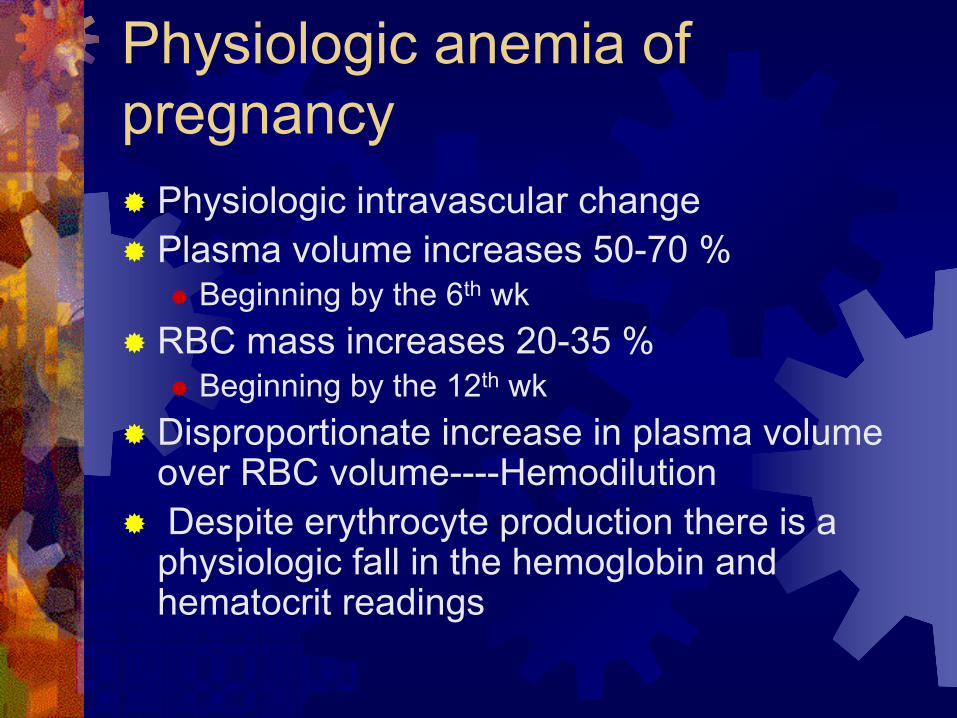
Physiologic anemia of pregnancy
Physiologic intravascular changePlasma volume increases 50-70 %
Beginning by the 6th wkRBC mass increases 20-35 %
Beginning by the 12th wkDisproportionate increase in plasma volume over RBC volume----Hemodilution Despite erythrocyte production there is a
physiologic fall in the hemoglobin and hematocrit readings

Patients without overt anemia & not given supplementation
deliv3rdTri
2nd
Tri1st
TriNonpregconcentration
27.614.722.297.463.0SerumFerritin
57.156.075.3106.590.0Serumiron
12.411.010.912.213.0HB
Williams 21edWide standard deviation
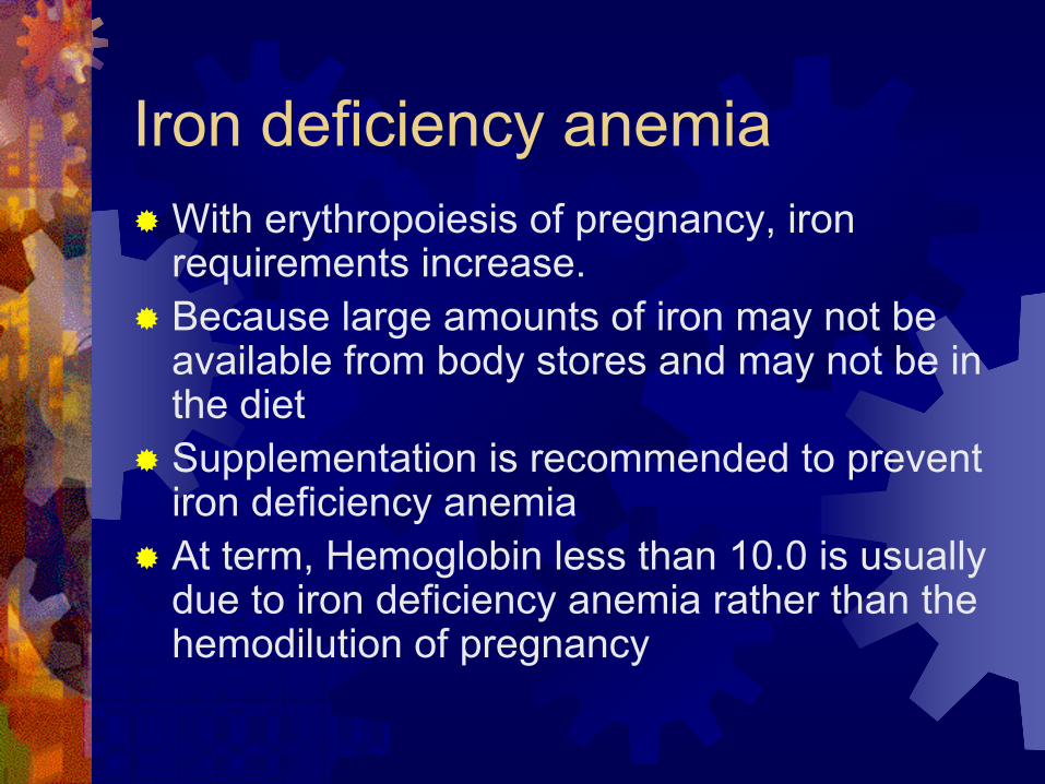
Iron deficiency anemiaWith erythropoiesis of pregnancy, iron requirements increase.Because large amounts of iron may not be available from body stores and may not be in the dietSupplementation is recommended to prevent iron deficiency anemiaAt term, Hemoglobin less than 10.0 is usually due to iron deficiency anemia rather than the hemodilution of pregnancy

Normal Iron RequirementsTotal body iron content average in normal adult females is 2gmIron requirement for normal pregnancy is 1 gm
200 mg is excreted300 mg is transferred to fetus500 mg is need for mom
Total volume of RBC inc is 450 ml1 ml of RBCs contains 1.1 mg of iron450 ml X 1.1 mg/ml = 500 mg
Daily average is 6-7 mg/daySmall intervals between pregnancies are most concerning

Cardiovascular systemTotal Body water Cardiac Output
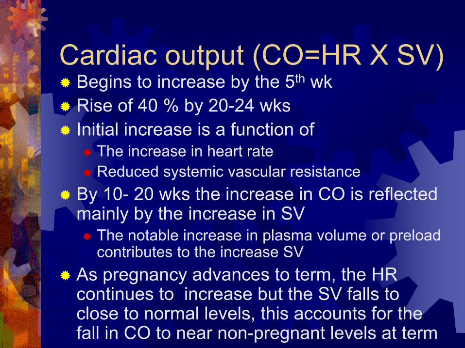
Cardiac output (CO=HR X SV)Begins to increase by the 5th wk Rise of 40 % by 20-24 wksInitial increase is a function of
The increase in heart rateReduced systemic vascular resistance
By 10- 20 wks the increase in CO is reflected mainly by the increase in SV
The notable increase in plasma volume or preload contributes to the increase SV
As pregnancy advances to term, the HR continues to increase but the SV falls to close to normal levels, this accounts for the fall in CO to near non-pregnant levels at term
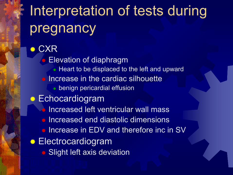
Interpretation of tests during pregnancy
CXRElevation of diaphragm
Heart to be displaced to the left and upwardIncrease in the cardiac silhouette
benign pericardial effusion
EchocardiogramIncreased left ventricular wall massIncreased end diastolic dimensionsIncrease in EDV and therefore inc in SV
ElectrocardiogramSlight left axis deviation

Respiratory systemMechanical
diaphragmConsumption
Increase in needed oxygenStimulation
Progesterone stimulation

RespiratoryMechanical
Diaphragm rises 4 cmLess negative intrathoracic pressureDec FRC-Functional Residual Capacity
volume after passive expirationDec ERV-Expiratory Reserve Volume
max volume expired after expirationDec RV-Residual Volume
volume after max expiration
No impairments in diaphragmatic or thoracic muscle motion
Lung compliance remains unaffected
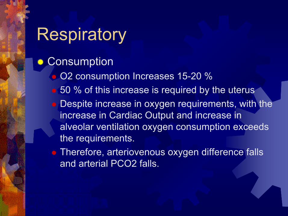
RespiratoryConsumption
O2 consumption Increases 15-20 % 50 % of this increase is required by the uterusDespite increase in oxygen requirements, with the increase in Cardiac Output and increase in alveolar ventilation oxygen consumption exceeds the requirements.Therefore, arteriovenous oxygen difference falls and arterial PCO2 falls.

RespiratoryStimulation
Progesterone is known to directly stimulate ventilationProgesterone increases the sensitivity of the respiratory centers to CO2 Also, it is thought to reduce total pulmonary resistance

RespiratoryMinute ventilation = RR X Tidal volumeTidal Volume-increases
Volume of air Inspired and expired with each breath
Minute ventilation-increasesVolume inspired or expired in 1 min
RR- remains unchangedVital capacity-remains unchanged
Max volume that can be forcibly inspired after max expiration

Physiologic changesDyspnea-increase in desire to breathe
70 % of pregnant women experience thisOccurs during 1st trimester without mechanical factorsNo change on PFTsThe lower PCO2 then paradoxically causes dyspneaThe marked change or marked decline in PCO2 results in the sensation of dyspnea

Genital TractIncreased vascularity and hyperemia
VaginaPerineumVulva
Increased secretionsCharacteristic violet color of the vagina
Chadwick’s signIncreased length to the vaginal wallHypertrophy of the papillae of the vaginal mucosa

Genital TractUterine hypertrophy of the myocytesHypertrophy can cause venous compression
Can result in fall in venous returnFurthermore a fall in COPhysiologic compensation
Rise in peripheral resistance to minimize fall in blood pressure
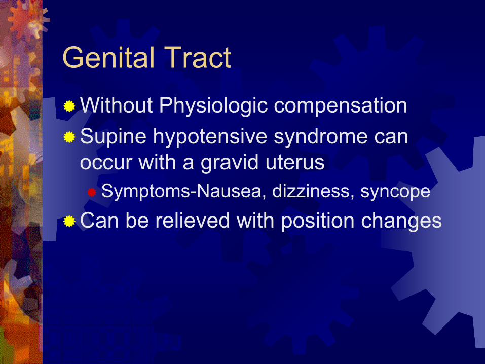
Genital TractWithout Physiologic compensationSupine hypotensive syndrome can occur with a gravid uterus
Symptoms-Nausea, dizziness, syncopeCan be relieved with position changes

Gravid uterus has limited autoregulation
Uterine blood flow is Increased 100 ml/min to 1200 ml/minBecause uterine vessels are maximally dilated little autoregulation can occur to improve flow during perfusion pressure changesWhen maternal Cardiac output declines, blood flow is shifted away from the uteroplacental circulation to the maternal brain, kidney and heart.

Urinary System-DilationCalyces, renal pelves, and ureters undergo marked dilatationMore prominent on the rightPartial obstruction of the ureters can occur at the pelvic brimProgesterone produces smooth muscle relaxation which is thought to cause the relaxation noted

Urinary System-inc GFRGFR and renal plasma flow increases 40 % by mid-gestationPlateaus, then remains unchanged until termElevated GFR is reflected in the lower serum levels of creatinine and blood urea nitrogenNL GFR 120-160 ml/min

Urinary System-ProteinuriaNormally not evidentAverage is 115 mg/day260 mg/day is in 95 percent confidence limitTherefore, our 300 mg screen would exceed most normal variations

EndocrineNormal pregnancy physiology shows
“lower lows and higher highs”Postprandial hyperglycemia
To ensure sustained glucose levels for fetusAccelerated starvation
Early switch from glucose to lipids for fuelsInsulin resistance promotes hyperglycemia
Resistance-Reduced peripheral uptake of glucose for a given dose of insulin
Mild fasting hypoglycemia occurs with elevated FFA, triglycerides,and cholesterol

Insulin resistanceAnti-insulin environment is aided by:placental lactogen
Like growth hormoneIncreases lipolysis and FFAIncreases tissue resistance to insulin
Increased unbound cortisol Estrogen and Progesterone may also exert some anti-insulin effects

ThyroidEstrogen stimulates Increase in TBG
Total T3 and T4 are increasedHowever the active hormones remains unchanged
hCG stimulates thyroid TSH is reduced
Iodine deficient stateDue to Increased renal clearance
To rule out pathologic changesEarly in pregnancy TSH can be used Later free T4 is needed

Gastrointestinal TractDisplacement of the stomach and intestines Appendix can be displaced to reach the right flankGastric emptying and intestinal transit times are delayed secondary to hormonal and mechanical factorsPyrosis is common due to the reflux of secretionsVascular swelling of the gumsHemorrhoids due to elevated pressure in veins

LiverLiver morphology unchangedLab Tests similar to liver disease
Alkaline phosphatase doublesAST, ALT, GGT and bilirubin are slightly lowerDecreased plasma albumin

GallbladderImpaired contractionHigh residual volumesPromotion of stasisStasis associated with increased cholesterol saturation of pregnancy, supports predisposition of stonesIntrahepatic cholestasisRetained bile salts-pruritus gravidarum

Skin changesChloasma or melasma gravidarumStriaeLinea nigra

Melasma

Melasma

MelasmaAlso known as the mask of pregnancyMore common in dark skin peopleMore pronounced in the summerFades a few months after deliveryRepeated pregnancy can intensifyCan occur in normal non-pregnant women with harmless hormonal imbalances or women on OCPs or depo

Striae

StriaeReddish slightly depressedBreasts, thighs, and abdomenIn future pregnancies they appear as glistening, silver lines

Linea nigra

HyperpigmentationMelasma and linea nigraEstrogen and progesterone Some melanocyte stimulating effect

The End-Go Tigers