physiol rev doi:10.1152/physrev.00006.2011 the brain basis ... brain basis... · the brain basis of...
TRANSCRIPT

THE BRAIN BASIS OF LANGUAGE PROCESSING:FROM STRUCTURE TO FUNCTIONAngela D. Friederici
Max Planck Institute for Human Cognitive and Brain Sciences, Leipzig, Germany; and Center for Advanced Studyin the Behavioral Sciences at Stanford University, Stanford, California
LFriederici AD. The Brain Basis of Language Processing: From Structure to Function.Physiol Rev 91: 1357–1392, 2011; doi:10.1152/physrev.00006.2011.—Lan-guage processing is a trait of human species. The knowledge about its neurobiologicalbasis has been increased considerably over the past decades. Different brain regionsin the left and right hemisphere have been identified to support particular language
functions. Networks involving the temporal cortex and the inferior frontal cortex with a clear leftlateralization were shown to support syntactic processes, whereas less lateralized temporo-frontalnetworks subserve semantic processes. These networks have been substantiated both by func-tional as well as by structural connectivity data. Electrophysiological measures indicate that withinthese networks syntactic processes of local structure building precede the assignment of gram-matical and semantic relations in a sentence. Suprasegmental prosodic information overtly avail-able in the acoustic language input is processed predominantly in a temporo-frontal network in theright hemisphere associated with a clear electrophysiological marker. Studies with patients suffer-ing from lesions in the corpus callosum reveal that the posterior portion of this structure plays acrucial role in the interaction of syntactic and prosodic information during language processing.
I. INTRODUCTION 1357II. A BRIEF VIEW OF SENTENCE... 1358III. THE LANGUAGE NETWORK 1358IV. PROCESS-SPECIFIC NEURAL... 1362V. TIME COURSE OF AUDITORY... 1377VI. LANGUAGE FUNCTION: BINDING... 1385
Our words are bound by an invisible grammar which isembedded in the brain.
Jonah Lehrer, in Proust Was a Neuroscientist.
I. INTRODUCTION
Since the first discovery that language functions are di-rectly related to brain tissue (28, 161, 258), people havebeen interested in understanding the neural basis of lan-guage. Starting with these early lesion studies, the adventof new methodologies such as electroencephalography(EEG), magnetoencephalography (MEG), and magnetresonance imaging (MRI), which can be used in vivo toimage cognitive functions in the brain (fMRI) as well asgray matter anatomy and white matter fiber tracts (dif-fusion-weighted MRI), has lead to a considerable in-crease in brain-based language studies (for recent re-views, see Refs. 15, 208, 251).
Despite the fact that there are hundreds of studies on thetopic, the description of the neural basis of language andspeech still remains difficult. It is hard to see the woodthrough the trees. In the last decade, various models haveproposed various paths through the wood (21, 67, 102,
117, 118). Although different in their perspective, there is aconsiderable and “hope-making” overlap of the differentpaths through the wood taken by the various models. Somemodels primarily focus on the neuroanatomy of speech per-ception (118, 213), whereas others try to specify the func-tional neuroanatomy of semantic and syntactic processes aswell as the time course of these (21, 67). Yet others haveconsidered different memory systems (247) or memory andcontrol systems (102) as major parts of language process-ing. Taken together, however, these models seem to coverthe different components of a language processing systemquite well.
The goal of the present article is to describe the structuraland functional neural network underlying sentence com-prehension and how this process evolves over time as asentence is perceived. We start the review by brieflysketching the time course of the different subprocessesconstituting the process of sentence comprehension.Then, the general network underlying language functionin the perisylvian cortex will be defined and its neuroana-tomical architecture will be specified. Based on this back-ground, the different processes taking place during com-prehension, such as acoustic-phonological analyses aswell as syntactic and semantic processes, will be de-scribed. These processes are hierarchically structured intime from the analysis of the auditory input to final inte-gration and sentence comprehension. While auditoryanalyses clearly take place in the auditory cortices in thetemporal lobes bilaterally, syntactic and semantic pro-
Physiol Rev 91: 1357–1392, 2011doi:10.1152/physrev.00006.2011
1357
on Novem
ber 1, 2011physrev.physiology.org
Dow
nloaded from

cesses are supported by separable temporo-frontal net-works strongly lateralized to the left hemisphere (LH) forsyntax and less so for semantics. Processing of sentence-level prosody is supported by a temporo-frontal networkin the right hemisphere (RH). These different processesand their respective neural implementation will be dis-cussed at the neuroanatomical macro-level, and when-ever possible also with respect to the neural structure atthe micro-level considering cytoarchitectonics and recep-torarchitectonics of the language-relevant cortices.
This review should be considered a critical one, but the goalis not to attack the position of single researchers. Rather, itis an attempt to provide a convergent view of what we knowabout the functional neuroanatomy of language up to nowand what recent debates focus on.
The review will mainly focus on neuroimaging studies(fMRI, EEG, MEG) and will not include full coverage of allpatient studies on language processing, although patientwork is considered. This decision was taken based on thefact that lesion data are not always restricted to small cir-cumscribed brain regions, and, moreover, on the findingthat performance depends on the time of lesion onset andon plastic reorganization of language functions that mayhave occurred.
II. A BRIEF VIEW OF SENTENCEPROCESSING
The present description of sentence processing cruciallydifferentiates three linguistic processing phases after aninitial phase of acoustic-phonological analysis (67). In afirst sentence-level processing phase, the local phrasestructure is built on the basis of word category informa-tion. In the second phase, syntactic and semantic rela-tions in the sentence are computed. These involve thecomputation of the relations between the verb and itsarguments, thereby leading to the assignment of thematicroles (i.e., the analysis of who is doing what to whom).Once both semantic and syntactic information lead to thecompatible interpretation, comprehension can easilytake place. For example, the interpretation of an animatenoun in sentence initial position as in “Mary cuts theflowers” is easy, as a person is a likely actor. For sen-tences in which semantic and syntactic information donot easily map, the processing system might need an ad-ditional third phase during which a final considerationand integration of the different information types isachieved, possibly including the context or world knowl-edge. During auditory sentence processing, these threedifferent phases interact with linguistic prosody provid-ing, for example, information about phrase boundariesrelevant for syntactic processes. Linguistic prosody canalso signal what is in the thematic focus of a sentence(indicated by stress in German and other Indo-European
languages) and whether an utterance is a declarative sen-tence or a question (indicated by pitch in German andother Indo-European languages). This information is ei-ther essential or modulatory to the syntactic and seman-tic processes in a given sentence.
The above description of the process of language under-standing is certainly only a sketch of what psycholinguisticshave to say about this very complex process, but it entailsthe basic processes that have to be considered when char-acterizing the neural basis of language comprehension.
III. THE LANGUAGE NETWORK
From different overviews (67, 118, 251), it is clear that thelanguage-relevant cortex includes Broca’s area in the infe-rior frontal gyrus (IFG), Wernicke’s area in the superiortemporal gyrus (STG), as well as parts of the middle tem-poral gyrus (MTG) and the inferior parietal and angulargyrus in the parietal lobe (see FIG. 1). Within these macro-anatomically defined regions, microanatomical subregionscan be specified.
A. Parcellation of the Language Cortex
Korbian Brodmann (29) was the first to provide a cyto-architectonic description of the human cortex. Novelneuroarchitectonic approaches provide detailed informa-tion about subdivisions of regions of the language net-work. These new neuroarchitectonic approaches are1) advanced objective cytoarchitectonic analysis basedon the density of different types of neurons in the cortex(5, 6), 2) receptorarchitectonic analysis based on the dis-tribution of different types of neuroreceptors in the cor-tex (3, 267), and 3) the connectivity-based parcellationapproach that subdivides brain regions according to theirarea-specific connectivity to other areas in the brain (8,132).
Interestingly, all these approaches propose a subdivisionof Broca’s area itself, and segregate it from adjacent ar-eas. This appears to be of importance as the larger regionof Broca’s area has often been discussed as supportingdifferent aspects of language processing (20, 102, 207).Broca’s area is usually defined as consisting of the cyto-architectonically defined Brodmann area (BA) 44, thepars opercularis and BA 45, and the pars triangularis (5,29) (see FIG. 1). Receptorarchitectonically, area 45 can besubdivided into two portions, a more anterior area 45abordering BA 47 and a more posterior area 45p border-ing BA 44 (3) (see FIG. 2). Moreover, area 44 can bereceptorarchitectonically subdivided into a dorsal (44d)and a ventral (44v) area. These subdivisions may be ofparticular functional importance as different languageexperiments have allocated different functions to area
THE BRAIN BASIS OF LANGUAGE PROCESSING
1358 Physiol Rev • VOL 91 • OCTOBER 2011 • www.prv.org
on Novem
ber 1, 2011physrev.physiology.org
Dow
nloaded from

45, and also to area 44 which now can possibly be as-signed to different subregions within 45 (45a versus 45p)and 44 (44d versus 44v) when considering the more fine-grained neuroanatomic parcellation of this area (com-pare with sect. IVC2).1
With the use of a connectivity based approach, the IFG hasbeen shown to separate into a subregion (BA 44) connectingto the temporal cortex via a dorsal pathway [which includesthe arcuate fasciculus (AF) and the superior longitudinalfasciculus (SLF)], a second region anterior to it (BA 45)connecting to the temporal cortex via the extreme fibercapsule system (EFCS) and a third region located more ven-
trally (frontal operculum, FOP) connecting via the uncinatefasciculus (UF) to the anterior temporal cortex (8). Thislatter article shows that there is variance between subjectswith respect to the absolute localization of each area, but italso reveals that the relative location of the three areas isstable across different subjects [see also Klein et al. (141) fora connectivity-based parcellation of the separation of BA 44and BA 45 and their probabilistic overlap].
The microanatomical description of the auditory andtemporal cortices provides the following picture. In theprimary auditory cortex (BA 41 in FIG. 1), cytoarchitec-tonic analyses have revealed different subregions in amedial-to-lateral direction (with Te1.0 in the middle,Te1.1 more medially located, and Te1.2 more laterallylocated) (176). The cytoarchitectonically defined regionBA 22 covers the posterior two-thirds of the lateral con-vexity of the STG (29) (see FIG. 1). Receptor and cyto-architectonic subdivisions have proposed a separation ofthe dorsal and ventral banks of the STG (175). It is sug-gested that the lateral STG proper excluding the dorsal
1It should be noted that these receptorarchitectonic analyses areperformed in post mortem brains and thus represent an analysis ofthe brain’s neuron receptors at a certain point in time. However, itis known that the density of neuron receptors is subject to dynamicmodulations over a millisecond time scale. Moreover, we shouldkeep in mind that up to now the functional relation between partic-ular neuron receptors and particular language functions is notknown.
FIGURE 1. Anatomical and cytoarchitectonic details of the left hemisphere. The different lobes (frontal,temporal, parietal, occipital) are marked by colored borders. Major language relevant gyri (IFG, STG, MTG) arecolor coded. Numbers indicate language-relevant Brodmann Areas (BA) which Brodmann (1909) defined onthe basis of cytoarchitectonic characteristics. The coordinate labels superior/inferior indicate the position ofthe gyrus within a lobe (e.g., superior temporal gyrus) or within a BA (e.g., superior BA 44; the superior/inferior dimension is also labeled dorsal/ventral). The coordinate labels anterior/posterior indicate the posi-tion within a gyrus (e.g., anterior superior temporal gyrus; the anterior/posterior dimension is also labeledrostral/caudal). Broca’s area consists of the pars opercularis (BA 44) and the pars triangularis (BA 45).Located anterior to Broca’s area is the pars orbitalis (BA 47). The frontal operculum (FOP) is located ventrallyand more medially to BA 44, BA 45. The premotor cortex is located in BA 6. Wernicke’s area is defined as BA42 and BA 22. The primary auditory cortex (PAC) and Heschl’s gyrus (HG) are located in a lateral to medialorientation.
ANGELA D. FRIEDERICI
1359Physiol Rev • VOL 91 • OCTOBER 2011 • www.prv.org
on Novem
ber 1, 2011physrev.physiology.org
Dow
nloaded from

and ventral banks is a functionally relevant area for lan-guage processing in humans. In the anterior-posteriordimension, there is no cytoarchitectonic parcellation ofBA 22 as it covers most of the STG, except its mostanterior portion (BA 38) (see FIG. 1).
As the cyto- and receptorachitectonic analysis cannot beconducted in the living brain, the team working with theseapproaches has calculated “probability maps” from postmortem brains of which the cytoarchitectonic analyses areavailable online (http://www.fz-juelich.de/inm/index.php?index�51).
B. Structural Connections Between theLanguage Cortices
The identification of fiber pathways between Broca’s areaand the temporal cortex (Wernicke’s area) dates back to thelate 19th century when Dejerine (47) defined the arcuatefasciculus as the dominant fiber tract connecting these tworegions. Nowadays, diffusion tensor imaging (DTI) allowsthe identification of structural connections between differ-
ent brain regions in the human in vivo (e.g., Refs. 11, 132).For a recent tractography atlas representing the major fiberconnections based on this method, see Catani and de Schot-ten (38). Note, however, that with this approach the direc-tionality of the connection cannot be determined. Concern-ing the connection between the language-relevant regions,i.e., the (pre)frontal cortex and the temporal cortex, theliterature generally agrees on two pathways, a dorsal and aventral pathway. Recently, there has been debate with re-spect to the particular functions of different pathways fromthe temporal cortex to other parts of the brain as well aswith respect to their end points in the other brain regions(see Refs. 65, 66, 256) (see FIG. 3).
Within “dual stream models” (117, 118, 213), the ventralpathway has been taken to support sound-to-meaning map-ping, whereas the dorsal pathway connecting the posteriordorsal-most aspect of the temporal lobe and the posteriorfrontal lobe has been suggested to support auditory-motorintegration (118). Using a deterministic fiber tracking ap-proach in which the two end points of the connection arepredefined on the basis of functional data, Saur and co-workers (227, 228) interpret the ventral pathway connect-ing the temporal cortex with the pars orbitalis (BA 47) andtriangularis (BA 45) via the EFCS as supporting sound-to-meaning mapping, and define the dorsal pathway as going
FIGURE 3. Structural connectivities between the language corti-ces. Schematic view of two dorsal pathways and two ventral path-ways. Dorsal pathway I connects the superior temporal gyrus (STG)to the premotor cortex via the arcuate fascile (AF) and the superiorlongitudinal fascicle (SLF). Dorsal pathway II connects the STG to BA44 via the AF/SLF. Ventral pathway I connects BA 45 and thetemporal cortex via the extreme fiber capsule system (EFCS). Ven-tral pathway II connects the frontal operculum (FOP) and the anteriortemporal STG/STS via the uncinate fascile (UF).
FIGURE 2. Receptorarchitectonic parcellation of the left posteriorprefrontal cortex. Extent of delineated areas projected to the lateralsurface of an individual post mortem brain. The following receptorbinding sites were studied by Amunts et al. (3) for the prefrontalcortex: glutamatergic AMPA and kainite receptors, GABAergicGABA
Areceptors, cholinergic muscarinic M1 and M2 receptors, and
noradrenergic receptors. The color coding indicates receptorarchi-tectonically defined borders. The borders between 44 d (dorsal) and44 v (ventral), for example, were differentiated mainly by �1 andmuscarinic M2 receptors. Area 45 can be subdivided receptorar-chitectonically into an anterior (45a) and a posterior (45p) part.Area 6 can be subdivided into three subparts. op, Operculum (num-bering indicates different subparts); ifs, inferior frontal sulcus; ifj,inferior frontal junction; prcs, precentral sulcus; cs, central sulcus.[From Amunts et al. (3).]
THE BRAIN BASIS OF LANGUAGE PROCESSING
1360 Physiol Rev • VOL 91 • OCTOBER 2011 • www.prv.org
on Novem
ber 1, 2011physrev.physiology.org
Dow
nloaded from

from the temporal lobe to the premotor cortex and continu-ing to the pars opercularis (BA 44) supporting sensory-motor mapping of sound-to-articulation. This functionalinterpretation stands in slight contrast to probabilistic fibertracking approach in which only one end of the connectionis defined as a seed point. Defining two seed points in theIFG on the basis of two functionally different activations,Friederici et al. (69) identified a dorsal pathway going frompars opercularis (BA 44) to the posterior temporal cortexvia the AF/SLF, and a ventral pathway from the FOP via theUF to the anterior temporal cortex. The function of thedorsal pathway was seen in the support of processing non-adjacent elements in syntactically complex sentences andthe ventral pathway taken to support combinations of ad-jacent elements in a sequence.
Thus these findings as well as additional data from intraop-erative deep stimulation (56) make it likely that there aretwo ventral pathways connecting the frontal to the tempo-ral cortex involved in language processing, one from BA 45via the EFCS to the temporal cortex (ventral pathway I) andone from the FOP via the UF (ventral pathway II). More-over, there is suggestive evidence that there are two paralleldorsal pathways, one from the temporal cortex to the pre-motor cortex (dorsal pathway I) and one from the temporalcortex to BA 44 (dorsal pathway II), with the former mainlysupporting sound-to-motor mapping and the latter sup-porting higher-level language processes (see Ref. 39, and fora recent debate, see Refs. 65, 66, 256).
This subdivision into two dorsal pathways is in line withrecent structural connectivity data from very young infantsshowing a dorsal fiber tract from the temporal lobe goingonly to the motor/premotor cortex (55). This pathway (dor-sal pathway I) subserving auditory-motor integration is al-ready of primary importance during early language acqui-sition, when tuning the system towards the target language(118). A dorsal fiber tract that connects the temporal lobewith Broca’s area in the IFG (dorsal pathway II) developsmuch later and appears to be functionally related to higher-level semantic and syntactic language functions (26). It is anopen issue whether these dorsal connections are direct orindirect with an intermediate stage in the inferior parietalcortex (39, 212, 213) whose role within the dorsal streammight be that of phonological working memory storage(198, 245).
In addition to these long-range connections, functional con-nectivity and structural connectivity analyses, moreover,have identified two short-range pathways within the tem-poral cortex, a first one from Heschl’s gyrus (HG) to theplanum polare and anterior STG via a rostral fiber pathwayand a second one from HG to the planum temporale (PT)and posterior STG via a caudal fiber pathway (248). Thesedata suggest two auditory processing streams within thetemporal cortex, 1) between the primary auditory cortex
(PAC) and the anterior auditory cortex (planum polare) and2) between the PAC and posterior auditory cortex (planumtemporale). Short-range connections have also been re-ported for the prefrontal cortex, interconnecting the infe-rior frontal sulcus and BA 44 (166).
To summarize, in addition to short-range structural con-nections within the language-related cortex, there are mul-tiple long-range structural connections between the lan-guage-relevant regions in the frontal and temporal cortices:two dorsal pathways and possibly two parallel ventral path-ways. Although the direction of the connectivity cannot bedetermined in humans using the DTI approach, data fromanimal studies using invasive tracer methods suggest strongdirectionality from sensory regions to the prefrontal cortexin the monkey (101, 221). The reverse information flow isalso considered, and the two directions are discussed interms of feed-forward and backward projections (212). Inthe domain of human language processing, projectionsfrom sensory to the premotor cortex (via dorsal pathway I)could support bottom-up information processes, whereasprojections from Broca’s area to the temporal context (viadorsal pathway II) could subserve top-down processesdrawing prediction about the incoming information,thereby easing its integration. Further research must showwhether these assumptions for language processing hold.
The precise function of these structural connections, how-ever, can only be defined indirectly, namely based on thefunction of the particular regions they connect. One way toestablish a closer relation between structural and functionalinformation might be to use the anatomical connectivity asa prior for dynamic causal modeling of fMRI data (240).
C. Functional Connections in the DefaultLanguage Network
Every brain-based study on language processing reportsat least one function-related activation in the left peri-sylvian cortex, which includes the prefrontal, frontal,temporal, and parietal cortices. The particular functionassigned to a given area in the perisylvian cortex as de-fined on the basis of functional imaging studies investi-gating different aspects of language processing, such asphonology, syntax, and semantics, will be discussed indetail in section IV.
Here we will first consider recent data which suggestthat the experimental variations in these studies onlyreflect the tip of the iceberg, since specific experimentalconditions can only explain �20% or less of the totalvariance of the activation of the brain in a given experi-ment (162). The rest of the variance represents activa-tion not induced by the specific experimental condi-tions. Interestingly, this “unexplained” activity is notrandom. For language experiments, it is located in the
ANGELA D. FRIEDERICI
1361Physiol Rev • VOL 91 • OCTOBER 2011 • www.prv.org
on Novem
ber 1, 2011physrev.physiology.org
Dow
nloaded from

perisylvian cortex. As this activation pattern was onlyobserved for language experiments and not for nonlan-guage experiments, it was taken to represent the defaultlanguage network (162). To identify this default activa-tion, a low-frequency fluctuation analysis of fMRI datacompared four language experiments with two nonlan-guage experiments from the same laboratory (formethod, see Ref. 162; for low-frequency fluctuationanalysis in general, see Refs. 17, 211).2 Moreover,when conducting a functional connectivity analysiswithin this default language network, a significant cor-relational connectivity was found between Broca’s areain the IFG and the posterior superior temporal lobe(162) (see FIG. 4).
Thus it is already within the default language network thatthere are functional connections between different languageregions, independent of the different conditions induced bya given experiment. To summarize, the particular activationpattern reported for specific experimental conditions aim-ing to test semantic or syntactic processes as reported in thedifferent language fMRI studies thus only represents a mod-ulation of this default language network.
IV. PROCESS-SPECIFIC NEURALNETWORKS
Spoken sentence comprehension requires a number of sub-processes to derive the meaning of a sentence from the au-ditory input, as there are acoustic-phonological, syntactic,and semantic processes. We will discuss the brain regionssupporting these different processes in turn.3
A. Acoustic-Phonological Analysis
The comprehension of spoken language starts with theacoustic-phonological analysis of the speech input. The ob-vious neural candidate to support this process is the audi-tory cortex and adjacent areas.
In an attempt to specify subregions in the auditory cortexand adjacent areas in humans, researchers have relied onneuroanatomical data from non-human primates for whicha core region in HG, a surrounding belt and parabelt regionhas been identified (213, 230). In humans, the PAC is lo-cated on the superior surface of the temporal lobe bilater-ally in HG. Three regions can be identified adjacent to HG.A region located posterior, the planum temporale (PT), aregion anterolateral to HG called planum polare (PP), and aregion at the lateral convexity of the cortex in the STGextending to the superior temporal sulcus (STS). All theseregions are involved in the acoustic analysis of speech. Cy-toarchitectonic studies have indicated that the PAC usuallycovers the medial two-thirds of the anterior HG (176), andthe identification of a subregion in the lateral convexity ofthe STG has been confirmed by a receptorarchitectonicanalysis (175).
Functionally, a primary step is to differentiate speechfrom nonspeech acoustic signals, and for a descriptionof the neuroanatomic basis of speech comprehension, itwould be of major interest to identify where in the pro-cessing stream this takes place. The primary auditoryanalysis is computed in HG. Functional neuroimagingstudies show that HG is activated by any type of sound(133, 177). The region lateral to HG at the convexityof the STG extending into the STS has been found torespond to acoustic features of phonetic parameters(16), but also to variations of frequency and spectralinformation in nonspeech sounds (109) and is thus notspecialized for speech. Functional imaging studies have,moreover, shown that PT also does not react specifi-cally to speech sounds, at least compared with equallycomplex nonspeech sounds (48, 261, 266). The infor-
2Earlier studies using the method of low-frequency fluctuationanalysis identified a general default network while subjects restedquietly in the scanner (17, 211). With data from such a restingstate, functional connectivities between different subregions of theIFG (i.e., pars orbitalis, pars triangularis, and pars opercularis) andsubregions in the parietal cortex and temporal cortex have beenreported (263).
3Note that the anatomic terminology varies from study to study.Here we used those anatomic terms provided by the authors of thestudy discussed. FIGURE 1 may help to orient the reader withrespect to the different anatomic terms.
FIGURE 4. Functional connectivities between the language corticeswithin the default language network. Results are of a conjunction anal-ysis involving 4 language experiments corrected for multiple compari-sons using FDR thresholded at P � 0.05. A: correlations with the seedregion BA 44. B: correlations with the seed region in FOP. For eachexperiment, the correlations were r-to-z transformed to ensure Gaussi-anity and then subjected to a voxelwise t-test across subjects. The mapshows the z values for the conjunction of all 4 language studies. The zvalues are color coded as indicated by the color bar. [Adapted fromLohmann et al. (162), by permission of Oxford University Press.]
THE BRAIN BASIS OF LANGUAGE PROCESSING
1362 Physiol Rev • VOL 91 • OCTOBER 2011 • www.prv.org
on Novem
ber 1, 2011physrev.physiology.org
Dow
nloaded from
mation flow from HG to PT has been demonstrated ina time-sensitive fMRI paradigm, indicating the involve-ment of HG and PT at different points in time (264). Ithas been concluded that HG is associated with analyz-ing the sound signal per se, whereas the PT may be in-volved in categorizational processes. The PT has beenproposed as the region for the segregation and match-ing of spectrotemporal patterns and as serving as a“computational hub” gating the information to higher-order cortical areas (95).
Speech perception of phonemes (consonants) was found toactivate a region anterolateral to HG in the STG/STS (189).This region differentiates between speech and nonspeechsounds. In contrast, the left posterior STG was found toprocess the basic acoustic characteristics of the signal.Given their respective responsibilities, the posterior STGwas defined as reflecting earlier processes than the antero-lateral STG/STS (146). The fMRI finding that the poste-rior STG houses an earlier processing level than the an-terolateral STG/STS is consistent with magnetoencepha-lographic evidence locating the relatively early N100response to consonants in HG and PT (188) and withpatient evidence showing that lesions in the posteriorSTG lead to word deafness as well as deficits in the per-ception of nonspeech sounds (204). Other neuroimagingstudies, however, reported the PT or the supramaginalgyrus to respond to speech compared with nonspeechsounds (46, 131, 174). These studies, in contrast toObleser et al. (189), who used a passive listening para-digm, used attention-demanding tasks. From these data,it appears that under specific task demands, the differen-tiation between speech and nonspeech sounds by meansof top-down processes may be shifted to an earlier pro-cessing level, in this case the PT.
Functionally, PAC in the left and the right hemispheres areresponding to speech and tonal pitch, but they appear tohave different computational preferences, with the left PACreacting specifically to speech sounds characteristics and theright PAC to characteristics of tonal pitch (265). The rela-tive specialization of the two auditory cortices for thesestimulus types, which differ in their temporal and spectralcharacteristics, is described as a specialization for rapidlychanging information with a limited frequency resolution inthe left hemisphere and a system with reverse characteristicsin the right hemisphere. The former system would be idealfor the perception and recognition of speech sounds, as thedetermination of these (i.e., phonemes in a sequence) re-quires a system with a time resolution of 20–50 ms. Thelatter system would be able to deal with suprasegmentalinformation (i.e., prosody requiring a system with a timeresolution of 150–300 ms). Hickok and Poeppel (118) pro-posed that the left and right hemisphere generally work atdifferent frequencies, leading to a relative lateralization offunctions. The left hemisphere primarily works in gamma
frequencies, whereas the right hemisphere works in thetheta range (93).
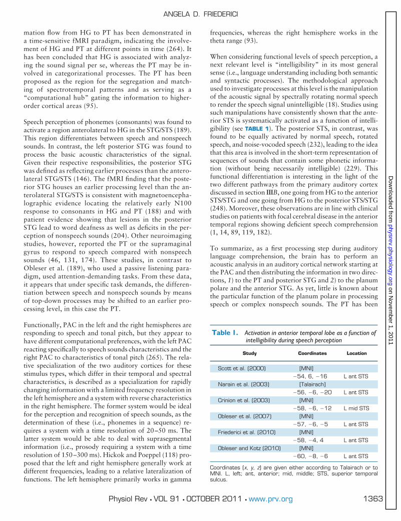
When considering functional levels of speech perception, anext relevant level is “intelligibility” in its most generalsense (i.e., language understanding including both semanticand syntactic processes). The methodological approachused to investigate processes at this level is the manipulationof the acoustic signal by spectrally rotating normal speechto render the speech signal unintelligible (18). Studies usingsuch manipulations have consistently shown that the ante-rior STS is systematically activated as a function of intelli-gibility (see TABLE 1). The posterior STS, in contrast, wasfound to be equally activated by normal speech, rotatedspeech, and noise-vocoded speech (232), leading to the ideathat this area is involved in the short-term representation ofsequences of sounds that contain some phonetic informa-tion (without being necessarily intelligible) (229). Thisfunctional differentiation is interesting in the light of thetwo different pathways from the primary auditory cortexdiscussed in section IIIB, one going from HG to the anteriorSTS/STG and one going from HG to the posterior STS/STG(248). Moreover, these observations are in line with clinicalstudies on patients with focal cerebral disease in the anteriortemporal regions showing deficient speech comprehension(1, 14, 89, 119, 182).
To summarize, as a first processing step during auditorylanguage comprehension, the brain has to perform anacoustic analysis in an auditory cortical network starting atthe PAC and then distributing the information in two direc-tions, 1) to the PT and posterior STG and 2) to the planumpolare and the anterior STG. As yet, little is known aboutthe particular function of the planum polare in processingspeech or complex nonspeech sounds. The PT has been
Table 1. Activation in anterior temporal lobe as a function ofintelligibility during speech perception
Study Coordinates Location
Scott et al. (2000) [MNI]�54, 6, �16 L ant STS
Narain et al. (2003) [Talairach]�56, �6, �20 L ant STS
Crinion et al. (2003) [MNI]�58, �6, �12 L mid STS
Obleser et al. (2007) [MNI]�57, �6, �5 L ant STS
Friederici et al. (2010) [MNI]�58, �4, 4 L ant STS
Obleser and Kotz (2010) [MNI]�60, �8, �6 L ant STS
Coordinates (x, y, z) are given either according to Talairach or toMNI. L, left; ant, anterior; mid, middle; STS, superior temporalsulcus.
ANGELA D. FRIEDERICI
1363Physiol Rev • VOL 91 • OCTOBER 2011 • www.prv.org
on Novem
ber 1, 2011physrev.physiology.org
Dow
nloaded from

suggested as the “computational hub” from which informa-tion is gated to higher-order cortical regions (95). A con-nection from the temporal cortex to the premotor cortexappears to support auditory-to-motor mapping and hasbeen claimed to represent part of the “phonological net-work” (228).
B. Initial Syntactic Processes
Several psycholinguistic models have proposed that thesentence parser processes syntactic information at differ-ent levels with an initial stage during which the simplestsyntactic structure based on word category informationis constructed and a second stage during which the rela-tions who is doing what to whom are established (63).These models called serial syntax-first models have beenchallenged by interactive and constraint-satisfactionmodels (163, 169), which assume that syntactic and se-mantic information interact at any time. Syntax-firstmodels, however, receive some support from neurocog-nitive models of language comprehension, which con-sider event-related brain potentials (ERPs) to providecrucial information about the temporal structure of lan-guage processing (21, 67).
As syntax-first models assume that the important syntacticprocesses relevant for the assignment of the grammatical struc-ture of a sentence to occur only a couple of hundred millisec-onds later than the initial syntactic parse, it is not easy toseparate these two stages of syntactic processing using fMRIdue to the low temporal resolution of this method. One way toinvestigate the different syntactic stages is to introduce viola-tions in natural sentences which tap either the initial or thelater syntactic processing stage. The initial processing stagewill clearly be affected by word category violations, since in-correct word category information would make the buildingup of an initial local phrase structure impossible while viola-tions of grammatical relations in the sentence will affect a laterprocessing stage. Another way of investigating local syntacticstructure building is to use artificial grammars which lack se-mantic relations. Initial local phrase structure building pro-cesses4 were found to be correlated with increased activationin the frontal operculum and the anterior STG both in studieson natural grammar processing (81) and on artificial grammarsequences (69). The natural grammar study in German intro-duced a word category error within a prepositional phrase byputting a verb instead of a noun after the preposition, e.g.,“The pizza was in the eaten” instead of “The pizza was in the
restaurant eaten” (literal translation). The past participle verbform is syntactically incorrect, disallowing local structurebuilding. The artificial grammar experiment used a probabi-listic grammar in which an element of the category A (a certainsyllable type) was always followed by an element of the cate-gory B (another syllable type), e.g., ABABAB. A violation wascreated by having an A syllable followed by another A syllablein the sequence. The processing of this syntactic error in theartificial grammar sequence led to activation in the FOP. Tak-ing the maximum of activation as a seed point for tractogra-phy analysis in each individual, a ventrally located fiber tractconnecting the FOP and the anterior STG via the uncinatefasciculus was found (69). On the basis of this finding, it hasbeen suggested that the FOP together with the anterior STGsupports local structure building. More generally, this net-work could be viewed as the system that supports rule-basedcombinatorics of adjacent elements.
During sentence processing, this initial stage of phrase struc-ture building is mandatory and should in principle be observ-able whenever a sentence is processed. Thus the FOP should beseen with increased activation not only for violations in sen-tences and sequences, but also when comparing sentences tononstructured word lists. Activation of the FOP was observedin a study comparing sentences to word lists without functionwords (78), but not in other studies using mixed word lists.Most of these other studies used word lists that allowed localstructure building partly due to syntactically legal combina-tions of two or three words in the list, for example, adjectivesand nouns (125, 127, 236, 241, 250). Interestingly, Vanden-berghe et al. (250) report activation in the FOP (�48, 22, 4)for different sentence conditions providing word category in-formation compared with control conditions in which unpro-nounceable letter sequences (providing no word category in-formation) were used. All these findings are thus generally inline with the view that local structure building is supported bythe FOP. However, it should be noted that local structurebuilding is quite automatic in adults only requiring small re-sources (as indicated by ERP studies; see sect. VB). Therefore,the FOP may not be seen to be significantly activated in eachstudy with native adult listeners. Moreover, given that theactivation in native listeners is very small, significant activa-tions may not be observable in grand averages across subjectsdue to the variability of the location of the FOP across indi-viduals as shown in a connectivity-based parcellation study(8). Further research taking individual subject data into ac-count must clarify this issue.
Studies investigating sentence processing under less profi-cient processing conditions as in language development (27)and second language learning (222) show that processingphrase structure violations involves the IFG, in particularBroca’s area, and not just the FOP. This suggests that theremay be a shift in the recruitment of necessary parts of theventral prefrontal cortex for local syntactic structure build-ing as a function of language proficiency.
4The low temporal resolution of fMRI, however, will not allow us todifferentiate early and late effects observed in the ERP in response toincorrect word category information (see sect. V, B and C), but incombination with ERP studies from patients with lesions in particularparts of the brain as well as MEG localization studies with healthyparticipants, conclusions about the localization of these effects arepossible.
THE BRAIN BASIS OF LANGUAGE PROCESSING
1364 Physiol Rev • VOL 91 • OCTOBER 2011 • www.prv.org
on Novem
ber 1, 2011physrev.physiology.org
Dow
nloaded from

C. Computation of Semantic and SyntacticRelations
Empirically, there are three basic methodological ap-proaches to investigate syntactic and semantic processesduring sentence comprehension. The first is to vary the pres-ence/absence of syntactic information (by comparing sen-tences to word list) or of semantic information (by compar-ing real word lists/sentences to pseudoword lists/sentences).The second approach is to introduce syntactic or semanticerrors in sentences. The third is to vary the complexity ofthe syntactic structure (including syntactic ambiguities) orthe difficulty of semantic interpretation (including semanticambiguities). All these approaches have been used in fMRIstudies published in the last 15 years.
In general, these studies found activations at different loca-tions in the anterior and posterior temporal cortex as wellas in the IFG. The picture that emerges from these studiesmay be less clear than some researchers had hoped (Ref. 59and a reply to this paper by Grodzinsky, Ref. 97). However,once we take both stimulus type and task as well as neuro-architectonic subdivisions of language-relevant brain re-gions into consideration, a picture emerges that is worthpresenting as a tentative state of the art model. Once thesedifferent aspects are considered, the reported activationpattern provides a surprisingly coherent picture even acrosstypologically different languages. We will first consider ac-tivations in the temporal lobe and then those in the IFG.
1. Role of the temporal lobe
Many of the neuroimaging studies on language comprehen-sion report activation in the anterior and posterior temporallobe. While some studies concluded that the anterior andposterior temporal regions react specifically to semantic orsyntactic aspects, others challenged this view by arguingeither that the anterior temporal lobe (218, 250) or theposterior temporal lobe is not domain specific (126).
A) ANTERIOR TEMPORAL LOBE. A number of fMRI studies re-porting activation in the temporal lobe investigated seman-tic and syntactic processes by systematically varying thepresence/absence of semantic and syntactic information in awithin-subject design. Those studies that compared sen-tences (syntax present) to word lists (syntax absent)found the lateral anterior temporal lobe to activate morestrongly for sentences than for word lists (for French,171; for German, 78; for English, 125) (see TABLE 2 formore studies). As this increase of activation in the ante-rior STG/STS is present even when comparing meaning-less pseudoword sentences (i.e., sentences in which func-tion words remain in their syntactic correct position, butcontent words are replaced by pseudowords) with mean-ingless pseudoword lists, this region has been interpretedto support the construction of phrase structure in partic-
ular (78, 125). One study investigating the processing ofsentences containing syntactic and semantic violationsfound that, compared with baseline, syntactic violationsled to an increased activation in the anterior STG,whereas semantic violations did not (81). Moreover,studies testing semantics by comparing real-word stimuli(sentences and word lists) with pseudo-word stimuli (sen-tences and word lists) reported no main effect of seman-tics in anterior STG/STS (78, 125).
However, activation in the anterior temporal lobe has beenreported to change as a function of sentence-level semanticprocesses (218, 250). This appears to be the case only undercertain experimental conditions. Vandenberghe, Nobre,and Price (250) used sentences that were either semanticallyincoherent and/or syntactically incorrect. The anterior tem-poral pole was found to be more active for semantic inco-herence, but only when syntactically incorrect versionswere compared with normal sentences. Thus the semanticeffect only carries once the syntax is incorrect. Rogalskyand Hickok (218) reported a direct comparison of activa-tion of two task conditions: subjects listening to sentencesincluding a semantic or syntactic violation had to detecteither the semantic violation or the syntactic violation, re-spectively. In a whole head analysis conducted over correctsentences, they were found to be activated during the syn-tactic task and the semantic task. A region of interest anal-ysis in the anterior temporal lobe including BA 38 revealeda large region that was equally modulated by the two tasks,but only a small subregion that was only modulated by thesemantic task. From these data the authors concluded thatthe anterior temporal lobe should be considered as a regionthat supports combinational processes both in the syntacticand the semantic domain.
From the studies discussed, we can conclude that the ante-rior STG is systematically involved whenever syntacticstructure has to be processed (sentences versus word lists).For a localization of the anterior STG, see FIGURE 1. Thesimple presence/absence of word semantics (real � pseudo-words) does not modulate this region. Sentence-level se-mantic aspects can activate the anterior temporal lobe butonly under certain stimulus conditions (250) or under spe-cific task conditions (218). It has been proposed that thereare two different subregions within the anterior STG/STSthat modulate their activation differentially as a function ofsemantic and syntactic processes, with the most anteriorportion of the STS responding to syntactic manipulations(sentence versus word list) and a region directly posterior toit showing an interaction of syntactic and semantic factors(125). Future studies will have to provide additional evi-dence of this functional separation within the anterior tem-poral lobe.
It should be noted that the anterior temporal lobe has longbeen discussed as supporting semantic tasks in general
ANGELA D. FRIEDERICI
1365Physiol Rev • VOL 91 • OCTOBER 2011 • www.prv.org
on Novem
ber 1, 2011physrev.physiology.org
Dow
nloaded from

(155). Evidence for this view mainly comes from patientswith dementia or lesions in the anterior temporal lobe, whoshow semantic impairments for word and picture process-ing and memory. We will not discuss these studies in detailas the focus of this review is on sentential processes, butrefer to recent meta-analyses. One recent meta-analysis (15)reviewed 120 fMRI studies on semantic processing at theword level and identified a left-lateralized semantic networkconsisting of seven regions, none of which, however, werein the anterior temporal lobe. Another recent meta-analysis(252) reviewed 164 functional imaging studies includingthose investigating words and sentences presented audito-rily and visually as well as pictures. This analysis revealedthat the likelihood of anterior temporal lobe activation isdependent on the type of stimuli, and that studies usingauditory sentences are more likely to find activation in thisregion than studies using other stimulus types, but the au-thors refrain from defining this region’s function in auditorysentence processing.
To conclude, it appears that the anterior temporal cortex isinvolved in semantic and syntactic processes. Its function
during sentence processing may be primarily combinatorialin nature.
B) POSTERIOR TEMPORAL LOBE. The posterior temporal lobe hasalso been found to be activated during language compre-hension. Activation in the left posterior STG/STS has beenreported for syntactic information across different studies,when comparing sentences to word lists (127, 236, 250),when comparing syntactically complex to less complex sen-tences (41, 77, 140, 184, 225), and when comparing sen-tences containing a syntactic violation with syntacticallycorrect sentences (76, 81) (see TABLE 3).
Activation in the posterior STG/STS has also been seen to bemodulated by specific semantic information at the senten-tial level, in particular, when the stimulus material involvesthe processing of the relation between the verb and its ar-guments, be it in correct sentences when considering a sen-tence’s semantic close probability with respect to the verb-argument relation (185), or in sentences which contain arestriction violation between the verb and its arguments(81). When different verb classes and their argument order
Table 2. Activation in the anterior temporal lobe
Study Coordinates Location
Mazoyer et al. (1993) [Talairach]syn (sent � WL, allowing local structure)sem (meaningful � meaningless sent)
no coordinatesno coordinates
L temp pole LMTG/STG
Stowe et al. (1998) visual [Talairach]syn (sent � WL, with local structure)no sem condition
�40, 2, �20 L ant MTG/STG
Friederici et al. (2000)syn (sent � WL) (real words, only nouns)syn (sent � WL) (pseudowords)sem (real � pseudowords)
no coordinatesno coordinatesno effect
L ant STGL ant STG
Humphries et al. (2005) [MNI]syn (sent � WL)no sem condition
�54, �1,- 24 L ant STS/MTG
Humphries et al. (2006) [MNI]syn (sent � scrambled WL)interaction (sem/syn)sem (real � pseudowords)
�47, 14, �28�54, 10, �8no effect
L ant STSL ant STG
Snijders et al. (2009) [MNI]syn (sent � WL, allowing local structure)no sem condition
�54, 18, �30 L temp pole
Friederici et al. (2003) [Talairach]syn violation (phrase structure)sem violation (select. restriction)
�53, �1, 0no effect
L ant STG
Vandenberghe et al. (2002) visual [Talairach]syn viol. (incorrect word order)sem viol. (sem random sent.)interaction (sem/syn)
�44, �6,�24�48, �4,�20�40, 8, �28
L ant temp poleL ant temp poleL ant temp pole
Presence/absence of syntactic (syn) (sentences � word list) or semantic (sem) information (real words �pseudowords), violation of syntactic, or semantic information. L, left; ant, anterior; temp, temporal; STG,superior temporal gyrus; MTG, middle temporal gyrus; STS, superior temporal sulcus.
THE BRAIN BASIS OF LANGUAGE PROCESSING
1366 Physiol Rev • VOL 91 • OCTOBER 2011 • www.prv.org
on Novem
ber 1, 2011physrev.physiology.org
Dow
nloaded from

were investigated, it was found that these two factors inter-act in the posterior STG/STS (22). Together, these studiessuggest that the left posterior STG/STS is a region in whichsyntactic information and verb-argument-based informa-tion are integrated (98).
Moreover, syntactic and semantic ambiguity involve theposterior temporal cortex. Syntactic ambiguity activates theposterior temporal lobe extending posteriorly to the infe-rior parietal lobe and the MTG anterior to Heschl’s gyrus(245). Semantic sentence ambiguity was found to activatethe left posterior temporal cortex including the STS, MTG,
and inferior temporal gyrus (215). However, it should benoted that both semantic and syntactic ambiguity are pro-cessed in a network which, in addition to the temporalcortex, also involves the left IFG, as evidenced by a func-tional connectivity analysis using a predictor time serieslocated in the left IFG (245). In this analysis, the activationdue to semantic ambiguity in the left IFG predicts the acti-vation in the left anterior STG, whereas the activation ofsyntactic ambiguity in the left IFG predicts the activation inthe anterior and posterior MTG/STG. Thus both ambiguitytypes activate a temporo-frontal network with type-specificmodulations in the temporal cortex. These modulations for
Table 3. Activation in the posterior temporal lobe variation of syntactic (syn) and semantic (sem) information
Study Coordinates Location
Vandenberghe et al. (2002) visual [Talairach]sem (sent with words randomly filled in)syn (sent � WL)
no effect�50, �58, 12
L post MTG
Snijders et al. (2009) visual [MNI]syn (sent � WL) �58, �56, 12
�44, �58, 18L post STG L post STG
Humphries et al. (2005) [MNI]syn (sent � WL) �63, �49, 7 L temp lobeCooke et al. (2001) [Talairach]syn complexity (short)syn complexity (long)
�48, �68, �8�40, �76, �4
L post temp-occL post temp-occ
Friederici et al. (2009) [Talairach]syn complexity �48, �54, 12 L STG/STSNewman et al. (2010) visual [Talairach]sem (relatedness of noun pairs)syn complexity
no effect�58, �36, 2
L temporal
Santi and Grodzinsky (2010) [Talairach]syn complexity �52, �34, 2 L post STGBen-Shachar et al. (2004) [Talairach]syn movement (exp. 1)syn movement (exp. 2)
�56, �42, 7�55, �41, 6
L post STSL post STS
Friederici et al. (2010) [MNI]syn violation �60, �42, 6 L post STSFriederici et al. (2003) [Talairach]syn (violation)sem (verb-noun violation)
�61, �40, 20�60, �42, 20
L post STGL post STG
Obleser and Kotz (2010) [MNI]sem (cloze: verb-noun) �50, �42, 2 L post STG/STSObleser et al. (2007) [MNI]sem (predict: verb-noun) [MNI] L AGHumphries et al. (2007)sem (congruent � random) no coordinates L AGBornkessel et al. (2005) [Talairach]word order x verb class �52, �43, 18
�47, �58, 24L post STGL post STS
Kinno et al. (2008) [MNI]syn complexity �51, �51, 3 L post STG/MTGSuzuki and Sakai (2003) [MNI]sem anomalysyn anomaly
�54, �42, 3�54, �42, 3
L post STG/MTG
Definitions are as in Table 2.
ANGELA D. FRIEDERICI
1367Physiol Rev • VOL 91 • OCTOBER 2011 • www.prv.org
on Novem
ber 1, 2011physrev.physiology.org
Dow
nloaded from

semantic and syntactic ambiguity are in line with the type-specific modulations observed in the language studies listedin TABLES 2 and 3.
Note, however, that the posterior STG is not specific tointegration processes in language or speech (230). Rather, ithas been implicated in the integration of different informa-tion types, for audiovisual integration (2, 31), for biologicalmotion (209), and for face processing (110). It has beenproposed that the function of the STS varies depending onthe coactivations of the network with regions in the medialtemporal lobe and in the frontal cortex (111).
There is one additional region in the left superior Sylvianfissure at the parietal-temporal boundary, called area Spt,which has been discussed as part of the auditory-motorintegration circuit, which involves left frontal regions andthe STS bilaterally (116, 118). The Spt is also not specific tospeech, as it is activated during the perception and repro-duction (humming) of tonal sequences as well (116). It isspeculated that Spt is more highly coupled to the motorsystem than to the sensory system. Thus the posterior tem-poral cortex is clearly involved in language processing, andits function appears to be primarily to integrate differenttypes of information. For sentence processing, this mightmean the integration of semantic and syntactic information.
2. Role of the IFG
The IFG, in particular Broca’s area, has long been known tosupport language production (28, 223) and comprehensionprocesses (269). For the localization of Broca’s area, definedas consisting of BA 44 and BA 45, see FIGURE 1. Its functionin language comprehension is still a matter of considerabledebate (99, 102, 103, 219). Although the different viewsagree upon the involvement of Broca’s area in languagecomprehension, they debate its particular role in this pro-cess. This discussion takes place on multiple levels. At themost general level, the claim is made that Broca’s regionsupports action observation and execution and that its partin language is related to motor-based speech productionand comprehension processes (210, 214). At the next level,the claim is that Broca’s region supports verbal workingmemory (235) and that this is why this region shows acti-vation when processing syntactically complex sentences(37, 220). At a linguistic level, subregions of Broca’s areahave been allocated to different aspects of language process-ing, either seeing BA 44 as supporting syntactic structurebuilding, BA 44/45 as supporting thematic role assignmentand BA 45/47 supporting semantic processes (67), or spec-ifying Broca’s area (BA 44/45) as the region supporting thecomputation of syntactic movement (96), or defining Bro-ca’s region (BA 44/45/47) as the space for the unification ofdifferent aspects in language (102). This debate was and isbased on a large number of neuroimaging studies as well asneurophysiological and behavioral studies with healthy in-dividuals and with patients suffering from circumscribed
brain lesions in the IFG. The majority of these are describedin different review articles published over the past decade(20, 67, 96, 98, 102, 219). This review will not reiterateeach of these studies, but will discuss recent studies thathave contributed possible solutions to the open issues at thelinguistic level and the related verbal working memory pro-cesses.
A) SYNTACTIC COMPLEXITY. A large number of studies in dif-ferent Indo-European languages have investigated the neu-ral substrate of syntactic processes by varying syntacticcomplexity. In these languages the canonical word order issubject-first either with a subject-verb-object or a subject-object-verb structure. Studies in these languages often com-pare brain activation for the processing of noncanonicalobject-first to canonical subject-first sentences using differ-ent sentence types in which the object-noun phrase is movedto a position in front of the subject-noun phase, calledmovement in linguistics (for studies in different languages,see TABLE 4 and FIG. 5). In linguistic terms, this means thatthe object-noun phrase (now antecedent) leaves an emptyposition in the original structure (gap) of the sentence. Whatis analyzed in the imaging studies is the difference in thebrain activation between sentences containing movementor not, or the difference between sentences varying the dis-tance of the antecedent-gap relation (short/long). The stud-ies listed in TABLE 4 show an activation increase in Broca’sarea (BA 44 and/or BA 45) for movement operations acrossdifferent languages with the exceptions of three studies.These are as follows: Caplan et al. (35), who presented thecritical sentences together with semantically implausiblesentences and employed a plausibility judgement task, andtwo studies (41, 60) which only found IFG activation for along, but not for a short antecedent-gap relation, suggestingan interaction between syntactic structure and distance assuch. However, the finding that these two studies only ob-served an effect for the long conditions could be explainedby the fact that their short conditions differed from the longconditions in the number of intervening noun phrases.
Supporting evidence for the view Broca’s area is crucial forthe processing of syntactic complexity comes from studiesinvestigating patients with focal lesions in Broca’s area (fora review, see Ref. 96). A recent study investigating patientswith the nonfluent, agrammatic variant of primary progres-sive aphasia (PPA), which is a clinical syndrome associatedwith degeneration of relevant language regions, providesadditional insights into the involvement of Broca’s area inprocessing syntactically complex sentences in English(259). In a functional and structural imaging experiment,these PPA patients, in contrast to controls, showed lowperformance for the processing of noncanonical sentences,i.e., sentences requiring movement operations. In controls,the left dorsal posterior IFG (BA 44) including IFS well asthe mid-posterior STS were modulated by syntactic com-plexity, and in patients, atrophy was observed in these very
THE BRAIN BASIS OF LANGUAGE PROCESSING
1368 Physiol Rev • VOL 91 • OCTOBER 2011 • www.prv.org
on Novem
ber 1, 2011physrev.physiology.org
Dow
nloaded from

Table 4. Areas of activation as a function of syntactic complexity
Study MOD
IFG TC
Languagex,y,z Anatomy x,y,z Anatomy
Studies on movement
Just et al. (1996)
Subj/Obj Center-Embedded RCsConjoined Sentences
Visual No information LIFG No information LSTG/LMTG English
Stromswold et al. (1996)
Embedded RCsRight-Branching RCs
VisualPET
[Talairach] �46.5, 9.8, 4.0 LPO English
Cooke et al. (2002)
Subj/Obj Embedded RCs Visual [Talairach] �52, 28, �8(Long Obj)
LIFG (BA47) [Talairach] �64, �56, 8(Long & Short Obj)
LSTG/LMTG English
Constable et al. (2004)
Subj/Obj Embedded RCs VisualAuditory
[Talairach] �49, 11, 13 LIFG [Talairach] �36, �64, 31[Talairach] �51, �58, 3
LSTCLpParietal
English
Ben-Shachar et al. (2003)
Obj Right-branching RCs Auditory [Talairach] �47, 18, 7 LIFG [Talairach] �37, 47, 20 LpSTS Hebrew
Ben-Shachar et al. (2004)
Topicalized D-Obj/ID-ObjDative Shift
Auditory [Talairach] �43, 21, 7[Talairach] �41, 11, 27
LIFGLvPCS
[Talairach] �56, �42, 7[Talairach] 58, �31, 6
LpSTSRpSTS
Hebrew
Fiebach et al. (2005)
Subj Wh-questions/Obj Wh-questions Visual [Talairach] �44, 21, 11[Talairach] �46, 17, 4[Talairach] 45, 21, 10
LIFGLIFGRIFG
[Talairach] �54, �27, �1[Talairach] �52, �46, 6[Talairach] 45, �18, �3
LmSTS/MTGLpMTGRSTS
German
Santi & Grodzinsky (2007)
BindingRelative Clauses
Auditory [MNI] �50, 32, 6 LIFG (movement) English
Caplan et al. (2008)
Unconstrained Subj/ObjConstrained Subj/Obj Center-embeddedRCs
Visual [MNI] �46, 24, �8[MNI] �38, 30, 0
LIFG (Obj-uncon)LIFG (Obj-uncon)
[MNI] �46, �56, 16�52, �44, �2�62, �18, �12
LMTG (Obj-uncon)LMTG (Obj-uncon)LMTG (Obj-uncon)
English
Newman et al. (2010)
Center-Embedded RCsConjoined Sentences
Visual [MNI] �40, 14, 24[MNI] �30, 22, 0
LIFGLaINS
[MNI] �58, �36, 2 LpSTG English
Santi & Grodzinsky (2010)
Embedding:Right-Branching vs. Center-EmbeddingMovement: Subject vs. Object
Auditory [Talairach] �41, 10, 31
[Talairach] �48, 29, 15
LIFG (embedding &movement)
LIFG (movement)
[Talairach] �52, �34, 2 LSTG (embedding &movement)
English
Studies on scrambling
Röder et al. (2002)
S-IO-DOS-DO-IOIO-DO-SDO-IO-SDO-V-IO-S
Auditory [Talairach] �45, 12, 16
[Talairach] �44, 3, 36
LIFG
LSFG
[Talairach] �47, �45, 9 LpSTG/MTG German
Bornkessel, et al. (2005)
SO/OS � activeSO/OS � obj-exp verb
Visual [Talairach] �43, 14, 18 LPO [Talairach] �47, �58, 24 LpSTS German
Grewe et al. (2005)
S-IO-DOPr-IO-DOIO-S-DOPr-S-DOPr-DO-S
Visual [Talairach] �52, 14, 15
[Talairach] �32, 20, 3[Talairach] �38, 8, 38
LPO
LdFO/aINSLIFJ
German
Friederici et al. (2006)
S-IO-DO-VIO-S-DO-VIO-DO-S-VS-V-IO-DO
Visual [Talairach] �49, 10, 4 LPO [Talairach] 7, 22, 44 preSMA German
Kinno et al. (2008)
Active sentence (AS)/Passive sentence (PS)/Scrambled sentence (SS)
Visual [MNI] �52, 21, 21 BA 45 [MNI] �54, �54, 3 pSTG/MTG Japanese
Continued
ANGELA D. FRIEDERICI
1369Physiol Rev • VOL 91 • OCTOBER 2011 • www.prv.org
on Novem
ber 1, 2011physrev.physiology.org
Dow
nloaded from

same brain regions including the left dorsal precentralgyrus, but sparing the most posterior portion of the STS.While the mid-posterior STS showed preserved modulationin the patient group, the posterior portion of IFG (BA 44)did not. These data suggest that BA 44 is the most criticalregion for processing syntactic complexity.
A second cluster of studies, those in free word order languagessuch as German and Japanese, investigated sentences withnoncanonical word order structures different from those inEnglish. Due to case marking in these languages, an object-noun can simply change position in the sentence (object-verb-subject) and is still grammatical (as in 1 and 2 below; nomina-tive case � NOM; accusative case � ACC).
1) Der Junge (NOM) grüßtden Mann (ACC).
The boy (subject) greets the man (object).
2) Den Mann (ACC) grüßtder Junge (NOM).
The man (object) greets the boy (subject) [literal].
The boy greets the man [nonliteral].
Clause-initial object-first order as in 2 is called topical-ization; clause-medial object-first order as in 3 is calledscrambling (when occurring in the so-called middle field).
3) Heute hat den Mann (ACC) der Junge (NOM) gegrüßt.
Today has the man (object) the boy (subject) greets [literal].
In linguistic theory, it is discussed whether topicalizationand scrambling can be considered as a type of movement ornot. At the neural level, it appears that scrambling activatesBroca’s area in quite a similar way to movement (see TABLE4 and FIG. 6).
A study in German investigated scrambling by parametricallyvarying the number of permutations in a sentence (70). Objectnoun phrases (indirect object � IO, direct object � DO) werescrambled in front of the subject noun (S) as in sentences 5 and 6,leading to sentences of varying syntactic complexity (nominativecase � NOM, dative case � DAT, and accusative case � ACC).
4) Low complexity (S-IO-DO).
Heute hat der Opa dem Jungen den Lutscher geschenkt.
Today has the grandfather (NOM) to the boy (DAT) thelollipop (ACC) given.
5) Medium complexity (IO-S-DO).
Heute hat dem Jungen der Opa ____ den Lutscher geschenkt.
Today has to the boy the grandfather the lollipop given.
6) High complexity (IO-CO-S).
Heute hat dem Jungen den Lutscher der Opa ____ ____geschenkt.
Today has to the boy the lollipop the grandfather given.
FIGURE 5. Syntax-related activations in the left hemisphere. Max-ima of activations for different types of syntactic complexity are colorcoded: studies investigating Movement (red), Scrambling (yellow),and Nesting (green). The studies reporting these maxima are listedin TABLE 4.
Table 4.—Continued
Study MOD
IFG TC
Languagex,y,z Anatomy x,y,z Anatomy
Study on nesting
Makuuchi et al. (2009)
Long Doubly-nested/Short Singly-nested/Long no nested/Short no nested
Visual [MNI] �45, 6, 24[MNI] �45, 27, 27[MNI] �45, 9, 36
LPO (structure)LIFS (distance)LIFS (distance)
German
MOD, modality; IFG, inferior frontal gyrus; PO, pars opercularis; SFG, superior frontal gyrus; INS, insula; PCS,precentral sulcus; TC, temporal cortex; RCs, relative clauses; S and Subj, Subject; O and Obj, Object; D-Obj,direct object; ID-Obj, indirect object. Other definitions are as in Table 2.
THE BRAIN BASIS OF LANGUAGE PROCESSING
1370 Physiol Rev • VOL 91 • OCTOBER 2011 • www.prv.org
on Novem
ber 1, 2011physrev.physiology.org
Dow
nloaded from

The brain activation in BA 44 increased systematically asthe syntactic complexity increased. Activation for thedifferent sentence types in Broca’s area is displayed inFIGURE 6.
A study on sentence embedding (nested structures) in Ger-man also showed activation in BA 44 (166). Comparingembedding and movement directly in English, it was foundthat embedding activated BA 44 and movement BA 45 aswell as BA 44 (225), suggesting BA 44 as the core region ofsyntactic complexity.
In sum, different studies indicate that the processing of syn-tactically complex sentences recruits Broca’s area. The par-ticular function of BA 44 and BA 45, however, still remainsto be specified across different languages.
B) SYNTACTIC COMPLEXITY AND WORKING MEMORY. With respectto the discussion on the role of Broca’s area, it is clear thatBroca’s area is involved in working memory (WM) in gen-eral (253) and that the processing of syntactically complexsentences requires some WM capacity (41, 92, 134). It isdebated whether the verbal WM involved in language com-prehension is specific for syntax or not (37, 58, 158, 255).Some authors see the role of Broca’s area in WM as specificto the processing of movement, since they found WM tointeract with the processing of sentences requiring move-ment in BA 45, but not with the processing of other sentencetypes (226).
The interplay between syntactic complexity, length ofsyntactic ambiguity, and working memory has been in-vestigated in a study involving participants with low andhigh reading span (61). This study found that the supe-rior portion of BA 44 bordering the IFS increased itsactivation as the length of the syntactically ambiguouspart of the sentence increased (requiring increased mem-ory resources), whereas the activation in the more infe-rior part of BA 44 increased as a function of syntacticcomplexity (but only for low span readers) (61). Thissuggests a possible subdivision of Broca’s area with itsmost dorsal part bordering the IFS responding as work-
ing memory demands increase, and with the more infe-rior part of BA 44 reacting to syntactic complexity. Morerecently, a study on processing syntactically complex,center-embedded, nested sentences varied the factorsWM and syntactic complexity systematically and wasable to segregate the two factors neuroanatomically. WMwas operationalized as the distance between the subjectnoun-phrase and its related verb, whereas syntax wasoperationalized as the number of hierarchical embed-dings (see FIG. 7). Example sentences for the long dis-tance condition are 1) embedded structure and 2) non-embedded structure.
1) Peter wusste, dass (Peter knew that).
Maria (S1), die (S2) Hans, der (S3) gut aussah (V3) liebte(V2) Johann geküsst hatte (V1).
Maria who Hans who was good looking loved Johannkissed. [literal]
Maria who loved Hans who was good looking kissed Jo-hann. [nonliteral]
2) Peter wusste, dass (Peter knew that).
Achim (S1) den großen Mann gestern am späten Abendgesehen hatte (V1).
Achim the tall man yesterday at late night saw. [literal]
Achim saw the tall man yesterday late at night. [nonlit-eral]
The main effect of distance reflecting WM was located inthe IFS, whereas the main effect of hierarchy reflectingsyntactic complexity was located in BA 44 proper (166)(see FIG. 7). Functionally, it was shown that the two areasstrongly interact during sentence comprehension. Al-though in this study the number of embeddings directlycorrelated with the number of subject-verb dependencies,the observed activation in BA 44 as a function of syntac-
FIGURE 6. Syntactic complexity effect in the IFG. Activation in Broca’s area (BA 44) increases systematicallyas the syntactic complexity of the structure of scrambled sentences increases. Plotted are the results of aregion of interest analysis for BA 44. A: activation location. B: activation over time for low, medium, and highlycomplex sentences. [Adapted from Friederici et al. (69).]
ANGELA D. FRIEDERICI
1371Physiol Rev • VOL 91 • OCTOBER 2011 • www.prv.org
on Novem
ber 1, 2011physrev.physiology.org
Dow
nloaded from

tic complexity is in line with earlier findings showing thatthe inferior portion of BA 44 parametrically increased itsactivation with increased syntactic complexity opera-tionalized as the number of permutations of nounphrases in scrambled sentence structures (70). Moreover,these data sets are perfectly compatible with a recentstudy which reports activation in BA 44 for syntacticcomplexity in general (embedding and movement), butactivation in BA 45 specifically only for syntactic move-ment (225). These functional subdivisions of Broca’s areainto BA 44 and BA 45, and even into a dorsal and ventralregion within BA 44 might be seen in the light of recentreceptorarchitectonic subdivisions of Broca’s area (3)(described in sect. IIIA). Future research might be able torelate different language functions to receptorarchitec-tonically defined subregions of Broca’s area in more de-tail.
Note that the proposed fine-grained segregation of a func-tional specification in subregions of Broca’s area into BA 45and BA 44 or its subregions during language processing isorthogonal to the dispute of whether Broca’s area has to beconceived to be language-specific or not, since Broca’s areamay receive domain-specific functions as part of differentdomain-specific neural networks. Within this dispute it isstill an open question whether Broca’s area subserves amore general function underlying the domain-specific func-
tions. Researchers who try to specify the brain structure-function relationship for the language domain have alreadypointed out that Broca’s area is involved in other nonlan-guage processing domains and suggested that Broca’s areamost general function is to support sequence processing inboth the language and the nonlanguage domain (67). In-deed, recent studies have shown that the processing of hi-erarchical sequences in artificial grammars (9, 69, 190, 192)and even in the visuospatial domain (10) activates Broca’sarea (BA 44), but as part of different neural networks.
Recently, however, the claim has again been made that theonly contribution of Broca’s area to sentence comprehen-sion is its role as a phonological short-term memory re-source and not more (219). This claim is based on a com-bination of behavioral and fMRI data. Behaviorally, it wasshown that the comprehension difference between difficultobject-relative sentences (OR) and easy subject-relative sen-tences (SR) (i.e., OR � SR, the syntactic complexity effect)is affected by a concurrent articulatory suppression taskperformed while listening to these sentences (220). How-ever, it is also affected by concurrent finger tapping, al-though to a somewhat less degree. The authors take theeffect of articulation on sentence comprehension to indicatethat verbal rehearsal, blocked by articulation, supports sen-tence processing. During sentence processing in the scannerwithout any task, both BA 44 and BA 45 showed the syn-
FIGURE 7. Syntactic complexity and working memory in the IFG. A: the factors syntactic complexity (hierar-chy) and working memory (distance) in sentence processing are segregable with complexity being located in BA44, i.e., left pars opercularis (LPO) and working memory in the left inferior frontal sulcus (LIFS). Duringsentence processing, these two brain regions closely interact with each other (indicated by red line). Thecytoarchitectonically based parcellation of BA 45 and BA 44 is color coded in yellow and green, respectively.B: schema of sentences constructed in a 2�2 design with the factors syntactic complexity (hierarchy) andworking memory (distance between subject-noun phrase and related verb). [Adapted from Makuuchi et al.(166).]
THE BRAIN BASIS OF LANGUAGE PROCESSING
1372 Physiol Rev • VOL 91 • OCTOBER 2011 • www.prv.org
on Novem
ber 1, 2011physrev.physiology.org
Dow
nloaded from

tactic complexity effect (OR � SR). With the concurrentarticulation task, the syntactic complexity effect is elimi-nated in BA 44 (not in BA 45), due to an increase in activa-tion for the easy-to-process SR sentences. The authors donot provide a compelling explanation for why articulatorysuppression should affect processing of the easy SR sen-tences rather than the difficult and complex OR sentences.Thus it is not entirely clear how their findings can be linkeddirectly to the claim that the role of Broca’s area (BA 44) insentence comprehension is nothing more than providing aphonological short-term memory resource, necessary forthe processing of syntactically complex sentences.
Moreover, this general claim is challenged by patient dataindicating that phonological rehearsal capacities are inde-pendent from sentence processing abilities (36). Thus thesedata rather lead to the assumption of two working memorysystems in the prefrontal cortex reflecting a phonologicalrehearsal component and a syntactic manipulation compo-nent (37).
3. Syntactic complexity and experimental demands
The majority of studies on syntactic complexity reportedactivation in the IFG, mostly in BA 45 and BA 44, but somealso in the more anteriorly located BA 47. The localizationof the syntactic complexity effect, manifested in more acti-vation for complex than simple sentences, appears to besubject to the experimental demands, such as task demandsor intelligibility of the stimulus.
A number of studies have demonstrated large effects of taskdemands on identical stimulus sets (33, 79). On the single-word processing level, a shift from BA 44 to BA 45 wasdemonstrated when words had to be judged for their syn-tactic word category (BA 44) or for their concreteness (BA45), respectively (79). On the sentence level, the effect ofsyntactic complexity was shown to differ as a function oftask (33). The complexity effect has repeatedly been shownto correlate with an increase of activation in Broca’s area,both in BA 45 and BA 44. Even across different tasks,activation in BA 44 and BA 45 was reported in studies usingplausibility judgement tasks (Ref. 243; and seven experi-ments reported in Ref. 32), as well as studies that had usedcomprehension verification tasks either by question an-swering (who did what to whom) (13, 22, 70, 166), bysentence probe verification (184), or by word probe verifi-cation (225, 226). Regions adjacent to Broca’s area wereobserved to be activated when the factor syntactic complex-ity and semantic constraint were mixed in sentence com-prehension experiments. Under such conditions, the syn-tactic effect (more activation for object-extracted thanfor subject-extracted sentences) was found in the moreanteriorly located BA 47 (34), an area which had previ-ously been allocated to the processing of semantic aspectsof a sentence rather than its syntactic aspects (20, 43).These data seem to raise the possibility that task demands
can lead to a shift in the activation focus or an additionalrecruitment of adjacent brain regions within the IFG. Astudy that directly compared three different tasks (plau-sibility judgement, sentence verification, and non-worddetection) in a within-subject design identified BA 44 asthe only region demonstrating a syntactic complexity ef-fect across the different tasks. Additional regions ob-served during sentence processing in the verification orplausibility judgement conditions were allocated to an-cillary cognitive operations (34). BA 44 was thus taken asthe core region of syntactic operations.
In a recent series of studies, it was shown that degradedauditory input can lead to a shift in locus of syntax effectsduring sentence processing. Across studies, it was foundthat syntactic phrase structure violations, which were seento correlate with activation in the frontal operculum undernormal auditory input conditions (81), activated BA 44when normal sentences were presented pseudo-randomlytogether with auditorily unintelligible sentences (76). Forsyntactically complex sentences, normally activating BA44, degradation of the auditory input (intelligibility) causeda shift in the maximum of activation in the IFG towards amore posterior and more superior region (inferior frontalsulcus) (187). This focal shift in activation towards regionsthat under normal auditory input are responsible for moreelaborate syntactic processes (from frontal operculum toBA 44 and from BA 44 to inferior frontal sulcus) has beentermed “upstream” delegation and is not only observed inthe IFG, but also in the temporal cortex, where activationsshift from the anterior and posterior STG/STS towards theauditory cortex (see FIG. 8). Notably, this upstream shift forsyntactic processes stands in clear contrast to the effect ofauditory degradation (intelligibility) on semantic processes,which leads to a more distributed neural network involvinga number of brain regions in addition to those observedunder normal auditory input conditions (185).
In sum, it appears that the syntax complexity effect canshift its maximum within the IFG. When semantic pro-cessing demands increase due to task or stimulus config-urations, more anterior portions of the IFG are recruited.When perceptual processing conditions induce increaseddemands during syntactic processes, more posterior-superior regions of the IFG towards the IFS are recruited.The data thus point towards a language processing sys-tem which allocates different subregions in the perisyl-vian default language network as needed. For syntacticprocessing, BA 44 appears to be central, but the involve-ment of adjacent areas in the IFG is observed as a func-tion of specific processing demands.
A) ARTIFICIAL GRAMMAR LEARNING. The role of Broca’s area asa central region for syntactic processes has also been dem-onstrated in the context of artificial grammar learning. Theidea behind the artificial grammar learning approach is
ANGELA D. FRIEDERICI
1373Physiol Rev • VOL 91 • OCTOBER 2011 • www.prv.org
on Novem
ber 1, 2011physrev.physiology.org
Dow
nloaded from
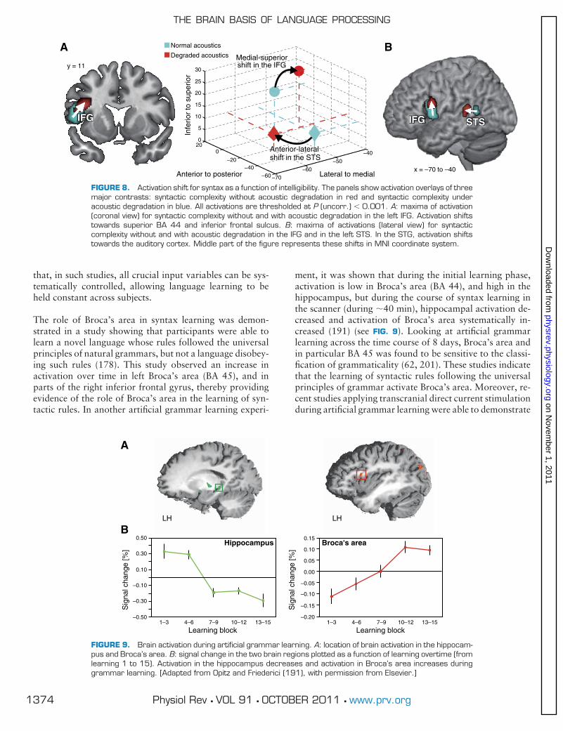
that, in such studies, all crucial input variables can be sys-tematically controlled, allowing language learning to beheld constant across subjects.
The role of Broca’s area in syntax learning was demon-strated in a study showing that participants were able tolearn a novel language whose rules followed the universalprinciples of natural grammars, but not a language disobey-ing such rules (178). This study observed an increase inactivation over time in left Broca’s area (BA 45), and inparts of the right inferior frontal gyrus, thereby providingevidence of the role of Broca’s area in the learning of syn-tactic rules. In another artificial grammar learning experi-
ment, it was shown that during the initial learning phase,activation is low in Broca’s area (BA 44), and high in thehippocampus, but during the course of syntax learning inthe scanner (during �40 min), hippocampal activation de-creased and activation of Broca’s area systematically in-creased (191) (see FIG. 9). Looking at artificial grammarlearning across the time course of 8 days, Broca’s area andin particular BA 45 was found to be sensitive to the classi-fication of grammaticality (62, 201). These studies indicatethat the learning of syntactic rules following the universalprinciples of grammar activate Broca’s area. Moreover, re-cent studies applying transcranial direct current stimulationduring artificial grammar learning were able to demonstrate
FIGURE 8. Activation shift for syntax as a function of intelligibility. The panels show activation overlays of threemajor contrasts: syntactic complexity without acoustic degradation in red and syntactic complexity underacoustic degradation in blue. All activations are thresholded at P (uncorr.) � 0.001. A: maxima of activation(coronal view) for syntactic complexity without and with acoustic degradation in the left IFG. Activation shiftstowards superior BA 44 and inferior frontal sulcus. B: maxima of activations (lateral view) for syntacticcomplexity without and with acoustic degradation in the IFG and in the left STS. In the STG, activation shiftstowards the auditory cortex. Middle part of the figure represents these shifts in MNI coordinate system.
FIGURE 9. Brain activation during artificial grammar learning. A: location of brain activation in the hippocam-pus and Broca’s area. B: signal change in the two brain regions plotted as a function of learning overtime (fromlearning 1 to 15). Activation in the hippocampus decreases and activation in Broca’s area increases duringgrammar learning. [Adapted from Opitz and Friederici (191), with permission from Elsevier.]
THE BRAIN BASIS OF LANGUAGE PROCESSING
1374 Physiol Rev • VOL 91 • OCTOBER 2011 • www.prv.org
on Novem
ber 1, 2011physrev.physiology.org
Dow
nloaded from

that Broca’s area is causally involved in the acquisition andthe processing of syntactic knowledge (44, 246).
These findings, in particular those of Musso et al. (178),require a view of Broca’s area that goes beyond that ofproviding “phonological working memory resources” asstated by Rogalsky and Hickok (219), since the learning ofboth rule types (as in Ref. 178) should require such re-sources. Rather, the findings point towards the specific roleBroca’s area plays in learning syntactic rules. In addition, anumber of artificial grammar studies with grammar thatfollows universal grammar principles have found BA 44activation, in particular for the processing of hierarchicaltree structures compared with local sequential dependen-cies (9, 69, 192). Together, these studies on grammar learn-ing and processing indicate the crucial role of Broca’s areain the processing of syntax.
Comparing artificial grammar sequence processing and sen-tence comprehension in an fMRI directly, Hoen et al. (121)found large parts of the perisylvian cortex activated. Basedon their data, they proposed the following functional sub-division of Broca’s area and its adjacent regions in the pre-frontal cortex (54, 121): superior and posterior regions (BA6/9/46/44) are engaged in sequential and structural aspectsof processing, whereas anterior and inferior regions (BA11/47/45) are implicated in context information insertioninto structural matrixes selected by the upper regions.
D. Localization of Integration: IFG or STG?
Psycholinguistic models on sentences comprehension as-sume a processing phase during which syntactic and seman-tic information interact with each other and are integratedto achieve interpretation. Some models hold that the differ-ent information types interact at any time during compre-hension (163, 169) or after an initial syntactic structurebuilding phase (63, 64). Neuroimaging approaches havediscussed two different regions as possible sites where inte-gration takes place. Some researchers (98) assume that thefinal integration of syntactic and semantic informationtakes place in the left posterior STG, whereas others (102,236) assume that unification of different language-relevantinformation types is located in the left IFG. Interestingly,the crucial neuroimaging studies these proposals are basedon all show activation in both the IFG and the STG (forlocalization, see FIG. 1).
The arguments in favor of the view that the STG is the locusof semantic-syntactic integration come from a cross-studycomparison revealing that activation in the STG is observedonly for sentences containing semantic information,whereas BA 44 is activated both for syntactic processes insentences (structural sequences) without semanticallymeaningful words (9, 190) and in sentences with meaning-ful words (70, 166). The argument for the IFG as the locus
of unification (integration) is based on findings reporting aninteraction of semantic and syntactic information in the leftIFG (e.g., Refs. 150, 217). The unification approach (102)subdivides the IFG functionally into BA 44/6 supportingphonological processes, BA 44/45 supporting syntactic pro-cesses and BA 45/47 supporting semantic processes is made,but defines the entire left IFG as the space where unificationtakes place. Empirical data providing direct evidence ofsuch a unification process are sparse, and a study testing thisview directly concludes that language understanding in-volves a dynamic interplay between the left inferior frontaland the posterior temporal regions (236). The role of theSTG and the IFG in the processes of integration or unifica-tion, respectively, cannot be ultimately defined on the basisof this study, but some additional specifications emergefrom the data available in the literature.
It is clear, however, that the posterior temporal cortex iscrucial in binding the verb and its arguments and moregenerally for integration across domains and that the infe-rior frontal gyrus support different language aspects withinits subregions (BA 47/45/ 44). Interactions between seman-tic aspects and syntax, as seen in studies manipulating se-mantics by lexical-semantic ambiguity (216), semantic re-latedness (184), or semantic constraint due to animacy (34),are located in the more anterior portions of the IFG (BA47/45), but not in BA 44 (184). From this, we may concludethat the IFG’s role as a region of combining semantic andsyntactic information may be restricted to its more anteriorparts.
E. Prosodic Processes
When processing spoken sentences, phonological informa-tion in addition to semantic and syntactic information mustbe processed. We have already discussed acoustic-phono-logical processes at the segmental level, i.e., phonemes andfeatures of these (see sect. IVA). But the acoustic signal alsoconveys suprasegmental phonological information, calledprosody. Two types of prosodic information are usuallydistinguished: emotional prosody and linguistic prosody.Emotional prosody is an extralinguistic cue signaling eitherthe speaker’s emotional state or emotional aspects of thecontent conveyed by the speaker. In the context of thisreview, we will focus on the brain basis of linguistic prosodyonly.
Prosodic information is mainly encoded in the intonationalcontour, which signals the separation of constituents (syn-tactic phrases) in a spoken sentence and the accentuation of(thematically) relevant words in a speech stream. By signal-ing constituent boundaries, this information becomes mostrelevant for sentence comprehension and the interpretationof who is doing what to whom. This can be gathered fromthe example below. In the example, # indicates the prosodicboundary (PB).
ANGELA D. FRIEDERICI
1375Physiol Rev • VOL 91 • OCTOBER 2011 • www.prv.org
on Novem
ber 1, 2011physrev.physiology.org
Dow
nloaded from

1) The man said # the woman is stupid.
2) The man # said the woman # is stupid.
The PBs in these sentences are crucial for the interpretationas they signal the noun phrase to which the attribute “to bestupid” has to be assigned, either to the woman (1) or to theman (2). As the example shows, the prosodic information isrelevant for syntactic processes, and there seems to be aclose relation between prosody and syntax. Indeed, almostevery PB is also a syntactic boundary, while the reverse doesnot hold.
The brain basis of prosodic information has initially beeninvestigated behaviorally in patients with cortical lesions inthe left hemisphere (LH) and the right hemisphere (RH).While some studies came to the conclusion that linguisticprosody is mainly processed in the RH (25, 257), othersfound that both LH and RH patients showed deficits inprocessing sentence level prosody (30). However, when seg-mental information was filtered, thereby increasing the re-liance on suprasegmental information, RH patients demon-strated significantly worse performance than LH patients(30). These and other studies (e.g., Ref. 200) suggest a rel-ative involvement of the RH in processing prosodic infor-mation. The less segmental information there is available,the more dominant the RH.
Neuroimaging studies provide support for this observation.Processing of pitch information (intonational contour) iscorrelated with an activation increase in the RH, but can bemodulated by task demands (205). An fMRI study thatsystematically varied the presence/absence of suprasegmen-tal and segmental information reported changes in brainactivation in the superior temporal and fronto-opercularcortices of the RH as a function of the presence/absence ofpitch information (172, 173) (see FIG. 10). Right dorsolat-eral prefrontal cortex and right cerebellar activation were
also reported for prosodic segmentation during sentenceprocessing (242). A study investigating sentences andword lists both with sentence prosody and word list pros-ody found bilateral activation in the anterior temporalcortex for syntactic and prosodic information, with theleft being more selective for sentence structure (127). Inthis study clear RH dominance was found for prosody,but the authors point out that the activation in the rightanterior temporal cortex may indicate prosody process-ing. Together, the studies suggest an involvement of theRH for the processing of intonational (pitch) informationduring sentence processing, but, in addition, indicate thatthe actual lateralization partly depends on task demands(90, 205) and on the presence of concurrent segmentalinformation (30, 68).
Moreover, it should be noted that the lateralization of lin-guistic prosody depends on the particular information pros-ody encodes in a given language. In tonal languages like, forexample, Thai, pitch patterns are used to distinguish lexicalmeaning. When encoding lexical information, pitch is pro-cessed in the LH, similar to lexical information in non-tonallanguages (91). From this, it appears that the localization oflanguage in the brain is determined by its function (lexicalinformation) and not its form (pitch information). Onlywhen intonation marks suprasegmental prosody, it is local-ized in the RH.
V. TIME COURSE OF AUDITORYLANGUAGE COMPREHENSION
Language undoubtedly unfolds in time. The data avail-able from the fMRI studies on language processing donot provide the sufficient time resolution to capture thiscrucial aspect. The cognitive description of the compre-hension process itself has been laid out in the introduc-tion as consisting of several subprocesses that take place
FIGURE 10. Brain activation during prosodic processes. Activation for sentence level prosodic information.Activation for prosodic speech (no segmental information, only suprasegmental information) vs. normal speech(segmental and suprasegmental information) is color coded in red-yellow, activation for normal speech vs.prosodic speech is color coded in green-blue. [Adapted from Meyer et al. (172), with permission from JohnWiley and Sons.]
THE BRAIN BASIS OF LANGUAGE PROCESSING
1376 Physiol Rev • VOL 91 • OCTOBER 2011 • www.prv.org
on Novem
ber 1, 2011physrev.physiology.org
Dow
nloaded from
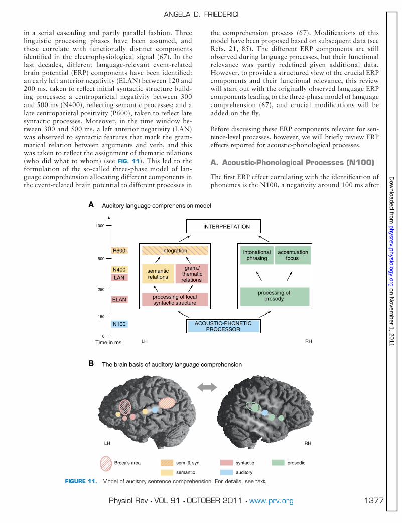
in a serial cascading and partly parallel fashion. Threelinguistic processing phases have been assumed, andthese correlate with functionally distinct componentsidentified in the electrophysiological signal (67). In thelast decades, different language-relevant event-relatedbrain potential (ERP) components have been identified:an early left anterior negativity (ELAN) between 120 and200 ms, taken to reflect initial syntactic structure build-ing processes; a centroparietal negativity between 300and 500 ms (N400), reflecting semantic processes; and alate centroparietal positivity (P600), taken to reflect latesyntactic processes. Moreover, in the time window be-tween 300 and 500 ms, a left anterior negativity (LAN)was observed to syntactic features that mark the gram-matical relation between arguments and verb, and thiswas taken to reflect the assignment of thematic relations(who did what to whom) (see FIG. 11). This led to theformulation of the so-called three-phase model of lan-guage comprehension allocating different components inthe event-related brain potential to different processes in
the comprehension process (67). Modifications of thismodel have been proposed based on subsequent data (seeRefs. 21, 85). The different ERP components are stillobserved during language processes, but their functionalrelevance was partly redefined given additional data.However, to provide a structured view of the crucial ERPcomponents and their functional relevance, this reviewwill start out with the originally observed language ERPcomponents leading to the three-phase model of languagecomprehension (67), and crucial modifications will beadded on the fly.
Before discussing these ERP components relevant for sen-tence-level processes, however, we will briefly review ERPeffects reported for acoustic-phonological processes.
A. Acoustic-Phonological Processes (N100)
The first ERP effect correlating with the identification ofphonemes is the N100, a negativity around 100 ms after
FIGURE 11. Model of auditory sentence comprehension. For details, see text.
ANGELA D. FRIEDERICI
1377Physiol Rev • VOL 91 • OCTOBER 2011 • www.prv.org
on Novem
ber 1, 2011physrev.physiology.org
Dow
nloaded from

stimulus onset (188). This ERP component is not specificto language, but reflects the discrimination of auditorycategories and can thus be used to investigate aspects ofvowel category perception. The same holds for the mis-match negativity (MMN), an ERP component occurringshortly after 100 ms, which has been shown to reflect thediscrimination of acoustic and phoneme categories(180). Studies investigating phoneme perception haveused single phonemes or syllables as stimulus material(for a review, see Refs. 179, 202, 260). The differentstudies indicate language-specific representations at thephoneme and syllable level (45, 203). The latter MMNstudy on the co-occurrence of phonemes in word-likeitems (pseudowords) compared French and Japanese lis-teners. The data suggest that during speech processing,the input signal is directly parsed into the language-specific phonological format of native language.
The N100 and the MMN have been located in or in thevicinity of the auditory cortex (51, 206), thereby indicat-ing that these processes take place early during speechperception in this region. This is compatible with resultsfrom neuroimaging studies on phoneme processing thatlocalize the N100 for vowels and consonants in the HGand PT (186, 188, 233). It has been proposed that the fastcomputation of a phonological representation from thespeech input facilitates lexical access (45) and access tosyntactically relevant morphological information.
B. Initial Syntactic Processes (ELAN)
The first sentence-level ERP component is the ELAN,correlating with the identification of the syntactic cate-gory of a word (e.g., verb, noun, preposition, etc.) occur-ring in response to a word category violation 120 –200ms after word onset or after the part of the word whichprovides the word category information (e.g., the inflec-tion as in refine versus refinement) (80, 129, 145, 183; fora recent review, see Ref. 85). Based on this word categoryinformation, the initial local phrase structure can be built(e.g., verb phrase, noun phrase, prepositional phrase).These phrases are the building blocks for larger sentencestructures. Within the three-phase model of languagecomprehension (67), this initial processing phase consti-tutes phase 1. The initial build up of local phrase struc-ture has been shown to be highly automatic as it is inde-pendent of attentional processes (107) and independentof task demands (106). The earliness of the componentwas attributed to the ease with which word categoryinformation can be extracted from the stimulus, be it tothe word’s shortness (e.g., function word, as in Ref. 183),or its morphological markedness (e.g., inflection, as inRef. 53), or more general for its atypical form properties(52). As this component has been reported mostly forconnected speech (but see Ref. 53, 183; and for otherstudies, see the review of Ref. 85), the question has been
asked to what extent this component might reflect pro-sodic aspects. However, it has been shown that prosodicviolations elicit a different component (right hemisphericanterior negativity) (57) and that changes in the prosodiccontour cannot account for the early syntactic effect(113).
A key question is where in the brain this initial processtakes place. One way to localize language processes on-line is to use MEG, as it provides the possibility for agood topographic resolution (depending on the numberof channels), although the method inherently has to dealwith the so-called inverse problem (i.e., calculation of theneural generator based on scalp distribution data). An-other approach is EEG in patients with circumscribedbrain lesion applying an ERP design known to elicit cer-tain language-related components. With the use of thelatter approach, it was found that the ELAN componentis absent in patients with left frontal cortical lesions (in-cluding left basal ganglia lesions), but present in patientsonly suffering from left basal ganglia lesions, indicatingthat the left frontal cortex plays a crucial role in thegeneration of the ELAN (82). The ELAN is also affectedin patients with lesions in the left anterior temporal lobe,but not in patients with lesions in the right temporal lobe,suggesting that the left frontal and left anterior temporalcortex are involved in early structure building processesas reflected in the ELAN (75). With the use of MEG, theELAN effect has been localized in the anterior temporalcortex and the inferior frontal cortex (84, 142) or solelyin the temporal cortex (115) for auditory language ex-periments. More fine-grained analyses revealed syntacticeffects in the temporal cortex already during the first 200ms after stimulus onset just anterior to the primary audi-tory cortex, i.e., in the anterior STG (114), but not in theprimary auditory cortex itself (see FIG. 12).
For visual experiments, the syntactic violation effectpresent around 100 ms was localized in the visual cortex,at least for sentences in which the word category infor-mation was morphologically marked (53). These datahave raised the possibility that clearly marked syntacticword category violations may be detected in the sensorycortices (53, 115) or in their direct vicinity (114). Thespeed of this process may be surprising. However, theprocess of building up a local structure such as nounphrase (determiner plus noun) or a prepositional phrase(preposition plus noun phrase) on the basis of word cat-egory information could be performed quickly once thepossible minimal local structures in a given language arelearned. Once learned, this process could be viewed as afast template-matching process taking place early in com-prehension (21). During this process, templates of localphrase structures are activated (e.g., a preposition wouldactivate a template of a prepositional phrase), againstwhich the incoming information is checked. If this infor-
THE BRAIN BASIS OF LANGUAGE PROCESSING
1378 Physiol Rev • VOL 91 • OCTOBER 2011 • www.prv.org
on Novem
ber 1, 2011physrev.physiology.org
Dow
nloaded from

mation does not match the template, a phrase structureviolation is detected and further processes are not syn-tactically licensed.
This would predict that sentences containing both aphrase structure violation and semantic violation shouldelicit only an ELAN, but no semantic effect. It has beenshown that this is indeed the case when combining aphrase structure violation with a semantic violation (72,106), and that it even holds when combining it with aviolation of the verb-argument information (86). Thesedata indicate that syntactic phrase structure violationsare processed prior to semantic information and canblock higher-level processes, thereby providing strongevidence for models assuming a crucial initial syntacticprocessing phase (21, 67). Although this conclusion wascalled into question on the basis of an experiment usingDutch language material (249), the data from this studydo not speak against the model’s assumption. This isbecause in this study the syntactic category informationof the critical word (given in the word’s suffix) only be-came available after the semantic information (given inthe word stem). A review of the literature on the timing ofsyntactic information and semantic information acrossthe different languages reveals that the absolute timingof the syntax-initial and other processes may vary, butthat the order of these processes in time is fixed across thedifferent languages with syntactic word category infor-mation being processed first (85).
C. Computation of Syntactic and SemanticRelations (LAN/N400)
A crucial part in the process of sentence comprehension isthe assignment of grammatical relations. To understandwho is doing what to whom, semantic features (e.g., ani-macy) as well as syntactic features (e.g., subject-verb agree-ment, case marking, etc.) have to be processed. Neurolin-guistic models assume that these processes take place afterinitial structure building. In the three-phase model of lan-guage comprehension of Friederici (67) this constitutesphase 2 (see FIG. 11). Bornkessel and Schlesewsky (21) sub-divide this phase 2 into two phases: phase 2a, during whichrelevant features are extracted, and phase 2b, during whichcomputation takes place. In their review they interpret dif-ferent ERP effects observed to different linguistic aspects asinvestigated in various languages in detail (21).
For the purpose of this review, we will summarize themajor findings observed across different languages, witha focus on two ERP components often reported in theliterature, i.e., the LAN found for syntactic and the N400found for semantic-thematic processes. Languages differas to whether they have a fixed word order, like English,for example, or a free word order, such as German orJapanese. To identify who is the subject of sentence, thestrategy in a language with fixed word order is to rely onpositional information (e.g., the first noun phrase is likelyto be the actor). However, in a language with free wordorder, morphosyntactic features must be considered. Sub-
FIGURE 12. Localization of syntactic phrase structure violation and auditory spatial violation. A: displayof the location of activations in each hemisphere for each violation type: strongest grand averageactivations for syntax (red), for space (blue), and for the double violation (syntax & space) (green). B: thetime-space distribution of processing. Results of the local maxima analysis in the temporal cortex forsyntax (red circle), space (blue circle) incongruent deviant condition, and syntax & space (green circle). Theyellow circle marks the auditory cortex as indicated by a sentence onset N100m; *P � 0.05; ***P �0.001. Syntax effects are found between 50 and 180 ms after stimulus onset; they are located anteriorto the auditory cortex in the superior temporal gyrus (STG). [Adapted from Herrmann et al. (114), withpermission from Elsevier.]
ANGELA D. FRIEDERICI
1379Physiol Rev • VOL 91 • OCTOBER 2011 • www.prv.org
on Novem
ber 1, 2011physrev.physiology.org
Dow
nloaded from

ject-verb number agreement [plural (PL) versus singular (SG)]determines who is the subject of the action, but assignment isonly possible if subject and object noun differ in numbermarking as in sentence 1, but not if the two noun phrases ina sentence carry the same number marking asin 2.
1) Die Männer [PL] grüßt [SG] der Junge [SG].
The men greet the boy [Actor] [literal].
The boy greets the men [nonliteral].
2) Die Männer [PL] grüßen [PL] die Jungen [PL].
The men greet the boys.
In an ambiguous situation as in 2, a subject-first strategy isapplied, taking the first noun as the actor. Case marking isan additional feature that can help to resolve ambiguity.There are a number of languages in which thematic roles(actor, patient, etc.) can be determined by case [nominative(NOM) assigns the actor, accusative (ACC) assigns the pa-tient, etc.], thereby allowing the assignment of who is doingwhat to whom as in 3, in which the boy is the actor.
3) Den Mann [SG, ACC] grüßt der Junge [SG, NOM].
The man greets the boy [Actor] [literal].
The boy greets the man [nonliteral].
If morphosyntactic cues are not available or are ambiguousas in 2, the system might rely on a simple subject-first wordorder strategy, or it might consider semantic features, suchas animacy. Since the prototypical actor is animate, thisinformation may help to assign the role of the actor, but notalways (e.g., in the sentence “The tree hit the man whenfalling,” the animacy-strategy could lead to an initial mis-assignment of the tree’s role, as tree is an inanimate noun).Nevertheless, the parsing system has to assign thematicroles on-line as the sentence is perceived in order to keep theworking memory demands low, even if initial assignmentsmust be reanalyzed later in the sentence.
1. Processing semantic and verb-argument relations
Sentence understanding crucially depends on extraction ofthe sentence’s meaning, that is on the meaning of differentwords and the relation between them. Since the first ERPpaper on language processing (152), a specific ERP compo-nent has been correlated with the processing of semanticinformation. This ERP component is a centro-parietal neg-ativity around 400 ms, called N400. An almost uncount-able number of papers have been published on semanticprocesses both at the word level and sentence level acrossdifferent languages (for recent reviews, see Refs. 153, 156).
The N400 is interpreted as reflecting difficulty of lexical-semantic integration, as its amplitude is known to increase1) when a word does not have a lexical status (i.e., a non-word or a pseudoword); 2) when the second word of a wordpair does not fit the first word semantically, and in a sen-tence 3) when the selectional restriction of verb-argumentrelations is violated; 4) when a word does not fit the pre-ceding sentence context with respect to world knowledge oris, moreover, simply unexpected; and 5) its amplitude isknown to decrease for words as the sentence unrolls due toincreased predictability of the upcoming word. Thus the N400is an indicator of 1) lexical processes, 2) lexical-semantic pro-cesses, 3) semantic contextual predictability, and 4) predict-ability due to world knowledge. Therefore, it reflects processesrelevant to language comprehension at different levels, but notonly those that are language internal but also those that con-cern world knowledge (105). The present review, however,will focus on the language internal level.
At this level, the N400 is correlated with semantic informa-tion carried by nouns and adjectives, and also with verb-internally information represented. This information isquite complex and partly concerns the semantic domain(i.e., selectional restriction information) and partly the syn-tactic domain (i.e., number and type of arguments). Selec-tional restriction information of the verb indicates whichtheoretically defined semantic features the related noun ar-gument(s) must have. For example, the verb “drink” re-quires the noun to have the feature of “liquid,” as in “drinkthe wine” and not “drink the chair.” For the latter type ofcombination, an N400 is observed at the violating noun(during reading, Ref. 152; and during listening, Refs. 80,123). Most interestingly, in one of the more recent studies,it has been shown that the amplitude of the N400 increasessystematically as a function of the number of semantic fea-tures violating the relation between the verb and its nounargument (159) (see FIG. 13). This is a strong demonstrationof the N400’s modulation by theoretically defined semanticaspects of a word.
The N400 has also been observed in the verb’s syntax-related domain when it comes to processing the informationof how many arguments a verb can take. For example,linguistic theory defines that the verb “cry” only takes oneargument, “she cries,” whereas the verb “give” takes threearguments, “she gave a letter to Peter.” Moreover, the verbencodes the type of the arguments (subject, direct object,indirect objects) which in some languages is marked byposition in the sentence (word order) and in other languagesby case (inflection or preposition), e.g., “to Peter.” Viola-tions of the number of arguments and types of arguments(incorrect case marking) in a sentence lead to an N400followed by a late positivity (71, 86, 88). Thus the ERPviolation of number and type of arguments (syntactic do-main) differs from that of violations of selectional restric-tions (semantic domain), as the former is reflected in a bi-
THE BRAIN BASIS OF LANGUAGE PROCESSING
1380 Physiol Rev • VOL 91 • OCTOBER 2011 • www.prv.org
on Novem
ber 1, 2011physrev.physiology.org
Dow
nloaded from

phasic pattern N400/P600, whereas the latter is reflected ina N400. (For variations of the particular realization of ar-gument-related negativity as a function of different lan-guage typologies, see Ref. 21.) Thus semantic and thematicprocesses during language comprehension are correlatedwith the N400 across different languages.
This leads to the question of where in the brain these pro-cesses take place. There are a number of MEG studies, bothat the word and sentential level, that have tried to localizethe semantic N400 effect. The main generators of the N400during speech processing have been located in the vicinity ofthe auditory cortex (108, 112), sometimes with an addi-tional generator in the inferior frontal cortex (164). FMRIexperiments using the same stimulus material as used in anERP experiment on the processing of selectional restrictionviolations (106) revealed activation mainly in the STG (midportion and posterior portion) (27, 81). The number andtype of verb-argument relations eliciting an N400 have notbeen investigated using the same material in ERP and fMRIexperiments. However, fMRI experiments on this issue sug-gest an involvement of the left posterior STG in addition tothe IFG (22). Further research must show whether the N400observed in response to semantic information and the N400in the N400/P600 pattern found for the syntax-related in-formation in the verb is a unitary component or whether theN400 differs as a function of information type.
2. Processing grammatical relations
In parallel to the processing of semantic and verb-argumentinformation, morphosyntactic information provided by theverb’s inflection (number and person) is most relevant forsentence comprehension, as it is essential for the assignmentof grammatical roles in a sentence. While this information isless important for sentence interpretation in languages with
fixed word order, it is crucial for languages with free wordorder (compare sect. IVC).
Violations of subject-verb agreement (singular versus plu-ral) in an inflecting language usually induce a LAN between300 and 500 ms (German, Ref. 199; Italian, Ref. 7; Spanish,Ref. 234). In a fixed word order language such as English,an LAN is found less systematically (LAN in Ref. 195, butnot in Refs. 151, 196). It has been argued that the presence/absence of the LAN should be viewed as a continuum acrossdifferent languages, and the likelihood of observing thiseffect increases with the amount of morphosyntactic mark-ing in a given language (85).
However, it is not the pure amount of morphosyntacticmarking that determines the presence of the LAN, butwhether this information is crucial for the assignment ofsyntactic roles. In some languages, determiner-noun agree-ment with respect to gender (masculine, feminine, neuter) iscrucial, and in others it is not. If this information is notcrucial for the assignment of grammatical relations betweena verb and its arguments in sentences, a violation of genderagreement between determiner and noun does not lead to astrong LAN effect. However, once gender agreement is rel-evant for the assignment of grammatical roles, as in He-brew, in which there is gender agreement between subjectnoun and verb, the LAN is clearly present (50). Thus, when-ever morphosyntactic marking is crucial for the assignmentof grammatical relations in a sentence, an LAN is observed.
D. Integration and Interpretation (P600)
Models on the time course of language processes have as-sumed a late processing phase during which different infor-mation types are mapped onto each other to achieve inter-pretation (21, 67, 75). Friederici (67) proposed that this last
FIGURE 13. Effects of semantic anomaly of object nouns in verb-noun combinations. ERP grand averages forthe four different conditions. The amplitude of the N400 increases as the “inappropriateness” of the targetobject noun in verb-noun combinations increases. The increase of “inappropriateness” is defined as the numberof mismatching semantic features of the noun. Semantic features are, for example, � human, � animate, etc.The most inappropriate noun for the verb in this case is an inanimate object noun leading to the largestamplitude of the N400. [Adapted from Li et al. (159), with permission from MIT Press.]
ANGELA D. FRIEDERICI
1381Physiol Rev • VOL 91 • OCTOBER 2011 • www.prv.org
on Novem
ber 1, 2011physrev.physiology.org
Dow
nloaded from

phase (phase 3) represents a phase during which processesof syntactic reanalysis and repair take place and that theseprocesses are reflected in a late centro-parietal positivity,called P600. This component, first observed for the process-ing of syntactic anomalies (193), was found for the process-ing of temporarily ambiguous sentences at the point of dis-ambiguation when reanalysis was necessary (194), and alsoafter a syntactic violation requiring repair (104), and some-times as part of a biphasic ELAN/P600 pattern (80, 107). Adirect comparison of the P600 topography in both instancesrevealed a differential pattern of distribution with a morefronto-central distribution for the reanalysis P600 and acentro-parietal distribution for the repair P600 (74) (seeFIG. 14).
The functional interpretation of the P600 has changed tosome degree over the past years. Initially, it was taken toreflect syntactic processes in general (104), processes of syn-tactic reanalysis and repair (73), or the difficulty of syntacticintegration (136). However, later studies found the P600 tovary not only as a function of syntactic variables, but also toreflect the interaction of syntactic and semantic anomaly atthe sentence level (100, 147, 148), suggesting that the P600might reflect sentence-level integration processes of syntac-
tic and semantic information. More recently, the status ofthe P600 as reflecting integration processes involving syn-tactic aspect was challenged by studies reporting P600 ef-fects for sentence-level semantic violations (120, 139, 144,149). For example, sentences like “The hearty meal wasdevouring” led to a P600 (139). Different explanationswere put forward for “semantic P600” effects: 1) plausibil-ity/semantic attraction between the verb and an argument(139), 2) thematic processing cost (120), and 3) interactionof thematic and semantic memory (149). Interestingly, allthese different interpretations concern aspects of thematicrole assignment in sentences and can be explained in anexisting linguistically based processing model (23).
The brain basis of P600 effects is still unclear, as the P600has not been localized using time-sensitive neuroimagingmeasures with the exception of a few MEG studies (154,231). These MEG studies localized the P600 in the mid-dle temporal gyrus and the posterior portion of the tem-poral cortex. Moreover, there is some indication that thebasal ganglia are part of the circuit supporting processesreflected in the syntax-related P600, since patients withlesions in the basal ganglia show reduced P600 ampli-tudes (82, 87). An involvement of the basal ganglia insyntactic processes has also been proposed in the modelby Ullman (247), although not specifically for the lateprocessing phase reflected by the P600. The localizationof the P600 in the fMRI is difficult, as the P600 oftenoccurs in close vicinity in time with the LAN or N400 andis thus difficult to separate from these effects. At present,therefore, the neural basis underlying the P600 effect hasnot yet been specified in much detail.
In summary, the data available on the neurotemporal dy-namics of language comprehension can be described as fol-lows. Language comprehension is incremental and takesplace in three sequential phases. In an initial phase (phase1), an initial phrase structure on the basis of word categoryinformation is built. This process is highly automatic, inde-pendent of semantic and verb argument information, andindependent of task demands. The process involves a por-tion of the left STG immediately anterior to the primaryauditory cortex, possibly connecting to the frontal opercu-lum located ventrally to Broca’s area. During a secondphase (phase 2), the relation between the verb and its argu-ments is computed to assign the thematic roles in a sentence.Morphosyntactic information (subject-verb agreement,LAN), case information (LAN or N400, depending on theparticular language), and lexical selectional restriction in-formation (N400) are taken into consideration to achieveassignment of the relation between the different elements ina sentence. The on-line assignment of semantic relationsmainly appears to involve the mid and posterior portion ofthe temporal cortex. Processes of subject-verb agreementhave not been clearly localized, but the distribution of theLAN suggests an involvement of the left frontal cortex.
FIGURE 14. Distribution of P600 for sentence repair and forsentence reanalysis. The distribution of the P600 differs as a func-tion of the underlying syntactic process. A: topographic maps of theP600 as a function of sentence repair, i.e., the difference betweenthe ERP response to incorrect and correct simple sentences forthree time windows. B: topographic maps of the P600 as a functionof syntactic complexity in correct sentences, i.e., the differencebetween the ERP response to complex and simple sentences forthree time windows. The distribution of the Repair P600 is centro-parietal, whereas the Reanalysis P600 is fronto-central. [Adaptedfrom Friederici et al. (74), with kind permission from Springer Sci-ence � Business Media.]
THE BRAIN BASIS OF LANGUAGE PROCESSING
1382 Physiol Rev • VOL 91 • OCTOBER 2011 • www.prv.org
on Novem
ber 1, 2011physrev.physiology.org
Dow
nloaded from

During a last phase (phase 3), the final interpretation takesplace, with semantic and syntactic information being takeninto account and mapped onto world knowledge. At thelinguistic level, the difficulty of integrating syntactic andsemantic information and the need for reanalysis is reflectedin a P600. The difficulty of mapping linguistic informationonto world knowledge also appears to elicit a P600 effect.At the moment it remains open whether these two P600effects are members of the same family of ERP componentsor not.
E. Prosodic Processes (CPS)
The processing of auditorily presented sentences not onlyrequires the processing of semantic and syntactic informa-tion but, moreover, the processing of prosodic information.The first electrophysiological correlate for the processing ofsentence-level prosodic information was found in a studythat recorded the EEG during the processing of Germansentences which either contained one intonational phraseboundary (IPB) or two. At the IPB, the ERPs revealed acentro-parietally distributed positive shift that was calledthe closure positive shift (CPS) since the IPB indicates theclosure of a phrase (238) (see FIG. 15). This effect was
replicated in other studies using a different language,namely, Dutch (19, 137), Japanese (262), Chinese (160),and English (130). Crucially, it was shown that the CPS isnot triggered by the pause at the IPB per se, but that the twoother parameters signaling the IPB, namely, the pitchchange and the lengthening of the syllable prior to the pauseare sufficient to evoke boundary perception. This was evi-denced in an experiment in which the pause at the IPB wasdeleted (238).
Interestingly, the latter does not hold for young children.In infants and toddlers, a boundary response is not elic-ited when the pause is deleted, but only when the pause ispresent (167, 168). However, in older children, whoshow a CPS as boundary response once sufficient syntac-tic knowledge is acquired, pitch information and sylla-ble-lengthening alone can trigger a CPS, just as in adults(167). This suggests that the pause initially serves as arelevant cue to structure the speech input, but that it isnot needed for intonational phrasing once sufficientknowledge about prosodic and syntactic structure areacquired. Additional experiments with adults showedthat the CPS can also be elicited when only prosodicinformation of a sentence is delivered (i.e., when segmen-tal information is deleted), under this condition the CPSis lateralized to the RH (197). Moreover, the CPS isreported for sentence reading triggered by the commaindicating the syntactic phrase boundary (138, 237,239). Thus the CPS can be viewed as an ERP componentto correlate with prosodic phrasing both when realizedopenly in the speech stream and when realized covertly inwritten sentences.
F. Interaction of Syntax and Prosody
Syntax and prosody are known to interact during languagecomprehension as indicated by behavioral studies on syn-tactic ambiguity resolution (170, 254). The studies cited insection IV indicate that syntax is mainly processed in theLH and prosody as such mainly in the RH. The two hemi-spheres are neuroanatomically connected via the corpuscallosum (122, 124). If the above view about the functionalrole of the LH and RH in language processing is valid, anyinteraction between syntactic (LH) and prosodic (RH) in-formation should be affected by a lesion to the corpus cal-losum (CC).
The prosody-syntax interaction may take place duringdifferent processing phases: 1) during the initial phase ofphrase structure building since the end of a syntacticphrase is marked prosodically, and/or 2) during the sec-ond processing phase during which the verb argumentstructure is processed, since the constituent structure isalso prosodically marked. In the following, we will takeup these issues in turn.
FIGURE 15. The closure positivity shift (CPS) for intonational phraseboundary processing. Intonational phrase boundary (IPB) processing iscorrelated with a particular ERP component, the CPS. Grand-averageERPs at the Pz electrode. The waveforms for sentences with twointonational phrases (IPh) and one intonational phrase boundary (IPB)(green) and sentences with three IPhs and two IPBs (blue) are super-imposed. The onsets of the sentence examples (black arrow) arealigned to the time axis in milliseconds (ms). Both conditions evoke aCPS (positive going wave form) at their respective IPBs indicated byarrows. [Adapted from Steinhauer et al. (238), with permission fromNature Publishing Group.]
ANGELA D. FRIEDERICI
1383Physiol Rev • VOL 91 • OCTOBER 2011 • www.prv.org
on Novem
ber 1, 2011physrev.physiology.org
Dow
nloaded from
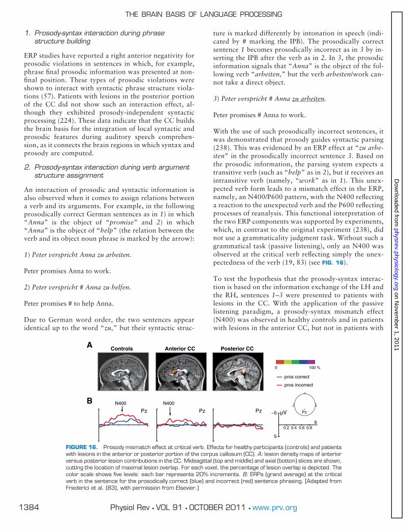
1. Prosody-syntax interaction during phrasestructure building
ERP studies have reported a right anterior negativity forprosodic violations in sentences in which, for example,phrase final prosodic information was presented at non-final position. These types of prosodic violations wereshown to interact with syntactic phrase structure viola-tions (57). Patients with lesions in the posterior portionof the CC did not show such an interaction effect, al-though they exhibited prosody-independent syntacticprocessing (224). These data indicate that the CC buildsthe brain basis for the integration of local syntactic andprosodic features during auditory speech comprehen-sion, as it connects the brain regions in which syntax andprosody are computed.
2. Prosody-syntax interaction during verb argumentstructure assignment
An interaction of prosodic and syntactic information isalso observed when it comes to assign relations betweena verb and its arguments. For example, in the followingprosodically correct German sentences as in 1) in which“Anna” is the object of “promise” and 2) in which“Anna” is the object of “help” (the relation between theverb and its object noun phrase is marked by the arrow):
1) Peter verspricht Anna zu arbeiten.
Peter promises Anna to work.
2) Peter verspricht # Anna zu helfen.
Peter promises # to help Anna.
Due to German word order, the two sentences appearidentical up to the word “zu,” but their syntactic struc-
ture is marked differently by intonation in speech (indi-cated by # marking the IPB). The prosodically correctsentence 1 becomes prosodically incorrect as in 3 by in-serting the IPB after the verb as in 2. In 3, the prosodicinformation signals that “Anna” is the object of the fol-lowing verb “arbeiten,” but the verb arbeiten/work can-not take a direct object.
3) Peter verspricht # Anna zu arbeiten.
Peter promises # Anna to work.
With the use of such prosodically incorrect sentences, itwas demonstrated that prosody guides syntactic parsing(238). This was evidenced by an ERP effect at “zu arbe-iten” in the prosodically incorrect sentence 3. Based onthe prosodic information, the parsing system expects atransitive verb (such as “help” as in 2), but it receives anintransitive verb (namely, “work” as in 1). This unex-pected verb form leads to a mismatch effect in the ERP,namely, an N400/P600 pattern, with the N400 reflectinga reaction to the unexpected verb and the P600 reflectingprocesses of reanalysis. This functional interpretation ofthe two ERP components was supported by experiments,which, in contrast to the original experiment (238), didnot use a grammaticality judgment task. Without such agrammatical task (passive listening), only an N400 wasobserved at the critical verb reflecting simply the unex-pectedness of the verb (19, 83) (see FIG. 16).
To test the hypothesis that the prosody-syntax interac-tion is based on the information exchange of the LH andthe RH, sentences 1–3 were presented to patients withlesions in the CC. With the application of the passivelistening paradigm, a prosody-syntax mismatch effect(N400) was observed in healthy controls and in patientswith lesions in the anterior CC, but not in patients with
FIGURE 16. Prosody mismatch effect at critical verb. Effects for healthy participants (controls) and patientswith lesions in the anterior or posterior portion of the corpus callosum (CC). A: lesion density maps of anteriorversus posterior lesion contributions in the CC. Midsagittal (top and middle) and axial (botton) slices are shown,cutting the location of maximal lesion overlap. For each voxel, the percentage of lesion overlap is depicted. Thecolor scale shows five levels: each bar represents 20% increments. B: ERPs (grand average) at the criticalverb in the sentence for the prosodically correct (blue) and incorrect (red) sentence phrasing. [Adapted fromFriederici et al. (83), with permission from Elsevier.]
THE BRAIN BASIS OF LANGUAGE PROCESSING
1384 Physiol Rev • VOL 91 • OCTOBER 2011 • www.prv.org
on Novem
ber 1, 2011physrev.physiology.org
Dow
nloaded from

lesions in the posterior CC (83) (see FIG. 16). This findingprovides clear evidence for the view that the interactionof prosody and syntax relies on communication betweenthe LH and the RH supported by the posterior portion ofthe CC through which the temporal cortices of the leftand the right hemisphere are connected.
VI. LANGUAGE FUNCTION: BINDINGSPATIALLY DISTRIBUTED NEURALACTIVITY IN TIME
A. The Model
The review has specified the brain areas in the temporal andinferior frontal cortex supporting different aspects of lan-guage processing, for example, phonetic, syntactic, sen-tence-level semantic, and prosodic processes (compare FIG.11; and for anatomical details, see Figs. 1 and 3). Acoustic-phonological processes taking place during the first 100 msafter acoustic stimulation crucially involve the primary au-ditory cortex (PAC) and the planum temporale (PT). Fromthese regions, the information is delivered to the anteriorand the posterior STG and STS, with the left anterior STSreacting generally as a function of the intelligibility of thestimulus. The anterior STG, together with the left frontaloperculum connected via a ventral pathway through theuncinate fasciculus (ventral pathway II), is seen as a possibleneural network for initial local structure building processestaking place between 120–200 ms.5 Semantic and syntacticrelations in a sentence are processed between 300–500 msafter the stimulus onset, possibly in parallel systems, acti-vating separable left-lateralized temporo-frontal networks.The semantic network involves the middle and posteriorSTG/MTG (sometimes extending into the anterior temporalcortex) and BA 45 (and BA 47) in the frontal cortex con-nected via another ventral pathway (ventral pathway I)through the extreme capsule fiber system (ECFS),6 whereasthe syntactic network dealing with complex sentencestructures involves the posterior STG/STS and BA 44 inthe frontal cortex connected via a dorsal pathway (dorsalpathway II). Note that dorsal pathway I connecting thetemporal cortex to the premotor cortex is supposed tosupport sensory-to-motor mappings. Syntactic and se-mantic integration processes take place �600 ms afterthe stimulus input and beyond, possibly under the in-volvement of the posterior STG/STS and the basal gan-glia. The processing of suprasegmental prosodic informa-tion is supported by the right hemisphere in close inter-
action with the left hemisphere through the posteriorportion of the CC, the structure which connects the tem-poral cortices of the two hemispheres.
B. Caveats and Open Issues
The model presented is a model based on empirical data,but it is a model and thereby subject to changes on the basisof new data. Moreover, it should be kept in mind that amodel tries to cover most of the data in the literature, butcertainly cannot include each and every data point pub-lished. A model always is a generalization.
With this in mind, we should now briefly consider the weak-nesses all such models might include.
1. Neuroanatomic variability
The model is based on data from imaging studies, whichusually present group data that are averaged over a group ofsubjects (usually using spatial smoothing algorithms) andmapped onto a standard brain. We know, however, that theneuroanatomic variability between subjects is quite consid-erable (4, 5, 268). Different approaches have been proposedto deal with this problem. One way is to discuss the ob-served group activation with respect to its probability to fallinto one or the other cytoarchitectonically defined area.Such probabilities have been calculated on the basis of 10brains analyzed post mortem in the “Jülich maps” (Ref. 5;compare sect. IIIA). This approach has already been suc-cessfully applied for language-related studies (9, 166, 225,226). A second approach would be to calculate connectivi-ty-based parcellations for each individual (8) and localizethe language-related activation according to this parcella-tion. So far, no study using this approach has been pub-lished, but there is work in progress (Amunts, Tittgemeyer,and Friederici, unpublished data). As a third approach, theuse of a functional localizer task has been proposed, and, inthe case of language studies, this would be a particularlanguage task (59). The idea is that a localizer task reliablyactivates locations across individuals, which can then betaken as the “same” functional region in different brains[see Grodzinsky (97) and Fedorenko and Kanwisher (59)for a discussion of this approach]. Such a localizer task hasbeen applied in a recent study to define a particular regionfor a region-of-interest analysis (218), and there is work inprogress applying this approach more broadly to languagestudies (59).
Thus several methodological approaches are being devel-oped to address the variability in neuroanatomy andthereby the functional neuroanatomy for a particular lan-guage function. This is of particular importance when try-ing to specify a fine-grained distinction in adjacent areas,such as activation in BA 44 versus BA 45 or activation in thefrontal operculum versus the anterior insula.
5This pathway may not only serve to support adjacent structuraldependencies but, moreover, to subserve semantic combinatorics.
6The processing of word semantics involves a large neural net-work including the middle and posterior part of the middle andsuperior temporal gyrus (including the angular gyrus and frontalassociation areas). For recent reviews, see Refs. 15 and 49.
ANGELA D. FRIEDERICI
1385Physiol Rev • VOL 91 • OCTOBER 2011 • www.prv.org
on Novem
ber 1, 2011physrev.physiology.org
Dow
nloaded from

2. Cross-linguistic variability
Another critical issue to be considered is to what extent afunctional neuroanatomic model of language processingbased on data mostly from English, German, Dutch, He-brew and, in a few cases, Japanese and Thai can be taken tobe valid in general. There is a “yes” and a “no” answer tothis question. The affirmative answer is based on the findingthat, across different languages, it is the particular languagefunction that determines particular activation patterns andnot its form. This is evidenced by the finding that syntacticprocesses in fixed word order languages such as English andDutch as well as in free word order languages such as Ger-man and Hebrew all show activation in Broca’s area (TABLE4), and moreover, by the finding that prosody (normallyprocessed in the RH) is processed in the LH when signalinga lexical function. The more negative answer to the gener-ality question is that there are certain neurocognitive pro-cessing differences observable in the language-related ERPpatterns, in particular when investigating the different cuesused to assign thematic role in a sentence. This issue hasbeen taken up by a recent neurotypological approach de-scribing the brain basis of language processing (24).
3. Domain specificity
This is a significant issue in the discussion of a functionalneuroanatomic model of language. The present model, aswell as an earlier version of it (67, 68), relates a particularfunction to a particular brain region within the languagesystem, leaving the option open that this same brain regionserves another function in another domain than language.The particular function the same region supports in theother domain may either be closely related, as for example,the syntactic function of Broca’s area in language and music(165), or the function in the other domain may not be thatsimilar, as, for example, the role of Broca’s area in languageand in processing simple chunks in goal-directed actions(143). The ongoing discussion about the specificity of aparticular area, be it the posterior STG or be it Broca’s area(see Refs. 99, 111), is hard to reconcile given the data avail-able.
In this article, we described the function of a given brainarea within the language processing domain. Taking amore general perspective, we suggest that a given area,for example, Broca’s area, receives its particular domain-specific function as part of a particular domain-specificnetwork which, for the language domain, involves theposterior STG and which, for the action domain, involvesthe parietal cortex (128). Thus the function of an areashould always be considered within a neural network ofwhich it is a part.
Future work will have to deal with these open issues toallow not only a more detailed description of the brainbasis of language, but moreover, to clarify the function of
certain brain regions in the concert of cognitive func-tions.
ACKNOWLEDGMENTS
I thank the Center for Advanced Study in the BehavioralSciences at Stanford University for providing the tranquilitythat allowed me to write the article. I am most thankful toMelanie Trümper and Margund Greiner for their patiencein going with me through several versions of the text andreference lists, and to Andrea Gast-Sandmann for designingthe figures. I also thank Jonas Obleser for his input on earlyauditory processes. Michiru Makuuchi and Emiliano Zac-carella were of great help in constructing tables and relatedfigures. Further thanks to Jens Brauer, Claudia Männel,Jutta Mueller, Lars Meyer, Björn Herrmann, Daniela Sam-mler, Sarah Gierhan, and Jürgen Weissenborn for theirhelpful comments and suggestions. I thank Rosie Wallis forher careful and thoughtful English editing activities. Finally,I am grateful to Cathy Price and one anonymous reviewerfor the helpful comments on the manuscript.
Address for reprint requests and other correspondence:A. D. Friederici, Max Planck Institute for Human Cognitiveand Brain Sciences, Stephanstraße 1A, 04103 Leipzig, Ger-many (e-mail: [email protected]).
GRANTS
The research reported here was partly supported by TheGerman Ministry of Education and Research (BMBF;Grant 01GW0773).
DISCLOSURES
No conflicts of interest, financial or otherwise, are declaredby the author.
REFERENCES
1. Alexander MP, Hiltbrunner B, Fischer RS. Distributed anatomy of transcortical sen-sory aphasia. Arch Neurol 46: 885–892, 1989.
2. Amedi A, von Kriegstein K, van Atteveldt NM, Beauchamp MS, Naumer MJ. Func-tional imaging of human crossmodal identification and object recognition. Exp BrainRes 166: 559–571, 2005.
3. Amunts K, Lenzen M, Friederici AD, Schleicher A, Morosan P, Palomero-Gallagher N,Zilles K. Broca’s region: novel organizational principles and multiple receptor map-ping. PLoS Biol 8: e1000489, 2010.
4. Amunts K, Malikovic A, Mohlberg H, Schormann T, Zilles K. Brodmann’s areas 17 and18 brought into stereotaxic space-where and how variable? NeuroImage 11: 66–84,2000.
5. Amunts K, Schleicher A, Bürgel U, Mohlberg H, Uylings HB, Zilles K. Broca’s regionrevisited: cytoarchitecture and intersubject variability. J Comp Neurol 412: 319–341,1999.
6. Amunts K, Zilles K. Advances in cytoarchitectonic mapping of the human cerebralcortex. Neuroimaging Clin N Am 11: 151–169, 2001.
THE BRAIN BASIS OF LANGUAGE PROCESSING
1386 Physiol Rev • VOL 91 • OCTOBER 2011 • www.prv.org
on Novem
ber 1, 2011physrev.physiology.org
Dow
nloaded from

7. Angrilli A, Penolazzi B, Vespignani F, De Vincenzi M, Job R, Ciccarelli L, Palomba D,Stegagno L. Cortical brain responses to semantic incongruity and syntactic violation inItalian language: an event-related potential study. Neurosci Lett 322: 5–8, 2002.
8. Anwander A, Tittgemeyer M, von Cramon DY, Friederici AD, Knösche TR. Connec-tivity-based parcellation of Broca’s area. Cereb Cortex 17: 816–825, 2007.
9. Bahlmann J, Schubotz R, Friederici AD. Hierarchical artificial grammar processingengages Broca’s area. NeuroImage 42: 525–534, 2008.
10. Bahlmann J, Schubotz RI, Mueller JL, Köster D, Friederici AD. Neural circuits ofhierarchical visuo-spatial sequence processing. Brain Res 1298: 161–170, 2009.
11. Behrens TEJ, Johansen-Berg H, Woolrich MW, Smith SM, Wheeler-Kingshott CAM,Boulby PA, Barker GJ, Sillery EL, Sheehan K, Ciccarelli O, Thompson AJ, Brady JM,Matthews PM. Non-invasive mapping of connections between human thalamus andcortex using diffusion imaging. Nat Neurosci 6: 750–757, 2003.
12. Ben-Shachar M, Hendler T, Kahn I, Ben-Bashat D, Grodzinsky Y. The neural reality ofsyntactic transformations-evidence from fMRI. Psychol Sci 13: 843–440, 2003.
13. Ben-Shachar M, Palti D, Grodzinsky Y. Neural correlates of syntactic movement:converging evidence from two fMRI experiments. NeuroImage 21: 1320–1336, 2004.
14. Berthier ML. Unexpected brain-language relationships in aphasia: evidence fromtranscortical sensory aphasia associated with frontal lobe lesions. Aphasiology 15:99–130, 2001.
15. Binder JR, Desai RH, Graves WW, Conant LL. Where is the semantic system? A criticalreview and meta-analysis of 120 functional neuroimaging studies. Cereb Cortex 19:2767–2796, 2009.
16. Binder JR, Frost JA, Hammeke TA, Bellgowan PS, Springer JA, Kaufman JN, PossingET. Human temporal lobe activation by speech and nonspeech sounds. Cereb Cortex10: 512–528, 2000.
17. Biswal B, Yetkin FZ, Haughton VM, Hyde JS. Functional connectivity in the motorcortex of resting human brain using echo-planar MRI. Magn Reson Med.34: 537–541,1995.
18. Blesser B. Speech perception under conditions of spectral transformation. I. Phoneticcharacteristics. J Speech Hear Res 15: 5–41, 1972.
19. Bögels S, Schriefers H, Vonk W, Chwilla DJ, Kerkhofs R. The interplay betweenprosody and syntax in sentence processing: the case of subject- and object-controlverbs. J Cogn Neurosci 22: 1036–1053, 2010.
20. Bookheimer S. Functional MRI of language: new approaches to understanding thecortical organization of semantic processing. Annu Rev Neurosci 25: 151–88, 2002.
21. Bornkessel I, Schlesewsky M. The extended Argument Dependency Model: a neuro-cognitive approach to sentence comprehension across languages. Psychol Rev 113:787–821, 2006.
22. Bornkessel I, Zyssett S, Friederici AD, von Cramon DY, Schlesewsky M. Who didwhat to whom? The neural basis of argument hierarchies during language compre-hension. NeuroImage 26: 221–233, 2005.
23. Bornkessel-Schlesewsky I, Schlesewsky M. An alternative perspective on “semanticP600” effects in language comprehension. Brain Res Rev 59: 55–73, 2008.
24. Bornkessel-Schlesewsky I, Schlesewsky M. The role of prominence information in thereal time comprehension of transitive constructions: a cross-linguistic approach. LangLinguist Compass 3: 19–58, 2009.
25. Bradvik B, Dravins C, Holtas S, Rosen I, Ryding E, Ingvar D. Disturbances of speechprosody following right hemisphere infarcts. Acta Neurol Scand 84: 114–126, 1991.
26. Brauer J, Anwander A, Friederici AD. Neuroanatomical prerequisites for languagefunctions in the maturing brain. Cereb Cortex 21: 459–466, 2011.
27. Brauer J, Friederici AD. Functional neural networks of semantic and syntactic pro-cesses in the developing brain. J Cogn Neurosci 19: 1609–1623, 2007.
28. Broca P. Remarques sur le siége de la faculté du langage articulé, suivies d’une obser-vation d’aphémie (parte de la parole). Bull Soc Anat Paris 6: 330–357, 1861.
29. Brodmann K. Beiträge zur histologischen Lokalisation der Grosshirnrinde. VI. DieCortexgliederung des Menschen. J Psychol Neurol 10: 231–246, 1909.
30. Bryan K. Language prosody and the right hemisphere. Aphasiology 3: 285–299, 1989.
31. Calvert GA. Crossmodal processing in the human brain: insights from functionalneuroimaging studies. Cereb Cortex 11: 1110–1123, 2001.
32. Caplan D. Functional neuroimaging studies of syntactic processing. J Psycholinguist Res30: 297–320, 2001.
33. Caplan D. Task effects on BOLD signal correlates of implicit syntactic processing. LangCogn Process 25: 866–901, 2010.
34. Caplan D, Chen E, Waters G. Task-dependent and task-independent neurovascularresponses to syntactic processing. Cortex 44: 257–275, 2008.
35. Caplan D, Vijayan S, Kuperberg G, West C, Waters G, Greve D, Dale AM. Vascularresponses to syntactic processing: event-related fMRI study of relative clauses. HumBrain Mapp 15: 26–28, 2002.
36. Caplan D, Waters GS. Short-term memory and language comprehension: a criticalreview of the neuropsychological literature. In: Neuropsychological Impairments ofShort-Term Memory, edited by Vallar G, Shallice T. Cambridge, UK: Cambridge Univ.Press, 1990, p. 337–389.
37. Caplan D, Waters GS. Verbal working memory and sentence comprehension. BehavBrain Sci 22: 77–94, 1999.
38. Catani M, de Schotten MT. A diffusion tensor imaging tractography atlas for virtual invivo dissections. Cortex 44: 1105–1132, 2008.
39. Catani M, Jones DK, Ffytche DH. Perisylvian language networks of the human brain.Ann Neurol 57: 8–16, 2005.
40. Constable RT, Pugh KP, Berroya E, Mencl WE, Westerveld M, Ni W, Shankweiler D.Sentence complexity and input modality effects in sentence comprehension: an fMRIstudy. NeuroImage 22: 11–21, 2004.
41. Cooke A, Zurif EB, Devita C, Alsop D, Koenig P, Detre J, Gee J, Pinango M, Balogh J,Grossman M. Neural basis for sentence comprehension: grammatical and Short-termmemory components. Hum Brain Mapp 15: 80–94, 2002.
42. Crinion JT, Lambon-Ralph MA, Warburton EA, Howard D, Wise RJS. Temporal loberegions engaged during normal speech comprehension. Brain 126: 1193–1201.
43. Dapretto M, Bookheimer SY. Form and content: dissociating syntax and semantics insentence comprehension. Neuron 24: 427–432, 1999.
44. De Vries MH, Barth AC, Maiworm S, Knecht S, Zwitserlood P, Flöel A. Electricalstimulation of Broca’s area enhances implicit learning of an artificial grammar. J CognNeurosci 22: 2427–2436, 2010.
45. Dehaene-Lambertz G, Dupoux E, Gout A. Electrophysiological correlates of phono-logical processing: a cross-linguistic study. J Cogn Neurosci 12: 635–647, 2000.
46. Dehaene-Lambertz G, Pallier C, Serniclaes W, Sprenger-Charolles Jobert A, DehaeneS. Neural correlates of switching from auditory to speech perception. NeuroImage 24:21–33, 2005.
47. Dejerine JJ. Anatomie des Centres Nerveux. Paris: Rueff et Cie, 1895.
48. Démonet JF, Chollet F, Ramsay S, Cardebat D, Nespoulous JL, Wise R, Rascol A,Frackowiak R. The anatomy of phonological and semantic processing in normal sub-jects. Brain 115: 1753–1768, 1992.
49. Démonet JF, Thierry G, Cardebat D. Renewal of the neurophysiology of language:functional neuroimaging. Physiol Rev 85: 49–95, 2005.
50. Deutsch A, Bentin S. Syntactic and semantic factors in processing gender agree-ment in Hebrew: evidence from ERPs and eye movements. J Mem Lang 45:200 –224, 2001.
51. Diesch E, Eulitz C, Hampson S, Ross B. The neurotopography of vowels as mirroredby evoked magnetic field measurements. Brain Lang 53: 143–168, 1996.
52. Dikker S, Rabagliati H, Farmer TA, Pylkkänen L. Early occipital sensitivity to syntacticcategory is based on form typicality. Psychol Sci 21: 629–634, 2010.
53. Dikker S, Rabagliati H, Pylkkänen L. Sensitivity to syntax in visual cortex. Cognition110: 293–321, 2009.
54. Dominey PF, Hoen M, Inui T. A neurolinguistic model of grammatical constructionprocessing. J Cogn Neurosci 18: 2088–2107, 2006.
ANGELA D. FRIEDERICI
1387Physiol Rev • VOL 91 • OCTOBER 2011 • www.prv.org
on Novem
ber 1, 2011physrev.physiology.org
Dow
nloaded from
55. Dubois J, Hertz-Pannier L, Dehaene-Lambertz G, Cointepas Y, Le Bihan D. Assess-ment of the early organization and maturation of infants’ cerebral white matter fiberbundles: a feasibility study using quantitative diffusion tensor imaging and tractogra-phy. NeuroImage 30: 1121–1132, 2006.
56. Duffau H, Gatignol P, Moritz-Gasser S, Mandonnet E. Is the left uncinate fasciculusessential for language? J Neurol 256: 382–389, 2009.
57. Eckstein K, Friederici AD. It’s early: event-related potential evidence for initial inter-action of syntax and prosody in speech comprehension. J Cogn Neurosci 18: 1696–1711, 2006.
58. Fedorenko E, Gibson E, Rohde D. The nature of working memory capacity in sen-tence comprehension: evidence against domain specific resources. J Mem Lang 54:541–553, 2006.
59. Fedorenko E, Kanwisher N. Neuroimaging of language: why hasn’t a clearer pictureemerged? Lang Linguist Compass 3: 839–865, 2009.
60. Fiebach CJ, Schlesewsky M, Lohmann G, von Cramon DY, Friederici AD. Revisitingthe role of Broca’s area in sentence processing: syntactic integration versus syntacticworking memory. Hum Brain Mapp 24: 79–91, 2005.
61. Fiebach CJ, Vos SH, Friederici AD. Neural correlates of syntactic ambiguity in sen-tence comprehension for low and high span readers. J Cogn Neurosci 16: 1562–1575,2004.
62. Forkstam C, Hagoort P, Fernandez G, Ingvar M, Petersson KM. Neural correlates ofartificial syntactic structure classification. NeuroImage 32: 956–967, 2006.
63. Frazier L. Theories of sentence processing. In: Modularity in Knowledge Representationand Natural-Language Processing, edited by Garfield J. Cambridge, MA: MIT Press,1987, p. 291–307.
64. Frazier L, Fodor JD. The sausage machine: a new two-stage model of the parser.Cogniton 6: 291–325, 1978.
65. Friederici AD. Allocating function to fiber tracts: facing its indirectness. Trends Cogn Sci9: 370–371, 2009.
66. Friederici AD. Pathways to language: fiber tracts in the human brain. Trends Cogn Sci13: 175–181, 2009.
67. Friederici AD. Towards a neural basis of auditory sentence processing. Trends Cogn Sci6: 78–84, 2002.
68. Friederici AD, Alter K. Lateralization of auditory language functions: a dynamic dualpathway model. Brain Lang 89: 267–276, 2004.
69. Friederici AD, Bahlmann J, Heim S, Schubotz RI, Anwander A. The brain differentiateshuman and non-human grammars: Functional localization and structural connectivity.Proc Natl Acad Sci USA 103: 2458–2463, 2006.
70. Friederici AD, Fiebach CJ, Schlesewsky M, Bornkessel ID, von Cramon DY. Process-ing linguistic complexity and grammaticality in the left frontal cortex. Cereb Cortex 16:1709–1717, 2006.
71. Friederici AD, Frisch S. Verb-argument structure processing: the role of verb-specificand argument-specific information. J Mem Lang 43: 476–507, 2000.
72. Friederici AD, Gunter TC, Hahne A, Mauth K. The relative timing of syntactic andsemantic processes in sentence comprehension. NeuroReport 15: 165–169, 2004.
73. Friederici AD, Hahne A, Mecklinger A. The temporal structure of syntactic parsing:early and late event-related brain potential effects. J Exp Psychol Learn Mem Cogn 22:1219–1248, 1996.
74. Friederici AD, Hahne A, Saddy D. Distinct neurophysiological patterns reflectingaspects of syntactic complexity and syntactic repair. J Psycholinguist Res 31: 45–63,2002.
75. Friederici AD, Kotz SA. The brain basis of syntactic processes: functional imaging andlesion studies. NeuroImage 20: S8–S17, 2003.
76. Friederici AD, Kotz SA, Scott SK, Obleser J. Disentangling syntax and intelligibility inauditory language comprehension. Hum Brain Mapp 31: 448–457, 2010.
77. Friederici AD, Makuuchi M, Bahlmann J. The role of the posterior superior temporalcortex in sentence comprehension. NeuroReport 20: 563–568, 2009.
78. Friederici AD, Meyer M, von Cramon DY. Auditory language comprehension: anevent-related fMRI study on the processing of syntactic and lexical information. BrainLang 74: 289–300, 2000.
79. Friederici AD, Opitz B, von Cramon DY. Segregating semantic and syntactic aspectsof processing in the human brain: an fMRI investigation of different word types. CerebCortex 10: 698–705, 2000.
80. Friederici AD, Pfeifer E, Hahne A. Event-related brain potentials during naturalspeech processing: effects of semantic, morphological and syntactic violations. CognBrain Res 1: 183–192, 1993.
81. Friederici AD, Rüschemeyer SA, Hahne A, Fiebach CJ. The role of left inferior frontaland superior temporal cortex in sentence comprehension: localizing syntactic andsemantic processes. Cereb Cortex 13: 170–177, 2003.
82. Friederici AD, von Cramon DY, Kotz SA. Language related brain potentials in patientswith cortical and subcortical left hemisphere lesions. Brain 122: 1033–1047, 1999.
83. Friederici AD, von Cramon DY, Kotz SA. Role of the corpus callosum in speechcomprehension: interfacing syntax and prosody. Neuron 53: 135–145, 2007.
84. Friederici AD, Wang Y, Herrmann CS, Maess B, Oertel U. Localization of earlysyntactic processes in frontal and temporal cortical areas: a magnetoencephalographicstudy. Hum Brain Mapp 11: 1–11, 2000.
85. Friederici AD, Weissenborn J. Mapping sentence form onto meaning: the syntax-semantic interface. Brain Res 1146: 50–58, 2007.
86. Frisch S, Hahne A, Friederici AD. Word category and verb-argument structure infor-mation in the dynamics of parsing. Cognition 91: 191–219, 2004.
87. Frisch S, Kotz SA, von Cramon DY, Friederici AD. Why the P600 is not just a P300: therole of the basal ganglia. Clin Neurophysiol 114: 336–340, 2003.
88. Frisch S, Schlesewsky M. The N400 reflects problems of thematic hierarchizing.NeuroReport 12: 3391–3394, 2001.
89. Galton CJ, Patterson K, Graham K, Lambon-Ralph MA, Williams G, Antoun N, Saha-kian BJ, Hodges JR. Differing patterns of temporal atrophy in Alzheimer’s disease andsemantic dementia. Neurology 57: 216–225, 2001.
90. Gandour J, Tong Y, Wong D, Talavage T, Dzemidzic M, Xu Y, Li X, Lowe M. Hemi-spheric roles in the perception of speech prosody. NeuroImage 23: 344–357, 2004.
91. Gandour J, Wong D, Hsieh L, Weinzapfel B, Van Lancker D, Hutchins GD. A cross-linguistic PET study of tone perception. J Cogn Neurosci 12: 207–222, 2000.
92. Gibson T, Perlmutter N. Constraints on sentence comprehension. Trends Cogn Sci 2:262–268, 1998.
93. Giraud AL, Kleinschmidt A, Poeppel D, Lund TE, Frackowiak RS, Laufs H. Endoge-nous cortical rhythms determine cerebral specialization for speech perception andproduction. Neuron 56: 1127–1134, 2007.
94. Grewe T, Bornkessel I, Zysset S, Wiese R, von Cramon DY, Schlesewsky M. Theemergence of the unmarked: a new perspective on the language-specific function ofBroca’s brea. Hum Brain Mapp 26: 178–190, 2005.
95. Griffiths TD, Warren JD. The planum temporale as a computational hub. TrendsNeurosci 25: 348–353, 2002.
96. Grodzinsky Y. The neurology of syntax: language use without Broca’s area. BehavBrain Sci 23: 1–71, 2000.
97. Grodzinsky Y. The picture of the linguistic brain: how sharp can it be? Reply toFedorenko and Kanwisher. Lang Linguist Compass 4: 605–622, 2010.
98. Grodzinsky Y, Friederici AD. Neuroimaging of syntax and syntactic processing. CurrOpin Neurobiol 16: 240–246, 2006.
99. Grodzinsky Y, Santi A. The battle for Broca’s region. Trends Cogn Sci 12: 474–480,2008.
100. Gunter TC, Friederici AD, Schriefers H. Syntactic gender and semantic expectancy:ERPs reveal early autonomy and late interaction. J Cogn Neurosci 12: 556–568, 2000.
101. Hackett TA. Information flow in the auditory cortical network. Hear Res 271: 133–146, 2011.
THE BRAIN BASIS OF LANGUAGE PROCESSING
1388 Physiol Rev • VOL 91 • OCTOBER 2011 • www.prv.org
on Novem
ber 1, 2011physrev.physiology.org
Dow
nloaded from

102. Hagoort P. On Broca, brain, and binding: a new framework. Trends Cogn Sci 9:416–423, 2005.
103. Hagoort P. The fractionation of spoken language understanding by measuring electri-cal and magnetic brain signals. Philos Trans R Soc Lond B Biol Sci 363: 1055–1069, 2008.
104. Hagoort P, Brown C, Groothusen J. The syntactic positive shift (sps) as an erp mea-sure of syntactic processing. Lang Cogn Proc 8: 439–483, 1993.
105. Hagoort P, Hald LA, Bastiaansen MCM, Petersson KM. Integration of word meaningand world knowledge in language comprehension. Science 304: 438–441, 2004.
106. Hahne A, Friederici AD. Differential task effects on semantic and syntactic processesas revealed by ERPs. Cogn Brain Res 13: 339–356, 2002.
107. Hahne A, Friederici AD. Electrophysiological evidence for two steps in syntacticanalysis: early automatic and late controlled processes. J Cogn Neurosci 11: 194–205,1999.
108. Halgren E, Boujon C, Clarke J, Wang C, Chauvel P. Rapid distributed fronto-parieto-occipital processing stages during working memory in humans. Cereb Cortex 12: 710–728, 2002.
109. Hall DA, Johnsrude IS, Haggard MP, Palmer AR, Akeroyd MA, Summerfield AQ.Spectral and temporal processing in human auditory cortex. Cereb Cortex 12: 140–149, 2002.
110. Haxby JV, Petit L, Ungerleider LG, Courtney SM. Distinguishing the functional roles ofmultiple regions in distributed neural systems for visual working memory. NeuroImage11: 380–391, 2000.
111. Hein G, Knight RT. Superior temporal sulcus: it’s my area: or is it? J Cogn Neurosci 20:2125–2136, 2008.
112. Helenius P, Salmelin R, Richardson U, Leinonen S, Lyytinen H. Abnormal auditorycortical activation in dyslexia 100 msec after speech onset. J Cogn Neurosci 14: 603–617, 2002.
113. Herrmann B, Maess B, Friederici AD. Violation of syntax and prosody: disentanglingtheir contributions to the early left anterior negativity (ELAN). Neurosci Lett 490:116–120, 2011.
114. Herrmann B, Maess B, Hahne A, Schröger E, Friederici AD. Syntactic and auditoryspatial processing in the human temporal cortex: an MEG study. NeuroImage 57:624–633, 2011.
115. Herrmann B, Maess B, Hasting AS, Friederici AD. Localization of the syntactic mis-match negativity in the temporal cortex: an MEG study. NeuroImage 48: 590–600,2009.
116. Hickok G, Buchsbaum B, Humphries C, Muftuler T. Auditory-motor interaction re-vealed by fMRI: speech, music, and working memory in area Spt. J Cogn Neurosci 15:673–682, 2003.
117. Hickok G, Poeppel D. Dorsal and ventral streams: a framework for understandingaspects of the functional anatomy of language. Cognition 92: 67–99, 2004.
118. Hickok G, Poeppel D. The cortical organization of speech perception. Nat Rev Neu-rosci 8: 393–402, 2007.
119. Hodges JR, Patterson K, Oxbury S, Funnell E. Semantic dementi. Progressive fluentaphasia with temporal lobe atrophy. Brain 115: 1783–1806, 1992.
120. Hoeks JC, Stowe LA, Doedens G. Seeing words in context: the interaction of lexicaland sentence level information during reading. Brain Res 19: 59–73, 2004.
121. Hoen M, Pachot-Clouard M, Segebarth C, Dominey PF. When Broca experiences theJanus syndrome: an ER-fMRI study comparing sentence comprehension and cognitivesequence processing. Cortex 42: 605–623, 2006.
122. Hofer S, Frahm J. Topography of the human corpus callosum revisited: comprehen-sive fiber tractography using diffusion tensor magnetic resonance imaging. NeuroIm-age 32: 989–994, 2006.
123. Holcomb PJ, Neville HJ. Auditory and visual semantic priming in lexical decision: acomparison using event-related brain potentials. Lang Cogn Proc 5: 281–312, 1990.
124. Huang H, Zhang J, Jiang H, Wakana S, Poetscher L, Miller MI, van Zijl PC, Hillis AE,Wytik R, Mori S. DTI tractography based parcellation of white matter: application tothe mid-sagittal morphology of corpus callosum. NeuroImage 26: 195–205, 2005.
125. Humphries C, Binder JR, Medler DA, Liebenthal E. Syntactic and semantic modulationof neural activity during auditory sentence comprehension. J Cogn Neurosci 18: 665–679, 2006.
126. Humphries C, Binder JR, Medler DA, Liebenthal E. Time course of semantic pro-cesses during sentence comprehension. NeuroImage 36: 924–932, 2007.
127. Humphries C, Love T, Swinney D, Hickok G. Response of anterior temporal cortex toprosodic and syntactic manipulations during sentence processing. Hum Brain Mapp 26:128–138, 2005.
128. Iacoboni M, Woods RP, Brass M, Bekkering H, Mazziotta JC, Rizzolatti G. Corticalmechanisms of human imitation. Science 286: 2526–2528, 1999.
129. Isel F, Hahne A, Maess B, Friederici AD. Neurodynamics of sentence interpretation:ERP evidence from French. Biol Psychol 74: 337–346, 2007.
130. Itzhak I, Pauker E, Drury JE, Baum SR, Steinhauer K. Event-related potentialsshow online influence of lexical biases on prosodic processing. NeuroReport 21:8 –13, 2010.
131. Jacquemot C, Pallier C, LeBihan D, Dehaene S, Dupoux E. Phonological grammarshapes the auditory cortex: a functional magnetic resonance imaging study. J Neurosci23: 9541–9546, 2003.
132. Johansen-Berg H, Behrens TEJ, Robson MD, Drobnjak I, Rushworth MFS, Brady JM,Smith SM, Higham DJ, Matthews PM. Changes in connectivity profiles define func-tionally distinct regions in human medial frontal cortex. Proc Natl Acad Sci USA 101:13335–13340, 2004.
133. Johnsrude IS, Giraud AL, Frackowiak RS. Functional imaging of the auditory system:the use of positron emission tomography. Audiol Neurootol 7: 251–276, 2002.
134. Just MA, Carpenter PA. A capacity theory of comprehension: individual differences inworking memory. Psychol Rev 99: 122–149, 1992.
135. Just MA, Carpenter PA, Keller TA, Eddy WF, Thulborn KR. Brain activation modu-lated by sentence comprehension. Science 274: 114–116, 1996.
136. Kaan E, Harris A, Gibson E, Holcomb P. The P600 as an index of syntactic integrationdifficulty. Lang Cogn Proc 15: 159–201, 2000.
137. Kerkhofs R, Vonk W, Schriefers H, Chwilla DJ. Discourse, syntax, and prosody: thebrain reveals an immediate interaction. J Cogn Neurosci 19: 1421–1434, 2007.
138. Kerkhofs R, Vonk W, Schriefers H, Chwilla DJ. Sentence processing in the visual andauditory modality: do comma and prosodic break have parallel functions? Brain Res 11:1224, 2008.
139. Kim A, Osterhout L. The independence of combinatory semantic processing: evi-dence from event-related potentials. J Mem Lang 52: 205–225, 2005.
140. Kinno R, Kawamura M, Shioda S, Sakai KL. Neural correlates of noncanonical syntacticprocessing revealed by a picture-sentence matching task. Hum Brain Mapp 29: 1015–1027, 2008.
141. Klein JC, Behrens TE, Robson MD, Mackay CE, Higham DJ, Johansen-Berg H. Con-nectivity-based parcellation of human cortex using diffusion MRI: establishing repro-ducibility, validity and observer independence in BA 44/45 and SMA/pre-SMA. Neu-roImage 34: 204–211, 2007.
142. Knösche TR, Maess B, Friederici AD. Processing of syntactic information monitoredby brain surface current density mapping based on MEG. Brain Topography 12: 75–87,1999.
143. Koechlin E, Jubault T. Broca’s area and the hierarchical organization of human behav-ior. Neuron 50: 963–974, 2006.
144. Kolk HH, Chwilla DJ, van Herten M, Oor PJ. Structure and limited capacity inverbal working memory: a study with event-related potentials. Brain Lang 85:1–36, 2003.
145. Kubota M, Ferrari P, Roberts TPL. Magnetoencephalography detection of early syn-tactic processing in humans: comparison between L1 speakers and L2 learners ofEnglish. Neurosci Lett 353: 107–110, 2003.
146. Kumar S, Stephan KE, Warren JD, Friston KJ, Griffiths TD. Hierarchical processing ofauditory objects in humans. PLoS Comput Biol 3: e100, 2007.
ANGELA D. FRIEDERICI
1389Physiol Rev • VOL 91 • OCTOBER 2011 • www.prv.org
on Novem
ber 1, 2011physrev.physiology.org
Dow
nloaded from

147. Kuperberg GR, Caplan D, Sitnikova T, Eddy M, Holcomb PJ. Neural correlates ofprocessing syntactic, semantic, and thematic relationships in sentences. Lang CognProc 21: 489–530, 2006.
148. Kuperberg GR, Holcomb PJ, Sitnikova T, Greve D, Dale AM, Caplan D. Distinctpatterns of neural modulation during the processing of conceptual and syntacticanomalies. J Cogn Neurosci 15: 272–293, 2003.
149. Kuperberg GR, Kreher DA, Sitnikova T, Caplan D, Holcomb PJ. The role of animacyand thematic relationships in processing active English sentences: evidence fromevent-related potentials. Brain Lang 100: 223–238, 2007.
150. Kuperberg GR, McGuire PK, Bullmore ET, Brammer MJ, Rabe-Hesketh S, Wright IC,Lythgoe DJ, Williams SC, David AS. Common and distinct neural substrates for prag-matic, semantic, and syntactic processing of spoken sentences: an fMRI study. J CognNeurosci 12: 321–341, 2000.
151. Kutas M, Hillyard SA. Event-related brain potentials to grammatical errors and se-mantic anomalies. Mem Cogn 11: 539–550, 1983.
152. Kutas M, Hillyard SA. Reading senseless sentences: brain potentials reflect semanticincongruity. Science 207: 203–205, 1980.
153. Kutas M, Van Petten CK, Kluender R. Psycholinguistics electrified II (1994–2005). In:Handbook of Psycholinguistics (2nd ed.), edited by Traxler MJ, Gernsbacher MA. NewYork: Elsevier, 2006, p. 659–724.
154. Kwon H, Kuriki S, Kim JM, Lee YH, Kim K, Nam K. MEG study on neural activitiesassociated with syntactic and semantic violations in spoken Korean sentences. Neu-rosci Res 51: 349–357, 2005.
155. Lambon Ralph MA, Patterson K. Generalization and differentiation in semantic mem-ory: insights from semantic dementia. Ann NY Acad Sci 1124: 61–76, 2008.
156. Lau EF, Phillips C, Poeppel D. A cortical network for semantics: (de)constructing theN400. Nat Rev Neurosci 9: 920–933, 2008.
157. Lehrer J. Proust Was a Neuroscientist. New York: Houghton Mifflin Harcourt, 2008, p.256.
158. Lewis RL, Vasishth S, Van Dyke JA. Computational principles of working memory insentence comprehension. Trends Cogn Sci 10: 44–54, 2006.
159. Li X, Shu H, Liu Y, Li P. Mental representation of verb meaning: behavioral andelectrophysiological evidence. J Cogn Neurosci 18: 1774–1787, 2006.
160. Li W, Yang Y. Perception of prosodic hierarchical boundaries in Mandarin Chinesesentences. Neuroscience 158: 1416–1425, 2009.
161. Lichtheim L. On aphasia. Brain 7: 433–484, 1884.
162. Lohmann G, Hoehl S, Brauer J, Danielmeier C, Bornkessel-Schlesewsky I, BahlmannJ, Turner R, Friederici AD. Setting the frame: the human brain activates a basiclow-frequency network for language processing. Cereb Cortex 20: 1286–1292, 2010.
163. MacDonald MC, Pearlmutter NJ, Seidenberg MS. Lexical nature of syntactic ambiguityresolution. Psychol Rev 101: 676–703, 1994.
164. Maess B, Herrmann CS, Hahne A, Nakamura A, Friederici AD. Localizing the distrib-uted language network responsible for the N400 measured by MEG during auditorysentence processing. Brain Res 1096: 163–172, 2006.
165. Maess B, Koelsch S, Gunter TC, Friederici AD. Musical syntax is processed in Broca’sarea: an MEG study. Nat Neurosci 4: 540–545, 2001.
166. Makuuchi M, Bahlmann J, Anwander A, Friederici A. Segregating the core computa-tional faculty of human language from working memory. Proc Natl Acad Sci USA 106:8362–8367, 2009.
167. Männel C, Friederici AD. Intonational phrase structure processing at different stagesof syntax acquisition: ERP studies in 2-, 3-, and 6-year-old children. Dev Sci 14: 2010.
168. Männel C, Friederici AD. Pauses and intonational phrasing: ERP studies in 5-month-old German infants and adults. J Cogn Neurosci 21: 1988–2006, 2009.
169. Marslen-Wilson W, Tyler LK. The temporal structure of spoken language understand-ing. Cognition 8: 1–71, 1980.
170. Marslen-Wilson WD, Tyler LK, Warren P, Grenier P, Lee CS. Prosodic effects inminimal attachment. Q J Exp Psychol A 45: 73–87, 1992.
171. Mazoyer BM, Tzourio N, Frak V, Syrota A, Murayama N, Levrier O. The corticalrepresentation of speech. J Cogn Neurosci 5: 467–479, 1993.
172. Meyer M, Alter K, Friederici AD, Lohmann G, von Cramon DY. FMRI reveals brainregions mediating slow prosodic modulations in spoken sentences. Hum Brain Mapp17: 73–88, 2002.
173. Meyer M, Steinhauer K, Alter K, Friederici AD, von Cramon DY. Brain activity varieswith modulation of dynamic pitch variance in sentence melody. Brain Lang 89: 277–289, 2004.
174. Meyer P, Mecklinger A, Grunwald T, Fell J, Elger CE, Friederici AD. Language pro-cessing within the human medial temporal lobe. Hippocampus 15: 451–459, 2005.
175. Morosan P, Rademacher J, Palomero-Gallagher N, Zilles K. Anatomical organizationof the human auditory cortex: cytoarchitecture and transmitter receptors. In: AuditoryCortex: Towards a Synthesis of Human and Animal Research, edited by Heil P, König E,Budinger E. Mahwah, NJ: Lawrence Erlbaum, 2005.
176. Morosan P, Rademacher J, Schleicher A, Amunts K, Schormann T, Zilles K. Humanprimary auditory cortex: cytoarchitectonic subdivisions and mapping into a spatialreference system. NeuroImage 13: 684–701, 2001.
177. Mummery CJ, Ashburner J, Scott SK, Wise RJS. Functional neuroimaging of speechperception in six normal and two aphasic subjects. J Acoust Soc Am 106: 449–457,1999.
178. Musso M, Moro A, Glauche V, Rijntjes M, Reichenbach J, Buchel C, Weiller C. Broca’sarea and the language instinct. Nat Neurosci 6: 774–781, 2003.
179. Näätänen R, Alho K. Higher-order processes in auditory change detection. TrendsCogn Sci 2: 44–45, 1997.
180. Näätänen R, Lehtokoski A, Lennes M, Cheour M, Huotilainen M, Iivonen A, Vainio M,Alku P, Ilmoniemi RJ, Luuk A, Allik J, Sinkkonen J, Alho K. Language-specific phonemerepresentations revealed by electric and magnetic brain responses. Nature 385: 432–434, 1997.
181. Narain C, Scott SK, Wise RJS, Rosen S, Leff A, Iversen SD, Matthews PM. Defining aleft-lateralized response specific to intelligible speech using fMRI. Cereb Cortex 13:1362–1368, 2003.
182. Neary D, Snowden JS, Gustafson L, Passant U, Stuss D, Black S, Freedman M, KerteszA, Robert PH, Albert M, Boone K, Miller BL, Cummings J, Benson DF. Frontotempo-ral lobar degeneration: a consensus on clinical diagnostic criteria. Neurology 51: 1546–1554, 1998.
183. Neville H, Nicol JL, Barss A, Forster KI, Garrett MF. Syntactically based sentenceprocessing classes: evidence from event-related brain potentials. J Cogn Neurosci 3:151–165, 1991.
184. Newman SD, Ikuta T, Burns T. The effect of semantic relatedness on syntactic anal-ysis: an fMRI study. Brain Lang 113: 51–58, 2010.
185. Obleser J, Kotz SA. Expectancy constraints in degraded speech modulate the languagecomprehension network. Cereb Cortex 20: 633–640, 2010.
186. Obleser J, Lahiri A, Eulitz C. Auditory-evoked magnetic field codes place of articula-tion in timing and topography around 100 milliseconds post syllable onset. NeuroImage20: 1839–1847, 2003.
187. Obleser J, Meyer L, Friederici AD. Dynamic assignment of neural resources in audi-tory comprehension of complex sentences. NeuroImage 56: 2310–2320, 2011.
188. Obleser J, Scott SK, Eulitz C. Now you hear it, now you don’t: transient traces ofconsonants and their nonspeech analogues in the human brain. Cereb Cortex 16:1069–1076, 2006.
189. Obleser J, Zimmermann J, Van Meter J, Rauschecker JP. Multiple stages of auditoryspeech perception reflected in event-related FMRI. Cereb Cortex 17: 2251–2257,2007.
190. Opitz B, Friederici AD. Brain correlates of language learning: the neuronal dissociationof rule-based versus similarity-based learning. J Neurosci 24: 8436–8440, 2004.
191. Opitz B, Friederici AD. Interactions of the hippocampal system and the prefrontalcortex in learning language-like rules. NeuroImage 19: 1730–1737, 2003.
192. Opitz B, Friederici AD. Neural basis of processing sequential and hierarchical syntacticstructures. Hum Brain Mapp 28: 585–592, 2007.
THE BRAIN BASIS OF LANGUAGE PROCESSING
1390 Physiol Rev • VOL 91 • OCTOBER 2011 • www.prv.org
on Novem
ber 1, 2011physrev.physiology.org
Dow
nloaded from

193. Osterhout L, Holcomb PJ. Event-related brain potentials elicited by syntactic anom-aly. J Mem Lang 31: 785–806, 1992.
194. Osterhout L, Holcomb PJ, Swinney DA. Brain potentials elicited by garden-pathsentences: evidence of the application of verb information during parsing. J Exp PsycholLearn Mem Cogn 20: 786–803, 1994.
195. Osterhout L, Mobley LA. Event-related brain potentials elicited by failure to agree. JMem Lang 34: 739–773, 1995.
196. Osterhout L, Nicol J. On the distinctiveness, independence, and time course of thebrain responses to syntactic and semantic anomalies. Lang Cogn Proc 14: 283–317,1999.
197. Pannekamp A, Toepel U, Alter K, Hahne A, Friederici AD. Prosody-driven sentenceprocessing: an event-related brain potential study. J Cogn Neurosci 17: 407–421, 2005.
198. Paulesu E, Frith CD, Frackowiak RSJ. The neural correlates of the verbal componentof working memory. Nature 362: 342–345, 1993.
199. Penke M, Weyerts H, Gross M, Zander E, Munte TF, Clahsen H. How the brainprocesses complex words: an event-related potential study of German verb inflec-tions. Cogn Brain Res 6: 37–52, 1997.
200. Perkins JM, Baran JA, Gandour J. Hemispheric specialization in processing intonationcontours. Aphasiology 10: 343–362, 1996.
201. Petersson KM, Forkstam C, Ingvar M. Artificial syntactic violations activate Broca’sregion. Cogn Sci 28: 383–407, 2004.
202. Phillips C. Levels of representation in the electrophysiology of speech perception.Cogn Sci 25: 711–731, 2001.
203. Phillips C, Pellathy T, Marantz A, Yellin E, Wexler K, Poeppel D, McGinnis M, RobertsT. Auditory cortex accesses phonological categories: an MEG mismatch study fullaccess. J Cogn Neurosci 12: 1038–1055, 2000.
204. Pinard M, Chertkow H, Black S, Peretz I. A case study of pure word deafness:modularity in auditory processing? Neurocase 8: 40–55, 2002.
205. Plante E, Creusere M, Sabin C. Dissociating sentential prosody from sentence pro-cessing: activation interacts with task demands. NeuroImage 17: 401–410, 2002.
206. Poeppel D, Phillips C, Yellin E, Rowley HA, Roberts TP, Marantz A. Processing ofvowels in supratemporal auditory cortex. Neurosci Lett 221: 145–148, 1997.
207. Poldrack RA, Wagner AD, Prull MW, Desmond JE, Glover GH, Gabrieli JDE. Func-tional specialization for semantic and phonological processing in the left inferior fron-tal cortex. NeuroImage 10: 15–35, 1999.
208. Price CJ. The anatomy of language: a review of 100 fMRI studies published in 2009.Ann NY Acad Sci 1191: 62–88, 2010.
209. Puce A, Syngeniotis A, Thompson JC, Abbott DF, Wheaton KJ, Castiello U. Thehuman temporal lobe integrates facial form and motion: evidence from fMRI and ERPstudies. NeuroImage 19: 861–869, 2003.
210. Pulvermüller F, Fadiga L. Active perception: sensorimotor circuits as a cortical basisfor language. Nat Rev Neurosci 11: 351–360, 2010.
211. Raichle ME, MacLeod AM, Snyder AZ, Powers WJ, Gusnard DA, Shulman GL. Adefault mode of brain function. Proc Natl Acad Sci USA 98: 676–682, 2001.
212. Rauschecker JP. An expanded role for the dorsal auditory pathway in sensorimotorcontrol and integration. Hear Res 271: 16–25, 2011.
213. Rauschecker JP, Scott SK. Maps and streams in the auditory cortex: nonhuman pri-mates illuminate human speech processing. Nat Neurosci 12: 718–724, 2009.
214. Rizzolatti G, Arbib MA. Language within our grasp. Trends Neurosci 21: 188–194,1998.
215. Rodd JM, Davis MH, Johnsrude IS. The neural mechanisms of speech comprehension:fMRI studies of semantic ambiguity. Cereb Cortex 15: 1261–1269, 2005.
216. Rodd JM, Gaskell MG, Marslen-Wilson WD. Modelling the effects of semantic ambi-guity in word recognition. Cogn Sci 28: 89–104, 2004.
217. Röder B, Stock O, Neville HJ, Bien S, Rösler F. Brain activation modulated by thecomprehension of normal and pseudo-word sentences of different processing de-
mands: a functional magnetic resonance imaging study. NeuroImage 15: 1003–1014,2002.
218. Rogalsky C, Hickok G. Selective attention to semantic and syntactic features modu-lates sentence processing networks in anterior temporal cortex. Cereb Cortex 19:786–796, 2009.
219. Rogalsky C, Hickok G. The role of Broca’s area in sentence comprehension. J CognNeurosci 23: 1664–1680, 2011.
220. Rogalsky C, Matchin W, Hickok G. Broca’s area, sentence comprehension, and work-ing memory: an fMRI Study. Front Hum Neurosci 2: 14, 2008.
221. Romanski LM, Tian B, Fritz J, Mishkin M, Goldman-Rakic PS, Rauschecker JP. Dualstreams of auditory afferents target multiple domains in the primate prefrontal cortex.Nat Neurosci 2: 1131–1136, 1999.
222. Rüschemeyer SA, Fiebach CJ, Kempe V, Friederici AD. Processing lexical semanticand syntactic information in first and second language: fMRI evidence from Germanand Russian. Hum Brain Mapp 25: 266–286, 2005.
223. Sahin NT, Pinker S, Cash SS, Schomer D, Halgren E. Sequential processing of lexical,grammatical, and phonological information within Broca’s area. Science 326: 445–449, 2009.
224. Sammler D, Kotz SA, Eckstein K, Ott D, Friederici AD. Prosody meets syntax: the roleof the corpus callosum. Brain 133: 2643–2655, 2010.
225. Santi A, Grodzinsky Y. FMRI adaptation dissociates syntactic complexity dimensions.NeuroImage 51: 1285–1293, 2010.
226. Santi A, Grodzinsky Y. Working memory and syntax interact in Broca’s area. Neuro-Image 37: 8–17, 2007.
227. Saur D, Kreher BW, Schnell S, Kümmerer D, Kellmeyer P, Vry MS, Umarova RM,Glauche V, Abel S, Huber W, Rijntjes M, Hennig J, Weiller C. Ventral and dorsalpathways for language. Proc Natl Acad Sci USA 105: 18035–18040, 2008.
228. Saur D, Schelter B, Schnell S, Kratochvil D, Kupper H, Kellmeyer P. Combiningfunctional and anatomical connectivity reveals brain networks for auditor languagecomprehension. NeuroImage 49: 3187–3197, 2010.
229. Scott SK, Blank CC, Rosen S, Wise RJS. Identification of a pathway for intelligiblespeech in the left temporal lobe. Brain 123: 2400–2406, 2000.
230. Scott SK, Johnsrude IS. The neuroanatomical and functional organization of speechperception. Trends Neurosci 26: 100–107, 2003.
231. Service E, Helenius P, Maury S, Salmelin R. Localization of syntactic and semanticbrain responses using magnetoencephalography. J Cogn Neurosci 19: 1193–1205,2007.
232. Shannon RV, Zeng FG, Kamath V, Wygonski J, Ekelid M. Speech recognition withprimarily temporal cues. Science 270: 303–304, 1995.
233. Shestakova A, Brattico E, Soloviev A, Klucharev V, Huotilainen M. Orderly corticalrepresentation of vowel categories presented by multiple exemplars. Brain Res CognBrain Res 21: 342–350, 2004.
234. Silva-Pereyra J, Carreiras M. An ERP study of PHI-features in Spanish. J Cogn Neurosci130: 2006.
235. Smith EE, Jonides J. Storage and executive processes in the frontal lobes. Science 283:1657–1661, 1999.
236. Snijders TM, Vosse T, Kempen G, Van Berkum JJA, Petersson KM, Hagoort P. Re-trieval and unification of syntactic structure in sentence comprehension: an fMRI studyusing word-category ambiguity. Cereb Cortex 19: 1493–1503, 2009.
237. Steinhauer K. Electrophysiological correlates of prosody and punctuation. Brain Lang86: 142–164, 2003.
238. Steinhauer K, Alter K, Friederici AD. Brain potentials indicate immediate use of pro-sodic cues in natural speech processing. Nat Neurosci 2: 191–196, 1999.
239. Steinhauer K, Friederici AD. Prosodic boundaries, comma rules, and brain responses:the Closure Positive Shift in the ERPs as a universal marker for prosodic phrasing inlisteners and readers. J Psychol Res 30: 267–295, 2001.
ANGELA D. FRIEDERICI
1391Physiol Rev • VOL 91 • OCTOBER 2011 • www.prv.org
on Novem
ber 1, 2011physrev.physiology.org
Dow
nloaded from

240. Stephan KE, Tittgemeyer M, Knoesche TR, Moran RJ, Friston KJ. Tractography-basedpriors for dynamic causal models. NeuroImage 47: 1628–1638, 2009.
241. Stowe LA, Broere CAJ, Paans AMJ, Wijers AA, Mulder G, Vaalburg W, Zwarts F.Localizing components of a complex task: sentence processing and working memory.Neuroreport 9: 2995–2999: 1998.
242. Strelnikov KN, Vorobyev VA, Chernigovskaya TV, Medvedev SV. Prosodic cluesto syntactic processing: a PET and ERP study. NeuroImage 29: 1127–1134,2006.
243. Stromswold K, Caplan D, Alpert N, Rauch S. Localization of syntactic comprehensionby positron emission tomography. Brain Lang 52: 452–473, 1996.
244. Suzuki K, Sakai KL. An event-related fMRI study of explicit syntactic processing ofnormal/anomalous sentences in contrast to implicit syntactic processing. Cereb Cortex13: 517–526, 2003.
245. Tyler LK, Marslen-Wilson W. Fronto-temporal brain systems supporting spoken lan-guage comprehension. Philos Trans R Soc Lond B Biol Sci 363: 1037–1054, 2008.
246. Uddén J, Folia V, Forkstam C, Ingvar M, Fernandez G, Overeem S, van Elswijk G,Hagoort P, Petersson KM. The inferior frontal cortex in artificial syntax processing: anrTMS study. Brain Res 1224: 69–78, 2008.
247. Ullman MT. A neurocognitive perspective on language: the declarative/proceduralmodel. Nat Rev Neurosci 2: 717–726, 2001.
248. Upadhyay J, Silver A, Knaus TA, Lindgren KA, Ducros M, Kim DS, Tager-Flusberg H.Effective and structural connectivity in the human auditory cortex. J Neurosci 28:3341–3349, 2008.
249. Van den Brink D, Hagoort P. The influence of semantic and syntactic context con-straints on lexical selection and integration in spoken-word comprehension as re-vealed by ERPs. J Cogn Neurosci 16: 1068–1084, 2004.
250. Vandenberghe R, Nobre AC, Price CJ. The response of left temporal cortex tosentences. J Cogn Neurosci 14: 550–560, 2002.
251. Vigneau M, Beaucousin V, Herve PY, Duffau H, Crivello F, Houde O, Mazoyer B,Tzourio-Mazoyer N. Meta-analyzing left hemisphere language areas: phonology, se-mantics, and sentence processing. NeuroImage 30: 1414–1432, 2006.
252. Visser M, Jefferies E, Lambon Ralph MA. Semantic processing in the anterior temporallobes: a meta-analysis of the functional neuroimaging literature. J Cogn Neurosci 22:1083–1094, 2009.
253. Wager TD, Smith EE. Neuroimaging studies of working memory: a meta-analysis.Cogn Affect Behav Neurosci 3: 255–274, 2003.
254. Warren P, Grabe E, Nolan F. Prosody, phonology and parsing in closure ambiguities.Lang Cogn Proc 10: 457–486, 1995.
255. Waters GS, Caplan D. The capacity theory of sentence comprehension: critique ofJust and Carpenter. Psychol Rev 103: 773–780, 1992.
256. Weiller C, Musso M, Rijntjes M, Saur D. Please don’t underestimate the ventralpathway in language. Trends Cogn Sci 13: 369–370, 2009.
257. Weintraub S, Mesulam MM, Kramer L. Disturbances in prosody: a right-hemispherecontribution to language. Arch Neurol 38: 742–744, 1981.
258. Wernicke C. Der aphasische Symptomencomplex. Berlin: Springer-Verlag, 1874.
259. Wilson SM, Dronkers NF, Ogar JM, Jang J, Growdon ME, Agosta F, Henry ML, MillerBL, Grono-Tempini ML. Neural correlates of syntactic processing in the nonfluentvariant of primary progressive aphasia. J Neurosci 30: 16845–16854, 2010.
260. Winkler I, Horvath J, Weisz J, Trejo LJ. Deviance detection in congruent audiovisualspeech: evidence for implicit integrated audiovisual memory representations. BiolPsychol 82: 281–292, 2009.
261. Wise RJS, Scott SK, Blank SC, Mummery CJ, Murphy K, Warburton EA. Separateneural subsystems within “Wernicke’s area.” Brain 124: 83–95, 2001.
262. Wolff S, Schlesewsky M, Hirotani M, Bornkessel-Schlesewsky I. The neural mecha-nisms of word order Processing revisited: electrophysiological evidence from Japa-nese. Brain Lang 107: 133–157, 2008.
263. Xiang HD, Fonteijn H, Norris DG, Hagoort P. Topographical functional connec-tivity pattern in the perisylvian language networks. Cereb Cortex 20: 549 –560,2010.
264. Zaehle T, Wustenberg T, Meyer M, Jancke L. Evidence for rapid auditory perceptionas the foundation of speech processing: a sparse temporal sampling fMRI study. Eur JNeurosci 20: 2447–2456, 2004.
265. Zatorre RJ, Belin P, Penhune VB. Structure and function of auditory cortex: music andspeech. Trends Cogn Sci 6: 37–46, 2002.
266. Zatorre RJ, Evans AC, Meyer E, Gjedde A. Lateralization of phonetic and pitch dis-crimination in speech processing. Science 256: 846–849, 1992.
267. Zilles K, Amunts K. Receptor mapping: architecture of the human cerebral cortex.Curr Opin Neurol 22: 331–339, 2009.
268. Zilles K, Schleicher A, Langemann C, Amunts K, Morosan P, PalomeroGallagher N,Schormann T, Mohlberg H, Burgel U, Steinmetz H, Schlaug G, Roland PE. Quantita-tive analysis of sulci in the human cerebral cortex: development, regional heteroge-neity, gender difference, asymmetry, intersubject variability and cortical architecture.Hum Brain Mapp 5: 218–221, 1997.
269. Zurif EB. Language mechanisms: a neuropsychological perspective. Am Sci 68: 305–311, 1980.
THE BRAIN BASIS OF LANGUAGE PROCESSING
1392 Physiol Rev • VOL 91 • OCTOBER 2011 • www.prv.org
on Novem
ber 1, 2011physrev.physiology.org
Dow
nloaded from

doi:10.1152/physrev.00006.2011 91:1357-1392, 2011.Physiol RevAngela D. FriedericiStructure to FunctionThe Brain Basis of Language Processing: From
You might find this additional info useful...
for this article can be found at:Supplemental material
http://physrev.physiology.org/content/suppl/2011/10/27/91.4.1357.DC2.html http://physrev.physiology.org/content/suppl/2011/10/25/91.4.1357.DC1.html
259 articles, 51 of which can be accessed free at:This article cites http://physrev.physiology.org/content/91/4/1357.full.html#ref-list-1
including high resolution figures, can be found at:Updated information and services http://physrev.physiology.org/content/91/4/1357.full.html
can be found at:Physiological Reviewsabout Additional material and information http://www.the-aps.org/publications/prv
This infomation is current as of November 1, 2011.
website at http://www.the-aps.org/.MD 20814-3991. Copyright © 2011 by the American Physiological Society. ISSN: 0031-9333, ESSN: 1522-1210. Visit ourpublished quarterly in January, April, July, and October by the American Physiological Society, 9650 Rockville Pike, Bethesda
provides state of the art coverage of timely issues in the physiological and biomedical sciences. It isPhysiological Reviews
on Novem
ber 1, 2011physrev.physiology.org
Dow
nloaded from