physicochemical methods of investigating natural …
TRANSCRIPT

PHYSICOCHEMICAL METHODS OFINVESTIGATING NATURAL PRODUCTS
H. W. THOMPSON
St. John's College, Oxford University, U.K.
INTRODUCTIONWhen one reflects on the achievements of organic chemists who studied
natural products more than twenty-five years ago, one cannot fail to beincreasingly impressed. Then, structural determination involved a lengthyprogramme of extraction and purification, chemical degradation andsynthesis. In the determination of purity, or proof of identity, a few simpleproperties were available, such as melting point, boiling point, mixedmelting point, refractive index, or specific optical rotatory power. Sincethat time, the introduction of new physical methods has revolutionized thesubject and opened up a new era, not only by speeding up the work, butalso by making it possible to deal with smaller quantities of material and byproviding detailed information of a kind not previously obtainable, such as,for example, certain aspects of stereochemistry.
The variety and scope of these physical methods are now vast, and eachhas its own particular sphere of application. Although in some respectsthe information provided by one method simply confirms that obtained byanother, it is usually desirable to have all available, since each has certainspecific advantages.
EXTRACTION AND SEPARATIONThe main applications of physical methods are: (I) for extraction,
separation and purification; (2) for structural determination; and (3) forquantitative assay. The older, standard procedure of solvent extractionhas been developed into the more powerful method of counter-currentseparation1, in which differences in the distribution of solutes between twoor more solvents are applied, greater volumes can be used, and repeatedpartition can be achieved by automatic and continuous mechanical operations. In this way, it has been possible to concentrate and extract suchsubstances as hormones, vitamins, antibiotics and peptides",
Some compounds, expecially those of high molecular weight, are wellsuited to separation by electrophoresis". This method is based upon thedifferences in mobility of charged colloidal particles under an electric fieldgradient, and commercial instruments are now available for the separationand estimation of very complex compounds.
Other methods of separation include molecular distillation, dialysis,sublimation and freeze-drying, diffusion, and ultracentrifugation. Mostimportant of all, however, is chromatography of one kind or another '.Simple absorption chromatography has now been extended by the use of alarge variety of column materials, such as metallic oxides, silica or silicic
439

H. W. THOMPSON
acid, or cellulose. The use of more than one liquid phase in partitionchromatography", and graded elution", has been much developed. Arecent advance in this connection is the use of capillary columns", whichmake it possible to use much smaller quantities and to obtain sharperseparation. The choice of column material and liquid phases is determinedsemi-empirically, although some general principles can be laid down withregard to the type of substances being separated. Paper chromatographyhas been improved in several ways, by using ascending flow, centrifugalmethods 8, or electrophoresis, and by the use-of automatic inspection devices.In many respects, however, and especially for volatile substances and whensmall quantities are involved, gas-liquid (vapour phase) chromatographyhas proved superior to other methods". Here again, the choice of columnmaterial and liquid phase is somewhat empirical and is decided by theparticular case. Recent developments have been mainly concerned withthe outflow detector equipment.. In a few cases, automatic titration canbe used. Hot wire or thermistor detectors rely on the difference in thermalconductivity of different vapours, and the gas density balance has beendeveloped to a high degree of sensitivity10. The electrical conductivity ofa flame of burning hydrogen through which an organic vapour is passinghas also been applied in the thermal ionization detector to give remarkablesensitivity11. In another very sensitive methodP, argon, used as the carriergas, is excited to a metastable state by a radioactive source such as strontium-Sf)or krypton-85, and then ionizes organic vapours by collision, the amountof ionization being recorded by the current under a constant applied voltage.When the separation of radioactive molecules is involved, such as arises withcompounds containing carbon-14, the effluent vapourscan be made toexcite a phosphor or scintillation counter, and the radiation thus emittedcan then be detected continuously with a photoelectric multiplier13. Allthese devices detect and estimate the amount of the emerging vapours.An obvious advance for some purposes would be the simultaneous qualitativeidentification of the compounds. Recently, it has been suggested thatdifferences in electron affinity of molecules might be used for this purpose-s,or at any rate to separate them into structural types.
X-RAY ANALYSISOf all the methods of structural determination, undoubtedly the most far
reaching in scope is X-ray diffraction by crystalline solids. In principle, itis capable of revealing the whole molecular structure, both the spatialatomic arrangement and the interatomic distances. X-rays are scatteredby the. electron clouds of atoms, and the intensity distribution in the diffraction pattern should, therefore, lead to an electron density contour map.Measurements in different dimensions will then serve to build up the entirestructure, although, as a rule, the hydrogen atoms cannot be seen and mustbe inferred. A remarkable analysis by Friedrichson and Mathieson15
established the formula of the alkaloid cryptopleurine without prior chemicalinformation about the substance.
Very considerable computations have usually been necessary to obtain afit between the observed pattern and an assumed molecular model. Yetthis computation is not the only difficulty. Calculation of the intensity
440

PHYSICOCHEMICAL METHODS OF INVESTIGATION
pattern involves both the atomic structural amplitude factors and thecorresponding phase angles. The latter cannot be determined directly,and this gives rise to what has been called the phase problem. If the mainfeatures of the structure are already known from chemical work, and thecrystals are centrosymmetric, the problem can sometimes be solved withouttoo much difficulty, but this is not usually the case. The difficulties may
.be avoided if one or more heavy atoms are present in, or can convenientlybe introduced into, the molecule: according to the particularly heavy atomand type of crystal structure, the molecular structure can be solved more orless exactly. Rough rules have been suggested for the most desirablerelative mass of the heavy atom. Another possible way of getting round thephase problem is by use of two or more isomorphous compounds.
Of course, in spite of these difficulties, the structure of many importantmolecules has been settled in recent years!". These include steroids,terpenes, antibiotics, alkaloids such as strychnine17, colchicine-" andcalycanthine-", natural pigments, proteins and nucleosides, and someprogress has been made with viruses. Some of these determinationshave been important in a wider sense. For example; in 1932 Bernal-" firstsuggested that the structure assumed earlier for the main steroid skeleton inergosterol was inconsistent with the X-ray pattern, and his results led tothe formula which is now accepted. Subsequently, complete X-ray determinations have been carried out on cholesterclv', calciferol-" and lumisterol-", in each case by use of a derivative containing a heavy atom. X-raypowder patterns are now available for a very large number of steroids as aguide to identity'v, supplementing other common properties if required.An exhaustive and refined X-ray analysis of penicillin25, using several heavyatom salts, gave the conclusive evidence for the ,B-Iactam structure; and therecent detailed solution26 of the complex vitamin B12 (containing nearly200 atoms) and other molecules studied by Hodgkin and her co-workershas shown the enormous power of the method. Very recently, remarkableresults have been reported for myoglobin-",
I t seems likely that significant improvements in the technique will bemade during the coming years, not only in diagnostic precision but also inthe speed of computation. If the intensities are to be measured moreaccurately, Geiger or scintillation counters should replace photographicmethods, although considerable labour will still be necessary. However,with more accurate intensity data and refinement of the Fourier analysis,hydrogen atoms may be more definitely located, and the new automaticcomputing instruments should speed up the whole operation.
It is certain, however, that, for reasons of convenience, other physicalmethods will be used for structural work for some time to come.
ULTRA-VIOLET ABSORPTION
Of the methods based upon optical transitions between molecular energylevels, visible or ultra-violet absorption in the region 7000-2000A is perhapsthe oldest. Absorption of electronic excitation energy is primarily involved,and, although the wavelength and intensity of the absorption band is fairlycharacteristic of the group of atoms forming the chrornophores'', it oftenhappens that different structures absorb at about the same wavelength.
441

H. W. THOMPSON
At present, the method is empirical, for it is impossible, in general, to calculatethe values of molecular electronic energy states and the transition probabilities. The absorption is primarily a function of the electron spacecloud, and the spectra are, therefore, far less characteristic of a nuclearskeleton than, for example, the infra-red absorption spectra. On the otherhand, the distinctive properties of the ultra-violet absorption band systemssometimes provide a more direct indication of a class of compound than thevibrational spectra. An example of this is the differentiation of aliphaticand aromatic structures, or of saturated and unsaturated systems.
Saturated compounds, as a rule, do not absorb at wavelengths above2oooA. Single chromophores such as 0=0,0=0, S=O, N0 2 absorb inthe ultra-violet, and the extinction coefficients vary over a wide range.They are sometimes so high that only very small (fLg) quantities of materialare needed to measure the absorption spectrum. Structural investigationsrely not only on the fact that the rough position and intensity of the absorption bands may indicate the main chromophore, but also on the fact thatthe specific small variations in these properties may indicate the relationshipof the key group to its particular environment. Factors which affect theposition and intensity include the nature and position of neighbouringsubstituents, cumulation or conjugation, the size and type of the ring inwhich the chromophore occurs, steric factors and even other structuralfeatures such as stereochemical conformation. Solvents, too, have distincteffects, and more work on these would be valuable, not only to providemore reference data, but also as a possible additional means of diagnosis.Sometimes a particular chromophore absorbs weakly, but conversion into aconvenient derivative leads to greatly intensified absorption. This occurs,for example, when a ketone is converted into its semicarbazone.
The introduction of photoelectric recording has greatly simplified themeasurement of ultra-violet absorption spectra, and routine instrumentsusing prisms or gratings, with single or double beam are now available.It is doubtful whether an extension of the conventional range into thevacuum ultra-violet below 2000A will be profitable for general purposes,since, apart from other experimental difficulties, some of the usual solventswill cease to be usable. Also, since it is known that ultra-violet absorptionspectra are sharpened up if measurements are made on substances in theform of solids at very low temperatures, it might be thought desirable toapply this method in structural work. Here again, however, it is unlikelythat much will be gained in most cases, for the use of low temperaturestends to bring out detailed vibrational structure of an electronic bandsystem, and for our present purpose this may be less important than thelocation of the main electronic absorption level.
Much work has concerned double or triple bond systems, especiallywhen they are conjugated, since conjugation shifts the absorption to longerwavelengths. In polyenes, such as
C6Hs • (CH=CH)n • C6Hs or CHa • (CH=CH)n • COOH,
the position and intensity of absorption is determined by the length of theconjugated chain. This has been useful in the investigation of carotenoids,and in a similar way with polyacetylenes-", Thus, the very characteristic
442

PHYSICOCHEMICAL METHODS OF INVESTIGATION
absorption of the triacetylene system in conjugation with a diene was usedto establish the structures of isomycomycin (I) 30 and of mycomycin, andthese are in accord with the equally characteristic infra-red absorption bandsof these compounds. In {1-ionone (III), the greater extent of conjugationcompared with that in ex-ionone. (II) leads to absorption at longer waveIengthss ', Similarly, it was possible to decide'" in favour of the structure(IV) rather than (V) for patulin. The ex,{1-unsaturated ketonic side-chain
CH3-C :;::::C-C =:C-C =:C-CH =CH-CH==CH-CH2-COOH
(1)
crCH"0
o cb(V)
in helvolic acid can be identifieds", but some infra-red and nuclear magneticresonance studiess! have shown that the ultra-violet absorption spectrumdoes not provide the whole story.
In steroids containing two C=C double bonds, the location of theabsorption band depends on whether these bonds are in the same or indifferent rings 35. For example, ergosterol (VI) and cholesta-3,5-diene (VII)have absorption maxima at 2820A and 2340A respectively. Similarly,ex, ,B-unsaturated ketones among the steroids can be distinguished.
~kNHO
(VI) (VII)
With polynuclear aromatic ring systems'", or with tropolones and azulenes,characteristic absorption occurs: as a general principle, the shift is to longerwavelengths as the complexity of the fused ring system increases. Similarresults are found with heterocyclic compounds. The spectrum has beenused to examine the position of attachment of a sugar residue to a purineskeleton in some nucleosidess", and spectral comparison with N-acyl indoleswas important in fixing the structure of strychninev". The pyrrole pigmentshave also been much studied, with special reference to chlorophyll andhaemins", The detections? of the 5,6-dimethyl-benziminazole skeleton by
443

H. W. THOMPSON
its ultra-violet absorption was important in the early work on vitamin Bl2 ;
and the presence of the 1,4-naphthoquinone structure was detectedv! inthe vitamins K by this means. In establishing the structure of terramycin,spectral differences between 5-hydroxy- and 7-hydroxy-indanones wereused to fix the position of the hydroxyl group42.
Some correlations between ultra-violet spectral features and stereochemical factors have been found. For example, in the polyenes, and inacyclic or monocyclic dienes, cis or trans structures have different spectralcharacteristics-", although the details are rather complex. In simplea-substituted cyclohexanones, the absorption maximum of the carbonylgroup is shifted slightly to shorter or longer wavelength, depending onwhether the substituent is equatorial or axial44. It is possible that othermethods described below provide more satisfactory criteria in these cases.
Quantitative determination of many natural products by ultra-violetabsorption is now a standard procedures". It has been invaluable withsome of the vitamins, sterols and chlorophylls, and recently for the determination of sugars and amino-acids after interaction or co-ordination' withreagents so as to bring the absorption band into a convenient spectral region.
INFRA-RED ABSORPTION
The infra-red absorption spectrum of a complex molecule can be used:(I) to discover the presence of particular groups, in the earlier stages ofenquiry; (2) to discriminate between the alternative overall structureswhich have been suggested by detailed chemical work, even between differentstereochemical forms; (3) to establish the identity of two specimens; and(4) for quantitative determination. Of these uses, the first two dependupon the principle that some of the nuclear skeletal vibrations can belocalized within a bond or small group of atoms, the oscillation taking placealmost independently of the remainder of the nuclear skeleton. This canonly be an approximation, but it is sufficiently satisfied in the case of X-Hvibrations, or those of multiple bonds such as C=O, C-N, P=O, or incertain other deformations of X-H bonds. There must be no " mass"effects or coupling between vibrations of different parts of the skeleton, norcomplications from Fermi resonance. Characteristic absorption bandsmay then be found, usually in the region 200-3500 cm-1 (2 ·8-50 J.t).
Proof of identity is based on the fact that any complex molecule givesrise to a vibrational array and spectral pattern which is characteristic forthe whole nuclear skeleton. This pattern involves both the positions and therelative intensities of absorption bands, and forms a "fingerprint" ofthe molecule. While a pair of optical enantiomorphs show identical spectra,all other structures should give different spectra; this often applies tostereoisomers which are closely similar, even with the same configurationbut different conformations-", In solution, if interactions between dextrorotatary and laevo-rotatary forms do not occur, a racemate should have thesame spectrum as either enantiomorph, and this may avoid the need foroptical resolution when only proof of identity is required.
Frequency correlation charts are, of course, well-knownv", They should,however, be used cautiously, for unexpected shifts of band frequency orchanges of band intensity often arise. To a spectroscopist, the dogmatic
444

PHYSICOCHEMICAL METHODS OF INVESTIGATION
assurance shown by some organic chemists in the interpretation of thespectra is sometimes surprising. Care is also needed in making comparisons ofspectra measured in different states of aggregation, or in differentsolvents, where one or another form of interaction may occur. Indeed,solvent effects may even prove useful in some specific cases for confirmatorydiagnosis. Unfortunately, all this work must at present remain empirical,for we cannot yet calculate and predict exactly either the molecular vibrationfrequencies or the band intensities. This difficulty arises from insufficientknowledge of the internal molecular free field rather than from the complexity of the mathematical problem itself.
In recent years there have been a number of advances on the experimentalside. Commercial recording spectrometers have been developed withbetter resolving power and speed, using prisms or gratings, not only for theconventional region 2 '5-15 I-t, but also tor longer wavelengths to 40 I-t andfor the region 1-3 I-'- in which overtone bands are sometimes useful-", Whereappropriate, the new fast detectors such as photoconductive cells have beenintroduced, and for much routine organic chemistry cheaper instruments ofhigh quality have proved invaluable. The pressed disc technique (inwhich solid samples are embedded in an alkali halide matrix) has beendeveloped, but great care must be exercised in using it owing to the effectsof absorbed water vapour, of grinding and other factors which are not yetfully understood. It has proved useful, combined with the freeze-dryingtechnique, in the study of lipids-". Cavity-type absorption cells (in whichthe sample is introduced into a hole drilled within a rock-salt crystal) havemade it possible to use very small quantities of material, and such cells arebeing adapted to take off successive fractions from a chromatographiccolumn. The reflecting microscopes? has also been more widely used, withsamples of 10 I-tg. Some of the difficulties of measuring infra-red spectra inaqueous solutions have also been overcome, and interesting results havebeen obtained with nucleoproteins, nucleic acids, polypeptides, amino-acidsand carbohydrates, studied in both water and deuterium oxides". Theintense absorption bands of water lie at 3500, 1650 and 800 cm- l but bythe use of very thin layers between plates of such materials as calciumfluoride and silver chloride, and double beam compensation methods, thespectral features of the solute can be pieced together. In some cases,variations with pH or temperature have been found. One difficulty ofusing deuterium oxide as a solvent is the possibility of exchange withhydrogen in the solute. In other cases, such deuterium exchange has beenused deliberately to help in sorting out complex spectral features andvibrational band assignments. Differential spectroscopy, by which partsof a complex spectrum can be cancelled out, leaving the features of lessdominant components clearer for examination, has been applied successfullyin the study of some natural oils.
In structural studies of natural products by means of infra-red absorptionspectra the groups most commonly sought are OH, NR, CH3, CH 2' CH,c=o and C=C, present in saturated or unsaturated systems, in open conjugated chains, or in rings of varying size and type. While a strong bandat 3300-700 crrr"! usually provides confirmatory evidence for an OR orNH group, ambiguity can arise as a result either of hydrogen bonding or of
445

H. W. THOMPSON
differences in the residue to which the OH or NH group is attached. Thestretching vibration bands of C-H bonds between 2800-3300 cm- l can tosome extent be used to distinguish between saturated, olefinic and acetylenictypes, and the higher frequency in a strained cyclopropane ring has beenparticularly useful (Figure 1). In all these cases, however, it seems that
oaeoN
aaa("')
-1em
aaN("')
aa-..:t("') 2 4
z p.p.rn.
6 8 10I I I l I I I
CH3-C(sat.) -- -CHJ-C=C -- -CH3-Q - --C-CHi-C - --CH 2 (Cyclopropane) - -CH2=C - --
-CH=.C «acyclic) - --CH =C<Jcyclic) - -
CH= - -CH (arorn) - -
Figure1. Characteristic C-H vibration frequencies and corresponding proton resonance shifts
greater discrimination can be achieved by using the chemical shift effect inthe nuclear magnetic resonance spectra which will be discussed below.This chemical shift effect not only serves to distinguish types of OH, NHand CH groups in most cases, but is also able to reveal details of, for example,the skeleton to which a CHa group is attached.
It is sometimes possible to resolve ambiguities in interpreting the C-Hstretching vibration bands by taking into account also the bands due todeformation vibrations at longer wavelengths. These bands are sometimessignificantly displaced by the contiguity of electronegative atoms or otherinductive influences, but here again the more recent nuclear resonancespectra may prove more convincing.
The stretching vibration of the C=O group has been exhaustively studied,and its absorption band shifts considerably in different classes of compound5 2• This has been much used to decide between possible alternativessuggested by chemical work. For example, ultra-violet irradiation ofverbonone (VIII) leads to the isomeric ketone chrysanthenone (IX); thevery high C=O frequency of this (1785 em-I) indicates a strained cyclobutanone ring, and other spectral features (frequenciesof3030 and 1660 cm- Iare associated with =CH and R 1R 2C=CHR3 groupings respectively)support the structure showns". In the early studies on penicillin, muchwork centred on the C=O absorption bands 1600-1800 cm-1 which mighthave been attributable to an amide, fused fi-Iactam (X) or oxazolone (XI).Detailed studies on many amides and oxazolones were rather indecisive, but
446

PHYSICOCHEMICAL METHODS OF INVESTIGATION
examination of some model fused ,B-lactams led Shell laboratories, Emeryville, to be the first to'support the lactam-amide formula (X) which wassubsequently confirmed by X-ray analysis'<.
~o(VIII)
~o(IX)
(X) (XI)
Among many other recent uses of C==O bands are the differentiation ofy- and 8-aldonolactones55, determination of the structure of ketoflavones'",studies on ketolactone oxidation products of camphors", and the analysis oftissue and serum Iipids'"; Sphingolipids show bands of an amide group at1655 and 1550 em"? which are absent with all other lipids. Cephalin andlecithin have an ester group band at 1740 crrr", and can themselves bedistinguished by other bands in the region of 1000 cmr '. All these bandscan be used for quantitative analysis.
The spectral characteristics of C=C and C==C bonds, alone, conjugatedor cumulated, are also useful. For example, they provide convincing supportfor the structure of mycomycin (XII)59 (Table 1). The characteristic
H-C==C-C-C-CH=C = CH-CH=CH-CH=CH-CH 2--COOR
(XII)
vibration of the C-N near 2250 cm- I was used''? to detect this group invitamin Bu.
Table 1. Spectral properties of mycomycin
Grouping Spectral characteristics
-C=o:=CHR'-C=o:=G-R"-CH=C=CH--C:==C-C:==C--COOR
447
3280,2040 cm- 1
2200 cm-1
1930 cm-1
V.V. 2560, 2670,2810 A1733 cm-1

H. W. THOMPSON
Another important set of bands is provided by the substituted olefins'",These occur in the region of 10 J-t and are associated with bending motionsof olefinic C-H bonds (Table 2). Either alone, or taken together with the
Table 2. Frequencies of characteristic bands in the infra-red absorption spectrum of o1efins,associated with C-H deformation (8C-H) and C= C stretching (vc=c)
Olefins OC-H (em-I) vC=C (em-I)
R-CH=CH z 905-915; 985-995 1635-1650R' H
)C-C< (trans) 960-970 1665-1675H R"
R' R"
H)C=C<H (cis) N690 1650-1660
R')C=CHz 885-895 1645-1655
R"
R' R")C-C/ 790-840 1665-1675
H - "'-Rill
R' R"')C-C/ 1665-1675
R" - "'-R'III
C=C stretching vibration bands, these bands have been widely used instudying complex molecules, for they appear to apply reliably whether theunsaturated group is in a side-chain or in a ring. In this way, the isopropenyl (XIII) or isopropylidene (XIV) end groups of simple terpenes
(Band frequencies: 890 and 1645 em-I)
(XIV)
(Band frequencies: 810 and 1670 em-I)
were investigatedv-. The cis or trans structure of the -CH=CH- grouphas been examined in many cases. Thus the double bond in the side-chainof ~22-ergostene (XV)63 is shown to be trans by the band at 970 em-I.The corresponding bonds in calciferol (XVI), in tachysterol, and in theprecursor of calciferol have also been examined in the same way64. Theabsence of the band at 965 cm-I indicates a cis structure for jasmone (XVII)and cinerolone'", and a band at 899 cm-I has been assigned to a terminalmethylene group in nyctanthic acid'" (XVIII) a seed extract related to thetetracyclic terpenes. The same characteristics have been used in attemptsto determine the cis-trans relationships in ~,w-diphenyl polyenes (XIX) andin ,B-carotene (XX)67. With simple cis or trans fatty acids and lipids thisdifferentiation is straightforward.
448

(XVI)(XV)
II
H
PHYSICOCHEMICAL METHODS OF INVESTIGATION
CH3 CH3I I /CHJCH--CH==CH--CH--CH
C~ 'C~
(XVII) II
H
G-(CH=CH)n -0 (XVIII)
(XIX)
(XX)Many of these simple correlations have been used, together with ultra
violet and nuclear resonance data, in establishing the structures of theinteresting new plant growth promoting factor, giberellic acid, and theassociated giberellins'", and, together with other characteristic vibrationfrequencies of linkages involving nitrogen atoms, in fixing the structure ofhighly complex alkaloids such as lycoctanine and its derivatives'",
Some intense infra-red bands in the region 11-15 JL occur with substituted aromatic rings 70. These are associated with out-of-plane bendingmotions of the residual C-H bonds, and sometimes provide immediateevidence about the positions of the substituents. Moreover, the rules oftenappear to apply for fused aromatic ring systems 71. The approximatefrequencies of these bands are given in Table 3. However, these bands aresomewhat unsatisfactory criteria for the presence of substituted aromaticrings, not only because of the rather wide frequency variation in some cases,but also since their intensity varies in an unpredictable way as the natureof the substituents is changed. A more reliable indication is the generalpattern of bands between 1600-2000 cm- I arising from combinations of theout-of-plane skeletal vibrations P (see Figure 2).
449

H. W. THOMPSON
Table 3. Characteristic bands in the infra-red absorption spectrum of aromatic rings,associated with C-H deformations
Grouping Frequency (cmr") I Grouping Frequency (cm-1)
Benzene 671 1,2,4~Trisubstituted 815 ± 10; 875 ± 5Monosubstituted 750 ± 20; 700 ± 10 1,3,5-Trisubstituted 835 ± 25; 700 ± 251,2-Disubstituted 750 ± 15 1,2,3,4-Tetrasubstituted 805 ± 51,3-Disubstituted 780 ± 30; 700 ± 10 1,2,3,5-Tetrasubstituted 845 ± 51,4-Disubstituted 820 ± 10 1,2,4,5-Tetrasubstituted 860 ± 101,2,3-Trisubstituted 770 ± 10; 725 ± 20 Pentasubstituted 870
Monosubstiluled Penlasubsli luted Hexasubstituled
~ J\ J\1,2 -Disubstiluled 1,3 -Disubstiluted 1,4- Disubsliluled
~n
~ ~I V\
1,2,3-TriSUbstituted 1,3, S -Trisubstituted 1,2,4 - Trisubstituted
" n n
'" MJ V UV Nl,2.3,l,-Tetrasubstiluled 1. 2,4,5 - Telrasubsftluted 1,2,3,S-Telrasubstituted
~ ~ rJ1900 1700 1900 1700 1900 1700
cm-1
Figure 2. Substituted benzenes, pattern 5-6 Il-
The class of natural products in which infra-red absorption has beenmost studied is the steroids?". Different side-chains attached at the C-17
. position in this perhydrocyclopentenophenanthrene system give rise tothe pregnanes, bile acids, cholestanes and ergostanes. With all these
450

PHYSICOCHEMICAL METHODS OF INVESTIGATION
compounds, information can be obtained about the structure and conformation of the nuclear skeleton, and about the side-chains. In saturatedsystems, C=O groups in rings A,B,C or at C-20 in the side-chain haveabout the same vibration frequency, but in the five-membered D ring thevalue is noticeably higher. Conjugated unsaturation in all cases lowers thecarbonyl group frequency significantly. In side-chain ester groups, thisfrequency is higher than that in the ketones, and may coincide with that inthe D ring, but the latter also has a band in the region 1100-1200 cmr',Some bands of the ring CH 2 groups are affected in definite ways by adjacentcarbonyl groups. Many other correlations, including some for the steroidlactones, have been worked out'", and more complex polycyclic terpenes arenow being examined in the same way.
The relation between the infra-red absorption spectrum and stereochemical factors is particularly interesting. First, closely related diastereoisomers have different spectra, although these are complex, Thus cholestane and coprostane, differing only in the trans or cis arrangement at thefusion of rings A and B, have different spectra46. Also, spectral differenceshave been established empirically between compounds containing a substituent group in the axial or equatorial position.
Thus the acetate band at 1240 cm"! in 3-acetoxy- steroids is single if thegroup is equatorial, but split into several peaks if it is axial.". Axial OHgroups in the 3-position usually give a band near 1000 crrr"! (probablydetermined by the C-O band vibration), but this lies higher at 1040 cm-1
if the group is equatorial. This variation of frequency can be interpreted interms of the likely effect of motions involving a greater or less amount ofbond stretching or bending. The rules are not without exceptions, buthave been applied to establish the axial orientation of the OH group inluminestan-Sji-ol".
Similar correlations between conformation and spectrum have been foundwith the decalols?", and in deuterium-substituted steroids the C-D bondfrequency varies for axial and equatorial positions 78• Also, when a halogenatom is introduced in an equatorial position on the carbon atom nextto a carbonyl group, the carbonyl group band shifts to higher frequency,whereas no such effect occurs when the group is axial 79. This result isparalleled by data on the ultra-violet absorption spectra referred to above.Similar conformation effects have been examined for the OH groups incarbohydrates'",
Reference has been made already to infra-red work on lipids, and thereare many applications in biochcmistrys-, Amino-acids, polypeptides andproteins have been much studied, and important spectral differences havebeen found between the open and closed chain forms of polypeptides'<.Measurements with polarized radiation on crystalline materials or orientedfibres can provide information about the relative orientation of N-H andc=o bonds and about the type of hydrogen bonding involved. There isa serious difficulty of principle here, since the directions of dipole vectorchange during a vibration may not coincide exactly with a preconceivedbond direction, but even semi-quantitative measurements from the bandintensities with polarized radiation are valuable. Deoxyribonucleic acidcan be distinguished from ribonucleic acid by its band at 1020 em-I, and
451

H. W. THOMPSON
its spectrum is consistent with the structure derived from X-ray work8 3•
Preliminary studies of bacteria and viruses show differences in compositionwhich are worth further examinationss.
Many ambiguities arise in the application· of the vibration frequencycorrelation rules. Attempts have been made to remove some of them byusing the intrinsic intensities of the absorption bands'", In some cases thismay be possible. For example, the intensity of the stretching vibrationband of the N-H group in different classes of compounds varies considerably'", In aliphatic secondary amines it is low, but in heterocyclicbases, such as carbazole, it is high. In fact, the very low intensity of thisgroup in certain alkaloids has caused anxiety. If the first overtone bandsare also studied, an even better differentiation of the NH types is obtained.Similarly, there appears to be a significant difference in the O-H bandintensity in phenols on the one hand and in alcohols on the other?", andregularities occur in the intensities of different kinds of C-H bands'",With steroids, intensities can also be used to discriminate between carbonylgroups in different ring positions and in the side-chains". An interestingexample has been found with cisoid and transoid ex,,B-unsaturated ketones'",With cisoid types, the intensities of C=C and C=O bands are about equal,but with transoid types the C=O band is relatively much stronger. Althoughthe attached groups give rise to minor variations, the rule seems to be reliableenough to apply in complex compounds. For instance, cholestan-5-en-4one (cisoid) can be distinguished from cholestan-4-en-3-one (transoid) inthis way.
However, accumulating evidence suggests that, in general, the bandintensities (which are, of course, determined by different factors from thosewhich control the vibration frequencies) are very susceptible to electroniceffects of neighbouring atoms and groups, so much indeed as to limit theirpresent value. A more detailed examination of these electronic influencesis being carried out in the hope that the variations can be predicted or atany rate reconciled with structural details'".
NUCLEAR MAGNETIC RESONANCENuclear magnetic resonance spectroscopy is, perhaps, the most important
new method for investigating details of molecular structure. It makes useof the magnetic effect set up by a spinning charged nucleus to get informationabout the chemical environment in which the particular nucleus occurs.Nuclei of even atomic mass and even atomic number, such as 12C, 160 and32S, do not spin, and this is indeed fortunate as far as applications to organicchemistry are concerned, since otherwise the nuclear resonance spectrumarising from a carbon chain might be too complex for general use. Nucleisuch as IH, 19F, 13C, 15N and 31p have a spin moment 1 = 1/2, and we areprimarily concerned with these. With other spinning nuclei, such as 2H,uN, 17 0 and 35Cl, complications arise, partly as a result of the effects oftheir quadrupole moments.
A nucleus ofspin angular momentum 1 gives rise to an equivalent magneticmoment JL which can be oriented in (21 + 1) directions if placed in a homogeneous magnetic field H. The energy difference between two suchadjacent levels is given by the relation
452

PHYSICOCHEMICAL METHODS OF INVESTIGATION
fLHhv=-
I
In a proton (lH) with a spin I = 1/2, only two orientations are possible,and the small difference in energy level associated with each of them leadsto a slight difference in population of the levels in accordance with theBoltzmann factor. In a given field strength H, therefore, there is a particularresonant frequency corresponding to the quantum ofenergy which is requiredto tip the nucleus from one orientation to the other. Alternatively, for agiven supplied frequency, there will be a specific field strength correspondingto the resonant condition. It is usual, for experimental convenience, tomaintain a constant frequency and vary the applied field strength.
Several factors can lead to broadening of the absorption line, especiallywith solids, but in liquids and gases these are minimized as a result ofmolecular motions, and sharp resonance lines arise.
The use of this phenomenon in molecular structural determination isbased upon two additional effects. First, the electronic space cloudssurrounding a spinning nucleus exert a screening effect which makes theeffective magnetic field strength different from that applied.
H = HappI. (1 - a)
A slightly different field strength from that relating to a " free" nucleusmust, therefore, be applied to obtain resonance at a fixed absorbed frequency. The extent of the so-called chemical shift varies with the particular skeletal environment, and can be correlated with the ionic characterof particular bonds, with the inductive and mesomeric effect of substituents,and with the magnetic anisotropy of the molecule concerned.
In practice, it is not possible to measure absolute values of field strengthwith the same accuracy as it is to determine differences of field strength.Chemical shifts are, therefore, measured with reference to standard referencelines, such as are provided by watery cyclohexane, benzene or tetramethylsilane. Also, since the chemical shift is proportional to the field strength,it is desirable to represent it as the dimensionless unit
S Hsubst. - Hrer.= Href. = asubst. - are!.
I t can also be expressed as
~
S = '1 fi X 106OSCI lator requency
in which ~ is the frequency shift between sample and reference correspondingto the change in field strength, S then being expressed in parts per million.In different nuclei, 8 may vary between a few parts per million (as withprotons) and a few per cent (as with cobalt). Recently, another quantity
453

H. W. THOMPSON
T (= 10 - 0) has been suggested for use with the proton taking tetra..methylsilane as the reference substance, so that a set of simple numbers areobtained for the shifts of all except the most acidic protons.
A difficulty arises here, however, which has led to some confusion in thecorrelation of many results so far obtained. If the reference substance isused internally, mixed with the sample, there must be no interactions. Ifit is used externally, in a separate probe, corrections must be applied toallow for differences in the bulk susceptibilities of the two samples. U nfortunately, both external and internal standards have been used in the past,as well as different reference substances, and it is not always possible tocorrect different recorded data so as to use them together in making comparisons of chemical shifts. In the illustrative examples, which follow,various scales of reference will be used.
The second effect which is important with organic molecules arises froma spin-spin interaction between neighbouring nuclei. As a result of this,the resonant line for a proton may be split by the magnetic disturbance ofthe other neighbouring protons into a pattern which is highly characteristic,and which makes it possible to designate the nature of the adjacent atomicgroupings. This effect, which is independent of the applied magneticfield, is transmitted through the electron space cloud of the molecule anddiminishes rapidly as the distance between the interacting nuclei is increased.
A point of particular importance is that the integrated intensity of anuclear resonance absorption line is a direct measure of the number ofnuclei of the type concerned. If the relative intensities of lines at differentresonant frequencies are compared, such as those which arise with protonssubject to different chemical shifts, it is at once possible to count the numberof groups of different kinds, and this is strikingly useful in solving certainstructures. Certainly here the method has considerable advantages overthe use of the infra-red absorption spectrum.
The design of equipment for these measurements is partly determined bywhether low or high resolution is required'<. It is essential, however, tohave a high degree of magnetic stability and homogeneity. Permanentmagnets, thermostatically controlled, have stability, but electromagnetscan have higher field strength, and may be required for less sensitive heaviernuclei. Greater field homogeneity over the required area can be obtainedwith correcting coils on the pole pieces, and it is usual to spin the sampleto obtain an even greater effective field homogeneity. The sweep rate usedin searching for the signal must be carefully controlled in relation to therelaxation time in the sample.
Usually 0·5 em" of liquid sample is adequate, but much smaller quantities have sometimes been used. For proton resonance, suitable solventsare carbon tetrachloride, tetrachloroethylene, carbon disulphide, deuterochloroform, trifluoroacetic acid, dimethyl sulphoxide, trichloroacetonitrile,dimethyl formamide, water, cyclohexane, benezene, dioxane and acetone.Many of these have been used to give an internal reference signal, althoughcyclohexane and tetramethylsilane are now regarded as the most suitable.A change of solvent sometimes causes shifts which are useful in splitting upoverlapping lines. The limited solubility of some compounds introduces adifficulty, since, in order to obtain a satisfactory signal strength, larger
454

PHYSICOCHEMICAL METHODS OF INVESTIGATION
samples or a higher field strength will be required, and both are undesirablein the interest of a uniform and stable field.
As already explained, the proton resonance signals in C-H bonds ofsaturated, unsaturated or aromatic hydrocarbons give very characteristicchemical shifts. In hept-I-ene (XXI), or in cis-propenylbenzene (XXII),for example, each type of proton gives a distinct signal'" (see Table 4).
4
CHa·CH2.CH2.CH2.CH2) /H2 C=C", aH "'H
(XXI)
Table 4. Characteristic chemical shifts of proton magnetic resonance signals in C--H bonds
4H
CH2-C=CCH 2 (sat.)CH.3
Hept-I-ene (XXI):2
H3
H
-1,1
-0-4
-0·2+2·8+3·5+4·0
2
cis-Propenylbenzene : Ha
HCH (arom.)CHa
-1,2
-0·5-2·0+3·5
Similarly in ethylphenylacetate, distinct signals are obtained for the CH3 ,
CH 2, CH 2CO and C6Hs protons, split in some cases by spin interactions'".In substituted benzene rings, the aromatic proton shifts differ according
to the relative position and nature of substituents, and in polynuclear fusedaromatic hydrocarbons or in azulenes the protons in different locations canbe distinguished. The same applies to protons in heterocyclic rings suchas furans, pyridines and thiophenes, and the chemical shifts are in somecases made more distinctive by spin interactions. The differences inpattern between glyoxalines and pyrazoles was used recently to confirm thepresence of a pyrazole ring in an amino-acid extracted from water-melonseed'", The presence of a 2,3-disubstituted pyridine ring in a new alkaloidhas also been established in this way?", and of a I,4-dihydropyridine skeletonin a phosphopyridine nucleotide? 7.
P.A.C. 2-(3-4)--8 455

H. W. THOMPSON
With protons attached to elements other than carbon, or with CHa orCH 2 groups attached to different kinds of group, the chemical shifts varyover a wide range'", Many of the earlier data require revision or correction,but it is certain that striking differences occur between groups which areoften difficult to distinguish or established by other methods. For example,unless hydrogen bonding complicates the situation too much, it is possibleto distinguish between the OR groups in alcohols and phenols, the SHgroups in mercaptans or thiophenols, or the NH groups in different typesof amine. Infra-red methods are not always conclusive in these instances.
The relative intensities of the signals often provide additional evidence.For example, a cresol derivative'" was shown to possess structure (XXIII)since, apart from the phenolic and ring protons, it gave three CHa resonancesof relative intensities 3:3 :6. The alternative structure (XXIV) wouldhave given five such resonances of intensities 1:2:3:3 :3.
B~*:HHaC-,-O-CHa
CH3
(XXIII)
B~*:HH3C-C -O-CHz-CH3
IH
(XXIV)
Thujic acid methyl ester provides a similar example-P'', This compoundgave three resonances of relative intensities 5:3:6 (due to =CH, COOCHaand C(CRa) 2 respectively), thus confirming structure (XXV) rather than(XXVI), since the latter would have given four resonances of relativeintensities 3:2 :3:6 (due to =CH, CH, COOCRa and C(CRa) 2 respectively).The spin-spin patterns are often complex, and can be much affected by therelative values of the chemical shifts of the groups between which spininteractions are occurring. The principles, however, can be illustrated bysome simple cases. For example, in the molecule HD, with the spins ofproton and deuteron 1/2 and 1 unit respectively, the proton can, so tospeak, " see" three orientations of the deuteron (+1,0, -1) and the protonsignal is a triplet. The deuteron "sees" two orientations of the proton(+ 1/2, - 1/2) and its signal is a doublet. In 3lp 19Fa, the fluorine nucleus
456

PHYSICOCHEMICAL METHODS OF INVESTIGATION
" sees" two orientations of the phosphorus nucleus (+ 1/2, - 1/2) givinga doublet, whereas the phosphorus nucleus "sees" four combinations ofthe fluorine species (+ 3/2, + 1/2, - 1/2, - 3/2), of which two have threetimes the statistical weight of the other pair, the P resonance therefore beinga quartet, 1:3:3:1.
CH
.s«.
CH
Figure 3. Spin-spin multiplets of proton resonance
In ethyl alcohol or diethyl ether, the CHa and CH 2 groups of the ethylradical split into three and four components, with relative intensitiescorresponding to the statistical weights of the levels (see Figure 3). n-Propyland isopropyl groups attached to a residue X can be distinguished. In thesimplest case, the iso-propyl group will give two lines for the eHa groupswhich " see H two orientations of the adjacent proton, and the CH groupwill have seven lines due to the sets of orientations of the six protons in themethyl groups (+ 3, + 2, + 1, 0, - 1, - 2, - 3), as shown in Figure 3.In the n-propyl group of eRg' CR 2' CH 2' X, the methyl group will give atriplet, the ex-methylene group a triplet, and the ,B-methylene group a setof twelve lines. Chemical shifts determined by X may complicate thegeneral appearance, however, and complete resolution of the multiplets isnot always obtained. In CHa' CHO, a quartet and doublet are foundassociated respectively with the CHO and CHa groups (see Figure 3). The-CH 2-CH2- or -CH2-CHa groups in ring ethers of the type (XXVII)or (XXVIII) can be distinguished; the ~CR-CH3 group gives a quartet(CH, 1:3:3:1) and doublet (CHa, 1:1), but the -CH2CH 2- group only
(XXVII)
457
,-c-o
I )H-CH3
-c-oI(XXVIII)

H. W. THOMPSON
a single line which may be split to a greater or less extent as the generalsymmetry of the attached residue is disturbed.
These principles have been applied in structural determinations of manynatural products. Evidence for the group ~C-CH-CH=CH- has
I0-
been important in fixing the structure of giberellic acid 10 1 (XXIX). Thecharacteristic chemical shifts of protons in the groups CH 2=C::::: and~CH found with Feist's acid, and the absence of CHa resonances at highfield, ShOW1 02 that this substance contains a saturated cyclopropane ringwith an exocyclic methylene group, and possesses structure (XXX) rather
H
H~C
I 0HO-C%
OC/ICH3
OH
(XXIX)
H" /COOH....'C/
H2C=<::1/c...
H/ <,eOOH
(XXX) (XXXI)
than (XXXI). This is confirmed by the intensity distribution, formula(XXX) giving three lines of equal intensity, and (XXXI) three lines ofrelative intensity 2: I :3.
Among alkaloids which have been studied, myosmine has a set of fournuclear resonance lines of equal intensity due to the four different pyridineprotons, with three other lines at higher field, each of which has twicethe intensity of the pyridine protonsP", This fixes the structure as(XXXII) rather than (XXXIII). The side-chain group -CH(CHa)2
and the position of the methoxy-group have been determined inlunacrine (XXXIV) 104, and the N-CHg group has been detected inaspidospermineV",
Much useful information has been obtained from the proton resonancespectra of essential oils and glycerides, by using the characteristics of groupssuch as CH (aromatic), OH, CHaCO, -CH=, CH 2 and CHa groups indifferent environments-v", Sterculic acid was recently shown to contain acyclopropene ring in the middle of a long fatty-acid chain107.
458

PHYSICOCHEMICAL METHODS OF INVESTIGATION
attH H ~
N H
N
(XXXII)
~H ~
N HH
N
(XXXIII)
~o H_H
/CH (CH3 >zN <,H
IH3CO CH3
(XXXIV)
The nature of the end-groups in carotenoid-type molecules such as aspirilloxanthin and the paprika pigments have been determined 108. Thetentative structure originally proposed for spirilloxanthin was (XXXV),where R = R'= (a). However, the spectrum is only explained if the endgroups are (b). In the paprika ketones-?", characteristics of CHa groups onsaturated quaternary carbon atoms have led to a similar revision of ideasabout the end groups. The stereochemistry of these conjugated chainsystems has also been examined in relation to the nuclear resonance spectraby reference to simpler related molecules such as the dimethyl muconates.
With methyl photosantonate (XXXVI) 110, the resonance signals due to(CHa) 2C= and =CH-CH 2-COOCHa proved invaluable in fixing thestructure; and in ¢J-santonin (XXXVII), a sesquiterpene lactone, oneolefinic proton has been recognized among a large number of saturatedgroup protons-P,
The chemical shifts of CHa groups in helvolic acid (XXXVIII) have ledto the determination of the end-group in the side-chain, one such groupbeing assigned to the C-24 position 34.
With steroids in general, characteristic resonances occur for CHa groupson quaternary carbon atoms, and for acetoxy-, olefinic CH=, and othergroups in different locations in the skeleton-t". These appear to offer great
R'R ~
(XXXV)
~co-& (2H(a) (b)
459

H. W. THOMPSON
(XXXVI) (XXXVII)
possibilities for structural determination. The chemical shifts are alsoaffected by stereochemical conformation, so that axial and equatorial Hatoms can be distinguished. Such conformational effects in the protonresonance spectra have also been found with acetylated pyranoses andinositols--", and are confirmed by results on similar conformations in suchcompounds as trioxane--", the cis- and trans,...decalins115, the dimethylcyclohexanes-!", and 2-acetoxy-l ,3-dimethoxycyclohexanones11 7•
eOOH
o
(XXXVIII)
The use of spinning nuclei other than protons is still in its early stages ofdevelopment, but this offers great possibilities, .especially perhaps with13C, 19F and 3lp which have a spin I = 1/2, and no quadrupole moment.The signal strengths are intrinsically weaker, so that larger samples areneeded, as well as better detecting equipment. Also, owing to the greaterrelaxation times, the sweep rate has to be controlled carefully. However,the chemical shifts are much greater than those for protons, and they arevery sensitive to structural changes. In 13C, the chemical shifts referred toa benzene carbon nucleus as zero vary from about + 120 in hydrocarbonsto - 80 in ketones-P, The location of these 13C resonances is far removedfrom that of protons, and the spin coupling with bonded hydrogen atomsleads to characteristic multiplets. With CRa· COOR, using the naturalabundance of 13C, the carbon nucleus of the COOH group appears as asingle resonance, and that of the CRa group as a quartet at higher fields.In dimethyl acetylene di-carboxylate there are resonances characteristic ofthe -CO-O-, -C==C-,and -OCR3 groups. In mesitylene, thechemical shifts of the 2, 4 and 6 carbon atoms are convincingly differentfrom those of the non-equivalent 1, 3 and 5 carbon atoms.
Resonances of 3lp nuclei in different environments show even larger
460

PHYSICOCHEMICAL METHODS OF INVESTIGATION
'chemical shifts 119• The experimental difficulties are at present considerable,but it may be possible to discover by this means features of phosphorylcompounds. Indeed, all these results encourage the hope that, by a studyof resonances ofseveral nuclei such as 1H, l3C and UN in the same molecule,it may be possible to determine a sufficiently large number of structuralunits in that molecule to fix its whole structure without recourse to muchchemical work on degradation or synthesis.
Other refinementsin technique seem likely to remove some of the presentdifficulties. One is the double irradiation method120. When spin interactions lead to a highly complex spectrum, it may be possible to decouplethese spins by irradiation with a field close to the resonant frequency ofone of the nuclei, thus simplifying the spectrum and its use for diagnosis.Another way of enhancing chemical shifts in certain cases is by addingparamagnetic salts which co-ordinate with hydroxyl or other groups12 1.
A still more interesting development is electronic-nuclear double resonance,in which the substance is irradiated with intense microwave radiation at afrequency which excites the electron resonance of any paramagnetic materialpresent. Strong interactions may come into play which lead to a largeincrease in the intensity of the nuclear resonance absorption, and theeffect may be useful in studying some of the heavier nuclei.
Electron paramagnetic resonance itself has been applied to the study ofcertain natural products-F'. These include, for example, the porphyrintypes in which a paramagnetic ion is surrounded by an organic skeleton, asin haemoglobin, myoglobin, chlorophyll or the phthalocyanines. In thisway, important information can be obtained about the symmetry of thecentral part of the structure and about the type of bonding to the centralmetallic atom.
OPTICAL ROTATORY DISPERSION
Optical rotation has long been used as a criterion for purity, proof ofidentity, for the determination of enantiomorphic type, and, to some extent,as an indication of the position of a functional group or of the relativeconfiguration of different asymmetric centres in a molecule. Empiricalrules have been proposed for computing the optical rotation of compoundswithin a class of compounds such as the steroids by addition of definiteamounts for different structural units present in the molecular skeleton-P,In this way, using rough measures for C=C, OH, CHaCO and C=Ogroups at different positions, useful corroborative evidence about thestructure of certain steroids was obtained.
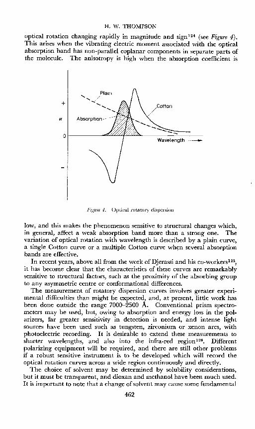
Optical rotation is equivalent to circular birefringence, and occurs whena substance transmits the left- and right-hand components of a beam ofcircularly polarized light with unequal velocity. If these components areabsorbed unequally, the optical rotation will vary with wavelength, andclassical equations have been proposed to express this. Indeed, someworkers have used the optical rotation at two wavelengths as a refinementfor structural work and for a criterion of purity. The variation of specificrotation with wavelength becomes greater as the absorption band isapproached, and, as it is traversed, a Cotton effect may be observed, the
461
H. W. THOMPSON
optical rotation changing rapidly in magnitude and sign 1 2 4 (see Figure 4).This arises when the vibrating electric moment associated with the opticalabsorption band has non-parallel coplanar components in separate parts ofthe molecule. The anisotropy is high when the absorption coefficient is
Wavelength ---
Plain" /...{
..............
............+
of-------dfZ.<:.:LL:.:.L4ULt.:L.:2ll....------=:.....- _
a Absorption >
Figure 4. Optical rotatory dispersion
low, and this makes the phenomenon sensitive to structural changes which,in general, affect a weak absorption band more than a strong one. Thevariation of optical rotation with wavelength is described by a plain curve,a single Cotton curve or a multiple Cotton curve when several absorptionbands are effective.
In recent years, above all from the work of Djerassi and his co-workers-P,it has become clear that the characteristics of these curves are remarkablysensitive to structural factors, such as the proximity of the absorbing groupto any asymmetric centre or conformational differences.
The measurement of rotatory dispersion curves involves greater experimental difficulties than might be expected, and, at present, little work hasbeen done outside the range 7000-2500 A. Conventional prism spectrometers may be used, but, owing to absorption and energy loss in the polarizers, far greater sensitivity in detection is needed, and intense lightsources have been used such as tungsten, zirconium or xenon arcs, withphotoelectric recording. I t is desirable to extend these measurements toshorter wavelengths, and also into the infra-red region126. Differentpolarizing equipment will be required, and there are still other problemsif a robust sensitive instrument is to be developed which will record theoptical rotation curves across a wide region continuously and directly.
The choice of solvent may be determined by solubility considerations,but it must be transparent, and dioxan and methanol have been much used.It is important to note that a change ofsolvent may cause some fundamental
462

PHYSICOCHEMICAL METHODS OF INVESTIGATION
alterations, especially when the solute molecule is flexible and can exist indifferent solvents' in different preferred conformations. The rotatorydispersion curves of 2-chloro-5-methylcyclohexanone are, for example,quite different in methanol and in octane solution127.
Plain rotatory dispersion curves down to 2600 A are found with compounds, such as amino-acids or carbohydrates, which have no chromophoreabsorbing at the longer wavelengths. Most work has been done withcompounds containing a carbonyl group, since its intrinsic absorptioncoefficient is low, and the band lies in a convenient spectral region. In thealiphatic aldehydes, the size and shape of the Cotton effect curve dependson the length of chain, the curve approaching a plain curve as the separationof the asymmetric centre from the absorbing group is increased. Far moreimportant work has been done with cyclic compounds, and two generalprinciples can be laid down: (1) that enantiomorphs give mirror-imageCotton effects; and (2) that similar stereochemical environments in theregion of the carbonyl group lead to similar dispersion curves.
The rise in absolute value of the optical rotation near the absorption bandprovides a new, more sensitive, method of quantitative analysis. Forexample, the pair of isomeric C-17 ketones (XXXIX), pregnane-So-ol-Zuone acetates, have Cotton effects of different sign, and at 3075 A theirspecific rotation differs by 3600°. A mixture can therefore be analysedeasilyl28.
CH .I 3i
CO
ct5~ pj H
..-AcO H
(XXXIX)
Another simple case arises when one component contains an absorbingchromophore leading to a large Cotton effect, while the second componenthas no such group and gives a plain curve. This occurs with hecogenin (XL)and tigogenin acetates (XLI) 129, a pair of saponins involved in the manufacture of cortisone. In some respects this method of detecting traceimpurities is more sensitive than the infra-red absorption spectrum, but itsuffers from the disadvantage that the Cotton curves in themselves give nodirect indication of the nature of the impurities.
With ring ketones such as the steroids, factors which affect the opticalrotatory dispersion (D.R.D.) curve are ring size, cis or trans fusion of rings,the relation of the carbonyl group to the ring junction, substituents at thejunction or a- to the carbonyl group, and conformation of c-substituents orother conformational effects. The steroid skeleton contains many asymmetric centres, and the carbonyl group is always near to one of them.
463

H. W. THOMPSON
AcO IH
(XL)
AcO II
H
(XLI )
The results confirm many deductions from infra-red and ultra-violetabsorption spectra, and from nuclear magnetic resonance, but sometimesreveal details unobtainable by other methods.
It is fortunate that, when a substituent is far removed from the carbonylabsorbing group, its nature has little effect upon the a.R.D. curve. Thus,in sapogenins, bile acids, and other classes with different groups at theC-17 position of the steroid skeleton, the rotatory dispersion characteristicsof the main AlB rings remain the same.
As already stated, the infra-red absorption spectra fail to determine thelocation of carbonyl groups in the A/B/C rings of steroids. The a.R.D.curves make this possible, and require only milligramme quantities ofmaterial. For example, the curves of cholestan-2-one (XLII) and -4-one
o
H
(XLII)
AcOII
H
(XLIV)
o
464
AcOII
H
(XLV)

PHYSICOCHEMICAL METHODS OF INVESTIGATION
(XLIII) differ in sign 130. A difference of amplitude is found with theacetates of ergostan-Sji-ol-l l-one (XLIV) and ergostan-3[j-ol-12-one1 31
(XLV). It seems also that, when more than one carbonyl group is presentin a steroid, the O.R.D. curves may give information about them, whereasthe infra-red absorption spectrum may be ambiguous. In the substituteddecalones (XLVI), the O.R.D. curve is markedly dependent upon thelocation of the carbonyl group in relation to the substituents-V,
6YowI I
H Hcb:oc+>
H H 0
(XLVI)
Optical rotatory dispersion appears to be more specifically valuable insorting out the stereochemical conformational problems of cyclic systems.For example, the 5cx- and 5[3- isomers, (XLVII) and (XLVIII), of androstan-17[3-o1-3-one differ only in the AlB ring junction, but have quitedifferent O.R.D. curvesP",
o
OH
(XLVII)
o
OH
H
(XLVIII)
m,H
(L)
Very different O.R.D. curves also arise between cholestan-4-one andcoprostan-4-one (5[3 cholestan-t-onej P", and between cholestan-3[j-ol-7one acetate and 3cx-hydroxy-7-ketocholanic acid, although, as alreadyexplained, changes in the C-17 substituent have no significant effect.
The O.R.D. curves for the cis- and trans-lO-methyl-2-decalones, (XLIX)and (L), are opposite in sign 13 4, and correspond closely to those of3-keto-5[3and 3-keto-5cx-steroids respectively, again showing that the configurationaround the absorbing carbonyl group is the dominant factor.
H
o£bH
(XLIX)
In the 5-hydroxy-l O-methyl- il1(9L 2-octalones, the orientation of theC-IO substituent similarly determines the sign of the O.R.D. curve, andthere is a correspondence with the curves of il4-3-keto-steroids1 35•
465

H. W. THOMPSON
As already explained, spectral differences in both the infra-red and ultraviolet regions occur with keto-steroids according to the axial or equatorialpositions of substituents. Similar effects are found with the rotatory dispersion curves. Axial and equatorial hydroxyl groups adjacent to thecarbonyl group in steroids shift the mid-point of the Cotton curves to longerand shorter wavelengths respectively-P", Acetoxy-groups have a similareffect, and, if axial, lead to enhanced amplitudes. The effect of ex-halogenatoms is similar, and may even change the sign of the Cotton curveP",
By consideration of these compounds, important rules have been formulated,and these bear upon the determination of absolute configurations and thesemi-quantitative prediction of a.R.D. curves-P",
Many principles suggested empirically by these illustrations have beenused to establish the stereochemistry of compounds such as lumisterol,luminsantonin and giberellic acid. Their extension to more complexsystems such as the polycyclic terpenes is at present difficult, since theintroduction of some substituents, gem-dialkyl groups, or extra fused ringsoften appears to affect the general conformation in unexpected ways.For example, the introdution of a pair of methyl groups at the C-4 positionin cholestan-3-one has a remarkable effect, and the specific orientation ofthe isopropenyl group in ex-cyperone (LI) leads to striking changes in thea.R.D. curve 139.
o~(LO
In a random conformation such as exists in a strongly hydrogen-bondingsolvent, polypeptides give plain a.R.D. curves. Only the ex-helical formsgive rise to a complex dispersion curve. The possibility of studying denaturation in this way has been examined, and this kind of work may beextended by using peptides containing an absorbing group such as a phenylnucleus.
All these empirical data on optical rotatory dispersion should stimulatethe development of a better theory ofoptical rotation in terms of the detailedelectronic structure along the lines visualized by Moffitt.
Another related phenomenon, the Kerr effect140 (the development ofbirefringence under a voltage gradient), promises to give structural information about molecular conformations and the axial or equatorial arrangement of substituents in saturated ring systems.
MASS SPECTROMETRYThe mass spectrometer has also been used for structural analysis. When
molecules are subjected to electron bombardment, cleavage may occurand positive ions of both the original molecule and of its fragments may beformed. These ions can be passed through a mass spectrometer and theirmasses determined. The method has, of course, been used in recent years
466

PHYSICOCHEMICAL METHODS OF INVESTIGATION
for the analysis of hydrocarbon mixtures, since, with appropriate calibration,the measured intensities of the different fragment masses can be used tocompute the composition of the original mixture. With complex molecules,this is being used to determine the molecular weights and also to estimatethe mass numbers for side-chains or other main fragments of the molecularskeleton. The strength of the applied voltage determines the degree offragmentation.
Steroids have been examined in this way. Cholestane-t', for example, ofmolecular weight 372, gives a set of fragments of molecular weight betweenabout 40 and 360, which correspond to the residues expected. By thismeans, steroids and triterpenoids can be distinguished, and valuable cluesobtained about side-chains. Recently, it has been applied to the study ofnew antibiotics such as lagosin14 2 and filipin 14 3, and flavonoids P", andseems likely to become more widely applied in preliminary studies onunknown structures which have molecular weights up to about 400.
OTHER PHYSICAL METHODS
Isotopes have been used in several ways in the study of natural products.As already explained, deuterium may be used in the interpretation of aninfra-red absorption spectrum. This exchange process has also beenapplied, in conjunction with infra-red measurements, in work on largemolecules such as cellulose or polypeptides; by this means it is possible onthe one hand to discriminate between crystalline and amorphous parts of acomplex structure, and on the other to differentiate between helical or openchain forms of the peptide chains82.
14C is now being widely used to elucidate the mechanism of biogenesis ofmany products, and has been invaluable in research on photosynthesis-V,Irradiation of vitamin B1 2 and subsequent measurement of the radioactivedecay gave a convincing confirmation of the presence of cobalt in thismolecule. Tritium also has been used as a tracer in many synthetic reactions.
Reaction kinetic measurements themselves have provided evidence aboutcertain structures and conformations. The electron microscope seems tobe providing new information about the viruses.
For quantitative determination, reference has already been made tostandard ultra-violet absorption methods for the assay of vitamins,porphyrins and other substances-v'. Polarography is sometimes convenientnot only for estimating organic compounds but for identifying structuressuch as a conjugated system. Flame photometry or ultra-violet emissionspectra with ashed products have been used for the determination of metals,but here the newer methods of X-ray fiuorescence-v? or atomic absorptionspectroscopyP" offer great possibilities, and technical improvements arerapidly increasing their sensitivity.
The whole range of physical methods is indeed wide. Which methodshould be used in a given case will be determined by such considerations asthe quantity of material available, its physical state and solubility, whetherit is necessary to recover the sample or not, the particular question beingasked, and whether it is required to predict the whole structure, or onlypart of it, or to confirm a structure already proposed.
467
H. W. THOMPSON
References
1 L. C. Craig and O. W. Post. Ind. Eng. Chem. Anal. Ed., 16, 413 (1944)L. C. Craig. J. Biol. Chem., 155, 519 (1944)E. S. Perry and W. H. Weber. Anal. Chem., 26, 498 (1954)
2 L. C. Craig, G. H. Hogeboom, F. H. Carpenter and V. du Vigneaud. J. Biol. Chem.,168, 665 (1947) .A. H. Livermore and V. du Vigneaud. J. Biol. Chem., 180, 365 (1949)L. L. Engel, W. R. Slaunwhite, P. Carter and I. T. Nathanson. J. Bioi. Chem., 185, 255(1950)H. Rosenkrantz. J. BioI. Chem., 192,9 (1951)P. von Tavel and R. Signer. Advances in Protein Chem., 11, 237 (1956)
3 See M. Bier. Electrophoresis, Academic Press, New York (1959)4 See E. Lederer and M. Lederer. Chromatography, Elsevier Press, Amsterdam (1953)
H. Rosenkrantz. Ann. x.r. Acad. Sci., 69, 5 (1957)H. P. Schwarz, L. Dreisbach, R. Childs and S. V. Mastrangelo. Ann. N.r. Acad. Sci.,69, 116 (1957)S. Moore and W. H. Stein. Advances in Protein Chem., 11, 191 (1956)
5 A. J. P. Martin and R. L. M. Synge. Biochem. ].,35, 1358 (1941)6 R. S. AIm, R. J. P. Williams and A. Tiselius, Acta Chern. Scand., 6, 826 (1952)
K. O. Donaldson, V. J. Tulane and L. M. Marshall. Anal. Chem., 24, 185 (1952)7 M. Golay. Instrument Society ofAmerica Symposium, p. 1, Academic Press, New York (1958)8 H. J. MacDonald, L. V. McKendell and E. W. Bermes. ]. Chromatog., 1,259 (1958)9 A. T. James and A. J. P. Martin. Biochem. J., 50, 679 (1952)
C. G. C. Phillips. Gas Chromatography, Butterworths, London (1956)10 A. J. P. Martin and A. T. James. Biochem. ]., 63, 138 (1956)11 1. G. McWilliam and R. A. Dewar. Nature, 181, 760 (1958)12 J. E. Lovelock. ]. Chrornatog., 1, 35 (1958)13 G. Popjak, A. E. Lowe, D. Moore, L. Brown and F. A. Smith. ]. Lipid Research, 1, 29
(1959)A. E. Lowe and D. Moore. Nature, 182, 133 (1958)
14 J. E. Lovelock and S. R. Lipsky. J. Arn. Chern. Soc., 82, 431 (1960)10 J. Fridrichsons and A. McL. Mathieson. Nature, 173, 732 (1954)16 A. McL. Mathieson. Revs. Pure Appl. Chern. (Australia), 5, 113 (1955)
P. J. Wheatley. Molecular Structure, Clarendon Press, Oxford (1958)17 C. Bokhoven,J. C. Schoone andJ. M. Bijvoet. Acta Cryst., 4, 275 (1951)
J. M. Robertson and C. A. Beevers. Nature, 165, 690 (1950)A. F. Peerdemann. Acta Cryst., 9, 824 (1956)
18 M. V. King, J. L. de Vries and R. Pepinsky. Acta Cryst., 5, 437 (1950)19 T. A. Hamer, J. M. Robertson, H. N. Shivestava and J. V. Silverton. Proc. Chern. Soc.,
1960, 7820 J. D. Bernal. Nature, 129, 277 (1930)
J. D. Bernal. Chem. & Ind. (London), 10, 466 (1932)J. D. Bernal, D. Crowfoot and I. Fankuchen. Phil. Trans. Roy. Soc. (London), A239,135 (1940)
21 C. H. Carlisle and D. Crowfoot. Proc, Roy. Soc. (London), A184, 64 (1945)22 D. Crowfoot and J. D. Dunitz. Nature, 162, 608 (1948)
D. C. Hodgkin, M. S. Webster andJ. D. Dunitz. Chern. & Ind. (London), 35, 1148 (1957)23 D. C. Hodgkin and D. Sayre. J. Chern. Soc., 1952,456124]. Parsons and W. T. Beher. Anal. Chem., 27, 514 (1955)
J. Parsons, W. T. Beher and G. D. Baker. Anal. Chem., 27, 1569 (1955); 28, 1514 (1956);29, 762 (1957)
25 D. Crowfoot, C. W. Bunn, B. W. Rogers-Low and A. Turner-Jones. The Chernistry ofPenicillin, p. 310, Princeton Press, Pa. (1949)
26 D. C. Hodgkin. Fortschr. Chem. org. Naturstoffe, 15, 167 (1958)D. C. Hodgkin, J. Kamper, J. Lindsey, M. Mackay,]. Pickworth, J. H. Robertson, C. B.Shoemaker, J. G. White, R. J. Prosen and K. N. Trueblood. Proc. Roy. Soc. (London),A242, 228 (1957)
27 J. C. Kendrew, G. Bodo, H. M. Dintzis, R. G. Parrish and H. Wyckoff. Nature, 181,662 (1958)
468
PHYSICOCHEMICAL METHODS OF INVESTIGATION
27 J. C. Kendrew. Nature, 182, 764 (1958)G. Bodo, H. M. Dintzis, J. C. Kendrew and H. W. Wyckoff. Proc. Roy. Soc. (London),A253, 70 (1959)
28 See A. E. Gillam and E. S. Stern. Electronic Absorption Spectroscopy in Organic Chemistry,Arnold, London (1958)E. A. Braude and E. S. Waight. Progress in Stereochemistry (ed. W. Klyne), Vol. 1, Butterworths, London (1954)
29 K. W. Hausser, R. Kuhn, A. Smakula and K. H. Kreuchen. Z. physik. Chem., B29,363 (1935)K. W. Hausser, R. Kuhn, A. Smakula and M. Hoffer. Z.physik. Chem., B29, 371 (1935)K. W. Hausser, R. Kuhn, A. Smakula and A. Deutsch. Z.physik. Chem., B29, 378 (1935)K. W. Hausser, R. Kuhn and A. Smakula. Z.physik. Chem., B29, 384 (1935)K. W. Hausser, R. Kuhn and G. Seitz. Z. physik. Chem., B29, 391 (1935)K. W. Hausser, R. Kuhn and E. Kuhn. Z. p~ysik. Chem., B29, 417 (1935)See A. E. Gillam and E. S. Stern. Electronic Absorption Spectroscopy in Organic Chemistry,p. 178, Arnold, London (1958)E. R. H. Jones. Proc. Chem. Soc., 1960, 199
80 W. D. Celmer and 1. A. Solomons. ]. Am. Chem. Soc., 74, 1870, 3838 (1952)31 E. A. Braude, E. R. H. Jones, H. P. Koch, R. W. Richardson, F. Sondheimer and J. B.
Toogood. ]. Chern. Soc., 1949, 1890 .32 R. B. Woodward and G. Singh. Experientia, 6, 238 (1950)
R. B. Woodward and G. Singh. ]. Am. Chem. Soc., 72, 1428,5351 (1950)33 D. J. Cram and N. L. Allinger. ]. Am. Chem. Soc., 78, 5275 (1956)
P. Bladon, H. B. Henbest and G. W. Wood. ]. Chem. Soc., 1952, 273734 N. L. Allinger. ]. Org. Chem., 21, 1180 (1956)35 See L. F. Fieser and M. A. Fieser. Steroids, Reinhold, New York (1959)
L. Dorfman. Chem. Revs., 53, 47 (1953)36 W. V. Mayneord and E. M. F. Roe. Proc. Roy. Soc. (London), A152, 299 (1935)
R. N. Jones. Chem. Revs., 32, 1 (1943); 41, 353 (1947)R. N. Jones. ]. Am. Chem. Soc., 67, 2127 (1945)
37 J. M. Gulland and E. R. Holiday. Nature, 132, 782 (1933)J. M. Gl.tlland, E. R. Holiday and T. F. Macrae. ]. Chem, Soc., 1934, 1639]. M. Gulland and E. R. Holiday. ]. Chem. Soc., 1936, 765
38 R. B. Woodward, W. J. Brehm and A. L. Nelson. ]. Am. Chem. Soc., 69, 2250 (1947)R. B. Woodward and W.]. Brehm. ]. Am. Chem. Soc., 70, 2107 (1948)
89 See S. Aronoff. Chem. Revs., 47,175 (1950)40 G. H. Beaven, E. R. Holiday, E. A. Johnson, B. Ellis and V. Petrow. ]. Pharm. and
Pharmacol., 2, 944 (1950)N. G. Brink and K. Folkers. ]. Am. Chem. Soc., 71, 2951 (1949)
41 H. Dam, A. Geiger, J. Glavind, P. Karrer, W. Karrer, E. Rothschild and H. Salomon.Helv. Chim. Acta, 22, 310 (1939)R. W. McKee, S. B. Binkley, D. W. MacCorquodale, S. A. Thayer and E. A. Doisy.]. Am. Chem. Soc., 61, 1295 (1939)
42 F. A. Hochstein, C. R. Stephens, L. H. Conover, P. P. Regna, R. Pasternack, K. J.Brunings and R. B. Woodward. ]. Am. Chem. Soc., 74, 3708 (1952)
43 L. Zechmeister. Chem. Revs., 34, 267 (1944)U R. C. Cookson. ]. Chem. Soc., 1954, 282; 1955, 352745 A. E. Gillam and 1. M. Heilbron. Biochem.J., 30, 1253 (1936)
See A. E. Gillam and E. S. Stern. Electronic Absorption Spectroscopy in Organic Chemistry,p. 195, Arnold, London (1958)
46 R. N. Jones and C. Sandorfy. Chemical Applications of Spectroscopy, p. 324, Interscience,New York (1956)
47 H. W. Thompson. ]~ Chem. Soc., 1948,328N. B. Colthup. ]. Opt. Soc. Am., 40, 397 (1950)L. J. Bellamy. Infrared Spectra a/Complex Molecules, Methuen, London (1958)]. Lecomte. Handbuch der Physik, Vol. 26, Springer (1958)R. N. Jones and C. Sandorfy. Chemical Applications qf Spectroscopy, p.324, Interscience,New York (1956)
48 W. Kaye. Spectrochim. Acta, 6, 257 (1954); 7, 181 (1955)R. F. Goddu. Anal. Chem., 29, 1790 (1957)
469

H. W. THOMPSON
49 H. P. Schwarz, L. Dreisbach, R. Childs and S. V. Mastrangelo. Ann. N.Y. Acad. Sci., 69,116 (1957)H. P. Schwarz, L. Dreisbach, R. Childs and S. V. Mastrangelo. Science, 123, 328 (1956)H. P. Schwarz, L. Dreisbach, R. Childs, S. V. Mastrangelo and A. Kleschik. Appl.Spectroscopy, 12, 35 (1958)
50 R. Barer, A. R. H. Cole and H. W. Thompson. Nature, 163, 198 (1949)A. R. H. Cole and R. N. Jones. ]. Opt. Soc. Am., 42,348 (1952)D. L. Wood. Ann. NY. Acad. Sci., 69, 194 (1957)
51 E. R. Blout. Ann. NY. Acad. Sci., 69, 84 (1957)E. R. Blout and H. Lenormant. ]. Opt. Soc. Am., 43, 1093 (1953)N. Wright. Appl. Spectroscopy, 9, 105 (1955)J. D. S. Goulden. Spectrochim. Acta, 15, 657 (1959)R. J. Koegel, R. A. McCallum, J. P. Greenstein, M. Winitz and S. M. Birnbaum. Ann.N.Y. Acad. Sci., 69, 94 (1957)
52 E. J. Hartwell, R. E. Richards and H. W. Thompson. ]. Chem. Soc., 1948, 1436J. Lecomte. Bull. soc. chim. France, 1955, 719R. N. Jones and C. Sandorfy. Chemical Applications of Spectroscopy, p. 443, Interscience,New York (1956)
53 J. J. Hurst and G. H. Whitham. Proc. Chem. Soc., 1959, 16054 H. W. Thompson, R. R. Brattain, H. M. Randall and R. S. Rasmussen. The Chemistry
ofPenicillin, p. 382, Princeton Press, Pa. (1949)R. S. Rasmussen. Fortschr. Chem. org. Natursto.fJe, 5, 331 (1948)
55 S. A. Barker, E. J. Bourne, R. M. Pinkard and D. H. Whiffen. Chem. & Ind. (London),1958, 658
56 W. Baker, W. D. Ollis and K. W. Robinson. Proc. Chem. Soc., 1959, 26957 J. D. Connolly and K. H. Overton. Proc. Chem. Soc., 1959, 18858 H. P. Schwarz, L. Dreisbach, R. Childs and S. V. Mastrangelo. Ann. N.Y. Acad. ss:
69, 116 (1957)G. Marinetti and E. Stotz. ]. Am. Chem. Soc., 76, 1347 (1954)N. K. Freeman. Ann. N.Y. Acad. Sci., 69, 131 (1957)
59 W. D. Celmer and 1. A. Solomons. ]. Am. Chem. Soc., 74, 3838 (1952); 75, 1372 (1953)60 N. G. Brink, F. A. Kuehl and K. Folkers. Science, 112, 354 (1950)
H. J. Callomon. Dissertation, Oxford University (1954)61 A. Andant, P. Lambert and J. Lecomte. Compt. rend., 201, 391 (1925)
P. Lambert and J. Lecomte. Compt. rend., 206, 1007 (1938)H. W. Thompson and P. Torkington. Proc. Roy. Soc. (London), A184, 3 (1945)R. S. Rasmussen and R. R. Brattain. J. Chem. Phys., 15, 120, 131 (1947)R. N. Jones and C. Sandorfy. Chemical Applications of Spectroscopy, p. 337. Interscience,New York (1956)
62 H. W. Thompson and D. H. Whiffen. ]. Chem. Soc., 1948, 1412M. F. Carroll, R. G. Mason, H. W. Thompson and R. C. S. Wood. ]. Chem. Soc., 1950,3457H. H. Gunthard and L. Ruzicka. Helv. Chim. Acta., 32, 2125 (1949)Y.-R. Naves and J. Lecomte. Bull. soc. chim. France, 1953, 112
63 J. H. Turnbull, D. H. Whiffen and W. Wilson. Chem. & Ind. (London), 1950, 626R. N. Jones. ]. Am. Chem. Soc., 72, 5322 (1950)
64 B. Lythgoe. Proc. Chem. Soc., 1959, 14265 L. Crombie, S. H. Harper, R. E. Stedman and D. Thompson. ]. Chem, Soc., 1951, 2445
S. H. Harper and L. Crombie. ]. Chem. Soc., 1952, 86966 G. H. Whitham. Proc. Chern. Soc., 1959, 27167 K. Lunde and L. Zechmeister. ]. Am. Chern. Soc., 77, 1647 (1955)68 B. E. Cross, J. F. Grove, J. MacMillan, T. P. C. Mulholland and N. Sheppard. Proc.
Chem. Soc., 1958, 221J. MacMillan, J. C. Seaton and P. J. Suter. Proc. Chern. Soc., 1959, 325
69 L. Marion, D. A. Ramsay and R. N. Jones. ]. Am. Chem. Soc., 73, 305 (1951)O. E. Edwards, M. Los and L. Marion. Proc. Chern. Soc., 1959, 192
70 R. B. Barnes, R. C. Gore, U. Liddel and van Zandt Williams. Infrared Spectroscopy,Reinhold, New York (1944)
470

PHYSICOCHEMICAL METHODS OF INVESTIGATION
70 D. H. Whiffen, P. Torkington and H. W. Thompson. Trans. FaradaySoc., 41,200 (1945)D. H. Whiffen and H. W. Thompson. J. Chem. Soc., 1945,268R. E. Richards and H. W. Thompson. J. Chern. Soc., 1947, 1260C. G. Cannon and G. B. B. M. Sutherland. Spectrochim. Acta, 4, 373 (1951)H. W. Thompson, E. E. Vago, M. C. Corfield and S. F. D. Orr. J. Chem. Soc., 1950,214L. J. Bellamy. Infrared Spectra ofComplex Molecules, p. 75, Methuen, London (1958)
71 S. F. D. Orr and H. W. Thompson. J. Chem.. Soc., 1950,21872 C. W. Young, R. B. DuVall and N. Wright. Anal. Chem., 23, 709 (1951)
D. H. Whiffen. Spectrochim. Acta, 7, 253 (1955)73 R. N. Jones and K. Dobriner, Vitamins and Hormones, 7, 293 (1949)
R. N. Jones and F. Herling. J. Org. Chem., 19, 1252 (1954)A. R. H. Cole. Revs. Pure Appl. Chem. (Australia), 4, III (1954)R. N.Jones, D. A. Ramsay, D. S. Keir and K. Dobriner. J. Am. Chern. Soc., 74, 80 (1952)R. N. Jones and A. R. H. Cole. J. Am. Chem. Soc., 74, 5648 (1952)R. N.Jones, A. R. H. Cole and B. Nolin. J. Am. Chem. Soc., 74, 5662 (1952)A. R. H. Cole. Fortschr. Chem. org. Naturstoffe, 13, 1 (1956)
74 R. N. Jones and B. S. Gallagher. J. Am. Chem. Soc., 81, 5242 (1959)75 R. N. Jones, P. Humphries, F. Herling and K. Dobriner. J. Am. Chem. Soc., 73, 3215
(1951)A. R. H. Cole, R. N. Jones and K. Dobriner. J. Am. Chem. Soc., 74, 5571 (1952)A. Furst, H. H. Kuhn, R. Scotoni and H. H. Gunthard. Helv. Chim. Acta, 35, 951 (1952)H. Rosenkrantz and L. Zablow, J. Am. Chem. Soc., 75, 903 (1953)
76 A. R. H. Cole. J. Chern. Soc., 1952, 496977 W. G. Dauben and E. Hoerger. J. Am. Chem. Soc., 73, 1504 (1951)
W. G. Dauben, E. Hoerger and N. K. Freeman. J. Am. Chem. Soc., 74, 5206"'(1952)78 M. Larnaudie. Compt. rend., 235, 154 (1952)
E.J. Corey, M. G. Howell, A. Boston, R. L. Young and R. A. Sneen. J. Am. Chem. Soc.,78, 5036 (1956)
79 R. N. Jones, D. A. Ramsay, F. Herling and K. Dobriner. J. Am. Chem. Soc., 74, 2828(1952)
80 S. A. Barker, E. J. Bourne, M. Stacey and D. H. Whiffen. J. Chem. Soc., 1954, 171S. A. Barker, E. J. Bourne, R. Stephens and D. H. Whiffen. J. Chem. Soc., 1954, 3468,4211
81 H. Rosenkrantz. Ann. u.r. Acad. Sci., 69, 5 (1957)C. Clark and M. Chianta. Ann. N.r. Acad. Sci., 69, 205 (1957)
82 See A. Elliott. Advances in Spectroscopy, p. 214, Interscience, New York (1959)83 G. B. B. M. Sutherland and M. Tsuboi. Proc. Roy. Soc. (London), A239, 446 (1957)84 S. H. Levine, H. J. R. Stevenson, L. A. Chambers and B. A. Kenner. J. Bacteriol., 65,
10 (1953)J. E. S. Greenstreet and K. P. Norris. Spectrochim. Acta, 9,177 (1957)D. W. Smith, H. M. Randall, M. M. Gastambide-Odier and A. L. Koevoet, Ann. u.r.Acad. Sci., 69, 145 (1957)A. A. Benedict. Ann. x.r. Acad. Sci., 69, 158 (1957)
85 H. W. Thompson. Molecular Spectroscopy, p. 94, Institute of Petroleum, London (1955)H. W. Thompson. Conference on Molecular Spectroscopy, p. 165, Pergamon Press, London(1959)H. W. Thompson and D. A. Jameson. Spectrochim. Acta, 13, 236 (1958)J. P. Jesson and H. W. Thompson. Spectrochim. Acta, 13, 217 (1958)
86 R. A. Russell and H. W. Thompson. J. Chern. Soc., 1955,483. R. A. Russell and H. W. Thompson. Proc. Roy. Soc. (London), A234, 318 (1956)87 R. Moccia and H. W. Thompson. Proc. Roy. Soc. (London), A243, 154 (1957)88 S. A. Francis. J. Chem. Phys., 18, 861 (1950); 19,942 (1951)89 R. N. Jones, E. Augdahl, A. Nickon, G. Roberts and D. J. Whittingham. Ann. s.r.
Acad. Sci., 69, 38 (1957)90 R. Mecke and K. Noack. Spectrochim. Acta, 12, 391 (1958)91 P. J. Krueger and H. W. Thompson. Proc. Roy. Soc. (London), A250, 22 (1958)92 H. S. Gutowsky, L. H. Meyer and R. E. McClure. Rev. Sci. Instr., 24, 644 (1953)
H. J. Bernstein, J. A. Pople and W. G. Schneider. High Resolution Nuclear MagneticResonance, McGraw-Hill, New York (1959)J. D. Roberts. Nuclear Magnetic Resonance, McGraw-Hill, New York (1959)
471

H. W. THOMPSON
92 R. E. Richards. Conference on MolecularSpectroscopy, p. 5, Pergamon Press, London (1959)R. H. Sands. Ann. u.r. Acad. Sci., 70, 780 (1958)J. B. Leane, R. E. Richards and T. P. Schaefer. ]. Sci. Instr., 36, 230 (1959)L. M. Jackman. Nuclear Magnetic Resonance Spectroscopy, Pergamon Press, London (1959)
93 H. J. Bernstein, J. A. Pople and W. G. Schneider. High Resolution Nuclear MagneticResonance, p. 245, McGraw-Hill, New York (1959)
94 R. E. Richards. Chem. Soc. (London) Spec. Publ., No. 12, 171 (1958)95 L. Fowden, F. F. Noe, J. H. Ridd and R. F. M. White. Proc. Chem. Soc., 1959, 13196 F. A. L. Anet and C. R. Eves. Can.J. Chem., 36, 902 (1958)97 R. F. Hutton and F. H. Westheimer. Tetrahedron, 3, 73 (1958)
A. F. E. Sims and P. W. G. Smith. Proc. Chem. Soc., 1958,28298 H. S. Gutowsky. Ann. N.r. Acad, Sci., 70, 786 (1958)
L. H. Meyer, A. Saika and H. S. Gutowsky. ]. Am. Chem. Soc., 75,4567 (1953)H. J. Bernstein, j. A. Pop1e and W. G. Schneider. High Resolution Nuclear MagneticResonance, p. 271, McGraw-Hill, New York (1959)
99 Varian Assoc. Technical Inform. Bull., 1, (4) (1956)100 J. Gripenberg. Acta Chem. Scand., 10,487 (1956)101 B. E. Cross, J. F. Grove, J. MacMillan, J. S. Moffatt, T. P. C. Mulholland and]. C.
Seaton. Proc. Chem. Soc., 1959, 302102 A. S. Kende. Chem. & Ind. (London), 1956, 544
A. T. Bottini and]. D. Roberts. ]. Am. Chem. Soc., 80,5203 (1958)103 H.]. Bernstein, J. A. Pop1e and W. G. Schneider. High Resolution Nuclear Magnetic
Resonance, p. 281, McGraw-Hill, New York (1959)104 Varian Assoc. Technical Inform. Bull., 2, (3) (1957)105 H. Conroy, P. R. Brook, M. K. Rout and N. Silverman. ]. Am. Chern. Soc., 79, 1763
(1957)106 M.]. Chisholm and C. Y. Hopkins. Can.]. Chem., 35, 358 (1957)
C. Y. Hopkins and H. J. Bernstein. Can.]. Chem., 37, 775 (1954)H. J. Bernstein, J. A. Pople and W. G. Schneider. High Resolution Nuclear MagneticResonance, p. 283, McGraw-Hill, New York (1959)
107 K. L. Rinehart, W. A. Nilsson and H. A. Whaley. ]. Am. Chem. Soc., 80, 503 (1958)108 M. S. Barber, L. M. Jackman and B. C. L. Weedon. Proc. Chem. Soc., 1959, 96109 M. S. Barber, L. M. Jackman, C. K. Warren and B. C. L. Weedon. Proc. Chem. Soc.,
1960, 19110 E. E. van Tame1en, S. H. Levin, G. Brenner. ]. Wolinsky and P. Aldrich. ]. Am. Chem.
Soc., 80, 501 (1958)111 W. G. Dauben and P. D. Hance. ]. Am. Chem. Soc., 77, 2451 (1955)112 J. N. Shooleryand M. T. Rogers. ]. Am. Chem. Soc., 80, 5121 (1958)113 R. U. Lemieux, R. K. Kullnig, H.]. Bernstein and W. G. Schneider. J. Am. Chem.
Soc., 79, 1005 (1957)R. U. Lemieux, R. K. Kullnig and R. Y. Moir. ]. Am. Chem. Soc., 80, 2237 (1958)
114 A. Novak and E. Whalley. Can.]. Chem., 36, 1116 (1958)115 J. Musher and R. E. Richards. Proc. Chem. Soc., 1958,230116 J. Musher, Spectrochim. Acta, in the press117 R. U. Lemieux, R. K. Kullnig and R. Y. Moir. J. Am. Chem. Soc., 80, 2237 (1958)118 P. C. Lauterbur. ]. Chem. Phys., 26, 217 (1957)
C. H. Holm. ]. Chem. Phys., 26, 707 (1957)P. C. Lauterbur. Ann. u.r. Acad. Sci., 70, 841 (1958)A. D. Cohen, N. Sheppard and J. J. Turner. Proc. Chem. Soc., 1958, 118
119 H. S. Gutowsky and D. W. McCall. ]. Chem. Phys., 22, 162 (1954)R. A. Jones, A. R. Katritzky and]. Michalski. Proc. Chem. Soc., 1959, 321H. Finegold. Ann. u.r. Acad. Sci., 70, 875 (1958)N. Muller, P. C. Lauterbur and]. Goldenson. ]. Am. Chem. Soc., 78, 3557 (1956)J. R. van Wazer, C. F. Callis, J. N. Shoo1ery and R. C. Jones. ]. Am. Chem. Soc., 78,5715 (1956)]. R. Parks. ]. Am. Chem. Soc., 79, 757 (1957)
120 A. L. Bloom andJ. N. Shoo1ery. Phys. Rev., 97,1261 (1955)A. Kastler. ]. Opt. Soc. Am., 47, 460 (1957)
121 W. D. Phillips, C. E. Looney and C. K. Ikeda. ]. Chem. Phys., 27, 1435 (1957)122 D.]. E. Ingram and]. E. Bennett. Discussions FaradaySoc., No. 19, 140 (1955)
472
PHYSICOCHEMICAL METHODS OF INVESTIGATION
122 P. George,]. E. Bennett and D.]. E. Ingram. ]. Chem. Phys., 24, 627 (1956)]. S. Griffith. Proc. Roy. Soc. (London), A235, 23 (1956)D.]. E. Ingram. ]. Am. Chem. Soc., 78, 3545 (1956)
123 D. H. R. Barton. ]. Chem. Soc., 1945, 813; 1946, 512, 1116D. H. R. Barton and]. D. Cox. ]. Chem. Soc., 1948, 783, 1354, 1357; 1949,214,219D. H. R. Barton and E. Miller. ]. Chem, Soc., 1949, 337D. H. R. Barton and W. Klyne. Chem. &, Ind. (London), 1948, 755W. Klyne. In Determination ofOrganicStructures by Physical Methods, Chapter 3, AcademicPress, New York (1955)
124 T. M. Lowry. Optical Rotatory Power, Longmans Green, London (1935)W. Kuhn. Ann. Rev. Phys. Chem., 9, 417 (1958)P. A. Levene and A. Rothen. In Organic Chemistry (ed. H. Gilman), Vol. 2, p. 1799,Wiley, New York (1938)W. Klyne. In Newer Methods of Organic Chemistry (ed. R. A. Raphael), Vol. I, Interscience, New York, in the press
125 C. Djerassi. Optical Rotatory Dispersion, McGraw-Hill, New York (1960)126 H.J. Redinger and H. H. Gtmthard. Helv. Chim. Acta, 41, 2216 (1958)127 C. Djerassi and L. E. Geller. Tetrahedron, 3, 319 (1958)128 C. Djerassi. Bull. soc. chim. France, 1957, 741129 C. Djerassi. Optical Rotatory Dispersion, p. 57, McGraw-Hill, New York (1960)130 C. Djerassi, W. Closson and A. E. Lippmann. ]. Am. Chem. Soc., 78, 3163 (1956)131 E. W. Foltz, A. E. Lippmann and C. Djerassi. ]. Am. Chem. Soc., 77, 4359 (1955)
C. Djerassi and W. Closson. ]. Am. Chem. Soc., 78, 3761 (1956)C. Djerassi,]. Osiecki, R. Riniker and B. Riniker. ]. Am. Chem, Soc" 80, 1216 (1958)
132 C. Djerassi and D. Marshall. ]. Am. Chem. Soc" 80, 3986 (1958)133 C. Djerassi and W. Closson. ]. Am. Chem. Soc., 78, 3761 (1956)134 C. Djerassi, R. Riniker and B. Riniker. ]. Am. Chern. Soc., 78, 6362 (1956)
C. Djerassi and D. Marshall. ]. Am. Chem. Soc., 80, 3986 (1958)136 C. Djerassi,]. Osiecki and W. Herz. ]. Org. Chem., 22, 1361 (1957)
V. Prelog and W. Acklin. Helv. Chim. Acta, 39, 748 (1956)136 C. Djerassi, O. Halpern, V. Halpern, O. Schindler and C. Tamm. Helv. Chim. Acta, 41,
250 (1958)137 C. Djerassi,]. Osiecki, R. Riniker and B. Riniker. ]. Am. Chem, Soc., 80, 1216 (1958)
C. Djerassi, 1. Fornagnera and O. Mancera. ]. Am. Chern. Soc., 81, 2383 (1959)138 C. Djerassi. Optical Rotatory Dispersion, Chapter 13, McGraw-Hill, New York (1960)139 C. Djerassi, R. Riniker and B. Riniker. ]. Am. Chem. Soc., 78, 6362 (1956)140 C. E. Le Fevre and R. J. W. Le Fevre, Revs. Pure Appl. Chem. (Australia), 5, 261 (1955)141 R. 1. Reed. ]. Chern. Soc., 1958, 3432
S. S. Friedland, G. H. Lane, R. T. Longman, K. E. Train and M. J. O'Neal. Anal.Chem., 31, 169 (1958)
142 M. L. Dhar, V. Thaller and M. C. Whiting. Proc. Chern. Soc., 1959, 154143 B. Berkoz and C. Djerassi. Proc, Chern. Soc., 1959, 316144 B. F. Burrows and W. D. Ollis. Proc. Chem. Soc., 1960, 177146]. A. Bassham, A. A. Benson, L. D. Key, A. Z. Harris, A. T. Wilson and M. Calvin.
]. Am. Chem. Soc., 76, 1760 (1954)146 See A. E. Gillam and E. S. Stern. Electronic Absorption Spectroscopy in Organic Chemistry,
Chapter 12, Arnold, London (1958)147 H. Friedman and L. S. Birks. Rev. Sci. Instr., 19, 323 (1948)
L. S. Birks. Rev. Sci. Instr., 22, 891 (1951)148 A. Walsh. Spectrochim. Acta, 7, 108 (1955)
B. J. Russell, J. P. Shelton and A. Walsh. Spectrochim. Acta, 8, 317 (1957)W. G. Jones and A. Walsh. Spectrochim. Acta, 16,249 (1960)G. F. Box and A. Walsh. Spectrochim. Acta, 16, 255 (1960)]. B. Willis. Spectrochim. Acta, 16, 259, 273 (1960)
473