physical therapy rehabilitation in a patient with guillain
TRANSCRIPT

University of North Dakota University of North Dakota
UND Scholarly Commons UND Scholarly Commons
Physical Therapy Scholarly Projects Department of Physical Therapy
5-2020
Physical Therapy Rehabilitation In A Patient With Guillain-Barre Physical Therapy Rehabilitation In A Patient With Guillain-Barre
Syndrome With Acute Respiratory Failure: A Case Report Syndrome With Acute Respiratory Failure: A Case Report
Daniel P. Torok University of North Dakota
Follow this and additional works at: https://commons.und.edu/pt-grad
Part of the Physical Therapy Commons
Recommended Citation Recommended Citation Torok, Daniel P., "Physical Therapy Rehabilitation In A Patient With Guillain-Barre Syndrome With Acute Respiratory Failure: A Case Report" (2020). Physical Therapy Scholarly Projects. 694. https://commons.und.edu/pt-grad/694
This Scholarly Project is brought to you for free and open access by the Department of Physical Therapy at UND Scholarly Commons. It has been accepted for inclusion in Physical Therapy Scholarly Projects by an authorized administrator of UND Scholarly Commons. For more information, please contact [email protected].
Physical Therapy Rehabilitation In A Patient With Guillain-Barre Syndrome With Acute Respiratory Failure: A Case Report
by
Daniel P. Torok Bachelor of General Studies with Health Emphasis
University of North Dakota, August 2018
A Scholarly Project Submitted to the Graduate Faculty of the Department of Physical Therapy
School of Medicine University of North Dakota
in partial fulfillment of the requirements for the degree of
Doctor of Physical Therapy
Grand Forks, North Dakota May 2020

This Scholarly Project, submitted by Daniel P. Torok in partial fulfillment of the requirements for the Degree of Doctor of Physical Therapy from the University of North Dakota, has been read by the Advisor and Chairperson of Physical Therapy under whom the work has been done and is hereby approved.
?1udi iLLJczsl</!;A-U,~ (Graduate School Advisor) U
'
~~ (Chairperson, Physical Therapy)
ii

PERMISSION
Title Physical Therapy Rehabilitation In A Patient With Guillain-Barre Syndrome With Acute Respiratory Failure: A Case Report
Department Physical Therapy
Degree Doctor of Physical Therapy
In presenting this Scholarly Project in partial fulfillment of the requirements for a graduate degree from the University of North Dakota, I agree that the Department of Physical Therapy shall make it freely available for inspection. I further agree that permission for extensive copying for scholarly purposes may be granted by the professor who supervised my work or, in her absence, by the Chairperson of the department. It is understood that any copying or publication or other use of this Scholarly Project or part thereof for financial gain shall not be allowed without my written permission. It is also understood that due recognition shall be given to me and the University of North Dakota in any scholarly use which may be made of any material in this Scholarly Project.
Signature
Date
iii
(
TABLE OF CONTENTS
LIST OF FIGURES ............................................ ......................................................... v
LIST OF TABLES ....................................................................................................... vi
ACKNOWLEDGEMENTS .................................................................................. ...... vii
ABSTRACT ............................................................................................................. viii
CHAPTER I.
II.
III.
IV.
V.
BACKGROUND AND PURPOSE .......................................... 1
CASE DESCRIPTION ............................................................ 6
Examination, Evaluation and Diagnosis ............................. 8
Prognosis and Plan of Care ................................................ 10
INTERVENTION ........................................................... ...... 12
OUTCOMES ......................................................................... 22
DISCUSSION ........................................................................ 25
Reflective Practice ............................................................... 2 9
APPENDIX A ....................................................................................................... ..... 34
APPENDIX B ............................................................................................................ 37
REFERENCES ........................................................................................................... 45
iv

LIST OF FIGURES
1. Multi Podus Boot ............................................................................................. 13
2. Sit to Stand Lift ................................................................................................. 16
V

LIST OF TABLES
1. Initial Lower Extremity PROM Limitations ....................................................... 9
vi

ACKNOWLEDGEMENTS
The Author would like to express gratitude to the patient representing this case
study and acknowledge the assistance of Michelle LaBrecque, PT/ Assistant
Professor; Kevin Hansen, SPT; Ragen Wilson, SPT; and McKenzie Klocke, SPT in
the editing and submission process.
vii
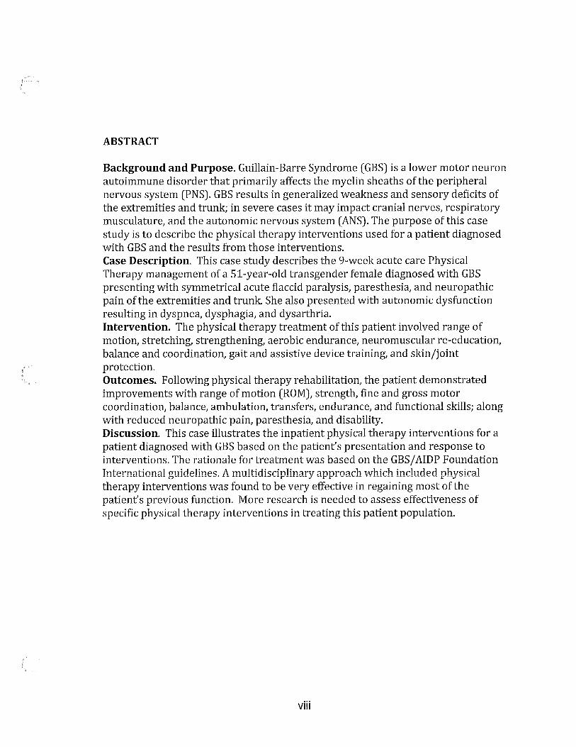
ABSTRACT
Background and Purpose. Guillain-Barre Syndrome (GBS) is a lower motor neuron autoimmune disorder that primarily affects the myelin sheaths of the peripheral nervous system (PNS). GBS results in generalized weakness and sensory deficits of the extremities and trunk; in severe cases it may impact cranial nerves, respiratory musculature, and the autonomic nervous system (ANS). The purpose of this case study is to describe the physical therapy interventions used for a patient diagnosed with GBS and the results from those interventions. Case Description. This case study describes the 9-week acute care Physical Therapy management of a 51-year-old transgender female diagnosed with GBS presenting with symmetrical acute flaccid paralysis, paresthesia, and neuropathic pain of the extremities and trunk. She also presented with autonomic dysfunction resulting in dyspnea, dysphagia, and dysarthria. Intervention. The physical therapy treatment of this patient involved range of motion, stretching, strengthening, aerobic endurance, neuromuscular re-education, balance and coordination, gait and assistive device training, and skin/joint protection. Outcomes. Following physical therapy rehabilitation, the patient demonstrated improvements with range of motion (ROM), strength, fine and gross motor coordination, balance, ambulation, transfers, endurance, and functional skills; along with reduced neuropathic pain, paresthesia, and disability. Discussion. This case illustrates the inpatient physical therapy interventions for a patient diagnosed with GBS based on the patient's presentation and response to interventions. The rationale for treatment was based on the GBS/ AIDP Foundation International guidelines. A multidisciplinary approach which included physical therapy interventions was found to be very effective in regaining most of the patient's previous function. More research is needed to assess effectiveness of specific physical therapy interventions in treating this patient population.
viii
CHAPTERI
BACKGROUND AND PURPOSE
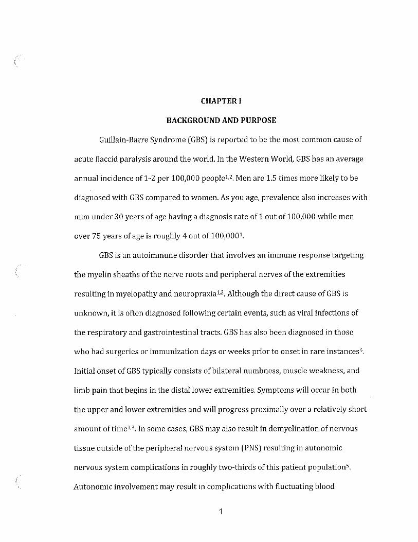
Guillain-Barre Syndrome (GBS) is reported to be the most common cause of
acute flaccid paralysis around the world. In the Western World, GBS has an average
annual incidence of 1-2 per 100,000 people1,2. Men are 1.5 times more likely to be
diagnosed with GBS compared to women. As you age, prevalence also increases with
men under 30 years of age having a diagnosis rate of 1 out of 100,000 while men
over 75 years of age is roughly 4 out of 100,0001.
GBS is an autoimmune disorder that involves an immune response targeting
the myelin sheaths of the nerve roots and peripheral nerves of the extremities
resulting in myelopathy and neuropraxia1,3• Although the direct cause of GBS is
unknown, it is often diagnosed following certain events, such as viral infections of
the respiratory and gastrointestinal tracts. GBS has also been diagnosed in those
who had surgeries or immunization days or weeks prior to onset in rare instances4.
Initial onset of GBS typically consists of bilateral numbness, muscle weakness, and
limb pain that begins in the distal lower extremities. Symptoms will occur in both
the upper and lower extremities and will progress proximally over a relatively short
amount of time1,3• In some cases, GBS may also result in demyelination of nervous
tissue outside of the peripheral nervous system (PNS) resulting in autonomic
nervous system complications in roughly two-thirds of this patient population 5.
Autonomic involvement may result in complications with fluctuating blood
1
pressure, arrhythmias, gastrointestinal dysregulation, and/ or vasomotor
dysfunction5. Cranial nerve (CN) involvement occurs in 45-75% of patients with the
facial nerve (CN VII) being the most commonly affected, resulting in deficits with
facial expression and taste6• According to a study from Lawn et. al.7, roughly one
quarter of GBS patients develop weakness of the inspiratory and expiratory muscles
with 17-30% requiring mechanical ventilation. Roughly 10-20% of patients develop
respiratory failure with a mortality rate of 2-5%3• A study conducted by the
University Medical Center in Rotterdam determined a mortality rate of 2.8% in GBS
patients within a 6-month period after discharge8• They also determined that risk
factors included increased age, severity of weakness, use of mechanical ventilation,
delayed onset of weakness, time to peak disability, increased rate of symptom
progression, and prior diarrhea8,9.
Progression of symptoms can vary widely, moving distal to proximal
anywhere from 12 hours up to four weeks from initial onset where symptoms reach
maximum severity1,10,11. At that time, the progression of symptoms ceases and
reaches a plateau phase at which point the symptoms remain unchanged for 1-2
weeks in most cases but can range anywhere from 2 days up to 6-months in more
severe cases3,10. Following the acute stage is a period of gradual recovery that
ranges from months to years depending on the severity of the GBS diagnosis,
administration of treatment, and rehabilitation. GBS can lead to permanent
disability, however, prognosis is typically good with 70% of GBS patients regaining
complete pre-morbidity functional status12,13,14,15. Roughly one-third of patients
develop mild GBS which is classified by maintaining the ability to ambulate while
2

severe GBS is classified as losing the ability to ambulate due to the neurological
symptoms16.
Diagnosis requires sound clinical judgment utilizing imaging and laboratory
testing to rule out other similarly presenting pathologies such as acute and sub
acute polyneuropathies, botulism and porphyria, basilar artery thrombosis, myelitis,
spinal compression, AIDS infection, and others3,10. Diagnosis of GBS requires lumbar
puncture to analyze cerebral spinal fluid (CSF) protein and white blood cell (WBC)
count. Typically, in the acute phase of GBS, findings from CSF and WBC analysis
would result in an albuminocytolgical dissociation, which demonstrates an elevation
in CSF protein (>0.55 g/L) without an elevation in white blood cells17. Another
diagnostic test that can be used to support GBS diagnosis would be decreased
peripheral nerve conduction velocities with nerve conduction studies (NCS). NCS
are not always conclusive in GBS patients as a study by Luigetti et. al.18 discovered
that 37% of patients who had NCS performed in the first four days of disease onset
had normal NCS findings 18. NCS was found to have increased reliability with
increased time following disease onset and increased severity of symptoms.
The typical course of treatment involves close monitoring for fluctuating
blood pressure (BP), oxygen saturation, cardiac arrythmias, respiratory function,
and signs of hypoxia. Forced vital capacity (FVC) ofless than 15mL/kg indicates
respiratory failure which often requires intubation and admission into the intensive
care unit (ICU)19. lmmunotherapy such as intravenous immunoglobulins (IVIG),
plasmapheresis, and immunosuppressive therapy are the most common treatment
options for GBS and are indicated based on the severity of initial presentation and
3

electrophysiological prognostic factors3,20 . Earlier administration of immunotherapy
following disease onset has shown to lead to better functional outcomes and
decreased demyelination of nerves21. Acute relapse of GBS following stabilization or
improvement of symptoms occurs in less than 10% of severe GBS cases22,23 . Some
research has been done by using corticosteroids, however, results were not
clinically significant in treatment of patients with GBS and is not widely used 24.
Rehabilitation for GBS patients admitted to the ICU requires a
multidisciplinary approach of specialists to monitor respiratory and cardiac
function, prevent secondary complication and disease progression, and initiate
physical rehabilitation. Fatigue is reportedly the most disabling and challenging
symptom to overcome during rehabilitation as it is a frequently occurring symptom
in disorders that involve the PNS25• Specifically, in GBS, the level of fatigue severity
tends to have a positive correlation with age and 60% of patients over 50-years-of
age report fatigue as their most limiting factor26. In many cases, increased fatigue
can remain for years after full recovery of muscle strength and function 27. During
rehabilitation, physical therapy plays an important role in managing fatigue. It takes
special care to improve activity tolerance, being careful not to overwork patients
which can cause increased fatigue and possibly slowing progress. According to
Garssen et. al.27, implementation of patient specific rehabilitation programs
managed by physical and occupational therapy resulted in a 20% improvement of
self-reported fatigue symptoms, which remained at a 2-year follow up.
In the recovery stages of GBS, physical therapy interventions are often
utilized to manage fatigue and pain, regain strength and neuromuscular control, and
4
address functional impairments associated with GBS. During the acute phase of
GBS, physical therapy plays a role in monitoring for the following complications:
deep vein thrombosis (DVT), dysautonomic disorders such as orthostatic
hypotension due to immobilization, respiratory disturbances, respiratory muscle
weakness, contractures, and sensory impairments28,29. To prevent joint
contractures, prescription of orthotics, patient positioning, and gentle range of
motion (ROM) is performed either passively or actively dependent on the ability of
the patient. Orthotic devices may also be used to prevent the development of
pressure sores from prolonged positioning. As the patient's symptoms stabilize and
improve, physical therapy will work to facilitate weight-bearing through the lower
extremities and improve upright tolerance in sitting and standing positions.
Throughout the course of treatment, exercise prescription is focused on regaining
muscular strength through resistive exercises and practice of functional activities.
Early physical therapy interventions have been shown to improve mobility
outcomes, decrease chronic fatigue symptoms, and improve mental function30.
The purpose of this case study is to discuss and review the role of physical
therapy as an intervention in the acute care setting of a patient with a diagnosis of
GBS, who exhibited severe fatigue along with bilateral paralysis and paresthesia of
the extremities and trunk due. This case report demonstrates that physical therapy
twice per day can improve fatigue, strength, motor control, and function with 9-
weeks of acute care rehabilitation.
5
CHAPTER II
CASE DESCRIPTION
Patient was a 51-year-old transgender female (biologically male but
identifies as female) who was self-referred into the Emergency Department (ED) at
the Veteran's Affairs Medical Center and eventually was diagnosed with GBS. The
patient reported her initial symptoms occurred three days prior to visiting the ED
and consisted of tingling, loss of sensation, and weakness of her toes and fingers.
The patient had a past medical history of Type II diabetes mellitus that she assumed
to be the source of her symptoms. However, she reported increasing fatigue and
progressively worsening symptoms that continued to move proximally in her
extremities. She sought medical attention three days following the initial symptoms
when she was unable to drive or negotiate stairs to her 2nd floor apartment.
Three days after admission to the hospital following her ED visit, she was
admitted to the ICU with complete loss of function and neuropathy of her
extremities. A complete blood count (CBCs) and lumbar puncture was ordered and
ruled out other pathologies and revealed elevated proteins in the CSF with normal
white blood cell count. These results, along with peripheral NCS resulted in a formal
diagnosis of GBS.
In the five days following her initial ED visit, her symptoms continued to
worsen with complete flaccid paralysis and neuropathy of the extremities, trunk,
and neck. Due to weakness of the neck and facial nerve involvement, the patient
6
became non-verbal and communicated using eye movements and weak facial and
head movements. The patient also had autonomic nervous system involvement that
led to loss of bowel and bladder control requiring catheterization. A nasogastric
(NG) tube was inserted in the ICU due to increased risk of choking and aspiration
from difficulty with swallowing. Increased respiratory weakness resulted in a
respiratory insufficiency requiring a tracheostomy and mechanically controlled
ventilation.
A referral for occupational and physical therapy services were placed after
the patient reached the plateau phase of the disease progression, which was eight
days after being admitted to the hospital from the ED. The patient's chief complaints
were fatigue, difficulty with breathing, severely diminished motor control of
extremities and trunk, loss of function, weakness, loss of sensation, and burning
sensation of extremities. The patient had no functional control of extremities with
trace muscle grades for all motions of bilateral upper and lower extremities. The
patient reported relief of burning pain in extremities only with pharmaceutical pain
medication. Patient's initial intake history revealed that she resided alone in a
second-floor apartment with two flights of stairs with handrails on both sides to
enter. Patient had two cats and had no immediate family in the area that could
provide support. Patient reported that she had several close friends and a strong
community support system that may provide assistance if needed. The patient was a
retired military Veteran with access to VA health care benefits to manage costs of
health care services. The patient reported working as a cross-country truck driver
7
that involved some heavy lifting and climbing, however, most time was spent in a
seated position driving.
Examination, Evaluation and Diagnosis
Evaluation criteria was based on GBS/CIDP Foundation International
guidelines for GBS/ AIDP evaluation and treatment31. The patient was 5'6" and
weighed 190lbs, which indicated a body mass index (BMI) of 30.7. A BMI of greater
than 30 placed the patient in the obese category32. Upon initial evaluation in ICU, the
patient was in Fowler's position with lower extremities slightly elevated. She had
bilateral compression stockings, a catheter, a left arm IV, a nasogastric tube, and
tracheostomy tube. She was non-verbal and could only communicate through facial
expression and weak head gestures. With palpation, the patient was unable to detect
light touch distal to her elbow and knee bilaterally. She was able to detect increased
pressure of her extremities however tenderness and burning sensation was noted
bilaterally at the gastrocnemius and plantar surface of both feet. Gross ROM was
assessed in the Fowler's position for upper and lower extremities. Limited passive
range of motion (PROM) and intense stretching sensation was experienced in
bilateral hamstrings with straight leg raise (SLR) and gastrocnemius with
dorsiflexion (see Table 1). All other upper and lower extremity motions were within
normal limits and no pain was elicited during PROM.
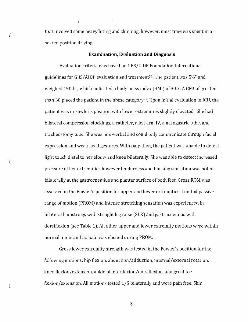
Gross lower extremity strength was tested in the Fowler's position for the
following motions: hip flexion, abduction/ adduction, internal/ external rotation,
knee flexion/ extension, ankle plantarflexion/ dorsiflexion, and great toe
flexion/ extension. All motions tested 1/5 bilaterally and were pain free. Skin
8
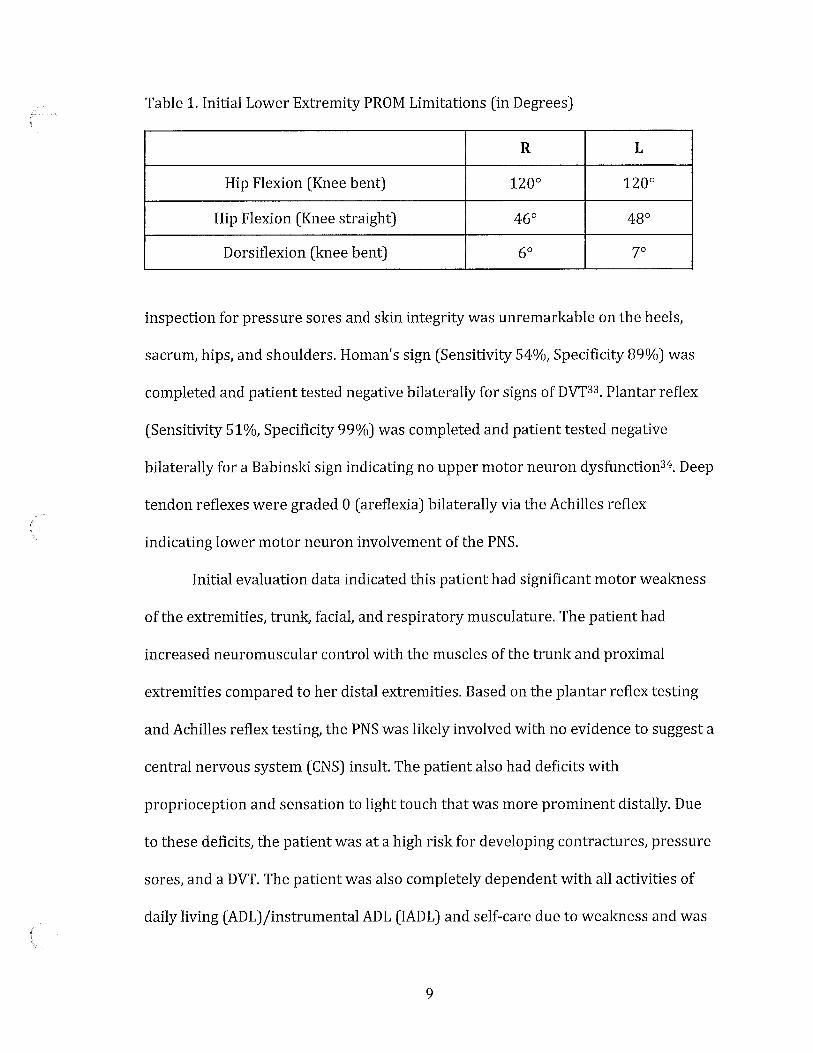
Table 1. Initial Lower Extremity PROM Limitations (in Degrees)
R L
Hip Flexion (Knee bent) 120° 120°
Hip Flexion (Knee straight) 46° 4go
Dorsiflexion (knee bent) 60 70
inspection for pressure sores and skin integrity was unremarkable on the heels,
sacrum, hips, and shoulders. Roman's sign (Sensitivity 54%, Specificity 89%) was
completed and patient tested negative bilaterally for signs of DVT33 . Plantar reflex
(Sensitivity 51 %, Specificity 99%) was completed and patient tested negative
bilaterally for a Babinski sign indicating no upper motor neuron dysfunction 34. Deep
tendon reflexes were graded O ( areflexia) bilaterally via the Achilles reflex
indicating lower motor neuron involvement of the PNS.
Initial evaluation data indicated this patient had significant motor weakness
of the extremities, trunk, facial, and respiratory musculature. The patient had
increased neuromuscular control with the muscles of the trunk and proximal
extremities compared to her distal extremities. Based on the plantar reflex testing
and Achilles reflex testing, the PNS was likely involved with no evidence to suggest a
central nervous system (CNS) insult. The patient also had deficits with
proprioception and sensation to light touch that was more prominent distally. Due
to these deficits, the patient was at a high risk for developing contractures, pressure
sores, and a DVT. The patient was also completely dependent with all activities of
daily living (ADL)/instrumental ADL (IADL) and self-care due to weakness and was
9

at a high fall risk. The patient required total assistance of two for bed mobility and
ceiling lift for transfers. The self-care, transfer, and locomotion portion of the
Functional Independence Measure (FIM) was completed and patient scored 1 in all
tested areas (see Appendix A). The FIM is an objective measuring tool to assess
patient's level of disability and response to treatment. Goals for this patient included
increasing strength of trunk and extremities, improving endurance, avoiding
formation of contractures, improving sensation and proprioception of extremities,
decreasing pain, improving balance and coordination, and improving mobility, gait,
and overall function. Completion of these goals would help the patient return to her
prior level of function allowing her to complete ADL/IADLs, ambulate, discharge to
home, and return to work activities.
Prognosis and Plan of Care
Mortality rates for GBS patients who have ICU management is roughly 2-12%
and is roughly 5% in tertiary care centers with medical professionals who have
previous experiences treating GBS35. The most common cause of GBS-related death
is tied to complications from ventilation such as pneumonia, sepsis, acute
respiratory distress syndrome, and autonomic dysfunction 36. The primary goal for
this patient while in the ICU was to restore optimal respiratory function as it is one
of the biggest predictors for long term disability and mortality in this population.
Other factors that have found to be associated with adverse patient outcomes
include older age (>57 years old), poor upper extremity strength, acute hospital stay
>11 days, an ICU admission, use of mechanical ventilation, and rapidly progressing
onset of muscle weakness37,3B_

Most GBS patients will return to their prior level offunction, however, 7-15%
of GBS patients suffer with chronic neurological sequelae such as, foot drop,
intrinsic hand and foot muscle wasting, sensory ataxia, dysesthesia, pain, increased
fatigue, and overall functional impairments39,40. The risk ofreoccurrence is rare and
only occurs in 2-5% of patientszz,23_
Given this information, the patient had a fair prognosis based on the relatively low
rate of mortality and reoccurrence within this population. Unfortunately, this
patient was positive for all of the factors associated with adverse outcomes listed
above. The patient was fortunately treated at a facility with a significant history of
treating GBS. Primary goals for this patient for rehabilitation were focused on early
bed mobility, transfers, and ambulation. Independent ambulation occurs in 20% of
GBS patients after 4 weeks and is a predictor for reaching full prior level of function
at 1 year while only 40% reach their prior level of function if they are ambulating
with an assistive device at 4 weeks.
Throughout the plan of care, the patient was reassessed utilizing the FIM,
MMT, and ROM. The patient's progress was also recorded and progressed based on
her response to treatment with an emphasis placed on reducing fatigue, improving
activity tolerance, strengthening, maintaining ROM, and functional mobility
(ambulation, stair ambulation, bed mobility, and transfers).
11

CHAPTER III
INTERVENTION
Patient was seen twice each day for 30-minute sessions in the morning and
the afternoon while in the ICU. She received the same frequency of physical therapy
and occupational therapy throughout her course of treatment in the acute care
setting. Physical therapy was primarily involved with regaining function of the
lower extremities and trunk, improving activity tolerance, and improving functional
mobility. Occupational therapy was primarily involved with regaining function of
the upper extremities including self-care, adaptive device training, and fine motor
skill reacquisition throughout the course of rehabilitation. This case study will focus
on the trunk and lower extremities, gait, and mobility interventions.
Since the patient was intubated, verbal communication from the patient was
impossible and use of facial and eye expressions was the main determinant for
communication. Facial expressions were subtle and inconsistent initially due to CN
VII involvement, therefore, close observation from the therapist was essential to
assess the patient's response to interventions. During the 1st week of intervention,
the patient received lower extremity PROM to end range for all motions of the hips,
knees, and ankles to prevent the development of contractures and maintain
flexibility due to the acute flaccid paralysis. The patient was encouraged to assist
with motion through verbal cues from the therapist. Each lower extremity motion
was completed through full range for 20 cycles with 3 second tempo in each
12
(
(
(
direction. The patient performed bed mobility training, rolling from supine to
sidelying with maximum assist +2. She was cued to assist with positioning and
rolling. While in sidelying, hip extension PROM exercises were performed. Passive
sustained stretches for the hamstrings and heelcords were held for 90 seconds for 2
sets during the treatment session to prevent the development of contractures ( see
Appendix B for specific week 1 interventions). The patient was fitted with Multi
Pod us boots to reduce the risk of developing plantar flexion contractures, hip
external rotation, and heel ulcers (see Figure 1.).
https:/ / cdn3.volusion.com/whfx.vmyur /v /vspfiles /photos/ 614 7ML-2T.jpg
Figure 1. Multi Podus Boot
During the 2nd week ( see Appendix B for specific week 2 interventions), the
patient completed transfers from supine to sitting at the edge of her hospital bed
with maximum assist +3. She was encouraged to assist with the transfer through
verbal cues to encourage neuromuscular facilitation. While sitting upright, she was
cued to maintain sitting balance to enable core stabilization. In this position, lower
extremity ROM exercises and stretching were also performed. She was unable to
13

maintain sitting balance with feet on the ground and required therapist assistance
to stay upright. During each therapy session, the patient's level of strength was
assessed through the amount of assistance needed with ROM activities. By the end
of the second week, the patient was able to provide gradually increasing levels of
effort but still required moderate assistance throughout the entire ROM with all hip
and knee motions. She was still unable to move her limbs against gravity and
quickly fatigued after 8-10 repetitions with therapist assistance. Bed mobility
exercises continued during the second week with increased participation from the
patient.
During the 3rd week ( see Appendix B for specific week 3 interventions), the
patient continued to perform transfers from supine to sit with therapist assistance
with verbal cues. She was able to maintain sitting balance without upper extremity
support with her feet planted on the floor for 20 seconds before losing her balance.
Lower extremity AAROM against gravity was performed in sitting with
demonstrating improved lower extremity strength. She had jerking concentric
movements with poor eccentric control against gravity. She was able to complete
two sit-to-stand transfers with maximum assistance of 2 with use of a standard
walker with upper extremity support. She was able to maintain a standing position
with maximum assistance of 2 with encouragement for two 30-second trials before
lower extremity fatigue set in. She required maximum assistance of 2 to block knees
from flexing and to prevent hip flexion. By the end of the 3rd week, she had 3 /5
strength with all hip motions and 2+/5 strength with knee flexion and extension
bilaterally. She continued to have difficulty with fatigue, coordination, and
14
proprioception of her lower extremities demonstrated by decreased strength and
control of movements. At this time, the patient was able to communicate verbally as
her ventilator was discontinued and replaced with supplemental oxygen. FIM scores
were reassessed at this time ( see Appendix A).
After 4 weeks ( see Appendix B for specific week 4 interventions), the patient
was discharged from the ICU and admitted into the acute care wing where she
received physical therapy twice daily in the physical therapy department. She
continued the same physical therapy scheduling with two 30-minute sessions for
transfers, gait, and functional training completed in the morning and strengthening
and endurance in the afternoon to avoid fatigue. With the use of bed rails and
trapeze bar, the patient performed log rolling and supine to sit transfers with
moderate assistance by the physical therapist and use of her upper extremities. Due
to her grip strength deficits, she required the use of Dycem pads and therapist
assisted hand holds to allow her to use her upper extremities for assistance. Balance
training in sitting was performed and the patient was able to maintain sitting
balance with feet on the floor with small perturbations for 30-second trials. She
continued to receive sustained passive stretches to her lower extremities in sitting
to maintain ROM and prevent the development of contractures since she continued
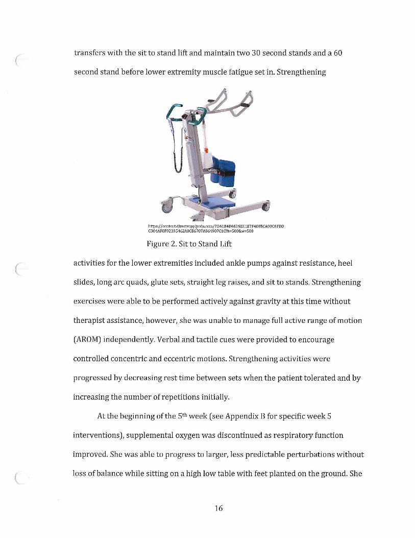
to spend most of her day in supine. A sit to stand lift ( see Figure 2.) was utilized
during a cotreatment session with occupational therapy to encourage concurrent
use of upper and lower extremities and encourage sustained weight bearing
through the lower extremities. The patient was able to tolerate 3 sit to stand
15
(
(
(
transfers with the sit to stand lift and maintain two 30 second stands and a 60
second stand before lower extremity muscle fatigue set in. Strengthening
https://contentdirectsupplycdn.com/7D61B4B6626EC1E7F4085CA03C6FDD C304AF8F92 39 5462A0CB6707 A064907C lC?h=S00&w=S00
Figure 2. Sit to Stand Lift
activities for the lower extremities included ankle pumps against resistance, heel
slides, long arc quads, glute sets, straight leg raises, and sit to stands. Strengthening
exercises were able to be performed actively against gravity at this time without
therapist assistance, however, she was unable to manage full active range of motion
(AROM) independently. Verbal and tactile cues were provided to encourage
controlled concentric and eccentric motions. Strengthening activities were
progressed by decreasing rest time between sets when the patient tolerated and by
increasing the number of repetitions initially.
At the beginning of the 5th week (see Appendix B for specific week 5
interventions), supplemental oxygen was discontinued as respiratory function
improved. She was able to progress to larger, less predictable perturbations without
loss of balance while sitting on a high low table with feet planted on the ground. She
16
Appendix A.
Functional Independence Measure (FIM) scores
34

FIM scores throughout treatment
Initial Week3 Week6 Discharge
Self-Care
A Eating 1 2 5 6
B. Grooming 1 2 4 7
C. Bathing 1 2 3 6
D. Dressing-Upper 1 2 4 6
E. Dressing-Lower 1 2 4 6
F. Toileting 1 3 3 7
Transfers
G. Bed, Chair, 1 3 4 6 Wheelchair
H. Toilet 1 3 4 6
I. Tub, Shower 1 2 3 6
Locomotion
J. Walk/Wheelchair 1 1 4 6
K. Stairs 1 1 1 6
Motor Subtotal Score 12 23 39 68
35

(
(
(
FIMTM instrument 7 Complete Indcpcndcru:e (Timely, Safely) 6 Modi.!ic::d Independence, ~cc) NO HELPER
Lt--------------------1-----------------1 E V ~ L s
ModHlt-d Dependen(:C 5 Supel"<isiori (Subject"' 100%+) 4 Mini~Assist (Subject= '15%◄·) 3 Modeta1e Assist (S.ubjcct i= SO%+)
Cmn1,1lct11 lkpeu.dence 2 Maxi.malA:ssi5t (Subject -25%+) I Total A5Sid (Subject- less lh~n 2S%)
-·
Self-Care ADMISSION A. Ealing
~ B Oroonling C. Bathing D. Drc8siag - Upper Body E:. Dressing - uiw,;:r Body F. Toileling
Spbindi:r Control
B G. Bladder Mmagcm~nt H. Bowel Maaagelllent
Tianslcn I. Bed, Chafr, Wheelchair § J. Toilet K. Tub, Sbowi:r
~moliou aeWA L. Walk/Whcclchai:r t;wi,o,i,,,,;,
M. Stairs ·-Motar Suht.otal Score CJ
36
HELPER
DISCHARGE FOLLOW•UP
~ D B B § § a□\1\8 aot-: C \lbo<l.a&l! C 'l,\,,d,,ui< ·- .,.,..
CJ D
maintained sitting balance without feet on the floor and progressed to maintaining
sitting balance on an Airex balance foam pad. Sit to stand transfer training
progressed significantly as she was able to transfer from wheelchair to standing in
parallel bars using her upper extremities. At the beginning of week 5, she required
moderate assistance of one and by the end of the week progressed to contact guard
assist with transfers. Standing balance required the use of her upper extremities and
minimal assistance for 30-60 seconds due to lower extremity fatigue. The patient
was also able to manage her first steps during the 5th week by ambulating 15 feet in
the parallel bars with minimum assistance of one. Strength and endurance were
progressed by increasing the number of repetitions before increasing resistance.
She was also encouraged to focus on slow and controlled eccentric movements to
improve coordination.
During the 6th week ( see Appendix B for specific week 6 interventions), the
patient was discharged from acute care and admitted to the transitional care unit
(TCU) where she continued to receive inpatient physical therapy twice daily. The
patient quickly improved her standing balance without upper extremity assistance
in the parallel bars with contact guard assist provided. Dynamic balance training
was completed with contact guard assist for timed trials from 30-60 seconds and
included narrowing base of support, tandem standing, Airex foam pad balance,
reaching outside her base of support, and light perturbations. She also made
significant progress with ambulation within the parallel bars using upper extremity
support. She was able to ambulate 30-feet at a time for 3 sets and progressed to 6
sets with contact guard of 1. She demonstrated the ability to pivot 180° within the
17
parallel bars to change direction. She continued to have difficulty with eccentric
control oflower extremities and dragging her feet that was corrected with verbal
cues. By the end of the 6th week, she demonstrated the ability to ambulate without
upper extremity support for 15 feet and minimal assist of 1. She required seated
rest breaks of 2-3 minutes between trials due to lower extremity muscular fatigue.
Bed mobility exercises continued with transfers from supine to sidelying, supine to
sitting, supine to prone, and prone to quadruped with no contact assistance but
continued to require verbal cues for lower extremities due to impaired
proprioception. Lower extremity strengthening exercises continued with emphasis
on slow, controlled concentric and eccentric movements. Patient was instructed to
not watch legs during exercise to improve proprioception and coordination. She was
able to perform exercises while maintaining conversation demonstrating improved
ability with performing dual tasks. Patient FIM score was reassessed at this time
(See Appendix A).
During the 7th week (see Appendix B for specific week 7 interventions), she
continued to perform and progress with standing dynamic balance activities. She
continued to received contact guard for safety, however, she was able to stand with
feet together on Airex foam balance pad, reach outside base of support, and
increased intensity and variation in perturbations. She was also able to play catch
with a foam ball while standing and using both hands to catch and toss the ball
without loss of balance. She progressed to ambulation with a front-wheeled-walker
(FWW) with contact guard but no assist for 300 feet at a time with lower extremity
fatigue being the limiting factor. She required seated rest breaks of 2-4 minutes for
18
recovery and at those times she was given verbal cues to avoid watching her feet
during ambulation to improve lower extremity proprioception and coordination.
She also completed activities in quadruped consisting of crawling, transfers to
kneeling, and reaching to improve balance, strength, and coordination. Lower
extremity strengthening exercises progressed by increasing number of repetitions
and implementation of yellow and red TheraBand. She demonstrated improved
neuromuscular control as eccentric motions were performed in a smoother, more
coordinated fashion with verbal cues. She demonstrated the ability to sit to stand
without upper extremity support from a low chair. She progressed to performing
squats to 90° knee flexion with upper extremity support in the parallel bars in a
slow and controlled manner with standing rest breaks. She also performed step-ups
and step-downs on a 6-inch step in the parallel bars with minimal upper extremity
support for balance and to prepare her for stair ambulation. By week 7, she started
endurance training on the NuStep bike for 5 minutes at level one at a slow pace with
an emphasis to avoid fatigue.
During the 8th week (see Appendix B for specific week 8 interventions), the
patient quickly progressed in all areas with treatment focused on addressing the
functional demands of discharging her back to her home. Special emphasis was
placed on improving the quality of her gait and endurance. She progressed to using
a 4WW with a seat for ambulation. Frequency of verbal cues continued to decrease
for dragging and watching feet during ambulation. At this time, she was able to
ambulate with her 4WW without rest breaks while negotiating slight inclines and
declines. She performed stair ambulation using a single handrail, contact guard, and
19
a step to gait pattern. She was able to ascend and descend a flight of stairs before
requiring a seated rest break due to fatigue. Floor transfers were started to ensure
the patient could get off the floor in the event of a fall. She required contact guard
with no assistance and verbal cues to transfer from quadruped to standing utilizing
a stable surface and upper extremity assistance. She continued to utilize the NuStep
bike twice daily at an intensity of 3-5 for 10-minute intervals with an emphasis to
avoid fatigue. Lower extremity strengthening exercises were discontinued at this
time to focus on compound movements related to improving functional tasks. For
example, she was instructed to pick small/light weight items off the floor using a
stable surface and upper extremities for support to maintain balance.
On the 9th and final week of therapy (see Appendix B for specific week 9
interventions), the patient was discharged home with a referral for home health
physical therapy. She was able to ambulate community distances ( +200m) without
fatigue or loss of balance. By the final week, she was ambulating with her 4WW to all
meals and to the PT department without supervision. She was also able to ambulate
without a walker by the final week for distances of +500 feet with contact guard no
assistance, however, she did fatigue, and her balance declined as a result. She could
ambulate step-over-step with use of a handrail, however, fatigue would set in after a
single flight. Strength and coordination continued to progress as evidenced by her
ability to transfer from quadruped to standing without the use of assistive devices
or use of a stable surface. By week 9, her endurance improved, as she was able to
ambulate +600 feet around the hospital without fatigue or lower extremity
weakness. She continued to improve her activity tolerance with the NuStep bike by
20

increasing her intervals to 15 minutes on level 5 with emphasis on avoiding fatigue.
By her final week, the patient's strength continued to improve with all lower
extremity motions improving to 5/5 aside from plantar flexion which was at 4/5.
Her lower extremity flexibility also improved to within normal limits by the final
week The final FIM score was taken at this time ( see Appendix A). The patient was
discharged from physical therapy after meeting all goals. She received a referral for
home health physical therapy to continue to improve endurance, gait, balance,
strength, and overall function.
21

CHAPTER IV
OUTCOMES
The overall outcome for this patient was excellent considering the level of
disability and function during the initial physical therapy evaluation. Outcomes for
this patient were based on objective measurements including lower extremity
strength, lower extremity ROM, and FIM scores. Her progress was also based on her
ability to perform functional skills, for example, ambulation, transfers, bed mobility,
and the number of rests breaks needed.
At discharge she no longer required the use of compression stockings on her
lower extremities, catheterization, nasogastric tube, or tracheostomy tube. She had
also regained function of her facial musculature and ability to communicate verbally
by the 3rd week. Bilateral ankle, knee, and hip ROM was assessed throughout the
plan of care using a goniometer. During the initial evaluation, she had limitations in
PROM with hip flexion (with full knee extension) and dorsiflexion bilaterally. Initial
measurements of hip flexion were 46° for the right hip and 48° for the left hip. At
discharge, hip flexion was measured at 69° for the right hip and 72° for the left hip.
Initial measurements of ankle dorsiflexion were 6° for the right ankle and 7° for the
left ankle. At discharge, ankle dorsiflexion was 11 ° for the right ankle and 10° for the
left ankle. Gross lower extremity strength, using manual muscle testing throughout
the plan of care, demonstrated significant improvement in all areas. The following
areas were tested every three weeks: hip flexion, abduction/adduction,
22
internal/ external rotation, knee flexion/ extension, ankle plantar/ dorsiflexion, and
great toe flexion/ extension. At the initial evaluation, all motions tested 1/5
indicating palpable muscle twitch with contraction but no visible motion at the joint.
At discharge, her plantarflexion tested 4/5 bilaterally indicating that she was able to
complete 10-24 single leg heel raises through full ROM. Remaining bilateral lower
extremity motions were graded 5 /5 indicating patient's ability to hold the test
position against gravity and with a maximal force from a physical therapist clinician.
Deep tendon reflexes of the lower extremity were tested bilaterally using the
Achilles Tendon reflex which primarily tests the Sl nerve root. At initial evaluation
the patient tested O (areflexia) bilaterally indicating lower motor neuron
involvement which was consistent with the patient's diagnosis. The Achilles Tendon
reflex was tested again at discharge and was graded 1 + ( diminished/low response)
indicating improvement with the lower motor neuron involvement. The FIM, which
has an excellent internal consistency (Conbach's alpha: 0.95 = FIM Motor, 0.95 =
Total FIM), was also used as an objective test to assess the patient's functional
performance throughout the plan of care42. FIM scores increased in all assessed
areas throughout the plan of care and be viewed in Appendix A The patient
progressed significantly with all functions of bed mobility, transfers, and
ambulation. Initially, the patient was completely dependent with bed mobility
requiring maximum assist of two. Throughout the course of treatment, she made
consistent progress with supine to sit, log rolling, and moving up/down the bed as
strength and neuromuscular control improved. At discharge she required no
assistance or supervision for any bed mobility. Significant improvement was made
23
with her ability to transfer as initially she required maximum assistance of two and
use of a ceiling lift for bed and chair transfers and toileting. At discharge, the patient
was capable of safe sit to/from stand transfers without assistance or supervision.
Ambulation was not possible during the initial evaluation with patient making
significant improvements by discharge. Ambulation was measured in feet
ambulated and assistive device needed. Initially, the patient was only able to
ambulate 15 feet within the parallel bars with minimum assistance of one at week 5.
As she regained strength, neuromuscular control, and endurance, she progressed
with the assistive device needed, gait quality, and ambulation distance. At discharge,
she was able to ambulate without an assistive device for 500+ feet with contact
guard assist for safety but required a 4WW to ambulate community distances
( +200m) without supervision or rest. Gait quality also improved; initially she
demonstrated an antalgic gait with poor eccentric knee extension control and foot
drag and required cues to not to watch her feet. At discharge, she increased her gait
speed and quality with increased eccentric knee extension and no foot dragging,
decreasing her risk of falls. Verbal cues were also not needed at this time.
The patient tolerated all interventions well with minimal complaints of
excessive fatigue that impeded the patient's rehabilitation. The patient continued to
have neuropathic pain of the lower extremities that primarily affected the dorsum of
the foot and anterior surface oflower leg; however, symptoms had greatly
improved. The patient was extremely motivated throughout the plan of care and did
not miss a physical therapy appointment at any time during her inpatient stay.
Overall, the patient was extremely happy with the progress she made in all areas.
24
CHAPTERV
DISCUSSION
The patient significantly reduced her disability and improved function
throughout her course of treatment as an inpatient. During the initial evaluation, the
patient was completely dependent and required maximum assistance for nearly all
motions and tasks. Although the patient was able to return home and demonstrated
improvements with strength, balance, and functional mobility, she was still not at
her prior level of function due to her limitations with mobility, fatigue, sensation
impairments, and fine motor skills. For this reason, she would benefit from
continued therapeutic interventions from occupational and physical therapy to
address these limitations.
Throughout the course of treatment, special emphasis was placed on
achieving functional mobility to promote independence as her goal was to return
home and back to work. Each week, interventions were performed to get her
prepared for standing and then progressed to walking. This was completed by
strengthening muscles of the trunk and lower extremities through transfer
exercises, quadruped activities, balance exercises, assistive device training, and gait
training. According to Lubenova et. at. 43, physical therapy interventions should be
formed and directed based on the current presentation of relative symptoms while
addressing specific functional tasks. By utilizing this principle, the physical therapy
interventions were successful in achieving that patient's goal of safe ambulation.
25

Bed mobility and functional transfers were another area that this patient
made great improvements in. At the initial evaluation, she was completely
dependent and required maximum assistance of two for any positional changes.
Ceiling lifts were used multiple times daily for toileting, bathing, and transfers. At
discharge, the patient was independent with all positional changes, including
transferring off the floor in the event of a fall. According to Khan et. al.44,
assessments and intervention should not primarily focus on movement quality. A
greater focus should be placed on promoting safe execution of skills to promote
functional independence44• This patient responded well to a systematic approach of
interventions, which included exercises that encouraged frequent positional
changes and required the patient to solve problems promoting independence45.
Lower extremity strengthening played a vital role in promoting
independence for this patient. Lower extremity strength improved in all areas from
1/5 (trace) muscle grades during the initial evaluation to 4/5 - 5/5 (good-normal)
at discharge. Isometric exercises were completed against gravity and increased in
both intensity and repetitions. Sit to stand exercises progressed to upper extremity
supported squats, which encouraged coordination between the lower extremity and
trunk musculature to promote synergy between these muscle groups. Cues were
required initially due to impairments with balance and proprioception. Cues
included proper positioning to initiate the movement sequence and increasing
movement speed. According to Davidson et. al.46, purposeful training of sit to stand
with emphasize on optimal positioning of the lower extremities and trunk improved
coordination, consistency of movement, and reduced energy demands.
26

Patients who are diagnosed with GBS tend to have high rates of depression
and anxiety throughout the entire rehabilitation process47• These same conditions
are also 4 times more likely to occur in trans gender individuals48 . As a result, this
patient was at a high risk of developing a depressive and/ or anxiety disorder during
the rehabilitation process. Effective communication with the patient and other
healthcare providers (physician, psychiatry, social work, etc.) played a vital role in
ensuring she had the support and resources needed throughout the rehabilitation
process. Recreational therapy became an option for the patient when she became
medically stable. A study by Gassaway et. al.49, demonstrated decreased rates of
depression (PHQ-9) in patients that had access to recreational therapies while in
inpatient settings. As a result, she was able to participate in group social activities
and community outings throughout the plan of care which may have contributed to
her high levels of motivation during her rehabilitation.
Although the results of physical therapy interventions positively impacted
the patient's functional progress throughout the plan of care, there were several
limitations regarding objective measures and documentation that would benefit
similar patients in the future. Utilizing more outcome measures when treating this
patient could have greatly benefited this patient in both the acute care setting and
for any post-discharge outpatient rehabilitation. One example of an outcome
measure that would have benefited the patient is the Fatigue Severity Scale (FSS) as
it is often used in patients with neuromuscular disorders. The FSS is an easy to
implement questionnaire that collects the patient's subjective assessment of how
fatigue impacts their daily functioning on a scale from 1 ( no fatigue) through 7
27
(extreme fatigue) 50. The FSS demonstrates good test-retest reliability (Cohen's
kappa vale= 0.84) and internal consistency (Cronbach's alpha coefficient= 0.88)51.
Two other objective measures that would be recommended to apply to
future cases would be the 10-Meter Walk Test (10MW) and the Timed Up and Go
Test (TUG). The 10MW test is a performance measure that can be utilized to assess
speed and quality of gait, functional mobility, and vestibular function in patients
who ambulate with or without assistive devices 52. The 10MW has excellent
neuromuscular test-retest reliability (ICC= 0.91) and is usually applied to monitor
patient progress and compare with normative data53. The TUG is an objective
measure to determine fall risk and track progress with balance, gait, sit-to-stand
transfers, and functional mobility in the rehabilitation setting. The TUG has a high
sensitivity and specificity of 0.87 for determining fall risk, however, it has its
limitations with predicting fall risks in community environments54,55.
Other areas of consideration and change for future patients would be
increasing the frequency of co-treatments with occupational therapy as a method of
conserving time and energy for the patient. Roughly 4 hours per day was spent
completing therapeutic activities between the two disciplines. Co-treatments were
initially performed in the ICU to maintain range of motion with upper and lower
extremities and to provide assistance with bed mobility. Co-treatments were also
performed during week 4 when utilizing the sit to stand lift. Use of neuromuscular
electrical stimulation (NMES) may have been a beneficial modality to use during the
initial acute phase of the diagnosis. A study by Harbo et. al. 56 demonstrated
decreased muscle wasting on extremities that were treated with NMES during the
28
acute phase. Functional electrical stimulation (FES) could have also been
implemented during the initial phases of gait training as a method of preventing foot
drop during the swing phase of gait. Although these modalities were not used, our
patient still had favorable outcomes from the implemented interventions.
Individually tailored physical therapy interventions are necessary for
successful outcomes when treating patients with GBS. Interventions must be
evaluated daily due to the frequently changing symptoms and condition of the
patient. Physical therapists must be cognizant to evaluate the patient's immediate
and daily responses to treatment to ensure proper prescription of exercise type and
intensity. Intensity of exercises must be high enough to promote a physiological and
therapeutic response while remaining below the threshold of irritation to prevent
unwanted fatigue and nervous stress57. Although quality research on this topic is
limited, this case study demonstrates that physical therapy interventions have a
positive impact on regaining function and decreasing disability in GBS patients in
the acute care setting.
Reflective Practice
Although the outcomes of this case were very good considering the state the
patient was in during the initial evaluation, there are several areas that could be
improved on and implemented to achieve the best outcome possible. During the
initial evaluation, most questions were directed toward the primary care team due
to the patient's inability to communicate verbally. More questions and specific
evaluations regarding the patient's perception of pain is an area that could have
required more attention. Although her pain was managed with pharmaceuticals, we
29

could have implemented more pain management interventions to reduce the risk of
developing a pharmaceutical dependence. We could have implemented the Visual
Analogue Scale for Pain (VAS Pain), Short Form-36 Bodily Pain Scale (SF36-BPS), or
the Chronic Pain Grade Scale (CPGS). Using these tests and measures would have
allowed us to document and observe her changes in perception of pain. Using this
information, we could assess how effective pain management modalities were and
also made referrals to pain specialists. There seems to be a lack of research on non
pharmaceutical pain management options for patients with GBS, therefore, more
research should be geared towards identifying more treatment options.
Sensory impairments are also a key feature in those diagnosed with GBS.
More questioning and attention toward the areas where sensation was impaired,
and characteristics of those impairments should be prioritized in the future. Asking
the patient about what areas are painful/aggravating and whether or not light
touch/vibration/deep pressure is being noticed. We could record and monitor the
progress of nerve reinnervation, monitor muscle soreness, and prevent any
unwanted injuries knowing this information. Filament testing would have been a
great tool to use for assessing sensation and should be implemented for future
cases.
As mentioned earlier, implementing more objective tests and measures
should be used for the following areas: fatigue, gait speed, gait quality, balance,
quality oflife, and function. Using these tests and measures would not only
contribute to better outcomes while in the inpatient setting, they would establish a
baseline for outpatient care after discharge and establish areas of need.
30
Unfortunately, documentation of objective information throughout the course of
treatment was a major weakness in the case and will be improved for future cases.
Although respiratory therapy was heavily involved for this patient, physical
therapy could have implemented more respiratory techniques to assist this patient
due to the respiratory failure that occurred in the acute stages. Recording oxygen
saturation with a pulse oximeter during exercise and recording the amount of
supplemental 02 during treatment sessions would have been a valuable method of
tracking oxygen demands of the body. Implementing co-treatments with respiratory
therapy could have been useful for practicing postural drainage, breathing and
coughing techniques, and resistive inspiratory exercises to prevent secondary
respiratory complications and improve quality of breathing.
Psychological support is another area that I will highlight regarding this
patient. Although she demonstrated a high level of motivation throughout the plan
of care, she was at a high risk of developing depression and anxiety disorders due to
her diagnosis, being transgender, and having a lack of family support. Although
social work was involved throughout the entire case, counseling and psychiatry
would have had a positive impact on the patient's overall wellbeing. The patient did
have periods of fear, anxiety, guilt, and shame during her treatment and through my
experience, physical therapy was an outlet for addressing her personal thoughts and
feelings. In the future, allowing the patient to "vent" is an effective method of
managing patients when they are struggling with their diagnosis. Asking the patient
about positive things can help pull them out of their negative mindset and build a
more positive, forward looking outlook. Referral to counseling or psychiatry would
31
have been the right option for this patient and will be utilized more readily in the
future.
Overall, this patient's outcomes were favorable due in part to the frequency
of physical therapy and occupational therapy interventions. Although this is ideal, it
is not always realistic for many patients. Fortunately, this patient was a Veteran
with VA medical benefits, therefore, she was able to receive treatment without
direct costs. Early mobilization with encouraged assistance from the patient was
essential with preventing secondary complications and regaining function. Another
area of emphasis during her 9-weeks of therapy was avoiding excessive fatigue.
Although we could regulate the amount of exercise during therapy, we could not
always account for the energy expenditure from busy days where the patient
attended many different appointments. Therefore, we had to regularly modify the
plan of care to compensate for increased fatigue when present.
Personally, treating this patient gave me a diverse treatment experience that
required me to adapt the plan of care daily due to the patient's quick return of
function. For example, during the first few weeks she was non-verbal which
required me to be creative with communication between the patient. Monitoring
facial expressions, lip reading, and writing on a whiteboard became the main
methods of communication. This was frustrating for the patient and myself at times
since we would not be on the same page and required more time, but it did not
affect the quality of the interventions. Overall, I was extremely excited to take part
in helping this patient regain most of her function and set her up to succeed with
continued outpatient physical therapy. It allowed me to utilize many different motor
32

learning interventions is a short period of time, which is perfect for a student
completing a 9-week clinical experience. The lessons and experience from this
patient helped me grow as a competent and effective clinician.
33
AppendixB.
Physical Therapy Interventions Weeks 1-9
37

Week 1- Physical Therapy Interventions
Lower extremity • All motions of the hip, knee, and ankle PROM in Supine • 20 repetitions at 3 second tempo
• Verbal cues to assist Hamstring and • Passive, low load stretch into dorsiflexion and hip flexion with heelchord stretch knee in full extension
• 90 second stretch, 2 sets each Log rolling/bed • 1-3 repetitions each direction mobility • Maximum assist +2 with verbal cues to assist with motion
Week 2 - Physical Therapy Interventions
Transfer supine to • Maximum assist +2 with verbal cues to assist with motion sitting • Seated position, feet on floor with cues to maintain sitting
balance Lower extremity • All motions of the hip, knee, and ankle PROM in Seated • 20 repetitions at 3 second tempo
• Verbal cues to assist
• Seated position with assistance to maintain balance Hamstring and • Passive, low load stretch into dorsiflexion and hip flexion with heelchord stretch knee in full extension
• 90 second stretch, 2 sets each Log rolling/bed • 1-3 repetitions each direction mobility • Maximum assist +2 with verbal cues to assist with motion
Week 3 - Physical Therapy Interventions
Transfer supine to e Maximum assist + 1 with verbal cues to assist with motion sitting • Seated position, feet on floor with cues to maintain sitting
balance 20 seconds, 3 sets Lower extremity • All motions of the hip, knee, and ankle AAROM in Seated • 20 repetitions at 3 second tempo
• Verbal cues to assist
• Seated position with assistance to maintain balance Hamstring and • Passive, low load stretch into dorsiflexion and hip flexion with heelchord stretch knee in full extension
• 90 second stretch, 2 sets each Log rolling/bed • 3-5 repetitions each direction mobility • Maximum assist + 1 with verbal cues to assist with motion
38
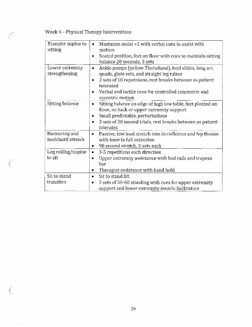
Week 4 - Physical Therapy Interventions
Transfer supine to • Maximum assist+ 1 with verbal cues to assist with sitting motion
• Seated position, feet on floor with cues to maintain sitting balance 20 seconds, 3 sets
Lower extremity • Ankle pumps (yellow TheraBand), heel slides, long arc strengthening quads, glute sets, and straight leg raises
• 2 sets of 10 repetitions, rest breaks between as patient tolerated
• Verbal and tactile cues for controlled concentric and eccentric motion
Sitting balance • Sitting balance on edge of high low table, feet planted on floor, no back or upper extremity support
• Small predictable, perturbations
• 3 sets of 30 second trials, rest breaks between as patient tolerates
Hamstring and • Passive, low load stretch into dorsiflexion and hip flexion heelchord stretch with knee in full extension
• 90 second stretch, 2 sets each Log rolling/supine • 3-5 repetitions each direction to sit • Upper extremity assistance with bed rails and trapeze
bar • Therapist assistance with hand hold
Sit to stand • Sit to stand lift transfers • 3 sets of 30-60 standing with cues for upper extremity
support and lower extremity muscle facilitation
39
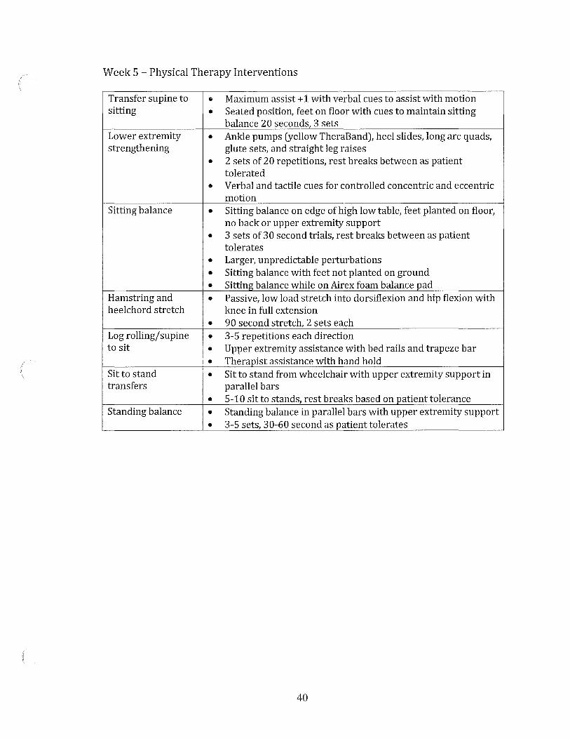
Week 5 - Physical Therapy Interventions
Transfer supine to • Maximum assist + 1 with verbal cues to assist with motion sitting • Seated position, feet on floor with cues to maintain sitting
balance 20 seconds, 3 sets Lower extremity • Ankle pumps (yellow TheraBand), heel slides, long arc quads, strengthening glute sets, and straight leg raises
• 2 sets of 20 repetitions, rest breaks between as patient tolerated
• Verbal and tactile cues for controlled concentric and eccentric motion
Sitting balance • Sitting balance on edge of high low table, feet planted on floor, no back or upper extremity support
• 3 sets of 3 0 second trials, rest breaks between as patient tolerates
• Larger, unpredictable perturbations
• Sitting balance with feet not planted on ground
• Sitting balance while on Airex foam balance pad Hamstring and • Passive, low load stretch into dorsiflexion and hip flexion with heelchord stretch knee in full extension
• 90 second stretch, 2 sets each Log rolling/supine • 3-5 repetitions each direction to sit • Upper extremity assistance with bed rails and trapeze bar
• Therapist assistance with hand hold Sitto stand • Sit to stand from wheelchair with upper extremity support in transfers parallel bars
• 5-10 sit to stands, rest breaks based on patient tolerance Standing balance • Standing balance in parallel bars with upper extremity support
• 3-5 sets, 30-60 second as patient tolerates
40
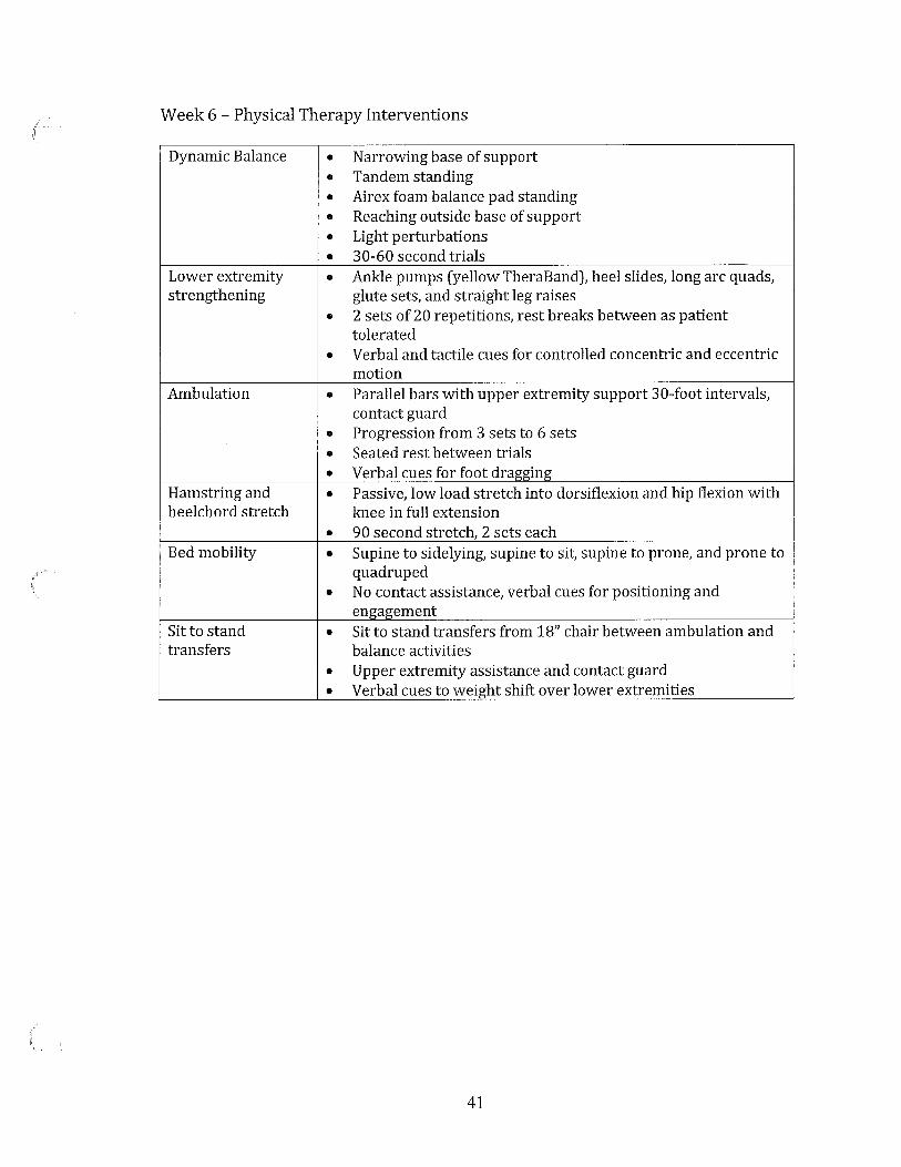
Week 6 - Physical Therapy Interventions
Dynamic Balance • Narrowing base of support
• Tandem standing
• Airex foam balance pad standing
• Reaching outside base of support
• Light perturbations
• 30-60 second trials Lower extremity • Ankle pumps (yellow TheraBand), heel slides, long arc quads, strengthening glute sets, and straight leg raises
• 2 sets of 20 repetitions, rest breaks between as patient tolerated
• Verbal and tactile cues for controlled concentric and eccentric motion
Ambulation • Parallel bars with upper extremity support 30-foot intervals, contact guard
• Progression from 3 sets to 6 sets
• Seated rest between trials
• Verbal cues for foot dragging Hamstring and • Passive, low load stretch into dorsiflexion and hip flexion with heelchord stretch knee in full extension
• 90 second stretch, 2 sets each Bed mobility • Supine to sidelying, supine to sit, supine to prone, and prone to
quadruped
• No contact assistance, verbal cues for positioning and engagement
Sitto stand • Sit to stand transfers from 18" chair between ambulation and transfers balance activities
• Upper extremity assistance and contact guard
• Verbal cues to weight shift over lower extremities
41
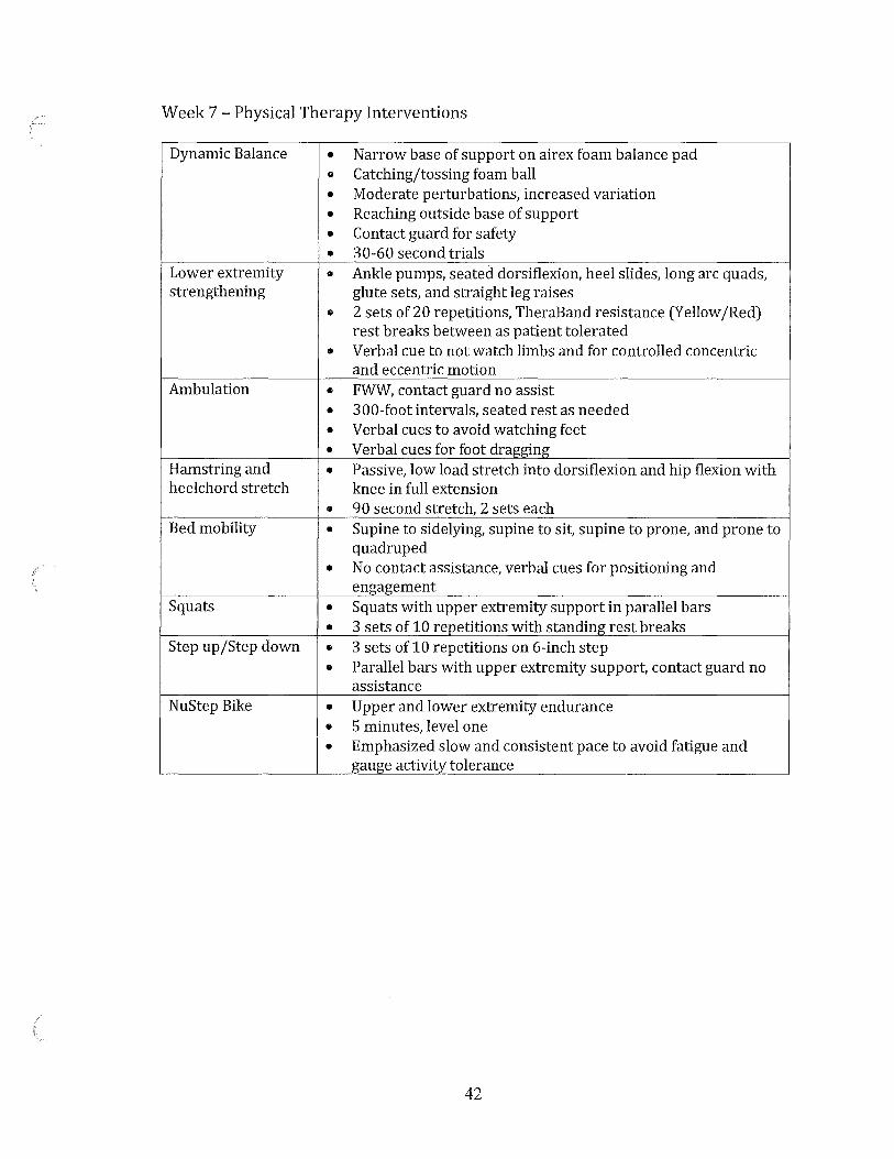
Week 7 - Physical Therapy Interventions
Dynamic Balance • Narrow base of support on airex foam balance pad
• Catching/tossing foam ball
• Moderate perturbations, increased variation
• Reaching outside base of support
• Contact guard for safety
• 30-60 second trials Lower extremity • Ankle pumps, seated dorsiflexion, heel slides, long arc quads, strengthening glute sets, and straight leg raises
• 2 sets of 20 repetitions, TheraBand resistance (Yellow /Red) rest breaks between as patient tolerated
• Verbal cue to not watch limbs and for controlled concentric and eccentric motion
Ambulation • FWW, contact guard no assist
• 300-foot intervals, seated rest as needed
• Verbal cues to avoid watching feet
• Verbal cues for foot drae-e:ing Hamstring and • Passive, low load stretch into dorsiflexion and hip flexion with heelchord stretch knee in full extension
• 90 second stretch, 2 sets each Bed mobility • Supine to sidelying, supine to sit, supine to prone, and prone to
quadruped
• No contact assistance, verbal cues for positioning and engagement
Squats • Squats with upper extremity support in parallel bars
• 3 sets of 10 repetitions with standing rest breaks Step up /Step down • 3 sets of 10 repetitions on 6-inch step
• Parallel bars with upper extremity support, contact guard no assistance
NuStep Bike • Upper and lower extremity endurance
• 5 minutes, level one
• Emphasized slow and consistent pace to avoid fatigue and gauge activity tolerance
42
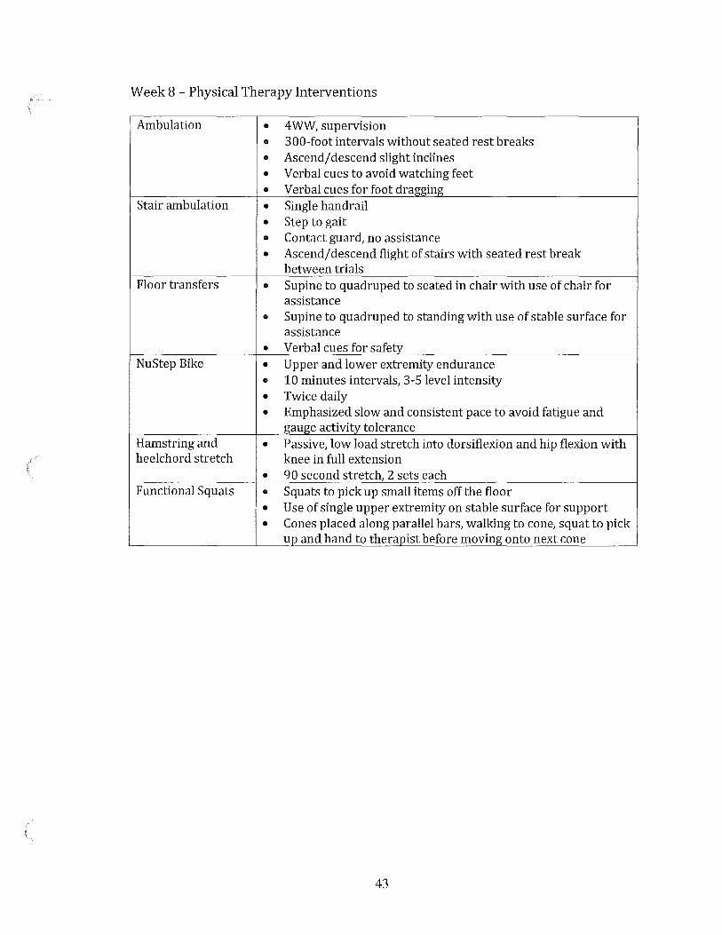
Week 8 - Physical Therapy Interventions
Ambulation • 4WW, supervision
• 300-foot intervals without seated rest breaks
• Ascend/ descend slight inclines
• Verbal cues to avoid watching feet
• Verbal cues for foot dragging Stair ambulation • Single handrail
• Step to gait
• Contact guard, no assistance
• Ascend/ descend flight of stairs with seated rest break between trials
Floor transfers • Supine to quadruped to seated in chair with use of chair for assistance
• Supine to quadruped to standing with use of stable surface for assistance
• Verbal cues for safety NuStep Bike • Upper and lower extremity endurance
• 10 minutes intervals, 3-5 level intensity
• Twice daily
• Emphasized slow and consistent pace to avoid fatigue and gauge activity tolerance
Hamstring and • Passive, low load stretch into dorsiflexion and hip flexion with heelchord stretch knee in full extension
• 90 second stretch, 2 sets each Functional Squats • Squats to pick up small items off the floor
• Use of single upper extremity on stable surface for support
• Cones placed along parallel bars, walking to cone, squat to pick up and hand to therapist before moving onto next cone
43

Week 9 - Physical Therapy Interventions
Ambulation with • 4WW, supervision while in therapy, no supervision between assistive device meals and within hospital hallways
• 600+ feet without rest breaks or verbal cues for safety /gait quality
Ambulation • 500+ feet without assistive device without assistive • Contact guard, no assistance from therapist (safety) device • Increased fatigue, required seated rest break Stair ambulation • Single handrail
• Step overstep gait
• Contact guard, no assistance
• Ascend/ descend flight of stairs with seated rest break between trials
Floor transfers • Supine to quadruped to seated in chair without assistive device or upper extremity support
• Supine to quadruped to standing with without assistive device or upper extremity support
• No verbal cues for safety NuStep Bike • Upper and lower extremity endurance
• 15 minutes intervals, 5 level intensity
• Twice daily
• Emphasized slow and consistent pace to avoid fatigue and gauge activity tolerance
Functional • Squats to pick up small items off the floor Squats • Upper extremity support from 4WW
• No upper extremity support, contact guard, no assistance
• Cones scattered around floor requiring navigation to objects in space
44
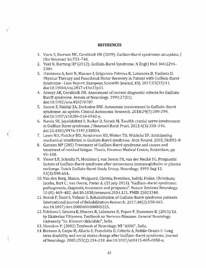
REFERENCES
1. Vucic S, Kiernan MC, Cornblath DR (2009). Guillain-Barre syndrome: an update. J Clin Neurosci 16:733-741.
2. Yuki N, Hartung HP (2012). Guillain-Barre Syndrome. N Engl J Med 366:2294-2304.
3. Dimitrova A, Izov N, Maznev I, Grigorova-Petrova K, Lubenova D, Vasileva D. Physical Therapy and Functional Motor Recovery in Patient with Guillain-Barre Syndrome - Case Report. European Scientific Journal, ESJ. 2017;13(33):11. doi:10.19044/ esj.2017.v13n33p11.
4. Asbury AK, Cornblath DR. Assessment of current diagnostic criteria for GuillainBarrl1l syndrome. Annals of Neurology. 1990;27(S1). doi:10.1002/ana.410270707.
5. Zaeem Z, Siddiqi ZA, Zochodne DW. Autonomic involvement in Guillain-Barre syndrome: an update. Clinical Autonomic Research. 2018;29(3):289-299. doi:10.1007 /s10286-018-0542-y.
6. Nanda SK, Jayalakshmi S, Ruikar D, Surath M. Twelfth cranial nerve involvement in Guillian Barre syndrome. J Neurosci Rural Pract. 2013;4(3):338-340. doi:10.4103/0976-3147.118804.
7. Lawn ND, Fletcher DD, Henderson RD, Wolter TD, Wijdicks EF. Anticipating mechanical ventilation in Guillain-Barre syndrome. Arch Neural. 2001;58:893-8
8. Garssen MP (205) Treatment of Guillain-Barre syndrome and causes and treatment of residual fatigue. Thesis, Erasmus Medical Centre, Rotterdam, pp 93-100.
9. Visser LH, Schmitz PI, Meulstee J, van Doorn PA, van der Meche FG. Prognostic factors of Guillain-Barre syndrome after intravenous immunoglobulin or plasma exchange. Dutch Guillain-Barre Study Group. Neurology. 1999 Aug 11. 53(3):598-604.
10. Van den Berg, Bianca; Walgaard, Christa; Drenthen, Judith; Fokke, Christiaan; Jacobs, Bart C.; van Doorn, Pieter A (15 July 2014). "Guillain-Barre syndrome: pathogenesis, diagnosis, treatment and prognosis". Nature Reviews Neurology. 10 (8): 469-482. doi:10.1038/nrneurol.2014.121. PMID 25023340.
11. Novak P, Smid S, Vidmar G. Rehabilitation of Guillain-Barre syndrome patients. International Journal of Rehabilitation Research. 2017;40(2):158-163. doi:10.1097 /mrr.0000000000000225.
12. Velcheva I, Genova K, Dimova R, Lubenova D, Popov P, Stamenov B. (2015) Ed. by Ekaterina Titianova. Textbook on Nervous Diseases. General Neurology. University "Sv. Kliment Okhridski", Sofia.
13. Shotekov P. (2002) Textbook of Neurology. MI "ARSO", Sofia. 14. Bersano A, Carpo M, Allaria S, Franciotta D, Citterio A, Nobile-Orazio E. Long
term disability and social status change after Guillain-Barre syndrome. Journal of Neurology. 2005;253(2):214-218. doi:10.1007 /s00415-005-0958-x.
45
15. Rees J, Thompson R, Hughes R 1998 Epidemiological study ofGuillain-Barre syndrome in south-east England. Journal ofNeurology, Neurosurgery and Psychiatry 64: 74-77.
16. Doorn PAV, Kuitwaard K, Walgaard C, Koningsveld RV, Ruts L, Jacobs BC. IVIG Treatment and Prognosis in Guillain-Barre Syndrome. Journal of Clinical Immunology. 2010;30(S1):74-78. doi:10.1007 /s10875-010-9407-4.
17. Lindenbaum Y, Kissel JT, Mendell JR. Treatment approaches for Guillain-Barre syndrome and chronic inflammatory demyelinating polyradiculoneuropathy. Neural Clin. 2001;19:187-204.
18. Luigetti M, Servidei S, Modoni A, Rossini PM, Sabatelli M, Monaco ML. Admission neurophysiological abnormalities in Guillain-Barre syndrome: A single-center experience. Clinical Neurology and Neurosurgery. 2015;135:6-10. doi:10.1016/j.clineuro.2015.05.001.
19. Wijdicks EF, Henderson RD, McClelland RL. Emergency intubation for respiratory failure in Guillain-Barre syndrome. Arch Neural. 2003 Jul. 60(7):947-8.
20. Shuang Liu, Chaoling Dong & Eroboghene Ekamereno Ubogu (2018) Immunotherapy of Guillain-Barre syndrome, Human Vaccines & Immunotherapeutics, 14:11, 2568 2579, DOI: 10.1080/21645515.2018.1493415
21. Ko K-J, Ha G-C, Kang S-J. Effects of daily living occupational therapy and resistance exercise on the activities of daily living and muscular fitness in Guillain-Barre syndrome: a case study. Journal of Physical Therapy Science. 2017;29(5) :950-953. doi:10.1589 /jpts.29.950.
22. Das A, Kalita J, Misra UK. Recurrent Guillain Barre' syndrome. Electromyogr Clin Neurophysiol. 2004 Mar. 44(2):95-102.
23. Roper TA, Alani SM. Recurrent Guillain-Barre syndrome: lightning does strike twice. Br J Hosp Med. 1995 Apr 19-May 2. 53(8):403-7.
24. Double-blind trial of intravenous methylprednisolone in Guillain-Barre syndrome. Guillain-Barre Syndrome Steroid Trial Group. Lancet. 1993 Mar 6. 341(8845) :586-90.
25. Wokke JH. Fatigue is part of the burden of neuromuscular diseases. J Neural. 2007;254(7):948-949. doi: 10.1007 /s00415-006-0436-0.
26. Garssen MJP, Koningsveld RV, Van Doorn PA. Residual fatigue is independent of antecedent events and disease severity in Guillain-Barre syndrome. J Neural 2006;253:1143-6.
27. Garssen MP, Bussmann JB, Schmitz PI, et al. Physical training and fatigue, fitness, and quality oflife in Guillain-Barre syndrome and CIDP. Neurology. 2004;63 (12) :2393-239 5.
28. Davidson I, Wilson Ch, Walton T, Brissenden Sh. (2009) Physiotherapy and Guillain-Barre syndrome: results of a national survey. Physiotherapy, 95:157-163.
29. Khan F. (2004) Clinical practice: Rehabilitation in Guillian Barre syndrome. Australian Family Physician, 33(12): 1013-7.
46

30. Bussmann JB, Garssen MP, van Doorn PA, et al. Analysing the favourable effects of physical exercise: relationships between physical fitness, fatigue and functioning in Guillain-Barre syndrome and chronic inflammatory demyelinating polyneuropathy. J Rehabil Med. 2007;39(2):121-5.
31. Hansen, Matthew DPT; Garcia, Santo MOTR/L. Guillain-Barre Syndrome, CIDP and Variants; Guidelines for Physical and Occupational Therapy. Guillain-Barre Syndrome, CIDP and Variants; Guidelines for Physical and Occupational Therapy, GBS/CIDP Foundation International, 2010.
32. Clinical guidelines on the identification, evaluation, and treatment of overweight and obesity in adults: The evidence report. PsycEXTRA Dataset. 1998. doi:10.1037 /e565682010-001.
33. McGee, Steven (2012). Evidence-Based Physical Diagnosis. Philadelphia, USA: Saunders. pp. 472-473. ISBN 978-1-4377-2207-9.
34. Jaramillo, Sandra Patricia Isaza, et al. "Accuracy of the Babinski Sign in the Identification of Pyramidal Tract Dysfunction." Journal of the Neurological Sciences, vol. 343, no. 1-2, 2014, pp. 66-68., doi: 10.1016/j.jns.2014.05.028.
35. Alshekhlee A, Hussain Z, Sultan B, Katirji B. Guillain-Barre syndrome: incidence and mortality rates in US hospitals. Neurology. 2008 Apr 29. 70(18):1608-13.
36. Fletcher DD, Lawn ND, Wolter TD, Wijdicks EF. Long-term outcome in patients with Guillain-Barre syndrome requiring mechanical ventilation. Neurology. 2000 Jun 27. 54(12):2311-5.
37. Mullings KR, Alleva JT, Hudgins TH. Rehabilitation of Guillain-Barre syndrome. Dis Mon. 2010 May. 56(5):288-92.
38. Winer JB, Hughes RA, Osmond C. A prospective study of acute idiopathic neuropathy. I. Clinical features and their prognostic value. J Neural Neurosurg Psychiatry. 1988 May. 51(5):605-12.
39. Dornonville de la Cour C, Jakobsen J. Residual neuropathy in long-term population-based follow-up of Guillain-Barre syndrome. Neurology. 2005 Jan 25. 64(2):246-53.
40. Rudolph T, Larsen JP, Farbu E. The long-term functional status in patients with Guillain-Barre syndrome. Eur J Neural. 2008 Dec. 15(12):1332-7.
41. RajaballyYA, Uncini A. Outcome and its predictors in Guillain-Barre syndrome. Journal of Neurology, Neurosurgery & Psychiatry. 2012;83(7):711-718. doi:10.1136/jnnp-2011-301882.
42. J.C. Hobart, D. L. Lamping, J. A. Freeman, D. W. Langdon, D. L. McLellan, R. J. Greenwood, A. J. Thompson Neurology Aug 2001, 57 (4) 639-644; DOI: 10.1212/WNL.57.4.639.
43. Lubenova D. (2011) Kinesitherapy of neurogical and psychiatric diseases. Betaprint-Petrovi i Sie, Sofia.
44. Khan F, Pallant JF, Ng L, Bhasker A. Factors associated with long-term functional outcomes and psychological sequelae in Guillain-Barre syndrome. J Neural. 2010 Dec. 257(12):2024-31.
45. Lubenova D, Titanova E. (2012) Principles of modern neurorehabilitation. Neuroscience and cerebral hemodynamics, 8(1): 45-55.
47

46. Davidson I, Wilson Ch, Walton T, Brissenden Sh. (2009) Physiotherapy and Guillain-Barre syndrome: results of a national survey. Physiotherapy, 95:157-163.
47. Merkies IS, Schmitz PI, Samijn JP, et al. Fatigue in immune-mediated polyneuropathies. European Inflammatory Neuropathy Cause and Treatment (INCAT) Group. Neurology. 1999;53(8):1648-1654.
48. Witcomb GL, Bouman WP, Claes L, Brewin N, Crawford JR, Arcelus J. Levels of depression in trans gender people and its predictors: Results of a large matched control study with trans gender people accessing clinical services. Journal of Affective Disorders. 2018;235:308-315. doi:10.1016/j.jad.2018.02.051.
49. Gassaway J, Sweatman M, Rider C, Edens K, Weber M. Therapeutic Recreation Outcomes during Inpatient SCI Rehabilitation: Propensity Score Analysis of SCIRehab Data. Therapeutic Recreation Journal. 2019;53(2):99-116. doi:10.18666 /trj-2019-v5 3-i2-9144.
50. Merkies IS, Kieseier BC. Fatigue, Pain, Anxiety and Depression in Guillain-Barre Syndrome and Chronic Inflammatory Demyelinating Polyradiculoneuropathy. European Neurology. 2016;75(3-4):199-206. doi:10.1159 /000445347.
51. Dittner AJ, Wessely SC, Brown RG. The assessment of fatigue: a practical guide for clinicians and researchers. J Psychosom Res. 2004;56(2):157-170. doi: 10.1016/S0022-3999(03)00371-4.
52. Fritz S, Lusardi M. White Paper: "Walking Speed: the Sixth Vital Sign." Journal of Geriatric Physical Therapy. 2009;32(2):2-5. doi:10.1519/00139143-200932020-00002.
53. Pirpiris, M., Wilkinson, A., et al. "Walking speed in children and young adults with neuromuscular disease: comparison between two assessment methods." Journal of Pediatric Orthopaedics 2003 23(3): 302.
54. Shumway-Cook A, Brauer S, Woollacott M. Predicting the probability for falls in community-dwelling older adults using the timed up & go test. Phys Ther. 2000;80(9):896-903.
55. Barry E, Galvin R, Keogh C, Horgan F, Fahey T. Is the Timed Up and Go test a useful predictor of risk of falls in community dwelling older adults: a systematic review and meta-analysis. BMC geriatrics. 2014 Dec;14(1):14.
56. Harbo T, Markvardsen LK, Hellfritzsch MB, Severinsen K, Nielsen JF, Andersen H. Neuromuscular Electrical Stimulation In Early Rehabilitation Of Guillain-Barre Syndrome: A Pilot Study. Muscle & Nerve. 2018. doi:10.1002/mus.26396.
57. Dimitrova A, Izov N, Maznev I, Grigorova-Petrova K, Lubenova D, Vasileva D. Physical Therapy and Functional Motor Recovery in Patient with Guillain-Barre Syndrome - Case Report. European Scientific Journal, ESJ. 2017;13(33):11. doi:10.19044/esj.2017.v13n33p11.
48