physical therapy ceu - c.ymcdn.com · si joint clinical examination, diagnosis and ... knee or...
TRANSCRIPT

The Symptomatic
SI Joint
Clinical Examination,
Diagnosis and Treatment
1
Physical Therapy CEU

Genne’ DeHenau McDonald PT• 28 years clinical experience (PT Degree Oakland University/Michigan State University)
• Former Adjunct Faculty and Clinical Lecturer• University of Florida DPT Program
• Former Faculty Herman and Wallace Pelvic Health Institute
• Publications• Musculoskeletal LBP During Pregnancy• Piriformis Syndrome• Co-Author and Editor Original Gynecological Manual SOWH
• APTA Member/SOWH Member since 1989
• Speaker for SOWH, SUNA
• Program/Project Manager Medical Affairs SI-BONE

Continuing Education Credits
Website - http://www.questionpro.com/t/AKQvQZbCqQ
**Certificates will be e-mailed the first and third week of the month after completion of survey
**Please check junk mail folder
Questions:Genné McDonald – Program/Project Manager - [email protected]
Confidential & Proprietary 3

1. Understand the prevalence of SI joint pain.
2. Review anatomy and biomechanics of the SI joint.
3. Review the integrated model of SI joint function.
4. Understand the relationship between the lumbar spine, SI joint and hip.
5. Review subjective history for patient with SI joint pain.
6. Correctly perform SI joint provocation testing
7. Review SI joint rehabilitation.
Course Objectives
4

Prevalence of SI Joint Pain
15-30%Component of chronic LBP
22.6%
30.0%
18.5%
27.0%
14.5%
Bernard1987
Schwarzer1995
Maigne1996
Irwin2007
Sembrano2009
32-43%Symptomatic Post-Lumbar Fusion
32% Katz 200335% Maigne 200543% DePalma 201140% Liliang 2011
DePalma – Pain Med 2011
Is the SI Joint truly a problem?
5

75% of post-lumbar fusion patients showed SI joint degenerative changes onCT scan 5 years aftervs.only 38% age- and gender-matched controls without prior lumbar fusion
Ha et al. 2008
Lumbar fusion leads to increases in angular motion and joint stress at the SI joint
Ivanov et al. 2009
SIJ & Adjacent Segment Degeneration1,2
6
1. Ha – Spine 20082. Ivanov – Spine 2009

Sacroiliac Joint
Articular Surface
Anatomy
7

Anterior Ligaments Posterior Ligaments
Anatomy
8

Biomechanics
NutationSacral base movement anteroinferior
CounternutationSacral base movement posterosuperior
TranslationLinear motion; motion in any one direction
Forst 2006
9
Nutation
Counternutation

SI Joint Motion
Multi-planar motion – Simultaneously rotate and
translate through 3 axes of motion
Motions (<4° in any plane)– Nutation / Counternutation
• Primary motionMales: 1 - 2°Females: 2 - 4°
Sacral Translation – (A-P motion) up to 1.6mm
Sturesson 1989
10

Integrated Model of SI Joint Function
• Describes effective transfer of force and load across the SI joint from the lower extremities to the spine.
Snijders & Vleeming 1993
• Requires a stable core (lumbar spine, pelvis and soft tissues)
• 3 Components of Functional StabilityPassive (form closure)
Active (force closure)
Neuromuscular Control (motor control)Panjabi 1992
11

Form Closure/Structural Integrity: The shape of the sacrum and the integrity of the supporting ligaments
contribute to SI joint stability
Lee 1998, Vleeming 1990a, Vleeming 1990b
The Integrated Model of SIJ Function
Force Closure/Joint Compression: The external dynamic forces created by
contraction of the stabilizing muscles and their fascial and
ligamentous attachmentsLee 1998, Vleeming 1990a, Vleeming 1990b
Motor Control: Nervous system coordination / co-activation of
deep stabilizing muscles (onset, duration, timing)
Snijders 1993, Hodges 1996, Richardson 2002
Components
Core muscles should contract before load reaches the low
back and pelvis to prepare the system for impending load.
12

Form ClosureOsseous Structures – Macro, Micro
Supporting Ligaments
13

The Integrated Model of SI Joint Function:Force Closure – Local Muscles
Local Core Muscles
• Transversus Abdominus• Multifidus• Pelvic Floor Muscles• Diaphragm
– More research being done on other contributing muscles
Local System: contracts prior to upper/lower limb movement regardless of direction Hodges 1997, 2003; Hodges & Richardson 1997; Hodges 1999; Moseley 2002,2003
14

The Integrated Model of SI Joint Function:Force Closure – Global Muscles
Vleeming’s slings:
Large Muscle Groups and their Fascial Connections
– Posterior Oblique System– Anterior Oblique System– Deep Longitudinal System– Lateral System
Global System: Contracts later and is direction dependent
Radebold 2000, 2001; Hodges 2003
Radebold 2000, 2001; Hodges 2003
15

Thoracolumbar Fascia & Paraspinal Muscles
Caudal part of the TLF, in combination with efficient paraspinal muscles, plays major role in force closure of SI joint
Macintosh & Bogduk – Spine 1993, Vleeming – Spine 1995
16

Motor Control
• Optimal force closure of the SI joint
• Studies showing alteration of muscle activation patterns in low back, groin, and SI joint pain populations– TrA Delayed in LBP Hodges & Richardson 1996
– TrA Delayed in Groin Injuries Cowen 2004
– Altered Motor Control with SI joint Pain O’Sullivan 2002
• Timing, sequence and amplitude of muscle firing– Afferent input from receptors in joints, ligaments, fascia and muscles – Efferent response of central and peripheral nervous systems Hodges 2003
17

Evaluation of the Patient with
Sacroiliac Joint Pain
18

Can we diagnose SI joint pain?
• Common pain patterns from multiple conditions– Spine (stenosis, facet, spondy, disc herniation, DDD, etc.)– SI Joint– Hip (OA, FAI, early AVN, etc.)– Pelvis (Glut tear, piriformis, pelvic floor)
• Imaging not routinely helpful
• History and Physical Examination− Provocative maneuvers Laslett 2005, Szadek 2009
− SI joint ROM & Position testing not reliable Freburger 1999, Robinson 2007
• Diagnostic Injection
19

Lower lumbar region (72%)
Devin – JAAOS 2012 Vanelderen – Evid Based Med 2010
Lateral hip – common
Thigh and Groin (55%)
Buttock pain (71%)
Lower extremity common (28%)
Groin not uncommon (14%)
Lateral hip & thigh common
Buttock, PSIS (94%)
Devin – JAAOS 2012
Spinal Stenosis
Lower extremity common
Lateral hip – common
Lumbar region common
Buttock common
Knee or below (47%)
Groin uncommon (except L1, L2)
Lumbar Spine – SI Joint – HipPain Referral Patterns
Lumbar pain uncommon
SI Joint IsolatedHip Pathology
20

Potential Causes of SIJ Pain: Traumatic
• MVA: Foot on Brake
• Slip and Fall
• Lifting and Twisting
• Traction Injuries
21
21

Potential Causes of SI Joint Pain: Gradual Onset
• Laxity of the SI joint / Pregnancy
• Repetitive Forces on SI joint and Supporting Structures
• Biomechanical Abnormalities– Leg length inequality– Pelvic obliquity/scoliosis– Iliac crest bone graft
• Adjacent Segment Degeneration – After lumbar spinal fusion
• Post Infection Degeneration
22
22

Hip and Adjacent Segment (SI Joint)
Hip and SI Joint76% (25/33) of patients with symptomatic SI joint pathology had at least one abnormal finding on hip radiograph
A significant number met the strict diagnostic criteria for FAI.
42% Hip OA
45% Subchondral cyst
21% Retroversion
12% Lateral CE* angle >40%
47% Coxa profunda
33% Cam impingement
Morgan 2013
*Center Edge
23

Positive Subjective
History
Positive SI Joint
Provocative Testing
Lumbar Spineand
Hip Exam
Positive Response to
Intra-articular Injection
24
Elements of the Diagnosis

HISTORY
• When did the pain start? • Prior trauma
– A fall on the buttock– Car accident
(T-bone, rear-end, head-on)– Lift/Twist– Other
• Prior lumbar fusion– Prior iliac bone graft harvest
• Pregnancy
COMPLAINTS• Lower back pain • Sensation of numbness, tingling or
weakness • Pelvis / buttock pain • Hip / groin pain • Feeling of leg instability, buckling, or
giving way • Disturbed sleep patterns • Disturbed sitting patterns (unable to sit
for long periods, on one side) • Pain going from sitting to standing
25
25
History and Complaints

Pain Diagram • Pain in buttock and posterior thigh
– Usually not midline
– Usually below L5
– At or lateral to PSIS
– Occasionally groin
• Secondary pain in lateral thigh, groin, and/or lateral calf
Fortin 1999
26
History: SI Joint Pain Presentation
26

Exacerbating Activities
Unilateral Weight Bearing- Putting on Socks/Shoes- Ascending/Descending Stairs- Getting in and out of Car- Prolonged Walking(85% of gait cycle is single leg stance)
Janda 1983
Sexual Intercourse
Pain with Transitional Motions- Supine to painful side - Sit to stand- Rolling over in bed- Getting in /out of bed
Pain while Stationary- Sitting on affected side- Prolonged standing/sitting
27
27

Relieving Activities
• Bearing weight on unaffected side
• Lying on unaffected side
• Manual or belt stabilization
28
28

Lumbar Spine – Hip – SI Joint Physical Examination
29

Assess Lumbar Spine: Facet Joint Pain
• Incidence 10-15% in patients with chronic LBP Saravanakumar 2008
• Low back pain from the facet joints: radiates down into the buttocks and posterior thigh.
• Clinical Findings – Point tenderness overlying the inflamed facet joints – Loss in spinal muscle flexibility (guarding)– More discomfort leaning backward than forward.
• No history findings or examination maneuver has been found to be unique or specific to this entity Kayser 2008, Cohen 2007, Jackson 1998
30
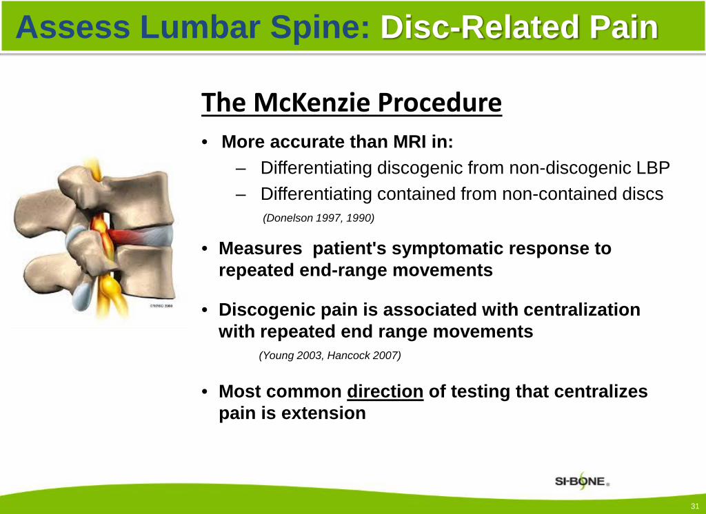
The McKenzie Procedure• More accurate than MRI in:
– Differentiating discogenic from non-discogenic LBP– Differentiating contained from non-contained discs
(Donelson 1997, 1990)
• Measures patient's symptomatic response to repeated end-range movements
• Discogenic pain is associated with centralization with repeated end range movements
(Young 2003, Hancock 2007)
• Most common direction of testing that centralizes pain is extension
Assess Lumbar Spine: Disc-Related Pain
31

These can all be performed quickly in supine before
performing provocative testing
for the SI joint.
Assess Lumbar Spine: Physical Exam
Standard tests to rule out lumbar radiculopathy
• DTRs• Neuro Exam
Passive Straight Leg Raise To rule out lumbar radicular pain + at 35-70°
• Sensitivity 91%• Specificity 26%
Deville 2000
Slump TestTo rule out lumbar disc herniation's
• Sensitivity 84%• Specificity 83%
Majlesi 2008
32

Assess Hip: Physical Examination
FABER: For Intra-articular hip pathology (if pain anterior)
• Hip flexion, abduction and ER with overpressure • 88% sensitive (Martin 2005)
Scour: For Hip OA and other pathologies• Sensitivity 62% - 91%• Specificity 43% - 75%
(Konin 2006, Magee 2008)
FADIR: For FAI and labral tears:• Hip flexion, adduction, IR• Sensitivity = 75%, • Specificity = 43% (Austin 2008)
FAIR: For Piriformis Syndrome (assure pain is posterior)
• Sensitivity 88%• Specificity 83% (Fishman, 2002)
33

Range of Motion Testing of the Hip (Birrell 2001)
• The most predictive finding for osteoarthritis is decreased range of motion with restriction in internal rotation
• For those patients with one plane of restricted movement, the sensitivity for severe osteoarthritis is 100% and specificity is 42%
• Used to rule in/out osteoarthritis
Femoral Neck Anteversion (FNA) Assessment Compare bilaterally
• If IR is 30 degrees > then external rotation, predictor of FNA (Swanson 1963)
• Trochanteric Prominence Angle Test (TPAT) or Craig testTPAT predicted the FNA measured intraoperatively, more accurately than either the CT or Magilligan method (Ruwe 1992)
Assess Hip: Physical Examination
34
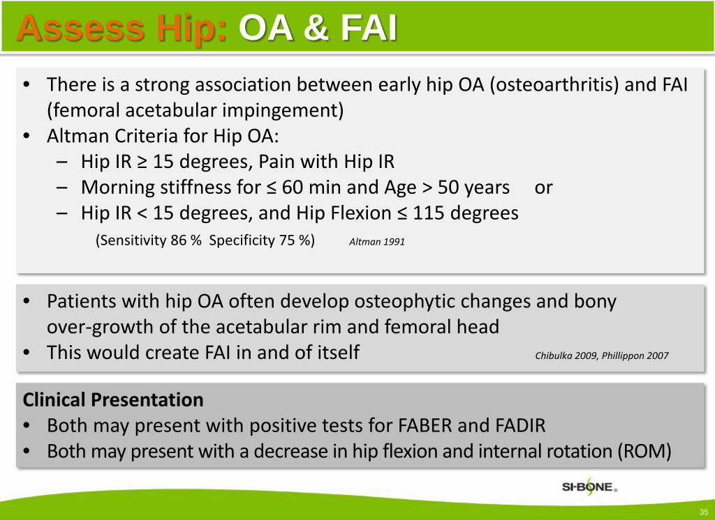
Assess Hip: OA & FAI• There is a strong association between early hip OA (osteoarthritis) and FAI
(femoral acetabular impingement)• Altman Criteria for Hip OA:
– Hip IR ≥ 15 degrees, Pain with Hip IR– Morning stiffness for ≤ 60 min and Age > 50 years or– Hip IR < 15 degrees, and Hip Flexion ≤ 115 degrees
(Sensitivity 86 % Specificity 75 %) Altman 1991
• Patients with hip OA often develop osteophytic changes and bony over-growth of the acetabular rim and femoral head
• This would create FAI in and of itself Chibulka 2009, Phillippon 2007
Clinical Presentation• Both may present with positive tests for FABER and FADIR• Both may present with a decrease in hip flexion and internal rotation (ROM)
35

Contact between the femoral head-neck junction and the acetabular rim
• Insidious onset 50% of cases Samora 2011
• Symptoms– Persistent insidious deep groin, lateral hip
or buttock pain – Pain increased with prolonged sitting or
standing and hip flexion-type movements– Decreased hip ROM
Two Types • CAM (More common in young males)
– Anterior-superior aspect • Pincer (More common in females)
– Anterior acetabular labrum and chondral injury in posterior-inferior acetabular rim
Ganz 2003
Confirmation• Arthroscopy: Gold Standard• MRI
Assess Hip: FAI (Femoral-Acetabular Impingement)
36
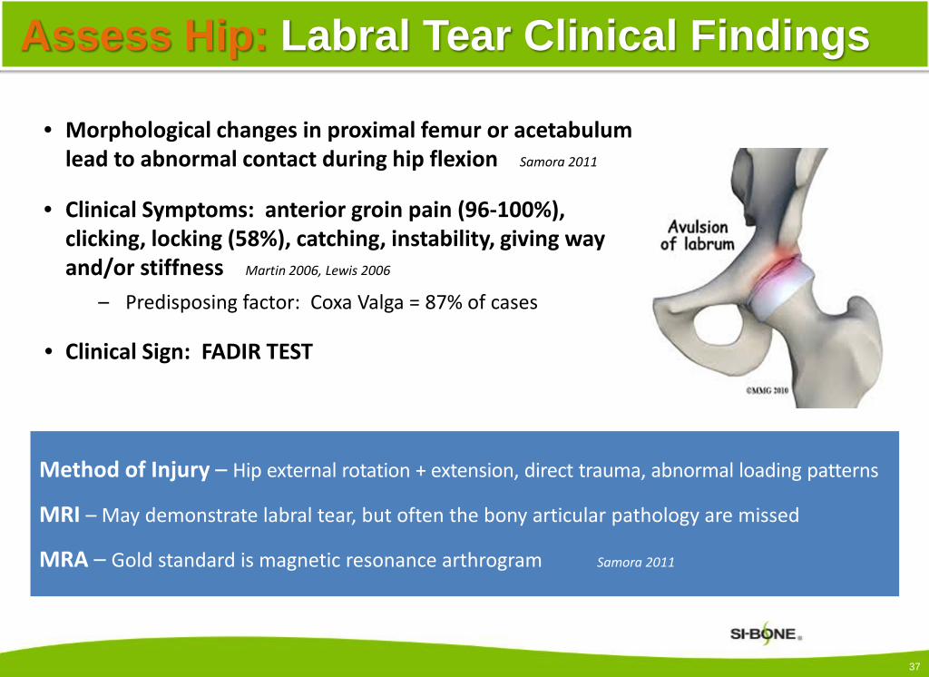
Assess Hip: Labral Tear Clinical Findings
• Morphological changes in proximal femur or acetabulum lead to abnormal contact during hip flexion Samora 2011
• Clinical Symptoms: anterior groin pain (96-100%), clicking, locking (58%), catching, instability, giving way and/or stiffness Martin 2006, Lewis 2006
– Predisposing factor: Coxa Valga = 87% of cases
• Clinical Sign: FADIR TEST
Method of Injury – Hip external rotation + extension, direct trauma, abnormal loading patterns
MRI – May demonstrate labral tear, but often the bony articular pathology are missed
MRA – Gold standard is magnetic resonance arthrogram Samora 2011
37

Piriformis compresses or irritates the sciatic nerve• Incidence: 17.2% among low back pain patient
Chen 2013
Clinical symptoms• A dull ache in the buttock.• Pain down the back of the thigh, calf and foot
(sciatica)• Pain when walking up stairs or inclines.• Increased pain after prolonged sitting.
Clinical Sign• FAIR Test: Reproduction of symptoms when
piriformis muscle is put on stretch.(hip flexion, adduction and internal rotation)
Fishman 2002, Loren 2010
Assess Hip: Piriformis Syndrome
Magnetic Resonance Neurography: Type of MRI that highlights
inflammation and compression of the nerves.
Filler 2005
38

Incidence• Common finding on MR imaging in patients
with buttock, lateral hip, or groin pain.Kingzett-Taylor 1999
Clinical Symptoms• Dull lateral hip pain, buttock or groin pain
Clinical Signs• Focal tenderness at the gluteal insertion• Weak hip abduction • Pain with passive and then resisted hip
internal rotation with the hip flexed to 90°Sen 88%, Spec 97.3%
• Pain on one-legged stance for 30 sec or moreSen 100 % Spec 97.3%
Lequesne 2008
Assess Hip:Gluteus Medius & Minimus Tears
39

Point to pain while standing • Able to localize pain with one finger• Within 1 cm of PSIS (inferomedial)• Consistent over at least 2 trials
Ask patient to point to location of primary pain
• Below L5: Consider SI joint
• Above L5: Consider lumbar spine etiologies
SI Joint: Physical ExamFortin Finger Test
Fortin & Falco 1997
40

SI Joint: Physical Exam
Active Straight Leg RaiseTo assess functional pelvic stability
• Sensitivity: 87%
• Specificity: 94%Mens 2001
41

SI Joint: Provocative Tests
The following five provocative tests, when performed in combination are proven to have a high degree of sensitivity and specificity:
1. Distraction* (Highest PPV**)
2. Thigh Thrust*
3. FABER4. Compression*
5. Gaenslen’s Maneuver
42
Laslett Szadek
3 or more positive tests
Sensitivity 91% 85%
Specificity 78% 76%
Laslett 2005, 2008Szadek 2009
* Most sensitive tests** PPV = positive predictive value
42

• Specificity increases when symptoms don’t centralize or peripheralize with thorough, multidirectional repeated movement assessment (McKenzie Assessment)
• In some cases a patient may not tolerate having five (5) tests performed. Therefore, it’s recommended that the three (3) most sensitive, specific and reliable tests be performed first.
Laslett 2005, Laslett 2008, Szadek 2009
How to Interpret Your Results
1 Positive Test = Suspicion
2 Positive Tests = Fair Confidence
3+ Positive Tests = High Confidence
SI Joint: Specificity of Provocative Tests
43

Distraction
Thigh Thrust
Compression
FABER
Gaenslen’s
3 of 5 positive testsprovides discriminative power
for diagnosing SI joint pain Szadek – J Pain 2009
Laslett – J Man Manip Ther 2008
SI Joint Provocative Tests
44
Assure the tester position is above the patient by lowering table or standing on a sturdy stool in order to provide adequate force.
Start with light pressure and gradually increase, keeping hands cupped to minimize local contact pressure (30 second max).
Keep arms straight and lean forward with your upper body to create gentle steady force.
Stabilize patient on the table to prevent muscle guarding.
Stabilize contralateral ASIS during Thigh Thrust and FABER tests.
If pain is provoked with test, ask patient to identify pain location to confirm it is their typical pain.
45
SI Joint: Provocative Test Tips

• Quadratus Lumborum
• Gluteus Maximus
• Piriformis
• Levator Ani
Travell and Simons 1992
46
SIJ Region Pain – Myofascial Causes

PositiveHistory
Positive Fortin Finger Test
and Physical Exam (Lumbar Spine,
SI Joint, and Hip)
Positive Provocation
Testing
47
Justification for SI Joint Injection
47

SI Joint Injections
Diagnostic Injection• Confirm with contrast and
imaging• Low volume, local anesthetic
Therapeutic Injection• Local anesthetic + corticosteroid • May provide intermediate or
long-term relief • Results of can be unpredictable
Injection Under Fluoroscopy
48
48

Assessment: Post Diagnostic Injection
• Positive clinical response ≥ 50% VAS reduction during anesthetic phase indicates
positive diagnosis of SI joint as pain generator.
Relief during previously painful functional / provocative movements.
• Minimal or no relief< 50% May have SI joint pain, but consider other pain sources.
49
ISASS and ASIPP utilize ≥ 50% reduction in pain as a threshold
NASS utilizes ≥ 75% reduction in pain as a threshold
Maugars – Br J Rheumatol 1996; Maigne - Spine 1996; Pauza – AAPM&R 2001; Fritz – AJR Am J Roentgenol 2008;Rupert – Pain Physician 2009; Liliang – Pain Med 2011; Manchikanti – Pain Physician 2013;

Conservative Treatment Options for
Sacroiliac Joint Pain
50

Conservative Treatment Options
Symptom Management
• Medications (non-steroidal anti-inflammatories, oral steroids, pain medications)
• External SI joint stabilization with belting
• Therapeutic SI joint injections (1-4 per year)
• Physical Therapy
Sembrano 2011Cohen 2005
51

Treat the joint by returning it to its normal relationships Fife 2008
• Optimal SI joint function occurs with the SI Joint in neutral (mid-range) position.
DonTigney 1990, 1994, 1999, 2005; Fujiwara 1999;Pool-Goudzwaard 2001, 2003; Snijders 1993, 2004; Vleeming 1996
• Treatment goals should include restoration of :– Optimal alignment of the lumbar spine, sacroiliac and
hip joints– Functional stability of the lumbopelvic region
52
Conservative Treatment Options:Physical Therapy

Conservative Treatment Options:Physical Therapy
• Modification of Activities of Daily Living (ADLs)– specific focus on activities that may create or exacerbate symptoms.
• Patient education regarding maintaining optimal alignment with positioning, posture and body mechanics
• Stabilization Exercise/ Neuromuscular Re-education – Specific focus on timing and engagement of local and global core muscles
Snijders 1993a, 1993b, Hodges 1996, Richardson 2002
53

Conservative Treatment Options:Physical Therapy• Achieve normal muscle strength balance where
existing deficits (include gluteus medius assessment)Lee 1998, Vleeming 1990, Vleeming 1989
• Achieve normal muscle length balance where existing imbalances exist – Consideration of muscles that attach to the ilium and sacrum directly and
indirectly, especially limiters of hip internal rotation.Cibulka 1998, Lee 1998, Vleeming 1990, Vleeming 1989
• Adjacent segment joint and soft tissue restriction mobilization and manipulation as needed*– Consider the hip structures, lumbar and thoracic regions, knee and ankle joints.
54

• Manual techniques to address myofascial pain
• Balance assessment and training
• Gait training
• Regain or maintain cardiovascular health
• Modalities for pain and muscle spasm
Conservative Treatment Options:Physical Therapy
55

Surgical Treatment Options for
Sacroiliac Joint Pain
56

Smith-Petersen 1926 Campbell 1927 Gaenslen 1927
Bloom 1937 iFuse 2008
Treatment Options: Surgical
57
57

MIS SI Joint Fusion Technologies
Examples of existing and/or developing technologies.
iFuse Implant is the ONLY SI joint fusion technology supported by multiple prospective clinical publications, including2 Randomized Controlled Trials. (May 2017)
SI-BONE:iFuse Implant System®
Globus:SI-LOK Joint Fixation System
Medtronic:Rialto Sacroiliac Joint Fusion System
VG Innovations:SiJoin Posterior Sacroiliac Joint Fusion System
X-spine Systems:Silex Sacroiliac Joint Fusion System
Zyga Technology:SImmetry Sacroiliac Joint Fusion System
58

• Unique Patented Design– Triangular shape (minimizes rotation) – Interference press fit (immediate stabilization)– Porous titanium surface
(promotes bony ongrowth/ingrowth for long-term fusion)*
• Strength of Experience25,000+ procedures worldwide (March 2017)
• Clinical Evidence– iFuse Implant is the ONLY device
for treatment of SI joint dysfunction supported by multiple prospective clinical studies including 2 RCTs
– More than 50 peer-reviewed publications
59
iFuse Implant System®
* MacBarb G, et al. Int J Spine Surg. 2017:11;116-28.

Incision Pin Soft Tissue Protector Measure
Drill(optional with
sharp-tip broach)
Broach Insert Implant Repeat
iFuse Procedure Overview
60

iFuse Implant System® Publications
61
RCT (INSITE, iMIA)…...…………………. 7Prospective, Multicenter…………………... 6Comparison…………….…. 5Retrospective Case Series….………. 17Systematic Review, Meta-analysis…………. 3Cost-effectiveness, Productivity, etc.………. 5Complications, Survivorship, etc.…... 7Stability, Implant Placement, etc.… 3
* Includes accepted articles that are pending publication (see iFuse bibliography)

62
Complete References in Bibliography

63
Complete References in Bibliography

64
Complete References in Bibliography

Post-Operative Considerations
Individual Treatment Plans
Considerations:• Age • Weight• Bone quality• Associated health factors
Post Surgical Decisions
• Plan for protected weight bearing
• Activity limitations• Post op rehab plans• Plan for return to activity
6565
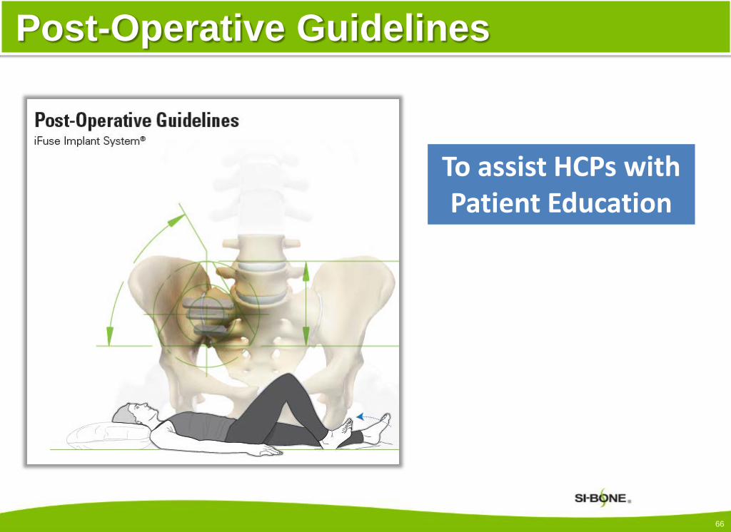
Post-Operative Guidelines
To assist HCPs with Patient Education
66

Weight-bearing Status
Post-Operative Swelling Prevention
Precautions and Activity Guidelines
Post-Operative Guidelines and Precautions
67

Description of Core Strengthening
Exercises for DVT Prevention combined with basic core
strengthening
Circulation and Stabilization Exercises
68

Bed Mobility, Transfers and Stairs
Patient is instructed in maintenance of neutral lumbopelvic mechanics with
movement and utilization of TrA muscle to assist with stabilization
69

Post-Operative PT Considerations
Review of common musculoskeletal problems affecting the SI joint or affected by chronic SI disorders
Suggested areas to address post-operatively based on best practice and
current evidence
70

Post-Operative Considerations
Patient Education: Positioning, Posture and Body Mechanics
Gait Training
Balance Assessment and Training
Timing and Engagement of Core Local/Global Stabilizers
Achieve Normal Muscle Strength and Length Balance
71

Post-Operative Considerations
Eliminate Restrictions in Adjacent Structures• Hip Capsule• Lumbar and Thoracic Spine / Knee and Ankle Joints
Retraining of Functional Movement Patterns/Motor Control
• With Activities of Daily Living• With Recreational Activities in Patient Population
Regain / Maintain Cardiovascular Health
72

Conclusion
• SI joint can be painful: pathology is prevalent and underdiagnosed
• SI joint stability depends on a complex integrated system:
Form and Force Closure, Motor Control
• Must understand the lumbar spine-SI joint-hip complex and how they interact
73

Conclusion
• Diagnosis of SI joint pain– History– Physical Examination of spine, hip and SI joint– Correct Performance of SI joint provocative tests– Diagnostic injection
• Treatment options for SI joint pathology– Non-surgical management– Surgical option – MIS SI joint fusion– Pre and post-operative considerations
74

SI-BONE, SI University and iFuse Implant System are registered trademarks of SI-BONE, Inc.© 2017 SI-BONE, Inc. All rights reserved. Patents www.si-bone.com
9228.062017
The iFuse Implant System is intended for sacroiliac fusion for conditions including sacroiliac joint dysfunction that is a direct result of sacroiliac joint disruption and degenerative sacroiliitis. This includes conditions whose symptoms began during pregnancy or in the peripartum period and have persisted postpartum for more than 6 months.
There are potential risks associated with the iFuse Implant System. It may not be appropriate for all patients and all patients may not benefit. For information about the risks, visit: www.si-bone.com/risks
One or more of the individuals named herein may be past or present SI-BONE employees, consultants, investors, clinical trial investigators, or grant recipients. Research described herein may have been supported in whole or in part by SI-BONE.
75

76

Altman R, Alarcon G, Appelrouth D, et al. The AmericanCollege of Rheumatology criteria for the classificationand reporting of osteoarthritis of the hip. Arthritis Rheum. 1991;34:505-5.
Austin AB., Souza R.B., Meyer J.L. and Powers C.M. (2008) Identification of abnormal hip motion associated with acetabular labral pathology. J Orthop Sports Phys Ther. 2008 Sep;38(9):558-65. [Epub 2008 Sep 1]
Barker P J, Briggs, CA, Bogeski G 2004 Tensile transmission across the lumbar fascia in unembalmed cadavers. Spine. 2004;29(2):129-38.
Beatty RA. The piriformis muscle syndrome: a simple diagnostic maneuver. Neurosurgery. 1994;34:512-514.
Bergmark A. Stability of the lumbar spine. A study in mechanical engineering. Acta Orthop Scand Suppl. 1989;230:1-54.
Bernard TN Jr, Kirkaldy-Willis WH. Recognizing Specific Characteristics of Nonspecific Low Back Pain. Clin Orthop Relat Res. 1987 Apr;(217):266-80.
Birrell F, Croft P, Cooper C, et al. Predicting radiographic hip osteoarthritis from range of movement. Rheumatology (Oxford). 2001;40:506–512
Boden SD, Davis DO, Dina TS, Patronas NJ, Wiesel SW. Abnormal magnetic-resonance scans of the lumbar spine in asymptomatic subjects. J Bone Joint Surg Am. 1990;72:403–8.
Bohl WR, Steffee AD. Lumbar spinal stenosis. A cause of continued pain and disability in patients after total hip arthroplasty. Spine. 1979;4:168–73.
Chen K, Nizar A.Prevalence of piriformis syndrome in chronic low back pain patients. A clinical diagnosis with modified FAIR test. Pain Pract. 2013 Apr;13(4):276-81.
Cher D, et al. Sacroiliac joint pain: burden of disease. Med Devices (Auckl). 2014;7:73-81.
Ciibulka MT, White DM, Woehrle J, Harris-Hayes M, Eneski K, Fagerson TL, Slover J, Godges JJ. Hip Pain and Mobility Deficits – Hip Osteoarthritis: Clinical Practice Guidelines Linked to the International Classification of Functioning, Disability, and Health from the orthopaedic Section of the American Physical Therapy Association. J Orthop Sports Phys Ther. 2009;39:A1-A25.
Cibulka M, et al. Unilateral Hip Rotation Range of Motion Asymmetry in Patients With Sacroiliac Joint Regional Pain. Spine. 1998;23(9):1009-15.
Cohen SP, Raja SN. Pathogenesis, diagnosis, and treatment of lumbar zygapophysial (facet) joint pain. Anesthesiology. Mar 2007;106(3):591-614.
Cohen SP. Sacroiliac joint pain; a comprehensive review of anatomy, diagnosis and treatment. Anesth Analg. 2005 Nov;101(5):1440-53. Review.
Cowan S M, Schache A, Brukner P, Bennell K, Hodges P, Coburn P, Crossley K, 2004 Delayed onset of transversus abdominus in long-standing groin pain. Med Sci Sports Exerc. 2004 Dec;36(12):2040-5.
Dar G, Peleg S, Masharawi Y, Steinberg N, Rothschild BM, Peled N, et al. Sacroiliac joint bridging: demographical and anatomical aspects. Spine. 2005;30:E429–32.
DePalma MJ, Ketchum JM, Saullo TR. Etiology of Chronic Low Back Pain in Patients Having Undergone Lumbar Fusion. Pain Med. 2011;12(5):732-9. doi: 10.1111/j.1526-4637.2011.01098.x.
Deville W, van der Windt D, Dzaferagic A, et al. The test of Lasegue. Systematic review of the accuracy in diagnosing herniated discs. Spine. 2000;25:1140-7.
Devin CJ, McCullough KA, Morris BJ, Yates AJ, Kang JD. Hip-spine syndrome. J Am Acad Orthop Surg. 2012;20:434-42.
Donelson R, et al. A prospective study of centralization of lumbar and referred pain. Spine. 1997;22:1115-22.
Donelson R, Grant W, Kamps C, et al. Pain response to sagittal end-range spinal motion. A prospective, randomized, multicentered trial. Spine. 1991;16:206–12.
Donelson R, Silva G, Murphy K. Centralization phenomenon. Its usefulness in evaluating and treating referred pain. Spine. 1990;15:211–3.
77
References

DonTigny RL. Anterior dysfunction of the sacroiliac joint as a major factor in the etiology of idiopathic low back pain syndrome. Phys Ther. 1990;70:250–65.
DonTigny RL. Critical analysis of the functional dynamics of the sacroiliac joints as they pertain to normal gait. J Orthopedic Medicine. 2005;27:3–10.
DonTigny RL. Critical analysis of the sequence and extent of the result of the pathological failure of self-bracing of the sacroiliac joint. J Man Manip Ther. 1999;7(4):173–181.
DonTigny RL. Function of the lumbosacroiliac complex as a self-compensating force couple with a variable, force dependent transverse axis: A theoretical analysis. J Man Manip Ther. 1994;2:87–93.
Duhon, B. et al. Safety and Midterm Effectiveness of Minimally Invasive Sacroiliac Joint Fusion: A Prospective Study. Med Devices (Auckl). 2013:6:219-29.
Fairbank JC, Pynsent PB. The Oswestry Disability Index. Spine. 2000;25(22):2940-52.
Fife S. J Man Manip 2008;16(4):E102. (Comment on Laslett – J Man Manip. 2008;16:142-52.)
Filler AG, Haynes J, Jordan SE, et al. Sciatica of nondisc origin and piriformis syndrome: diagnosis by magnetic resonance neurography and interventional magnetic resonance imaging with outcome study of resulting treatment. Journal of Neurosurgery. Spine. 2005;2(2):99–115.
Fishman LM, et al., Piriformis syndrome: diagnosis, treatment, and outcome--a 10-year study. Arch Phys Med Rehabil. 2002 Mar;83(3):295-301.
Fishman LM, Dombi GW, Michaelsen C, et al. Piriformis syndrome: diagnosis, treatment, and outcome--a 10-year study. Arch Phys Med Rehabil. 2002 Mar;83(3):295-301.
Fishman LM, Wilkins AN. Functional Electrodiagnosis. New York: Springer. 2010.
Fitzgerald RH Jr. Acetabular labrum tears: diagnosis and treatment. Clin Orthop. 1995;311:60–8.
Fortin JD, Kissling RO, O’Connor BL, Vilensky JA. Sacroiliac joint innervation and pain. Am J Orthop (Belle Mead NJ). 1999;28:687–90.
Fortin JD, Falco FJ. The Fortin Finger Test: An Indication of Sacroiliac pain. Am J Orthop (Bell Mead NJ). 1997 Jul;26(7):477-80.
Fortin JD, Washington WJ, Falco FJ. Three Pathways Between the Sacroiliac Joint and Neural Structures. AJNR Am J Neuroradiol. 1999 Sep;20(8):1429-34.
Freburger JK, Riddle DL. Measurement of sacroiliac joint dysfunction: a multicenter intertester reliability study. Phys Ther. 1999;79:1134–41.
Forst SL, Wheeler MT, Fortin JD, Vilensky JA. The Sacroiliac Joint: Anatomy, Physiology and Clinical Significance. Pain Physician. 2006 Jan;9(1):61-7.
Fujiwara A, Tamai K, Kurihashi A, Yoshida H, Saotome K. Relationship between morphology of iliolumbar ligament and lower lumbar disc degeneration. J Spinal Disord. 1999 Aug;12(4):348-52.
Ganz R, Parvizi J, Beck M, Leunig M, Nötzli H, Siebenrock KA. Femoroacetabular impingement: a cause for osteoarthritis of the hip. Clin Orthop Relat Res. 2003 Dec;(417):112-20. Review.
Ha KY, Lee JS, Kim KW. Degeneration of sacroiliac joint after instrumented lumbar or lumbosacral fusion: a prospective cohort study over five-year follow-up. Spine. 2008 May 15;33(11):1192-8.
Hancock M et al., Systematic review of tests to identify the disc, SIJ or facet joint as the source of low back pain. Eur Spine J. 2007 Oct;16(10):1539-50. [Epub 2007 Jun 14].
Hase T, Ueo T. Acetabular labral tear: arthroscopic diagnosis and treatment. Arthroscopy. 1999;15:138–41.
Hitselberger WE, Whitten RM, Abnormal myelograms in asymptomatic patients. J Neurosurg. 1968 Mar;28(3):204-6.
78
References

Hodges PW 1997 Feedforward contraction of transversus abdominis is not influenced by the direction of arm movement. Exp Brain Res. 1997 Apr;114(2):362-70.
Hodges PW. Neuromechanical control of the spine. PhD thesis. 2003. Karolinska Institutet, Stockholm, Sweden.
Hodges PW, Cresswell AG, Thortensson A. Prepatory trunk motion accompanies rapid upper limb movement. Exp Brain Res. 1999 Jan;124(1):69-79.
Hodges PW, Richardson CA. Inefficient muscular stabilization of the lumbar spine associated with low back pain. A motor control evaluation of transversus abdominis. Spine. 1996;21(22):2640-50.
Hodges PW, Richardson CA. Contraction of the abdominal muscles associated with movement of the lower limb. Phys Ther. 1997 Feb;77(2):132-42; discussion 142-4.
Hodges PW. Core stability exercise in chronic low back pain. Orthop Clin North Am. 2003;34:245–54.
Ikeda R. [Innervation of the sacroiliac joint. Macroscopical and histological studies]. Nihon Ika Daigaku Zasshi. 1991;58:587–96.
Illgen RL, Honkamp NJ, Weisman MH. The diagnostic and predictive value of hip anesthetic arthrograms in selected patients before total hip arthroplasty. J Arthroplasty. 2006;5:724-30.
Irwin RW, Watson T, Minick RP, Ambrosius WT. Age, body mass index, and gender differences in sacroiliac joint pathology. Am J Phys Med Rehabil. 2007 Jan;86(1):37-44.
Ivanov et al: Lumbar Fusion Leads to Increases in Angular Motion and Joint Stress Across the Sacroiliac Joint: A finite element analysis. Spine. 2009;34(5):E162-9.
Jackson RP, Jacobs RR, Montesano PX. 1988 Volvo award in clinical sciences. Facet joint injection in low-back pain. A prospective statistical study. Spine. Sep 1988;13(9):966-71.
Janda V. On the concept of postural muscles and posture in man. Aust J Physiotherapy. 1983;29:83-90.
Jensen MC, et al. Magnetic Resonance Imaging of the Lumbar Spine in People without Back Pain. N Engl J Med. 1994 Jul 14;331(2):69-73.
Katz V, Schofferman J, Reynolds J. The sacroiliac joint: A potential cause of pain after lumbar fusion to sacrum. J Spinal Disord Tech. 2003 Feb;16(1):96-9.
Kayser R, Mahlfeld K, Heyde CE. [Concepts of in-patient gradual diagnostics for patients with lumbar back-pain] [German]. Orthopade. Apr 2008;37(4):285-99.
Kingzett-Taylor AJR. Tendinosis and tears of gluteus medius and minimus muscles as a cause of hip pain: MR imaging findings. Am J Roentgenol. 1999 Oct;173(4):1123-6.
Konin JG, et al. Special Tests for orthopedic Examination. 3rd ed. Thorofare, NJ; 2006.
Kim SR, Lee MJ, Lee SJ, Suh YS, Kim DH, Hong JH. Thoracolumbar Junction Syndrome Causing Pain around Posterior Iliac Crest: A Case Report. Korean J Fam Med. 2013 Mar;34(2):152-5.
Laslett M, et al. Diagnosis of sacroiliac joint pain: validity of individual and composite provocation tests. Man Ther. 2005 Aug;10(3):207-8.
Laslett M. Evidence Based Diagnosis and treatment of the painful Sacroiliac Joint. J Man Manip Ther. 2008;16(3):142-52.
Lee DG, Vleeming A. Impaired load transfer through the pelvic girdle- a new model of altered neutral zone function. In: Proceedings from the 3rd interdisciplinary world congress on low back and pelvic pain. Vienna, Austria. 1998
Lequesne M, Mathieu P, Vuillemin-Bodaghi V, Bard H, Djian P. Gluteal tendinopathy in refractory greater trochanter pain syndrome: Diagnostic value of two clinical tests. Arthritis Rheum. 2008;59:241-6.
Lewis CL, Sahrmann SA. Acetabular labral tears. Phys Ther. 2006;86(1):110-21.
Liliang PC, et al. Sacroiliac Joint Pain after Lumbar and Lumbosacral Fusion: Findings Using Dual Sacroiliac Joint Blocks. Pain Med. 2011 April;12(4):565-70.
Macintosh JE, Bogduk N, Pearcy MJ. The effects of flexion on the geometry and actions of the lumbar erector spinae. Spine. 1993;18:884–93.
Magee DJ. Orthopedic Physical Assessment. 5th ed. St. Louis, MO: Saunders Elsevier; 2008.
Maigne JY, Planchon CA. Sacroiliac joint pain after lumbar fusion: A study with anesthetic blocks, Spine. 2005;14:654-8.
Majlesi J, Togay H, Unalan H, Toprak S The sensitivity and specificity of the Slump and the Straight Leg Raising tests in patients with lumbar disc herniation. J Clin Rheumatol. 2008 Apr;14(2):87-91.
References
79

Martin DE, Tashman S. The biomechanics of femoroacetabular impingement. Oper Tech Orthop. 2010;20:248-54.
Martin RL, Enseki KR, Draovitch P, Trapuzzano T, Philippon MJ. Acetabular labral tears of the hip: Examination and diganostic challenges. J Orthop Sports Phys Ther. 2006 Jul;36(7):503-15. Review.
Mendoza-Lattes S, Ries Z, Gao Y, Weinstein SL. Natural history of spinopelvic alignment differs from symptomatic deformity of the spine. Spine. 2010;35:E792–8.
Mens JM, Vleeming A, Snijders CJ, Koes BW, Stam HJ Reliability and validity of the active straight leg raise test in posterior pelvic pain since pregnancy. Spine. 2001 May 15;26(10):1167-71.
Morgan H. Regarding the article entitled “The utility of routine postoperative radiographs after cervical spine fusion” by Grimm et al. in The Spine Journal 13 (2013, 764-769). Spine J. 2013;13:1713.
Niu CC, Lai PL, Fu TS, et al. Ruling out piriformis syndrome before diagnosing lumbar radiculopathy. Chang Gung Med J. Mar-Apr 2009;32(2):182-7.
O’Sullivan PB, Beakes D, Beetham JA, et al. Altered motor control strategies in subjects with sacroiliac joint pain during the active straight leg raise test. Spine. 2002;27(1):E1-8.
Panjabi MM. The stabilizing system of the spine. Part I. Function, dysfunction, adaptation, and enhancement. J Spinal Disord. 1992;5:383–9; discussion 397.
Panjabi MM. The stabilizing system of the spine. Part II. Neutral zone and instability hypothesis. J Spinal Disord. 1992;5:390–6; discussion 397.
Petersilge CA. MR arthography for evaluation of the acetabular labrum. Skeletal Radiol. 2001;30(8):423-30.
Philippon MJ, Maxwell RB, Johnston TL, Scheneker M, Briggs KK. Clinical presentation of femoracetabular impingement. Knee Surg Traum Artho. 2007;15:1041-7.
Pool-Goudzwaard A, Van Dijke GH, Mulder P, Spoor C, Snijders C, Stoeckart R. The iliolumbar ligament: Its influence on stability of the sacroiliac joint. Clin Biomech (Bristol, Avon). 2003;18:99–105
Radebold A, Cholewicki J, Panjabi MM, Patel TC. Muscle response pattern to sudden trunk loading in healthy individuals and in patients with chronic low back pain. Spine. 2000;25(8):947-54.
Radebold A, Cholewicki J, Polzhofer G K, Greene HS. Impaired postural control of the lumbar spine is associated with delayed muscle response times in patients with chronic idiopathic low back pain. Spine. 2001;26(7):724-30.
Richardson CA, Snijders CJ, Hides JA, Damen L, Pas MS, Storm J. The relationship between the transversely oriented abdominal muscles, sacroiliac joint mechanics and low back pain. Spine. 2002;27(4):399-405.
Robinson HS, Brox JI, Robinson R, Bjelland E, Solem S, Telje T. The reliability of selected motion- and pain provocation tests for the sacroiliac joint. Man Ther. 2007;12:72–9.
Rudolf L, Capobianco R. Five-Year Clinical and Radiographic Outcomes After Minimally Invasive Sacroiliac Joint Fusion Using Triangular Implants. Open Orthop J. 2014;8:375–83.
Ruwe PA, Gage JR, Ozonoff MB, DeLuca PA. Clinical determination of femoral anteversion: a comparison with established techniques. J Bone Joint Surg Am. 1992;74:820–830
Samora JB, Ng VY, Ellis TJ. Femoroacetabular impingement: A common cause of hip pain in young adults. Clin J Sport Med. 2011;21:51-56.
Saravanakumar K , Harvey A. Lumbar Zygapopphyseal (Facet) Joint Pain. British Journal of Pain. 2008 September;2(1):8-13.
Schwarzer AC, Aprill CN, Bogduk N. The Sacroiliac Joint in Chronic Low Back Pain. Spine.1995 Jan 1;20(1):31-7.
Sembrano JN, Polly DW Jr. How Often is Low Back Pain Not Coming From the Back? Spine. 2009 Jan 1;34(1):E27-32.
Sembrano JN, Reiley MA, Pollw DW Jr, Garfin SR. Diagnosis and Treatment of SI Joint Pain. Current Orthopaedic Practice. 2011 Jul/Aug;22(4):344-50.
Smith AG, Capobianco R, Cher D, Rudolf L, Sachs D, Gundanna M, Kleiner J, Mody MG, Shamie AN. Open versus minimally invasive sacroiliac joint fusion: a multi-center comparison of perioperative measures and clinical outcomes. Ann Surg Innov Res. 2013 Oct 30;7(1):14.
80
References

Snijders CJ, Hermans PFG, Niesing R, Spoor CW, Stoeckart R. The influence of slouching and lumbar support on iliolumbar ligaments, intervertebral discs and sacroiliac joints. Clin Biomech (Bristol, Avon). 2004;19:323–9.
Snijders CJ, Vleeming A, Stoeckart R. Transfer of lumbosacral load to iliac bones and legs. Part 1: Biomechanics of self-bracing of the sacroiliac joints and its significance for treatment and exercise. Clin Biomech(Bristol, Avon). 1993 Nov;8(6):285-94.
Snijders CJ, Vleeming A, Stoeckart R. Transfer of lumbosacral load to iliac bones and legs. Part 2: Loading of the sacroiliac joints when lifting in a stooped posture. Clin Biomech (Bristol, Avon). 1993;8:295–301.
Speed S, Sims K, Weinrauch P. Entrapment of the Medial Branch of the Superior Cluneal Nerve – A Previously Unrecognized Cause of Lower Back Pain, in Cricket Fast Bowlers Journal of Medical cases. 2011 June;2(3):101-3.
Sturesson B, Selvik G, Udén A. Movements of the sacroiliac joints. A roentgen stereophotogrammetric analysis. Spine. 1989 Feb;14(2):162-5.
Swanson AB, Greene PW Jr, Allis HD. Rotational deformities of the lower extremity in children and their clinical significance. Clin Orthop. 1963;27:157–175.
Szadek KM, van der Wurff P, van Tulder MW, Zuurmond WW, Perez RS. Diagnostic Criteria for Sacroiliac Pain, A Systemic Review. J Pain. 2009;10:354-68.
Szadek KM, Hoogland PV, Zuurmond WW, de Lange JJ, Perez RS. Nociceptive nerve fibers in the sacroiliac joint in humans. Reg Anesth Pain Med. 2008;33:36-43.
Travell & Simons. Myofascial Pain and Dysfunction: The Trigger Point Manual, Volume 2 (The Lower Extremities). Williams & Wilkins, Philadelphia, 1992, pages 28-31.
Van Wingerden JP, Vleeming A, Buyruk HM, Raissadat K. Stabilization of the SIJ in vivo: verification of muscular contribution to force closure of the pelvis. Eur Spine J. 2004;13(3):199-205.
Vanelderen P, Szadek K, Cohen SP, De Witte J, Lataster A, Patijn J, et al. 13. Sacroiliac Joint Pain. Pain Pract. 2010 Sep-Oct;10(5):470–8.
Vleeming A, Pool-Goudzwaard AL, Stoeckart R, van Wingerden JP, Snijders CJ. The posterior layer of the thoracolumbar fascia. Its function in load transfer from spine to legs. Spine. 1995;20:753–8.
Vleeming A, Stoeckart R, Volkers ACW, Snijders CJ. Relation between form and function in the sacroiliac joint. Part 1: Clinical anatomical aspects. Spine. 1990 Feb;15(2):130-2.
Vleeming A, Stoeckart R, Volkers ACW, Snijders CJ. Relation between form and function in the sacroiliac joint. Part 2: Biomechanical aspects. Spine. 1990 Feb;15(2):133-6.
Vleeming A, Van Wingerden JP, Snijders CJ, Stoeckart R, Stijnen T. Load application to the sacrotuberous ligament; influences on sacroiliac joint mechanics. Clin Biomech. 1989 Nov;4(4):204–9.
Wiesel SW, Tsourmas N, Fetter HL, Citrin CM, Patronas N. A Study of Computer-Assisted Tomography. I. The Incidence of Positive CAT Scan in an Asymptomatic Group of Patients. Spine. 1984 Sep;9(6):549-51.
Young S, Aprill C, Laslett M. Correlation of clinical examination characteristics with three sources of chronic low back pain. Spine J. 2003;3:460-5.
References
81

iFuse Implant System – BibliographyLEVEL I – Randomized Clinical Trial [7]• Dengler J, Kools D, Pflugmacher R, Gasbarrini A, Prestamburgo D, Gaetani P, Cher D, Van Eeckhoven E, Sturesson B. Low back pain originating
from the sacroiliac joint – 1 year results from a randomized controlled trial of conservative management vs. minimally invasive surgical treatment. Pain Physician. 2017 [Accepted, publication pending]
• Dengler J, Sturesson B, Kools D, Prestamburgo D, Cher D, van Eeckhoven E, Erk E, Pflugmacher R, Vajkoczy P; and the iMIA study group. Referred leg pain originating from the sacroiliac joint: 6-month outcomes from the prospective randomized controlled iMIA trial. Acta Neurochir (Wien). 2016;158(11):2219-2224. Epub 2016 Sep 15. DOI: 10.1007/s00701-016-2953-7.
• Polly DW, Swofford J, Whang PG, Frank CJ, Glaser JA, Limoni RP, Cher DJ, Wine KD, Sembrano JN, and the INSITE Study Group. Two-Year Outcomes from a Randomized Controlled Trial of Minimally Invasive Sacroiliac Joint Fusion vs. Non-Surgical Management for Sacroiliac Joint Dysfunction. Int J Spine Surg. 2016;10.Article 28. DOI: 10.14444/3028.
• Sturesson B, Kools D, Pflugmacher R, Gasbarrini A, Prestamburgo D, Dengler J. Six-Month Outcomes from a Randomized Controlled Trial of Minimally Invasive SI Joint Fusion with Triangular Titanium Implants vs Conservative Management. Eur Spine J. 2016 May 14 [Epub]. DOI: 10.1007/s00586-016-4599-9.
• Polly D, Cher D, Whang PG, Frank C, Sembrano J, for the INSITE Study Group. Does Level of Response to SI Joint Block Predict Response to SI Joint Fusion? Int J Spine Surg. 2016;10:Article 4. DOI: 10.14444/3004.
• Polly DW, Cher DJ, Wine KD, Whang PG, Frank CJ, Harvey CF, Lockstadt H, Glaser JA, Limoni RP, Sembrano JN, and the INSITE Study Group. Randomized Controlled Trial of Minimally Invasive Sacroiliac Joint Fusion Using Triangular Titanium Implants vs. Non-Surgical Management for Sacroiliac Joint Dysfunction: 12-Month Outcomes. Neurosurgery. 2015;77:674-91. [Epub 2015 Aug 19]. DOI: 10.1227/NEU.0000000000000988.
• Whang P, Cher D, Polly D, Frank C, Lockstadt H, Glaser J, Limoni R, Sembrano J, on behalf of the INSITE Study Group. Sacroiliac Joint Fusion Using Triangular Titanium Implants vs. Non-Surgical Management: Six-Month Outcomes from a Prospective Randomized Controlled Trial. Int J Spine Surg. 2015;9:Article 6. DOI: 10.14444/2006.
82

iFuse Implant System – BibliographyLEVEL II/IIb – Prospective, Multicenter [6]• Dengler J, Duhon B, Whang P, Frank C, Glaser J, Sturesson B, Garfin S, Cher D, Rendahl A, Polly D, on behalf of the INSITE, iMIA and SIFI study
groups. Predictors of Outcome in Conservative and Minimally Invasive Surgical Management of Pain Originating from the Sacroiliac Joint – a Pooled Analysis. Spine. 2017 March 27 [Epub ahead-of-print]. DOI: 10.1097/BRS.0000000000002169.
• Duhon B, Bitan F, Lockstadt H, Kovalsky D, Cher D, Hillen T, on behalf of the SIFI Study Group. Triangular Titanium Implants for Minimally Invasive Sacroiliac Joint Fusion: 2-Year Follow-Up from a Prospective Multicenter Trial. Int J Spine Surg. 2016;10:Article 13. DOI: 10.14444/3013.
• Capobianco R, Cher D. Safety and Effectiveness of Minimally Invasive Sacroiliac Joint Fusion in Women with Persistent Post-partum Posterior Pelvic Girdle Pain: 12-month Outcomes from a Prospective, Multi-center Trial. SpringerPlus. 2015 Oct 5;4(1):570. DOI: 10.1186/s40064-015-1359-y
• Duhon B, Cher D, Wine K, Kovalsky D, Lockstadt H. Triangular Titanium Implants for Minimally Invasive Sacroiliac Joint Fusion: A Prospective Study. Global Spine J. 2016;6(3):257-69. [Epub 2015 Aug 11]. DOI: 10.1055/s-0035-1562912.
• Cher DJ, Polly DW. Improvement in Health State Utility after Sacroiliac Joint Fusion: Comparison to Normal Populations. Global Spine J. 2016;6(2):100-7. [Epub 2015 Jun 25]; DOI: 10.1055/s-0035-1556581.
• Duhon B, Cher D, Wine K, Lockstadt H, Kovalsky D, Soo C-L. Safety and 6-month Effectiveness of Minimally Invasive Sacroiliac Joint Fusion: A Prospective Study. Med Devices (Auckl). 2013;6:219–29. DOI: 10.2147/MDER.S55197.
LEVEL III – Clinical Comparisons [5]• Vanaclocha-Vanaclocha V, Herrera JM, Sáiz-Sapena N, Rivera-Paz M, Verdú-López F. Minimally Invasive Sacroiliac Joint Fusion, Radiofrequency
Denervation and Conservative Management for Sacroiliac Joint Pain: Six Year Comparative Study. Neurosurgery. 2017 Apr 20. [Epub ahead of print]. DOI: 10.1093/neuros/nyx185.
• Spain K, Holt T. Surgical Revision after Sacroiliac Joint Fixation or Fusion. Int J Spine Surg. 2017;11(1):24-30. DOI: 10.14444/4005.• Graham Smith A, Capobianco R, Cher D, Rudolf L, Sachs D, Gundanna M, et al. Open Versus Minimally Invasive Sacroiliac Joint Fusion: A Multi-
center Comparison of Perioperative Measures and Clinical Outcomes. Ann Surg Innov Res. 2013;7:14. DOI: 10.1186/1750-1164-7-14.• Ledonio CGT, Polly DW, Swiontkowski MF. Minimally Invasive Versus Open Sacroiliac Joint Fusion: Are They Similarly Safe and Effective? Clin
Orthop Relat Res. 2014;472:1831–8. DOI: 10.1007/s11999-014-3499-8.• Ledonio C, Polly D, Swiontkowski MF, Cummings J. Comparative Effectiveness of Open Versus Minimally Invasive Sacroiliac Joint Fusion. Med
Devices (Auckl). 2014;2014:187–93. DOI: 10.2147/MDER.S60370.
83
iFuse Implant System – BibliographyLEVEL IV – Clinical [17]• Bornemann R, Roessler PP, Strauss A, Sander K, Rommelspacher Y, Wirtz DC, Pflugmacher R, Frey SP. 2-year clinical results of patients with
sacroiliac joint syndrome treated by arthrodesis using a triangular implant system. Technol Health Care. 2016 Nov 4. [Epub ahead of print]. DOI: 10.3233/THC-161272
• Sachs D, Kovalsky D, Redmond A, Limoni R, Meyer SC, Harvey C, Kondrashov D. Durable intermediate- to long-term outcomes after minimally invasive transiliac sacroiliac joint fusion using triangular titanium implants. Med Devices (Auckl). 2016;9:213-22. DOI: 10.2147/MDER.S109276.
• Bornemann R, Pflugmacher R, Webler M, Koch EM, Dengler J, Wirtz DC, Frey SP. [Clinical Trial to Test the iFuse Implant System® in Patients with Sacroiliac Joint Syndrome: One Year Results]. Z Orthop Unfall. 2016 Jul 7. [Epub ahead of print] [Article in German]. DOI: 10.1055/s-0042-110207.
• Manfré L. Percutaneous Sacroiliac Joint Fixation in Sacroiliac Instability. The First Case Report Using a Fully CT-Guided Technique. Interv Neuroradiol. 2014;20:621–5. DOI: 10.15274/INR-2014-10049.
• Rudolf L, Capobianco R. Five-Year Clinical and Radiographic Outcomes After Minimally Invasive Sacroiliac Joint Fusion Using Triangular Implants. Open Orthop J. 2014;8:375–83. DOI: 10.2174/1874325001408010375.
• Vanaclocha-Vanaclocha V, Verdú-López F, Sánchez-Pardo M, Gozalbes-Esterelles L, Herrera JM, Rivera-Paz M, Martínez-Gómez D. Minimally Invasive Sacroiliac Joint Arthrodesis: Experience in a Prospective Series with 24 Patients. J Spine. 2014;03. DOI: 10.4172/2165-7939.1000185.
• Sachs D, Capobianco R, Cher D, Holt T, Gundanna M, Graven T, Shamie AN, Cummings J Jr. One-year Outcomes After Minimally Invasive Sacroiliac Joint Fusion with A Series of Triangular Implants: A Multicenter, Patient-level Analysis. Med Devices (Auckl). 2014;2014:299–304.
• Scheyerer MJ, Hüllner MS, Pietsch C, Veit-Haibach P, Werner CML. Implant-Bone Interface of Sacroiliac Joint Fusion Using iFuse Implant System. ISRN Minimally Invasive Surgery. 2014;2014:Article ID 571014. DOI: 10.1155/2014/571014.
• Schroeder JE, Cunningham ME, Ross T, Boachie-Adjei O. Early Results of Sacro–Iliac Joint Fixation Following Long Fusion to the Sacrum in Adult Spine Deformity. Hosp Spec Surg J. 2014;10:30–5. ePub 2013 Dec 11. DOI: 10.1007/s11420-013-9374-4.
• Gaetani P, Miotti D, Risso A, Bettaglio R, Bongetta D, Custodi V, Silvani V. Percutaneous Arthrodesis of Sacro-iliac Joint: A Pilot Study. J Neurosurg Sci. 2013;57:297–301.
84

iFuse Implant System – BibliographyLEVEL IV – Clinical [17] (cont.)• Cummings J, Capobianco RA. Minimally Invasive Sacroiliac Joint Fusion: One-year Outcomes in 18 Patients. Ann Surg Innov Res. 2013;7:12.
DOI: 10.1186/1750-1164-7-12.
• Sachs D, Capobianco R. Minimally Invasive Sacroiliac Joint Fusion: One-year Outcomes in 40 patients. Adv Orthop. 2013;2013:536128. DOI: 10.1155/2013/536128.
• Rudolf L. MIS Fusion of the SI Joint: Does Prior Lumbar Spinal Fusion Affect Patient Outcomes? Open Orthop J. 2013;7:163–8.
• Kim JT, Rudolf LM, Glaser JA. Outcome of percutaneous sacroiliac joint fixation with porous plasma-coated triangular titanium implants: an independent review. Open Orthop J. 2013;7:51-6. DOI: 10.2174/1874325001307010051.
• Sachs D, Capobianco R. One Year Successful Outcomes for Novel Sacroiliac Joint Arthrodesis System. Ann Surg Innov Res. 2012;6:13. DOI: 10.1186/1750-1164-6-13.
• Lokietek J-C, Gaspar B-S. L’Articulation sacro-iliaque “adjacent level”: Un probleme frequent et frequemment neglige. Le Rachis. 2012;24:11–6.
• Rudolf L. Sacroiliac Joint Arthrodesis-MIS Technique with Titanium Implants: Report of the First 50 Patients and Outcomes. Open Orthop J. 2012;6:495–502. DOI: 10.2174/1874325001206010495.
REVIEWS [3]• Lingutla KK, Pollock R, Ahuja S. Sacroiliac joint fusion for low back pain: a systematic review and meta-analysis. Eur Spine J. 2016;25(6):1924-31.
[Epub 2016 Mar 8]. DOI: 10.1007/s00586-016-4490-8.
• Heiney J, Capobianco R, Cher D. A Systematic Review of Minimally Invasive Sacroiliac Joint Fusion Utilizing A Lateral Transarticular Technique. Int J Spine Surg. 2015;9:Article 40. DOI: 10.14444/2040.
• Zaidi HA, Montoure AJ, Dickman CA. Surgical and clinical efficacy of sacroiliac joint fusion: a systematic review of the literature. J Neurosurg Spine. 2015;23:59-66. [Epub 2015 Apr 3]. DOI: 10.3171/2014.10.SPINE14516. Review.
85

iFuse Implant System – BibliographyECONOMICS [5]• Frank C, Kondrashov D, Meyer SC, Dix G, Lorio M, Kovalsky D, Cher D. Work Intensity in SI Joint Fusion and Lumbar Microdiscectomy. Clinicoecon
Outcomes Res. 2016;8:367-76. DOI: 10.2147/CEOR.S112006.• Saavoss JD, Koenig L, Cher DJ. Productivity benefits of minimally invasive surgery in patients with chronic sacroiliac joint dysfunction. Clinicoecon
Outcomes Res. 2016;8:77-85. DOI: 10.2147/CEOR.S101607.• Polly DW, Cher D. Ignoring the sacroiliac joint in chronic low back pain is costly. Clinicoecon Outcomes Res. 2016;8:23–31.
DOI: 10.2147/CEOR.S97345.• Cher DJ, Frasco MA, Arnold RJG, Polly DW. Cost-effectiveness of minimally invasive sacroiliac joint fusion.
Clinicoecon Outcomes Res. 2016;8:1-14. DOI:10.2147/CEOR.S94266.• Garber T, Ledonio CG, Polly DW Jr. How Much Work Effort is Involved in Minimally Invasive Sacroiliac Joint Fusion?
Int J Spine Surg. 2015;9:Article 58. DOI: 10.14444/2058. eCollection 2015.
OTHER [7]• MacBarb RF, Lindsey DP, Woods SA, Lalor PA, Gundanna MI, Yerby SA. Fortifying the Bone-Implant Interface Part 2: An In Vivo Evaluation of 3D-
Printed and TPS-Coated Triangular Implants. Int J Spine Surg. 2017;11:116-28. DOI: 10.14444/4016.• Vanaclocha-Vanaclocha V, Verdú-López F, Sáiz-Sapena N, Herrera JM, Rivera-Paz M. Biplanar x-ray fluoroscopy for sacroiliac joint fusion.
Neurosurg Focus. 2016;41(Video Suppl 1):1. DOI: 10.3171/2016.2.FocusVid.1687.• Cher DJ, Reckling WC, Capobianco RA. Implant Survivorship Analysis after Minimally Invasive Sacroiliac Joint Fusion using the iFuse Implant
System. Med Devices (Auckl). 2015;8:485-92. DOI: 10.2147/MDER.S94885.• Copay AG, Cher DJ. Is the Oswestry Disability Index a valid measure of response to sacroiliac joint treatment? Qual Life Res. 2015 Aug 6.• Woods M, Birkholz D, MacBarb R, Capobianco R, Woods A. Utility of Intraoperative Neuromonitoring During Minimally Invasive Fusion of the
Sacroiliac Joint. Adv Orthop. 2014;2014:e154041. DOI: 10.1155/2014/154041.• Geisler F. Stabilization of the sacroiliac joint with the SI-bone surgical technique. Neurosurg Focus. 2013;35(2 Suppl):Video 8.
DOI: 10.3171/2013.V2.FOCUS13195.• Miller L, Reckling WC, Block JE. Analysis of Postmarket Complaints Database for the iFuse SI Joint Fusion System: A Minimally Invasive Treatment
for Degenerative Sacroiliitis and Sacroiliac Joint Disruption. Med Devices (Auckl). 2013;6:77–84. DOI: 10.2147/MDER.S44690.
86

iFuse Implant System – BibliographyBIOMECHANICS [3]• Lindsey DP, Kiapour A, Yerby SA, Goel VK. Sacroiliac Joint Fusion Minimally Affects Adjacent Lumbar Segment Motion: A Finite Element Study. Int J
Spine Surg. 2015;9:Article 64. DOI: 10.14444/2064.
• Soriano-Baron H, Lindsey DP, Rodriguez-Martinez N, Reyes PM, Newcomb A, Yerby SA, Crawford NR. The Effect of Implant Placement on Sacroiliac Joint Range of Motion: Posterior vs Trans-articular. Spine. 2015;40:E525–30. DOI: 10.1097/BRS.0000000000000839.
• Lindsey D, Perez-Orribo L, Rodriquez-Martinez N, Reyes PM, Newcomb A, Cable A, Hickam G, Yerby SA, Crawford NR. Evaluation of A Minimally Invasive Procedure for Sacroiliac Joint Fusion – An in vitro Biomechanical Analysis of Initial and Cycled Properties. Med Devices (Auckl). 2014;2014:131–7. DOI: 10.2147/MDER.S63499.
87