physical mapping of 5s and 18s ribosomal dna in … · mosomes, and then grouped in metacentric...
TRANSCRIPT
Physical mapping of 5S and 18S ribosomal DNA in three species of Agave... 191
Physical mapping of 5S and 18S ribosomal DNA in three species of Agave (Asparagales, Asparagaceae)
Victor Manuel Gomez-Rodriguez1, Benjamin Rodriguez-Garay1, Guadalupe Palomino2, Javier Martínez2, Rodrigo Barba-Gonzalez1
1 Centro de Investigación y Asistencia en Tecnología y Diseño del Estado de Jalisco A.C., Unidad de Biotec-nología Vegetal. Av. Normalistas No. 800. C.P. 44270. Guadalajara, Jalisco. Mexico 2 Instituto de Biología, Jardín Botánico, Universidad Nacional Autónoma de México, México D. F., C.P. 04510, Mexico
Corresponding author: Rodrigo Barba-Gonzalez ([email protected])
Academic editor: L. Peruzzi | Received 16 April 2013 | Accepted 26 June 2013 | Published 12 August 2013
Citation: Gomez-Rodriguez VM, Rodriguez-Garay B, Palomino G, Martínez J, Barba-Gonzalez R (2013) Physical mapping of 5S and 18S ribosomal DNA in three species of Agave (Asparagales, Asparagaceae). Comparative Cytogenetics 7(3): 191–203. doi: 10.3897/CompCytogen.v7i3.5337
AbstractAgave Linnaeus, 1753 is endemic of America and is considered one of the most important crops in Mexico due to its key role in the country’s economy. Cytogenetic analysis was carried out in A. tequilana Weber, 1902 ‘Azul’, A. cupreata Trelease et Berger, 1915 and A. angustifolia Haworth, 1812. The analysis showed that in all species the diploid chromosome number was 2n = 60, with bimodal karyotypes composed of five pairs of large chromosomes and 25 pairs of small chromosomes. Furthermore, different karyotypical formulae as well as a secondary constriction in a large chromosome pair were found in all species. Fluores-cent in situ hybridization (FISH) was used for physical mapping of 5S and 18S ribosomal DNA (rDNA). All species analyzed showed that 5S rDNA was located in both arms of a small chromosome pair, while 18S rDNA was associated with the secondary constriction of a large chromosome pair. Data of FISH analysis provides new information about the position and number of rDNA loci and helps for detection of hybrids in breeding programs as well as evolutionary studies.
KeywordsAgave, Fluorescent In Situ Hybridization, Ribosomal DNA, Karyotype, Physical mapping
CompCytogen 7(3): 191–203 (2013)
doi: 10.3897/CompCytogen.v7i3.5337
www.pensoft.net/journals/compcytogen
Copyright V. M. Gomez-Rodriguez et al. This is an open access article distributed under the terms of the Creative Commons Attribution License 3.0 (CC-BY), which permits unrestricted use, distribution, and reproduction in any medium, provided the original author and source are credited.
ReSeARCh ARtiCle
Comparative
CytogeneticsInternational Journal of Plant & Animal Cytogenetics,
Karyosystematics, and Molecular Systematics
A peer-reviewed open-access journal

Victor Manuel Gomez-Rodriguez et al. / Comparative Cytogenetics 7(3): 191–203 (2013)192
introduction
Agave Linnaeus, 1753 is a genus of the monocotyledonous family Asparagaceae, be-longing to the subfamily Agavoideae (APGIII 2009). It is distributed from southern U.S.A. to Colombia and Venezuela, including the Caribbean Islands (García-Mendoza 2002). The genus has a basic chromosome number x = 30 (Doughty 1936, Brandham 1969, Ruvalcaba-Ruiz and Rodriguez-Garay 2002) and diploid to hexaploid species have been reported (Banerjee and Sharma 1987, Castorena-Sánchez et al. 1991, Palo-mino et al. 2005, Palomino et al. 2012). Species of this genus are characterized by asymmetric and highly conserved bimodal karyotypes, which consist in five pairs of large chromosomes and 25 pairs of small chromosomes, maintaining the same karyo-type structure (Castorena-Sánchez et al. 1991, Brandham and Doherty 1998, Moreno-Salazar et al. 2007, Palomino et al. 2010).
Fluorescent in situ hybridization (FISH) is a very useful technique in plant cytoge-netics for the physical mapping of multigene families (Mukai et al. 1991) and DNA sequences to plant chromosomes (Rayburn and Gill 1985) as well as chromosome identification (Brown et al. 1999, Hizume et al. 2002, Koo et al. 2004, Kato et al. 2004). The ribosomal RNA (rRNA) genes have been used as probes in FISH because of the high copy number of repeat units, specific position in chromosomes and highly conserved sequences (Liu and Davis 2011). Plant rDNA consists of the 18S, 5.8S and 26S (45S) and 5S genes; in yeasts, these genes are juxtaposed in the same locus, whereas in higher eukaryotes, they are organized as families of tandemly repeated units located at one or a few chromosomal sites (Lavania et al. 2005, Garcia et al. 2009). 45S rRNA genes are clustered in tandem arrays of repeat units of 18S, 5.8S and 26S genes, in-ternal transcribed spacers (ITS) and external non-transcribed spacers (NTS), with an approximate size of 7.5–18.5 Kb in plants (Mizuochi et al. 2007). 5S rRNA genes also occur in high numbers as tandem repeats, usually independent of 45S rDNA, how-ever, co-localization of 45S and 5S rDNA have been reported in some angiosperms as Silene chalcedonica E.H.L. Krause, 1901 (Siroky et al. 2001) and Artemisia Linnaeus, 1753 (Garcia et al. 2007); 5S rDNA repeat unit size ranges between 0.2-0.9 Kb, with a highly conserved region (120 bp in length) separated by a NTS (Specht et al. 1997). These genes are highly conserved, so they have been used as molecular markers in a large number of plant species, such as Triticum Linnaeus, 1753 (Jiang and Gill 1994), Gossypium hirsutum Linnaeus, 1763 (Ji et al. 1999), Hordeum vulgare Linnaeus, 1753 ‘Plaisant’ (Cuadrado and Jouve 2010); however, comparative studies using rDNA as markers in Agave have been limited, such as those by Robert et al. (2008), where they reported the number of rDNA loci in a few species and demonstrated the existence of additivity in the number of loci with increasing ploidy.
The aim of this work was to identify the number and chromosomal location of rDNA sites in three different species of the genus Agave including A. tequilana Weber, 1902 ‘Azul’, A. angustifolia Haworth, 1812 ‘Lineño’ and ‘Cimarron’ and A. cupreata Trelease et Berger, 1915 by physical mapping of 5S and 18S rDNA from A. tequilana ‘Azul’.

Physical mapping of 5S and 18S ribosomal DNA in three species of Agave... 193
Methods
Plant material
Plants were collected in the Denomination of Origin Zone for Agave tequilana ‘Azul’ and in southern Jalisco, México (municipality of Tolimán) for A. angustifolia ‘Lineño’ and ‘Cimarron’ and in Miraval, Guerrero for A. cupreata. Three accessions of each spe-cies and varieties were used in this work; the accessions were planted in pots containing a mixture of organic soil:sand:vermiculite (3:3:1) and kept under standard greenhouse conditions.
Mitotic chromosome counts
Elongating secondary root tips were treated with 2 mM 8-hydroxyquinoleine for 6 hours at 18 °C, in darkness. Later, root tips were fixed in ethanol:acetic acid (3:1) for 24 hours. Root tips were hydrolyzed with 1 N HCl for 15 minutes at 60 °C, transferred to Schiff’s reagent for 1 hour, and then to 1.8% propionic orcein to stain chromosomes (Moreno-Salazar et al. 2007). Slides were frozen with dry ice (Conger and Fairchild 1953), and mounted in Canada balsam. Twelve of the best cells of each population were photographed by using Technical Pan Film and a Zeiss photomicro-scope II (Carl Zeiss AG, Germany).
Karyotype analysis
A negative film was used to draw and measure the chromosome arms and the total genome length. The centromere position was obtained following Levan et al. (1964); arm ratio (r = long arm/short arm) was calculated for each chromosome. Chromosome homology was assigned according to similarities in length and centromere position. In addition, secondary constrictions were useful to distinguish homologous pairs in all populations. Idiograms were constructed according to the arm ratio of the chro-mosomes, and then grouped in metacentric (m), submetacentric (sm), subtelocentric (st) and telocentric (t) chromosomes. The number of homologous chromosomes was sequentially assigned following chromosome length, for a total number of 30.
Chromosome preparations
Root tips of each three accessions of A. tequilana ‘Azul’, A. angustifolia ‘Lineño’ and A. angustifolia ‘Cimarron’ and A. cupreata were collected early in the morning, pretreated with satured α-bromonaphthalene solution and kept in ice water overnight, then fixed in ethanol:acetic acid (3:1), for at least 12 hours and stored at -20 °C until use. Root
Victor Manuel Gomez-Rodriguez et al. / Comparative Cytogenetics 7(3): 191–203 (2013)194
tips were incubated in a pectolytic-enzyme mixture, containing 0.2% (w/v) pectolyase (Sigma, USA), 0.2% (w/v) cellulase Onozuka RS (Yakult, Japan), and 0.2% (w/v) cytohelicase (Sigma) in 10 mM citrate buffer (pH 4.5), at 37 °C for approximately 2 hours. Squash preparations were made in a drop of 45% acetic acid and frozen in liquid nitrogen; the cover slips were removed with a razor blade and slides were dehy-drated in absolute ethanol and then air-dried. The best slides were stored at 2–3 °C for up to 1 month.
Amplification and cloning of rDNA from A. tequilana ‘Azul’
Total genomic DNA from A. tequilana ‘Azul’ was extracted from fresh young leaves using the CTAB method (Murray and Thompson 1980). The 5S and 18S rRNA genes were amplified by PCR using the following set of primers as follows: 5SF (5’-CACCA-GATCCCATCAGAACT-3’); 5SR (5’-TTAGTCTGGTATGATCGCAC-3’); 18SF (5’-CAAAGATTAAGCCATGCATG-3’) and 18SR (5’-CCCAGAACATCTAA-GGGCAT-3’) (Integrated DNA Technologies, USA). Both PCR reactions were per-formed in 20 µl reactions containing: 5.2 µl mQ water, 2µl Taq buffer 10×, 1 µl 50 mM MgCl2, 1.6 µl 2.5 mM dNTPs, 2 U Taq polimerase (Life Technologies Corpora-tion, USA), 2.5µl 1 mM of each primer and 50 ng DNA (5µl). Cycling conditions for 5S rDNA were: 94 °C for 4 minutes; 35 cycles of 94 °C for 30 s, 55 °C annealing temperature for 30 s and 72 °C for 30 s, followed by a final extension of 72 °C for 10 minutes. Cycling conditions for 18S rDNA were: 94 °C for 5 minutes; 35 cycles of 94 °C for 30 s, 60 °C annealing temperature for 30 s and 72 °C for 90 s, followed by a final extension of 72 °C for 10 minutes. PCR products were separated by 1% agarose gel electrophoresis in 1× TAE running buffer. Products were visualized by staining with ethidium bromide and the most prominent bands (~1400 bp for 18S and 300-500 bp for 5S) were purified by QIAquick Gel Extraction kit (Qiagen, Germany) accord-ing to the manufacturer’s instructions. The purified bands were cloned into pGem®-T Easy Vector System I (Promega, USA), incubated overnight at 4 °C. Ligation products were transformed into electrocompetent E. coli DH5α cells (Life Technologies Cor-poration). The recombinant clones were sequenced by LANGEBIO (Cinvestav, Irap-uato, Mexico). The sequences were edited with BioEdit version 7.0.9 (Ibis Biosciences, USA) and compared with other sequences available in GenBank (http://www.ncbi.nlm.nih.gov/).
Probe labeling
5S and 18S rDNA probes were isolated with the High Pure Plasmid Isolation kit (Roche Diagnostics GmbH, Germany) and labeled with biotin-16-dUTP by nick translation according to the manufacturer’s instructions (Roche Diagnostics GmbH).
Physical mapping of 5S and 18S ribosomal DNA in three species of Agave... 195
Fluorescent in situ hybridization
Slide pretreatment. Slides were incubated in RNase A (100 µg ml-1 in 2× SSC) for 1 hour at 37 °C, and washed with 2× SSC for 15 minutes. Then, the slides were incu-bated in 0.01 M HCl for two minutes and followed by treatment in pepsin (5 µg ml-1) in 0.01M HCl for 10 minutes at 37 °C. Afterwards, the slides were washed in 2× SSC for 10 minutes and incubated in 4% paraformaldehyde for 10 minutes at room tem-perature. Finally, the slides were dehydrated in ethanol series (70%, 90%, and absolute ethanol for 3 minutes each), and air-dried.
Probe hybridization. Hybridization was carried by using a mixture consisting of 20× SSC, formamide, 50% sodium dextran sulphate, 10% sodium dodecyl sulphate, and 25-50 ng/slide of each probe. DNA probes were denatured by heating the hybridiza-tion mixture at 70 °C for 10 minutes and then placing it on ice for at least 10 minutes. For each slide, 40 µl of the hybridization mixture were used. Slides were denatured at 80 °C for 5 minutes. The slides were then placed in a pre-warmed humid chamber and incubated overnight at 37 °C. Slides were washed at 37 °C in 2× SSC for 15 minutes, 0.1× SSC at 42 °C for 30 minutes, and 2× SSC at room temperature for 10 minutes.
Signal detection. Biotin-labeled probes were detected with streptavidin-Alexa Fluor
546 conjugate (Life Technologies Corporation) and amplified with biotinylated goat-antistreptavidin (Vector Laboratories, USA). Chromosomes were counterstained with DAPI solution (1 µg ml-1), and one drop of Vectashield antifade (Vector Laboratories) was added before examination under a Leica DMRA2 microscope (Leica Microsys-tems, Germany) equipped with epifluorescent illumination and coupled to an Evo-lution QEi Camera (Media-Cybernetics, USA), and the images were analyzed with the Image-Pro software (Media-Cybernetics) and enhanced with Photoshop (Adobe Systems Incorporated, USA).
Results
Agave tequilana ‘Azul’ rDNA cloned sequences
The partial amplification of 18S rDNA generated one band, which was cloned into electrocompetent E. coli DH5α cells and a single clone was isolated, which after se-quencing showed a fragment of 1424 bp (GenBank: KF159807) and a maximal iden-tity of 100 % with A. tequilana cultivar Azul (GenBank: GU980213.1) and A. ghies-breghtii K.Koch, 1862 voucher Chase 3467(K) (GenBank: HM640709.1) according to BLASTn analysis (nucleotide blast) at the NCBI database. The partial amplification of 5S rDNA generated one band, which was cloned into electrocompetent E. coli DH5α cells and one clone was isolated, which after sequencing showed a fragment of 436 bp (GenBank: KF159808) and a maximal identity of 97% with Arabidopsis thaliana (Linnaeus, 1753) clone CIC YAC 9A12 and 9A5 5S ribosomal RNA gene (GenBank: AF198223.1), according to BLASTn analysis (nucleotide blast) at the NCBI database.
Victor Manuel Gomez-Rodriguez et al. / Comparative Cytogenetics 7(3): 191–203 (2013)196
In situ hybridization
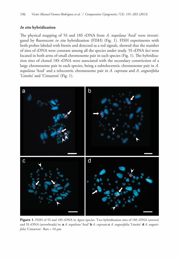
The physical mapping of 5S and 18S rDNA from A. tequilana ‘Azul’ were investi-gated by fluorescent in situ hybridization (FISH) (Fig. 1). FISH experiments with both probes labeled with biotin and detected as a red signals, showed that the number of sites of rDNA were constant among all the species under study. 5S rDNA loci were located in both arms of small chromosome pair in each species (Fig. 1). The hybridiza-tion sites of cloned 18S rDNA were associated with the secondary constriction of a large chromosome pair in each species, being a subtelocentric chromosome pair in A. tequilana ‘Azul’ and a telocentric chromosome pair in A. cupreata and A. angustifolia ‘Lineño’ and ‘Cimarron’ (Fig. 1).
Figure 1. FISH of 5S and 18S rDNA in Agave species. Two hybridization sites of 18S rDNA (arrows) and 5S rDNA (arrowheads) in: a A. tequilana ‘Azul’ b A. cupreata c A. angustifolia ‘Lineño’ d A. angusti-folia ‘Cimarron’. Bars = 10 µm.
Physical mapping of 5S and 18S ribosomal DNA in three species of Agave... 197
Karyotype analysis
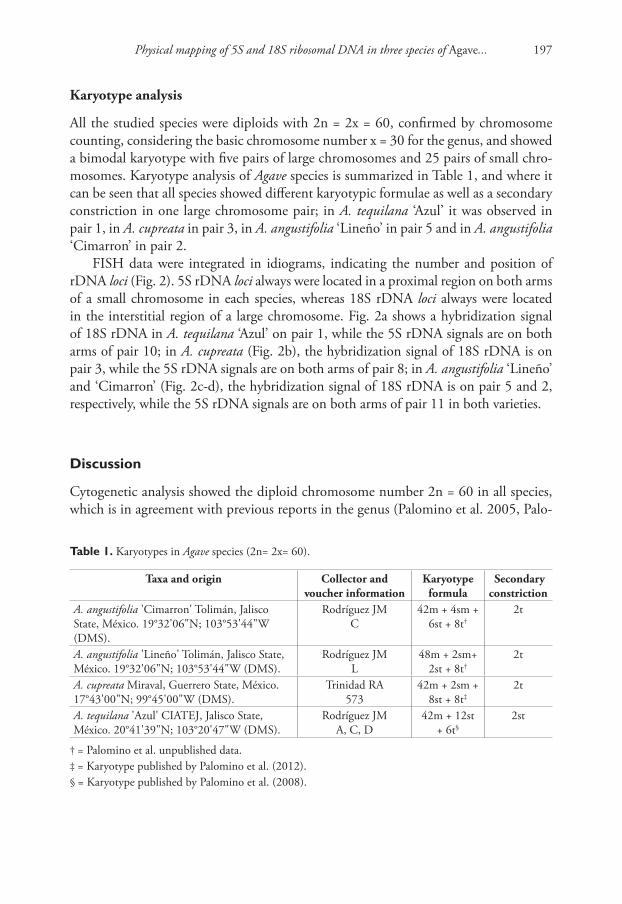
All the studied species were diploids with 2n = 2x = 60, confirmed by chromosome counting, considering the basic chromosome number x = 30 for the genus, and showed a bimodal karyotype with five pairs of large chromosomes and 25 pairs of small chro-mosomes. Karyotype analysis of Agave species is summarized in Table 1, and where it can be seen that all species showed different karyotypic formulae as well as a secondary constriction in one large chromosome pair; in A. tequilana ‘Azul’ it was observed in pair 1, in A. cupreata in pair 3, in A. angustifolia ‘Lineño’ in pair 5 and in A. angustifolia ‘Cimarron’ in pair 2.
FISH data were integrated in idiograms, indicating the number and position of rDNA loci (Fig. 2). 5S rDNA loci always were located in a proximal region on both arms of a small chromosome in each species, whereas 18S rDNA loci always were located in the interstitial region of a large chromosome. Fig. 2a shows a hybridization signal of 18S rDNA in A. tequilana ‘Azul’ on pair 1, while the 5S rDNA signals are on both arms of pair 10; in A. cupreata (Fig. 2b), the hybridization signal of 18S rDNA is on pair 3, while the 5S rDNA signals are on both arms of pair 8; in A. angustifolia ‘Lineño’ and ‘Cimarron’ (Fig. 2c-d), the hybridization signal of 18S rDNA is on pair 5 and 2, respectively, while the 5S rDNA signals are on both arms of pair 11 in both varieties.
Discussion
Cytogenetic analysis showed the diploid chromosome number 2n = 60 in all species, which is in agreement with previous reports in the genus (Palomino et al. 2005, Palo-
table 1. Karyotypes in Agave species (2n= 2x= 60).
Taxa and origin Collector and voucher information
Karyotype formula
Secondary constriction
A. angustifolia 'Cimarron' Tolimán, Jalisco State, México. 19°32'06"N; 103°53'44"W (DMS).
Rodríguez JM C
42m + 4sm + 6st + 8t†
2t
A. angustifolia 'Lineño' Tolimán, Jalisco State, México. 19°32'06"N; 103°53'44"W (DMS).
Rodríguez JM L
48m + 2sm+ 2st + 8t†
2t
A. cupreata Miraval, Guerrero State, México. 17°43'00"N; 99°45'00"W (DMS).
Trinidad RA573
42m + 2sm + 8st + 8t‡
2t
A. tequilana 'Azul' CIATEJ, Jalisco State, México. 20°41'39"N; 103°20'47"W (DMS).
Rodríguez JMA, C, D
42m + 12st + 6t§
2st
† = Palomino et al. unpublished data.‡ = Karyotype published by Palomino et al. (2012).§ = Karyotype published by Palomino et al. (2008).

Victor Manuel Gomez-Rodriguez et al. / Comparative Cytogenetics 7(3): 191–203 (2013)198
Figure 2. Idiograms of Agave karyotypes showing the 5S (green) and 18S (red) rDNA loci. a A. tequilana ‘Azul’ b A. cupreata; c A. angustifolia ‘Lineño’ d A. angustifolia ‘Cimarron’. Bars = 10 µm.
mino et al. 2008, Palomino et al. 2010). All species showed a bimodal karyotype with small and large chromosomes (n = S + L); this bimodal karyotype is shared among multiple genera in Asphodeloideae (Brandham and Doherty 1998, Adams et al. 2000, Vosa 2005) and Agavoideae (McKelvey and Sax 1933, Brandham 1969) and recently, McKain et al. (2012) demonstrated that the Agavoideae bimodal karyotype was origi-nated by an allopolyploid event, where the progenitor species seems to be extinct. De-spite maintaining the same karyotype in all species, it was also found different karyo-
Physical mapping of 5S and 18S ribosomal DNA in three species of Agave... 199
type formulae. This inter- and intraspecific variation shown here has been reported in other species and varieties in the genus (Banerjee and Sharma 1988, Moreno-Salazar et al. 2007, Palomino et al. 2008), leading to the formation of different cytotypes. Moreno-Salazar et al. (2007) studied three wild populations of A. angustifolia and found two different cytotypes; Palomino et al. (2008) analyzed eight varieties of A. tequilana and reported the same number of cytotypes. The presence of different cy-totypes in Agave genus could be originated by heterozygous chromosomal exchange (Moreno-Salazar et al. 2007, Palomino et al. 2008, Palomino et al. 2010), which can modify the structure of chromosomes and maintaining at the same time their diploid number (Lima-Cardoso et al. 2013).
FISH with rDNA probes showed that loci of 18S and 5S rDNA in Agave species were located in different chromosomes and on similar position in all species; this finding suggests that the chromosomes bearing the rDNA loci are homeologous and the dif-ference in numerical assignment is due to chromosomal rearrangements as mentioned before. 18S rDNA locus always was located in the interstitial region on the large arm of a large chromosome and associated to the secondary constriction, whereas the 5S rDNA loci were located in a proximal region on both arms of a small chromosome in all species. These results differ from Robert et al. (2008) because they reported that Agave species have one locus of 5S rDNA by monoploid genome in some diploid and polyploid spe-cies in the genus, including A. tequilana ‘Azul’ and A. angustifolia ‘Letona’ (tetraploid) and A. angustifolia ‘Chelem ki’ (hexaploid). The presence of 5S rDNA loci on both arms of a small chromosome in all species can be resulted from an unequal recombination or an event of transpositions; the latter have been reported previously in other monocots such as Allium Linnaeus, 1753 (Schubert and Wobus 1985), Oryza Linnaeus, 1753 (Shishido et al. 2000) and Alstroemeria Linnaeus, 1762 (Chacón et al. 2012). Recently, Khaliq et al. (2012), reported that Ty1-Copia retrotransposons are a major component of the A. tequilana genome (approximately 32 %) and might played a vital role in the organization and evolution of it, which could explain the results reported here.
To the best of our knowledge, here we reported the number and location of rDNA loci in two species with no previous report, A. cupreata and A. angustifolia ‘Lineño’ and ‘Cimarron’ as well as a different locus of 5S rDNA in all species studied. Data of FISH analysis provides new information about physical mapping of rDNA in Agave and such identified sites can be useful as chromosome markers for chromosome identification in hybrids in breeding programs as well as in evolutionary studies.
Conclusions
Despite the great diversity of the genus Agave which includes 166 species, the physical mapping of rDNA or other molecular markers are scarce, since just about five species have been described. The different karyotype formulae found in all species indicated the presence of cytotypes and data of FISH of rDNA allowed the physical mapping of A. cupreata and two new varieties of A. angustifolia. This work provides new informa-
Victor Manuel Gomez-Rodriguez et al. / Comparative Cytogenetics 7(3): 191–203 (2013)200
tion about the position and number of rDNA loci in Agave species through compara-tive karyotype analysis, however, further cytogenetic research must be conducted to understand the evolution of this genus and develop breeding programs to preserve its biodiversity.
Acknowledgements
The authors would like to thank SEP-CONACYT-Mexico Project 24554, CONA-CYT, FOMIX-JAL Project 99210, who supported this research, and to Jose Manuel Rodriguez Dominguez for his technical assistance in field work. Also the authors would like to thank Dr. Ignacio del Real Laborde (Tequila Sauza, S. de R.L. de C.V.) for providing Agave tequilana plant material. VMGR is a graduate student and finan-cially supported by CONACYT-Mexico (Reg. 45382).
References
Adams SP, Leitch IJ, Bennett MD, Chase MW, Leitch AR (2000) Ribosomal DNA evolution and phylogeny in Aloe (Asphodelaceae). American Journal of Botany 87(11): 1578–1583. doi: 10.2307/2656733
APG III (2009) An update of the Angiosperm Phylogeny Group classification for the orders and families of flowering plants: APG III. Botanical Journal of the Linnean Society 161(2): 105–121. doi: 10.1111/j.1095-8339.2009.00996.x
Banerjee S, Sharma AK (1987) Cytophotometric estimation of nuclear DNA in different spe-cies and varieties of Agave. Cytologia 52(1): 85–90.
Banerjee S, Sharma AK (1988) Structural differences of chromosomes in diploid Agave. Cyto-logia 53: 415–420.
Brandham PE (1969) Inversion heterozygosity and sub-chromatid exchange in Agave stricta. Chromosoma 26(3): 270–286. doi: 10.1007/BF00326522
Brandham PE, Doherty MJ (1998) Genome size variation in the Aloaceae, an angiosperm fam-ily displaying karyotypic orthoselection. Annals of Botany 82(1): 67–73. doi: 10.1006/anbo.1998.0742
Brown SE, Stephens Jl, Lapitan NLV, Knudson DL (1999) FISH landmarks for barley chromo-somes (Hordeum vulgare L). Genome 42(2): 274–281. doi: 10.1139/g98-127
Castorena-Sánchez I, Escobedo RM, Quiroz A (1991) New cytotaxonomical determinants recognized in six taxa of Agave in the sections Rigidae and Sisilanae. Canadian Journal of Botany 69(6): 1257–1264. doi: 10.1139/b91-163
Chacón J, Sousa A, Baeza CM, Renner SS (2012) Ribosomal DNA distribution and a genus-wide phylogeny reveal patterns of chromosomal evolution in Alstroemeria (Alstroemer-iaceae). American Journal of Botany 99(9): 1501–1512. doi: 10.3732/ajb.1200104
Choi YA, Tao R, Yonemori K, Sugiura A (2003) Simultaneous visualization of 5S and 45S rD-NAs in Persimmon (Diospyros kaki) and several wild relatives (Diospyros spp.) by fluorescent
Physical mapping of 5S and 18S ribosomal DNA in three species of Agave... 201
in situ hybridization (FISH) and multicolor FISH (MCFISH). Journal of the American Society for Horticultural Science 128(5): 736–740.
Conger DD, Fairchild LM (1953) A quick-freeze method for making smear slides permanent. Stain Technology 28(6): 281–283. doi: 10.3109/10520295309105555
Cuadrado A, Jouve N (2010) Chromosomal detection of simple sequence repeats (SSRs) using nondenaturing FISH (ND-FISH). Chromosoma 119(5): 495–503. doi: 10.1007/s00412-010-0273-x
Doughty LR (1936) Chromosome behaviour in relation to genetics of Agave. I. Seven species of fibre Agave. Journal of Genetics 33(2): 198–205. doi: 10.1007/BF02982532
D’Hont A, Ison D, Alix K, Roux C, Glaszmann JC (1998) Determination of basic chromo-some numbers in the genus Saccharum by physical mapping of ribosomal RNA genes. Genome 41(2): 221–225. doi: 10.1139/g98-023
García-Mendoza A (2002) Distribution of Agave (Agavaceae) in Mexico. Cactus and Succulent Journal 74(4): 177–188.
Garcia S, Garnatje T, Hidalgo O, McArthur ED, Siljak-Yakovlev S, Valles J (2007) Extensive ribosomal DNA (18S-5.8S-26S and 5S) colocalization in the North American endemic sagebrushes (subgenus Tridentatae, Artemisia, Asteraceae) revealed by FISH. Plant System-atics and Evolution 267(1–4): 79–92. doi: 10.1007/s00606-007-0558-6
Garcia S, Yoong-Lim K, Chester M, Garnatje T, Pellicer J, Vallès J, Leitch AR, Kovařík A (2009) Linkage of 35S and 5S rRNA genes in Artemisia (family Asteraceae): first evidence from angiosperms. Chromosoma 118(1): 85–97. doi: 10.1007/s00412-008-0179-z
Hizume H, Shibata F, Matsusaki Y, Garajova Z (2002) Chromosome identification and com-parative karyotypic analyses of four Pinus species. Theoretical and Applied Genetics 105(4): 491–497. doi: 10.1007/s00122-002-0975-4
Ji Y, De Donato M, Crane CF, Raska WA, Islam-Faridi MN, McKnight TD, Price HJ, Stelly DM (1999) New ribosomal RNA gene locations in Gossypium hirsutum mapped by mei-otic FISH. Chromosoma 108(3): 200–207. doi: 10.1007/s004120050369
Jiang J, Gill BS (1994) New 18S.26S ribosomal RNA gene loci: chromosomal landmarks for the evolution of polyploid wheats. Chromosoma 103(3): 179–185. doi: 10.1007/BF00368010
Khaliq I, Awais-Khan M, Pearce S (2012) Ty1-Copia retrotransposons are heterogeneous, ex-tremely high copy number and are major players in the genome organization and evolution of Agave tequilana. Genetic Resources and Crop Evolution 59(4): 575–587. doi: 10.1007/s10722-011-9705-6
Kato A, Lamb JC, Birchler JA (2004) Chromosome painting using repetitive DNA sequences as probes for somatic chromosome identification in maize. Proceedings of the National Acad-emy of Sciences of the United States of America 101(37): 13554–13559. doi: 10.1073/pnas.0403659101
Koo DH, Plaha P, Lim YP, Hur Y, Bang JW (2004) A high-resolution karyotype of Brassica rapa ssp. pekinensis revealed by pachytene analysis and multicolor fluorescence in situ hy-bridization. Theoretical and Applied Genetics 109(7): 1346–1352. doi: 10.1007/s00122-004-1771-0
Victor Manuel Gomez-Rodriguez et al. / Comparative Cytogenetics 7(3): 191–203 (2013)202
Lavania UC, Basu AS, Srivastava S, Mukai Y, Lavania S (2005) In situ chromosomal localiza-tion of rDNA sites in ‘‘safed musli’’ Chlorophytum Ker-Gawl and their physical measure-ment by fiber FISH. Journal of Heredity 96(2): 155–160. doi: 10.1093/jhered/esi018
Levan A, Fredga K, Sandberg AA (1964) Nomenclature for centromeric position on chromo-somes. Hereditas 52(2): 201–220. doi: 10.1111/j.1601-5223.1964.tb01953.x
Lima-Cardoso A, Holanda-Sales KA, Yoshiko-Nagamachi C, Pieczarka JC, Rodrigues-Noronha RC (2013) Comparative cytogenetics of two species of genus Scobinancistrus (Siluriformes, Loricariidae, Ancistrini) from the Xingu River, Brazil. Comparative Cytogenetics 7(1): 43–51. doi: 10.3897/CompCytogen.v7i1.4128
Liu B, Davis TM (2011) Conservation and loss of ribosomal RNA gene sites in diploid and polyploid Fragaria (Rosaceae). BMC Plant Biology 11: 157 pp. doi: 10.1186/1471-2229-11-157
McKain MR, Wickett N, Zhang Y, Ayyampalayam S, McCombie WR, Chase MW, Pires JC, DePamphilis CW, Leebens-Mack J (2012) Phylogenomic analysis of transcriptome data elucidates co-occurrence of a paleopolyploid event and the origin of bimodal karyotypes in Agavoideae (Asparagaceae). American Journal of Botany 99(2): 397–406. doi: 10.3732/ajb.1100537
McKelvey SD, Sax K (1933) Taxonomic and cytological relationships of Yucca and Agave. Jour-nal of the Arnold Arboretum 14: 76–81.
Mizuochi H, Marasek A, Okazaki K (2007) Molecular cloning of Tulipa fosteriana rDNA and subsequent FISH analysis yields cytogenetic organization of 5S rDNA and 45S rDNA in T. gesneriana and T. fosteriana. Euphytica 155(1–2): 235–248. doi: 10.1007/s10681-006-9325-y
Moreno-Salazar SF, Esqueda M, Martínez J, Palomino G (2007) Tamaño del genoma y cariot-ipo en Agave angustifolia y A. rhodacantha de Sonora, México. Revista Fitotecnia Mexicana 30(1): 13–23.
Mukai Y, Endo TR, Gill BS (1991) Physical mapping of the 18S.26S rRNA multigene fam-ily in common wheat: Identification of a new locus. Chromosoma 100(2): 71–78. doi: 10.1007/BF00418239
Murray MG, Thompson WF (1980) Rapid isolation of high molecular weight plant DNA. Nucleic Acids Research 8(19): 4321–4326. doi: 10.1093/nar/8.19.4321
Palomino G, Martínez J, Méndez I (2005) Citotipos en Agave angustifolia Haw. determinados por citometría de flujo y análisis de sus cariotipos. Revista Internacional de Contaminación Ambiental 21 (Suppl. 1): 49–54.
Palomino G, Martínez J, Méndez I (2008) Karyotype studies in cultivars of Agave tequilana Weber. Caryologia 61(2): 144–153.
Palomino G, Martínez J, Méndez I (2010) Análisis del tamaño del genoma y cariotipo de Agave aktites Gentry (Agavaceae) de Sonora, México. Revista Mexicana de Biodiversidad 81: 655–662.
Palomino G, Martínez J, Cepeda-Cornejo V, Pimienta-Barrios E (2012) Nuclear genome size and cytotype analysis in Agave cupreata Trel. & Berger (Agavaceae). Caryologia 65(4): 281–294. doi: 10.1080/00087114.2012.752915
Physical mapping of 5S and 18S ribosomal DNA in three species of Agave... 203
Pinkava DJ, Baker MA (1985) Chromosome and hybridization studies of Agaves. Desert Plants 7(2): 93–100.
Raina SN, Mukai Y (1999) Detection of a variable number of 18S-5.8S-26S and 5S ribosomal DNA loci by fluorescent in situ hybridization in diploid and tetraploid Arachis species. Genome 42(1): 52–59. doi: 10.1139/g98-092
Raina SN, Mukai Y, Kawaguchi K, Goel S, Jain A (2001) Physical mapping of 18S-5.8S-26S and 5S ribosomal RNA gene families in three important vetches (Vicia species) and their allied taxa constituting three species complexes. Theoretical and Applied Genetics 103(6–7): 839–845. doi: 10.1007/s001220100706
Rayburn AL, Gill BS (1985) Use of biotin-labeled probes to map specific DNA sequences on wheat chromosomes. Journal of Heredity 76(2): 78–81.
Robert ML, Lim KY, Hanson L, Sanchez-Teyer F, Bennett MD, Leitch AR, Leitch IJ (2008) Wild and agronomically important Agave species (Asparagaceae) show proportional in-creases in chromosome number, genome size, and genetic markers with increasing ploidy. Botanical Journal of the Linnean Society 158(2): 215–222. doi: 10.1111/j.1095-8339.2008.00831.x
Ruvalcaba-Ruiz D, Rodriguez-Garay B (2002) Aberrant meiotic behavior in Agave tequilana Weber var. Azul. BMC Plant Biology 2: 10 pp. doi: 10.1186/1471-2229-2-10
Schubert I, Wobus U (1985) In situ hybridization confirms jumping nucleolus organizing re-gions in Allium. Chromosoma 92(2): 143–148. doi: 10.1007/BF00328466
Shishido R, Sano Y, Fukui K (2000) Ribosomal DNAs: an exception to the conservation of gene order in rice genomes. Molecular and General Genetics 263(4): 586–591. doi: 10.1007/s004380051205
Siroky J, Lysak MA, Dolozel J, Kejnovsky E, Vyskot B (2001) Heterogeneity of rDNA dis-tribution and genome size in Silene spp. Chromosome Research 9(5): 387–393. doi: 10.1023/A:1016783501674
Specht T, Szymanski M, Barciszewska MZ, Barciszewski J, Erdmann VA (1997) Compilation of 5S rRNA and 5S rRNA gene sequences. Nucleic Acids Research 25(1): 96–97. doi: 10.1093/nar/25.1.96
Vosa CG (2005) On chromosome uniformity, bimodality and evolution in the tribe Aloineae (Asphodelaceae). Caryologia 58(1): 83–85.