phylum arthropoda: subphylum pancrustacea: class … · know basic mosquito anatomy. use a...
TRANSCRIPT
PHYLUM ARTHROPODA: SUBPHYLUM PANCRUSTACEA: CLASS INSECTA
ORDER DIPTERA: FAMILY CULICIDAE Know basic mosquito anatomy. Use a slide-mounted specimen to draw and label the parts of the mosquito. Most specimens are females. Use the overhead image to identify the different parts of the mosquito before proceeding with a taxonomic identification. Know the functions of the different head appendages: Antennae = olfaction, thermal (heat) sensors Palps = gas sensors (CO2) Proboscis (many mouthparts combined into a single compound organ) = blood sucking ID your mosquitoes. Everyone is provided with a dish containing 6 mosquitoes representing 3 species. Both sexes should be present. There is strong sexual dimorphism in mosquitoes, so males and females are distinguishable. Most taxonomic IDs are based on females since they are the vectors for disease. If a female mosquito is damaged, in most cases she can still be identified by a combination of characters. First things first - identify the sexes of your specimens. 1A. Antennae bushy or feather-like = male mosquito (do not worry about species ID). Set aside. 1B. Antennae not bushy or feather-like = female mosquito. Go to 2. Taxonomy of Female mosquitoes 2A. Palps as long as proboscis, not bushy and without paddles = Anopheles quadrimaculatus (known to transmit Malaria, Heartworm, and Triple E virus) 2B. Palps shorter than proboscis..........................................................................................3 3A. Abdomen light brown...................................................................................................4 3B. Abdomen often dark brown, with metallic scales.........................................................5 4A. Antennae not longer than proboscis (may appear about the same length as proboscis, but never longer) = Culex quiquefasciatus (known to transmit Lymphatic filariasis, St Louis encephalitis, West Nile virus) 4B. Antennae longer than proboscis, antennal flagellomere 1 elongate, twice as long as flagellomere 2 = Deinocerites pseudes (not known to function as a vector) 5A. Abdomen scales bright metallic violet and silver, thorax with broad flat metallic scales. Hamagogus sp (known to transmit Mayaro virus) 5B. Abdomen scales metallic white. Horizontal white stripes on abdomen. Aedes aegypti (known to transmit yellow fever, Zika, dengue fever, Chikungunya)
PHYLUM ARTHROPODA: SUBPHYLUM PANCRUSTACEA: CLASS INSECTA
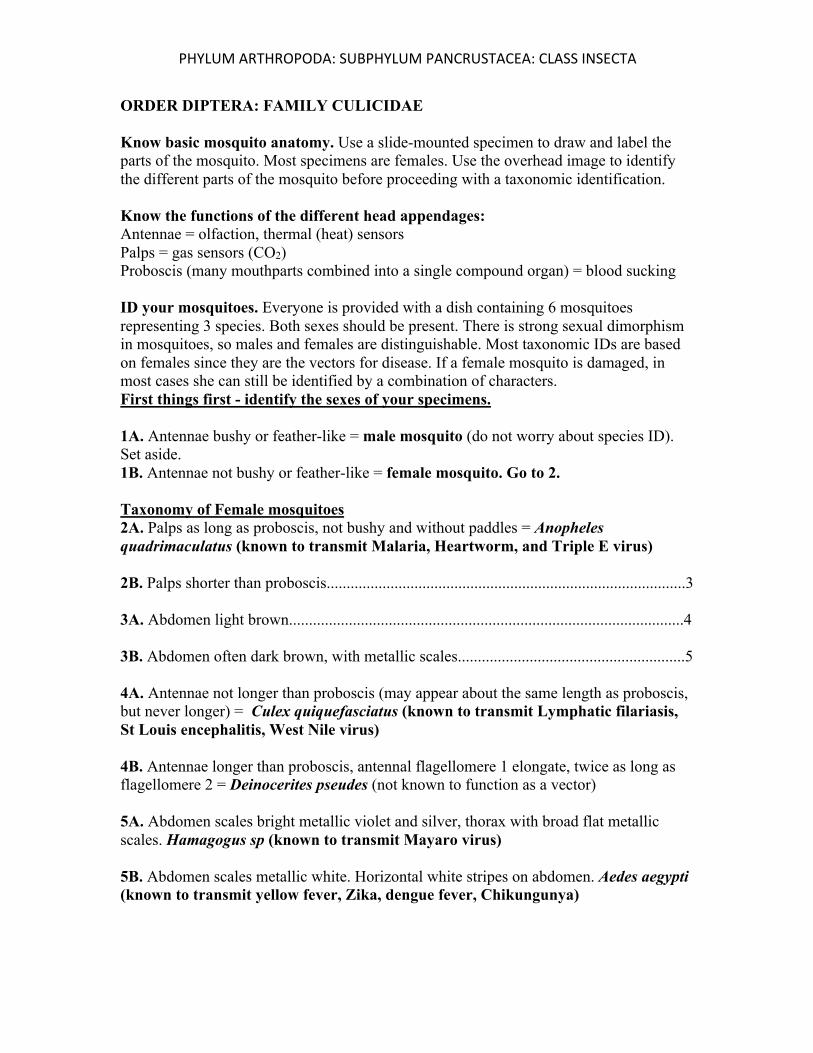
IDENTIFYING LARVAL (INSTAR) STAGES OF MOSQUITOES
Examine the cultured larvae and observe how they behave. Use the guide below to attempt to identify the species. At present, none are in the pupa stage. Anopheles quadrimaculatus Larvae: Larvae lie horizontally at the surface of the water where they filter feed on organics. They do not possess the breathing siphon present in other mosquitos. They obtain oxygen through hairs along the abdomen. The food sources include a variety of plant and animal matter suspended at the surface of the water and small enough to eat Aedes aegypti Larvae: Mosquito larvae are often called "wrigglers" or "wigglers," because they appear to wiggle in the water when disturbed. Larvae breathe oxygen through a posteriorly located siphon, which is held above the water surface while the rest of the body hangs vertically. Most Aedes larvae can be distinguished from other genera by their short siphon (compare to C. quinquefasciatus). Culex quinquefasciatus Larvae: The larval head is short and stout becoming darker toward the base. The mouth brushes have long yellow filaments that are used for filtering organic materials. The abdomen consists of eight segments, the siphon, and the saddle. Each segment has a unique setae pattern. The siphon is on the dorsal side of the abdomen, and in Culex quinquefasciatus the siphon is four times longer than it is wide with multiple setae tufts. The saddle is barrel shaped and located on the ventral side of the abdomen with four long anal papillae protruding from the posterior end 2. Use a transfer pipette to suck up a small larva and place it in a small bowl of water. Place the bowl on your stereomicroscope and attempt to draw the larva if it ever stops moving. You should be able to see a head, thorax, and abdomen. Unlike adults, there are no appendages on the thorax of a larva. The abdomen has many segments. At the posterior end may be a siphon, depending on the species. Small hairs and gills may also be present. Illustrate and label.
eggs
larvae
pupa
PHYLUM ARTHROPODA: SUBPHYLUM PANCRUSTACEA: CLASS INSECTA
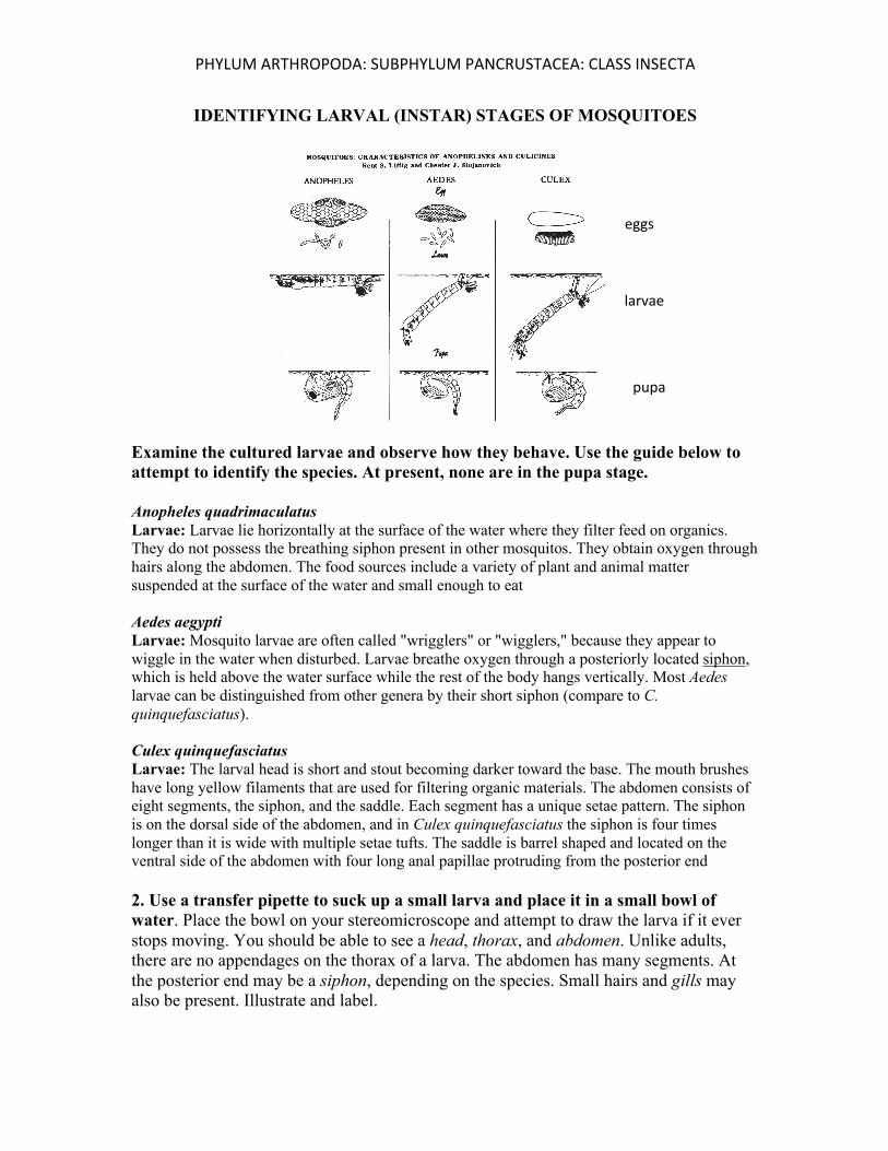
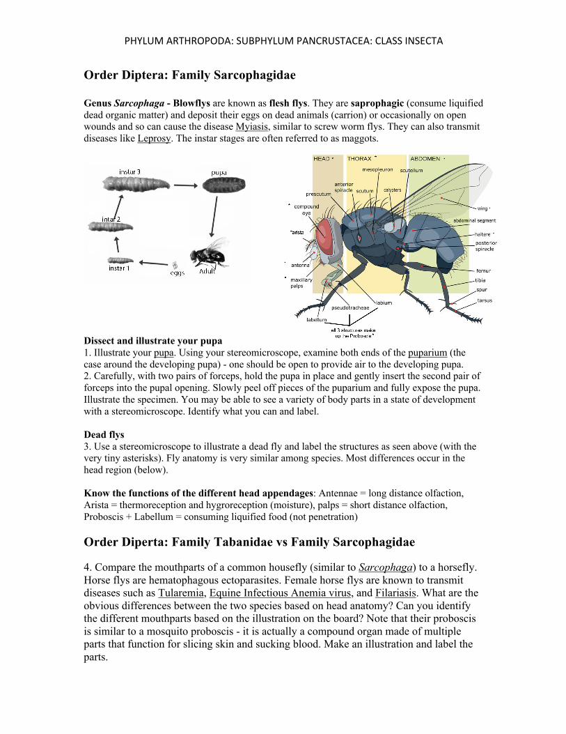
Order Diptera: Family Sarcophagidae Genus Sarcophaga - Blowflys are known as flesh flys. They are saprophagic (consume liquified dead organic matter) and deposit their eggs on dead animals (carrion) or occasionally on open wounds and so can cause the disease Myiasis, similar to screw worm flys. They can also transmit diseases like Leprosy. The instar stages are often referred to as maggots. Dissect and illustrate your pupa 1. Illustrate your pupa. Using your stereomicroscope, examine both ends of the puparium (the case around the developing pupa) - one should be open to provide air to the developing pupa. 2. Carefully, with two pairs of forceps, hold the pupa in place and gently insert the second pair of forceps into the pupal opening. Slowly peel off pieces of the puparium and fully expose the pupa. Illustrate the specimen. You may be able to see a variety of body parts in a state of development with a stereomicroscope. Identify what you can and label. Dead flys 3. Use a stereomicroscope to illustrate a dead fly and label the structures as seen above (with the very tiny asterisks). Fly anatomy is very similar among species. Most differences occur in the head region (below). Know the functions of the different head appendages: Antennae = long distance olfaction, Arista = thermoreception and hygroreception (moisture), palps = short distance olfaction, Proboscis + Labellum = consuming liquified food (not penetration) Order Diperta: Family Tabanidae vs Family Sarcophagidae 4. Compare the mouthparts of a common housefly (similar to Sarcophaga) to a horsefly. Horse flys are hematophagous ectoparasites. Female horse flys are known to transmit diseases such as Tularemia, Equine Infectious Anemia virus, and Filariasis. What are the obvious differences between the two species based on head anatomy? Can you identify the different mouthparts based on the illustration on the board? Note that their proboscis is similar to a mosquito proboscis - it is actually a compound organ made of multiple parts that function for slicing skin and sucking blood. Make an illustration and label the parts.