phylogeny and diversification of bryophytesinstruct.uwo.ca/biology/3404f/shaw2004.pdf · directions...
TRANSCRIPT
1557
American Journal of Botany 91(10): 1557–1581. 2004.
PHYLOGENY AND DIVERSIFICATION OF BRYOPHYTES1
JONATHAN SHAW2,4 AND KAREN RENZAGLIA3
2Duke University, Department of Biology, Box 90338, Durham, North Carolina 27708 USA; and 3Department of Plant Biology,Southern Illinois University, Carbondale, Illinois 62901-6509 USA
The bryophytes comprise three phyla of embryophytes that are well established to occupy the first nodes among extant lineages inthe land-plant tree of life. The three bryophyte groups (hornworts, liverworts, mosses) may not form a monophyletic clade, but theyshare life history features including dominant free-living gametophytes and matrotrophic monosporangiate sporophytes. Because oftheir unique vegetative and reproductive innovations and their critical position in embryophyte phylogeny, studies of bryophytes arecrucial to understanding the evolution of land plant morphology and genomes. This review focuses on phylogenetic relationships withineach of the three divisions of bryophytes and relates morphological diversity to new insights about those relationships. Most previouswork has been on the mosses, but progress on understanding the phylogeny of hornworts and liverworts is advancing at a rapid pace.Multilocus multigenome studies have been successful at resolving deep relationships within the mosses and liverworts, whereas single-gene analyses have advanced understanding of hornwort evolution.
Key words: Anthocerophyta; Bryophyta; bryophyte phylogeny; hornworts; liverworts; Marchantiophyta; mosses; tree of life.
As the only land plants with a dominant gametophyte gen-eration, liverworts, mosses, and hornworts exhibit structuraland reproductive attributes that are exclusive, unifying, andinnovative. Their persistent gametophyte is responsible for ex-ploratory growth as well as for proliferation of a new gener-ation through either sexual or asexual processes. As a conse-quence, bryophyte gametophytes exhibit a degree of diversityand complexity unparalleled in tracheophytes. They are char-acterized by modular growth (repeated patterns) from a gen-erative apex, range in habit from upright to procumbent, andinclude thalloid to leafy forms (Mishler and DeLuna, 1991).Within mosses and liverworts, leafy gametophytes are thenorm, rivaling the leafy sporophytic growth forms of sometracheophytes, especially lycophytes (Renzaglia et al., 2000).However, because they depend on water for sexual reproduc-tion, the gametophytes of bryophytes are small relative to mostvascular plant sporophytes. Sexual reproduction in bryophytesinvolves release of motile male gametes into the environmentand requires successful navigation of these naked cells fromthe male to the female sex organs via an external water source.
Sporophytes of bryophytes are without exception monospo-rangiate and matrotrophic throughout their life span (Grahamand Wilcox, 2000). Ephemeral and dependent on the game-tophyte for nutrition and protection, they never exhibit themodular, indeterminate growth form of the gametophyte gen-eration. In their greatest structural complexity, bryophyte spo-rophytes consist of a nutritive foot, elongating pedicel or seta,and a single terminal sporangium or capsule. Formative divi-sions in the embryo produce all precursor components of thesporophyte; i.e., distinct embryonic regions are determined todevelop into the three organographic zones of the mature spo-
1 Manuscript received 5 January 2004; revision accepted 15 June 2004.Various aspects of the research presented here were supported by NSF
grants from the Systematic Biology and Assembling the Tree-of-Life Pro-grams (DEB-0089131 to AJS and DEB-0235985 and DEB-0228679 to KSR).We are grateful to Christine Cargill, Cymon Cox, Barbara Crandall-Stotler,Christine Davis, R. Joel Duff, Laura Forrest, Bernard Goffinet, Xiaolan He-Nygren, and Juan Carlos Villarreal for providing data and sharing unpublishedmanuscripts. We are also indebted to Andrew Blackwell and Scott Schuettefor contributions of images and expert technical assistance. Molly McMullenkindly edited an earlier manuscript draft.
4 E-mail: [email protected].
rophyte: sporangium, seta, and foot. In contrast, an apical mer-istem initial develops in the embryo of tracheophytes and issubsequently responsible for continuous production of repeat-ed shoot and root modules in these plants (Bierhorst, 1971;Kato and Imaichi, 1997). Capsules of bryophytes are structur-ally elaborate and, in some instances, exhibit complicatedmechanisms for spore production and dispersal. Basal sporo-phyte elongation with nonsynchronized spore production inhornworts, elaters in liverworts, and peristomes of mosses pro-vide examples of this complexity.
General treatments of bryophyte morphology can be foundin Leitgeb (1874–1881), Campbell (1895), Goebel (1905),Smith (1955), Parihar (1965), Watson (1971), Puri (1973),Richardson (1981), Schofield (1985), and Crum (2001). TheManual of Bryology, edited by Verdoorn (1932), contains au-thoritative treatments of selected bryology topics that sum-marized the state of our knowledge at that time, and the NewManual of Bryology, edited by Schuster (1984), provided ex-panded updates more than fifty years later. Both manuals arestill useful. Other edited volumes on various aspects of bryo-phyte biology especially relevant to the tree of life includeClarke and Duckett (1979), Smith (1982), and Shaw and Gof-finet (2000).
The crucial position of bryophytes in embryophyte evolu-tion—An unambiguous conclusion from the multitude of con-temporary phylogenetic investigations of streptophytes is thatbryophytes are the first green plants to successfully radiate intoterrestrial niches. These small, inconspicuous plants have ex-isted for several hundreds of millions of years and have playeda prominent role in shaping atmospheric and edaphic changeand the subsequent evolution of all forms of plant life on land.Explorations of life history phenomena in bryophytes and asolid understanding of interrelationships among them are nec-essary to reconstruct the early evolution of embryophytes.
The concept that the embryo/sporophyte evolved in landplants through intercalation of mitotic divisions between fer-tilization and meiosis is widely accepted (Graham, 1993; Gra-ham and Wilcox, 2000). Based on this axiom, land plant evo-lution proceeded in the direction of progressively more elab-orate sporophytes. Although generally true, unconditional ac-ceptance of this trend leads to conclusions that ignore
1558 [Vol. 91AMERICAN JOURNAL OF BOTANY
processes of reduction and parallel/convergent evolution, phe-nomena that have occurred repeatedly during bryophyte di-versification (Schuster, 1992; Niklas, 1997; Boisselier-Dubayleet al., 2002). A defining characteristic of embryophytes is themeiotic production of spores in tetrads and sporopollenin-im-pregnated spore walls. Because of their resistance to degra-dation, fossil spores have provided valuable clues to the initialstages of land colonization (Taylor, 1995; Wellman and Gray,2000; Wellman et al., 2003). The earliest confirmed land plantfossils are spores, speculated to be from an ancient liverwortdating to the middle Ordovician, some 475 million years ago(mya) (Wellman et al., 2003).
Gametophytes of bryophytes also provide critical cluesabout land plant evolution. Thalloid and filamentous growthforms are shared with pteridophytes, but the completely sub-terranean and nonphotosynthetic life histories found in manylycophytes and some ferns show no homology in bryophytes(Bierhorst, 1971). The achlorophyllous gametophyte of the liv-erwort Cryptothallus is a recent acquisition within a strictlyphotosynthetic lineage (Renzaglia, 1982). Unlike pterido-phytes, bryophyte gametophytes frequently show organ de-velopment (leaf, stem, and rhizome) and extensive tissue dif-ferentiation, including conducting and supportive tissues (He-bant, 1977; Ligrone et al., 2000). Production of multicellulargametangia was an innovation in embryophytes that was anecessary precursor to embryo development (Graham and Wil-cox, 2000). Among land plants, only mosses and liverwortsproduce superficial gametangia, which are variously protectedby elaborate appendages, including leaves. Hornworts seques-ter vulnerable organs in internal compartments (Renzaglia etal., 2000; Renzaglia and Vaughn, 2000).
A lack of intermediate forms in both life history phases andthe potential to interpret morphological transitions in oppositedirections have obscured relationships among bryophytes andpteridophytes. Understanding morphological evolution re-quires unambiguous establishment of phylogenetic relation-ships among and within bryophyte lineages. Over the past de-cade, great strides have been made toward reaching this goal;however, fundamental questions remain.
In addition to elucidating early patterns of morphologicaldiversification in embryophytes, bryophytes are crucial to un-derstanding plant genome evolution. Approximately 66% ofgenes identified from expressed sequence tag analyses of geneexpression in gametophytes of Physcomitrella patens have ho-mologues in the Arabidopsis genome, consistent with the hy-pothesis that genes expressed in the diploid plant body of an-giosperms were expressed in the gametophytes of early landplants and were recruited for sporophytic morphogenesis laterin plant phylogeny (Nishiyama et al., 2003). Phylogenetic andfunctional analyses of genes expressed in Physcomitrella ga-metophytes have clarified the phylogenetic history of severalimportant gene families, including MIKC-type MADS-boxgenes (Krogan and Ashton, 2000; Henschel et al., 2002; Hoheet al., 2002) and homeobox genes (Champagne and Ashton,2001). Phylogenetic analyses of the KNOX (homeobox) genefamily across the land plant tree of life have provided insightsinto the history of gene duplication and functional divergenceduring embryophyte history (Champagne and Ashton, 2001).Because KNOX genes are involved in expression of meriste-matic activity in vascular plant sporophytes, functional anal-yses of KNOX genes in mosses, liverworts, and hornworts arecentral to understanding evolution of plant development in em-bryophytes. Comparable studies of genes involved in flower
development are underway, and, in the context of phylogeneticanalyses of bryophytes, the early evolution of these genes isnow a tractable problem for investigation (Himi et al., 2001).
Relationships among the three lineages—Relationshipsamong the three lineages of bryophytes remain one of themajor unresolved questions in plant evolutionary biology(Goffinet, 2000). Virtually every conceivable hypothesis hasbeen put forth in regards to primary branching patterns at thebase of embryophytes. Most commonly, bryophytes areviewed as a grade of three monophyletic lineages, with anuncertain branching order (Mishler et al., 1994; Qiu et al.,1998). Controversy often focuses on which bryophyte groupis sister to all other embryophytes, with two hypotheses mostfrequently supported: liverworts as sister to other embryo-phytes vs. hornworts as the sister group (Mishler et al., 1994;Hedderson et al., 1996, 1998; Malek et al., 1996; Garbary andRenzaglia, 1998; Qiu et al., 1998; Beckert et al., 1999; Duffand Nickrent, 1999; Nishiyama and Kato, 1999; Soltis et al.,1999; Nickrent et al., 2000; Renzaglia et al., 2000; Stech etal., 2003). A moss-plus-liverwort clade has been recovered inseveral of these analyses (Hedderson et al., 1996, 1998; Ni-shiyama and Kato, 1999; Nickrent et al., 2000; Renzaglia etal., 2000). Recently, it was postulated that hornworts, notmosses, are the closest living relative of tracheophytes. Thisspeculation finds support in sequence data as well as in struc-tural genomic features (Samigullin et al., 2002; Kelch et al.,in press). In contrast, recent analyses of amino acid sequencesbased on entire plastid genomes provided support for a mono-phyletic bryophyte assemblage; however, these results must beviewed with caution because of severe limitations in taxonsampling (Nishiyama et al., in press).
The focus of this review is to present the current state ofknowledge on phylogenetic relationships within, not among,hornworts, liverworts, and mosses. Emphasis is placed on syn-thesizing results of recent molecular investigations that haverevolutionized interpretations of genetic and morphological di-versification within each of these groups. Intriguing new per-spectives on character evolution have emerged from thesestudies.
ANTHOCEROTOPHYTA
Hornwort classification and relationships—For centuries,botanists have marveled at the structural peculiarities of horn-worts (Hofmeister, 1851; Leitgeb, 1879; Campbell, 1895,1917, 1924; Goebel, 1905; Lang, 1907; Bower, 1935). In noother branch of the green tree of life does extension of eachsporophyte involve continuous, presumably indeterminate, ba-sipetal growth of a single elongated sporangium. All stages ofspore development, from undifferentiated cells through pre-meiotic/meiotic spore mother cells to sequentially more maturespores, can be found in a single hornwort sporangium. A con-stant production of spores therefore ensures dispersal through-out the growing season for as long as the gametophyte persists.This mode of sporophyte development has no counterpart inother plant groups, thus obscuring the phylogenetic positionof hornworts among green plants.
Hornworts have remained relatively unexplored at all levelsof phylogenetic inquiry (Renzaglia and Vaughn, 2000; Stechet al., 2003; Duff et al., in press). The perception that horn-worts are invariable, elusive, and difficult to identify has con-tributed to the paucity of systematic studies within the group.

October 2004] 1559SHAW AND RENZAGLIA—PHYLOGENY OF BRYOPHYTES
Fig. 1. ‘‘Backbone’’ phylogeny of the one most parsimonious tree basedon rbcL sequences from seven of the 11 named genera of hornworts (Antho-cerophyta). Bootstrap percentages are shown below branches. Tree providedby R. Joel Duff, University of Akron.
Fig. 2. Diversity in growth forms among hornworts. A. Photograph of Anthoceros punctatus L. Small orbicular gametophyte with both immature and almostripe sporophytes, growing on soil. Image provided by Christine Cargill. B and C Scanning electron micrographs (SEM) of gametophyte of Dendroceros crispatus(Hook.) Nees. B. Ventral surface showing monostromatic wings and thickened midrib with bulging Nostoc colonies (arrow). Note the numerous small pores(mucilage clefts) along either side of the midrib. C. Dorsal surface showing sunken archegonia (arrow) on the midrib and developing sporophytes enclosedwithin gametophytic involucre. D. SEM of Notothylas orbicularis (Schwein.) Sull. Small orbicular gametophytes growing on bare soil; note the numerous small,horizontally oriented sporophytes enclosed in involucres. Bar 5 0.2 mm, except in A, bar 5 3 mm.
This small, homogeneous assemblage contains 100–150 poor-ly delineated species (Schuster, 1992). Concepts of interrela-tionships among hornworts based on morphology, and the re-sulting classification schemes, show virtually no consensus atthe generic, familial, and ordinal levels (Mishler and Churchill,1984; Hasegawa, 1988; Hassel de Menendez, 1988; Schuster,1992; Hyvonen and Piippo, 1993; Renzaglia and Vaughn,2000). Twelve genera of hornworts have been named, Antho-ceros, Dendroceros, Folioceros, Notothylas, Megaceros,Phaeoceros, Aspiromitus, Hattorioceros, Leiosporoceros,Nothoceros, Mesoceros, and Sphaerosporoceros, of whichonly the first six are widely recognized.
Even with the advent of molecular systematics and a re-newed interest in early land plant phylogeny, hornwort sam-pling has been sparse, with one to three taxa included in mostanalyses (Katoh et al., 1983; van de Peer et al., 1990; Mishleret al., 1994; Bopp and Capesius, 1996, 1998; Hedderson etal., 1996, 1998; Malek et al., 1996; Qiu et al., 1998; Beckertet al., 1999; Duff and Nickrent, 1999; Nishiyama and Kato,1999; Soltis et al., 1999; Nickrent et al., 2000). Among thedozens of papers on bryophyte phylogeny over the past tenyears, there is only one comprehensive molecular analysis ofwithin-hornwort relationships, based on rbcL gene sequencesfrom 20 hornworts (Duff et al., in press). A second study uti-lizing the plastid trnL intron sampled nine hornworts but fo-cused on the position of the group among land plants (Stechet al., 2003). Results of these analyses are congruent and re-veal novel but intuitive relationships. The rbcL analysis pro-vided much greater resolution of hornwort interrelations be-cause of more extensive sampling, including additional speciesof the five genera included in the trnL study and representa-tives of three other genera (Folioceros, Leiosporoceros, andNothoceros). The discussion that follows will focus on taxo-nomic inferences and morphological character evolution thatemerge from scrutiny of the consensus phylogenetic patternsupported by these pioneering studies (Fig. 1).
Diagnostic characters of hornworts are found in both lifehistory generations and are variably emphasized by systema-tists (Cargill et al., in press). Growth form (Fig. 2), chloroplaststructure and number (Fig. 3), antheridial number and jacketcell organization, Nostoc colony organization, and presence ofmucilage canals and thallus outgrowths are taxonomically use-ful gametophytic characters. Taxonomically informative fea-tures of the sporophyte include degree of development of his-

1560 [Vol. 91AMERICAN JOURNAL OF BOTANY
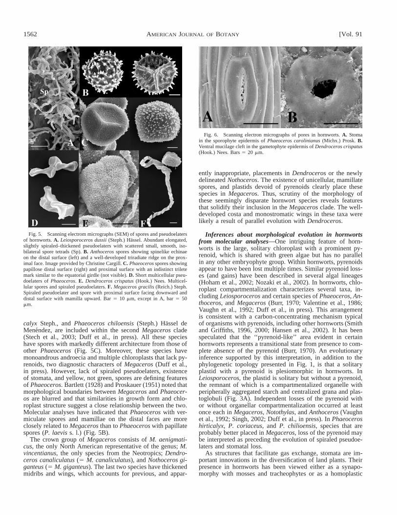
Fig. 3. Transmission electron micrographs of chloroplasts in hornworts.A. Leiosporoceros dussii (Steph.) Hassel. Chloroplast in the assimilative layerof the sporophytes showing peripheral starch and centralized grana. B. Foli-oceros fuciformis Baradw. Central pyrenoid with lens-shaped subunits sepa-rated by narrow grana and surrounded by starch grains. Bar 5 0.5 mm.
togenic regions (Fig. 4), spore and pseudoelater architectureand ultrastructure (Fig. 5), and the presence of columella andstomata (Fig. 6).
The backbone of hornwort phylogenetic relationships—Molecular evidence corroborates morphological inferencesthat the hornworts are monophyletic (Fig. 1). The genus Leios-poroceros, which was named for its unusually small, mono-lete, smooth spores produced in isobilateral tetrads (Figs. 4A,5A), is sister to the remaining hornworts. The position ofLeiosporoceros among hornworts has been controversial. Has-sel de Menendez (1986, 1988) segregated it into an autono-mous family and order, whereas Hasegawa (1988) and Schus-ter (1992) recognized it as a subgenus of Phaeoceros. In con-trast, Hyvonen and Piippo (1993) supported a sister relation-ship between Leiosporoceros and Folioceros, based primarilyon morphologically similar pseudoleaters (Fig. 5A). More de-tailed examination of Leiosporoceros dussii reveals morpho-logical and molecular features heretofore undescribed in anyhornwort. Moreover, Leiosporoceros gene sequences have ex-tremely low levels of RNA editing (J. Duff, University of Ak-ron, unpublished data) and thus differ from other hornwortsthat have been shown to have extensive editing (Yoshinga etal., 1996; Duff et al., in press). The gametophyte resemblesthat of Phaeoceros and Megaceros in that it is fleshy and lacks
internal mucilage canals. Number of antheridia per cavity isgreater than 20, a feature shared only with Anthoceros andFolioceros (Cargill et al., in press). However, unlike otherhornworts in which Nostoc is in discrete spherical colonieswithin the ventral thallus (Fig. 2B), those in Leiosporocerosoccur in branching strands that run longitudinally and are se-questered in the thallus midregion. Ventral mucilage clefts thatenable Nostoc to enter and establish colonies in other taxa(Fig. 6B) are lacking in Leiosporoceros. Chloroplasts of Leio-sporoceros are also readily differentiated from those in otherhornworts. Starch is neatly aggregated around the peripheryof the organelle, with a central elaboration of photosyntheticmembranes; the chloroplasts have no pyrenoids (Fig. 3A).
The sporophyte of Leiosporoceros is elongated and robust,and its anatomy departs significantly from that of other horn-worts (Fig. 4A). The suture is highly differentiated and visibleas a deep longitudinal groove. The assimilative and sporoge-nous regions are massive when compared with other horn-worts. Several layers of small spore tetrads are surrounded bymucilage and interspersed with groups of large, elongatedpseudoelaters (Fig. 5A). Stomata are abundant and appear sim-ilar to those in more derived taxa (Fig. 6A). Clearly, the sharedtraits between Leiosporoceros and other hornworts provide in-sight about plesiomorphies within the group. For example, sto-mata and large numbers of antheridia are best interpreted asancestral hornwort traits. On the other hand, unique morpho-logical traits that characterize Leiosporoceros are presumedautapomorphies and likely reflect the deep evolutionary sep-aration of this genus from other hornworts. Further molecularand morphological studies are required to evaluate these hy-potheses.
After Leiosporoceros, Anthoceros plus Folioceros form aclade sister to other hornworts (Fig. 1). Taxonomic treatmentshave generally recognized a sister relationship between Antho-ceros (including Folioceros) and Phaeoceros, placing them inthe same family or subfamily. Thus genetic divergence be-tween Anthoceros plus Folioceros and the remaining taxa ap-pears problematic at first glance. Similarities between Antho-ceros, Folioceros, and Phaeoceros include rosette-like habits(Fig. 2A), large solitary chloroplasts with well-developed py-renoids (Fig. 3B), and comparable sporophyte anatomy (Fig.4B). However, clearly defined features distinguish Anthocerosand Folioceros from other hornworts; these include dorsal la-mellae, schizogenous mucilage cavities, and antheridia in largegroups of up to 50 per cavity, as compared to 1–4 (–6) an-theridia per cavity in other hornworts. Darkly pigmentedspores with well-defined trilete marks also serve to differen-tiate these taxa from other hornworts (Fig. 5B). Molecular ev-idence that Anthoceros plus Folioceros form a clade sister toall other hornworts (except Leiosporoceros) has reinforced thetaxonomic value of diagnostic morphological features that arerestricted to these two genera.
Close affinity between Folioceros and Anthoceros is wellsupported by rbcL data and reflected in most current classifi-cations (Hasegawa, 1988, 1994; Schuster, 1992; Hyvonen andPiippo, 1993). In contrast, Hassel de Menendez (1988) seg-regated Folioceros into a monotypic family and order basedon spore ornamentation and pseudoelaters. Folioceros hasthick-walled, reddish brown, highly elongated pseudoelaters,whereas Anthoceros has short, thin-walled multicellular pseu-doelaters similar to those of Phaeoceros (Fig. 5D). Additionaldifferences are found in the placenta and chloroplasts of thesetwo taxa (Vaughn et al., 1992; Vaughn and Hasegawa, 1993).

October 2004] 1561SHAW AND RENZAGLIA—PHYLOGENY OF BRYOPHYTES
Fig. 4. Cross sections of hornwort sporophytes. A. Light micrograph of Leiosporoceros dussii (Steph.) Hassel. Tissue is differentiated from outside to insideas follows: single-layered epidermis, 9–10 layers of assimilative cells, abundant sporogenous tissue with several layers of tetrads intermixed with elaters, andan indistinct columella. The suture is clearly defined as a longitudinal groove that extends nearly to the sporogenous tissue. Bar 5 100 mm. B. Scanning electronmicrograph of Phaeoceros carolinianus (Michx.) Prosk. In contrast to Fig. 4A, this sporophyte contains an assimilative zone of four cell layers, sporogenoustissue with one layer of large tetrads intermixed with small elaters, and a columella of 16 cells. Bar 5 1 mm.
With only a single species included in the rbcL sequence anal-ysis, it is not possible to evaluate monophyly of Folioceros.
The remaining hornworts form a monophyletic group thatincludes two well-supported assemblages: Phaeoceros laevissensu lato (represented in Fig. 1 by P. carolinianus) plus No-tothylas and Megaceros plus Dendroceros. A close affinitybetween Phaeoceros and Notothylas was suggested by Hasselde Menendez (1988), who placed these two genera in the fam-ily Notothyladaceae. Both genera have chloroplasts withprominent pyrenoids, spores with an equatorial girdle (Fig.5C), and 2–4 (–6) antheridia per chamber. However, becauseof the distinctive sporophyte of Notothylas (Fig. 2D), mostsystematists have segregated this genus into a monotypic sub-family, family, or order (Singh, 2002). Notothylas is the onlyhornwort taxon in which growth of the sporophyte is abbre-viated, spore production appears synchronized, stomata are ab-sent, and the columella is normally absent to poorly developed,a combination of characters that indicate affinities with liver-worts. Consequently, it has been suggested that the Notothylassporophyte is plesiomorphic, representing a structural ‘‘link’’with other bryophytes. Under this interpretation, hornwort ra-diation involved an elaboration of sporophytes in more derivedtaxa (Campbell, 1895; Mishler and Churchill, 1984; Graham,1993; Hyvonen and Piippo, 1993; Hasegawa, 1994). An al-ternative hypothesis, supported by molecular data, is that spo-rophytes in Notothylas are not representative of the ancestralcondition in hornworts but are highly reduced and specialized(Lang, 1907; Bartlett, 1928; Proskauer, 1960; Renzaglia, 1978;Schofield, 1985; Schuster, 1992). Features such as the exis-tence of a relictual and largely nonfunctional suture in somespecies support the derived nature of the Notothylas sporo-phyte. If parallel reduction in sporophye complexity occurredamong hornwort genera, Notothylas may be polyphyletic
(Lange, 1907; Proskauer, 1960). An evaluation of this hypoth-esis requires increased taxon sampling across the hornworts.
Diversity within Phaeoceros is particularly evident in sporemorphology (Schuster, 1992). Phaeoceros laevis s. l., includesspecies with spiny papillate spores, whereas ornamentation inthe remaining species varies from vermiculate to blunt, wart-like projections. As described later, the three representativesof Phaeoceros with vermiculate spores included in molecularanalyses are more closely related to Megaceros than to P. lae-vis s. l.
A close relationship between Megaceros and Dendrocerosis evident in morphological characters such as spiraled pseu-doelaters (Fig. 5E, F), absence of stomata, and solitary an-theridia. The only epiphytic hornwort, Dendroceros, has athickened, central midrib with perforated wings (Fig. 2B, C);large, central pyrenoids in each plastid; and multicellularspores (Fig. 5E) (Hasegawa, 1980; Renzaglia and Vaughn,2000). Diagnostic features of Megaceros include unicellulargreen spores with distal mammilla (Fig. 5F), the absence of apyrenoid, and multiple plastids per cell (Hasegawa, 1983; Val-entine et al., 1986; Vaughn et al., 1992). However, as discussednext, the demarcation between Megaceros and Dendroceros isnot always well defined, especially with regard to growth form(Proskauer, 1953; Hassel de Menendez, 1962).
A clade containing two species of Dendroceros is sister toa monophyletic assemblage that includes species previouslyplaced in Megaceros, Phaeoceros, Nothoceros, and Dendro-ceros (Fig. 1). This taxonomically heterogeneous group in turnconsists of two clades: the first includes two Old World speciesof Megaceros, the Austral-Asian M. flagellaris and M. denti-culatus (Hasegawa, 1983; Glenny, 1998), and the second is anassemblage of species from four generic segregates. ThreePhaeoceros species, P. coriaceus (Steph.) Campbell, P. hirti-
1562 [Vol. 91AMERICAN JOURNAL OF BOTANY
Fig. 5. Scanning electrom micrographs (SEM) of spores and pseudoelatersof hornworts. A. Leiosporoceros dussii (Steph.) Hassel. Abundant elongated,slightly spiraled–thickened pseudoelaters with scattered small, smooth, iso-bilateral spore tetrads (Sp). B. Anthoceros spores showing spinelike echinaeon the distal surface (left) and a well-developed triradiate ridge on the prox-imal face. Image provided by Christine Cargill. C. Phaeoceros spores showingpapillose distal surface (right) and proximal surface with an indistinct triletemark similar to the equatorial girdle (not visible). D. Short multicellular pseu-doelaters of Phaeoceros. E. Dendroceros crispatus (Hook.) Nees. Multicel-lular spores and spiraled pseudoelaters. F. Megaceros gracilis (Reich.) Steph.Spiraled pseudoelater and spore with proximal surface facing downward anddistal surface with mamilla upward. Bar 5 10 mm, except in A, bar 5 50mm.
Fig. 6. Scanning electron micrographs of pores in hornworts. A. Stomain the sporophyte epidermis of Phaeoceros carolinianus (Michx.) Prosk. B.Ventral mucilage cleft in the gametophyte epidermis of Dendroceros crispatus(Hook.) Nees. Bars 5 20 mm.
calyx Steph., and Phaeoceros chiloensis (Steph.) Hassel deMenendez, are included within the second Megaceros clade(Stech et al., 2003; Duff et al., in press). All these specieshave spores with markedly different architecture from those ofother Phaeoceros (Fig. 5C). Moreover, these species havemonoandrous androecia and multiple chloroplasts that lack py-renoids, two diagnostic characters of Megaceros (Duff et al.,in press). However, lack of spiraled pseudoelaters, existenceof stomata, and yellow, not green, spores are defining featuresof Phaeoceros. Bartlett (1928) and Proskauer (1951) noted thatmorphological boundaries between Megaceros and Phaeocer-os are blurred and that similarities in growth form and chlo-roplast structure suggest a close relationship between the two.Molecular analyses have indicated that Phaeoceros with ver-miculate spores and mamillae on the distal faces are moreclosely related to Megaceros than to Phaeoceros with papillatespores (P. laevis s. l.) (Fig. 5B).
The crown group of Megaceros consists of M. aenigmati-cus, the only North American representative of the genus; M.vincentianus, the only species from the Neotropics; Dendro-ceros canaliculatus (5 M. canaliculatus), and Nothoceros gi-ganteus (5 M. giganteus). The last two species have thickenedmidribs and wings, which accounts for previous, and appar-
ently inappropriate, placements in Dendroceros or the newlydelineated Nothoceros. The existence of unicellular, mamillatespores, and plastids devoid of pyrenoids clearly place thesespecies in Megaceros. Thus, scrutiny of the morphology ofthese seemingly disparate hornwort species reveals featuresthat solidify their inclusion in the Megaceros clade. The well-developed costa and monostromatic wings in these taxa werelikely a result of parallel evolution with Dendroceros.
Inferences about morphological evolution in hornwortsfrom molecular analyses—One intriguing feature of horn-worts is the large, solitary chloroplast with a prominent py-renoid, which is shared with green algae but has no parallelin any other embryophyte group. Within hornworts, pyrenoidsappear to have been lost multiple times. Similar pyrenoid loss-es (and gains) have been described in several algal lineages(Hoham et al., 2002; Nozaki et al., 2002). In hornworts, chlo-roplast compartmentalization characterizes several taxa, in-cluding Leiosporoceros and certain species of Phaeoceros, An-thoceros, and Megaceros (Burr, 1970; Valentine et al., 1986;Vaughn et al., 1992; Duff et al., in press). This arrangementis consistent with a carbon-concentrating mechanism typicalof organisms with pyrenoids, including other hornworts (Smithand Griffiths, 1996, 2000; Hansen et al., 2002). It has beenspeculated that the ‘‘pyrenoid-like’’ area evident in certainhornworts represents a transitional state from presence to com-plete absence of the pyrenoid (Burr, 1970). An evolutionaryinference supported by this interpretation, in addition to thephylogenetic topology presented in Fig. 1, is that a solitaryplastid with a pyrenoid is plesiomorphic in hornworts. InLeiosporoceros, the plastid is solitary but without a pyrenoid,the remnant of which is a compartmentalized organelle withperipherally aggregated starch and centralized grana and plas-toglobuli (Fig. 3A). Independent losses of the pyrenoid withor without organellar compartmentalization occurred at leastonce each in Megaceros, Notothylas, and Anthoceros (Vaughnet al., 1992; Singh, 2002; Duff et al., in press). In Phaeoceroshirticalyx, P. coriaceus, and P. chilioensis, species that areprobably better placed in Megaceros, loss of the pyrenoid maybe interpreted as preceding the evolution of spiraled pseudoe-laters and stomatal loss.
As structures that facilitate gas exchange, stomata are im-portant innovations in the diversification of land plants. Theirpresence in hornworts has been viewed either as a synapo-morphy with mosses and tracheophytes or as a homoplastic

October 2004] 1563SHAW AND RENZAGLIA—PHYLOGENY OF BRYOPHYTES
Fig. 7. ‘‘Backbone’’ tree showing phylogenetic relationships among themajor clades of liverworts, redrawn from Davis (in press). The topology isfrom a maximum likelihood analysis of 12 nuclear, plastid, and mitochondrialgenes. Broken-bold branch indicates uncertainty in the placement of Haplomi-trium.
acquisition within hornworts (Mishler and Churchill, 1984;Kenrick and Crane, 1997; Renzaglia et al., 2000). The pres-ence of stomata in Leiosporoceros, Anthoceros, and Foliocer-os supports the contention that these structures are plesiomor-phic in hornworts and may be homologous to those in mossesand/or tracheophytes. A clear case of homoplasy is the loss ofstomata in at least three, possibly four, hornwort lineages: No-tothylas, Dendroceros, and Megaceros. Stomatal loss mayhave accompanied modifications in sporophyte development,e.g., maturation of the sporophyte within the protective ga-metophytic involucre where gas exchange is limited (Notothy-las, Fig. 2D and Dendroceros, Fig. 2C). Stomatal loss in Me-gaceros is associated with occurrence of these species in pe-riodically inundated habitats. The existence of P. coriaceus,P. hirticalyx, and P. chiloensis, three terrestrial species withstomata, supports this hypothesis. The topology presented inFig. 1 necessitates at least two losses of stomata in the Me-gaceros clade.
The interpretation set forth by Proskauer (1951) and Schus-ter (1992) that mucilage clefts on the ventral side of the ga-metophyte in hornworts (Fig. 6B) are homologous to sporo-phytic stomata (Fig. 6A) is not supported by molecular anal-ysis. Absence of mucilage clefts in Leiosporoceros and thespecialized function of these structures in all other hornwortsindicate that gametophytic ‘‘stomata’’ evolved after hornwortdiversification simply as an entryway for the cyanobacterium,Nostoc.
It is reasonable to hypothesize that the habit of extant mem-bers of Anthoceros, Leiosporoceros, and Phaeoceros representthe ancestral condition in hornworts and may be related totheir common occurrence on exposed soil. Morphological di-versity in other taxa likely results from radiation into and con-sequent adaptations to specialized habitats; e.g., Dendrocerosis an epiphyte, and Megaceros is restricted to tropical or tem-perate sites where it often occurs submerged in streams. Di-versification of Dendroceros may be correlated with the evo-lution of angiosperms, which provided abundant new bark andleaf habitats (Ahonen et al., 2003). Notothylas is an ephemeralhornwort that grows as a pioneer on soil. Unlike other generain which spores are wind dispersed, Notothylas spores are dis-persed by water or facultatively by insects or other animals,thus eliminating the ‘‘need’’ for vertical elongation of the spo-rophyte.
MARCHANTIOPHYTA
Liverwort classification and relationships—The immensemorphological diversity among the 377 genera and 6000–8000species of liverworts has presented significant challenges tosystematists (Schljakov, 1972; Schuster, 1984; Crandall-Stotlerand Stotler, 2000). Within this monophyletic assemblage areseveral morphologically isolated elements that represent prod-ucts of deep divergences (Garbary and Renzaglia, 1998; Ren-zaglia et al., 2000). Morphological heterogeneity in the groupis particularly evident in growth form of the gametophyte,which shows the greatest range of variability among bryo-phytes. Since the starting point of liverwort nomenclature(Linnaeus, 1753) and the beginning of their systematic treat-ment (Endlicher, 1841), hepatics have been organized intothree groups based on growth form: (1) complex thalloids, (2)simple thalloids, and (3) leafy liverworts. Conflicting conceptsof diversification have led to opposing views on the direction-ality of change within liverworts; that is, whether thalloid or
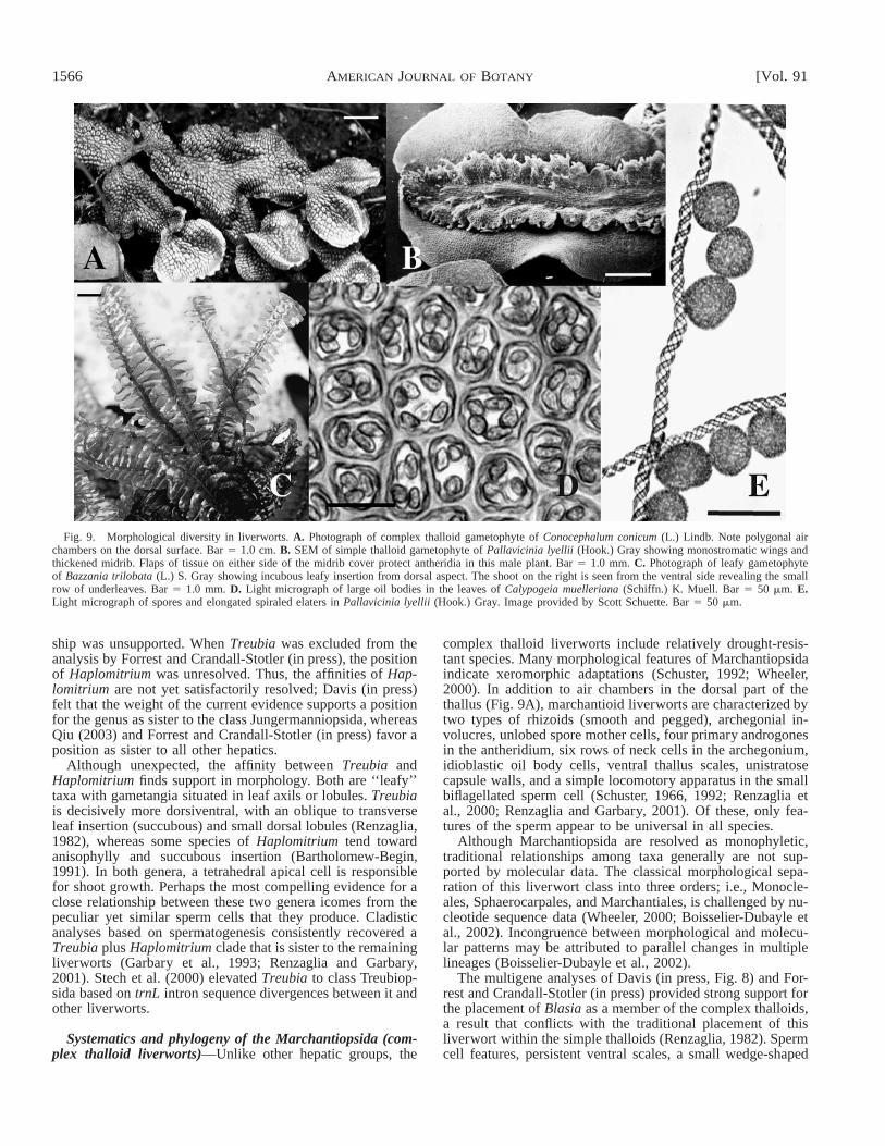
leafy forms are viewed as ancestral (see literature review inCrandall-Stotler and Stotler, 2000 and Davis, in press [Figs. 7,8]). Morphological studies supported the concept that simplethalloid liverworts are more closely related to leafy types thanto complex thalloids. Classification schemes reflect this inter-pretation with hepatics typically divided into two groups: mar-chantioid or complex thalloid liverworts (Marchantiopsida,Marchantiidae) and jungermannioid liverworts, including theleafy (Jungermanniopsida, Jungermanniidae) and simple thal-loid taxa (Jungermanniopsida, Metzgeriidae). Complex thal-loid types usually have air chambers with dorsal pores anddifferentiated internal tissues (Fig. 9A). Less commonly, thethallus resembles the simple thalloid type in the lack of inter-nal or epidermal differentiation (e.g., Sphaerocarpos, Mono-clea, and Dumortiera). Gametophytes of leafy liverwortsrange from radially symmetrical with three rows of morpho-logically similar leaves (isophyllous) to dorsiventral with tworows of lateral leaves and an additional row of reduced (toabsent) ventral underleaves or amphigastria (anisophyllous;Fig. 9C). Simple thalloid (metzgerialean) organizations showless variability, from fleshy undifferentiated thalli to those withprominent midribs and monostromatic wings (Fig. 9B). Leaf-like lobes or lobules in some taxa blur the distinction betweenleafy and simple thalloid forms. Internal differentiation of wa-ter-conducting tissue is restricted to Haplomitrium and certainsimple thalloid taxa, whereas conducting parenchyma is wide-spread among both complex and simple thalloid forms, but notleafy taxa (Hebant, 1977; Kobiyama and Crandall-Stotler,1999; Ligrone et al., 2000).
Liverworts are distinguished from hornworts and mosses bythe possession of oil bodies, unique organelles in which ter-penoids accumulate (Fig. 9D). All other embryophytes, in-cluding mosses and hornworts, produce cytoplasmic oil drop-lets (usually triglycerides), but they are not sequestered in spe-cialized organelles. Although the function of the oil body iscontroversial, these single-membrane-bound organelles are re-stricted to hepatics and occur in approximately 90% of taxa.Derived from endoplasmic reticulum in meristematic cells(Duckett and Ligrone, 1995), oil bodies provide valuable tax-onomic information because their size, shape, number, and col-or are taxon specific (Crandall-Stotler and Stotler, 2000).
Unlike hornworts, but comparable to mosses, is the produc-tion of a variety of organized external appendages, most ofwhich function in protecting fragile tissues. For example, var-
1564 [Vol. 91AMERICAN JOURNAL OF BOTANY

October 2004] 1565SHAW AND RENZAGLIA—PHYLOGENY OF BRYOPHYTES
←
Fig. 8. Phylogenetic relationships among liverworts, especially the Jungermanniidae (leafies). Homogeneous Bayesian 95% majority rule tree from a four-gene data matrix (Davis, in press). Bold branches indicate significant support for the clade in all Bayesian analyses (homogeneous and heterogeneous posteriorprobabilites $95). Parsimony bootstrap values $50 are shown on the tree.
ious mucilage papillae, hairs, scales, bracts, cups, or flask-shaped structures protect the meristem, gemmae, and othervegetative organs. Especially vulnerable are the superficial sexorgans that often occur in clusters protected by flaps of tissue,leaf lobes, young leaves, or modified branches (Fig. 9B).
The uniformity and uniqueness of liverwort sporophytesprovide compelling evidence for monophylly of hepatics. Un-like mosses and hornworts, sporophytes of liverworts reachmaturity within the confines of protective gametophytic tissuethat develops from the shoot/thallus (5 perigynium or coelo-caule) and/or archegonium (5 calyptra). Additional gameto-phytic structures such as perianths, pseudoperianths, bracts,scales, and involucral flaps may further surround the sporo-phyte and associated protective tissue. In such a milieu, pho-tosynthesis is limited, and the sporophyte derives nourishmentfrom the gametophyte through a placenta. The seta is pale tohyaline, and the capsule is devoid of stomata. The majority ofliverwort sporophytes are differentiated into foot, seta, andcapsule; in the occasional marchantioid taxon (e.g., Riccia,Corsinia), the seta and/or foot is vestigial or absent. At com-pletion of meiosis and spore development, cells of the setatypically undergo rapid elongation through water imbibitionand thus elevate the capsule away from the substrate. Sterile,elongated elaters have hygroscopic, spiraled, inner-wall thick-enings, that are strategically interspersed among spores to fa-cilitate their separation and dispersal (Fig. 9E). Capsule de-hiscence normally entails a patterned separation into four lon-gitudinal valves, but variations range from two valves throughirregular fragments or plates to cleistocarpous capsules.
The backbone of liverwort phylogenetic relationships—Crandall-Stotler and Stotler (2000) used morphological char-acters in a cladistic analysis of liverworts. Their analyses in-cluded 34 taxa and 61 characters, and they resolved two mainlineages: complex thalloids (Marchantiopsida) and simple thal-loids plus leafies (Jungermanniopsida: Metzgeriidae, Junger-manniidae, respectively). However, their sampling was not ex-tensive enough to address phylogenetic issues within any ofthe major clades. There are a few taxa for which placementrelative to the three large groups is ambiguous on the basis ofmorphological, ultrastructural, and chemical features. Theseinclude Treubia and Apotreubia (Treubiales), Monoclea (Mon-ocleales), Sphaerocarpos, Geothallus and Riella (Sphaerocar-pales), Blasia and Cavicularia (Blasiales), and Haplomitrium(Haplomitriales [Calobryales]). Early molecular analyses ofthe liverworts were limited to single genomic regions withlimited taxon sampling (e.g., Lewis et al., 1997; Bopp andCapesius, 1998; Beckert et al., 1999; Stech and Frey, 2001)but recent multigene analyses with increased sampling havebegun to clarify phylogenetic relations among (and within) themajor groups of liverworts. Phylogenetic relationships withinMarchantiopsida (complex thalloids) from DNA sequence datawere analyzed by Bischler (1998), Wheeler (2000), and Bois-selier-Dubayle et al. (2002). Forrest and Crandall-Stotler (inpress) focused on Metzgeriidae (simple thalloids), whereas He-Nygren et al. (in press) sampled a wide diversity of liverwort
taxa. Davis (in press) provided the most extensive analysis ofrelationships among leafy liverwort genera available to date.
Davis (in press) reconstructed ‘‘backbone’’ relationshipsamong liverworts based on a combined data set including twonuclear, three mitochondrial, and eight loci sequenced from 20liverworts and three outgroup mosses (Fig. 7). The data wereanalyzed using maximum parsimony, maximum likelihood,and Bayesian inference, and most of the results were robustto these alternative methods. The liverworts are resolved asmonophyletic, as are class Marchantiopsida (complex thal-loids) and Jungermanniidae (leafies). Metzgeriidae are re-solved as a grade paraphyletic to Jungermanniidae, in agree-ment with earlier studies. Although Forrest and Crandall-Sto-tler (in press) sampled different species, results of their anal-ysis of five plastid loci are congruent with those of Davis (inpress).
Although Haplomitrium has generally been regarded as anearly-diverging lineage within the liverworts (Smith, 1955;Schuster, 1984; Renzaglia et al., 1994), the precise placementof this genus remains problematic. The gametophyte of Hap-lomitrium is erect and radially symmetrical and therefore rem-iniscent of both jungermannialean liverworts and mosses. Pri-or to the discovery of antheridia and sporophytes in Takakia(Smith and Davison, 1993; Renzaglia et al., 1997), Haplomi-trium was considered closely related to Takakia because ofgametophytic similarities (Schuster, 1972, 1984). More recentmolecular and morphological data have come together to so-lidify the placement of Takakia among mosses (see later). Di-vergent opinions have been expressed with regard to the re-lationship of Haplomitrium to other hepatics. A conclusionfrom Bartholomew-Began’s (1990, 1991) extensive morpho-genetic reevaluation of Haplomitrium was that the genus is amember of the simple thalloid lineage. In their analysis of landplant relationships based on rbcL sequences, Lewis et al.(1997) noted that the precise position of the genus dependedon the data set analyzed (1st and 2nd vs. 3rd positions, all po-sitions, ‘‘ts/tv’’ weighting); Haplomitrium fell out sister to allother embryophytes, sister to all other liverworts, or nestedwithin the liverworts and sister to the leafy taxa. Nuclear 18SrDNA sequences resolved Haplomitrium (without bootstrapsupport) as sister to the class Jungermanniopsida (i.e., leafiesplus simple thalloids; Hedderson et al., 1996).
Recent multigene analyses have focused on two hypotheses:Haplomitrium is either sister to Jungermanniopsida or sister toall other liverworts. In contrast to almost all other nodes onher tree, Davis (in press) reported that the placement of Hap-lomitrium varied among analyses. Under parsimony, likeli-hood, and Bayesian methods, Haplomitrium is resolved withstrong support as sister to Jungermanniopsida (simple thalloidsplus leafies), and this inclusive clade is in turn sister to Mar-chantiopsida (complex thalloids; Fig. 7). However, the mostcomplex heterogeneous Bayesian substitution model, with 21partitions, yielded Haplomitirum as the sister group to all otherliverworts. Forrest and Crandall-Stotler (in press) and Qiu(2003) reported that Haplomitrium plus Treubia form a cladesister to all other hepatics. However, the sister-group relation-
1566 [Vol. 91AMERICAN JOURNAL OF BOTANY
Fig. 9. Morphological diversity in liverworts. A. Photograph of complex thalloid gametophyte of Conocephalum conicum (L.) Lindb. Note polygonal airchambers on the dorsal surface. Bar 5 1.0 cm. B. SEM of simple thalloid gametophyte of Pallavicinia lyellii (Hook.) Gray showing monostromatic wings andthickened midrib. Flaps of tissue on either side of the midrib cover protect antheridia in this male plant. Bar 5 1.0 mm. C. Photograph of leafy gametophyteof Bazzania trilobata (L.) S. Gray showing incubous leafy insertion from dorsal aspect. The shoot on the right is seen from the ventral side revealing the smallrow of underleaves. Bar 5 1.0 mm. D. Light micrograph of large oil bodies in the leaves of Calypogeia muelleriana (Schiffn.) K. Muell. Bar 5 50 mm. E.Light micrograph of spores and elongated spiraled elaters in Pallavicinia lyellii (Hook.) Gray. Image provided by Scott Schuette. Bar 5 50 mm.
ship was unsupported. When Treubia was excluded from theanalysis by Forrest and Crandall-Stotler (in press), the positionof Haplomitrium was unresolved. Thus, the affinities of Hap-lomitrium are not yet satisfactorily resolved; Davis (in press)felt that the weight of the current evidence supports a positionfor the genus as sister to the class Jungermanniopsida, whereasQiu (2003) and Forrest and Crandall-Stotler (in press) favor aposition as sister to all other hepatics.
Although unexpected, the affinity between Treubia andHaplomitrium finds support in morphology. Both are ‘‘leafy’’taxa with gametangia situated in leaf axils or lobules. Treubiais decisively more dorsiventral, with an oblique to transverseleaf insertion (succubous) and small dorsal lobules (Renzaglia,1982), whereas some species of Haplomitrium tend towardanisophylly and succubous insertion (Bartholomew-Begin,1991). In both genera, a tetrahedral apical cell is responsiblefor shoot growth. Perhaps the most compelling evidence for aclose relationship between these two genera icomes from thepeculiar yet similar sperm cells that they produce. Cladisticanalyses based on spermatogenesis consistently recovered aTreubia plus Haplomitrium clade that is sister to the remainingliverworts (Garbary et al., 1993; Renzaglia and Garbary,2001). Stech et al. (2000) elevated Treubia to class Treubiop-sida based on trnL intron sequence divergences between it andother liverworts.
Systematics and phylogeny of the Marchantiopsida (com-plex thalloid liverworts)—Unlike other hepatic groups, the
complex thalloid liverworts include relatively drought-resis-tant species. Many morphological features of Marchantiopsidaindicate xeromorphic adaptations (Schuster, 1992; Wheeler,2000). In addition to air chambers in the dorsal part of thethallus (Fig. 9A), marchantioid liverworts are characterized bytwo types of rhizoids (smooth and pegged), archegonial in-volucres, unlobed spore mother cells, four primary androgonesin the antheridium, six rows of neck cells in the archegonium,idioblastic oil body cells, ventral thallus scales, unistratosecapsule walls, and a simple locomotory apparatus in the smallbiflagellated sperm cell (Schuster, 1966, 1992; Renzaglia etal., 2000; Renzaglia and Garbary, 2001). Of these, only fea-tures of the sperm appear to be universal in all species.
Although Marchantiopsida are resolved as monophyletic,traditional relationships among taxa generally are not sup-ported by molecular data. The classical morphological sepa-ration of this liverwort class into three orders; i.e., Monocle-ales, Sphaerocarpales, and Marchantiales, is challenged by nu-cleotide sequence data (Wheeler, 2000; Boisselier-Dubayle etal., 2002). Incongruence between morphological and molecu-lar patterns may be attributed to parallel changes in multiplelineages (Boisselier-Dubayle et al., 2002).
The multigene analyses of Davis (in press, Fig. 8) and For-rest and Crandall-Stotler (in press) provided strong support forthe placement of Blasia as a member of the complex thalloids,a result that conflicts with the traditional placement of thisliverwort within the simple thalloids (Renzaglia, 1982). Spermcell features, persistent ventral scales, a small wedge-shaped

October 2004] 1567SHAW AND RENZAGLIA—PHYLOGENY OF BRYOPHYTES
Fig. 10. Diagrammatic representation of apical cell shapes in liverwortsas oriented in plants growing horizontally. The shoot tip is directed towardthe left. A. Tetrahedral cell with three cutting faces. B. Wedge-shaped orcuneate cell with two lateral, one dorsal and one ventral cutting face. C.Lenticular or lens-shaped cell with two lateral cutting faces. D. Hemidiscoidcell with two lateral and one posterior cutting face. This cell is rare in liv-erworts and was developmentally and evolutionarily derived from a wedge-shaped cell.
Fig. 11. Formative divisions in the lateral derivative from apical cells ofliverworts. Apices are vertically oriented so that the shoot tip is facing up.A. Lateral thallus, wing and ‘‘leaf’’ development from a central wedge-shapedcell (single initial) (L) in a three-celled derivative. This type of developmentcharacterizes all simple thalloid and complex thalloid taxa and occurs in de-rivatives from all four types of apical cells. B. ‘‘True’’ leaf development inleafy liverworts from two initials. In this five-celled derivative, two leaf ini-tials (L) are determined to develop into the bifid and conplicate–bilobedleaves.
apical cell, and a Monoclea-like female involucre provide mor-phological evidence for the inclusion of Blasia in the complexthalloid lineage (Renzaglia and Duckett, 1987; Pass and Ren-zaglia, 1995; Renzaglia and Garbary, 2001). Previous molec-ular analyses based on one or two gene sequences do not agreein the placement of Blasia. Stech and Frey (2001) resolvedBlasia as sister to Jungermanniopsida (simple thalloids plusleafies) and described the new class, Blasiopsida. Their studywas based solely on trnL intron sequences (ca. 500 bp), andthe relationship was without bootstrap support. Wheeler(2000) found that Blasia grouped with the simple thalloids(Metzgeriidae) based on 26S nrDNA (also without bootstrapsupport), and He-Nygren et al. (in press) resolved Blasia assister to the remaining liverworts.
After Blasia, Sphaerocarpos is the next divergent taxon(Fig. 8). A position for Sphaerocarpales (Sphaerocarpos, Riel-la, Geothallus) among complex thalloids is generally support-ed by morphology (Smith, 1955; Bishler, 1998; Crandall-Sto-tler and Stotler, 2000; Boisselier-Dubayle et al., 2002). How-ever, with additional taxon sampling, the sister relationshipbetween Sphaerocarpales and the remaining Marchantiopsidais called into question. Based on LSU rDNA sequences,Wheeler (2000) and Boisselier-Dubayle et al. (2002) reportedthat Sphaerocarpales were placed within Marchantiaceae. Sim-ilarly, Sphaerocarpos nested between Neohodgsonia and Mar-chantia in the five-gene analysis of Forrest and Crandall-Sto-tler (in press). The implication from these results is that therelatively simple morphology of both generations in Spharo-carpales may not be plesiomorphic but rather the product ofextreme simplification in ephemeral or aquatic habitats.
Air chambers are found in the crown group taxa (Marchan-tia, Preissia, Targionia, Riccia) (Wheeler, 2000). One lineage,Monoclea plus Dumortiera, has secondarily reverted to a mor-phologically simple thallus devoid of chambers, perhaps ad-aptations to the semi-aquatic habit of these plants (Wheeler,2000). The production of archegoniophores (carpocephala)that elevate sporophytes above the gametophyte also evolvedwithin the crown Marchantiopsida group. Independent lossesof these structures occurred in riccioid taxa (Riccia, Riccio-carpos, Oxymitra) and Monoclea (Wheeler, 2000). Reductionin sporophyte complexity is likewise a derived feature of ric-cioid liverworts (Renzaglia et al., 2000; Boisselier-Dubayle etal., 2002). With a jungermannioid-like sporophyte elevated ona fragile and highly elongated seta, Monoclea seems inappro-priately placed within this crown group. Additional charactersof the genus, including a free nuclear embryo and monoplas-tidic meiosis in some species (Schofield, 1985; Renzaglia etal., 1994), support a more traditional placement of Monoclea
in Marchantiopsida. However, congruence among the multi-gene analyses provided support for Monoclea close to Du-mortiera.
Systematics and phylogeny of Metzgeriidae (simple thal-loid liverworts)—Clearly not a monophyletic group, Mezger-iidae traditionally include some 30 highly diverse genera of‘‘leafy’’ and thalloid forms. Although four apical cell types arefound in the group (Fig. 10), a unifying feature of apicalgrowth in these plants is development of wings and leavesfrom a central wedge cell (single initial) that forms in thenewly produced apical derivative (Fig. 11A) (Renzaglia,1982). Simple thalloid genera are distinguished from leafy liv-erworts (Jungermanniidae) in that they are anacrogynous: ar-chegonia are produced along the mid-thallus of either themain, lateral, or ventral shoots. Consequently, the apical cellis not transformed into permanent tissue after archegonial de-velopment, and sporophytes do not terminate the shoot as inacrogynous Jungermanniidae. Additional features that unifythe simple thalloid taxa, but are also found in leafy liverworts,are the development of antheridia from two primary andro-gones, oil bodies in all cells, lobed sporocytes, smooth rhi-zoids, and five rows of neck cells per archegonium.
Two assemblages of simple thalloid taxa are paraphyletic(simple thalloid I and II in Fig. 8) within Jungermanniopsida.The first group (simple thalloid I) is the most diverse andincludes Phyllothallia, generally placed in Treubiales, mostmembers of Fossombroniales, and suborder Pallaviciniineae ofMetzgeriales (classification according to Crandall-Stotler andStotler, 2000). Placement of Phyllothallia, Pellia, Calycularia,and Noteroclada is not resolved in the five-gene analysis ofForrest and Crandall-Stotler (in press); all represent geneticallyand morphologically divergent taxa. Phyllothallia and Noter-oclada are distinctly ‘‘leafy’’ in habit, but development is froma wedge-shaped cell (Fig. 10B) in the former and a tetrahedralapical cell (Fig. 10A) in the latter (Renzaglia, 1982). Pelliaand Calycularia are fleshy thalloid types, both with wedge(Fig. 10B) and hemidiscoid apical cells (Fig. 10D). Althoughsupport for an assemblage that includes Fossombronia, Aus-trofossombronia, Petalophyllum, and Allisonia is weak, thisgroup includes most of the genera traditionally placed in Fos-sombroniales (Crandall-Stotler and Stotler, 2000). All havespheroidal capsules, which are typically irregular or nonval-vate in dehiscence. Most exhibit a ‘‘leafy’’ growth form witheither lens-shaped (Fig. 10C) or tetrahedral (Fig. 10A) apicalcells. Suborder Pallaviciniinae of Metzgeriales are recovered

1568 [Vol. 91AMERICAN JOURNAL OF BOTANY
as monophyletic and include Hymenophyton, Moerckia, Hat-torianthus, Podomitrium, Pallavicinia, Jensenia, Xenothallus,and Symphyogyna (Crandall-Stotler and Stotler, 2000; Forrestand Crandall-Stotler, in press). This morphologically uniformgroup contains upright or procumbent taxa, most with prom-inent midribs and monostromatic wings (e.g., Pallavicinia,Fig. 9B). Lens-shaped apical cells (Fig. 10C) are responsiblefor vegetative growth. An autapomorphy of this group is theproduction of specialized strands of dead, water-conductingcells that predominate in most taxa (Ligrone et al., 2000). Ex-tensive variability is seen in position and type of protectivestructure associated with gametangia and sporophytes (Ren-zaglia, 1982; Fig. 9B).
The apparent affinity between suborder Metzgeriinae (sim-ple thalloid II) and Jungermanniidae (‘‘true’’ leafy liverworts)in the multigene analyses of both Davis (Fig. 8) and Forrestand Crandall-Stotler (in press) was unexpected. Members ofMetzgeriinae epitomize the simple thalloid condition, withfleshy (Aneuraceae) and midrib-plus-wing (Metzgeriaceae) or-ganizations. All of these thalli develop from a lens-shaped api-cal cell (Fig. 10C), and no ‘‘leafy’’ forms exist (except perhapsPleurozia, discussed next). Endogenous branches in Metzger-iaceae are reminiscent of those in leafy liverworts (Renzaglia,1982).
One of the most surprising results from the Davis (in press)analyses was the placement of Pleurozia in Metzgeriinae (sim-ple thalloid II) rather than among the ‘‘true’’ leafy liverworts(Figs. 7, 8). Pleurozia is composed of about 11 species dis-tributed primarily in the tropics. Leaves are complicate-bi-lobed, and for that reason, Pleurozia has traditionally beenincluded in or near Porellales within the leafy liverworts(Schuster, 1984; Crandall-Stotler and Stotler, 2000). However,leaf morphology in Pleurozia is unique in that the leaf lobuleis dorsal in orientation, not ventral (Thiers, 1993), and theplants grow from a lenticular apical cell (Crandall-Stotler,1976) rather than a tetrahedral cell as in all ‘‘true’’ leafy liv-erworts. The placement of Pleurozia in the metzgerioid liv-erworts indicates that the ‘‘leafy’’ gametophytes of Pleurozia,with their complicate–bilobed leaves, may have evolved con-vergently in a group otherwise characterized by thalloid ga-metophytes. In contrast to the single leaf initial in simple thal-loids that have ‘‘leafy’’ gametophytes (Fig. 11A), leaves ofPleurozia develop from two initial cells, as is typical of leafyliverworts (Fig. 11B; Crandall-Stotler, 1976). The phyloge-netic position of Pleurozia should be further investigated, al-though its placement within subclass Metzgeriidae is stronglysupported by both the 12- and four-locus analyses of Davis (inpress).
Systematics and phylogeny of the Jungermanniidae (leafyliverworts)—The leafy liverworts, with some 4000–6000 spe-cies, are by far the largest of the liverwort groups. They occurin most terrestrial and aquatic habitats but are especially di-verse in high-moisture environments. Many species are epi-phytic on bark, and in the tropics, epiphyllous liverworts maycover the leaves of angiosperm trees and shrubs in shaded,high-humidity forests. More than 75% of the liverworts oftropical lowland forests and almost all the epiphylls belong toLejeuneaceae (Gradstein, 1994, 1997). Lejeuneaceae compriseapproximately 93 of the 307 genera (30%) of leafy liverworts,and well over 1000 species (Gradstein, 1979, 1994, 1997;Crandall-Stotler and Stotler, 2000). In lowland equatorial for-ests, as many as 20 species of Lejeuneaceae may co-occur on
a single leaf (Zartman, 2003). These organisms are importantcomponents of tropical forest diversity, and the diversity ofepiphylls (almost exclusively Lejeuneaceae) is a sensitive in-dicator of habitat change associated with forest fragmentation(Zartman, 2003).
Jungermanniidae are distinguished from Metzgeriidae inhaving tetrahedral apical cells, gametophytic shoots with (usu-ally) well-differentiated stems and leaves, leaves formed fromtwo initial cells, acrogynous perichaetia (terminating the mainstem or branch), bracts and perianths (modified, fused leaves)associated with the perichaetium, and capsules that regularlydehisce into four valves (Crandall-Stotler and Stotler, 2000).The perianths of leafy liverworts are diverse and provide im-portant taxonomic characters in many genera and families.
Leaves of leafy liverworts may be entire or more often havetwo large lobes or teeth. They are most commonly differen-tiated as two rows of lateral leaves and a single row of ventralunderleaves (amphigastria; Fig. 9C). Underleaves are frequent-ly small or lacking. Insertion of the lateral leaves may be trans-verse, or, more commonly, they are oblique and the plants aremore or less flattened because the leaves overlap. In plantswith incubous leaf orientation, the forward leaf margin over-laps the trailing margin of the next younger leaf, resemblingthe arrangement of roof shingles (Fig. 9C). In sucubous ori-entation, forward margins of older leaves are covered by over-lapping trailing margins of the younger leaves. In species withcomplicate–bilobed leaves, lateral leaves are each folded toform ventral and dorsal lobes. The dorsal lobe is larger in mosttaxa, and the ventral lobe may be highly modified into theform of a pouch or helmet-shaped lobule that holds water.
Schuster (1966, 1984) assumed that the most primitive liv-erworts would be the most mosslike, with leafy, radially sym-metric gametophytes and therefore placed leafy taxa at thebase of his subjectively derived ‘‘phylogenetic trees’’ (e.g.,Schuster, 1966, pp. 406, 696). He considered leafy taxa withradial symmetry and three rows of transversely (or nearly so)inserted leaves (e.g., Herbertineae) to be early diverginggroups, and from these he showed the branching of lineagesor clusters of lineages with increased anisophylly and moreobliquely inserted leaves (Schuster, 1966, 1972, 1984). Onegroup includes Schistochilaceae, Cephaloziaceae, Lepidozia-ceae, and Pleuroziaceae, whereas the other progresses throughPtidiaceae to Jungermanniaceae, Frullaniaceae and Lejeune-aceae. (His diagram shows extant families ancestral to otherfamilies.) The classification of Crandall-Stotler and Stotler(2000) has a sequence of families in five orders, Lepicoleales(including Ptilidiaceae, Lepicoleaceae, Schistochilaceae, andLepidolaenaceae), Jungermanniales (including Herbertaceae,Balantiopsidaceae, Geocalycaceae, Lepidoziaceae, Cephalo-ziaceae, Jungermanniaceae, and Gymnomitriaceae), Porellales(including Porellaceae, Jubulaceae, and Lejeuneaceae), and themonotypic Radulales and Pleuroziales. Their classification im-plies similar concepts of evolution in leafy liverworts to thoseof Schuster.
In a liverwort backbone tree based on 12 loci, Davis (inpress) resolved two major clades within subclass Jungerman-niidae (Fig. 7). One clade contains most of the taxa with com-plicate–bilobed, incubous (or transverse) leaves (mainly Po-rellaceae, Jubulaceae, Radulaceae, and Lejeuneaceae), whereasthe other contains the remaining families of leafy liverworts.In a more taxon-extensive analysis that included 81 liverworts,two mosses, and a hornwort, based on sequences from 26SnrDNA, two plastid loci ( psbA and rps4), and mitochondrial
October 2004] 1569SHAW AND RENZAGLIA—PHYLOGENY OF BRYOPHYTES
nad5, the same two leafy liverwort clades were resolved (Fig.8). The noncomplicate-bilobed group consists of three sub-clades for which sister group relationships are ambiguous (A,B, and C in Fig. 8). Species in clade A have incubous ortransverse leaf insertion, well-developed underleaves, and mul-tilobed lateral leaves (Davis, in press). Herbertus, assumed bySchuster (1984) to be primitive among leafy liverworts, is re-solved in a derived position within clade A (Fig. 8). Moreover,other isophyllous taxa (e.g., Anthelia, Triandrophyllum) arealso resolved in relatively derived phylogenetic positions. Taxain clade B have sucubous or transverse leaves and generallylack underleaves; however, lateral leaf shape is variable. Leafshape, insertion, and underleaf development are highly vari-able in clade C, but many of the species are characterized byhaving perichaetia formed in fleshy perigynia or marsupia,which do not occur elsewhere in the leafy liverworts.
Among suborders of leafy liverworts recognized by Schus-ter (1984), only Radulineae and Balantiopsidineae are mono-phyletic based on the four-locus analysis of Davis (in press).The classification of Crandall-Stotler and Stotler (2000) is alsoin conflict with many of the phylogenetic inferences from Da-vis’s analysis (Fig. 8). Notably, Lepicoleales are extensivelypolyphyletic, and Radulales are nested within Porellales. Her-bertaceae, Lepidoziaceae, Balantiopsidaceae, Cephaloziaceae,Porellaceae, and Radulaceae are supported as monophyletic.Lejeuniaceae are monophyletic only if Bryopteris is includedwithin them (Bryopteridaceae, fide Crandall-Stotler and Sto-tler, 2000). Jungermanniaceae, Gymnomitriaceae, Geocalyca-ceae, Cephaloziaceae, Lepidolaenaceae are paraphyletic (Da-vis, in press).
Inferences about morphological evolution in liverwortsfrom molecular analyses—Leaves or leaflike lobes haveevolved in every major group of hepatics. Haplomitrium andTreubia have leafy appendages. Blasia and Sphaerocarpos,taxa within the marchantioid line, have leafy habits. Phylloth-allia, Noteroclada, and Pleurozia, with leafy gametophytes,are scattered among simple thalloid taxa. The leaves of theseplants are typically succubous to transversely inserted and maybe formed from any one of three apical cell types: wedge-shaped, lenticular, or tetrahedral. In Phyllothallia and Noter-oclada (Fig. 10A), each leaf develops from a single initial,whereas in Pleurozia there are two initials. A single initial isalso responsible for development of wings and lateral thallusin all simple thalloids, complex thalloids, hornworts, and manypteridophyte gametophytes and is thus best viewed as plesio-morphic.
An autapomorphy of the Jungermanniidae is the productionof bifid leaves from two leaf initials in a derivative from atetrahedral apical cell (Fig. 10B). Once ‘‘locked’’ into this pat-tern of cell divisions, a number of variations on the ‘‘typical’’bifid leaf of hepatics evolved, including complicate–bilobedleaves. A narrower ventral cutting face in the apical cell isassociated with a smaller size or absence of underleaves thatoriginate from it. Incubous leaf insertion results from a ventral(downward) tilt of the apical cell (Crandall-Stotler and Stotler,2000), a feature that is often correlated with taxa that grow onvertical substrates such as tree bark (e.g., Leujeuniaceae). Con-versely, succubous leaf arrangements are correlated with a dor-sal (upward) tilt of the growing tip.
Few conclusions can be drawn at present about the evolu-tion of apical cell shapes and growth forms in liverworts.Transformation from one apical cell type to another readily
occurs during development, and this plasticity may have pro-vided fuel for evolutionary change (Renzaglia et al., 2000).Depending on the position of Haplomitrium in the trees, eithera tetrahedral or wedge-shaped cell is plesiomorphic. Similarly,either an upright ‘‘leafy’’ habit or a flattened thallus is ances-tral in hepatics; both hypotheses have garnered support(Schuster, 1992; Mishler and Churchill, 1984). Outgroup com-parisons provide no further resolution of this issue as hornwortand pteridophyte gametophytes are thalloid with wedge-shaped apical cells, whereas mosses are leafy with tetrahedralcells.
Within liverworts, significant evolutionary changes can beinferred at the cellular level based on the consensus topologyof recent molecular analyses. Monoplastidic meiosis occurs inall mosses and hornworts. However, it is restricted in liver-worts to Haplomitrium, Blasia, and Monoclea (Renzaglia etal., 1994) and is best interpreted as plesiomorphic. Monoplas-tidic meiosis involves precise control of plastid division andmigration prior to chromosomal separation (Brown and Lem-mon, 1990). Polyplastidic meiosis predominates in liverwortsand is a derived state. Similarly, lobed spore mother cells thatoccur in liverworts such as Haplomitrium, Treubia, and Blasiaare shared with other bryophytes and represent a plesio-morphic condition (Brown and Lemmon, 1988). Sporocytelobing was lost within Marchantiopsida, whereas spores unitedin permanent tetrads are viewed as derived within Sphaero-carpales. Among bryophytes, pre-meiotic patterning of sporewall ornamentation occurs in Apotreubia and Haplomitriumand presumably has been lost in more derived liverwort line-ages (Brown et al., 1986). Further ultrastructural studies acrossa range of hepatic groups are likely to provide new insightsinto the nature and direction of changes in cellular processesduring early land plant evolution.
BRYOPHYTA
Moss classification and relationships—Division Bryophy-ta, or mosses, include about 10 000 species (Crosby et al.,2000). Systematic knowledge about the mosses has grownsteadily since Hedwig (1801), the starting point for moss no-menclature (excluding Sphagnum), recognized 32 genera.Most classifications of the 19th century depended on gameto-phyte characters for defining the major groups of mosses (e.g.,Bruch et al., 1851–1855; Kindberg, 1897). Mosses (excludingSphagnum and Andreaea) were divided into acrocarpous andpleurocarpous taxa (Mitten, 1859). Acrocarpous mosses havearchegonia terminating the main stems, which tend to besparsely if at all branched. Pleurocarpous mosses, in contrast,have archegonia borne laterally along relatively highlybranched, generally procumbent or pendent, extensively inter-woven stems. The two forms of gametophyte architecture areoften obvious, but some taxa are confusingly intermediate(e.g., Rhizogoniaeae, Orthotrichaceae, Hedwigiaceae) becausethey have moderately branched stems with archegonia termi-nating short to long lateral branches. La Farge-England (1996)clarified the definitions of these forms of gametophytic archi-tecture and discussed possible phylogenetic relations betweentaxa characterized by acrocarpous, pleurocarpous, and clado-carpous gametophyte architecture (the latter including theseemingly intermediate forms).
Philibert (1884–1902) published a series of seminal papersdescribing variation in the structure of the moss peristome(sporophytic) and distinguished several basic types character-
1570 [Vol. 91AMERICAN JOURNAL OF BOTANY
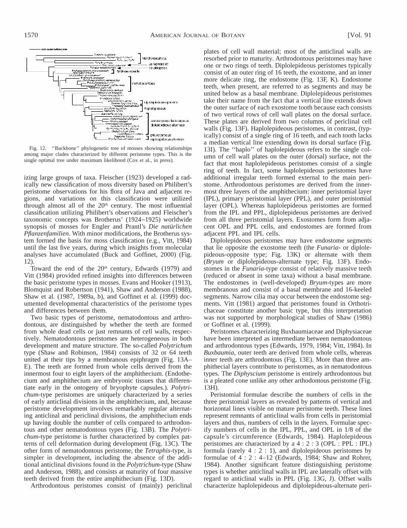
Fig. 12. ‘‘Backbone’’ phylogenetic tree of mosses showing relationshipsamong major clades characterized by different peristome types. This is thesingle optimal tree under maximum likelihood (Cox et al., in press).
izing large groups of taxa. Fleischer (1923) developed a rad-ically new classification of moss diversity based on Philibert’speristome observations for his flora of Java and adjacent re-gions, and variations on this classification were utilizedthrough almost all of the 20th century. The most influentialclassification utilizing Philibert’s observations and Fleischer’staxonomic concepts was Brotherus’ (1924–1925) worldwidesynopsis of mosses for Engler and Prantl’s Die naturlichenPflanzenfamilien. With minor modifications, the Brotherus sys-tem formed the basis for moss classification (e.g., Vitt, 1984)until the last five years, during which insights from molecularanalyses have accumulated (Buck and Goffinet, 2000) (Fig.12).
Toward the end of the 20th century, Edwards (1979) andVitt (1984) provided refined insights into differences betweenthe basic peristome types in mosses. Evans and Hooker (1913),Blomquist and Robertson (1941), Shaw and Anderson (1988),Shaw et al. (1987, 1989a, b), and Goffinet et al. (1999) doc-umented developmental characteristics of the peristome typesand differences between them.
Two basic types of peristome, nematodontous and arthro-dontous, are distinguished by whether the teeth are formedfrom whole dead cells or just remnants of cell walls, respec-tively. Nematodontous peristomes are heterogeneous in bothdevelopment and mature structure. The so-called Polytrichumtype (Shaw and Robinson, 1984) consists of 32 or 64 teethunited at their tips by a membranous epiphragm (Fig. 13A–E). The teeth are formed from whole cells derived from theinnermost four to eight layers of the amphithecium. (Endothe-cium and amphithecium are embryonic tissues that differen-tiate early in the ontogeny of bryophyte capsules.). Polytri-chum-type peristomes are uniquely characterized by a seriesof early anticlinal divisions in the amphithecium, and, becauseperistome development involves remarkably regular alternat-ing anticlinal and periclinal divisions, the amphithecium endsup having double the number of cells compared to arthrodon-tous and other nematodontous types (Fig. 13B). The Polytri-chum-type peristome is further characterized by complex pat-terns of cell deformation during development (Fig. 13C). Theother form of nematodontous peristome, the Tetraphis-type, issimpler in development, including the absence of the addi-tional anticlinal divisions found in the Polytrichum-type (Shawand Anderson, 1988), and consists at maturity of four massiveteeth derived from the entire amphithecium (Fig. 13D).
Arthrodontous peristomes consist of (mainly) periclinal
plates of cell wall material; most of the anticlinal walls areresorbed prior to maturity. Arthrodontous peristomes may haveone or two rings of teeth. Diplolepideous peristomes typicallyconsist of an outer ring of 16 teeth, the exostome, and an innermore delicate ring, the endostome (Fig. 13F, K). Endostometeeth, when present, are referred to as segments and may beunited below as a basal membrane. Diplolepideous peristomestake their name from the fact that a vertical line extends downthe outer surface of each exostome tooth because each consistsof two vertical rows of cell wall plates on the dorsal surface.These plates are derived from two columns of periclinal cellwalls (Fig. 13F). Haplolepideous peristomes, in contrast, (typ-ically) consist of a single ring of 16 teeth, and each tooth lacksa median vertical line extending down its dorsal surface (Fig.13I). The ‘‘haplo’’ of haplolepideous refers to the single col-umn of cell wall plates on the outer (dorsal) surface, not thefact that most haplolepideous peristomes consist of a singlering of teeth. In fact, some haplolepideous peristomes haveadditional irregular teeth formed external to the main peri-stome. Arthrodontous peristomes are derived from the inner-most three layers of the amphithecium: inner peristomial layer(IPL), primary peristomial layer (PPL), and outer peristomiallayer (OPL). Whereas haplolepideous peristomes are formedfrom the IPL and PPL, diplolepideous peristomes are derivedfrom all three peristomial layers. Exostomes form from adja-cent OPL and PPL cells, and endostomes are formed fromadjacent PPL and IPL cells.
Diplolepideous peristomes may have endostome segmentsthat lie opposite the exostome teeth (the Funaria- or diplole-pideous-opposite type; Fig. 13K) or alternate with them(Bryum or diplolepideous-alternate type; Fig. 13F). Endo-stomes in the Funaria-type consist of relatively massive teeth(reduced or absent in some taxa) without a basal membrane.The endostomes in (well-developed) Bryum-types are moremembranous and consist of a basal membrane and 16-keeledsegments. Narrow cilia may occur between the endostome seg-ments. Vitt (1981) argued that peristomes found in Orthotri-chaceae constitute another basic type, but this interpretationwas not supported by morphological studies of Shaw (1986)or Goffinet et al. (1999).
Peristomes characterizing Buxbaumiaceae and Diphysiaceaehave been interpreted as intermediate between nematodontousand arthrodontous types (Edwards, 1979, 1984; Vitt, 1984). InBuxbaumia, outer teeth are derived from whole cells, whereasinner teeth are arthrodontous (Fig. 13E). More than three am-phithecial layers contribute to peristomes, as in nematodontoustypes. The Diphyscium peristome is entirely arthrodontous butis a pleated cone unlike any other arthodontous peristome (Fig.13H).
Peristomial formulae describe the numbers of cells in thethree peristomial layers as revealed by patterns of vertical andhorizontal lines visible on mature peristome teeth. These linesrepresent remnants of anticlinal walls from cells in peristomiallayers and thus, numbers of cells in the layers. Formulae spec-ify numbers of cells in the IPL, PPL, and OPL in 1/8 of thecapsule’s circumference (Edwards, 1984). Haplolepideousperistomes are characterized by a 4 : 2 : 3 (OPL : PPL : IPL)formula (rarely 4 : 2 : 1), and diplolepideous peristomes byformulae of 4 : 2 : 4–12 (Edwards, 1984; Shaw and Rohrer,1984). Another significant feature distinguishing peristometypes is whether anticlinal walls in IPL are laterally offset withregard to anticlinal walls in PPL (Fig. 13G, J). Offset wallscharacterize haplolepideous and diplolepideous-alternate peri-
October 2004] 1571SHAW AND RENZAGLIA—PHYLOGENY OF BRYOPHYTES
stomes but not the diplolepideous-opposite (Funaria-) type.Anticlinal walls in the Polytrichum-type nematodontous peri-stomes are not offset (Wenderoth, 1931), and there is little ifany offsetting of the walls in Tetraphis-type nematodontousperistomes (Shaw and Anderson, 1988). The peristome of Di-physcium has a developmental pattern that conforms in all de-tails to the haplolepideous peristomial type and has a 4 : 2 :3 formula at maturity despite the unique structure of the ma-ture peristome (Shaw et al., 1987).
Developmental studies have succeeded (even if based onfew taxa) in defining when and how the basic peristome typesdiffer from one another. These studies do not, however, claritfyphylogenetic relationships among the types. One of the centralgoals of higher-level phylogenetic analyses for mosses hasbeen to resolve these relationships. Much progress has beenmade, but a full resolution is still forthcoming.
The backbone of moss phylogenetic relationships—Vari-ous approaches to resolving relationships among mosses haveincluded taxon-extensive analyses using a single plastid gene(rps4: Goffinet et al., 2001; rbcL: Tsubota et al., 2002), andanalyses of multigene, multigenomic data sets with more syn-optic taxon sampling (Cox et al., 2004). Both approaches havetheir merits, but it is clear that resolution of well-supportedrelationships among the major groups of mosses will not beaccomplished using one or a few genes, even if such analysessucceed in placing more genera into monophyletic groups. Thebest-supported ‘‘backbone’’ for mosses was derived from ananalysis of eight genes representing the mitochondrial, plastid,and nuclear genomes of 30 exemplars that represent majorlineages based on previous studies (Cox et al., 2004). Thefollowing synopsis is based on that analysis, with discussionof supportive and/or contradictory evidence when appropriate.
With sequences from four species of liverworts as the out-group, the Bayesian reconstruction presented by Cox et al.(2004) indicated that Sphagnum and Takakia form a clade sis-ter to all remaining mosses (Fig. 12). A close relationship be-tween Sphagnum and Takakia was also resolved by Heddersonet al. (1998), Newton et al. (2000), and Yatsentyuk (2001)from nucleotide sequences, although Newton et al. (2000)were not able to identify any morphological synapomorphiesuniting the two genera. Gametophytes of Sphagnum and Tak-akia could not be more divergent: those of Takakia are tiny,simple in structure, and reminiscent of liverworts, whereasthose of Sphagnum are large and characterized by a numberof autapomorphies. The sporophyte of Takakia is mosslike indevelopment (Renzaglia et al., 1997) with a well-developedseta, a cylindrical capsule, and spiraled dehiscence (Fig. 14),whereas capsules of Sphagnum are ovoid in shape, open byan apical operculum, and are elevated on gametophytic pseu-dopodia. In the analyses of Cox et al. (2004), support for theclade containing Sphagnum and Takakia was lower when sub-stitution patterns were modeled separately for each of the eightgenomic regions than when a single model was applied, thusraising the possibility that resolution of the Sphagnum-Takakiaclade may be an artifact. Phylogenetic relationships amongspecies within Sphagnopsida (Sphagnum and Ambuchanania)have been described by Shaw (2000) and Shaw et al. (2003a).
After Sphagnum and Takakia, the next diverging clade ofmosses contains the two genera, Andreaea and Andreaeo-bryum. These two mosses, although similar in gross morphol-ogy, differ in several seemingly fundamental morphologicalfeatures including development of a seta (absent in Andreaea
but present in Andreaeobryum), mode of capsule dehiscence,and timing of perichaetium differentiation relative to sporo-phyte development. Newton et al. (2000), Goffinet et al.(2001), and Cox et al. (2004) resolved Andreaea and An-dreaeobryum in a single clade, but without impressive supportfrom the bootstrap, Bremer support indices, and Bayesian pos-terior probabilities. It remains possible, though not likely, thatAndreaea and Andreaeobryum form a paraphyletic grade lead-ing to the ‘‘true’’ (peristomate) mosses. Murray (1988) notedmorphological similarities that might link Andreaeobryumwith Takakia.
One of the most exciting insights from phylogenetic anal-yses of mosses is that the monospecific genus, Oedopodium,appears to be sister to all remaining peristomate taxa (Fig. 15).A critical position for Oedopodium corroborates the results ofNewton et al. (2000) from combined analyses of morphologyand four plastid DNA regions and of Goffinet et al. (2001)based on a taxon-extensive analysis of plastid rps4 sequences.Goffinet et al. (2001) resolved Oedopodium as sister to a cladecontaining Tetraphidaceae and Polytrichaceae at or near thebase of peristomate mosses, but relationships were not fullyresolved and without bootstrap support. Newton et al. (2000)and Magombo (2003), based on four plastid DNA regions,resolved Oedopodium, both with moderate to strong bootstrapsupport, as sister to all peristomate mosses.
Oedopodium griffitheanum (Dicks.) Schwaegr. is a small ac-rocarpous moss with soft obovate to spathulate leaves, thin-walled hexagonal leaf cells, erect capsules with a well-devel-oped, long-tapered sterile neck region, a well-developed oper-culum but no peristome (Fig. 15). Stalked multicellular gem-mae are sometimes formed in the leaf axils (Smith, 1978). Thespecies is uncommon, but reported from Alaska, Greenland,Britain, Scandinavia, and Japan, and it is disjunct in the South-ern Hemisphere on the Falkland Islands, where it grows onpeaty soil, typically in rock crevices (Smith, 1978; Mahu,1979; Noguchi, 1988). Oedopodium has previously been clas-sified near Funariaceae, mainly because of similarities in ga-metophyte morphology (especially the broad, soft-texturedleaves with large, thin-walled cells). The absence of a peri-stome in Oedopodium may well be plesiotypic, although thepossibility of secondary loss cannot be eliminated (Cox et al.,2004).
Mosses characterized by nematodontous peristomes form agrade paraphyletic to the arthrodontous taxa (Fig. 12). Poly-trichales form a monophyletic group sister to the rest of theperistomate mosses; Tetraphis (representing the small family,Tetraphidaceae) is next diverging, then Buxbaumia, and Di-physcium. Monophylly of Polytrichales based on the eight-gene data set corroborated earlier results of Hyvonen et al.(1998), Newton et al. (2000), and Magombo (2003). Tetra-phidaceae, characterized by four massive, nematodontous peri-stome teeth, are not part of the monophyletic Polytrichaceae.This result makes sense in terms of peristome structure anddevelopment; Tetraphis does not have the ‘‘extra’’ anticlinaldivision that characterizes the amphithecial layers of Polytri-chaceae (Shaw and Anderson, 1988) nor the complex patternof cell malformation that occurs during peristome developmentin Polytrichaceae. Aside from their nematodontous structure,peristomes of Tetraphidaceae and Polytrichaceae have little incommon.
Molecular phylogenetic analyses, as well as peristome struc-ture and development, support the interpretation that Buxbau-mia and Diphyscium are intermediate between nematodontous

1572 [Vol. 91AMERICAN JOURNAL OF BOTANY

October 2004] 1573SHAW AND RENZAGLIA—PHYLOGENY OF BRYOPHYTES
Fig. 14. Sporophyte of Takakia ceratophylla (Mitt.) Grolle. Scanningelectron micrograph showing cylindrical capsule with a spiraled suture (S)and persistent terminal calyptra (C). At the base of the sporophyte is theremnant of the archegonium and spirally inserted terete phyllids (leaves) ofthe gametophyte. Inset (light micrograph) shows dehisced capsule with sporesadhering along the spiraled opening. Bar 5 0.2 mm.
←
Fig. 13. Anatomy and development of moss peristomes. A. Polytrichum juniperinum Hedw. Bar 5 45 mm. B. Transverse section of a young Polytrichumcapsule showing two innermost amphithecial layers of 32 cells (arrows) resulting from ‘‘extra’’ periclinal divisions early in development. Bar 5 100 mm. C.Transverse section of an older Polytrichum capsule showing cell deformation. Bar 5 30 mm. D. Tetraphis peristome. Bar 5 350 mm. E. Buxbaumia aphyllaHedw. peristome. Bar 5 425 mm. F. Bryum pseudotriquetrum (Hedw.) Gaertn. et al. (diplolepideous-alternate) peristome showing 16 outer exostome teethsurrounding a keeled endostome. Bar 5 200 mm. G. Transverse section of a young Funaria hygrometrica Hedw. capsule showing symmetric anticlinal divisions(arrows) in the inner peristomial layer (IPL). Bar 5 100 mm. H. Diphyscium peristome. Bar 5 450 mm. I. Dicranum scoparium Hedw. (haplolepideous)peristome. Bar 5 75 mm. J. Transverse section of a young Bryum peristome showing asymmetric anticlinal divisions (arrows) in the IPL. Bar 5 100 mm. K.Funaria peristome (diplolepideous-opposite). Bar 5 125 mm. L. Timmia peristome, viewed from the inside, showing the endostome consisting of cilia but nosegments. Bar 5 55 mm.
and arthodontous mosses. The two families are paraphyletic tothe rest of the arthrodontous clade, forming a bridge from taxawith nematodontous peristomes (Fig. 12). Newton et al. (2000)resolved Buxbaumia and Tetraphis as a monophyletic groupbetween Polytrichaceae and Diphyscium, but their topologywas otherwise similar in the intermediate placement of thesetaxa between nematodonts and arthrodonts. Goffinet et al.(2001) resolved Buxbaumia in an unsupported clade with Te-traphidaceae and Oedopodium (in contrast to Fig. 12), but theiranalysis resolved Diphyscium as sister to arthrodontous taxa(as in Fig. 12). With more extensive taxon sampling withinDiphysiaceae, Magombo (2003) confirmed that the family ismonophyletic and corroborated its phylogenetic position be-tween Buxbaumiacae and arthrodontous mosses. A placementof Buxbaumia near the basal node of the Diphyscium-arthro-dontous clade is supported by a shared deletion of approxi-mately 200 nucleotides in the rps4 gene (Goffinet et al., 2001;Cox et al., 2004). The deletion is absent in nematodonts (Po-lytrichaceae, Tetraphidaceae) as well as in Oedipodium,Sphagnum, Takakia, Andreaea, and Andreaeobryum.
Relationships within arthrodontous mosses are less well es-tablished, and internal branches down the backbone of the ar-throdonts in the eight-gene tree are notably short (Fig. 12). Ifthese short branches reflect time rather than a shift in substi-tution rate, the shape of the tree indicates a rapid radiation ofarthrodontous mosses. Arthrodontous taxa are resolved in twolineages. One includes the genus Timmia plus the Encalypta-ceae and Funariaceae. Timmiaceae are a small family ofNorthern Hemisphere mosses with one genus and fewer than10 species (Brassard, 1979). The peristome of Timmia isunique, although unambiguously arthrodontous. It has typicaldiplolepideous exostome teeth, but the endostome consists ofa membranous basal membrane from which approximately 64cilia arise (Fig. 13L); normal endostome segments are notformed. There has been much speculation about homology ofcilia in the Timmia endostome (Vitt, 1984) but little consensus.In the eight-gene tree in Fig. 12, Timmia is sister to a cladecontaining Encalyptaceae and Funariaceae, which is consistentwith the topology recovered from analyses of plastid and mi-tochondrial sequences (Beckert et al., 1999, 2001; Goffinet andCox, 2000; Magombo, 2003). It is possible that sequencesfrom the nuclear genome produce a different position for Tim-mia as sister to all arthrodontous mosses (Cox and Hedderson,1999; Newton et al., 2000), but support for this potential con-flict is weak at present.
Interpretations of peristome structure in Encalypta (Enca-lyptaceae) have been controversial. Vitt (1984) suggested thatthe genus encompasses characteristics of nematodontous andarthrodontous peristomes, including species with diplolepi-deous-alternate and diplolepideous-opposite types (Vitt, 1984).Developmental patterns in the sporophytes of and phylogeneticrelationships among the peristomially diverse species of En-
calypta have not been investigated but might offer insights intoperistome evolution. Funariaceae are also diverse in peristomestructure, ranging from well developed to absent. When pre-sent, however, they consistently have a Funaria-type diplole-pideous-opposite morphology. The absence of peristomes insome Funariaceae is generally interpreted as secondary reduc-tion, and phylogenetic analyses do not contradict this conclu-sion. Some Funariaceae, including the ‘‘model organism’’Physcomitrella patens (Hedw.) Bruch & Schimp., have cleis-tocarpous capsules (i.e., no differentiated operculum or peri-stome).
The eight-gene tree in Fig. 12 indicates a sister-group re-

1574 [Vol. 91AMERICAN JOURNAL OF BOTANY
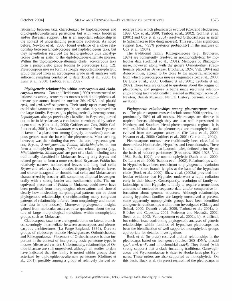
October 2004] 1575SHAW AND RENZAGLIA—PHYLOGENY OF BRYOPHYTES
←
Fig. 15. Oedipodium griffitheanum (Dicks.) Schwaegr. habit. Drawing by C. Zartman.
lationship between taxa characterized by haplolepideous anddiplolepideous-alternate peristomes but with weak bootstrapand/or Bayesian support. This is an important relationship inthe context of understanding peristome evolution. As notedbefore, Newton et al. (2000) found evidence of a close rela-tionship between Encalyptaceae and haplolepideous taxa, butthey nevertheless resolved the haplolepideous plus Encalyp-taceae clade as sister to the diplolepideous-alternate mosses.Within the diplolepideous-alternate clade, acrocarpous taxaform a paraphyletic grade leading to pleurocarps (Fig. 12).Pleurocarpous mosses form a strongly supported monophyleticgroup derived from an acrocarpous grade in all analyses withsufficient sampling conducted to date (Buck et al., 2000; DeLuna et al., 2000; Tsubota et al., 2002).
Phylogenetic relationships within acrocarpous and clado-carpous mosses—Cox and Hedderson (1999) reconstructed re-lationships among acrocarpous mosses with diplolepideous-al-ternate peristomes based on nuclear 26s rDNA and plastidrps4, and trnL-trnF sequences. Their study upset many long-established taxonomic concepts. In particular, they showed thatthe large family, Bryaceae, is phylogenetically heterogeneous.Leptobryum, always previously classified in Bryaceae, turnedout to be in Meesiaceae, a conclusion corroborated by subse-quent studies (Cox et al., 2000; Goffinet and Cox, 2000; Gof-finet et al., 2001). Orthodontium was removed from Bryaceaein favor of a placement among (largely unresolved) acrocar-pous genera near the base of the pleurocarps. Most striking,however, was their finding that even the core bryaceous gen-era, Bryum, Brachymenium, Pohlia, Mielichhoferia, do notform a monophyletic group. Pohlia and related genera (e.g.,Mielichhoferia, Mniobryum) are part of a clade including taxatraditionally classified in Mniaceae, leaving only Bryum andrelated genera to form a more restricted Bryaceae. Pohlia hasrelatively narrow, nonbordered leaves and long leaf cells;Bryum and relatives have broader, frequently bordered leavesand shorter hexagonal or rhombic leaf cells; and Mniaceae arecharacterized by broader still, sometimes elliptical leaves gen-erally with a strong border and isodiametric cells. The un-equivocal placement of Pohlia in Mniaceae could never havebeen predicted from morphological observations and showedclearly how misleading morphological patterns can be aboutphylogenetic relationships (notwithstanding many congruentpatterns of relationship inferred from morphology and molec-ular data in the mosses). Moreover, phylogenetic insightsgained from molecular analyses raise questions about the na-ture of large morphological transitions within monophyleticgroups such as Mniaceae.
Cladocarpous taxa have archegonia borne on lateral branch-es, seemingly intermediate between acrocarpous and pleuro-carpous architectures (La Farge-England, 1996). Diversegroups of cladocarps include Hedwigiaceae, Orthotrichaceae,and Rhizogoniaceae. Placement of Orthotrichaceae is also im-portant in the context of interpreting basic peristome types inmosses (discussed earlier). Unfortunately, relationships of Or-thotrichaceae are still unresolved, although all studies to datehave indicated that the family is nested within groups char-acterized by diplolepideous-alternate peristomes (Goffinet etal., 2001), possibly among a group of relatively derived ac-
rocarps from which pleurocarps evolved (Cox and Hedderson,1999; Cox et al., 2000; Tsubota et al., 2002). Goffinet et al.(2001) and Cox et al. (2004) resolved Orthotrichacae as sisterto Splachnaceae (the dung mosses). This result has significantsupport (i.e., .95% posterior probability) in the analyses ofCox et al. (2004).
The traditional family Rhizogoniaceae (e.g., Brotherus,1924) are consistently resolved as nonmonophyletic by mo-lecular data (Goffinet et al., 2001). Members of Rhizogon-iaeae, however, along with the genera Orthodontium (tradi-tionally placed in Bryaceae; Brotherus, 1924; Vitt, 1984) andAulacomnium, appear to be close to the ancestral acrocarpsfrom which pleurocarpous mosses originated (Cox et al., 2000;De Luna et al., 2000; Goffinet et al., 2001; Tsubota et al.,2002). These taxa are critical to questions about the origins ofpleurocarpy, and progress is being made resolving relation-ships among taxa traditionally classified in Rhizogoniaceae (A.Newton, British Museum, Natural History, personal commu-nication).
Phylogenetic relationships among pleurocarpous moss-es—The pleurocarpous mosses include some 5000 species, ap-proximately 50% of all mosses. Pleurocarps are diverse intropical forests, although they are also well represented inNorthern and Southern Hemisphere temperate regions. It iswell established that the pleurocarps are monophyletic andevolved from acrocarpous ancestors (De Luna et al., 2000;Newton et al., 2000; Goffinet et al., 2001; Cox et al., 2004).
Pleurocarpous mosses have traditionally been classified inthree orders: Hookeriales, Hypnales, and Leucodontales. Thereis now little question that Leucodontales, defined primarily onthe basis of reduced peristomes (Brotherus, 1924–1925; Vitt,1984; Buck, 1991), are nonmonophyletic (Buck et al., 2000;De Luna et al., 2000; Tsubota et al., 2002). Relationships with-in Hypnales have been recalcitrant to phylogenetic resolutionbecause of short branch lengths at the base of the hypnaleanclade (Buck et al., 2000). Shaw et al. (2003a) provided mo-lecular evidence that Hypnales underwent a rapid radiationearly in their history. Consequently, resolution of family re-lationships within Hypnales is likely to require a tremendousamounts of nucleotide sequence data and/or comparative in-formation about genome structure. Although relationshipsamong hypnalean families are largely unresolved at present,some apparently monophyletic groups have been identifiedand generic relationships within them investigated (Chiang andSchaal, 2000; Quandt et al., 2000; Tsubota et al., 2001a, b;Blocher and Capesius, 2002; Pedersen and Hedenas, 2002;Stech et al., 2002; Vanderpoorten et al., 2002a, b). A difficultbut critical issue confronting phylogenetic analyses of genericrelationships within families of hypnalean pleurocarps hasbeen the identification of well-supported monophyletic groupsappropriate for detailed investigations.
Buck et al. (in press) resolved ordinal relationships in thepleurocarps based on four genes (nuclear 26S rDNA, plastidrps4, trnL-trnF, and mitochondrial nad4). They found (withstrong support) that a clade including traditional Garovagli-aceae and Ptychomniaceae is sister to Hookeriales plus Hyp-nales. These orders are also supported as monophyletic. Onthis basis, Buck et al. (in press) reclassified the pleurocarps in
1576 [Vol. 91AMERICAN JOURNAL OF BOTANY
two superorders, the Pychomnianae and Hypnanae. Ptychom-nianae include the single order, Ptychomniales (with one fam-ily: Ptychomniaceae), whereas Hypnanae encompass Hooker-iales and Hypnales. They also resolved familial and genericrelationships within the Hookeriales, recognizing seven fami-lies, and reconstructed the evolution of morphological char-acters on the basis of their results. Obtaining phylogenetic res-olution within Hookeriales proved less problematic than inHypnales because Hookeriales do not appear to have under-gone the sort of rapid radiation that characterizes Hypnales(Shaw et al., 2003b). Branch lengths along Hookerialean back-bone are substantially longer than in Hypnales.
Inferences about morphological evolution in mosses frommolecular phylogenies—Cox et al. (2004) conservatively stat-ed the morphological implications of their phylogenetic resultsfor the mosses. These inferences are briefly summarized here.
Taxa near the base of the moss tree have the capsule ele-vated on a gametophytic pseudopodium rather than on a spo-rophytic seta (i.e., Sphagnum and Andreaea), but the ancestralcondition in mosses is ambiguous because Takakia and An-dreaeobryum have a seta. Cox et al. (2004) concluded that thepseudopodium evolved independently in Sphagnum and An-dreaea. Because stomata are absent in Takakia, Andreaea, andAndreaeobryum and those of Sphagnum are nonfunctional,Cox et al. also concluded that stomata-like structures in Sphag-num may not be homologous with stomata of more derivedmosses or to those of tracheophytes and hornworts. Their pres-ence in some hornworts indicates, to the contrary, that stomatamay be homologous in mosses and hornworts, which impliesmultiple losses in mosses. Alternatively, stomata could havebeen lost once in the early evolution of mosses and regainedin class Bryopsida.
The acrocarpous habit is clearly pleisiotypic in peristomatemosses, but acrocarps are a paraphyletic group within whichpleurocarps are nested. Although resolution among cladocar-pous taxa is poor, it appears that cladocarpy evolved severaltimes. It may be that hypnalean pleurocarps evolved from acladocarpous ancestor, but additional resolution among acro-carps near the origin of pleurocarps is needed.
Absence of a peristome in Oedopodium makes the phylo-genetic node at which peristomes originated ambiguous, andit is not clear whether nematodontous peristomes of the Po-lytrichum-type evolved independently of arthrodontous peri-stomes. The unique anticlinal divisions in the IPL of Polytri-chaceae, leading to twice the ‘‘normal’’ number of cells in thislayer, may be an apomorphy for that clade. These divisionsappear to be characteristic not only of Polytrichum, but alsoAtrichum and Pogonatum, also in Polytrichaceae (Shaw, L.Anderson, Duke University, and B. Mishler, University of Cal-ifornia, Berkeley, unpublished data). These divisions do notoccur in the developing sporophyte of Sphagnum or Andreaea(Shaw, unpublished data). Phylogenetic considerations lendsupport to the hypothesis of Vitt (1984) that the Funaria-typearthrodontous peristome with opposite exostome and endo-stome teeth is primitive in arthrodonts. Cox et al. (2004) notedthat although Timmia lacks endostome segments, the most par-simonious interpretation of its endostome (which consists of abasal membrane and cilia), given its phylogenetic position, isthat it conforms to the opposite type. Developmental studiesof peristomial layers in Timmia are sorely needed. Of criticalimportance is whether or not anticlinal walls in the PPL andIPL are offset.
The teeth of haplolepideous peristomes develop in positionsthat would be opposite exostome teeth were the latter formed(clearly shown in Vitt, 1981). Thus, in terms of development,the single row of teeth in haplolepideous peristomes are ho-mologous with the opposite endostome segments of the Fu-naria-type. Like Funaria-type endostomes, haplolepideousperistomes also lack a basal membrane and are relatively mas-sive. Groups characterized by haplolepideous and diplolepi-deous-alternate peristomes appear to be sister groups, implyingthat asymmetric anticlinal cell divisions in the IPL during peri-stome development may be a synapomorphy for that clade.Diplolepideous-alternate peristomes have additional anticlinalIPL walls that become offset during development relative tothose in the PPL. This pattern is likely a synapomorphy forthe clade characterized by such peristomes.
CONSPECTUS
We are currently in a period of exponential change in ourunderstanding of bryophyte phylogeny. Relationships amongthe major moss clades are relatively well resolved in compar-ison to the liverworts and hornworts. However, current workon all three groups is progressing at such a fast pace that evenby the time this review is in print, new discoveries and insightsare likely.
The molecular hypothesis presented here on hornworts (Fig.1) is a critical first step toward a modern phylogenetic under-standing for the group. Comprehensive analyses using genesfrom all three genomes in combination with morphologicaldata, with sampling from all 12 genera of hornworts, are re-quired to verify the novel relationships described earlier. Sys-tematic studies of hornworts have lagged so far behind thoseof other land plants that any molecular analysis must first be-gin with a clear delineation of morphological characteristics inthe specimens/species that are examined. Worldwide collectingand basic taxonomic evaluations are essential. With theirunique adaptations to land, including basal elongation of thesporophyte and internalization of vulnerable tissues, hornwortswill continue to provide essential information about early landplant evolution.
A general understanding of liverwort relationships hasemerged from recent molecular studies, and this has led tomajor reinterpretations of evolutionary changes in the group.For example, isophylly in leafy liverworts and conducting tis-sue in simple thalloid taxa are now clearly seen as derived,not ancestral. However, critical unanswered questions in liv-erwort phylogeny remain, and these include the position ofHaplomitrium (and Treubia), the placement of Pleuroziaamong the simple thalloid vs. leafy clades, and the precisepositions of Sphaerocarpos and Lunularia within the complexthalloid lineage. Relationships among simple thalloid taxa, es-pecially Pellia, Phyllothallia, Calycularia, and Cavicularia(the last sister to Blasia, Renzaglia, 1982) have not been re-solved using multilocus studies to date and will require addi-tional sequences and more taxon sampling. Most striking isthe lack of representation of critical genera and families in anyone study and the need for a concerted, collaborative effort toobtain and share specimens of poorly known taxa.
Still outstanding questions with regard to moss phylogenyinclude the relationship among Takakia, Sphagnum, and An-dreaea (do they form a monophyletic group sister to all othermosses?), the origin and evolution of the major peristometypes, and the nature of the acrocarpous ancestors of pleuro-

October 2004] 1577SHAW AND RENZAGLIA—PHYLOGENY OF BRYOPHYTES
carpous taxa. Fundamental morphological data on Takakia, in-cluding embryology, sporophyte development, and apicalgrowth are necessary to identify structural changes withinmosses. Additional work on developmental anatomy of peri-stomes is also needed in conjunction with ongoing phyloge-netic work. Development of peristomial cell layers in Oedi-podium (which lacks a peristome) is critical, as is the devel-opment of the unique Timmia peristome. Resolution of family-level relationships in mosses, especially in closely relatedpleurocarps, will require expanded data sets based on not justone or two genes, nor even five, but probably 15 to 20. Avail-ability of primers for such multilocus analyses are probablynot far off, given the intensive genomic work currently un-derway on a wide diversity of land plants, including mosses.
Molecular technologies are improving at a rate that is un-predictable and incomprehensible; we speculate that within thenext decade most of the phylogenetic questions raised in thisreview will be resolved. In addition to sequences from mul-tiple genes, complete organellar genomes will soon be avail-able for representatives of each major lineage within bryo-phytes. Structural features of genomes, including intron pres-ence, and gene order and deletions, likewise will continue toprovide informative phylogenetic evidence. Further under-standing of the existence and expression of developmentalgenes, especially homeobox and MADS-box genes, in all threegroups of bryophytes will provide clues to the evolution ofstructural complexity within these plants and evolutionary re-lationships to more complicated organ systems of tracheo-phytes. Comparative morphological/ultrastructural studies ofliving and fossil taxa are required to fill in the gaps in knowl-edge as well as to fully comprehend structural changes. Inaddition to perfecting data collection methodologies, the pri-mary challenge that lies ahead is in developing methods ofanalyzing and combining the large and diverse data sets thatare rapidly materializing. Only then will the intricate detailsof early land plant interrelationships be clearly illuminated.
LITERATURE CITED
AHONEN, I., J. MUONA, AND S. PIIPPO. 2003. Inferring the phylogeny of theLejeuneaceae (Jungermanniopsida): a first appraisal of molecular data.Bryologist 106: 297–308.
BARTHOLOMEW-BEGAN, S. E. 1990. Classification of the Haplomitriales andMetzgeriales into the subclass Metzgeriidae, subclass nov. (Hepatophyta,Jungermanniopsida). Phytologia 69: 464–466.
BARTHOLOMEW-BEGAN, S. E. 1991. A morphogenetic re-evaluation of Hap-lomitrium Nees (Hepatophyta). Bryophytorum Bibliotheca 41: 1–297.
BARTLETT, E. M. 1928. A comparative study of the development of the spo-rophyte in the Anthocerotae, with special reference to the genus Antho-ceros. Annals of Botany 42: 409–430.
BECKERT, S., H. MUHLE, D. PRUCHNER, AND V. KNOOP. 2001. The mito-chondrial nad2 gene as a novel marker locus for phylogenetic analysisof early land plants: a comparative analysis in mosses. Molecular Phy-logenetics and Evolution 18: 117–126.
BECKERT, S., S. STEINHAUSER, H. MUHLE, AND V. KNOOP. 1999. A molecularphylogeny of the bryophytes based on nucleotide sequences of the mi-tochondrial nad5 gene. Plant Systematics and Evolution 218: 179–192.
BIERHORST, D. W. 1971. Morphology of vascular plants. Macmillan Co., NewYork, New York, USA.
BISCHLER, H. 1998. Systematics and evolution of the genera of the Mar-chantiales. Bryophytorum Bibliotheca 51: 1–201.
BLOCHER, R., AND I. CAPESIUS. 2002. The systematic position of the Hy-popterygiaceae (Bryopsida) inferred from rps4 gene sequences. Cryp-togamie, Bryologie 23: 191–207.
BLOMQUIST, H. L., AND L. L. ROBERTSON. 1941. The development of theperistome in Aulacomnium heterostichum. Bulletin of the Torrey Botan-ical Club 68: 569–584.
BOISSELIER-DUBAYLE, M.-C., J. LAMBOURDIERE, AND H. BISHLER. 2002.Molecular phylogenies support multiple morphological reductions in theliverwort subclass Marchantiidae (Bryophyta). Molecular Phylogeneticsand Evolution 24: 66–77.
BOPP, M., AND I. CAPESIUS. 1996. New aspects of bryophyte taxonomy pro-vided by a molecular approach. Botanica Acta 106: 368–372.
BOPP, M., AND I. CAPESIUS. 1998. A molecular approach to bryophyte sys-tematics. In J. W. Bates, N. W. Ashton, and J. G. Duckett [eds.], Bryologyfor the twenty-first century, 79–88. Maney and the British BryologicalSociety, Leeds, UK.
BOWER, F. O. 1935. Primitive land plants. Macmillan and Company, London,UK.
BRASSARD, G. R. 1979. The moss genus Timmia. I. Introduction, and revisionof T. norvegica and allied taxa. Lindbergia 5: 39–53.
BROTHERUS, V. A. 1924–1925. Musci (Laubmoose). III. Unterklasse Bryales:II. Spezieller Teil. In A. Engler [ed.], Die Naturlichen Pflanzenfamilien,277–700. Engelmann, Leipzig, Germany.
BROWN, R. C., AND B. L. LEMMON. 1988. Sporogenesis in bryophytes. Ad-vances in Bryology 3: 159–223.
BROWN, R. C., AND B. L. LEMMON. 1990. Monoplastidic cell division inlower land plants. American Journal of Botany 77: 559–571.
BROWN, R. C., B. L. LEMMON, AND K. S. RENZAGLIA. 1986. Sporocyticcontrol of spore wall patterns in liverworts. American Journal of Botany73: 593–596.
BRUCH, P., W. P. SCHIMPER, AND T. GUMBEL. 1851–1855. Bryologia Europaeaseu Generum Muscorum Europaeorum Monographice Illustrata.Schweizerbart, Stuttgart, Germany.
BUCK, W. R. 1991. The basis for familial classification of pleurocarpousmosses. Advances in Bryology 4: 169–185.
BUCK, W. R., C. J. COX, A. J. SHAW, AND B. GOFFINET. In press. Ordinalrelationships of pleurocarpous mosses, with special emphasis on theHookeriales. Systematics and Biodiversity.
BUCK, W. R., AND B. GOFFINET. 2000. Morphology and classification ofmosses. In A. J. Shaw and B. Goffinet [eds.], Bryophyte biology, 71–123. Cambridge University Press, New York, New York, USA.
BUCK, W. R., B. GOFFINET, AND A. J. SHAW. 2000. Testing morphologicalconcepts of orders of pleurocarpous mosses (Bryophyta) using phylo-genetic reconstructions based on trnL-trnF and rps4 sequences. Molec-ular Phylogenetics and Evolution 16: 180–198.
BURR, F. A. 1970. Phylogenetic transitions in the chloroplast numbers of theAnthocerotales. I. The number and ultrastructure of the mature plastids.American Journal of Botany 57: 97–110.
CAMPBELL, D. H. 1895. The structure and development of mosses and ferns(Archegoniatae). Macmillan and Co., London, UK.
CAMPBELL, D. H. 1917. Growth of isolated sporophytes of Anthoceros. Pro-ceedings of the National Academy of Science, USA 3: 494–496.
CAMPBELL, D. H. 1924. A remarkable development of the sporophyte ofAnthoceros fusiformis Aust. Annals of Botany 38: 473–483.
CARGILL, D. C., K. S. RENZAGLIA, J. C. VILLAREAL, AND R. J. DUFF. Inpress. Generic concepts within hornworts (Phylum Anthocerotophyta).Australian Journal of Botany.
CHAMPAGNE, C. E. M., AND N. W. ASHTON. 2001. Ancestry of KNOX genesrevealed by bryophyte (Physcomitrella patens) homologs. New Phytol-ogist 150: 23–36.
CHIANG, T.-Y., AND B. A. SCHAAL. 2000. The internal transcribed spacer 2region of the nuclear ribosomal DNA and the phylogeny of the mossfamily Hylocomiaceae. Plant Systematics and Evolution 224: 127–137.
CLARKE, G. C. S., AND J. G. DUCKETT [EDS.]. 1979. Bryophyte systematics.The Systematics Association Special Volume 14. Academic Press, NewYork, New York, USA.
COX, C. J., B. GOFFINET, A. E. NEWTON, A. J. SHAW, AND T. A. J. HEDDER-SON. 2000. Phylogenetic relationships among the diplolepideous-alter-nate mosses (Bryidae) inferred from nuclear and chloroplast DNA se-quences. Bryologist 103: 224–240.
COX, C. J., B. GOFFINET, A. J. SHAW, AND S. BOLES. 2004. Phylogeneticrelationships among the mosses based on heterogeneous Bayesian anal-ysis of multiple genes from multiple genomic compartments. SystematicBotany 29: 234–250.
COX, C. J., AND T. A. J. HEDDERSON. 1999. Phylogenetic relationships amongthe ciliate arthrodontous mosses: evidence from chloroplast and nuclearDNA sequences. Plant Systematics and Evolution 215: 119–139.
CRANDALL-STOTLER, B. 1976. The apical cell and early development of Pleu-rozia purpurea Lindb. Lindbergia 32: 197–208.
1578 [Vol. 91AMERICAN JOURNAL OF BOTANY
CRANDALL-STOTLER, B. 1981. Morphology/anatomy of hepatics and antho-cerotes. Advances in Bryology 1: 315–398.
CRANDALL-STOTLER, B., AND R. E. STOTLER. 2000. Morphology and clas-sification of the Marchantiophyta. In A. J. Shaw and B. Goffinet [eds.],Bryophyte biology, 21–70. Cambridge University Press, Cambridge, UK.
CROSBY, M. R., R. E. MAGILL, B. ALLEN, AND S. HE. 2000. A checklist ofthe mosses. Missouri Botanical Garden, St. Louis, Missouri, USA, web-site: www.mobot.org/MOBOT/tropicos/most/checklist.shtml.
CRUM, H. A. 2001. Structural diversity of bryophytes. University of MichiganHerbarium, Ann Arbor, Michigan, USA.
DAVIS, E. C. In press. A molecular phylogeny of leafy liverworts (Junger-manniidae: Marchantiophyta). In B. Goffinet, V. C. Hollowell, and R. E.Magill [eds.], Molecular systematics of bryophytes: progress, problems& perspectives. Monographs in systematic botany from the Missouri Bo-tanical Garden, St. Louis, Missouri, USA.
DE LUNA, E., W. R. BUCK, H. AKIYAMA, T. ARIKAWA, H. TSUBOTA, D.GONZALEZ, A. E. NEWTON, AND A. J. SHAW. 2000. Ordinal phylogenywithin the hypnobryalean pleurocarpous mosses inferred from cladisticanalyses of three chloroplast DNA sequence data sets: trnL-F, rps4, andrbcL. Bryologist 103: 242–256.
DUCKETT, J. G., AND R. LIGRONE. 1995. The formation of catenate foliargemmae and the origin of the oil bodies in the liverwort Odontoschismadenudatum (Mart.) Dum. (Jungermanniales): a light and electron micro-scope study. Annals of Botany 76: 406–419.
DUFF, R. J., AND D. L. NICKRENT. 1999. Phylogenetic relationships amongland plants using mitochondrial small-subunit rDNA sequences. Ameri-can Journal of Botany 86: 372–386.
DUFF, R. J., D. C. CARGILL, J. C. VILLARREAL, AND K. S. RENZAGLIA. Inpress. Phylogenetic relationships of the hornworts based on rbcL se-quence data: novel relationships and new insights. In B. Goffinet, V. C.Hollowell, and R. E. Magill [eds.], Molecular systematics of bryophytes:progress, problems & perspectives. Monographs in systematic botanyfrom the Missouri Botanical Garden, St. Louis, Missouri, USA.
EDWARDS, S. R. 1979. Taxonomic implications of cell patterns in haplole-pidous moss peristomes. In G. C. S. Clarke and J. G. Duckett [eds.],Bryophyte systematics, 317–346. Systematics Association Special Vol-ume 14. Academic Press, New York, New York, USA.
EDWARDS, S. R. 1984. Homologies and inter-relationships of moss peri-stomes. In R. M. Schuster [ed.], New manual of bryology, vol. 2, 658–695. Hattori Botanical Laboratory, Nichinan, Japan.
ENDLICHER, S. 1841. Enchiridion botanicum. W. Engelmann, Leipzig, Ger-many.
EVANS, A. W., AND H. D. HOOKER, JR. 1913. Development of the peristomein Ceratodon purpureus. Bulletin of the Torrey Botanical Club 40: 97–109.
FLEISCHER, M. 1923. Die Moose der Flora von Buitenzorg 4: XI–XXIII. E.J. Brill, Leiden, Netherlands.
FORREST, L. L., AND B. J. CRANDALL-STOTLER. In press. A phylogeny ofthe simple thalloid liverworts (Jungermanniopsida, subclass Metzgeri-idae) as inferred from five chloroplast genes. In B. Goffinet, V. C. Hol-lowell, and R. E. Magill [eds.], Molecular systematics of bryophytes:progress, problems & perspectives. Monographs in systematic botanyfrom the Missouri Botanical Garden, St. Louis, Missouri, USA.
GARBARY, D. J., AND K. S. RENZAGLIA. 1998. Bryophyte phylogeny and theevolution of land plants: evidence from development and ultrastructure.In J. W. Bates, N. W. Ashton, and J. G. Duckett [eds.], Bryology for thetwenty-first century, 45–63. Maney and British Bryological Society,Leeds, UK.
GARBARY, D. J., K. S. RENZAGLIA, AND J. G. DUCKETT. 1993. The phylogenyof land plants: a cladistic analysis based on male gametogenesis. PlantSystematics and Evolution 188: 237–269.
GLENNY, D. 1998. A revised checklist of New Zealand liverworts and horn-worts. Tuhinga 10: 11–149.
GOEBEL, K. 1905. Organography of plants, especially of the archegoniataeand spermatophyta. II. Special organography. Clarendon Press, Oxford,UK.
GOFFINET, B. 2000. Origin and phylogenetic relationships of bryophytes. InJ. Shaw and B. Goffinet [eds.], The biology of bryophytes, 124–149.Cambridge University Press, Cambridge, UK.
GOFFINET, B., AND C. J. COX. 2000. Phylogenetic relationships among basal-most arthrodontous mosses with special emphasis on the evolutionarysignificance of the Funariineae. Bryologist 103: 212–223.
GOFFINET, B., C. J. COX, L. E. ANDERSON, AND B. D. MISHLER. 1999. Peri-stome development in the Orthotrichaceae. Bryologist 102: 581–594.
GOFFINET, B., C. J. COX, A. J. SHAW, AND T. A. J. HEDDERSON. 2001. Thebryophyta (mosses): systematic and evolutionary inferences from an rps4gene (cpDNA) phylogeny. Annals of Botany 87: 191–208.
GRADSTEIN, S. R. 1979. The genera of the Lejeuneaceae: past and present.In G. C. S. Clarke and J. G. Duckett [eds.], Bryophyte systematics, 83–107. Academic Press, New York, New York, USA.
GRADSTEIN, S. R. 1994. Lejeuneaceae: Ptychantheae, Brachiolejeuneae. Flo-ra Neotropica Monograph 62: 1–225.
GRADSTEIN, S. R. 1997. The taxonomic diversity of epiphyllous bryophytes.Abstracta Botanica 21: 15–19.
GRAHAM, L. E. 1993. Origin of land plants. John Wiley and Sons, Inc., NewYork, USA.
GRAHAM, L. E., AND L. W. WILCOX. 2000. The origin of alternation of gen-erations in land plants: a focus on matrotrophy and hexose transport.Philosophical Transactions of the Royal Society, London, B, BiologicalSciences 355: 757–767.
HANSEN, D., T. J. ANDREWS, AND M. R. BADGER. 2002. Variability of thepyrenoid-based CO2 concentrating mechanism in hornworts. FunctionalPlant Biology 29: 407–416.
HASEGAWA, J. 1980. Taxonomical studies on Asian Anthocerotae. III. SomeAsian species of Dendroceros. Journal of the Hattori Botanical Labo-ratory 47: 287–309.
HASEGAWA, J. 1983. Taxonomical studies on Asian Anthocerotae. III. Asianspecies of Megaceros. Journal of the Hattori Botanical Laboratory 54:227–240.
HASEGAWA, J. 1988. A proposal for a new system of the Anthocerotae, witha revision of the genera. Journal of the Hattori Botanical Laboratory 74:105–119.
HASEGAWA, J. 1994. New classification of Anthocerotae. Journal of the Hat-tori Botanical Laboratory 76: 21–34.
HASSEL DE MENENDEZ, G. G. 1962. Estudio de los Anthocerotales y Mar-chantiales de Argentina. Opera Lilloana 7: 1–297.
HASSEL DE MENENDEZ, G. G. 1986. Leiosporoceros Hassel n. gen. and Leios-porocerotaceae Hassel n. fam. of Anthocerotopsida. Journal of Bryology14: 255–259.
HASSEL DE MENENDEZ, G. G. 1988. A proposal for a new classification ofthe genera within the Anthocerotophyta. Journal of the Hattori BotanicalLaboratory 64: 71–86.
HEBANT, C. 1977. The conducting tissues of bryophytes. Bryophytorum Bib-liotheca 10: 1–157, 80 plates, J. Cramer, Vaduz, Lichtenstein.
HEDDERSON, T. A., R. L. CHAPMAN, AND C. J. COX. 1998. Bryophytes andthe origins and diversification of land plants: new evidence from mole-cules. In J. W. Bates, N. W. Ashton, and J. G. Duckett [eds.], Bryologyfor the twenty-first century, 65–77. Maney Publishing, Leeds, UK.
HEDDERSON, T. A., R. L. CHAPMAN, AND W. L. ROOTES. 1996. Phylogeneticrelationships of bryophytes inferred from nuclear encoded rRNA genesequences. Plant Systematics and Evolution 200: 213–224.
HEDWIG, J. 1801. Species Muscorum frondosorum Descriptae et TabulisAeneid 1xxvii Colratis Illustratae. Barthii, Leipzig, Germany.
HENSCHEL, K., R. KUFUJI, M. HASEBE, H. SAEDLER, T. MUNSTER, AND G.THEISSEN. 2002. Two ancient classes of MIKC-type MADS-box genesare present in the moss Physcomitrella patens. Molecular Biology andEvolution 19: 801–814.
HE-NYGREN, X., I. AHONEN, A. JUSLEN, D. GLENNY, AND S. PIIPPO. In press.Dichotomous evolution in leafy liverworts. In B. Goffinet, V. C. Hollow-ell, and R. E. Magill [eds.], Molecular systematics of bryophytes: pro-gress, problems & perspectives. Monographs in systematic botany fromthe Missouri Botanical Garden, St. Louis, Missouri, USA.
HIMI, S., R. SANO, T. NISHIYAMA, T. TANAHASHI, M. KATO, K. UEDA, AND
M. HASEBE. 2001. Evolution of MADS-box gene induction by FLO/LFY genes. Journal of Molecular Evolution 53: 387–393.
HOFMEISTER, W. 1851. Vergleichende Untersuchungen der Keimung, Entfal-tung und Fruchtbildung hoherer Kryptogamen. F. Hofmeister, Liepzig,Germany.
HOHAM, R. W., T. A. BONOME, D. W. MARTIN, AND J. H. LEEBENS-MACK.2002. A combined 18S rDNA and rbcL phylogenetic analysis of Chlo-romonas and Chlamydomonas (Chlorophyceae, Volvocales) emphasizingsnow and other cold-temperature habitats. Journal of Phycology 38:1051–1064.
HOHE, A., S. A. RENSING, M. MILDNER, D. LANG, AND R. RESKI. 2002. Daylength and temperature strongly influence sexual reproduction and ex-

October 2004] 1579SHAW AND RENZAGLIA—PHYLOGENY OF BRYOPHYTES
pression of a novel MADS-box gene in the moss Physcomitrella patens.Plant Biology 4: 595–202.
HYVONEN, J., T. A. HEDDERSON, G. L. SMITH MERRILL, J. G. GIBBINS, AND
S. KOSKINEN. 1998. On phylogeny of the Polytrichales. Bryologist 101:489–504.
HYVONEN, J., AND S. PIIPPO. 1993. Cladistic analysis of the hornworts (An-thocerotophyta). Journal of the Hattori Botanical Laboratory 74: 105–119.
KATO, M., AND R. IMAICHI. 1997. Morphological diversity and evolution ofvegetative organs in pteridophytes. In K. Iwatsuki and P. H. Raven [eds.],Evolution and diversification of plants, 27–43. Springer-Verlag, NewYork, New York, USA.
KATOH, K., H. HORI, AND S. OSAWA. 1983. The nucleotide sequences of 5Sribosomal RNAs from four Bryophyta-species. Nucleic Acids Research11: 5671–5674.
KELCH, D. G., B. D. MISHLER, AND A. DRISKELL. In press. Inferring phy-logeny using genomic characters: a case study using land plant plas-tomes. In B. Goffinet, V. C. Hollowell, and R. E. Magill [eds.], Molecularsystematics of bryophytes: progress, problems & perspectives. Mono-graphs in systematic botany from the Missouri Botanical Garden, St.Louis, Missouri, USA.
KENRICK, P., AND P. R. CRANE. 1997. The origin and early diversification ofland plants: a cladistic study. Smithsonian Institution Press, WashingtonD.C., USA.
KINDBERG, N. C. 1897. Genera of European and North American Bryineae(Mosses) synoptically disposed. Bonniers Boktryckeri Aktiebolag, Go-teborg, Sweden.
KOBIYAMA, Y., AND B. J. CRANDALL-STOTLER. 1999. Studies of specializedpitted parenchyma cells of the liverwort Conocephalum Hill and theirphylogenetic implications. International Journal of Plant Sciences 160:351–370.
KROGAN, N. T., AND N. W. ASHTON. 2000. Ancestry of plant MADS-boxgenes revealed by bryophyte (Physcomitrella patens) homologues. NewPhytologist 147: 505–517.
LA FARGE-ENGLAND, C. 1996. Growth form, branching pattern, and peri-chaetial position in mosses: cladocarpy and pleurocarpy redefined. Bry-ologist 99: 170–186.
LANG, W. H. 1907. On the sporangium of Notothylas. Annals of Botany 21:201–210.
LEITGEB, H. 1874–1881. Untersuchungen uber die Lebermoose, I–V. I. Blasiapusilla, 1874; II. Die Foliosen Jungermannieen, 1877; IV. Die Riccieen,1879; V. Die Anthoceroteen, 1879; VI. Die Marchanteen, 1881, vols. I–III, Jena: O. Deistungs Buchhandlungs, vols. IV–VI. Leuschner and Lu-bensky, Graz, Austria.
LEWIS, L., B. D. MISHLER, AND R. VILGALYS. 1997. Phylogenetic relation-ships of the liverworts (Hepaticae), a basal embryophyte lineage, inferredfrom nucleotide sequence data of the chloroplast gene rbcL. MolecularPhylogenetics and Evolution 7: 377–393.
LIGRONE, R., J. G. DUCKETT, AND K. S. RENZAGLIA. 2000. Conducting tis-sues and phyletic relationships of bryophytes. Philosophical Transac-tions of the Royal Society 355: 795–813.
LINNAEUS, C. 1753. Species plantarum. Laurentii Salvii, Stockholm, Sweden.MAGOMBO, Z. L. K. 2003. The phylogeny of basal peristomate mosses: ev-
idence from cpDNA, and implications for peristome evolution. System-atic Botany 28: 24–38.
MAHU, M. 1979. Familias y generos de musgos Chilenos. Bryologist 82: 513–524.
MALEK, O., K. LATTIG, R. HIESEL, A. BRENNICKE, AND V. KNOOP. 1996.RNA editing in bryophytes and a molecular phylogeny of land plants.European Molecular Biology Organization 15: 1403–1411.
MISHLER, B. D., AND S. P. CHURCHILL. 1984. A cladistic approach to thephylogeny of the ‘‘bryophytes.’’ Brittonia 36: 406–424.
MISHLER, B. D., AND E. DE LUNA. 1991. The use of ontogenetic data inphylogenetic analyses of mosses. Advances in Bryology 4: 121–167.
MISHLER, B. D., L. A. LEWIS, M. A. BUCHHEIM, K. S. RENZAGLIA, D. J.GARBARY, C. F. DELWICHE, F. W. ZECHMAN, T. S. KANTZ, AND R. L.CHAPMAN. 1994. Phylogenetic relationships of the ‘‘green algae’’ and‘‘bryophytes.’’ Annals of the Missouri Botanical Garden 81: 451–483.
MITTEN, W. 1859. Musci Indiae Orientalis. An enumeration of the mosses ofthe East Indies. Journal of the Proceedings of the Linneaen Society (Lon-don), Supplement Botany 1: 1–171.
MURRAY, B. M. 1988. Systematics of the Andreaopsida (Bryophyta): twoorders with links to Takakia. Beiheft zur Nova Hedwigia 90: 289–336.
NEWTON, A. E., C. J. COX, J. G. DUCKETT, J. WHEELER, B. GOFFINET, B.D. MISHLER, AND T. A. J. HEDDERSON. 2000. Evolution of the majormoss lineages. Bryologist 103: 187–211.
NICKRENT, D. L., C. L. PARKINSON, J. D. PALMER, AND R. J. DUFF. 2000.Multigene phylogeny of land plants with special reference to bryophytesand the earliest land plants. Molecular Biology and Evolution 17: 1885–1895.
NIKLAS, K. J. 1997. The evolutionary biology of plants. University of Chi-cago Press, Chicago, Illinois, USA.
NISHIYAMA, T., AND M. KATO. 1999. Molecular phylogenetic analysis amongbryophytes and tracheophytes based on combined data of plastid codedgenes and the 18S rRNA gene. Molecular Biology and Evolution 16:1027–1036.
NISHIYAMA, T., T. FUJITA, T. SHIN-I, M. SEKI, H. NISHIDE, I. UCHIYAMA, A.KAMIYA, P. CARNINCI, Y. HAYASHIZAKI, K. SHINOZAKI, Y. KOHARA,AND H. HASEBE. 2003. Comparative genomics of Physcomitrella patensgametophytic transcriptome and Arabidopsis thaliana: implication forland plant evolution. Proceedings of the National Academy of Science,USA 100: 8007–8012.
NISHIYAMA, T., P. G. WOLF, M. KUGHITA, R. B. SINCLAIR, M. SUGITA, C.SUGIURA, T. WAKASUGI, K. YAMADA, K. YOSHINAGA, K. YAMAGUCHI,K. EUDA, AND M. HASEBE. In press. Chloroplast phylogeny indicatesthat bryophytes are monophyletic. Molecular Biology and Evolution.
NOGUCHI, A. 1988. Illustrated moss flora of Japan, part 2. Hattori BotanicalLaboratory, Nichinan, Japan.
NOZAKI, N., K. ONISHI, AND E. MORITA. 2002. Differences in pyrenoid mor-phology are correlated with differences in the rbcL genes of membersof the chloromonas lineage (Volvocales, Chlorphyceae). Journal of Mo-lecular Evolution 55: 414–430.
PARIHAR, N. S. 1965. An introduction to Embryophyta, vol. 1, Bryophyta,5th ed. Central Book Dept., Allahabad, India.
PASS, J. M., AND K. S. RENZAGLIA. 1995. Comparative microanatomy of thelocomotory apparatus of Conocephalum conicum (Hepaticae, Conoce-phalaceae). Fragmenta Floristica et Geobotanica 40: 365–377.
PEDERSEN, N., AND L. HEDENAS. 2002. Phylogeny of the Plagiotheciaceaebased on molecular and morphological evidence. Bryologist 105: 310–324.
PHILIBERT, H. 1884–1902. De l’importance du peristome pour les affinitiesnaturelles des mousses. Revue Bryologique 11: 49–52, 65–72 (1884).Etudes sur le peristome. Revue Bryologique 11: 80–87 (1884); 12: 67–77, 81–85 (1885); 13: 17–26, 81–86 (1886); 14: 9–11, 81–90 (1887);15: 6–12, 24–28, 37–44, 50–60, 65–69, 90–93 (1888); 16: 1–9, 39–44,67–77 (1889); 17: 8–12, 25–29, 39–42 (1890); 23: 36–38, 41–56 (1896);28: 56–59, 127–130 (1901); 29: 10–13 (1902).
PROSKAUER, J. 1951. Studies on Anthocerotales. III. The genera Anthocerosand Phaeoceros. Bulletin of the Torrey Botanical Club 78: 331–349.
PROSKAUER, J. 1953. Studies on Anthocerotales. IV. Bulletin of the TorreyBotanical Club 80: 65–75.
PROSKAUER, J. 1960. Studies on Anthocerotales. VI. On spiral thickenings inthe columella and its bearing on phylogeny. Phytomorphology 10: 1–19.
PURI, P. 1973. Bryophytes. A broad perspective. Atma Ram & Sons, Dehli,India.
QIU, Y.-L., Y. CHO, J. C. COX, AND J. D. PALMER. 1998. The gain of threemitochondrial introns identifies liverworts as the earliest land plants. Na-ture 394: 671–674.
QUANDT, D., R. S. TANGNEY, J.-P. FRAHM, AND W. FREY. 2000. A molecularcontribution for understanding the Lembophyllaceae (Bryopsida) basedon noncoding chloroplast regions (cpDNA) and ITS2 (nrDNA) sequencedata. Journal of the Hattori Botanical Laboratory 89: 71–92.
RENZAGLIA, K. S. 1978. A comparative morphology and developmental anat-omy of the Anthocerotophyta. Journal of the Hattori Botanical Labo-ratory 44: 31–90.
RENZAGLIA, K. S. 1982. A comparative developmental investigation of thegametophyte generation in the Metzgeriales (Hepatophyta). Bryophythe-ca Bibliotheca 24: 1–253.
RENZAGLIA, K. S., R. C. BROWN, B. E. LEMMON, J. G. DUCKETT, AND R.LIGRONE. 1994. The occurrence and phylogenetic significance of mon-oplastidic meiosis in liverworts. Canadian Journal of Botany 72: 65–72.
RENZAGLIA, K. S., AND J. G. DUCKETT. 1987. Spermatogenesis of Blasiapusilla, from antheridial initial through mature spermatozoid. Bryologist90: 419–449.
RENZAGLIA, K. S., R. J. DUFF, D. L. NICKRENT, AND D. GARBARY. 2000.Vegetative and reproductive innovations of early land plants: implica-
1580 [Vol. 91AMERICAN JOURNAL OF BOTANY
tions for a unified phylogeny. Transactions of the Royal Society, London355: 769–793.
RENZAGLIA, K. S., AND D. J. GARBARY. 2001. Motile male gametes of landplants: diversity, development and evolution. Critical Reviews in PlantSciences 20: 107–213.
RENZAGLIA, K. S., K. D. MCFARLAND, AND D. K. SMITH. 1997. Anatomyand ultrastructure of the sporophyte of Takakia ceratophylla (Bryophyta).American Journal of Botany 84: 1337–1350.
RENZAGLIA, K. S., AND K. C. VAUGHN. 2000. Anatomy, development andclassification of hornworts. In J. Shaw and B. Goffinet [eds.], The biol-ogy of bryophytes, 1–35. Cambridge University Press, Cambridge, UK.
RICHARDSON, D. H. S. 1981. The biology of bryophytes. Blackwell ScientificPublications, Oxford, UK.
SAMIGULLIN, T. K., S. P. YACENTYUK, G. V. DEGTYARYEVA, K. M. VALIEHO-ROMAN, V. K. BOBROVA, I. CAPESIUS, W. F. MARTIN, A. V. TROITSKY,V. R. FILIN, AND A. S. ANTONOV. 2002. Paraphyly of bryophytes andclose relationship of hornworts and vascular plants inferred from analysisof chloroplast rDNA ITS (cpITS) spacer sequences. Arctoa 11: 31–43.
SCHLJAKOV, R. N. 1972. On the higher taxa of liverworts—class Hepaticaes. str. Botanicheskii Zhurnal 57: 496–508.
SCHOFIELD, W. B. 1985. Introduction to bryology. Macmillan, New York,New York, USA.
SCHUSTER, R. M. 1966. The Hepaticae and Anthocerotae of North America,vol. I. Columbia University Press, New York, New York, USA.
SCHUSTER, R. M. 1972. Phylogenetic and taxonomic studies on Jungerman-nidae. Journal of the Hattori Botanical Laboratory 36: 321–405.
SCHUSTER, R. M. 1984. New manual of bryology, vols. 1–2. The HattoriBotanical Laboratory, Nichinan, Miyazaki, Japan.
SCHUSTER, R. M. 1992. The Hepaticae and Anthocerotae of North America,vol. VI. Field Museum of Natural History, Chicago, Illinois, USA.
SHAW, J. 1986. Peristome structure in the Orthotrichaceae. Journal of theHattori Botanical Laboratory 60: 119–136.
SHAW, J. 2000. Phylogeny of the Sphagnopsida based on nuclear and chlo-roplast DNA sequences. Bryologist 103: 277–306.
SHAW, J., AND L. E. ANDERSON. 1988. Peristome development in mosses inrelation to systematics and evolution. II. Tetraphis pellucida (Tetraphi-daceae). American Journal of Botany 75: 1019–1032.
SHAW, J., L. E. ANDERSON, AND B. D. MISHLER. 1987. Peristome develop-ment in mosses in relation to systematics and evolution. I. Diphysciumfoliosum (Buxbaumiaceae). Memoirs of the New York Botanical Garden45: 55–70.
SHAW, J., L. E. ANDERSON, AND B. D. MISHLER. 1989a. Peristome devel-opment in mosses in relation to systematics and evolution. III. Funariahygrometrica, Bryum bicolor, and B. pseudocapillare. Systematic Botany14: 24–36.
SHAW, J., L. E. ANDERSON, AND B. D. MISHLER. 1989b. Peristome devel-opment in mosses in relation to systematics and evolution. IV. Haplole-pideae: Ditrichaceae and Dicranaceae. Bryologist 92: 314–325.
SHAW, J., C. J. COX, AND S. B. BOLES. 2003a. Polarity of peatmoss (Sphag-num) evolution: who says mosses have no roots? American Journal ofBotany 90: 1777–1787.
SHAW, J., C. J. COX, S. B. BOLES, AND B. GOFFINET. 2003b. Phylogeneticevidence for a rapid radiation of pleurocarpous mosses (Bryopsida). Evo-lution 57: 2226–2241.
SHAW, J., AND B. GOFFINET. 2000. Bryophyte biology. Cambridge UniversityPress, Cambridge, UK.
SHAW, J., AND H. ROBINSON. 1984. The development, evolution, and functionof peristomes in mosses. Journal of the Hattori Botanical Laboratory57: 319–335.
SHAW, J., AND J. ROHRER. 1984. Endostomial architecture in diplolepideousmosses. Journal of the Hattori Botanical Laboratory 57: 41–61.
SINGH, D. K. 2002. Notothylaceae of India and Nepal (a morpho-taxonomicrevision). Bishen Singh Mahendra Pal Singh, Dehra Dun, India.
SMITH, A. J. E. 1978. Moss flora of Britain and Ireland. Cambridge UniversityPress, Cambridge, UK.
SMITH, A. J. E. [ED.]. 1982. Bryophyte ecology. Chapman and Hall, NewYork, New York, USA.
SMITH, D. K., AND P. G. DAVISON. 1993. Antheridia and sporophytes inTakakia ceratophylla (Mitt.) Grolle.: evidence for reclassification amongthe mosses. Journal of the Hattori Botanical Laboratory 73: 263–271.
SMITH, E. C., AND H. GRIFFITHS. 1996. A pyrenoid-based carbonic-concen-trating mechanism is present in terrestrial bryophytes of the class An-thocerotae. Planta 200: 203–212.
SMITH, E. C., AND H. GRIFFITHS. 2000. The role of carbonic anhydrase inphotosynthesis and the activity of the carbon-concentrating-mechanismin bryophytes of the class Anthocerotae. New Phytologist 145: 29–37.
SMITH, G. M. 1955. Cryptogamic botany, vol. II, Bryophytes and pterido-phytes. McGraw-Hill Book Co., New York, New York, USA.
SOLTIS, P. S., D. E. SOLTIS, P. G. WOLF, D. L. NICKRENT, S. M. CHAW, AND
R. L. CHAPMAN. 1999. The phylogeny of land plants inferred from 18SrDNA sequences: pushing the limits of rDNA signal? Molecular Biologyand Evolution 16: 1774–1784.
STECH, M., AND W. FREY. 2001. CpDNA—relationship and classification ofthe Jungermanniopsida (Hepaticophytina, Bryophyta). Nova Hedwigia72: 45–58.
STECH, M., J.-P. FRAHM, H. H. HILGER, AND W. FREY. 2000. Molecularrelationships of Treubia Goebel. (Treubiaceae, Treubiopsida) and hightaxonomic level classification of the Hepaticophytina. Nova Hedwigia71: 195–208.
STECH, M., T. PFEIFFER, AND W. FREY. 2002. Molecular generic classificationof the Hypopterygiaceae (Bryopsida), with the description of a new ge-nus, Arbusculohypopterygium gen. nov. Studies of austral temperate rainforest bryophytes 10. New Zealand Journal of Botany 40: 207–221.
STECH, M., D. QUANDT, AND W. FREY. 2003. Molecular circumscription ofthe hornworts (Anthocerotophyta) based on the chloroplast DNA trnL-trnF region. Journal of Plant Research 116: 389–398.
TAYLOR, W. A. 1995. Spores in earliest land plants. Nature 373: 391–392.THIERS, B. M. 1993. A monograph of Pleurozia (Hepaticae; Pleuroziaceae).
Bryologist 96: 517–554.TSUBOTA, H., H. AKIYAMA, T. YAMAGUCHI, AND H. DEGUCHI. 2001a. Mo-
lecular phylogeny of the genus Trismegistia and related genera (Sema-tophyllaceae, Musci) based on chloroplast rbcL sequences. Hikobia 13:529–549.
TSUBOTA, H., H. AKIYAMA, T. YAMAGUCHI, AND H. DEGUCHI. 2001b. Mo-lecular phylogeny of Sematophyllaceae (Hypnales, Musci) based onchloroplast rbcL sequences. Journal of the Hattori Botanical Laboratory90: 221–240.
TSUBOTA, H., T. ARIKAWA, H. AKIYAMA, E. DE LUNA, D. GONZALEZ, M.HIGUCHI, AND H. DEGUCHI. 2002. Molecular phylogeny of hypnobry-alian mosses as inferred from a large-scale dataset of chloroplast rbcL,with special reference to the Hypnaceae and possibly related families.Hikobia 13: 645–665.
VALENTINE, L. J., E. O. CAMPBELL, AND D. H. HOPCROFT. 1986. Study ofchloroplast structure in 3 Megaceros species and 3 Dendroceros species(Anthocerotae) indigenous to New Zealand. New Zealand Journal of Bot-any 24: 1–8.
VAN DE PEER, Y., R. DE BAER, J. CAUWENBERGHS, AND R. DE WACHTER.1990. Evolution of green plants and their relationship with other pho-tosynthetic eukaryotes as deduced from 5S ribosomal RNA sequences.Plant Systematics and Evolution 170: 85–96.
VANDERPOORTEN, A., L. HEDENAS, C. J. COX, AND A. J. SHAW. 2002a. Phy-logeny and morphological evolution of the Amblystegiaceae (Bryopsida).Molecular Phylogenetics and Evolution 23: 1–21.
VANDERPOORTEN, A., L. HEDENAS, C. J. COX, AND A. J. SHAW. 2002b. Cir-cumscription, classification, and taxonomy of Amblystegiaceae (Bryop-sida) inferred from nuclear and chloroplast DNA sequence data and mor-phology. Taxon 2002: 115–122.
VAUGHN, K. C., AND J. HASEGAWA. 1993. Ultrastructural characteristics ofthe placental region of Folioceros and their taxonomic significance. Bry-ologist 96: 112–121.
VAUGHN, K. C., R. LIGRONE, H. A. OWENS, J. HASEGAWA, E. O. CAMPBELL,K. S. RENZAGLIA, AND J. MONGE-NAJERA. 1992. The anthocerote chlo-roplast: a review. New Phytologist 120: 169–190.
VERDOORN, F. [ED.]. 1932. Manual of bryology. Martinus Nijhoff, TheHague, Netherlands.
VITT, D. H. 1981. Adaptive modes of the moss sporophyte. Bryologist 84:166–186.
VITT, D. H. 1984. Classification of the Bryopsida. In R. M. Schuster [ed.],New Manual of Bryology, vol. 2, 676–759. Hattori Botanical Laboratory,Nichinan, Japan.
WATSON, E. V. 1971. The structure and life of bryophytes, 3rd ed. Hutchinson& Co., Ltd., London, UK.
WELLMAN, C. H., AND J. GRAY. 2000. The microfossil record of early landplants. Philosophical Transactions of the Royal Society, London, B 355:717–732.

October 2004] 1581SHAW AND RENZAGLIA—PHYLOGENY OF BRYOPHYTES
WELLMAN, C. H., P. OSTERLOFF, AND U. MOHLUDDIN. 2003. Fragments ofthe earliest land plants. Nature 425: 282–285.
WENDEROTH, H. 1931. Beitrage zur Kenntniss des Sporophyten von Polytri-chum juniperinum Willdenow. Planta 14: 244–385.
WHEELER, J. A. 2000. Molecular phylogenetic reconstruction of the mar-chantioid liverwort radiation. Bryologist 103: 314–333.
YATSENTYUK, S. P. 2001. Molecular phylogeny of the Bryophyta and Ly-copodophyta, according to results from some sequences of chloroplast
DNA. Ph.D. dissertation, Moscow State University, Moscow, Idaho,USA.
YOSHINGA, K., H. IINUMA, T. MASUZAWA, AND K. UEDAL. 1996. ExtensiveRNA editing of U to C in addition to C to U substitution in the rbcLtranscripts of hornwort chloroplasts and the origin of RNA editing ingreen plants. Nucleic Acids Research 24: 1008–1014.
ZARTMAN, C. 2003. Habitat fragmentation impacts on epiphyllous bryophytecommunities in central Amazonia. Ecology 84: 948–954.