photoreceptor displaced nuclei (pdn) in cynomolgus … of ocular injection procedures on background...
TRANSCRIPT
Photoreceptor Displaced Nuclei (PDN) in Cynomolgus Monkeys:
Influence of Ocular Injection Procedures on Background Finding
James A. Render NAMSA, Northwood, Ohio, USA
Leslie E. Lemke, Brenda B. Smith, Rebecca Rice, Nicholas Langevin
Alcon, Fort Worth, Texas, USA
Thank you to Pamela Buie, Joel Ellis, Heather Floyd, Guangming Li, Mark Reimers and Kelly Stout for directing and/or planning the studies reviewed in this effort.
INTRODUCTION • Photoreceptor displaced nuclei (PDN) are
photoreceptors – lost almost all of their cytoplasm – retained their nuclei – occur in the layer of the photoreceptor inner
and outer segments • Occur in areas of
– retinal degeneration – unremarkable retina
• Most PDN have – non-condensed chromatin – resemble nuclei in the outer nuclear layer (ONL)
• Some have condensed chromatin (appear pyknotic).
PURPOSE OF RETROSPECTIVE ANALYSIS • Characterize the features of PDN in histologic sections of
the globes of control cynomolgus monkeys – appearance – retinal location – occurrence
• Approximately half the globes received no injection • Approximately half the globes received intravitreal
injections with an innocuous control vehicle

OCULAR SECTIONS
• Control, male and female, cynomolgus monkeys • Age: 3 to 6 years old • Nine separate toxicology studies conducted
– at Alcon Laboratories between 2008 and 2010 • All monkeys had prestudy funduscopic examinations
and none had evidence of retinal degeneration. • The globes were fixed in Davidson’s fixative followed by
storage in 10% neutral buffered formalin until trimming. • Sectioned globes to obtain three vertical sections along
the following planes* • (1) central midsagittal plane contained optic disc • (2) temporal sagittal plane • (3) nasal sagittal plane
*Method for intravitreal injection studies used by Dr. Margarita Gruebbel, EPL

• Left globe (OS) was not injected (NIG)
• Right globe (OD) was injected (IG)
• Various intravitreal vehicles were formulated in sterile water for injection and included well-qualified, innocuous excipients.
• Intravitreal injections varied in volume from 20 to 100 µL. When the injection volume exceeded 50 µL, an equal amount of vitreous was aspirated prior to administration of the vehicle.
• The intravitreal injections were consistently administered in the inferior-temporal quadrant, either once or up to four times, generally with at least three weeks between injections.
INTRAVITREAL INJECTIONS
• 3 sections / globe were examined
• Retina of each globe was roughly divided into three areas:
- Superior peripheral retina
- Central retina
- Inferior peripheral retina
• Peripheral retina – (1) portion of the anterior retina that was thinner than the central
retina
– (2) had distinct inner and outer nuclear layers
– (3) identifiable layer of inner and outer photoreceptor segments
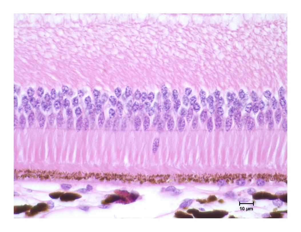
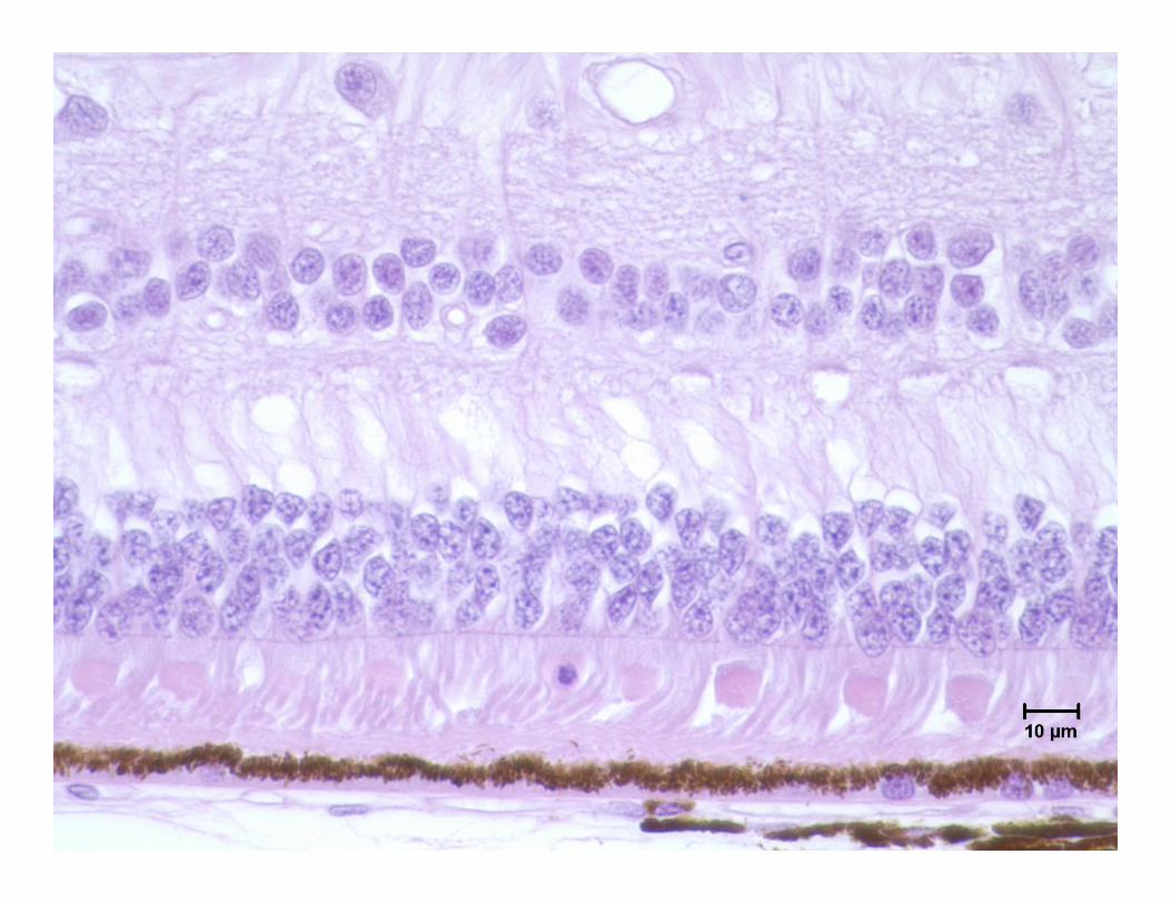
RETINAL EXAMINATION FOR PDN


• PDN were not counted in the disorganized area of peripheral retina and only counted in the peripheral retina that had clearly identifiable retinal layers.
• Only PDN located external to the outer limiting membrane (OLM) and in the layer of inner and outer segments (IS and OS) were counted.
• Because the transition point is arbitrary, the actual number of PDN in the peripheral retina was considered to be a relative number.
RETINAL EXAMINATION FOR PDN
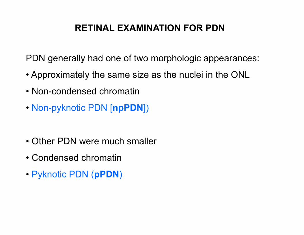
PDN generally had one of two morphologic appearances:
• Approximately the same size as the nuclei in the ONL
• Non-condensed chromatin
• Non-pyknotic PDN [npPDN])
• Other PDN were much smaller
• Condensed chromatin
• Pyknotic PDN (pPDN)
RETINAL EXAMINATION FOR PDN

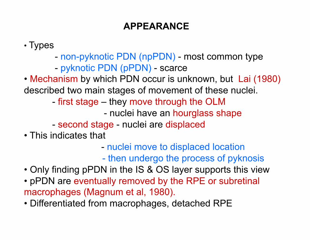
• Types - non-pyknotic PDN (npPDN) - most common type - pyknotic PDN (pPDN) - scarce
• Mechanism by which PDN occur is unknown, but Lai (1980) described two main stages of movement of these nuclei.
- first stage – they move through the OLM - nuclei have an hourglass shape - second stage - nuclei are displaced
• This indicates that - nuclei move to displaced location
- then undergo the process of pyknosis • Only finding pPDN in the IS & OS layer supports this view • pPDN are eventually removed by the RPE or subretinal macrophages (Magnum et al, 1980). • Differentiated from macrophages, detached RPE
APPEARANCE
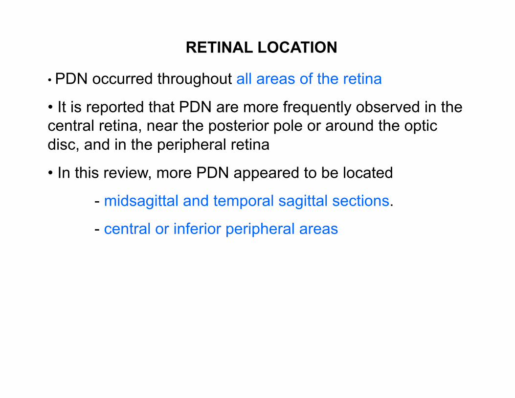
• PDN occurred throughout all areas of the retina
• It is reported that PDN are more frequently observed in the central retina, near the posterior pole or around the optic disc, and in the peripheral retina
• In this review, more PDN appeared to be located
- midsagittal and temporal sagittal sections.
- central or inferior peripheral areas
RETINAL LOCATION

• Displaced photoreceptor nuclei (PDN) have been reported – humans – monkeys (cynomolgus monkeys) – dogs – cats – pigs – rabbits – guinea pigs – rats – mice – chickens
OCCURENCE
• Non-injected globes indicating spontaneous cause. • Injected globes, so possibly induced cause.
Study Number Number of Animals
(Total = 32)
Globe Total (Average number
of npPDN)
Total (Average
number of pPDN)
1-N- 09-159
4 Left (NIG)
2 (<1)
0 (0)
Right (IG)
2 (<1)
0 (0)
2- N- 10-031
4 Left (NIG)
13 (3)
0 (0)
Right (IG)
40 (10)
0 (0)
3- N- 10-121
6 Left (NIG)
10 (2)
0 (0)
Right (IG)
12 (2)
0 (0)
4- N- 10-104
6 Left (NIG)
17 (3)
0 (0)
Right (IG)
21 (4)
2 (<1)
5- N- 09-104
12 Left (IG)
61 (5)
0 (0)
Right (IG)
50 (4)
1 (<1)
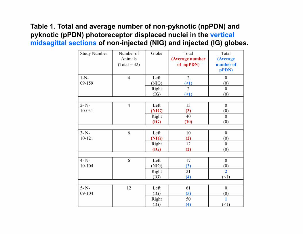
Table 1. Total and average number of non-pyknotic (npPDN) and pyknotic (pPDN) photoreceptor displaced nuclei in the vertical midsagittal sections of non-injected (NIG) and injected (IG) globes.
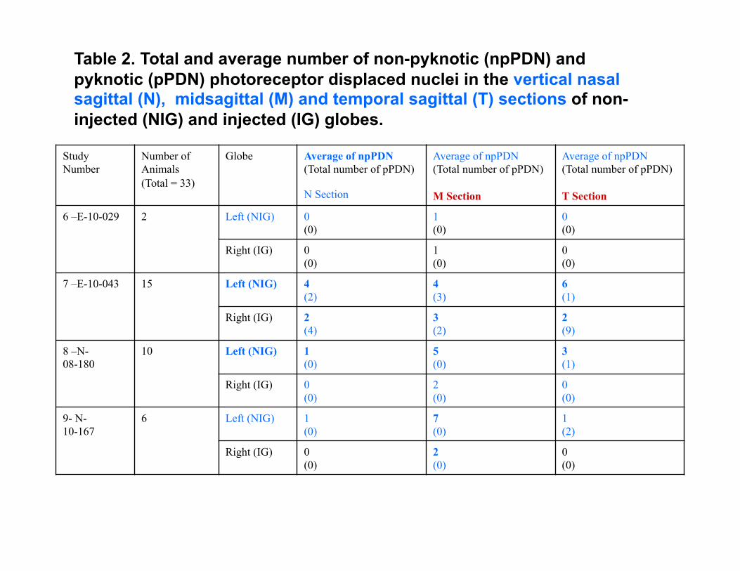
Table 2. Total and average number of non-pyknotic (npPDN) and pyknotic (pPDN) photoreceptor displaced nuclei in the vertical nasal sagittal (N), midsagittal (M) and temporal sagittal (T) sections of non-injected (NIG) and injected (IG) globes.
Study Number
Number of Animals (Total = 33)
Globe Average of npPDN (Total number of pPDN)
N Section
Average of npPDN (Total number of pPDN)
M Section
Average of npPDN (Total number of pPDN)
T Section
6 –E-10-029 2 Left (NIG) 0 (0)
1 (0)
0 (0)
Right (IG) 0 (0)
1 (0)
0 (0)
7 –E-10-043 15 Left (NIG) 4 (2)
4 (3)
6 (1)
Right (IG) 2 (4)
3 (2)
2 (9)
8 –N- 08-180
10 Left (NIG) 1 (0)
5 (0)
3 (1)
Right (IG) 0 (0)
2 (0)
0 (0)
9- N- 10-167
6 Left (NIG) 1 (0)
7 (0)
1 (2)
Right (IG) 0 (0)
2 (0)
0 (0)

Globe Retinal Area Total (Average) number of
npPDN
N Section
Total (Average) number of
npPDN
M Section
Total (Average) number of
npPDN
T Section Left (NIG) Superior 9
(<1) 30
(<1) 27
(<1) Central 44
(1) 89 (2)
26 (<1)
Inferior 26 (<1)
34 (1)
90 (2)
Right (IG) Superior 3 (<1)
16 (<1)
10 (<1)
Central 5 (<1)
19 (<1)
18 (<1)
Inferior 21 (<1)
29 (<1)
22 (<1)
Table 3. Total and average whole number of non-pyknotic (npPDN) photoreceptor displaced nuclei in superior (S), central (C), or inferior (I) areas of the vertical nasal sagittal (N), midsagittal (M) and temporal sagittal (T) sections of 33 non-injected (NIG) and 33 injected (IG) globes from four studies.
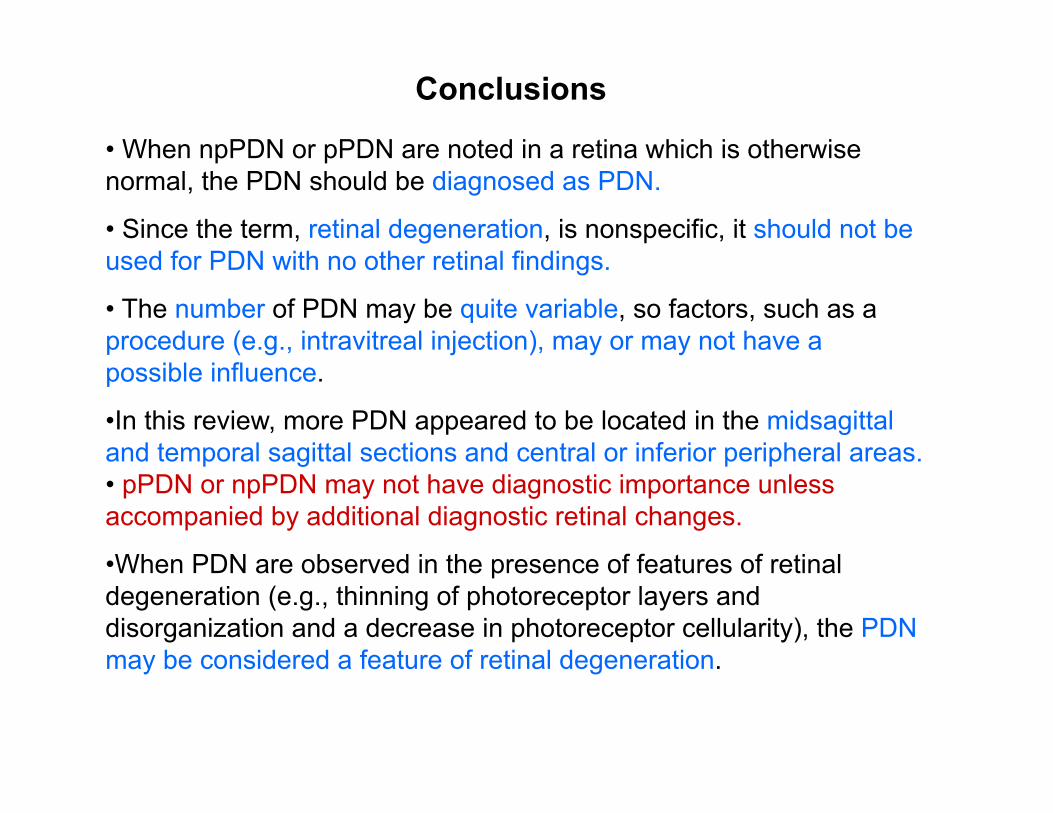
• When npPDN or pPDN are noted in a retina which is otherwise normal, the PDN should be diagnosed as PDN.
• Since the term, retinal degeneration, is nonspecific, it should not be used for PDN with no other retinal findings.
• The number of PDN may be quite variable, so factors, such as a procedure (e.g., intravitreal injection), may or may not have a possible influence.
• In this review, more PDN appeared to be located in the midsagittal and temporal sagittal sections and central or inferior peripheral areas. • pPDN or npPDN may not have diagnostic importance unless accompanied by additional diagnostic retinal changes.
• When PDN are observed in the presence of features of retinal degeneration (e.g., thinning of photoreceptor layers and disorganization and a decrease in photoreceptor cellularity), the PDN may be considered a feature of retinal degeneration.
Conclusions
REFERENCES • Armengol, J.A., Prada, F., Génis-Gálvez, J.M. (1985). Photoreceptor nuclei outside of
the limiting membrane in the chick retina. Acta anat 122, 201-204. • Gartner, S., Henkind, P. (1981). Aging and degeneration of the human macula.1.
Outer nuclear layer and photoreceptors. Brit J Ophthalmol 65, 23-28. • Geiss, V., Yoshitomi, K. (1999). Eyes, In: Pathology of the Mouse. Reference and
Atlas. (eds. R.R. Maronpot, G.A. Boorman, B.W.Gaul), Cache River Press, Vienna, pg 479.
• Hao, W., Wenzel, A., Obin, M.S., Ching-Kang, C., Brill, E., Krasnoperova, N.V., Eversole-Cire, P., Kleyner, Y., Taylor, A., Simon, M.I., Grim, C., Remé, C.E., Lem, J. (2002). Evidence for two apoptotic pathways in light-induced retinal degeneration. Nature Genetics 32, 254-260.
• Lai, Y-L. (1980). Outward movement of photoreceptor cells in normal rat retina. Invest Ophthalmol Vis Sci 19, 849-856.
• Lai, Y-L., Jacoby, R.O., Jonas, A.M. (1978). Age-related and light-associated retinal changes in Fischer rats. Invest Ophthalmol Vis Sci 17, 634-638.
• Lai, Y-L., Jonas, A.M. (1977). Rat model for hereditary retinal degeneration. Adv Exp Med. 77, 115-136.
• Lai, Y-L., Masuda, K., Mangum, M.D., Lug, R., Macrae, D.W., Fletcher, G., Liu, Y-P. (1982). Subretinal displacement of photoreceptor nuclei in human retina. Exp Eye Res 34, 219-228.
• Mangum, M.D., Lug R., Lai Y-L. (1980). Subretinal photoreceptor cells. Invest Ophthalmol Vis Sci 19 (Suppl), 191.
• Saunders L.Z., Rubin L.F. (1975). The Retina, In: Ophthalmic Pathology of Animals. An atlas and reference book. S Karger, Basel, pp 122-123.