photochemical & photobiological...
TRANSCRIPT
Photochemical &Photobiological Sciences
PAPER
Cite this: Photochem. Photobiol. Sci.,2015, 14, 583
Received 13th July 2014,Accepted 16th October 2014
DOI: 10.1039/c4pp00272e
www.rsc.org/pps
Combined cytotoxic effect of UV-irradiation andTiO2 microbeads in normal urothelial cells,low-grade and high-grade urothelial cancer cells
Roghayeh Imani,a,b Peter Veranič,c Aleš Iglič,b Mateja Erdani Kreft,c Meysam Pazokid
and Samo Hudoklin*c
The differentiation of urothelial cells results in normal terminally differentiated cells or by alternative path-
ways in low-grade or high-grade urothelial carcinomas. Treatments with traditional surgical and chemo-
therapeutical approaches are still inadequate and expensive, as bladder tumours are generally highly
recurrent. In such situations, alternative approaches, using irradiation of the cells and nanoparticles, are
promising. The ways in which urothelial cells, at different differentiation levels, respond to UV-irradiation
(photolytic treatment) or to the combination of UV-irradiation and nanoparticles (photocatalytic treat-
ment), are unknown. Here we tested cytotoxicity of UV-irradiation on (i) normal porcine urothelial cells
(NPU), (ii) human low-grade urothelial cancer cells (RT4), and (iii) human high-grade urothelial cancer
cells (T24). The results have shown that 1 minute of UV-irradiation is enough to kill 90% of the cells in
NPU and RT4 cultures, as determined by the live/dead viability assay. On the other hand, the majority of
T24 cells survived 1 minute of UV-irradiation. Moreover, even a prolonged UV-irradiation for 30 minutes
killed <50% of T24 cells. When T24 cells were pre-supplemented with mesoporous TiO2 microbeads and
then UV-irradiated, the viability of these high-grade urothelial cancer cells was reduced to <10%, which
points to the highly efficient cytotoxic effects of TiO2 photocatalysis. Using electron microscopy, we
confirmed that the mesoporous TiO2 microbeads were internalized into T24 cells, and that the cell’s ultra-
structure was heavily compromised after UV-irradiation. In conclusion, our results show major differences
in the sensitivity to UV-irradiation among the urothelial cells with respect to cell differentiation. To
achieve an increased cytotoxicity of urothelial cancer cells, the photocatalytic approach is recommended.
Introduction
The urothelium is a unique three layered epithelium thatcovers most of the mammalian urinary tract, including theurinary bladder, and is responsible for maintaining the tight-est permeability barrier in the human body, the so-calledblood–urine permeability barrier.1–4 In a normal urothelium,the formation and maintenance of the blood–urine barrierdepends on the processes of urothelial differentiation. Theseinclude: (i) the characteristic structure of the cell’s apicalplasma membrane, (ii) the low-level of molecule internaliz-
ation into cells, and (iii) the highly resistant tightjunctions.5–10 Superficial urothelial cells synthesize uro-thelium-specific transmembrane proteins, uroplakins(UPs),11,12 which are glycosylated in the Golgi apparatus,arranged into 16 nm particles and are organized into detergentresistant 2D crystals, called urothelial plaques.13–17 During celldifferentiation and during the filling of the bladder withurine, urothelial plaques are being transported to the apicalplasma membrane with the fusiform vesicles.18,19 70–90% ofthe apical plasma membrane is therefore covered with theplaques, which significantly contributes to the transcellularbarrier of the urothelial cells and gives their luminal mem-brane a characteristic scalloped appearance.20
Moreover, internalization studies have shown that endo-cytotic activity is 43% to 86% lower in differentiated superficialurothelial cells in comparison with the partially differentiatedurothelial cells, and 5 to 15-times lower than in polarizedMDCK cells, which contain no urothelial plaques.21 Therefore,for the intact blood–urine permeability barrier the normalurothelial differentiation with the urothelial plaque formationand a reduced internalization rate is necessary.22–24
aLaboratory of Clinical Biophysics, Faculty of Health Sciences, University of
Ljubljana, Zdravstvena 5, Ljubljana, SloveniabLaboratory of Biophysics, Faculty of Electrical Engineering, University of Ljubljana,
Tržaška 25, Ljubljana, SloveniacInstitute of Cell Biology, Faculty of Medicine, University of Ljubljana, Vrazov trg 2,
Ljubljana, Slovenia. E-mail: [email protected] of Chemistry, Ångström Laboratory, Physical Chemistry, University of
Uppsala, Box 523, SE 75120 Uppsala, Sweden
This journal is © The Royal Society of Chemistry and Owner Societies 2015 Photochem. Photobiol. Sci., 2015, 14, 583–590 | 583
Ope
n A
cces
s A
rtic
le. P
ublis
hed
on 1
7 O
ctob
er 2
014.
Dow
nloa
ded
on 2
6/03
/201
5 06
:59:
08.
Thi
s ar
ticle
is li
cens
ed u
nder
a C
reat
ive
Com
mon
s A
ttrib
utio
n 3.
0 U
npor
ted
Lic
ence
.
View Article OnlineView Journal | View Issue
On the other hand, urothelial differentiation can also takealternative pathways, which result in the urinary bladdercancer with an inverse relation of the differentiation level tothe cancer cell grade.3,25 The urothelial cancer is the 4th mostcommon type of cancer in men, with 430 000 new diagnosesworldwide every year.26,27 Current approaches to the treatmentof the most common urothelial cancer in humans include thetransurethral papilloma resection and the local application ofchemotherapy or immunotherapy.28,29 The high rate of recur-rence makes these treatments rather inadequate and the life-time treatment and monitoring costs of patients withurothelial carcinomas are the highest among of all cancers.26
To this end, alternative or supplemental approaches to treat-ment are being tested, including photocatalytic treatments.The rationale behind the photocatalytic treatment of bladdercancer consists of 2 steps: (i) urothelial cancer cells are at alower level of differentiation and they can therefore internalizemore nanoparticles than normal, highly differentiated uro-thelial cells, and (ii) the irradiation of cells with sufficientenergy (e.g. UV-irradiation) would cause a photocatalytic gene-ration of reactive oxidative species (ROS) in the cells with inter-nalized nanoparticles, which would in turn cause sufficientcytotoxic effects (e.g. lipid, protein and DNA lesions) to lethallydamage cancer cells.
Recently, titanium dioxide (TiO2) has emerged as an excel-lent biocompatible photocatalyst material.19,30,31 In particular,TiO2 has been useful as a catalyst for the photodegradation oforganic compounds and the deactivation of microorganismsby photogenerated ROS.32,33 It has been reported that variousROS, such as superoxide (O2
•−), singlet oxygen (1O2), thehydroxyl radical (•OH), the hydroperoxyl radical (HO2
•), andhydrogen peroxide (H2O2), are generated on the TiO2 surfaceand react with organic or inorganic compounds in the gas andliquid phases.34 Studies have shown that the geometry ofnanoparticles may have a significant effect on the photo-electrolysis activity.34 In addition, the photocatalytic activity ofTiO2 crystals is heavily dependent on the surface structure,including surface atomic arrangement and coordination,especially when the particle size is reduced to the nanometerscale, leading to a large effective surface area.35,36
UV-irradiation is well known for its cytotoxic effects, amongwhich DNA lesions and their consequences have been studiedin great detail.37 In general, DNA molecules exhibit strong UVabsorption in the wavelength range of ∼220–300 nm, with themaximum peak at 260 nm and act as the major cellularchromophores for the UV-C spectrum of irradiation.38 Thedirect absorption of UV-irradiation induces the formation ofcyclobutane pyrimidine (CPD) dimers, (6-4) pyrimidine-pyrimi-done photoproducts and their Dewar isomers,39 which if un-repaired would block the transcription of DNA genes to RNAand would eventually lead to the cell death or initiatephotocarcinogenesis.40,41
To the best of our knowledge, the cytotoxic effects of UV-irradiation on differently differentiated urothelial cells havenot yet been studied. In this paper, we studied the sensitivityof three differently differentiated cultures of urothelial cells to
UV-irradiation. To increase the photocatalytic damage and theselectivity of treatment to high-grade urothelial cancer cells,the cells were supplemented with mesoporous TiO2 micro-beads before exposing them to UV-irradiation.
Results and discussion
The UV-spectrum is divided into three regions UV-A(315–400 nm), UV-B (280–315) nm and UV-C (100–280 nm).For phototherapies used in clinics for the treatment of patho-logies, such as psoriasis, vitiligo, atopic dermatitis or mycosisfungoides, relatively non-harmful UV-A and UV-B spectra aregenerally used.42,43 On the other hand, for the purpose ofefficiently killing urothelial cancer cells, UV-C irradiation waschosen, which has the highest energy content among UV-spectra and causes the most damaging effects to the cells.44
Cell treatment showed that normal porcine urothelial cells(NPU) and human low-grade non-invasive cancer RT4 cells aresignificantly more prone to UV-irradiation damage thanhuman high-grade and invasive urothelial cancer T24 cells.Twenty-four hours after 1 minute of UV-irradiation, themorphological and ultrastructural appearance of NPU and RT4cell cultures changed from confluent with polygonal cells(Fig. 1a and e) to sporadic with frequently rounded cells(Fig. 1c and g). Many cells were detached and floated in thegrowth medium. The live/dead viability assay indicated a highlevel of cytotoxicity of UV-irradiation for NPU and RT4 cells: inthe control cultures the cells were >95% live (labelled green;Fig. 1b and f), while in the UV-irradiated cultures thereremained <10% live cells (Fig. 1d and h). On the other hand,the morphological appearance of irradiated T24 cells remainedunchanged 24 hours after 1 minute of UV-irradiation (figuresnot shown). The live/dead viability assay showed that T24 cellswere still viable after such a treatment. Moreover, even afterprolonged UV-irradiation (30 minutes), the morphology of theT24 cultures remained mainly unchanged (Fig. 1i and k), withthe majority of cells being viable (Fig. 1j and l). Neither NPUnor RT4 cells survived 30 minutes of UV-irradiation.
Our results showed that even relatively high doses of veryphotolytic UV-C irradiation did not eliminate all high-gradeurothelial cancer cells. It is not likely that the DNA of high-grade urothelial cancer cells is better protected or less suscep-tible to UV-C irradiation damage in comparison to the DNA ofmore differentiated cells.45 In some cell types, very proficientDNA repair systems were found to cope with various kinds ofDNA damage: mismatch repair, base excision repair, directdamage reversal, double strand break repair and nucleotideexcision repair.46,47 The better survival of less differentiatedT24 cells is in accordance with the studies that show that theDNA repair system is differentiation dependent.48 In general,the repair system is attenuated with the progression of celldifferentiation: in rat neurons, chicken striated muscles,human macrophages, mouse keratinocytes and others.48,49 Wesuggest that urothelial high-grade cancer cells, which are at alower differentiation stage than the RT4 and NPU cells, are
Paper Photochemical & Photobiological Sciences
584 | Photochem. Photobiol. Sci., 2015, 14, 583–590 This journal is © The Royal Society of Chemistry and Owner Societies 2015
Ope
n A
cces
s A
rtic
le. P
ublis
hed
on 1
7 O
ctob
er 2
014.
Dow
nloa
ded
on 2
6/03
/201
5 06
:59:
08.
Thi
s ar
ticle
is li
cens
ed u
nder
a C
reat
ive
Com
mon
s A
ttrib
utio
n 3.
0 U
npor
ted
Lic
ence
.View Article Online

also more resistant to structural lesions and apoptosis,50
which makes for the difference in the cells’ survival rate.To test the cytotoxic potential of mesoporous TiO2
microbead photocatalysis and to increase the selectivity indamaging the predominantly less differentiated urothelialcancer cells with an elevated level of endocytotic activity, thegrowth medium of T24 cells was supplemented with meso-porous TiO2 microbeads. The mesoporous TiO2 microbeadswere prepared by the solvothermal method. These microbeadsare monodispersed TiO2 with a diameter of 600 ± 100 nm(Fig. 2). The microbeads have rough surfaces made of ∼15 nmsized TiO2 nanocrystals organized in such a way that they formpores into the internal structure of the microbeads. Highsurface area, light harvesting and scattering efficiency togetherwith their high crystallinity make them very promising forkilling the cancer cells (data under publication elsewhere).
Crystallinity and low trap density of the microbeads, whichallows the fast diffusion of electrons, facilitate electron donat-ing properties (i.e. higher reactive oxidative species generation)
and impede the electron–hole recombination processes, arepossible reasons for the high efficiency of the here used TiO2
microbeads compared to commercial nanoparticles.51
Such mesoporous TiO2 microbeads were left to be interna-lized by T24 urothelial cancer cells. The leftovers of the
Fig. 1 Morphology and viability of normal porcine urothelial cells (NPU), and cancer RT4 and T24 cells in control and UV-irradiated cultures. Notethe changed morphology and the reduced number of NPU (c) and RT4 cells (g) 24 hours after 1 minute of UV-irradiation in comparison to theirnon-irradiated controls (a, e). The number of NPU and RT4 cells labeled green (live cells) has significantly decreased after the UV-irradiation (d, hversus b, f ). The cells labelled red (dead cells) were detached from the growth medium and are therefore not seen on the panels (d, h). On the otherhand, the high fraction of T24 cells retained their morphology and survived 24 hours after 30 minutes of UV-irradiation (k, l versus i, j). Legend:green – live cells, red – dead cells. Bars: 100 µm.
Fig. 2 Scanning electron microscopy image of mesoporous TiO2
microbeads in (a) low and (b) high magnification. Legend: asterisks –
microbeads, black arrow – individual TiO2 particle, white arrow – poresin the surface structure of the microbead. Bars: 250 nm.
Photochemical & Photobiological Sciences Paper
This journal is © The Royal Society of Chemistry and Owner Societies 2015 Photochem. Photobiol. Sci., 2015, 14, 583–590 | 585
Ope
n A
cces
s A
rtic
le. P
ublis
hed
on 1
7 O
ctob
er 2
014.
Dow
nloa
ded
on 2
6/03
/201
5 06
:59:
08.
Thi
s ar
ticle
is li
cens
ed u
nder
a C
reat
ive
Com
mon
s A
ttrib
utio
n 3.
0 U
npor
ted
Lic
ence
.View Article Online

microbeads were removed from the growth medium, and sub-sequently the cultures were UV-irradiated for 30 minutes. Thelive/dead viability assay revealed that 24 hours after irradiationthe viability of T24 cells pre-supplemented with mesoporousTiO2 microbeads was significantly reduced in comparison withT24 cells that were UV-irradiated, but contained no meso-porous TiO2 microbeads (Fig. 3a and d). The mesoporous TiO2
microbeads alone proved to be non-toxic for the cells (Fig. 3b).When the T24 cultures were UV-irradiated, 58% of the cellswere labelled green (Fig. 3c), and were thus live, while in theUV-irradiated cultures pretreated with mesoporous TiO2
microbeads there were <10% of the green labelled cells(Fig. 3d). Scanning and transmission electron microscopy wasused to localize the mesoporous TiO2 microbeads in the cellculture and determine their photocatalytic effects on the cell’sultrastructure (Fig. 3e–l). Mesoporous TiO2 microbeads werelocated at the apical side of the plasma membranes and intra-cellularly, in the membrane compartments (Fig. 3h, j and l).
Occasionally, mesoporous TiO2 microbeads were located inT24 cells in the invaginations characteristic of phagocytosis(Fig. 3l). The ultrastructure of the examined cells pre-treatedwith TiO2 microbeads and UV-irradiated (Fig. 3l) was signifi-cantly changed in comparison to control cells and to the TiO2
microbead pre-treated or UV-irradiated only cells (Fig. 3i–k). Inthe UV-irradiated cells loaded with mesoporous TiO2 micro-beads, the plasma membrane was discontinuous, clearlyshowing holes (Fig. 3h). Their cytoplasm lost its fine, homo-geneous appearance and looked washed out (Fig. 3l). Thecell’s intracellular membrane compartments were distended,with ruptured internal membrane structures, which is aknown characteristic of the necrotic cells. The emphasizednecrotic cell death is most likely a consequence of the photo-catalytic effects of mesoporous TiO2 microbeads pre-sup-plemented to the cells. The photocatalysis of TiO2 generallyinvolves four processes: (i) the generation of electrons andholes by photoexcitation; (ii) the migration of the photo-
Fig. 3 Viability, scanning- and transmission electron microscopy of high-grade urothelial cancer T24 cells 24 hours after the treatment. The firstcolumn shows control cells, the second column the cells pre-treated with mesoporous TiO2 microbeads, but not exposed to UV-irradiation, thethird column shows the cells exposed to UV-irradiation for 30 minutes, and the fourth column shows the cells pre-treated with mesoporous TiO2
microbeads and UV-irradiated for 30 minutes. Note that in the last column there is a significantly increased number of cells labelled red (dead cells;d), and that perforations (white arrow; h) can be seen in the apical plasma membrane of the cells and TiO2 microbeads (black arrow; l), associatedwith the damaged cell’s ultrastructure. Legend: green – live cells, red – dead cells, asterisk – remains of the cell, black arrows – TiO2 microbeads,white arrow – perforation in the plasma membrane, M – mitochondria. Bars: a–d – 100 µm, e–h – 10 µm, i–l – 1 µm.
Paper Photochemical & Photobiological Sciences
586 | Photochem. Photobiol. Sci., 2015, 14, 583–590 This journal is © The Royal Society of Chemistry and Owner Societies 2015
Ope
n A
cces
s A
rtic
le. P
ublis
hed
on 1
7 O
ctob
er 2
014.
Dow
nloa
ded
on 2
6/03
/201
5 06
:59:
08.
Thi
s ar
ticle
is li
cens
ed u
nder
a C
reat
ive
Com
mon
s A
ttrib
utio
n 3.
0 U
npor
ted
Lic
ence
.View Article Online
generated charge carriers to the surface of TiO2 microbeads;(iii) the subsequent reduction/oxidization of the adsorbed reac-tants directly by electrons/holes or indirectly by ROS; and (iv)the recombination of the photogenerated electron–hole pairs.The efficient photocatalytic material is expected to promoteprocesses (i), (ii), and (iii) and to suppress process (iv).36
The mesoporous microbeads used here consist of a bundleof ∼15 nm sized TiO2 particles, which form sub-micron porousspheres with the superior light-harvesting properties in com-parison to the photocatalytic material made of small individ-ual particles. Moreover, mesoporous TiO2 microbeads were notonly shown to exhibit strong UV-light scattering properties, butthe interface of small TiO2 crystallites in the microbeads canalso lead to a faster diffusion of electrons, which is beneficialfor photocatalysis and may also affect the electron–hole recom-bination.52,53 Therefore, the selective internalization of TiO2
microbeads used here facilitates the production of ROS afterUV-irradiation, and can be recommended for efficient andselective treatment of cancer cells.54
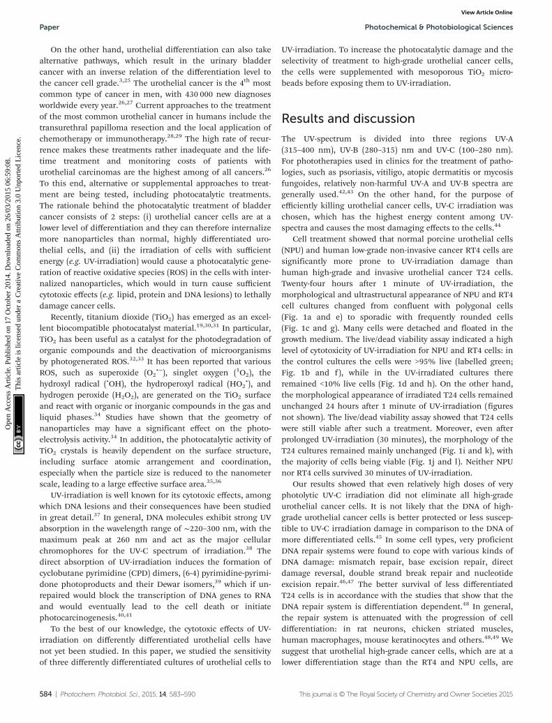
The synergistic effect of the here tested combined treatmentwith TiO2 and UV-irradiation suits well to the clinicaldemands and recent experimental findings in the treatment ofbladder cancer. In clinical praxis the main problem in treatingbladder cancer is not how to eliminate the main population ofcancer cells, but in the accessibility of drugs to the remainingcancer cells, as they represent seeds for the new urothelialtumours.55 After the conventional treatment these remainingcancer cells are supposed to spread within the normal uro-thelium where they are protected from chemotherapeuticdrugs by the tight blood–urine barrier of normal urothelialcells (Fig. 4a). Such persisting cancer cells are supposed to bemainly responsible for the relapse of bladder tumours.56
Therefore, in order to treat hidden cancer cells, it is necessaryto remove the superficial layer of differentiated urothelial cells(umbrella cells; Fig. 4b) in the first step. UV-C irradiation usedhere was proved to be successful in the removal of normalsuperficial cells, while the cell irradiated with longer wave-lengths (e.g. UV-A) failed to remove these cells (unpublisheddata). In the second step, the highly efficient light harvestingphotocatalytic material introduced into urinary bladder lumen(i.e. TiO2 microbeads) should be internalized into the cancercells (Fig. 4c). We have recently proven that less differentiatedhave a highly increased potential of endocytosis in comparisonto normal urothelial cells (ref. 7 and unpublished results)giving a strong emphasis on the selectivity of such a treatment.Next, the enhanced phototoxicity of UV-irradiation in cells thatendocytosed TiO2 microbeads speaks well in favour of theselective treatment of exposed cancer cells after the removal ofumbrella cell shield by UV-C irradiation (Fig. 4d and e). Sincethe energy for generating the photocatalytic effects of TiO2
microbeads can be lower than the one needed for the removalof the umbrella cells, one might reduce the intensity of UV-Cirradiation to harm only the cancer cells or use UV-Airradiation in this step. That would selectively kill cancer cellscontaining microbeads, but preserve normal cells in theurothelium. Finally, the regeneration and differentiation of the
remaining normal urothelial cells restores the urothelium andits blood–urine barrier function (Fig. 4f). Exceedingly rapidregeneration of the urothelial tissue, recovering within lessthan an hour, also prevents unfavourable effects of the barrierremoval and the potential toxic effect on regenerating normalcells, caused by UV-C irradiation.57
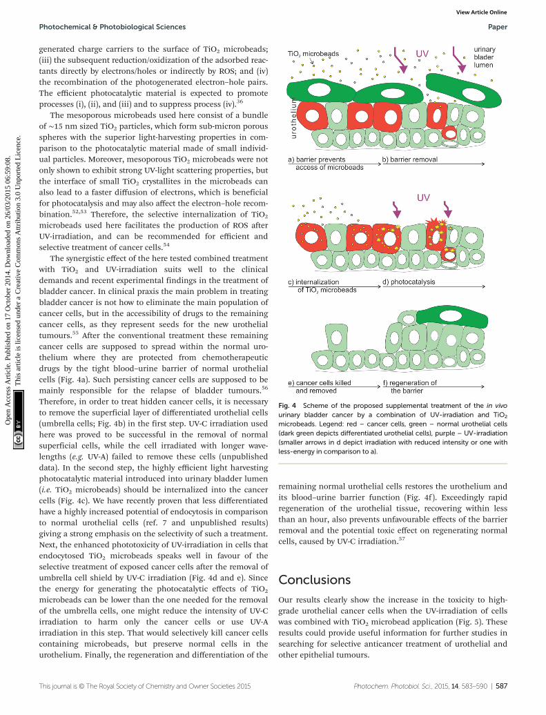
Conclusions
Our results clearly show the increase in the toxicity to high-grade urothelial cancer cells when the UV-irradiation of cellswas combined with TiO2 microbead application (Fig. 5). Theseresults could provide useful information for further studies insearching for selective anticancer treatment of urothelial andother epithelial tumours.
Fig. 4 Scheme of the proposed supplemental treatment of the in vivourinary bladder cancer by a combination of UV-irradiation and TiO2
microbeads. Legend: red – cancer cells, green – normal urothelial cells(dark green depicts differentiated urothelial cells), purple – UV-irradiation(smaller arrows in d depict irradiation with reduced intensity or one withless-energy in comparison to a).
Photochemical & Photobiological Sciences Paper
This journal is © The Royal Society of Chemistry and Owner Societies 2015 Photochem. Photobiol. Sci., 2015, 14, 583–590 | 587
Ope
n A
cces
s A
rtic
le. P
ublis
hed
on 1
7 O
ctob
er 2
014.
Dow
nloa
ded
on 2
6/03
/201
5 06
:59:
08.
Thi
s ar
ticle
is li
cens
ed u
nder
a C
reat
ive
Com
mon
s A
ttrib
utio
n 3.
0 U
npor
ted
Lic
ence
.View Article Online
ExperimentalCell cultures
Three types of urinary bladder epithelial cells were used forthe experiments: normal porcine urothelial cells (NPU; cellsisolated from a healthy pig and further prepared and differen-tiated as described previously),58 the RT4 cell line (human low-grade and noninvasive urothelial carcinoma cells), and the T24cell line (human high-grade and invasive urothelial carcinomacells). Each type of cells was seeded on glass coverslips withinPetri dishes and cultured to >85% confluence in the UroM oradvanced-DMEM-F12 medium as described previously.6,59
Petri dishes were then divided into 2 groups: (1) the controlgroup and (2) the experimental group (+UV). Normal porcineurothelial cells, RT4 and T24 cells of the experimental groupwere irradiated for 1 or 30 minutes with the UV-C light (Sylva-nia Ultra Violet G15W; 15 W cm−2); the irradiation of the cellsin the control group was omitted. In the next step, cells weregrown for additional 24 hours in the CO2-incubator at 37 °Cunder a humidified atmosphere of 5% CO2 (v/v), and were sub-sequently sampled for morphology, live/dead viability assay,and ultrastructural analysis.
Mesoporous TiO2 microbeads synthesis and characterization
Mesoporous TiO2 microbeads were synthesized by the sol-vothermal method according to our previous report.51 Themesoporous TiO2 microbead particle morphology was exam-ined with a S4700 scanning electron microscope (Hitachi).
Morphological characterization of the cells
The samples of the control and experimental cell cultures wereexamined unprocessed. The samples were taken from the CO2-incubator and were immediately inspected with the T300phase-contrast light microscope (Nikon).
Live/dead viability assay
To evaluate the cytotoxic effects of the mesoporous TiO2
microbeads and UV-light irradiation, a Live/Dead Viability Kit(Invitrogen, Life Technologies) was used. Cells attached to thecoverslips were processed according to the manufacturer’s pro-tocol, and visualized and photographed 25 minutes afteradding the kit with a T300 fluorescence-light microscope(Nikon). The green signal characterized the live cells and thered signal characterized the dead cells. For each cell group,four coverslips and five fields on each coverslip wereexamined.
To evaluate the cytotoxic effect of the mesoporous TiO2
microbeads in combination with UV-irradiation, T24 cells wereincubated for 2 hours in the cell growth medium, sup-plemented with 50 µg ml−1 of mesoporous TiO2 microbeads.Afterwards the medium was changed for a fresh one withoutthe TiO2 microbeads, and the cell cultures were irradiated for30 minutes as above. After 24 hours, the cultures weresampled for the morphological study, the live/dead viabilityassay, transmission electron microscopy (TEM) and scanningelectron microscopy (SEM) analysis.
Ultrastructural analysis
Samples were fixed with 4% formaldehyde and 2% glutar-aldehyde in 0.1 M cacodylate buffer and subsequently pro-cessed for TEM and SEM. For TEM, samples were embeddedin Epon, sectioned, counterstained and examined with aCM100 TEM (Philips) operating at 80 kV. For SEM, thesamples were dehydrated, dried, sputter-coated and examinedwith a JSM840A SEM (Jeol) at 15 kV.
Acknowledgements
This work was in part supported by the Slovenian ResearchAgency (ARRS; grant no. P3-0108, P2-0232, P3-0388, J1-4136,J3-4108 and J1-4109). The funders had no role in study design,data collection and analysis, decision to publish, or prepa-ration of the manuscript. The authors would like to thankSanja Čabraja, Linda Štrus, Sabina Železnik and especiallyNada Dubarič Pavlica for all the technical assistance and help.
Notes and references
1 M. E. Kreft, S. Hudoklin, K. Jezernik and R. Romih, Proto-plasma, 2010, 246, 3–14.
2 F. X. Liang, M. C. Bosland, H. Huang, R. Romih,S. Baptiste, F. M. Deng, X. R. Wu, E. Shapiro and T. T. Sun,J. Cell Biol., 2005, 171, 835–844.
Fig. 5 Schematic presentation of the mechanism for the cytotoxicpotential of photocatalytic TiO2 microbeads in high grade urothelialcancer cells. (a) Mesoporous TiO2 microbeads are supplemented intothe lumen of urinary bladder, where they interact with (b) and are endo-cytosed (c) preferably by cancer cells, which are at the lower stage ofdifferentiation. (d) The UV-irradiation of microbeads triggers the photo-catalytic production of reactive oxidative species, which highly increasesthe efficiency of killing cancer cells.
Paper Photochemical & Photobiological Sciences
588 | Photochem. Photobiol. Sci., 2015, 14, 583–590 This journal is © The Royal Society of Chemistry and Owner Societies 2015
Ope
n A
cces
s A
rtic
le. P
ublis
hed
on 1
7 O
ctob
er 2
014.
Dow
nloa
ded
on 2
6/03
/201
5 06
:59:
08.
Thi
s ar
ticle
is li
cens
ed u
nder
a C
reat
ive
Com
mon
s A
ttrib
utio
n 3.
0 U
npor
ted
Lic
ence
.View Article Online
3 X. R. Wu, X. P. Kong, A. Pellicer, G. Kreibich and T. T. Sun,Kidney Int., 2009, 75, 1153–1165.
4 H. O. Negrete, J. P. Lavelle, J. Berg, S. A. Lewis andM. L. Zeidel, Am. J. Physiol., 1996, 271, F886–F894.
5 S. A. Lewis and J. M. Diamond, J. Membr. Biol., 1976, 28,1–40.
6 T. Visnjar and M. E. Kreft, In Vitro Cell. Dev. Biol.: Anim,2013, 49, 196–204.
7 M. E. Kreft, K. Jezernik, M. Kreft and R. Romih, Ann. N. Y.Acad. Sci., 2009, 1152, 18–29.
8 R. Romih, P. Korosec, W. de Mello Jr. and K. Jezernik, CellTissue Res., 2005, 320, 259–268.
9 R. Romih, P. Veranic and K. Jezernik, Appl. Immunohisto-chem. Mol. Morphol., 2002, 10, 339–343.
10 P. Veranic, R. Romih and K. Jezernik, Eur. J. Cell Biol.,2004, 83, 27–34.
11 X. R. Wu, M. Manabe, J. Yu and T. T. Sun, J. Biol. Chem.,1990, 265, 19170–19179.
12 J. Yu, M. Manabe, X. R. Wu, C. Xu, B. Surya and T. T. Sun,J. Cell Biol., 1990, 111, 1207–1216.
13 B. Kachar, F. Liang, U. Lins, M. Ding, X. R. Wu,D. Stoffler, U. Aebi and T. T. Sun, J. Mol. Biol., 1999, 285,595–608.
14 G. Min, H. Wang, T. T. Sun and X. P. Kong, J. Cell Biol.,2006, 173, 975–983.
15 F. X. Liang, I. Riedel, F. M. Deng, G. Zhou, C. Xu, X. R. Wu,X. P. Kong, R. Moll and T. T. Sun, Biochem. J., 2001, 355,13–18.
16 S. Hudoklin, D. Zupancic and R. Romih, Cell Tissue Res.,2009, 336, 453–463.
17 C. C. Hu, T. Bachmann, G. Zhou, F. X. Liang, J. Ghiso,G. Kreibich and T. T. Sun, Biochem. J., 2008, 414, 195–203.
18 S. Hudoklin, K. Jezernik, J. Neumuller, M. Pavelka andR. Romih, PLoS One, 2012, 7, e32935.
19 P. Kocbek, K. Teskac, M. E. Kreft and J. Kristl, Small, 2010,6, 1908–1917.
20 S. A. Lewis, Am. J. Physiol. Renal Physiol., 2000, 278, F867–F874.
21 M. E. Kreft, R. Romih, M. Kreft and K. Jezernik, Differen-tiation, 2009, 77, 48–59.
22 P. Hu, F. M. Deng, F. X. Liang, C. M. Hu, A. B. Auerbach,E. Shapiro, X. R. Wu, B. Kachar and T. T. Sun, J. Cell Biol.,2000, 151, 961–972.
23 P. Hu, S. Meyers, F. X. Liang, F. M. Deng, B. Kachar,M. L. Zeidel and T. T. Sun, Am. J. Physiol. Renal Physiol.,2002, 283, F1200–F1207.
24 X. T. Kong, F. M. Deng, P. Hu, F. X. Liang, G. Zhou,A. B. Auerbach, N. Genieser, P. K. Nelson, E. S. Robbins,E. Shapiro, B. Kachar and T. T. Sun, J. Cell Biol., 2004, 167,1195–1204.
25 P. Khandelwal, S. N. Abraham and G. Apodaca,Am. J. Physiol. Renal Physiol., 2009, 297, F1477–F1501.
26 K. D. Sievert, B. Amend, U. Nagele, D. Schilling, J. Bedke,M. Horstmann, J. Hennenlotter, S. Kruck and A. Stenzl,World J. Urol., 2009, 27, 295–300.
27 L. W. Fei Ye, M. Castillo-Martin, R. McBride, M. D. Galsky,J. Zhu, P. Boffetta, D. Y. Zhang and C. C.-C. Zhang,Am. J. Clin. Exp. Urol., 2014, 2, 1–14.
28 J. Bhatt, N. Cowan, A. Protheroe and J. Crew, Expert Rev.Anticancer Ther., 2012, 12, 929–939.
29 R. K. Lee, H. Abol-Enein, W. Artibani, B. Bochner,G. Dalbagni, S. Daneshmand, Y. Fradet, R. E. Hautmann,C. T. Lee, S. P. Lerner, A. Pycha, K. D. Sievert, A. Stenzl,G. Thalmann and S. F. Shariat, BJU Int., 2014, 113,11–23.
30 M. Li, G. Huang, Y. Qiao, J. Wang, Z. Liu, X. Liu and Y. Mei,Nanotechnology, 2013, 24, 305706.
31 A. L. Linsebigler, G. Lu and J. T. Yates, Chem. Rev., 1995,95, 735–758.
32 G. Gogniat and S. Dukan, Appl. Environ. Microbiol., 2007,73, 7740–7743.
33 Y. Guo, C. Cheng, J. Wang, Z. Wang, X. Jin, K. Li, P. Kangand J. Gao, J. Hazard Mater., 2011, 192, 786–793.
34 M. R. Hoffmann, S. T. Martin, W. Choi andD. W. Bahnemann, Chem. Rev., 1995, 95, 69–96.
35 G. K. Mor, K. Shankar, M. Paulose, O. K. Varghese andC. A. Grimes, Nano Lett., 2005, 6, 215–218.
36 N. Wu, J. Wang, N. Tafen de, H. Wang, J. G. Zheng,J. P. Lewis, X. Liu, S. S. Leonard and A. Manivannan, J. Am.Chem. Soc., 2010, 132, 6679–6685.
37 M. Ljungman and F. Zhang, Oncogene, 1996, 13, 823–831.38 J. S. Taylor, Acc. Chem. Res., 1994, 27, 76–82.39 J. L. Ravanat, T. Douki and J. Cadet, J. Photochem. Photobiol.
B, 2001, 63, 88–102.40 R. M. Costa, V. Chigancas, S. Galhardo Rda, H. Carvalho
and C. F. Menck, Biochimie, 2003, 85, 1083–1099.41 L. Proietti De Santis, C. L. Garcia, A. S. Balajee, P. Latini,
P. Pichierri, O. Nikaido, M. Stefanini and F. Palitti, DNARepair, 2002, 1, 209–223.
42 V. Bulat, M. Situm, I. Dediol, I. Ljubicic and L. Bradic, Coll.Antropol., 2011, 35(Suppl. 2), 147–151.
43 W. Lapolla, B. A. Yentzer, J. Bagel, C. R. Halvorson andS. R. Feldman, J. Am. Acad. Dermatol., 2011, 64, 936–949.
44 T. D. Cutler and J. J. Zimmerman, Anim. Health Res. Rev.,2011, 12, 15–23.
45 R. Yin, T. Dai, P. Avci, A. E. Jorge, W. C. de Melo,D. Vecchio, Y. Y. Huang, A. Gupta and M. R. Hamblin,Curr. Opin. Pharmacol., 2013, 13, 731–762.
46 T. Lindahl and R. D. Wood, Science, 1999, 286, 1897–1905.
47 A. Sancar, L. A. Lindsey-Boltz, K. Unsal-Kacmaz andS. Linn, Annu. Rev. Biochem., 2004, 73, 39–85.
48 T. Nouspikel, Neuroscience, 2007, 145, 1213–1221.49 T. Nouspikel and P. C. Hanawalt, DNA Repair, 2002, 1,
59–75.50 K. Wu, J. Zeng, J. Zhou, J. Fan, Y. Chen, Z. Wang,
T. Zhang, X. Wang and D. He, Urol. Oncol., 2013, 31,1751–1760.
51 M. Pazoki, N. Taghavinia, A. Hagfeldt and G. Boschloo,J. Phys. Chem. C, 2014, 118(30), 16472–16478.
Photochemical & Photobiological Sciences Paper
This journal is © The Royal Society of Chemistry and Owner Societies 2015 Photochem. Photobiol. Sci., 2015, 14, 583–590 | 589
Ope
n A
cces
s A
rtic
le. P
ublis
hed
on 1
7 O
ctob
er 2
014.
Dow
nloa
ded
on 2
6/03
/201
5 06
:59:
08.
Thi
s ar
ticle
is li
cens
ed u
nder
a C
reat
ive
Com
mon
s A
ttrib
utio
n 3.
0 U
npor
ted
Lic
ence
.View Article Online

52 J. A. Wang, R. Limas-Ballesteros, T. López, A. Moreno,R. Gómez, O. Novaro and X. Bokhimi, J. Phys. Chem. B,2001, 105, 9692–9698.
53 F. Sauvage, D. Chen, P. Comte, F. Huang, L. P. Heiniger,Y. B. Cheng, R. A. Caruso and M. Graetzel, ACS Nano, 2010,4, 4420–4425.
54 J. Rauch, W. Kolch, S. Laurent and M. Mahmoudi, Chem.Rev., 2013, 113, 3391–3406.
55 C. Hafner, R. Knuechel, R. Stoehr and A. Hartmann,Int. J. Cancer, 2002, 101, 1–6.
56 M. G. Wientjes, R. A. Badalament, R. C. Wang, F. Hassanand J. L. Au, Cancer Res., 1993, 53, 3314–3320.
57 P. Veranic, A. Erman, M. Kerec-Kos, M. Bogataj, A. Mrharand K. Jezernik, Histochem. Cell Biol., 2009, 131, 129–139.
58 T. Visnjar and M. E. Kreft, Histochem. Cell Biol., 2014, DOI:10.1007/s00418-014-1265-3.
59 R. Imani, D. Kabaso, M. Erdani Kreft, E. Gongadze,S. Penic, K. Elersic, A. Kos, P. Veranic, R. Zorec and A. Iglic,Croat. Med. J., 2012, 53, 577–585.
Paper Photochemical & Photobiological Sciences
590 | Photochem. Photobiol. Sci., 2015, 14, 583–590 This journal is © The Royal Society of Chemistry and Owner Societies 2015
Ope
n A
cces
s A
rtic
le. P
ublis
hed
on 1
7 O
ctob
er 2
014.
Dow
nloa
ded
on 2
6/03
/201
5 06
:59:
08.
Thi
s ar
ticle
is li
cens
ed u
nder
a C
reat
ive
Com
mon
s A
ttrib
utio
n 3.
0 U
npor
ted
Lic
ence
.View Article Online