phosphoinositide signaling in plant development · primer phosphoinositide signaling in plant...
TRANSCRIPT
PRIMER
Phosphoinositide signaling in plant developmentIngo Heilmann*
ABSTRACTThe membranes of eukaryotic cells create hydrophobic barriers thatcontrol substance and information exchange between the inside andoutside of cells and between cellular compartments. Besides theirroles as membrane building blocks, some membrane lipids, such asphosphoinositides (PIs), also exert regulatory effects. Indeed,emerging evidence indicates that PIs play crucial roles in controllingpolarity and growth in plants. Here, I highlight the key roles of PIs asimportant regulatory membrane lipids in plant development andfunction.
KEY WORDS: Lipids, Membranes, Phosphoinositides, Plant,Polarity, Recruitment
IntroductionBiological membranes act as a barrier limiting the free exchange ofmolecules and information. They also serve as a primary contact sitebetween intracellular and extracellular spaces and, in eukaryoticcells, between subcellular compartments. Proteins embedded in, orassociated with, membranes enable membranes or membrane areasto serve particular functions (Nicolson, 2014). These functionsinclude the controlled exchange of substance and information acrossmembranes, but also the attachment of proteins and cytoskeletalstructures to membranes, and the insertion or recycling ofmembrane components (Fig. 1A). Membranes are composed of adiverse array of lipids (van Meer et al., 2008). Owing to theirstructural complexity and various membrane systems, eukaryoticcells require an extra level of control of subcellular membrane trafficand have evolved to contain specialized classes of membrane lipidsthat exert regulatory influences on membrane-associated processes(Lee, 2004). Such regulatory lipids can exert their influence byacting as ligands for membrane-associated proteins (Eyster, 2007)or by influencing the biophysical properties (Lundbaek et al., 2010)of the membrane itself, or both (Fig. 1B).Phosphoinositides (PIs) are an example of such regulatory
membrane lipids found in eukaryotic membranes. Unlike themajority of membrane lipids, which serve a structural role, PIs are ofonly minor abundance and their dynamic formation occurs withinnarrow spatial and temporal limits. Although plant PIs werediscovered two or three decades ago, their function long remainedelusive. However, in recent years, PIs have been shown to havecrucial roles in plant development and function (Boss and Im, 2012;Heilmann and Heilmann, 2015; Munnik and Nielsen, 2011; Tholeand Nielsen, 2008). Here, I provide an overview of PI structure,biogenesis and modes of action, and I discuss the key functions ofPIs in plants.
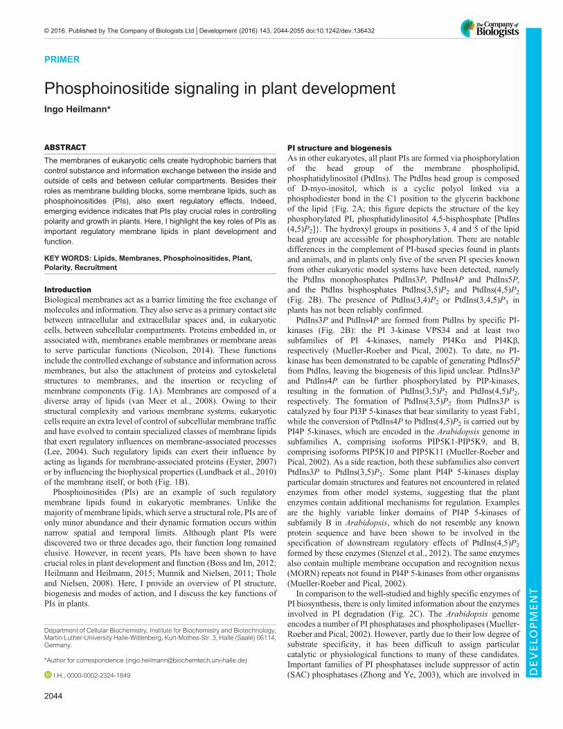
PI structure and biogenesisAs in other eukaryotes, all plant PIs are formed via phosphorylationof the head group of the membrane phospholipid,phosphatidylinositol (PtdIns). The PtdIns head group is composedof D-myo-inositol, which is a cyclic polyol linked via aphosphodiester bond in the C1 position to the glycerin backboneof the lipid {Fig. 2A; this figure depicts the structure of the keyphosphorylated PI, phosphatidylinositol 4,5-bisphosphate [PtdIns(4,5)P2]}. The hydroxyl groups in positions 3, 4 and 5 of the lipidhead group are accessible for phosphorylation. There are notabledifferences in the complement of PI-based species found in plantsand animals, and in plants only five of the seven PI species knownfrom other eukaryotic model systems have been detected, namelythe PtdIns monophosphates PtdIns3P, PtdIns4P and PtdIns5P,and the PtdIns bisphosphates PtdIns(3,5)P2 and PtdIns(4,5)P2
(Fig. 2B). The presence of PtdIns(3,4)P2 or PtdIns(3,4,5)P3 inplants has not been reliably confirmed.
PtdIns3P and PtdIns4P are formed from PtdIns by specific PI-kinases (Fig. 2B): the PI 3-kinase VPS34 and at least twosubfamilies of PI 4-kinases, namely PI4Kα and PI4Kβ,respectively (Mueller-Roeber and Pical, 2002). To date, no PI-kinase has been demonstrated to be capable of generating PtdIns5Pfrom PtdIns, leaving the biogenesis of this lipid unclear. PtdIns3Pand PtdIns4P can be further phosphorylated by PIP-kinases,resulting in the formation of PtdIns(3,5)P2 and PtdIns(4,5)P2,respectively. The formation of PtdIns(3,5)P2 from PtdIns3P iscatalyzed by four PI3P 5-kinases that bear similarity to yeast Fab1,while the conversion of PtdIns4P to PtdIns(4,5)P2 is carried out byPI4P 5-kinases, which are encoded in the Arabidopsis genome insubfamilies A, comprising isoforms PIP5K1-PIP5K9, and B,comprising isoforms PIP5K10 and PIP5K11 (Mueller-Roeber andPical, 2002). As a side reaction, both these subfamilies also convertPtdIns3P to PtdIns(3,5)P2. Some plant PI4P 5-kinases displayparticular domain structures and features not encountered in relatedenzymes from other model systems, suggesting that the plantenzymes contain additional mechanisms for regulation. Examplesare the highly variable linker domains of PI4P 5-kinases ofsubfamily B in Arabidopsis, which do not resemble any knownprotein sequence and have been shown to be involved in thespecification of downstream regulatory effects of PtdIns(4,5)P2
formed by these enzymes (Stenzel et al., 2012). The same enzymesalso contain multiple membrane occupation and recognition nexus(MORN) repeats not found in PI4P 5-kinases from other organisms(Mueller-Roeber and Pical, 2002).
In comparison to the well-studied and highly specific enzymes ofPI biosynthesis, there is only limited information about the enzymesinvolved in PI degradation (Fig. 2C). The Arabidopsis genomeencodes a number of PI phosphatases and phospholipases (Mueller-Roeber and Pical, 2002). However, partly due to their low degree ofsubstrate specificity, it has been difficult to assign particularcatalytic or physiological functions to many of these candidates.Important families of PI phosphatases include suppressor of actin(SAC) phosphatases (Zhong and Ye, 2003), which are involved in
Department of Cellular Biochemistry, Institute for Biochemistry and Biotechnology,Martin-Luther-University Halle-Wittenberg, Kurt-Mothes-Str. 3, Halle (Saale) 06114,Germany.
*Author for correspondence ([email protected])
I.H., 0000-0002-2324-1849
2044
© 2016. Published by The Company of Biologists Ltd | Development (2016) 143, 2044-2055 doi:10.1242/dev.136432
DEVELO
PM
ENT

the degradation of PtdIns bisphosphates at the plasma membrane orthe tonoplast, and phosphatase and tensin homolog deleted onchromosome 10 (PTEN)-related enzymes, which mediate thedegradation of 3-phosphorylated PtdIns monophosphates andPtdIns bisphosphates (Pribat et al., 2012). Further relevantphosphatases are enzymes of the Arabidopsis PI 5-phosphatase(5PTase) family, which exhibit varying specificities and may alsoact on soluble inositol polyphosphates (IPPs) (Gunesekera et al.,2007). PIs can also be degraded by PI-specific phospholipase C (PI-PLC) enzymes, which are encoded in the Arabidopsis genome as afamily of nine isoforms (Mueller-Roeber and Pical, 2002). All plantPI-PLCs reported so far are activated by Ca2+ and display similarityto the human ζ-family of PLCs that cannot be activated byheterotrimeric G-proteins (Pokotylo et al., 2014).The enzymes listed above together mediate the biosynthesis and
degradation of PIs. PIs are constantly formed and degraded, resultingin rapid and dynamic turnover (reviewed by Heilmann, 2016). Thishigh turnover contributes to the dynamic changes in PI levels thathave been observed in numerous studies in different plant models.For instance, the levels of PtdIns(4,5)P2 change in response toenvironmental stresses, including wounding (Mosblech et al., 2008),salt or osmotic stress (DeWald et al., 2001; Einspahr et al., 1988a,b;Heilmann et al., 1999, 2001; König et al., 2008, 2007; Pical et al.,1999) or heat (Mishkind et al., 2009). PI formation also changestransiently during gravitropic curvature (Perera et al., 1999, 2001),and it has also been shown that PI levels respond to exogenouslyapplied auxin (Tejos et al., 2014) or salicylic acid (Krinke et al.,2007b). Notably, Arabidopsis PLC2 has been reported to act in theendoplasmic reticulum (ER) stress response (Kanehara et al., 2015).The modes of regulation underlying the dynamic interplay betweenPI biosynthesis and degradation in plants remain unclear. Theenzymes catalyzing PI interconversions are expressed in differentpatterns (see Table 1), with some being expressed ubiquitously and at
all developmental stages analyzed, whereas others are restricted intheir expression patterns to certain organs or cell types (Heilmann,2016). The transcription of some enzymes of PI metabolism is alsoinduced upon perception of environmental stresses. For instance, inArabidopsis transcription of the PLC1 gene is induced upondehydration and salt stress (Hirayama et al., 1995) and that ofPIP5K1 by exogenous auxin (Tejos et al., 2014). However, a reviewof publicly available transcriptomic data (Zimmermann et al., 2004)indicates that the transcription of the majority of PI-related genesdoes not change dramatically upon challenge by a wide range ofstresses, suggesting that transcriptional control might in sumcontribute only little to the dynamic changes in PI levels observedduring plant stress responses (Heilmann, 2016). The control of PIdynamics might thus not take place at the transcriptional level, butrather via post-translational modifications such as phosphorylation(Westergren et al., 2001). However, to date the relevance of post-translationalmodifications of PI pathway enzymes during plant stressresponses has not been addressed in detail.
Overall, plants contain a complex network of enzymes thatmediate the dynamic formation and degradation of PIs. As inanimals, plant PIs appear to be continuously formed and degradedwith a high rate of turnover, resulting in a dynamic equilibrium thatis easily perturbed when plants are exposed to stress conditions.This dynamic equilibrium enables fast and transient or localizedsignaling events to be mediated by PIs in response to developmentalcues or stimulation.
PI modes of actionPIs exert their functions via various mechanisms. For example, theycan act as intact lipids, binding to target proteins, or they caninfluence the properties of the membrane in which they areembedded. Alternatively, they can act as precursors for theformation of soluble IPPs (Fig. 3).
Transport
Signaltransduction
Cytoskeleton
Recruitment
Trafficking
Secretion Endocytosis
A B
Structural role
Regulatory roles • Protein
recruitment
• Hydrophobicbarrier
• Membraneproperties
Regulatory lipid
Structural lipid
Cytoplasm
Vacuole
Plasma membrane Apoplasticspace
Golgi
TGN
Lyticvacuole
Key
Fig. 1. Cellular membranes and their functions. (A) Cellular membranes restrict the exchange of substance and information between cells and, in eukaryoticcells, between subcellular compartments. The properties of membranes are defined by embedded or associated proteins that carry out various functions, allowingseveral membrane-associated cellular processes to take place including transport, signal transduction, cytoskeletal attachment, protein recruitment and dynamicmembrane trafficking. (B) Membranes can play both structural and regulatory roles. They consist of bilayer-forming lipids that generate a hydrophobic barrier.Although the body of the membrane comprises structural lipids, there are also membrane lipids of low abundance that exert predominantly regulatory effects onmembrane functions. These regulatory effects can be mediated by lipids acting as ligands for protein partners or through direct effects on the biophysicalproperties of membranes. TGN, trans-Golgi network; yellow, cargo; yellow triangles, soluble cargo.
2045
PRIMER Development (2016) 143, 2044-2055 doi:10.1242/dev.136432
DEVELO
PM
ENT

Intact PIs are found predominantly at the cytosolic face of cellularmembranes. It has also been reported that under certain conditions PIsmay occur in the luminal/extracellular leaflets of vesicles or theplasma membrane (Gonorazky et al., 2012, 2008; Kale et al., 2010),although it is currently unclear how PIs would manifest in theselocations. Within the membrane, PIs act as regulators of integralmembrane proteins or mark membrane areas for the recruitment ofperipheral membrane proteins. In this context, PIs act mainly throughtheir head groups, which are characterized by their phosphorylationpatterns and protrude from the plane of the membrane further into thecytosol than the head groups of other, structural lipids (Takenawa,2010). These distinctive phosphorylated PI head groups can berecognized and bound by PI-binding protein domains, such asPleckstrin homology (PH) domains, Fab1 YOTB Vac1 EEA1(FYVE) domains and phagocytic oxidase (PX) domains (Lemmon,
2003). The interactions between PIs and such domains can resultin the recruitment of particular proteins to membrane areas enrichedin PIs.
A number of proteins regulated by PI binding have beenidentified; in some studies, these proteins have been termedPI-‘modulins’ (McLaughlin et al., 2002). It should be noted thatnot all PI-binding domains of such modulins display a high degreeof specificity towards a particular PI species (Fig. 3A). In fact, only aselect few PI-binding domains have been found to be highlyspecific, including the PH domain of human PLCδ1, whichpreferentially binds PtdIns(4,5)P2, and the PH domain of humanfour-phosphate adaptor protein 1 (FAPP1, also known asPLEKHA3), which preferentially binds PtdIns4P (Takenawa andItoh, 2006). Nonetheless, even though most PI-binding proteins donot display exquisite specificity, protein recruitment to PIs does
A PI structure (example)
CDP-DAG
PtdIns
PtdIns3P PtdIns4P
PtdIns(3,5)P2 PtdIns(4,5)P2
PIS1-At1g68000 PIS2-At4g38570
VPS34-At1g56600
Fab1a-At4g33240 Fab1b-At3g14270 Fab1c-At1g71010 Fab1d-At1g34260
B PI biosynthesis
PIP5K1-At1g21980 PIP5K2-At1g77740 PIP5K3-At2g26420 PIP5K4-At3g56960 PIP5K5-At2g41210 PIP5K6-At3g07960 PIP5K7-At1g10900 PIP5K8-At1g60890 PIP5K9-At3g09920 PIP5K10-At4g01190 PIP5K11-At1g01460
PI4Kα1-At1g49340 PI4Kα2-At1g51040 PI4Kβ1-At5g64070 PI4Kβ2-At5g09350
PI-synthase
PI 3-kinase
PI3P 5-kinase
PI 4-kinase
PI4P 5-kinase
1 2
3 4
5
6
Fattyacids
Glycerolbackbone
Inositolphosphatehead group
PtdIns
PtdIns3P PtdIns4P PtdIns5P
PtdIns(3,5)P2 PtdIns(4,5)P2 ?
PTEN1-At5g39400 PTEN2-At3g19420 PTEN3-At3g50110
SAC1-At1g22620 SAC2-At3g14205 SAC3-At3g43220 SAC4-At5g20840 SAC5-At1g17340 SAC6-At5g66020 SAC7-At3g51460 SAC8-At3g51830 SAC9-At3g59770
PTEN
SAC
At5PTase1-At1g34120 At5PTase2-At4g18010 At5PTase3-At1g71710 At5PTase4-At3g63240 At5PTase5-At5g65090 At5PTase6-At1g05470 At5PTase7-At2g32010 At5PTase8-At2g37440
5-phosphatase
PLC1-At5g58670 PLC2-At3g08510 PLC3-At4g38530 PLC4-At5g58700 PLC5-At5g58690 PLC6-At2g40116 PLC7-At3g55940 PLC8-At3g47290 PLC9-At3g47220
PI-PLC
At5PTase9-At2g01900 At5PTase10-At5g04980 At5PTase11-At1g47510 At5PTase12-At2g43900 At5PTase13-Át1g05630 At5PTase14-At2g31830 At5PTase15-At1g65580
DAG + IPP
C PI degradation
Fig. 2. The plant phosphoinositide network. (A) Structure of phosphatidylinositol 4,5-bisphosphate [PtdIns(4,5)P2], a key regulatory phosphoinositide (PI). Themolecule contains two fatty acids esterified to a glycerol backbone that convey its hydrophobic properties. The inositol-phosphate head group is hydrophilicand mediates the amphiphilic properties of the membrane lipid. The inositol-phosphate head group is the characteristic moiety that can be recognized and boundby PI-binding protein domains. (B,C) Schematic representation of the PI biosynthesis (B) and degradation (C) pathways in Arabidopsis. Arrows indicate theindividual enzymatic steps. The Arabidopsis genes encoding the enzymes that catalyze each step are highlighted in boxes matching the colors of thearrow. Please note that the specificities for some of the degrading enzymes are not clear in all cases and that not all isoforms represented in each box mightperform the reactions specified by the colors. The formation of phosphatidylinositol 5-phosphate (PtdIns5P) is still a matter of debate. CDP-DAG, cytidinediphosphodiacylglycerol; DAG, diacylglycerol; IPP, inositol polyphosphate; PI-PLC, PI-specific phospholipase C; PtdIns, phosphatidylinositol; PtdIns3P, PtdIns3-phosphate; PtdIns(3,5)P2, PtdIns 3,5-bisphosphate; PtdIns4P, PtdIns 4-phosphate; PtdIns(4,5)P2, PtdIns 4,5-bisphosphate; PTEN, phosphatase and tensinhomolog deleted on chromosome 10; SAC, suppressor of actin.
2046
PRIMER Development (2016) 143, 2044-2055 doi:10.1242/dev.136432
DEVELO
PM
ENT

Table 1. Phenotypes reported upon perturbation of the PI signaling system in Arabidopsis thaliana
Enzyme IsoformExpressionpattern
Subcellulardistribution
Experimentalperturbation Reported phenotype References
PI-synthase PIS1 Ubiquitous ER/Golgi PIS1 OE Enhanced growth (Löfke et al., 2008)PIS2 Ubiquitous ER/Golgi PIS2 OE Enhanced growth, pollen tube
defects(Ischebeck et al.,2010; Löfke et al.,2008)
PI 3-kinase VPS34 Ubiquitous Endomembranes pi3k (T-DNA) Homozygous lethal,heterozygous defective pollendevelopment, root hairdefects, vacuolar defects
(Lee et al., 2008a,b;Welters et al., 1994)
PI 4-kinase PI4Kα1 Ubiquitous Plasma membrane/plastid outerenvelope
Mutant;knockdown
Homozygous lethal, knockdownincreased plastid division
(Delage et al., 2013;Okazaki et al., 2015)
PI4Kα2 Ubiquitous – – – –
PI4Kβ1 Ubiquitous TGN (Antignani et al., 2015;Preuss et al., 2006)
PI4Kβ2 Ubiquitous TGN (Antignani et al., 2015;Preuss et al., 2006)
pi4kβ1 pi4kβ2double mutant(T-DNA)
Reduced growth, root hairdefects, enhanced defense,increased plastid division
(Antignani et al., 2015;Preuss et al., 2006)
PI3P 5-kinase FAB1A Ubiquitous – fab1a (T-DNA) Vacuolar defects in pollen (Whitley et al., 2009)FAB1B Ubiquitous – fab1b (T-DNA) Vacuolar defects in pollen,
reduced stomatal closure(Bak et al., 2013)
FAB1C Ubiquitous – fab1c (T-DNA) Reduced stomatal closure (Bak et al., 2013)FAB1D Ubiquitous – – No reports –
PI4P 5-kinase PIP5K1 Ubiquitous Plasma membrane/vesicles/nucleus
pip5k1 (T-DNA) Reduced growth (Ischebeck et al.,2013; Tejos et al.,2014)
PIP5K1 OE Wavy root growth, defects in PINtrafficking
(Ischebeck et al.,2013)
PIP5K2 Ubiquitous Plasma membrane/vesicles/nucleus
pip5k2 (T-DNA) Reduced growth, defects in PINtrafficking
(Camacho et al., 2009;Ischebeck et al.,2013; Mei et al.,2012; Tejos et al.,2014)
PIP5K2 OE Wavy root growth, defects in PINtrafficking
(Ischebeck et al.,2013)
pip5k1 pip5k2 (T-DNA)
Defects in early embryodevelopment; defects invascular development;defects in PIN trafficking andclathrin-mediated endocytosis
(Ischebeck et al.,2013; Tejos et al.,2014)
PIP5K3 Root epidermisand cortex
Plasma membrane/vesicles
pip5k3 (T-DNA) Root hair defects (Kusano et al., 2008;Stenzel et al., 2008)
PIP5K3 OE Root hair defects (Kusano et al., 2008;Stenzel et al., 2008)
PIP5K4 Flowers/pollen Plasma membrane/vesicles
pip5k4 (T-DNA) Reduced pollen germination (Ischebeck et al.,2008; Sousa et al.,2008)
PIP5K4 OE Pollen tube defects, enhancedpectin secretion
(Ischebeck et al.,2008)
PIP5K5 Flowers/pollen Plasma membrane/vesicles
pip5k5 (T-DNA) Reduced pollen germination (Ischebeck et al.,2008; Sousa et al.,2008)
PIP5K5 OE Pollen tube defects, enhancedpectin secretion
(Ischebeck et al.,2008)
pip5k4 pip5k5double mutant(T-DNA)
Reduced pollen germination (Ischebeck et al.,2008; Sousa et al.,2008)
PIP5K6 Ubiquitous Plasma membrane/vesicles
pip5k6 (T-DNA) Defects in clathrin-mediatedendocytosis
(Zhao et al., 2010)
PIP5K7 Ubiquitous – – – –
PIP5K8 Ubiquitous – – – –
PIP5K9 Ubiquitous Plasma membrane/nucleus
pip5k9 (T-DNA;resulting in OE)
Altered sugar metabolism (Lou et al., 2007)
Continued
2047
PRIMER Development (2016) 143, 2044-2055 doi:10.1242/dev.136432
DEVELO
PM
ENT

Table 1. Continued
Enzyme IsoformExpressionpattern
Subcellulardistribution
Experimentalperturbation Reported phenotype References
PIP5K10 Pollen Plasma membrane/nucleus
pip5k10 (T-DNA) Increased sensitivity to LatB (Ischebeck et al.,2011)
pip5k10 OE Pollen tube defects, stabilizedactin cytoskeleton
(Ischebeck et al.,2011)
PIP5K11 Pollen Plasma membrane/nucleus
pip5k11 (T-DNA) Increased sensitivity to LatB (Ischebeck et al.,2011)
pip5k11 OE Pollen tube defects, stabilizedactin cytoskeleton
(Ischebeck et al.,2011)
pip5k10 pip5k11double mutant(T-DNA)
Increased sensitivity to LatB (Ischebeck et al.,2011)
PI-PLC PLC1 Roots, leaves Plasma membrane – – (Pokotylo et al., 2014)PLC2 Roots, leaves,
stems, flowersPlasma membrane plc2 (T-DNA) Growth defects (Kanehara et al., 2015;
Pokotylo et al.,2014)
PLC3 Roots, leaves Plasma membrane plc3 (T-DNA) – (Pokotylo et al., 2014;Zheng et al., 2012)
PLC4 Roots, stems,flowers
Plasma membrane – – (Pokotylo et al., 2014)
PLC5 Roots, siliques – – – (Pokotylo et al., 2014)PLC6 Flowers (Pokotylo et al., 2014)PLC7 – Plasma membrane plc7 (T-DNA) – (Pokotylo et al., 2014;
Zheng et al., 2012)PLC8 Leaves, siliques,
flowers– plc8 (T-DNA) – (Pokotylo et al., 2014;
Zheng et al., 2012)PLC9 Roots, siliques – plc9 (T-DNA) Impaired thermotolerance (Pokotylo et al., 2014;
Zheng et al., 2012)SACphosphatase
SAC1/FRA7 Ubiquitous Golgi sac1/fra7 (T-DNA) Cell wall defects (Zhong et al., 2005)SAC2 Ubiquitous Tonoplast (Novakova et al.,
2014)SAC3 Ubiquitous Tonoplast (Novakova et al.,
2014)SAC4 Ubiquitous Tonoplast (Novakova et al.,
2014)SAC5 Ubiquitous Tonoplast (Novakova et al.,
2014)sac2 sac3 sac4sac5 quadruplemutant (T-DNA)
Vacuolar defects (Novakova et al.,2014)
SAC6/SAC1b No report ER – – (Despres et al., 2003)SAC7/RHD4/SAC1c
Ubiquitous ER/TGN sac7/rhd4/sac1c(EMS; T-DNA)
Root hair defects (Despres et al., 2003;Thole et al., 2008)
SAC8/SAC1a No report ER – – (Despres et al., 2003)SAC9 Ubiquitous No report sac9 (T-DNA) Growth defects, constitutive
stress response(Williams et al., 2005)
PTEN PTEN1 Flowers/pollen Vesicles PTEN1 OE Pollen defects (Zhang et al., 2011)PTEN2a Ubiquitous – – – (Pribat et al., 2012)PTEN2b Ubiquitous – – – (Pribat et al., 2012)
5PTase At5PTase1 Ubiquitous No report 5ptase1-1,5ptase1-2(T-DNA)
Defective seedlingdevelopment, hypocotyldefects
(Gunesekera et al.,2007)
At5PTase2 Ubiquitous No report 5ptase2-1(T-DNA)
Defective seedlingdevelopment, hypocotyldefects
(Gunesekera et al.,2007)
5ptase1-15ptase2-1double mutant(T-DNA)
Defective seedlingdevelopment, hypocotyldefects
(Gunesekera et al.,2007)
At5PTase3 Ubiquitous – – –
At5PTase4 Shoot apex,ubiquitous
– – –
At5PTase5/BST1
Pollen, weakubiquitous
– bst1 (EMS) Root hair defects (Parker et al., 2000)
At5PTase6/CVP2
Ubiquitous,vasculartissue
No report cvp2-1, cvp2-2,cvp2-3 (EMS)
Vascular defects (Carland and Nelson,2004; Rodriguez-Villalon et al., 2015)
Continued
2048
PRIMER Development (2016) 143, 2044-2055 doi:10.1242/dev.136432
DEVELO
PM
ENT

exhibit substantial specificity depending on the local abundance ofcertain PIs and on coincident binding to other (protein) factors andcoincidence detection (Carlton and Cullen, 2005), a conceptdiscussed in more detail below. It should also be noted that PI-binding domains, such as PH domains, are not well conserved withregards to primary sequence and are instead characterized by theirthree-dimensional fold. These domains are therefore not readilyidentified by mere sequence analysis. Extensive bioinformaticsanalyses, however, have revealed that the Arabidopsis genomeencodes numerous proteins with recognizable PI-binding domains(van Leeuwen et al., 2004), including: the PI 3-kinase VPS34(At1g60490), the PI 4-kinases α1 (At1g49340) and α2(At1g51040), the 3-phosphoinositide-dependent protein kinase 1(PDK1; At3g10540), the dynamin-related proteins 2A (DRP2A;At1g10290) and B (DRP2B; At1g59610) and the Rho-of-plantsGTPase-activating proteins 1-3 (ROP-GAPs 1-3; At4g24580,At5g12150 and At5g19390), all of which contain a PH domain;FAB1A (At4g33240) and FAB1B (At3g14270), which contain aFYVE domain; and the sorting nexin-like protein 2 (SNX2B;At5g07120), which contains a PX domain (van Leeuwen et al.,2004). Interestingly, there are several instances in which proteinscontain more than one PI-binding domain, for example thephospholipases D ζ1 (PLDζ1; At3g16785-90) and ζ1 (PLDζ2;At3g05630), which contain both a PH domain and a PX domain(van Leeuwen et al., 2004). It appears likely that the number ofproteins with PI-binding domains is still underestimated, asPI-binding domains may form transiently upon dynamicinteraction of protein partners, thus escaping detection viasequence annotation (van Rossum et al., 2005). Furthermore,despite the seeming abundance of PI-modulins, the directexperimental identification of PI-binding proteins has so far not
yielded a substantial amount of data. A notable exception is theidentification of numerous candidate binding partners for 3-phosphorylated PIs from Arabidopsis, which were determined byagarose-phosphatidylinositol-phosphate affinity chromatographyand subsequent mass spectrometric analysis (Oxley et al., 2013).
Intact PIs can also influence a membrane directly by modifyingits biophysical properties, inducing membrane curvature orstabilizing curved membrane areas (Lundbaek et al., 2010). Thus,PIs can both exert an intrinsic influence on the membrane andmediate the recruitment of proteins that stabilize certain membranestructures, although the relative contributions of these individualeffects remain unclear.
Besides their roles as intact lipids, PIs can serve as precursors forPLC-mediated cleavage into diacylglycerol (DAG) and solubleIPPs (Gillaspy, 2013). In mammals, DAG acts as a potent activatorof protein kinase C (Amadio et al., 2006), but this enzyme ismissing in plants (Mueller-Roeber and Pical, 2002) and a signalingfunction for plant DAG has not been reported. Instead, DAG inplants is rapidly phosphorylated to phosphatidic acid (PtdOH),which has been shown to have a number of regulatory functions(Meijer and Munnik, 2003). PtdOH can be further phosphorylatedto diacylglycerol pyrophosphate (DGPP), a lipid of unknownrelevance (van Schooten et al., 2006). Soluble IPPs, such as inositolhexakisphosphate (InsP6) or inositol pentakisphosphate (InsP5), arepresent within the crystal structures of receptor proteins for auxin(Tan et al., 2007) or jasmonic acid (Sheard et al., 2010). However,the role of these IPPs in the perception of auxin or jasmonic acid asstructural or possibly functional co-factors is currently unclear. Ithas been reported that Arabidopsis ipk1-1 mutants with decreasedlevels of InsP6 and increased levels of InsP5 display an enhancedwounding response and enhanced resistance to herbivory
Table 1. Continued
Enzyme IsoformExpressionpattern
Subcellulardistribution
Experimentalperturbation Reported phenotype References
At5PTase7/CVL1
Ubiquitous,weak in shootsand siliques
No report cvl1-1, cvl1-2(T-DNA)
Vascular defects, altered ROSproduction and salt tolerance
(Carland and Nelson,2009; Kaye et al.,2011)
At5PTase8 Young roots,weakubiquitous
– – –
At5PTase9 Young roots,weakubiquitous
No report At5ptase9(T-DNA),At5ptase9 OE
Defects in salt tolerance andendocytosis
(Golani et al., 2013)
At5PTase10 Late flowers,senescentleaves, weakubiquitous
– – –
At5PTase11 Developingseeds,seedlings
Cytosol, plasmamembrane
5ptase11-1,5ptase11-2(T-DNA)
Defective seedlingdevelopment, hypocotylgrowth
(Ercetin et al., 2008)
At5PTase12 Ubiquitous(leaves weak)
No report 5pt12 (T-DNA) Altered pollen dormancy/germination
(Wang et al., 2012;Zhong andYe, 2004)
At5PTase13 Ubiquitous – Atptase13(T-DNA)
Vascular defects, alteredCa2+ signatures, alteredphototropin signaling
(Chen et al., 2008; Linet al., 2005; Zhongand Ye, 2004)
At5PTase14 Seedlings, shootapices
– – – (Zhong and Ye, 2004)
At5PTase15/FRA3
Ubiquitous – fra3 (T-DNA) Cell wall defects (Zhong et al., 2004)
Sec14-like SFH1 Ubiquitous Plasma membrane sfh1 (T-DNA) Root hair defects (Ghosh et al., 2015;Vincent et al., 2005)
ER, endoplasmic reticulum; LatB, latrunculin B; OE, overexpression; ROS, reactive oxygen species; TGN, trans-Golgi network.
2049
PRIMER Development (2016) 143, 2044-2055 doi:10.1242/dev.136432
DEVELO
PM
ENT

(Mosblech et al., 2011). More recently, other IPPs, such as thepyrophosphorylated IPPs (PP-IPPs) InsP7 or InsP8, have also beenimplicated in modulating the function of the jasmonic acid receptor(Laha et al., 2015), possibly suggesting a role for IPPs inphytohormone perception and the control of hormone-inducedgene expression patterns. Other IPPs, such as inositol 1,4,5-trisphosphate (InsP3), are also implicated in mediating Ca2+
responses, although an InsP3-sensitive Ca2+ channel has not beenreported in plants (Krinke et al., 2007a).
PIs and membrane microdomainsFrom the current data it is evident that plant PIs exert a number ofdifferent functions by interacting with a variety of binding partners.However, it is also clear that not all cellular processes that can inprinciple be controlled by PIs will manifest whenever PIs areformed. In fact, the physiological consequences of enhanced PIformation tend to be rather limited and highly specific, raising thequestion of how the specificity of PI functions is achieved(Heilmann and Heilmann, 2013). A key piece of information forunderstanding PI function is that these lipids do not display uniformdistribution within membranes (Ischebeck et al., 2008, 2011, 2013;Kost et al., 1999; Kusano et al., 2008; Sousa et al., 2008; Tejos et al.,2014; van Leeuwen et al., 2007; Vermeer et al., 2009; Vincent et al.,
2005). Instead, PIs are enriched in certain membrane domains, andit is likely that such microdomains play a part in mediating thespecific effects of PIs (Fig. 4A). Plants form particular molecularspecies of PIs under certain conditions (e.g. when challenged bysalt) that are characterized by an increased degree of unsaturation intheir associated fatty acids (König et al., 2007). The degree to whichlipids are unsaturated is known to influence their lateral mobilitywithin the plane of the membrane and to guide their association withparticular planar or curved membrane domains (Cho et al., 2006;Mukherjee et al., 1999). The degree of unsaturation of PI-associatedfatty acids might, therefore, be an as yet underestimated factorgoverning the lateral mobility of PIs within membranes, withpossible implications for their association with certain partnerproteins and for defining the specificity of PI effects. The lateraldistribution of PIs within a membrane is also likely to be influencedby other membrane lipids, such as sphingolipids and sterols (Furtet al., 2010).
An additional aspect contributing to the specificity of PI effects isthe concept of coincidence detection (Carlton and Cullen, 2005).Although a number of plant proteins have been demonstrated tobind PIs, as discussed above, it is known from other model systemsthat there can be additional factors guiding the effects of PIs (Balla,2005; Várnai et al., 2005). Regulation by coincidence detection(Fig. 4B) suggests that PI binding is necessary for the regulation ofcertain processes but might not be sufficient by itself, and otherfactors must coincide for an effect to manifest. Thus, in addition tothe binding of a PI, a membrane-associated protein partner, forexample, must also be bound and only membrane areas containingboth recruitment factors will be targeted by modulins that requirecoincidence detection to function. An interesting aspect of
Lipid-bindingdomains
A Intact PIs: ligands for partner proteins
B Hydrolyzed PIs: precursors for further signaling factors
PLC
PP-IPPs
PtdIns(4,5)P2
Ins(1,4,5)P3 InsP5 InsP6
DAG PtdOH DGPP
PtdIns(4,5)P2
PtdIns4P
OH
Key
Fig. 3. Modes of PI action. PIs can act through the binding of theircharacteristic head groups to partner proteins containing specific PI-recognition domains (A), or as substrates for hydrolyzing enzymes thatgenerate a range of lipid messengers and soluble inositol polyphosphates(IPPs) (B). (A) The binding of a PI ligand to a proteinmay serve as a recruitmentsignal or have effects on the activity or function of the target protein. Some PI-recognition domains display a high degree of specificity for particular PIs. Forexample, some pleckstrin homology (PH) domain-containing proteins (darkblue) have a strong preference for PtdIns(4,5)P2, whereas others (red) bindpreferentially to PtdIns4P. However, most PH domains do not exhibit suchpreferences, and proteins harboring these domains (gray) can bind to anygiven PI. The specificity of PI function is thus likely to be exerted by additionalfactors, including locally increased levels of certain PIs and coincidencedetection (see Fig. 4). (B) The hydrolysis of PtdIns(4,5)P2 by PLC yields DAGand Ins(1,4,5)P3. In plants, DAG is rapidly phosphorylated to PtdOH andpossibly further to DGPP. An Ins(1,4,5)P3-sensitive Ca2+ channel is absent inplants and Ins(1,4,5)P3 instead appears to act as a precursor for IPPs,including InsP5, InsP6 or pyrophosphorylated IPPs (PP-IPPs). DAG,diacylglycerol; DGPP, diacylglycerol pyrophosphate; Ins(1,4,5)P3, inositol1,4,5-trisphosphate; InsP5, inositol pentakisphosphate; InsP6, inositolhexakisphosphate; PtdIns(4,5)P2, phosphatidylinositol 4,5-bisphosphate;PtdIns4P, PtdIns 4-phosphate; PtdOH, phosphatidic acid.
PI degradation PI biosynthesis
PI degradation PI degradation
PI biosynthesis PI biosynthesis
Area enriched in PIs
Cytosol
Apoplastic space
Area of coincidencedetection
Cytosol
Apoplastic space
A
B
Sphingolipid Sterol PtdIns(4,5)P2
X X
Physiologicaleffect
Key
Fig. 4. PI membrane domains and localized potentiation of PI function.(A) PIs are not uniformly distributed within membranes; instead, concentratedareas of increased PI abundance are observed and are associated withspecific PI functions. This localized accumulation of PIs in a membrane mightbe due to increased PI biosynthesis but is also possibly linked to the presenceof other membrane lipids, such as sphingolipids with very long-chain fatty acidsor sterols. (B) The presence of a PI ligand (left) or a membrane-associatedfactor (red, middle) that can bind to a target protein (dark blue) might each bythemselves not be sufficient to recruit the target protein and exert a regulatoryeffect. Only when the PI ligand and the membrane-associated factor coincideat a membrane domain (the ‘area of coincidence detection’) and both bind tothe target protein will a regulatory effect be exerted.
2050
PRIMER Development (2016) 143, 2044-2055 doi:10.1242/dev.136432
DEVELO
PM
ENT

coincidence detection involves the possible role of proteins thatpotentiate PI functionality by facilitating their interaction withcertain target proteins. For instance, recent evidence in Arabidopsisindicates that Sec14-like lipid transfer proteins aid PI-proteininteraction in a specific manner (Bankaitis et al., 2010; Ghosh et al.,2015; Schaaf et al., 2008; Vincent et al., 2005). As a consequence,membrane areas that contain Sec14-like proteins will exhibitpotentiated PI effects, possibly similar to the effects of localizedPI overproduction.
PI functions in plantsIn recent years, PIs have emerged as central regulators of plantfunction and development, and a number of important and severeplant phenotypes have been reported when plant PI metabolismis perturbed (Carland and Nelson, 2009, 2004; Chen et al., 2008;Ercetin et al., 2008; Golani et al., 2013; Gunesekera et al., 2007;Ischebeck et al., 2008, 2011, 2013; Kaye et al., 2011; Kusano et al.,2008; Löfke et al., 2008; Mei et al., 2012; Novakova et al., 2014;Parker et al., 2000; Preuss et al., 2006;Rodriguez-Villalon et al., 2015;Sousa et al., 2008; Stenzel et al., 2008; Tejos et al., 2014;Wang et al.,2012; Williams et al., 2005; Zhong et al., 2004, 2005; Zhong and Ye,2004). The perturbation of PI signaling components in Arabidopsis,for example, gives rise to a variety of phenotypes affecting multipletissue types and aspects of plant development (summarized inTable 1). A number of studies utilizing model systems other thanArabidopsis have also highlighted a role for PIs in plant developmentand function. For instance, the overexpression of PI-synthase inmaizeresults in altered drought tolerance (Liu et al., 2013).Workon petunia,tobacco and the moss Physcomitrella patens supports an importantrole for PIs in the control of polar tip growth (Dowd et al., 2006;Helling et al., 2006; Saavedra et al., 2011, 2015). Although some ofthe reported phenotypes are severe, the molecular mechanismsunderlying PI functions in plants remain mostly unclear.In the absence of detailed information on the molecular targets of
PIs in plants, the interpretation of reported phenotypes poses achallenge. Based on the available data, the phenotypes can becategorized according to the defects observed, enabling a firstapproximation of how the elements of the plant PI network might beinterlinked at the cellular level (Fig. 5). For example, phenotypesresulting from the perturbation of PI-synthases (Löfke et al., 2008),PI4Kβ1/PI4Kβ2 (Preuss et al., 2006), PIP5K1-PIP5K6 (Ischebecket al., 2008, 2013; Kusano et al., 2008; Sousa et al., 2008; Stenzelet al., 2008; Tejos et al., 2014; Zhao et al., 2010) or of SAC1 (Zhonget al., 2005) or SAC7 (Thole and Nielsen, 2008) are all related tofunctional changes in membrane trafficking between the Golgi andthe plasma membrane. Based on the subcellular localization of theindividual enzymes, it appears that an increasing degree ofphosphorylation [PtdIns→PtdIns4P→PtdIns(4,5)P2] is associatedwith the progressive delivery or retrieval of vesicles to and from theplasma membrane. Another category of phenotypes indicates thecontrol of cytoskeletal dynamics by PIs. For instance, the perturbationof PIP5K2 (Stenzel et al., 2012) or PIP5K10 and PIP5K11(Ischebeck et al., 2011) results in altered actin dynamics and theloss of cellular polarity. Yet a different category of phenotypesindicates that 3-phosphorylated PIs, which are perturbed by alteredlevels of VPS34 (Lee et al., 2008a,b), Fab1-like proteins (Bak et al.,2013) or SAC2-SAC5 (Novakova et al., 2014), might be involved inendomembrane trafficking and the control of vacuolar functions. Theoverall scheme suggests that PIs are involved in controlling the centralmachinery for membrane trafficking and protein sorting in plants. Asa consequence, PIs have an impact on several key cellular processes,such as the establishment of cell polarity (Ischebeck et al., 2008,
2011, 2013; Kusano et al., 2008; Mei et al., 2012; Preuss et al., 2006;Sousa et al., 2008; Stenzel et al., 2008; Tejos et al., 2014) and cellwall deposition (Krishnamoorthy et al., 2014; Zhong et al., 2004,2005). These processes influence embryo development, vasculardevelopment and, ultimately, plant growth.
In addition to their cytosolic effects on trafficking and membranerecruitment, emerging evidence indicates that PIs may also have adirect or indirect influence on nuclear function in plants. PI reporters(see Box 1) have been observed in the nuclei of plant cells(Mishkind et al., 2009), as have fluorescently tagged variants ofenzymes involved in PI metabolism (Ischebeck et al., 2011, 2013;Lou et al., 2007; Tejos et al., 2014). However, so far it is unclearwhether these observations have biological relevance or if theysimply reflect fluorescence patterns ensuing after fluorescent fusionproteins have been degraded. The expression of human PI4P5-kinase in plant cells resulted in changes to the cell cycle controlmachinery and histone modifications (Dieck et al., 2012b), but it isunclear whether this finding has relevance for endogenous plantPI-based processes. Further information comes from the analysis offactors involved in the control of stem cell populations in plantmeristems. For instance, the protein phosphatase POLTERGEIST,an enzyme that functionally links plasma membrane perception ofCLAVATA peptides and nuclear regulation, has been shown to bindPtdIns4P (Gagne and Clark, 2010). This binding to PtdIns4P resultsin the activation of phosphatase activity in vitro, suggesting thatPtdIns4P might be involved in the signaling pathways controllingstem cell maintenance. So far, there is only very limited informationabout the effects of PIs on nuclear function in plants, and a role forPIs in plant nuclei (Dieck et al., 2012a), as has been proposed for PIsin animal cells, is currently unclear.
PtdIns
PtdIns4P
PtdIns(4,5)P2
PtdIns3P
PtdIns(3,5)P2
PtdIns4P
Cytoplasm
Vacuole
Plasma membrane Apoplasticspace
Golgi
TGN
Lyticvacuole
Fig. 5. Model of subcellular PI distribution. Based on the effects ofperturbed PI metabolism, a number of conclusions can be drawn with regard tothe subcellular organization of PIs and their function in plants. PtdIns, theprecursor lipid of all PIs, is formed in the ER or Golgi. PtdIns can bephosphorylated to PtdIns4P on vesicles of the trans-Golgi network (TGN) enroute to the plasma membrane. PtdIns4P is further phosphorylated at theplasma membrane to PtdIns(4,5)P2. PtdIns(4,5)P2 influences membranetrafficking events at the plasma membrane, such as exocytosis andendocytosis. It is also likely to have regulatory effects on membranerecruitment of proteins controlling cytoskeletal dynamics and on transporters.The 3-phosphorylated PIs PtdIns3P and PtdIns(3,5)P2 are likely to be involvedin the control of endomembrane trafficking in the context of autophagy and thecontrol of vacuolar/tonoplast functions.
2051
PRIMER Development (2016) 143, 2044-2055 doi:10.1242/dev.136432
DEVELO
PM
ENT

Conclusions and future perspectivesThe perturbation of PI metabolism in plants results in severephenotypes, highlighting the fact that PIs play central roles in plantdevelopment and plant adaptation to the environment. The profoundinfluences of PIs on diverse cellular processes indicate that the plantPI network is an important field for future studies. Our currentunderstanding of the plant PI network is still limited and the precisemolecular targets of PI action in plants are largely unknown. Amassive amount of data has already accumulated on the plant PIsystem, but to date it seems that not all dots have been connected andthe available information has not been used to the fullest extent. Forinstance, numerous proteins have been proposed to contain PI-binding domains, making these proteins prime candidates forregulation by PIs (van Leeuwen et al., 2004). However, only aminority of these potential PI-modulins have been experimentallytested for PI binding by in vitro lipid overlay assays (Munnik andWierzchowiecka, 2013) or liposome binding tests (Julkowska et al.,2013), leaving the field open for further scrutiny. Whereas many ofthe potential PI-modulins are proteins of unknown function, othersare well annotated, linking potential PI binding to relevant cellularprocesses. In combination with the review of PI-dependent plantphenotypes related to membrane trafficking or cytoskeletal control,this information might indicate future avenues of research toelucidate how and when PIs are formed and what their respectivemodulins in each context might be. The study of the molecularmechanisms by which PIs act will require a combination ofcomplementary in vitro and in vivo data to demonstrate a precisemode of action. In particular, information on the specificity andaffinityof PI-protein binding is currentlymissing.Moreover, the studyof PI-protein binding should expand to include other lipid classes
known to influence membrane biophysics, such as sphingolipids andsterols, becausePIs are unlikely to act individually in acell but rather inthe context of membrane areas defined by amixture of different lipids(van Meer et al., 2008). In this sense, it will be a major goal of futurestudies to determine the coincident properties of mixed membranemicrodomains with regard to protein recruitment and binding. On thein vivo side, plant researchers have started using fluorescent probes forthe detection of membrane lipid-ordered domains (Frescatada-Rosaet al., 2014; Gerbeau-Pissot et al., 2016), which might prove apowerful tool to link in vitro and in vivo studies. Considering thecurrent status of sequence information and the range of available invitro and in vivo methods, I am confident that the coming years willbring substantial advances in the field of plant PI signaling.
AcknowledgementsThe author apologizes to all authors whose relevant contributions might not havebeen cited owing to space restrictions and wishes to thank Dr Mareike Heilmann andDr Praveen Krishnamoorthy (both Martin-Luther-University Halle-Wittenberg) forhelpful discussion.
Competing interestsThe author declares no competing or financial interests.
FundingFunding from the German Research Foundation (DeutscheForschungsgemeinschaft) [grants He3424/1, He3424/2, He3424/3 and He3424/6]and the European Regional Development Fund of the European Commission(EFRE) is gratefully acknowledged.
ReferencesAmadio, M., Battaini, F. and Pascale, A. (2006). The different facets of protein
kinases C: old and new players in neuronal signal transduction pathways.Pharmacol. Res. 54, 317-325.
Antignani, V., Klocko, A. L., Bak, G., Chandrasekaran, S. D., Dunivin, T. andNielsen, E. (2015). Recruitment of PLANT U-BOX13 and the PI4Kbeta1/beta2
Box 1. PI reporters: now you see me, now you don’t
GFP
GFP
Displacementof endogenous binding partners
No displacement,no reporter
binding
CompensatoryPI changes
GFP
A B C D Reporter binding
GFP
The highly specific binding of PIs can be visualized using genetically encoded reporters consisting of PI-binding domains fused to fluorescent proteins(Hammond et al., 2014; Kost et al., 1999; Simon et al., 2014; van Leeuwen et al., 2007; Várnai and Balla, 1998; Vermeer et al., 2009, 2006). Although at firstthis appears to be a straight-forward approach, there are a number of catches that render it difficult to interpret data obtained using these reporters (Balla,2007; Balla et al., 2000). The difficulty arises largely from the fact that in living cells the binding of the reporters to PIs occurs in competition with the binding ofendogenous binding partners. Consequently, the reporters might not bind to their target PIs, or they may perturb PI-dependent processes, as illustratedabove. The desired effect is that a fluorescent reporter specifically binds its PI and relocalizes from the cytosol to a cellular membrane (A). However, aninherent problem of the reporter binding to a PI is that the reporter will compete for PI occupancy with endogenous proteins. If the apparent affinity of thereporter for a PI is higher than that of the endogenous protein, the reporter will displace the endogenous protein from the membrane (B), abolishing thefunction of the endogenous protein and causing undesirable side effects that might complicate interpretation. On the other hand, an endogenous bindingprotein with a high affinity for a PI might not be displaced by the reporter, resulting in the absence of any informative fluorescence distribution pattern (C). Afurther complication of reporter expression is that cells might respond to the displacement of endogenous proteins with compensatory upregulation of PIbiosynthesis, resulting in an overall change to PI levels (D). The relative affinities of reporters and endogenous partner proteins for their specific ligands arethus of great importance and reporters should be used at the minimal expression levels required for adequate imaging. It is also important to note that PIsbound to overexpressed PI reporters no longer contribute to a functional PI system; this has led researchers to harness reporter expression to functionallyinhibit certain PIs (Lee et al., 2008a). All these caveats have to be considered when interpreting data obtained using genetically encoded PI reporters, withparticular care required when expression levels and/or apparent affinities of the reporters are high, for instance in reporters usingmore than one lipid-bindingdomain for enhanced affinity (Simon et al., 2014).
2052
PRIMER Development (2016) 143, 2044-2055 doi:10.1242/dev.136432
DEVELO
PM
ENT

phosphatidylinositol-4 kinases by the small GTPase RabA4B plays importantroles during salicylic acid-mediated plant defense signaling in Arabidopsis. PlantCell 27, 243-261.
Bak, G., Lee, E.-J., Lee, Y., Kato, M., Segami, S., Sze, H., Maeshima, M., Hwang,J.-U. and Lee Y. (2013). Rapid structural changes and acidification of guard cellvacuoles during stomatal closure require phosphatidylinositol 3,5-bisphosphate.Plant Cell 25, 2202-2216.
Balla, T. (2005). Inositol-lipid binding motifs: signal integrators through protein-lipidand protein-protein interactions. J. Cell Sci. 118, 2093-2104.
Balla, T. (2007). Imaging and manipulating phosphoinositides in living cells.J. Physiol. 582, 927-937.
Balla, T., Bondeva, T. and Varnai, P. (2000). How accurately can we image inositollipids in living cells? Trends Pharmacol. Sci. 21, 238-241.
Bankaitis, V. A., Mousley, C. J. and Schaaf, G. (2010). The Sec14 superfamily andmechanisms for crosstalk between lipid metabolism and lipid signaling. TrendsBiochem. Sci. 35, 150-160.
Boss, W. F. and Im, Y. J. (2012). Phosphoinositide signaling. Annu. Rev. Plant Biol.63, 409-429.
Camacho, L., Smertenko, A. P., Perez-Gomez, J., Hussey, P. J. and Moore, I.(2009). Arabidopsis Rab-E GTPases exhibit a novel interaction with a plasma-membrane phosphatidylinositol-4-phosphate 5-kinase. J. Cell Sci. 122,4383-4392.
Carland, F. M. and Nelson, T. (2004). Cotyledon vascular pattern2–mediatedinositol (1,4,5) triphosphate signal transduction is essential for closed venationpatterns of Arabidopsis foliar organs. Plant Cell 16, 1263-1275.
Carland, F. and Nelson, T. (2009). CVP2- and CVL1-mediated phosphoinositidesignaling as a regulator of the ARF GAP SFC/VAN3 in establishment of foliar veinpatterns. Plant J. 59, 895-907.
Carlton, J. G. and Cullen, P. J. (2005). Coincidence detection in phosphoinositidesignaling. Trends Cell Biol. 15, 540-547.
Chen, X., Lin, W.-H., Wang, Y., Luan, S. and Xue, H.-W. (2008). An inositolpolyphosphate 5-phosphatase functions in PHOTOTROPIN1 signaling inArabidopis by altering cytosolic Ca2+. Plant Cell 20, 353-366.
Cho, H., Kim, Y. A. and Ho, W. K. (2006). Phosphate number and acyl chain lengthdetermine the subcellular location and lateral mobility of phosphoinositides. Mol.Cells 22, 97-103.
Delage, E., Puyaubert, J., Zachowski, A. and Ruelland, E. (2013). Signaltransduction pathways involving phosphatidylinositol 4-phosphate andphosphatidylinositol 4,5-bisphosphate: convergences and divergences amongeukaryotic kingdoms. Prog. Lipid Res. 52, 1-14.
Despres, B., Bouissonnie, F., Wu, H.-J., Gomord, V., Guilleminot, J., Grellet, F.,Berger, F., Delseny, M. and Devic, M. (2003). Three SAC1-like genes showoverlapping patterns of expression in Arabidopsis but are remarkably silent duringembryo development. Plant J. 34, 293-306.
DeWald, D. B., Torabinejad, J., Jones, C. A., Shope, J. C., Cangelosi, A. R.,Thompson, J. E., Prestwich, G. D. and Hama, H. (2001). Rapid accumulation ofphosphatidylinositol 4,5-bisphosphate and inositol 1,4,5-trisphosphate correlateswith calcium mobilization in salt-stressed Arabidopsis. Plant Physiol. 126,759-769.
Dieck, C. B., Boss, W. F. and Perera, I. Y. (2012a). A role for phosphoinositides inregulating plant nuclear functions. Front. Plant Sci. 3, 50.
Dieck, C. B., Wood, A., Brglez, I., Rojas-Pierce, M. and Boss, W. F. (2012b).Increasing phosphatidylinositol (4,5) bisphosphate biosynthesis affects plantnuclear lipids and nuclear functions. Plant Physiol. Biochem. 57, 32-44.
Dowd, P. E., Coursol, S., Skirpan, A. L., Kao, T. H. and Gilroy, S. (2006). Petuniaphospholipase c1 is involved in pollen tube growth. Plant Cell 18, 1438-1453.
Einspahr, K. J., Maeda, M. and Thompson, G. A., Jr (1988a). Concurrent changesin Dunaliella salina ultrastructure and membrane phospholipid metabolism afterhyperosmotic shock. J. Cell Biol. 107, 529-538.
Einspahr, K. J., Peeler, T. C. and Thompson, G. A., Jr. (1988b). Rapid changes inpolyphosphoinositide metabolism associated with the response of Dunaliellasalina to hypoosmotic shock. J. Biol. Chem. 263, 5775-5779.
Ercetin, M. E., Ananieva, E. A., Safaee, N. M., Torabinejad, J., Robinson, J. Y.and Gillaspy, G. E. (2008). A phosphatidylinositol phosphate-specific myo-inositol polyphosphate 5-phosphatase required for seedling growth. Plant Mol.Biol. 67, 375-388.
Eyster, K. M. (2007). The membrane and lipids as integral participants in signaltransduction: lipid signal transduction for the non-lipid biochemist. Adv. Physiol.Educ. 31, 5-16.
Frescatada-Rosa, M., Stanislas, T., Backues, S. K., Reichardt, I., Men, S.,Boutte, Y., Jurgens, G., Moritz, T., Bednarek, S. Y. and Grebe, M. (2014). Highlipid order of Arabidopsis cell-plate membranes mediated by sterol andDYNAMIN-RELATED PROTEIN1A function. Plant J. 80, 745-757.
Furt, F., Konig, S., Bessoule, J. J., Sargueil, F., Zallot, R., Stanislas, T., Noirot,E., Lherminier, J., Simon-Plas, F., Heilmann, I. et al. (2010).Polyphosphoinositides are enriched in plant membrane rafts and formmicrodomains in the plasma membrane. Plant Physiol. 152, 2173-2187.
Gagne, J. M. and Clark, S. E. (2010). The Arabidopsis stem cell factorPOLTERGEIST is membrane localized and phospholipid stimulated. Plant Cell22, 729-743.
Gerbeau-Pissot, P., Der, C., Grebe, M. and Stanislas, T. (2016). Ratiometricfluorescence live imaging analysis of membrane lipid order in Arabidopsis mitoticcells using a lipid order-sensitive probe. Methods Mol. Biol. 1370, 227-239.
Ghosh, R., de Campos, M. K. F., Huang, J., Huh, S. K., Orlowski, A., Yang, Y.,Tripathi, A., Nile, A., Lee, H.-C., Dynowski, M. et al. (2015). Sec14-nodulinproteins and the patterning of phosphoinositide landmarks for developmentalcontrol of membrane morphogenesis. Mol. Biol. Cell 26, 1764-1781.
Gillaspy, G. E. (2013). The role of phosphoinositides and inositol phosphates inplant cell signaling. Adv. Exp. Med. Biol. 991, 141-157.
Golani, Y., Kaye, Y., Gilhar, O., Ercetin, M., Gillaspy, G. and Levine, A. (2013).Inositol polyphosphate phosphatidylinositol 5-phosphatase9 (At5ptase9) controlsplant salt tolerance by regulating endocytosis. Mol. Plant 6, 1781-1794.
Gonorazky, G., Laxalt, A. M., Testerink, C., Munnik, T. and de la Canal, L. (2008).Phosphatidylinositol 4-phosphate accumulates extracellularly upon xylanasetreatment in tomato cell suspensions. Plant Cell Environ. 31, 1051-1062.
Gonorazky, G., Laxalt, A. M., Dekker, H. L., Rep, M., Munnik, T., Testerink, C.and de la Canal, L. (2012). Phosphatidylinositol 4-phosphate is associated toextracellular lipoproteic fractions and is detected in tomato apoplastic fluids. PlantBiol. 14, 41-49.
Gunesekera, B., Torabinejad, J., Robinson, J. andGillaspy, G. E. (2007). Inositolpolyphosphate 5-phosphatases 1 and 2 are required for regulating seedlinggrowth. Plant Physiol. 143, 1408-1417.
Hammond, G. R. V., Machner, M. P. and Balla, T. (2014). A novel probe forphosphatidylinositol 4-phosphate reveals multiple pools beyond the Golgi. J. CellBiol. 205, 113-126.
Heilmann, I. (2016). Plant phosphoinositide signaling - dynamics on demand.Biochim. Biophys. Acta (in press).
Heilmann, M. and Heilmann, I. (2013). Arranged marriage in lipid signalling? Thelimited choices of PtdIns(4,5)P 2 in finding the right partner. Plant Biol. 15,789-797.
Heilmann, M. and Heilmann, I. (2015). Plant phosphoinositides—complexnetworks controlling growth and adaptation. Biochim. Biophys. Acta 1851,759-769.
Heilmann, I., Perera, I. Y., Gross, W. and Boss, W. F. (1999). Changes inphosphoinositide metabolism with days in culture affect signal transductionpathways in Galdieria sulphuraria. Plant Physiol. 119, 1331-1340.
Heilmann, I., Perera, I. Y., Gross, W. and Boss, W. F. (2001). Plasma membranephosphatidylinositol 4,5-bisphosphate levels decrease with time in culture. PlantPhysiol. 126, 1507-1518.
Helling, D., Possart, A., Cottier, S., Klahre, U. and Kost, B. (2006). Pollen tube tipgrowth depends on plasma membrane polarization mediated by tobacco PLC3activity and endocytic membrane recycling. Plant Cell 18, 3519-3534.
Hirayama, T., Ohto, C., Mizoguchi, T. and Shinozaki, K. (1995). A gene encodinga phosphatidylinositol-specific phospholipase C is induced by dehydration andsalt stress in Arabidopsis thaliana. Proc. Natl. Acad. Sci. USA 92, 3903-3907.
Ischebeck, T., Stenzel, I. and Heilmann, I. (2008). Type B phosphatidylinositol-4-phosphate 5-kinases mediate Arabidopsis and Nicotiana tabacum pollen tubegrowth by regulating apical pectin secretion. Plant Cell 20, 3312-3330.
Ischebeck, T., Vu, L. H., Jin, X., Stenzel, I., Lofke, C. and Heilmann, I. (2010).Functional cooperativity of enzymes of phosphoinositide conversion according tosynergistic effects on pectin secretion in tobacco pollen tubes. Mol. Plant 3,870-881.
Ischebeck, T., Stenzel, I., Hempel, F., Jin, X., Mosblech, A. and Heilmann, I.(2011). Phosphatidylinositol-4,5-bisphosphate influences Nt-Rac5-mediated cellexpansion in pollen tubes of Nicotiana tabacum. Plant J. 65, 453-468.
Ischebeck, T., Werner, S., Krishnamoorthy, P., Lerche, J., Meijon, M., Stenzel,I., Lofke, C., Wiessner, T., Im, Y. J., Perera, I. Y. et al. (2013).Phosphatidylinositol 4,5-bisphosphate influences PIN polarization by controllingclathrin-mediated membrane trafficking in Arabidopsis. Plant Cell 25, 4894-4911.
Julkowska, M. M., Rankenberg, J. M. and Testerink, C. (2013). Liposome-bindingassays to assess specificity and affinity of phospholipid–protein interactions.Methods Mol. Biol. 1009, 261-271.
Kale, S. D., Gu, B., Capelluto, D. G. S., Dou, D., Feldman, E., Rumore, A.,Arredondo, F. D., Hanlon, R., Fudal, I., Rouxel, T. et al. (2010). External lipidPI3P mediates entry of eukaryotic pathogen effectors into plant and animal hostcells. Cell 142, 284-295.
Kanehara, K., Yu, C.-Y., Cho, Y., Cheong, W.-F., Torta, F., Shui, G., Wenk, M. R.and Nakamura, Y. (2015). Arabidopsis AtPLC2 is a primary phosphoinositide-specific phospholipase C in phosphoinositide metabolism and the endoplasmicreticulum stress response. PLoS Genet. 11, e1005511.
Kaye, Y., Golani, Y., Singer, Y., Leshem, Y., Cohen, G., Ercetin, M., Gillaspy, G.and Levine, A. (2011). Inositol polyphosphate 5-phosphatase7 regulates theproduction of reactive oxygen species and salt tolerance in Arabidopsis. PlantPhysiol. 157, 229-241.
Konig, S., Mosblech, A. and Heilmann, I. (2007). Stress-inducible and constitutivephosphoinositide pools have distinctive fatty acid patterns in Arabidopsis thaliana.FASEB J. 21, 1958-1967.
Konig, S., Ischebeck, T., Lerche, J., Stenzel, I. and Heilmann, I. (2008). Salt-stress-induced association of phosphatidylinositol 4,5-bisphosphate with clathrin-coated vesicles in plants. Biochem. J. 415, 387-399.
2053
PRIMER Development (2016) 143, 2044-2055 doi:10.1242/dev.136432
DEVELO
PM
ENT

Kost, B., Lemichez, E., Spielhofer, P., Hong, Y., Tolias, K., Carpenter, C.and Chua, N.-H. (1999). Rac homologues and compartmentalizedphosphatidylinositol 4, 5-bisphosphate act in a common pathway to regulatepolar pollen tube growth. J. Cell Biol. 145, 317-330.
Krinke, O., Novotna, Z., Valentova, O. and Martinec, J. (2007a). Inositoltrisphosphate receptor in higher plants: is it real? J. Exp. Bot. 58, 361-376.
Krinke, O., Ruelland, E., Valentova, O., Vergnolle, C., Renou, J.-P., Taconnat, L.,Flemr, M., Burketova, L. and Zachowski, A. (2007b). Phosphatidylinositol 4-kinase activation is an early response to salicylic acid in Arabidopsis suspensioncells. Plant Physiol. 144, 1347-1359.
Krishnamoorthy, P., Sanchez-Rodriguez, C., Heilmann, I. and Persson, S.(2014). Regulatory roles of phosphoinositides in membrane trafficking and theirpotential impact on cell-wall synthesis and re-modelling. Ann. Bot. 114,1049-1057.
Kusano, H., Testerink, C., Vermeer, J. E. M., Tsuge, T., Shimada, H., Oka, A.,Munnik, T. and Aoyama, T. (2008). The Arabidopsis phosphatidylinositolphosphate 5-kinase PIP5K3 is a key regulator of root hair tip growth. Plant Cell20, 367-380.
Laha, D., Johnen, P., Azevedo, C., Dynowski, M., Weiss, M., Capolicchio, S.,Mao, H., Iven, T., Steenbergen, M., Freyer, M. et al. (2015). VIH2 regulates thesynthesis of inositol pyrophosphate InsP8 and jasmonate-dependent defenses inArabidopsis. Plant Cell 27, 1082-1097.
Lee, A. G. (2004). How lipids affect the activities of integral membrane proteins.Biochim. Biophys. Acta 1666, 62-87.
Lee, Y., Bak, G., Choi, Y., Chuang, W.-I., Cho, H.-T. and Lee, Y. (2008a). Roles ofphosphatidylinositol 3-kinase in root hair growth. Plant Physiol. 147, 624-635.
Lee, Y., Kim, E.-S., Choi, Y., Hwang, I., Staiger, C. J., Chung, Y.-Y. and Lee, Y.(2008b). The Arabidopsis phosphatidylinositol 3-kinase is important for pollendevelopment. Plant Physiol. 147, 1886-1897.
Lemmon, M. A. (2003). Phosphoinositide recognition domains. Traffic 4, 201-213.Lin, W.-H., Wang, Y., Mueller-Roeber, B., Brearley, C. A., Xu, Z.-H. and Xue, H.-W. (2005). At5PTase13 modulates cotyledon vein development throughregulating auxin homeostasis. Plant Physiol. 139, 1677-1691.
Liu, X., Zhai, S., Zhao, Y., Sun, B., Liu, C., Yang, A. and Zhang, J. (2013).Overexpression of the phosphatidylinositol synthase gene (ZmPIS) conferringdrought stress tolerance by altering membrane lipid composition and increasingABA synthesis in maize. Plant Cell Environ. 36, 1037-1055.
Lofke, C., Ischebeck, T., Konig, S., Freitag, S. and Heilmann, I. (2008).Alternative metabolic fates of phosphatidylinositol produced by PI-synthaseisoforms in Arabidopsis thaliana. Biochem. J. 413, 115-124.
Lou, Y., Gou, J.-Y. and Xue, H.-W. (2007). PIP5K9, an Arabidopsisphosphatidylinositol monophosphate kinase, interacts with a cytosolic invertaseto negatively regulate sugar-mediated root growth. Plant Cell 19, 163-181.
Lundbaek, J. A., Collingwood, S. A., Ingolfsson, H. I., Kapoor, R. andAndersen, O. S. (2010). Lipid bilayer regulation of membrane protein function:gramicidin channels as molecular force probes. J. R. Soc. Interface 7, 373-395.
McLaughlin, S., Wang, J., Gambhir, A. and Murray, D. (2002). PIP(2) andproteins: interactions, organization, and information flow. Annu. Rev. Biophys.Biomol. Struct. 31, 151-175.
Mei, Y., Jia, W.-J., Chu, Y.-J. and Xue, H.-W. (2012). Arabidopsisphosphatidylinositol monophosphate 5-kinase 2 is involved in root gravitropismthrough regulation of polar auxin transport by affecting the cycling of PIN proteins.Cell Res. 22, 581-597.
Meijer, H. J. G. and Munnik, T. (2003). Phospholipid-based signaling in plants.Annu. Rev. Plant Biol. 54, 265-306.
Mishkind, M., Vermeer, J. E. M., Darwish, E. and Munnik, T. (2009). Heat stressactivates phospholipase D and triggers PIP 2 accumulation at the plasmamembrane and nucleus. Plant J. 60, 10-21.
Mosblech, A., Konig, S., Stenzel, I., Grzeganek, P., Feussner, I. and Heilmann, I.(2008). Phosphoinositide and inositolpolyphosphate signalling in defenseresponses of Arabidopsis thaliana challenged by mechanical wounding. Mol.Plant 1, 249-261.
Mosblech, A., Thurow, C., Gatz, C., Feussner, I. and Heilmann, I. (2011).Jasmonic acid perception by COI1 involves inositol polyphosphates inArabidopsis thaliana. Plant J. 65, 949-957.
Mueller-Roeber, B. and Pical, C. (2002). Inositol phospholipid metabolism inArabidopsis. Characterized and putative isoforms of inositol phospholipid kinaseand phosphoinositide-specific phospholipase C. Plant Physiol. 130, 22-46.
Mukherjee, S., Soe, T. T. and Maxfield, F. R. (1999). Endocytic sorting of lipidanalogues differing solely in the chemistry of their hydrophobic tails. J. Cell Biol.144, 1271-1284.
Munnik, T. and Nielsen, E. (2011). Green light for polyphosphoinositide signals inplants. Curr. Opin. Plant Biol. 14, 489-497.
Munnik, T. and Wierzchowiecka, M. (2013). Lipid-binding analysis using a fat blotassay. Methods Mol. Biol. 1009, 253-259.
Nicolson, G. L. (2014). The Fluid—Mosaic Model of Membrane Structure: stillrelevant to understanding the structure, function and dynamics of biologicalmembranes after more than 40 years. Biochim. Biophys. Acta 1838, 1451-1466.
Novakova, P., Hirsch, S., Feraru, E., Tejos, R., van Wijk, R., Viaene, T.,Heilmann, M., Lerche, J., De Rycke, R., Feraru, M. I. et al. (2014). SAC
phosphoinositide phosphatases at the tonoplast mediate vacuolar function inArabidopsis. Proc. Natl. Acad. Sci. USA 111, 2818-2823.
Okazaki, K., Miyagishima, S.-y. and Wada, H. (2015). Phosphatidylinositol4-phosphate negatively regulates chloroplast division in Arabidopsis. Plant Cell27, 663-674.
Oxley, D., Ktistakis, N. and Farmaki, T. (2013). Differential isolation andidentification of PI(3)P and PI(3,5)P2 binding proteins from Arabidopsis thalianausing an agarose-phosphatidylinositol-phosphate affinity chromatography.J. Proteomics 91, 580-594.
Parker, J. S., Cavell, A. C., Dolan, L., Roberts, K. and Grierson, C. S. (2000).Genetic interactions during root hair morphogenesis in Arabidopsis. Plant Cell 12,1961-1974.
Perera, I. Y., Heilmann, I. and Boss, W. F. (1999). Transient and sustainedincreases in inositol 1,4,5-trisphosphate precede the differential growth responsein gravistimulated maize pulvini. Proc. Natl. Acad. Sci. USA 96, 5838-5843.
Perera, I. Y., Heilmann, I., Chang, S. C., Boss, W. F. and Kaufman, P. B. (2001). Arole for inositol 1,4,5-trisphosphate in gravitropic signaling and the retention ofcold-perceived gravistimulation of oat shoot pulvini. Plant Physiol. 125,1499-1507.
Pical, C., Westergren, T., Dove, S. K., Larsson, C. and Sommarin, M. (1999).Salinity and hyperosmotic stress induce rapid increases in phosphatidylinositol4,5-bisphosphate, diacylglycerol pyrophosphate, and phosphatidylcholine inArabidopsis thaliana cells. J. Biol. Chem. 274, 38232-38240.
Pokotylo, I., Kolesnikov, Y., Kravets, V., Zachowski, A. and Ruelland, E. (2014).Plant phosphoinositide-dependent phospholipases C: variations around acanonical theme. Biochimie 96, 144-157.
Preuss, M. L., Schmitz, A. J., Thole, J. M., Bonner, H. K. S., Otegui, M. S. andNielsen, E. (2006). A role for the RabA4b effector protein PI-4Kbeta1 in polarizedexpansion of root hair cells in Arabidopsis thaliana. J. Cell Biol. 172, 991-998.
Pribat, A., Sormani, R., Rousseau-Gueutin, M., Julkowska, M. M., Testerink, C.,Joubes, J., Castroviejo, M., Laguerre, M., Meyer, C., Germain, V. et al. (2012).A novel class of PTEN protein in Arabidopsis displays unusual phosphoinositidephosphatase activity and efficiently binds phosphatidic acid. Biochem. J. 441,161-171.
Rodriguez-Villalon, A., Gujas, B., van Wijk, R., Munnik, T. and Hardtke, C. S.(2015). Primary root protophloem differentiation requires balancedphosphatidylinositol-4,5-biphosphate levels and systemically affects rootbranching. Development 142, 1437-1446.
Saavedra, L., Balbi, V., Lerche, J., Mikami, K., Heilmann, I. and Sommarin, M.(2011). PIPKs are essential for rhizoid elongation and caulonemal celldevelopment in the moss Physcomitrella patens. Plant J. 67, 635-647.
Saavedra, L., Catarino, R., Heinz, T., Heilmann, I., Bezanilla, M. and Malho, R.(2015). Phosphatase and tensin homolog is a growth repressor of both rhizoid andgametophore development in themoss Physcomitrella patens.Plant Physiol. 169,2572-2586.
Schaaf, G., Ortlund, E. A., Tyeryar, K. R., Mousley, C. J., Ile, K. E., Garrett, T. A.,Ren, J., Woolls, M. J., Raetz, C. R. H., Redinbo, M. R. et al. (2008). Functionalanatomy of phospholipid binding and regulation of phosphoinositide homeostasisby proteins of the sec14 superfamily. Mol. Cell 29, 191-206.
Sheard, L. B., Tan, X., Mao, H., Withers, J., Ben-Nissan, G., Hinds, T. R.,Kobayashi, Y., Hsu, F.-F., Sharon, M., Browse, J. et al. (2010). Jasmonateperception by inositol-phosphate-potentiated COI1–JAZ co-receptor. Nature 468,400-405.
Simon, M. L. A., Platre, M. P., Assil, S., van Wijk, R., Chen, W. Y., Chory, J.,Dreux, M., Munnik, T. and Jaillais, Y. (2014). A multi-colour/multi-affinity markerset to visualize phosphoinositide dynamics in Arabidopsis. Plant J. 77, 322-337.
Sousa, E., Kost, B. and Malho, R. (2008). Arabidopsis phosphatidylinositol-4-monophosphate 5-kinase 4 regulates pollen tube growth and polarity bymodulating membrane recycling. Plant Cell 20, 3050-3064.
Stenzel, I., Ischebeck, T., Konig, S., Holubowska, A., Sporysz, M., Hause, B.and Heilmann, I. (2008). The type B phosphatidylinositol-4-phosphate 5-kinase 3is essential for root hair formation in Arabidopsis thaliana. Plant Cell 20, 124-141.
Stenzel, I., Ischebeck, T., Quint, M. and Heilmann, I. (2012). Variable regions ofPI4P 5-kinases direct PtdIns(4,5)P2 toward alternative regulatory functions intobacco pollen tubes. Front. Plant Sci. 2, 114.
Takenawa, T. (2010). Phosphoinositide-binding interface proteins involved inshaping cell membranes. Proc. Jpn. Acad. Ser. B Phys. Biol. Sci. 86, 509-523.
Takenawa, T. and Itoh, T. (2006). Membrane targeting and remodeling throughphosphoinositide-binding domains. IUBMB Life 58, 296-303.
Tan, X., Calderon-Villalobos, L. I. A., Sharon, M., Zheng, C., Robinson, C. V.,Estelle, M. and Zheng, N. (2007). Mechanism of auxin perception by the TIR1ubiquitin ligase. Nature 446, 640-645.
Tejos, R., Sauer, M., Vanneste, S., Palacios-Gomez,M., Li, H., Heilmann, M., vanWijk, R., Vermeer, J. E.M., Heilmann, I., Munnik, T. et al. (2014). Bipolar plasmamembrane distribution of phosphoinositides and their requirement for auxin-mediated cell polarity and patterning in Arabidopsis. Plant Cell 26, 2114-2128.
Thole, J. M. and Nielsen, E. (2008). Phosphoinositides in plants: novel functions inmembrane trafficking. Curr. Opin. Plant Biol. 11, 620-631.
Thole, J. M., Vermeer, J. E. M., Zhang, Y., Gadella, T. W. J., Jr. and Nielsen, E.(2008). ROOT HAIR DEFECTIVE4 encodes a phosphatidylinositol-4-phosphate
2054
PRIMER Development (2016) 143, 2044-2055 doi:10.1242/dev.136432
DEVELO
PM
ENT

phosphatase required for proper root hair development in Arabidopsis thaliana.Plant Cell 20, 381-395.
van Leeuwen, W., Okresz, L., Bogre, L. and Munnik, T. (2004). Learning the lipidlanguage of plant signalling. Trends Plant Sci. 9, 378-384.
van Leeuwen, W., Vermeer, J. E. M., Gadella, T. W. J., Jr. and Munnik, T. (2007).Visualization of phosphatidylinositol 4,5-bisphosphate in the plasmamembrane ofsuspension-cultured tobacco BY-2 cells and whole Arabidopsis seedlings. PlantJ. 52, 1014-1026.
vanMeer, G., Voelker, D. R. and Feigenson, G.W. (2008). Membrane lipids: wherethey are and how they behave. Nat. Rev. Mol. Cell Biol. 9, 112-124.
van Rossum, D. B., Patterson, R. L., Sharma, S., Barrow, R. K., Kornberg, M.,Gill, D. L. and Snyder, S. H. (2005). Phospholipase Cgamma1 controls surfaceexpression of TRPC3 through an intermolecular PH domain. Nature 434, 99-104.
van Schooten, B., Testerink, C. and Munnik, T. (2006). Signalling diacylglycerolpyrophosphate, a new phosphatidic acid metabolite. Biochim. Biophys. Acta1761, 151-159.
Varnai, P. and Balla, T. (1998). Visualization of phosphoinositides that bindpleckstrin homology domains: calcium- and agonist-induced dynamic changesand relationship to myo-[3H]inositol-labeled phosphoinositide pools. J. Cell Biol.143, 501-510.
Varnai, P., Bondeva, T., Tamas, P., Toth, B., Buday, L., Hunyady, L. and Balla, T.(2005). Selective cellular effects of overexpressed pleckstrin-homology domainsthat recognize PtdIns(3,4,5)P3 suggest their interaction with protein bindingpartners. J. Cell Sci. 118, 4879-4888.
Vermeer, J. E. M., van Leeuwen, W., Toben a-Santamaria, R., Laxalt, A. M.,Jones, D. R., Divecha, N., Gadella, T. W. J., Jr. and Munnik, T. (2006).Visualization of PtdIns3P dynamics in living plant cells. Plant J. 47, 687-700.
Vermeer, J. E. M., Thole, J. M., Goedhart, J., Nielsen, E., Munnik, T. andGadella,T. W. J.Jr. (2009). Imaging phosphatidylinositol 4-phosphate dynamics in livingplant cells. Plant J. 57, 356-372.
Vincent, P., Chua, M., Nogue, F., Fairbrother, A., Mekeel, H., Xu, Y., Allen, N.,Bibikova, T. N., Gilroy, S. andBankaitis, V. A. (2005). A Sec14p-nodulin domainphosphatidylinositol transfer protein polarizes membrane growth of Arabidopsisthaliana root hairs. J. Cell Biol. 168, 801-812.
Wang, Y., Chu, Y.-J. and Xue, H.-W. (2012). Inositol polyphosphate 5-phosphatase-controlled Ins(1,4,5)P3/Ca
2+ is crucial for maintaining pollendormancy and regulating early germination of pollen. Development 139,2221-2233.
Welters, P., Takegawa, K., Emr, S. D. and Chrispeels, M. J. (1994). AtVPS34, aphosphatidylinositol 3-kinase of Arabidopsis thaliana, is an essential protein with
homology to a calcium-dependent lipid binding domain. Proc. Natl. Acad. Sci.USA 91, 11398-11402.
Westergren, T., Dove, S. K., Sommarin, M. and Pical, C. (2001). AtPIP5K1, anArabidopsis thaliana phosphatidylinositol phosphate kinase, synthesizes PtdIns(3,4)P(2) and PtdIns(4,5)P(2) in vitro and is inhibited by phosphorylation.Biochem. J. 359, 583-589.
Whitley, P., Hinz, S. and Doughty, J. (2009). Arabidopsis FAB1/PIKfyve proteinsare essential for development of viable pollen. Plant Physiol. 151, 1812-1822.
Williams, M. E., Torabinejad, J., Cohick, E., Parker, K., Drake, E. J., Thompson,J. E., Hortter, M. and Dewald, D. B. (2005). Mutations in the Arabidopsisphosphoinositide phosphatase gene SAC9 lead to overaccumulation of PtdIns(4,5)P2 and constitutive expression of the stress-response pathway.Plant Physiol.138, 686-700.
Zhang, Y., Li, S., Zhou, L.-Z., Fox, E., Pao, J., Sun, W., Zhou, C. andMcCormick,S. (2011). Overexpression of Arabidopsis thaliana PTEN caused accumulation ofautophagic bodies in pollen tubes by disrupting phosphatidylinositol 3-phosphatedynamics. Plant J. 68, 1081-1092.
Zhao, Y., Yan, A., Feijo, J. A., Furutani, M., Takenawa, T., Hwang, I., Fu, Y. andYang, Z. (2010). Phosphoinositides regulate clathrin-dependent endocytosis atthe tip of pollen tubes in Arabidopsis and tobacco. Plant Cell 22, 4031-4044.
Zheng, S.-Z., Liu, Y.-L., Li, B., Shang, Z.-l., Zhou, R.-G. and Sun, D.-Y. (2012).Phosphoinositide-specific phospholipase C9 is involved in the thermotolerance ofArabidopsis. Plant J. 69, 689-700.
Zhong, R. and Ye, Z.-H. (2003). The SAC domain-containing protein gene family inArabidopsis. Plant Physiol. 132, 544-555.
Zhong, R. and Ye, Z. H. (2004). Molecular and biochemical characterization of threeWD-repeat-domain-containing inositol polyphosphate 5-phosphatases inArabidopsis thaliana. Plant Cell Physiol. 45, 1720-1728.
Zhong, R., Burk, D. H., Morrison, W. H., III and Ye, Z. H. (2004). FRAGILEFIBER3, an Arabidopsis gene encoding a type II inositol polyphosphate 5-phosphatase, is required for secondary wall synthesis and actin organization infiber cells. Plant Cell 16, 3242-3259.
Zhong, R., Burk, D. H., Nairn, C. J., Wood-Jones, A., Morrison, W. H., III and Ye,Z. H. (2005). Mutation of SAC1, an Arabidopsis SAC domain phosphoinositidephosphatase, causes alterations in cell morphogenesis, cell wall synthesis, andactin organization. Plant Cell 17, 1449-1466.
Zimmermann, P., Hirsch-Hoffmann, M., Hennig, L. and Gruissem, W. (2004).GENEVESTIGATOR. Arabidopsis microarray database and analysis toolbox.Plant Physiol. 136, 2621-2632.
2055
PRIMER Development (2016) 143, 2044-2055 doi:10.1242/dev.136432
DEVELO
PM
ENT