phosphate treatment of hypercalcaemia to carcinoma
TRANSCRIPT

14 5 October 1968
Phosphate Treatment of Hypercalcaemia Due to Carcinoma
N. THALASSINOS,* M.D.; G. F. JOPLIN,* PH.D., M.R.C.P.
Brit. med.J7., 1968, 4, 14-19
Summary: Thirteen patients with hypercalcaemia dueto carcinoma received inorganic phosphate, orally or
intravenously, as palliative treatment for their highserum calcium levels. The serum calcium promptly fellin all patients fully treated, and there was a striking clini-cal improvement in most patients. The blood urea was
usually unchanged or became nearer to normal, while theserum phosphate altered variably. Only two of the eightpatients who were studied at necropsy had microscopicalnephrocalcinosis; corneal calcification was evident inboth before phosphate treatment was started.This oral inorganic phosphate (1 gramme thrice daily) is
a safe and effective means of treating hypercalcaemia dueto carcinoma. An intravenous infusion of 1 gramme over
eight hours may sometimes be required initially forpatients who are vomiting.
Introduction
Part of the palliation of the effects of cancer is the treatmentof any associated hypercalcaemia, estimated to occur in 10-20% of patients with the more common cancers (Watson, 1966).Clearly, the most effective means of relieving the hypercalcaemiais either by removal of the tumour (Plimpton and Gellhorn,1956) or by temporary suppression of the malignant growthsuch as by radiotherapy (Albright and Reifenstein, 1948), cyto-toxic drugs (Drivsholm and Videbaek, 1966), or alteration of theendocrine environment as is seen in some patients with dis-seminated breast cancer after yttrium-90 pituitary implantation(Joplin, 1965). Unfortunately, these measures often fail witha cancer which has reached the stage of causing hypercalcaemia,and so the relief of the latter becomes a problem in its own
right.Though the clinical effects of a raised serum calcium are well
known, they are unfortunately rather non-specific; lassitude,anorexia, nausea, and vomiting are indistinguishable from thegeneral effect of advanced cancer, though polyuria, thirst, anddehydration may prompt a check on the serum calcium. Ulti-mately stupor and coma (Henson, 1966) follow with uraemia iand indeed it was not until this critical stage had been reachedthat some patients of the present study were found to behypercalcaemic.A variety of measures have been used for the relief of hvper-
calcaemia of malignant disease persisting after rehydration.These include corticosteroids (Connor et al., 1956 ; Watson,1964), intravenous sodium citrate (Kennedy et al., 1953) or
sodium sulphate (Lemann and Mehr, 1965; Chakmakjian andBethune, 1966), calcitonin (Foster et al., 1968), and intra-venous or oral inorganic phosphate. The present paper de-scribes the results from phosphate administration in 13 suchpatients with malignant disease.
The effect of phosphate, whether given orally or intra-venously, on the serum calcium concentration in normalanimals has long been recognized (Binger, 1917-18 ; Salvesenet al., 1924). The therapeutic use of phosphate to lower a
raised serum calcium in humans was first reported by Bulger
* Department of Medicine, Royal Postgraduate Medical School, LondonW.12.
et al. (1931) in two hyperparathyroid patients, and its effective-ness was confirmed by Albright et al. (1932) in another twohyperparathyroid patients. Phosphate therapy was then largelyabandoned, possibly because of the theoretical fear that it mightproduce extraskeletal calcification. Recently, however, it hasagain come into use in the treatment of otherwise untreatablehypercalcaemia (Goldsmith and Ingbar, 1966; Hebert et al.,1966; Kahil et al., 1967; Kistler, 1967 ; Massry et al., 1968).
Methods
There were 13 patients with hypercalcaemia due to malignantdisease. Certain clinical particulars are shown in the Table.In five patients skeletal x-ray films showed no evidence of bonedeposits; nor were bone metastases found in any of these atnecropsy.
Clinical Procedure
As soon as hypercalcaemia was found all patients were placedon a low calcium diet, consisting of a normal menu but exclud-ing milk and milk products (<500 mg. calcium/day); in fact,few patients were eating any food at all. In all cases at leasttwo days had elapsed between formalizing the calcium restric-tion and beginning phosphate. At the same time a fluid intakeof at least 3 litres per day was begun, initially using normalsaline intravenously in the 10 cases where vomiting precludedthe oral route. In most of the cases (10) rehydration had beenstarted at least three days before instituting phosphate.
Oral phosphate was administered when the clinical conditionpermitted; otherwise the intravenous route was used. In allthe patients a fluid intake of 3 litres daily was aimed at, andcontinued during the phosphate treatment.
Oral Preparation.-A neutral phosphate mixture was used-namely, Na2HPO4 (anhydrous) 3.66 g., NaH2PO4.2H2O 1 g.,
orange syrup 16 ml., purified water to 60 ml. 60 ml. of thismixture contains 1 g. of elemental phosphorus. The standarddaily dose was 180 ml., distributed as evenly as possible aroundthe 24 hours. Seven patients were able to take at least thisamount, but sometimes only 60 ml. could be taken in a day,owing to nausea or diarrhoea.
Intravenous Preparation.-The formulation of Goldsmithand lngbar (1966) was used-namely, Na2HPO4 0.081 moleand KH21'04 0.019 mole in 1 litre of distilled water. One litreof tlus solution contains 100 millimoles of elemental phosphorus(3.1 g.), 162 mEq of sodium, and 19 mEq of potassium; theosmolality is 240 mOsm/kg. and the pH 7.4. Our standardintravenous dose consisted of 330 ml. of this solution (supply-ing about 1 g. of elemental phosphorus) given as an infusionover six to eight hours. In no patient was more than one suchdose given in one day, and the most that was ever required toreduce the serum calcium to near normal was two days oftreatment. Once vomiting ceased the oral route was used.
Courses of Treatment Studied.-Once a normal serum cal-cium was achieved, phosphate co:ld be discontinued. Aftera varying interval, depending on whether other treatment waseffective, recurrence of hypercalcaemrLia necessitated a furthercourse in five patients. Reduction in oral phosphate dosage,
BRmSHMEDICAL JOURNAL
on 9 February 2022 by guest. P
rotected by copyright.http://w
ww
.bmj.com
/B
r Med J: first published as 10.1136/bm
j.4.5622.14 on 5 October 1968. D
ownloaded from

5 October 1968 Hypercalcaemia due. to Carcinoma-Thalassinos and 7oplin
EOffect of Phosphate Treatment
Therapy ___ ___ Biochemical Effects Seen*
Total Phosphate Serum Ca Serum P04 Blood Serum Alkaline I DaysBone given (g. of P) mEq/1. mEq/1. Urea Phosphatase Drop in Required to
Case Age DpsttoPdue (4-5-55) (1-4-2-6) m.100 ml. K.A. u./100 ml. Serum Ca ProduceNo. ad TpofCrioa on No. of MaximalEffect ('440) ( 13) Fall in
Sex ~~~~X-ray Course Serum CsPre- On Pre- On Pre- On Pre- On
Oral I.V. phos- phos- phos- phos- phos- phos- phos- phos- mEq/1. % Max. Firstphate phate phate phate phate phate phate phate Fall Effect
1b2Mrc~aonus cel . No 1 29 - 7-9 5-3 2-9 2-8 78 54 7 6 2-6 33 9 32 52 M Squamous cell,
bronchus .. Yes 1 11 - 7-2 6-0 2-5 2-6 46 42 - - 1-2 17 3 23 60 F Breast, inappropriate
ADH * *. Yes 1 - 2 7-2 5-3 1.9 1.3 27 21 - - 1-9 26 2 14 75 F Squiamous cell,
bronchus .. No 1 1 2 7-5 5-4 1-6 1-6 52 16 9 8 2-1 28 3 35 65 F Breast .. . Yes4 1 10 - 10-8 7-5 2-1 2-8 106 110 12 12 3-3 30 6 5
L 2 - 1 7-5 5-0 2-8 5-3 110 110 12 15 2.5 33 1 16 53 F Breast . .. Yes 1 15 - 9.1 5-2 1-3 2-1 130 22 19 50 3-9 43 9 1
r 1 24 - 8-2 4-9 2-7 2-1 180 50 7 7 3-3 40 6 12Y9 - 8-7 5-8 2-8 3-4 86 64 - - 2-9 33 6 3
3 5-8 6-6 2-7 2-1 60 55 - - -0-8 - 5 -7 59 FBreast Ys 4 4 - 7-3 7-8 2-0 2-6 52 56 - - -0.5 - 3 -1
- 2-3 7-8 6-8 2-6 2-6 56 32 - - 1.0
L 6 - 1.5 8.5 8-1 1-6 2-8 34 34 - 0-4 2
8 45 FBreast Yes 1 5 1-5 8-8 6-8 2-4 2-7 70 43 10 - 2-0 23 2 184SFBreast .. .. es-~~~ 2 16 - 8-6 6-1 1-8 5.1 56 64 21 23 2-5 30 7 1
b 1 9 - 8-1 5-4 3.4 5-7 102 124 - - 2-7 33 368M Bladder .. .. rob.- 2 9 - 7-1 5-7 5-1 5-8 224 148 - - 1-4 20 6 3
F Breast~ ~ ~~~~ 1 11 7.4 6-2 2-1 2-0 66 53 - - 1-2 16 3 310 45 F.rea.tYes~ 2 4 - 8-7 5.9 1-9 4-5 63 100 46 33 2-8 32 3 111 70M Lung; partly differ-
entiated adeno-carcinoma No 1 24 - 8-6 6-9 1-7 2-8 73 72 - - 1-7 20 6 1
12 66 M Brocus ell typenotknown .. No 1 4 - 6-8 6-6 1-6 1-35 16 12 15 16 0-2 3 3 3
13 62 M Squamous cell,bronchus .. No 1 2 1 7-6 6-3 1-4 iS5 38 26 22 24 1-3 18 3 2
On phosphate " values are those at maximum calcium fall, as defined for Fig. 1. Normal range shown in parenthesis.
or even its discontinuance, was necessitated occasionally by
gastrointestinal symptoms. The Table shows the response to
18 first or second courses of oral or' intravenous therapy fol-
lowed for two to nine days and the response to four further
.courses in one patient. Only the responses of the serum calcium
and other measurements from the first two courses are included
in the analyses that follow, as later courses do not reflect the
phosphate treatment alone but the combined effect of ancillary
measures and the patient's terminal state.
Measurements Made
Serum calcium, inorganic phosphate, alkaline phosphatase,
urea, and sometimes electrolytes were followed as closely as
possible. Venepuncture was with minimal or no stasis, usually
at 8 to 9 a~m., before breakfast. The estimations were done
in the department of chemical pathology, using flame spectro-
photometry for serum calcium and autoanalyser for serum phos-
phate, serum alkaline phosphatase, and blood urea (Wootton,
1964). Therapy was monitored by daily serum calcium deter-
minations until a downward trend was shown, and then about
twice weekly to follow the serum calcium level after cessation
of phosphate treatment. Reliable urine collections were pre-
cluded by our patients' poor general condition.
Results
The effects of phosphate therapy in each patient are sum-
marized in the Table. The route of administration is indicated,
and where multiple courses were given each has a new entry.
Effect of Phosphate on Serum Calcium Level
From the extent of the maximum fall observed (Fig. 1) it is
evident that the treatment was nearly always very effective.
Seven patients reached normal serum calcium levels ; the rest
showed decreases of 16 to 33 % to reach levels of 5.7 to 7.5
mEq/1. (except Case 12, whose gastrointestinal symptoms per-
mitted only about 1 g. of oral phosphorus daily for three days).
Fiq.1
I.1
9.-
a,-uJ
.2
.U
5.
phosphate phosphate'Maximarfaii) .Days on phosphate'
FIG. 1.-Extent of "maximal" fall in serum calcium (18 courses in 13
patients). The " before phosphate " sample was the last one taken before
treatment. The "on phosphate " sample was either the first normal
value achieved or, failing normalization, the lowest achieved. All of the
patients' first and second courses are shown. FIG. 2.-Rapidity of
response of hypercalcaemia (18 courses in 13 patients). The last sampletaken before beginning phosphate is plotted at time zero for each course.
The next value plotted is from the first venepuncture done after starting
phosphate: a fall had invariably occurred.
The fall in the serum calcium was rapid, being evident at
24 hours in all of the nine courses sampled at that time., as is
shown in Fig. 2.
In an uninterrupted course the maximal fall was attained at
varying times. In nine courses (four of oral phosphate only)
the maximum fall was achieved by three days, while the other
eight courses (all oral) took four to nine days of treatment.
The response to intravenous phosphate was probably a little
faster than that to oral in that all patients treated intravenouslyhad achieved maximal effect within three days of phosphate
BRrssH
MEDICAL. JOURNAL. 15
Fig._Z
on 9 February 2022 by guest. P
rotected by copyright.http://w
ww
.bmj.com
/B
r Med J: first published as 10.1136/bm
j.4.5622.14 on 5 October 1968. D
ownloaded from
16 5 October 1968
Fig. 3_I I
cr KLUE
E 9.2 9U
aU
E0) 8
0~
Hypercalcaemia due to Carcinoma-Thalassinos and 7oplin
.
LI_9_A9.
8
x0 0
* 0
x xx
rO-77; pO.000Iy-079x 6b3*-Oral phosphate.X I.V. oral phosphate
cru-IE
0U-
E
C,)
7.
5.
4-
O 1 2 3 4Fall in serum calcium (mEq/l.)
4 8. O 12 14Days after discontinuing phosphate
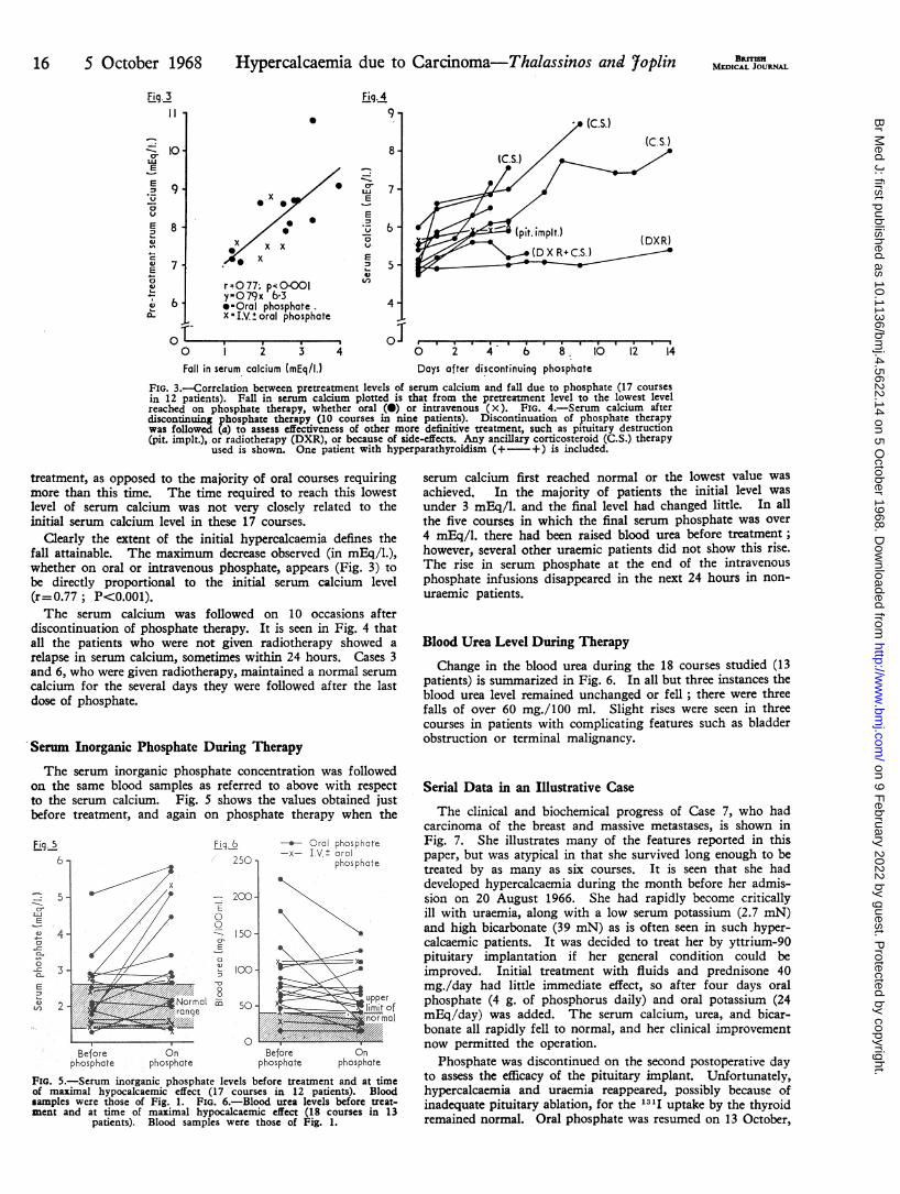
FIG. 3.-Correlation between pretreatment levels of serum calcium and fall due to phosphate (17 coursesin 12 patients). Fall in serum calcium plotted is that from the pretreatment level to the lowest levelreached on phosphate therapy, whether oral (@) or intravenous (x). FIG. 4.-Serum calcium afterdiscontinuing phosphate therapy (10 courses in nine patients). Discontinuation of phosphate therapywas followed (a) to assess effectiveness of other more definitive treatment, such as pituitary destruction(pit. implt.), or radiotherapy (DXR), or because of side-effects. Any ancillary corticosteroid (C.S.) therapy
used is shown. One patient with hyperparathyroidism (+ +) is included.
treatment, as opposed to the majority of oral courses requiringmore than this time. The time required to reach this lowestlevel of serum calcium was not very closely related to theinitial serum calcium level in these 17 courses.
Clearly the extent of the initial hypercalcaemia defines thefall attainable. The maximum decrease observed (in mEq/l.),whether on oral or intravenous phosphate, appears (Fig. 3) tobe directly proportional to the initial serum calcium level(r=0.77; P<0.001).The serum calcium was followed on 10 occasions after
discontinuation of phosphate therapy. It is seen in Fig. 4 thatall the patients who were not given radiotherapy showed arelapse in serum calcium, sometimes within 24 hours. Cases 3and 6, who were given radiotherapy, maintained a normal serumcalcium for the several days they were followed after the lastdose of phosphate.
Serum Inorganic Phosphate During Therapy
The serum inorganic phosphate concentration was followedon the same blood samples as referred to above with respectto the serum calcium. Fig. 5 shows the values obtained justbefore treatment, and again on phosphate therapy when the
Before Onphosphate phosphate
LI9&( 250'
200.
8I-' 150.d.-a.
,.4 loo2la
008M 50
O
-_- Oral phosphate-x- I.V.± oral
phosphate
Before u.Onphosphate phosphate
FIG. 5.-Serum inorganic phosphate levels before treatment and at timeof maximal hypocalcaemic effect (17 courses in 12 patients). Bloodsamples were those of Fig. 1. FIG. 6.-Blood urea levels before treat-ment and at time of maximal hypocalcaemic effect (18 courses in 13
patients). Blood samples were those of Fig. 1.
serum calcium first reached normal or the lowest value wasachieved. In the majority of patients the initial level wasunder 3 mEq/l. and the final level had changed little. In allthe five courses in which the final serum phosphate was over4 mEq/l. there had been raised blood urea before treatment;however, several other uraemic patients did not show this rise.The rise in serum phosphate at the end of the intravenousphosphate infusions disappeared in the next 24 hours in non-uraemic patients.
Blood Urea Level During Therapy
Change in the blood urea during the 18 courses studied (13patients) is summarized in Fig. 6. In all but three instances theblood urea level remained unchanged or fell; there were threefalls of over 60 mg./100 ml. Slight rises were seen in threecourses in patients with complicating features such as bladderobstruction or terminal malignancy.
Serial Data in an Illustrative Case
The clinical and biochemical progress of Case 7, who hadcarcinoma of the breast and massive metastases, is shown inFig. 7. She illustrates many of the features reported in thispaper, but was atypical in that she survived long enough to betreated by as many as six courses. It is seen that she haddeveloped hypercalcaemia during the month before her admis-sion on 20 August 1966. She had rapidly become criticallyill with uraemia, along with a low serum potassium (2.7 mN)and high bicarbonate (39 mN) as is often seen in such hyper-calcaemic patients. It was decided to treat her by yttrium-90pituitary implantation if her general condition could beimproved. Initial treatment with fluids and prednisone 40mg./day had little immediate effect, so after four days oralphosphate (4 g. of phosphorus daily) and oral potassium (24mEq/day) was added. The serum calcium, urea, and bicar-bonate all rapidly fell to normal, and her clinical improvementnow permitted the operation.
Phosphate was discontinued on the second postoperative dayto assess the efficacy of the pituitary implant. Unfortunately,hypercalcaemia and uraemia reappeared, possibly because ofinadequate pituitary ablation, for the 13'I uptake by the thyroidremained normal. Oral phosphate was resumed on 13 October,
BRfsHMEDICAL JOURNAL
6-
5.I-
*, 4.a
0.
-o 3.so,0
m 2
J-
o1 .,--
0
on 9 February 2022 by guest. P
rotected by copyright.http://w
ww
.bmj.com
/B
r Med J: first published as 10.1136/bm
j.4.5622.14 on 5 October 1968. D
ownloaded from

Ist. .i.. implant 2nd. pit.,implant I 1 1.1 I.I I I I
I I II I I II I - I I
I I Ia . f
)sphatc (g./day).IM4r~ 1 i|.w 2 =1 ir2mIV. Phosphate ( .55g.) 1
Prednisone ER.:.:.:.4f: : ,,. .
.7 20 25 30 5 10 15 20 25 30 4 9Aug.-I| Sep. I Ct.-. I
* - 19b{> ..~~~~~~~*1966,FIG. 7.-Effect of phosphate therapy on hypercalcaemia and blood urea in Cbreast cancer). Oral phosphate was initially effective in doses of 4 and 2daily. Later, neither oral nor intravenous phosphorus prevented advancing
Prednisone 40 mg. daily was probably ineffective, as was pituitary ir
2 g. of phosphorus being given daily because diarrhoea hadoccurred with 4 g. of phosphorus daily in the first course. Againthere was a brisk fall in serum calcium to normal. Reimplanta-tion was done on 20 October, but by this time she was in theterminal phase of the disease and henceforth neither oral norintravenous phosphate was effective in preventing the serumcalcium from increasing, though uraemia did not recur.
Clinical Effects of Phosphate Therapy
Side-effects.-These were confined to gastrointestinal symp-toms from oral administration, comprising epigastric pain,nausea, and diarrhoea. The higher the dose of oral phosphatethe more frequent and more obvious the symptoms. Some-times these could be better controlled by distributing the phos-phate in smaller and more frequent doses, or by symptomatictreatment, but the only successful measure sometimes was todiscontinue administration temporarily and, if necessary, con-tinue by the intravenous route.
Effect on Symptoms Due to Hypercalcaemia.-The bene-ficial effect of the phosphate treatment on the hypercalcaemicsymptoms was apparent in all the treated patients whose serumcalcium fell-that is, all except Case 12. The patients' clinicalimprovement paralleled the biochemical improvement, andseemed to antedate the latter by about a day.
Necropsy FindingsEight of the 13 hypercalcaemic patients with malignant
hypercalcaemia came to necropsy. None showed evidence ofexcessive calcification within the blood vessels and only twoshowed -microscopical nephrocalcinosis. These two patientshad had severe hypercalcaemia (Case 11, for more than onemonth and Case 10 for more than two months before beingseen by us) and corneal calcification was noted in both beforetreatment with inorganic phosphate; both patients also hadmild uraemia when first seen and both were treated with oralphosphate only. Unfortunately they succumbed from othereffects of cancer after only a few days of phosphate treatment.Of the eight patients who came to necropsy six had. had
corneal calcification noted by us in life before any treatmentwith inorganic phosphate had been given; this clinical obser-vation was confirmed by microscopy in the three cases in whichit was looked for. In the other two patients who came tonecropsy in whom we had not found corneal calcification in
BMMs=MEDICAL JOURNAL 17
Death life, no evidence of corneal calcification wasfound microscopically.
Discussion
I All of these hypercalcaemic patients with+ -| cancer responded favourably to the admin-
+ + I istration of an adequate course of inorganicii, DiarrhoeMa phosphate; seven achieved a normal serum
1 calcium and the rest showed a significantupper limit decrease. Concomitant with the fall in the
I_01 serum calcium the serum phosphate waslargely unchanged, while the blood urea alsooften fell and only rarely rose. Along with
1[ the biochemical evidence of amelioration ofOm the hypercalcaemia, in most patients there
. - . * was a striking clinical improvement. Some14 19 24, patients-for example, Cases 1 and 3-who
Nov. looked moribund before treatment were able
to eat and drink after 48 hours on phos--ase 7 (metastatic phate, and later became ambulant.g. of phosphorus Our findings with respect to the responsemplantation. of the serum calcium are in general agree-
ment with other authors who have usedinorganic phosphate treatment of hypercalcaemia (Bulgeret al., 1931 ; Albright et al., 1932; Dent, 1962; Goldsmith andIngbar, 1966; Hebert et al., 1966; Kahil et al., 1967; Kistler,1967; Massry et al., 1968). While the effective reduction ofthe rise in serum calcium has been a universal finding, the effecton the serum phosphate has been controversial. We have foundlittle change in serum phosphate with non-uraemic patients.Bulger et al. (1931) found an increase in the serum phosphatewhen they used oral inorganic phosphate in the treatment ofhyperparathyroidism, but that increase was from subnormal tonormal values, and our case of inoperable primary hyperpara-thyroidism responded similarly (see Appendix). Goldsmithand Ingbar (1966) found no significant change in the serum
phosphate in their patients with cancer at the stage that theserum calcium became normal, though some of them were
treated with repeated intravenous infusions. Likewise, Hebertet al. (1966), Kahil et al. (1967), and Kistler (1967) all foundthat in their cancer patients the increase in serum phosphatenear the end of an infusion came back to preinfusion valueswithin hours.There was no striking difference in the effect of oral or
intravenous administration; probably the intravenous route ismore rapidly effective. Thus phosphate need be given intra-venously only in those few cases where the patients conditiondoes not at first permit the oral route. Such ill patients mayhave the highest levels of serum calcium, and so may benefitespecially from the prompt reduction of the hypercalcaemia.We have never found more than a total of 2 g. of phosphorusrequired intravenously. Similarity in the effect of oral andintravenous phosphate was also found by Goldsmith and Ingbar(1966) and Massry et al. (1968), but not by Kahil et al. (1967)and Kistler (1967), who found no effect from oral administra-tion on serum calcium, though they did find a reduction inurinary calcium. It is difficult to explain their negative results,as our patients obtained a hypocalcaemic effect with even
smaller daily doses of phosphate.The therapeutic effect of inorganic phosphate on hyper-
calcaemia of malignant disease persisting after the generalmeasures of dietary calcium restriction and rehydration can bequite dramatic. Most of our patients had a preliminary periodof treatment confined to these conventional measures; henceit is surprising that, although it was used successfully 35 years
ago in connexion with hyperparathyroidism (Bulger et al.,1931; Albright et al., 1932), it had been largely abandoned untilrecently. This may have been partly due to a general unaware-ness of the frequency and clinical importance of " malignant "hypercalcaemia. In the meantime chelating agents, and
5 October 1968 Hypercalcaemia due to Carcinoma-Thalassinos and 7oplin
on 9 February 2022 by guest. P
rotected by copyright.http://w
ww
.bmj.com
/B
r Med J: first published as 10.1136/bm
j.4.5622.14 on 5 October 1968. D
ownloaded from

18 5 October 1968 Hypercalcaemia due to Carcinoma-Thalassinos and 7oplin MEBRITISHespecially edetic acid, have been used (Spencer et al., 1952;Holland et al., 1953), though it is clear that repeated infusionshad deleterious effects on the renal function (Dudley et al.,1955 ; Foreman et al., 1956). Sodium citrate infusions (Ken-nedy et al., 1953) have been used in an attempt to treat highserum calcium, but their effect lasts for only a few hours.
Corticosteroids have been widely accepted as a useful treat-ment of the hypercalcaemia of malignant disease (Connor et al.,1956 ; Watson, 1964) but are clearly less generally effective thanphosphate, as instanced in Fig. 7. Also the response is tooslow to be useful for critically severe hypercalcaemia (Gleckler,1956), and a number of cases have failed to respond to thistreatment (Connor et al., 1956; Thomas et al., 1958), as wealso have found. Sodium sulphate (Lemann and Mehr, 1965;Chakmakjian and Bethune, 1966) has been fairly effective, butits practical use is limited by the need to confine its administra-tion to intravenous infusions, while hypernatraemia is a hazardfrom the very large sodium content (Heckman and Walsh,1967).
Infusions of chelating agents, sodium citrate, and sodiumsulphate all act partly or wholly by producing an increase inurinary calcium (Wolf and Ball, 1950; Chakmakjian andBethune, 1966). On the contrary, inorganic phosphate lowersthe urine calcium (Hebert et al., 1966; Kistler, 1967) and thecalcium clearance (Massry et al., 1968), and so must act by anincrease in calcium deposition in bone or ectopic sites, or by areduction in bone resorption. Ignorance regarding which ofthese mechanisms is taking place has been the cause for hesita-tion in the use of inorganic phosphate.
Hebert et al. (1966) suggested that the observed decrease inthe serum calcium ought to be attributed to increased precipita-tion of calcium salts, due to tile increased solubility productresulting from a phosphate infusion. On the other hand, Pechetet al. (1967), working with thyroparathyroidectomized rats,concluded that the mechanism of action is by increasing truebone formation as evidenced by radioisotope and histologicalstudies. In addition, an enhancement of the action of calci-tonin, as reported by Hirsch (1968), may reduce the boneresorption rate. Some reported evidence in man suggests thatextraskeletal calcification is not increased after phosphate ad-ministration: Goldsmith and Ingbar (1966) considered thatthe calcification found in some of their phosphate-treatedpatients was not excessive in view of the long-standing hyper-calcaemia; Kahil et al. (1967) compared five hypercalcaemicpatients treated by intravenous inorganic phosphate with fivehypercalcaemic patients who were never treated with phosphate,and found no increased incidence at necropsy of extraskeletalcalcification, while in only two of them was there any evidenceof microscopical nephrocalcinosis.
Neither Dent's (1967) case of inoperable hyperparathyroidismon long-term phosphate treatment nor our own case (seeAppendix) has shown clinical or radiological evidence of ectopiccalcification. Furthermore, Bernstein and Newton (1966) havenow reported on long-term phosphate therapy for stone-formers; up to five years of treatment has not produced anyclinical or radiological evidence of ectopic calcification. Hebertet al. (1966) demonstrated radiologically some calcium deposi-tion in the vein into which phosphate was infused in three outof seven patients with cancer and hypercalcaemia ; this in itselfis not a proof of general extraskeletal calcification due to phos-phate, since calcification is a non-specific response to injury,and these veins were exposed to extremely high concentrationsof phosphate.
It therefore seems to us that oral inorganic phosphate (1 g.of phosphorus three times daily) is the treatment of choicefor the short-term palliation of malignant hypercalcaemia, occa-sionally preceded by an intravenous infusion (1 g. of phosphorusover eight hours) where the general clinical condition neces-sitates this route. Treatment is adequately monitored by dailyserum calcium estimations until a downward trend is shown,
and then it must be followed about twice weekly, after dis-continuing phosphate to detect any subsequent relapse. Hypo-kalaemia, which occurs quite often, should also be sought whenhypercalcaemia is found.
AppendixLong-term Administration of Phosphate in Patient with In-
operable Primary Hyperparathyroidism.-This patient has hadlong-standing hypercalcaemia due to hyperparathyroidism, butoperation has twice failed to reveal an adenoma. No skeletalchanges of hyperparathyroidism are seen radiologically. In view ofthe raised serum calcium (mean value 6.2 mEq/l.) and his vaguelack of energy, he was started on phosphate at a dose of 3 g. ofelemental phosphorus per day in October 1964. Fig. 8 summarizesthe progress of this patient, who has been on oral phosphate treat-ment for more than three years. The serum calcium soon fell andhe felt fitter. When treatment was withheld in January 1965 tocheck its efficacy, the serum calcium rose to the pretreatment levelwithin five days and serum phosphate dropped to subnormal levels.On restarting treatment near-normal levels of both serum calciumand phosphate concentrations were obtained. During a four-monthtrial on a lower dose of 1.5 g. of phosphorus per day both serumcalcium and phosphate became more abnormal, but reverted againto nearly normal when the previous dose of 3 g. of phosphorus perday was resumed. During phosphate treatment the urinary calciumwas 5 to 10 mEq/day, but on the temporary discontinuation oftreatment it shot up to the highly abnormal output of 20 mnEq/day.Seven days after reinstitution of treatment urine calcium again fell(14 mEq/day). During the whole course of treatment the bloodurea has been normal, and no evidence of corneal or renal calcifica-tion has appeared. He occasionally has to omit a dose of phosphateon account of diarrhoea.
Serum 6:2 pctalcium
2n0 L ^ I C
Ser'um
Phosphate 3O_
q./dayl OAct.-Jon. JulJun. n. . Jan..1964 1965 1966 196
FIG. 8.-Long-term phosphate treatment in inoperable primary hyper-parathyroidism. Near normal levels of serum calcium and phosphateare maintained by oral phosphate therapy at 3 g. of phosphorus daily.
Acknowledgements.-We are grateful to the physicians andsurgeons of Hammersmith Hospital who encouraged us in the studyand treatment of patients under their care, and to the nursing andjunior medical staff who exceeded their duty in assisting us withrecord-keeping and patient-management. We are also grateful tothe Department of Morbid Anatomy for the use of their post-mortem reports. We wish to thank Professor Russell Fraser for hishelpful criticism of the manuscript.One of us (N. T.) was supported by a World Health Organization
Fellowship, and the other (G. F. J.) by a Wellcome Senior ClinicalResearch Fellowship.
REFERENCES
Albright, F., Bauer, W., Claflin, D., and Cockrill, J. R. (1932). 7. clin.Invest., 11, 411.
Albright, F., and Reifenstein, E. C. (1948). The Parathyroid Glands andMetabolic Bone Disease. Baltimore.
Bernstein, D. S., and Newton, R. (1966). Lancet, 2, 1105.Binger, C. (1917-18). 7. Pharmnacol. exp. Ther., 10, 105.Bulger, H. A., Dixon, H. H., Barr, D. P., and Schregardus, 0. (1931).
7. clin. Invest., 9, 143.
on 9 February 2022 by guest. P
rotected by copyright.http://w
ww
.bmj.com
/B
r Med J: first published as 10.1136/bm
j.4.5622.14 on 5 October 1968. D
ownloaded from

5 October 1968 Hypercalcaemia due to Carcinoma-Thalassinos and ?oplin MEDICAL JOURNAL 19
Chakmakjian, Z. H., and Bethune, J. E. (1966). New Engl. 7. Med., 275,862.
Connor, T. B., Hopkins, T. R., Thomas, W. C., jun., Carey, R. A., andHoward, J. E. (1956). 7. clin. Endocr., 16, 945.
Dent, C. E. (1962). Brit. med. 7., 2, 1495.Dent, C. E. (1967). Lancet, 2, 613.Drivsholm, Aa., and Videbzk, Aa. (1966). Acta med. scand., 179, SuppL
No. 445, p. 187.Dudley, H. R., Ritchie, A. C., Schilling, A., and Baker, W. H. (1955).
New Engl. 7. Med., 252, 331.Foreman, H., Finnegan, C., and Lushbaugh, C. C. (1956). 7. Amer. med.
Ass., 160, 1042.Foster, G. V., et al. (1968). Calcitonin: Proceedings of the Symposium
on Thyrocalcitonin and the C Cells, edited by S. Taylor, p. 379.London.
Gleckler, W. J. (1956). Ann. intern. Med., 44, 174.Goldsmith, R. S., and Ingbar, S. H. (1966). New Engl. 7. Med., 274, 1.Hebert, L. A., Lemann, J., jun., Petersen, J. R., and Lennon, E. J. (1966).
7. clin. Invest., 45, 1886.Heckman, B. A., and Walsh, J. H. (1967). New Engl. 7. Med., 276, 1082.Henson, R. A. (1966). 7. roy. Coll. Phycns, 1, 41.Hirsch, P. F. (1968). Calcitonin: Proceedings of the Symposium on
Thyrocalcitonin and the C Cells, edited by S. Taylor, p. 11. London.Holland, J. F., Danielson, E., and Sahagian-Edwards, A. (1953). Proc.
Soc. exp. Biol. (N.Y.), 84, 359.
Joplin, G. F. (1965). Thesis submitted to University of London forPh.D., p. 302.
Kahil, M., Orman, B., Gyorkey, F., and Brown, H. (1967). 7. Amer.med. Ass., 201, 721.
Kennedy, B. J., Tibbetts, D. M., Nathanson, I. T., and Aub, J. C. (1953).Cancer Res., 13, 445.
Kistler, H. J. (1967). Helv. med. Acta, 33, 447.Lemann, J., jun., and Mehr, M. P. (1965). 7. Amer. med. Ass., 194,
1126Massry, S. G., Mueller, E., Silverman, A. G., and Kleeman, C. R. (1968).
Clin. Res., 16, 128.Pechet, M. M., Bobadilla, E., Carroll, E. L., and Hesse, R. H. (1967).
Amer. 7. Med., 43, 696.Plimpton, C. H., and Gellhorn, A. (1956). Amer. 7. Med., 21, 750.Salvesen, H. A., Hastings, A. B., and McIntosh, J. F. (1924). 7. biol.
Chem., 60, 311.Spencer, H., Vankinscott, V., Lewin, I., and Laszlo, D. (1952). 7. clin.
Invest., 31, 1023.Thomas, W. C., jun., Connor, T. B., and Morgan, H. G. (1958). 1. Lab.
clin. Med., 52, i1.Watson, L. (1964). Quart. 7. Med., 33, 525.Watson, L. (1966). Aust. Ann. Med., 15, 359.Wolf, A. V., and Ball, S. M. (1950). Amer. 7. Physiol., 160, 353.Wootton, I. D. P. (1964). Micro-analysis in Medical Biochemistry, 4th ed.
London.
Puerperal Thromboembolism in Relation to the Inhibition ofLactation by Oestrogen Therapy
T. N. A. JEFFCOATE,* M.D., F.R.C.S.ED., F.R.C.O.G.; JANINE MILLER,* M.B., CH.B., M.R.C.O.G.
R. F. ROOS,* M.B., CH.B., M.R.C.O.G.; V. R. TINDALLt M.D., F.R.C.S.ED., M.R.C.O.G.
Brit. med. J., 1968, 4, 19-25
S ummary: An analysis was made of 111 consecutivecases of puerperal thromboembolism by the age,
parity, mode of delivery, and lactation habit of the womenconcerned, and the findings were compared with thosefrom control groups.The statistics show that inhibition of lactation by means
of ethinyloestradiol is associated with a threefoldincrease in thromboembolism, although the effect is seenmainly in women who have an operative delivery and whoare aged more than 25 years. Among women aged morethan 3S years who have an assisted delivery, inhibition oflactation is accompanied by a tenfold increase in the inci-dence of puerperal thromboembolism.
Advancing age and operative intervention (especiallycaesarean section) are in themselves predisposing causesof deep venous thrombosis and embolism. They can alsoconstitute indications for inhibiting lactation. Thismakes it difficult to assess whether the relation ofthromboembolism to inhibition of lactation or to the ad-ministration of oestrogen is real or apparent. Doubts onthe interpretation of the findings are raised by the factthat the number of fatal cases of puerperal thrombo-embolism in England and Wales, and of non-fatal cases inthe hospitals under review, has not increased in recentyears despite a progressive decrease in breast-feeding.Nevertheless, the evidence suggests that although theadministration of ethinyloestradiol is not by itself enoughto cause puerperal thromboembolism, it may be a factorwhich can tip the scales in women who are already pre-disposed to suffer this condition.Any thromboembolic hazard associated with admini-
stration of oestrogens for inhibiting lactation is probablyacceptable except in women known to be at special riskby reason of age, operative delivery, obesity, and a pasthistory of thromboembolic episodes.
Introduction
It was first suggested by Daniel, Campbell, and Turnbull (1967)that suppression (or inhibition) of lactation favours the devel-opment of deep venous thrombosis and embolism in puerperalwomen. From information derived from the Cardiff BirthSurvey these workers concluded that the inhibition of lactationin mothers aged 25 and more is associated with a tenfoldincrease in the incidence of thromboembolism, and postulatedthat the administration of oestrogen may play a part in this.Following this, Daniel, Bloom, Giddings, Campbell, and Turn-bull (1968) showed that the administration of relatively largeamounts of diethylstilboestrol increases the coagulability ofblood by raising the level of factor IX. In the original Cardiffseries the oestrogen used was diethylstilboestrol administered individed doses up to a total of 210-330 mg. during the courseof nine days. This dosage, it may be noted, is higher than isusually given, and work carried out many years ago indicatedthat a total of 45-50 mg. of diethylstilboestrol during fourto seven days is generally adequate to inhibit lactation (Jeff-coate, Lister, Hargreaves, and Roberts, 1948).
There are some who question the efficacy of oestrogen therapyto inhibit lactation, irrespective of dosage, and the literaturepertaining to this was reviewed by Hodge (1967). Differencesin experience and opinion may in large measure reflect thetiming of the administration of the hormone. The evidencesuggests that the sooner after delivery oestrogen is giventhe more likely is it to inhibit breast activity. Oestrogens willnot suppress lactation which is already established. Whateverbe their efficacy, there is no question that oestrogens are widelyused with the object of inhibiting or suppressing lactation, andit is therefore of considerable importance to determine whetherall or certain of the preparations available predispose to puer-
*Department of Obstetrics and Gynaecology, University of Liverpool,Liverpool 3.
t Department of Obstetrics and Gynaecology, the Welsh National Schoolof Medicine, Cardiff.
on 9 February 2022 by guest. P
rotected by copyright.http://w
ww
.bmj.com
/B
r Med J: first published as 10.1136/bm
j.4.5622.14 on 5 October 1968. D
ownloaded from