phenolic compounds in flaxseed - slu.sepub.epsilon.slu.se/570/1/pernillajohnsson.pdfabstract...
TRANSCRIPT
Phenolic Compounds in Flaxseed
Chromatographic and Spectroscopic Analyses of Glucosidic Conjugates
Pernilla Johnsson Department of Food Science
Uppsala
Licentiate thesis Swedish University of Agricultural Sciences
Uppsala 2004

ISSN 1101-5411 ISBN 91-576-6630-x © 2004 Pernilla Johnsson, Uppsala Tryck: SLU Service/Repro, Uppsala 2004

Abstract
Johnsson, P. 2004. Phenolic compounds in flaxseed: Chromatographic and spectroscopic analyses of glucosidic conjugates. Licentiate thesis. ISSN 1101-5411, ISBN 91-576-6630-x
The dietary lignan secoisolariciresinol diglucoside (SDG), present in high concentrations in flaxseed, and its metabolites enterolactone and enterodiol are thought to decrease the risk of hormone dependent cancers, cardiovascular disease and other “welfare” diseases. Flaxseed also contains other biologically active phenolic compounds, such as phenolic acids. The understanding of the nature of these compounds is crucial for their possible exploitation in drugs and functional foods. Until the mid 1990’s, the number of methods available for analysis of lignans and other compounds in flaxseed was limited and were divergent in terms of yields. Moreover, the structure of a “flaxseed polymer” to which these phenolic compounds are bound was not known. Thus, the aim of the work presented in this thesis was to develop a quantitative method for analysis of SDG in flaxseed and to unravel the structure of the “polymer”.
A high performance liquid chromatography (HPLC) method for quantification of SDG in a base hydrolysed dioxane/ethanol flaxseed extract was developed. The method was applied to study the SDG content in 14 Swedish and 15 Danish flaxseed cultivars. The SDG content in defatted flour from the cultivars varied between 11.7 and 24.1 mg/g (6.1 – 13.3 mg/g in dried seeds).
Two chromatographic peaks eluting before SDG in the developed HPLC system were identified as 4-O-β-D-glucopyranosyl-p-coumaric acid and 4-O-β-D-glucopyranosyl-ferulic acid by liquid chromatography-mass spectrometry and nuclear magnetic resonance (NMR) analyses. Moreover, by the use of different NMR techniques, a strait chain oligomeric structure with an average composition of five SDG residues interlinked with four 3-hydroxy-3-methyl glutaric acid residues was assigned to the “polymer”.
Keywords: Linum usitatissimum, lignans, phenolic acids, polymer, oligomer, complex, glucosides, HPLC, NMR.
Author’s address: Pernilla Johnsson, Department of Food Science, P.O. Box 7051, Swedish University of Agricultural Sciences, S-750 07 Uppsala, Sweden.

Sammanfattning
Linfrö innehåller höga halter av lignanen secoisolariciresinol diglukosid (SDG) som som sådan, eller omvandlad till de metaboliska produkterna enterolakton och enterodiol, tros kunna hämma vissa hormonberoende cancersjukdomar, hjärtkärlsjukdomar och andra “välfärdssjukdomar”. Linfrö innehåller också andra biologiskt aktiva fenolföreningar, som t. ex. fenoliska syror. Kännedom om dessa ämnens egenskaper är nödvändig för deras eventuella användning i sk. mervärdesmat eller mediciner. Vid mitten av 1990-talet så var antalet metoder för analys av lignaner och andra föreningar i linfrö fortfarande begränsat, och de gav starkt varierande resultat i fråga om halter. Dessutom kände man inte till strukturen på det “linfröpolymer” i vilket dessa ämnen är bundna. Arbetet som presenteras i denna avhandling syftade därför till att utveckla en kvantitativ metod för SDG i linfrö och att utreda “polymer”-strukturen. En HPLC-metod (high performance liquid chromatography) för kvantitativ bestämning av SDG i dioxan/etanol-extraherade bashydrolysat av linfrö utvecklades. Metoden användes sedan för att analysera SDG-innehållet i 14 svenska och 15 danska linfrösorter. SDG-innehållet i avfettat linfrömjöl från de olika sorterna varierade mellan 11.7 and 24.1 mg/g (6.1 – 13.3 mg/g i torkade hela frön).
Två kromatografiska toppar som elueras före SDG i den framtagna HPLC-metoden identifierades som 4-O-β-D-glukopyranosyl-p-coumarsyra och 4-O-β-D-glukopyranosyl-ferulasyra med hjälp av LC-MS (liquid chromatography-mass spectrometry) och NMR (nuclear magnetic resonance). Med hjälp av olika NMR-tekniker tillskrevs “polymeren” en oligomerisk strukur bestående av en rak kedja med ett genomsnittligt innehåll av fem SDG molekyler sammanbundna av fyra molekyler av 3-hydroxy-3-metylglutarsyra.

Contents
Background 11
Dietary Lignans 12 Flaxseed – a rich source of plant lignans 12 Metabolism 14 Physiological and health effects 15
Other phenols in flaxseed 17 Analysis of phenolic compounds in flaxseed 18 Objectives 21 Analytical procedures 22 Results and discussion 25 Conclusions and future research 29 References 30 Acknowledgements 36

Appendix The present thesis is based on the following papers, which will be referred to by their Roman numerals: I. Johnsson, P., Kamal-Eldin, A., Lundgren, L.N., Åman, P. (2000). HPLC
method for analysis of secoisolariciresinol diglucoside in flaxseeds. Journal of agricultural and food chemistry, 48:5216-5219.
II. Johnsson, P., Peerlkamp, N., Kamal-Eldin, A., Andersson, R.E.,
Andersson, R., Lundgren, L.N., Åman, P. (2002). Polymeric fractions containing phenol glucosides in flaxseed, Food chemistry, 76:207-212.
III. Kamal-Eldin, A., Peerlkamp, N., Johnsson, P., Andersson, R., Andersson,
R.E., Lundgren, L.N., Åman, P. (2001). An oligomer from flaxseed composed of secoisolariciresinoldiglucoside and 3-hydroxy-3-methyl glutaric acid residues, Phytochemistry, 58:587-590.
Papers I-III are reproduced by permission of the publishers.

11
Background
The incidence and mortality of cancers of the breast, prostate and colon are higher in the western world compared to Asian countries (World Cancer Research Fund, 1997). In some western countries, e.g. Finland, there is a regional difference in cancer incidence possibly caused by differences in diet and lifestyle (Adlercreutz, 1990). Moreover, population and immigrant studies have shown that the incidence of the mentioned cancers has increased for Japanese who have immigrated to USA and changed their dietary habits (Dunn, 1975; Tominaga, 1985). There is now convincing evidence that a diet rich in whole grain, fruits and vegetables decreases the risk of developing coronary artery diseases, diabetes and cancer (Adlercreutz, 1990; Kushi et al., 1999; Adlercreutz, 2002; Bhathena and Velasquez, 2002; Kris-Etherton et al., 2002). Thus, the Swedish National Food Administration, along with other international authorities, recommends an increased consumption of these food products (National Food Administration, 2004).
The “western world” diet, typically high in fat and protein and low in fibre and whole-grain products, is associated with high levels of endogenous sex hormones and low levels of sex hormone binding globulin (SHBG) (Adlercreutz, 1990). These factors may play an important role in the development of hormone dependent cancers such as breast, prostate and endometrial cancer. Also, coronary heart disease and colon cancer may result from such a dietary pattern due to unfavourable plasma lipid levels and intestinal bile acid metabolism.
In the 1970’s, a class of compounds called lignans, especially abundant in fibre rich parts of plants, was recognised by the National Cancer Institute (USA) as high-interest compounds with regard to antitumour activity (Hartwell, 1976; Barclay, 1976). A few years later, the “mammalian lignans” enterolactone and enterodiol, were discovered in human urine (Setchell et al., 1980 b; Stich et al., 1980; Setchell et al, 1981). Their excretion in urine from human females was shown to vary during the reproductive cycle, which suggested a physiological function related to hormone metabolism (Setchell et al., 1979; Stich et al., 1980). Eventually, this lead to the assumption that fibre-rich plant foods containing hormone like compounds such as lignans and isoflavonoids, also known as phyto-oestrogens, may influence oestrogen metabolism and reduce the incidence of colon and breast cancer and possibly other diseases (Adlercreutz, 1984; Horwitz and Walker, 1984; Adlercreutz, 1990).
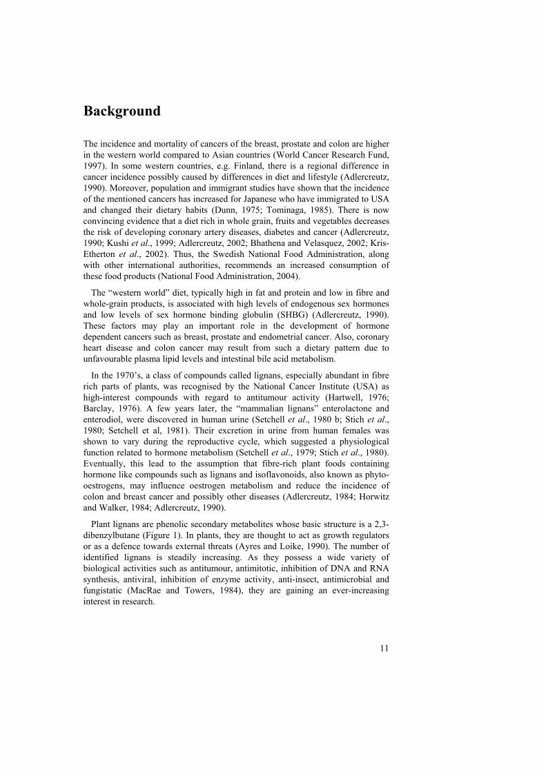
Plant lignans are phenolic secondary metabolites whose basic structure is a 2,3-dibenzylbutane (Figure 1). In plants, they are thought to act as growth regulators or as a defence towards external threats (Ayres and Loike, 1990). The number of identified lignans is steadily increasing. As they possess a wide variety of biological activities such as antitumour, antimitotic, inhibition of DNA and RNA synthesis, antiviral, inhibition of enzyme activity, anti-insect, antimicrobial and fungistatic (MacRae and Towers, 1984), they are gaining an ever-increasing interest in research.
12
Figure 1. The basic structure of lignans.
Dietary lignans
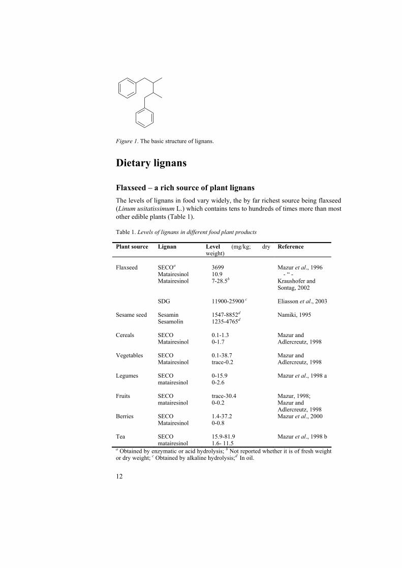
Flaxseed – a rich source of plant lignans The levels of lignans in food vary widely, the by far richest source being flaxseed (Linum usitatissimum L.) which contains tens to hundreds of times more than most other edible plants (Table 1). Table 1. Levels of lignans in different food plant products Plant source Lignan Level (mg/kg; dry
weight) Reference
Flaxseed
SECOa Matairesinol Matairesinol SDG
3699 10.9 7-28.5b 11900-25900 c
Mazur et al., 1996 - “ - Kraushofer and Sontag, 2002 Eliasson et al., 2003
Sesame seed Sesamin Sesamolin
1547-8852d 1235-4765d
Namiki, 1995
Cereals SECO Matairesinol
0.1-1.3 0-1.7
Mazur and Adlercreutz, 1998
Vegetables SECO Matairesinol
0.1-38.7 trace-0.2
Mazur and Adlercreutz, 1998
Legumes SECO matairesinol
0-15.9 0-2.6
Mazur et al., 1998 a
Fruits SECO matairesinol
trace-30.4 0-0.2
Mazur, 1998; Mazur and Adlercreutz, 1998
Berries SECO Matairesinol
1.4-37.2 0-0.8
Mazur et al., 2000
Tea SECO matairesinol
15.9-81.9 1.6- 11.5
Mazur et al., 1998 b
a Obtained by enzymatic or acid hydrolysis; b Not reported whether it is of fresh weight or dry weight; c Obtained by alkaline hydrolysis;d In oil.
13
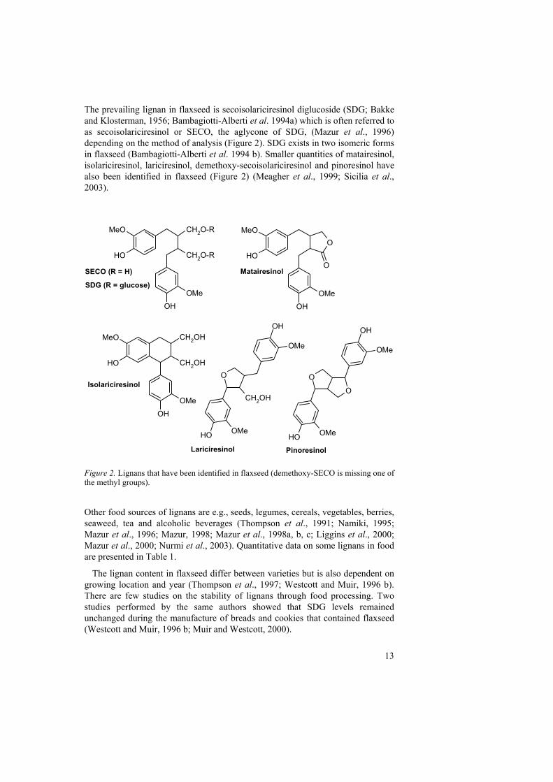
The prevailing lignan in flaxseed is secoisolariciresinol diglucoside (SDG; Bakke and Klosterman, 1956; Bambagiotti-Alberti et al. 1994a) which is often referred to as secoisolariciresinol or SECO, the aglycone of SDG, (Mazur et al., 1996) depending on the method of analysis (Figure 2). SDG exists in two isomeric forms in flaxseed (Bambagiotti-Alberti et al. 1994 b). Smaller quantities of matairesinol, isolariciresinol, lariciresinol, demethoxy-secoisolariciresinol and pinoresinol have also been identified in flaxseed (Figure 2) (Meagher et al., 1999; Sicilia et al., 2003).
Figure 2. Lignans that have been identified in flaxseed (demethoxy-SECO is missing one of the methyl groups).
Other food sources of lignans are e.g., seeds, legumes, cereals, vegetables, berries, seaweed, tea and alcoholic beverages (Thompson et al., 1991; Namiki, 1995; Mazur et al., 1996; Mazur, 1998; Mazur et al., 1998a, b, c; Liggins et al., 2000; Mazur et al., 2000; Nurmi et al., 2003). Quantitative data on some lignans in food are presented in Table 1.
The lignan content in flaxseed differ between varieties but is also dependent on growing location and year (Thompson et al., 1997; Westcott and Muir, 1996 b). There are few studies on the stability of lignans through food processing. Two studies performed by the same authors showed that SDG levels remained unchanged during the manufacture of breads and cookies that contained flaxseed (Westcott and Muir, 1996 b; Muir and Westcott, 2000).
CH2O-R
CH2O-ROH
OH
MeO
OMe
O
OH
OH
OMe
MeO
O
CH2OH
CH2OHOH
OH
MeO
OMe
OH
CH2OH
O
OH
OMe
OMe
OH
OMe
O
OH OMe
O
SECO (R = H)
SDG (R = glucose)
Matairesinol
Isolariciresinol
Lariciresinol Pinoresinol
14
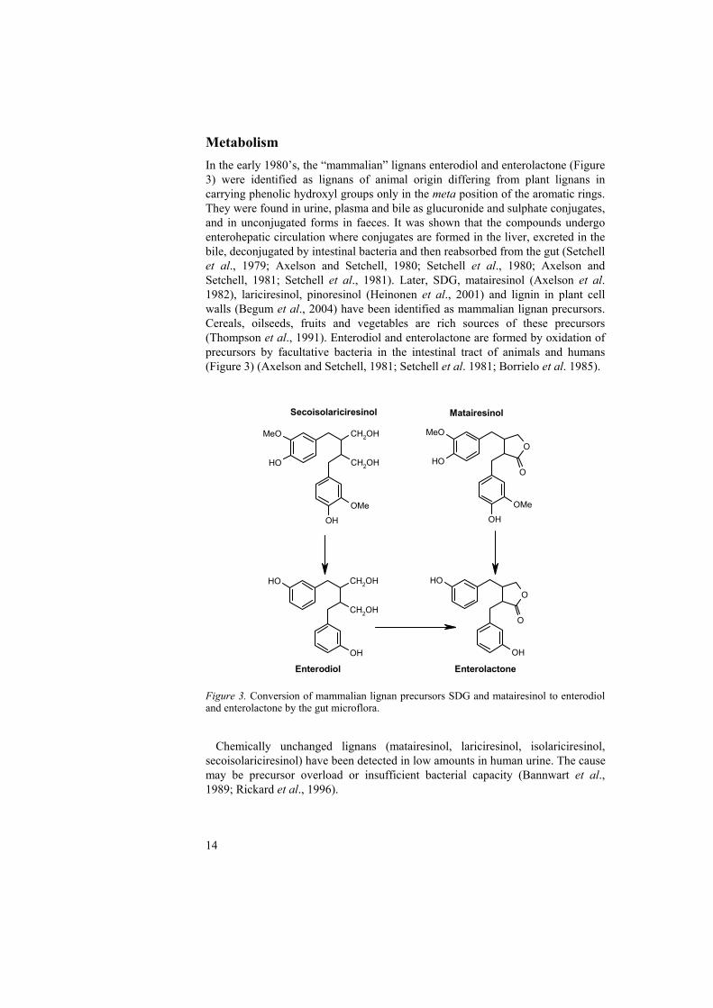
Metabolism In the early 1980’s, the “mammalian” lignans enterodiol and enterolactone (Figure 3) were identified as lignans of animal origin differing from plant lignans in carrying phenolic hydroxyl groups only in the meta position of the aromatic rings. They were found in urine, plasma and bile as glucuronide and sulphate conjugates, and in unconjugated forms in faeces. It was shown that the compounds undergo enterohepatic circulation where conjugates are formed in the liver, excreted in the bile, deconjugated by intestinal bacteria and then reabsorbed from the gut (Setchell et al., 1979; Axelson and Setchell, 1980; Setchell et al., 1980; Axelson and Setchell, 1981; Setchell et al., 1981). Later, SDG, matairesinol (Axelson et al. 1982), lariciresinol, pinoresinol (Heinonen et al., 2001) and lignin in plant cell walls (Begum et al., 2004) have been identified as mammalian lignan precursors. Cereals, oilseeds, fruits and vegetables are rich sources of these precursors (Thompson et al., 1991). Enterodiol and enterolactone are formed by oxidation of precursors by facultative bacteria in the intestinal tract of animals and humans (Figure 3) (Axelson and Setchell, 1981; Setchell et al. 1981; Borrielo et al. 1985).
Figure 3. Conversion of mammalian lignan precursors SDG and matairesinol to enterodiol and enterolactone by the gut microflora.
Chemically unchanged lignans (matairesinol, lariciresinol, isolariciresinol,
secoisolariciresinol) have been detected in low amounts in human urine. The cause may be precursor overload or insufficient bacterial capacity (Bannwart et al., 1989; Rickard et al., 1996).
CH2OH
CH2OH
OH
MeO
OH
OMe
O
OH
OOH
MeO
OMe
CH2OH
CH2OH
OH
OHO
OH
OH
O
Enterodiol Enterolactone
MatairesinolSecoisolariciresinol
15
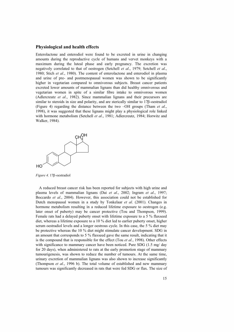
Physiological and health effects Enterolactone and enterodiol were found to be excreted in urine in changing amounts during the reproductive cycle of humans and vervet monkeys with a maximum during the luteal phase and early pregnancy. The excretion was negatively correlated to that of oestrogen (Setchell et al., 1979; Setchell et al., 1980; Stich et al., 1980). The content of enterolactone and enterodiol in plasma and urine of pre- and postmenopausal women was shown to be significantly higher in vegetarian compared to omnivorous subjects. Breast cancer patients excreted lower amounts of mammalian lignans than did healthy omnivorous and vegetarian women in spite of a similar fibre intake to omnivorous women (Adlercreutz et al., 1982). Since mammalian lignans and their precursors are similar to steroids in size and polarity, and are sterically similar to 17β-oestradiol (Figure 4) regarding the distance between the two –OH groups (Tham et al., 1998), it was suggested that these lignans might play a physiological role linked with hormone metabolism (Setchell et al., 1981; Adlercreutz, 1984; Horwitz and Walker, 1984).
Figure 4. 17β-oestradiol
A reduced breast cancer risk has been reported for subjects with high urine and
plasma levels of mammalian lignans (Dai et al., 2002; Ingram et al., 1997; Boccardo et al., 2004). However, this association could not be established for Dutch menopausal women in a study by Tonkelaar et al. (2001). Changes in hormone metabolism resulting in a reduced lifetime exposure to oestrogen (e.g. later onset of puberty) may be cancer protective (Tou and Thompson, 1999). Female rats had a delayed puberty onset with lifetime exposure to a 5 % flaxseed diet, whereas a lifetime exposure to a 10 % diet led to earlier puberty onset, higher serum oestradiol levels and a longer oestrous cycle. In this case, the 5 % diet may be protective whereas the 10 % diet might stimulate cancer development. SDG in an amount that corresponds to 5 % flaxseed gave the same result, indicating that it is the compound that is responsible for the effect (Tou et al., 1998). Other effects with significance to mammary cancer have been noticed. Pure SDG (1.5 mg/ day for 20 days), when administered to rats at the early promotion stage of mammary tumourigenesis, was shown to reduce the number of tumours. At the same time, urinary excretion of mammalian lignans was also shown to increase significantly (Thompson et al., 1996 b). The total volume of established and new mammary tumours was significantly decreased in rats that were fed SDG or flax. The size of
OH
OHCH3
16
established tumours was inversely related to urinary excretion of mammalian lignans (Thompson et al., 1996 a). According to a review by Adlercreutz (2002), a plasma enterolactone concentration of 30-70 nmol/l is likely to protect against breast cancer. Plasma levels of enterolactone were 8.3-38.2 nmol/l in premenopausal women who ingested 25 g flaxseed/day for eight days (Nesbitt et al., 1999) and has been shown to increase by 37 % for each 10 g increase in fibre intake (Horner et al., 2002).
Dietary lignans may also protect against prostate and colon cancer (Adlercreutz, 2002). High levels of enterolactone in prostatic fluid may indicate a lower risk of prostate cancer (Morton et al., 1997). A 5 % flaxseed diet inhibited growth and development of prostatic carcinoma in mice (Lin et al., 2002) possibly due to hormonal effects of the flaxseed lignans. In male rats, a 5 % lifetime diet reduced the prostate cell proliferation rate and prostate weight without a raise in sex hormone levels. A 10 % lifetime diet on the other hand resulted in higher serum testosterone and oestradiol levels and prostate cell proliferation. Again, the 5 % diet seemed to be protective whereas the 10 % diet might be at risk of stimulating cancer development. Results indicated that SDG is responsible for the effect (Tou et al., 1998). Diets supplemented with flaxseed, flaxmeal (2.5 or 5 %) or SDG (daily gavage of 1.5 mg) showed a protective effect against colon cancer in rats (Jenab and Thompson, 1996).
The reason for the cancer inhibiting activity of mammalian lignans and their precursor may be that they possess oestrogenic/anti-oestrogenic effects. A decreased formation and circulation of endogenous oestrogens limit stimulation of oestrogen dependent tumours. A compound with weak oestrogenic effect can be anti-oestrogenic when competing with a compound that has a stronger oestrogenic effect. The effect is dependent on the dose, timing and duration of exposure (Tou et al., 1998). In a study by Mousavi and Adlercreutz (1992), oestradiol and enterolactone, per se, stimulated the growth of MCF-7 breast cancer cells in vitro, but together they inhibited each others effect. Enterolactone was found to inhibit the binding of oestradiol and testosterone to SHBG in vitro in a dose-dependent manner with effective concentration range of 5-50 µM (Martin, et al., 1996). Aromatase (oestrogen synthetase) is an enzyme which is involved in oestrogen formation. Adlercreutz et al. (1993) showed that enterolactone caused a 50 % inhibition of aromatase at a concentration of 14 µM. Also, demethylated forms of matairesinol and enterolactone as well as demethylated secoisolariciresinol and enterodiol respectively, were shown to be moderate to weak inhibitors of aromatase activity in human preadipocytes (Wang et al., 1994). Moreover, enterolactone has been shown to depress oestrogen-stimulated RNA synthesis in rats (Waters and Knowler, 1982).
Flaxseed low in α-linolenic acid (<3%), was shown to decrease the level of atherosclerotic plaques in rabbits (Prasad et al, 1998). High serum enterolactone concentrations were correlated with a decreased level of in vivo lipid peroxidation (Vanharanta et al., 2002), and were also protective against acute coronary heart events in men as suggested by a case-control study (Vanharanta et al., 1999). Cardio protective effects may be due to inhibition of lipid peroxidation or changes in SHBG production, and circulation of cholesterol (Vanharanta et al., 1999;
17
Vanharanta et al., 2002). A low rate of lipid peroxidation may lower the risk of atherogenesis (Chisolm and Steinberg, 2000). Decreases in lipid peroxidation is perhaps not caused by enterolactone but by its precursors matairesinol, and SECO or SDG (Prasad, 1997; Prasad, 1999). Primarily SDG, SECO, and matairesinol but also enterolactone and enterodiol have been shown to possess antioxidant activity in vitro (Kitts et al., 1999; Prasad, 2000; Niemeyer and Metzler, 2003). Moreover, SDG was demonstrated to prevent the development of type I and II diabetes in diabetes prone rats probably by decreasing oxidative stress in body tissues (Prasad, 2000; Prasad, 2001). There are, however, results that may have a negative influence on the development of cardiovascular disease. SDG and its oligomer, when fed to rats, were shown to significantly increase the amount of cholesterol in the liver and liver lipids (p<0.05), and to decrease rat plasma content of α- and γ–tocopherols (p<0.001) (Frank et al., 2004). Also flaxseed was found to cause a significant reduction in α- and γ–tocopherol levels in rats (Ratnayake, et al., 1992). Reduced levels of vitamin E, γ-tocopherol in particular, have been associated with an increased risk for cardiovascular disease (Hensley et al., 2004).
In spite of all the evidence pointing towards health protective effects of enterolactone and enterodiol, the results are sometimes confusing and contradictive. The determinants or causes of their excretion may be a more important factor than the single compounds. Also, a low fat intake and a healthy and viable gut microflora may be crucial for a decreased risk of many diet-related diseases. Whether mammalian lignans really are effective or if they are merely indicators of a healthy diet has yet to be established (Adlercreutz, 2002; Adlercreutz, 2003).
Other phenols in flaxseed
Apart from lignans, flaxseed has been reported to contain free phenolic acids, glycosylated phenolic acids and flavonoids. Due to varying methodologies, reports on quantities and types (e.g. free, ester bound etc.) of phenolic acids in flaxseed are diverging and confusing. Kozlowska et al. (1983), showed that the highest proportion of phenolic acids in flaxseed and other oil seeds were ester bound. By extraction with 80 % methanol, the content of ester bound phenolic acids in flaxseed was 320 mg/kg defatted flaxseed flour (DFF), the main constituents being p-hydroxybenzoic, trans-ferulic and trans-p-coumaric acids. Free phenolic acids, present at 200-280 mg/kg defatted flaxseed flour, were mainly composed of trans- and cis-sinapic, o-coumaric, p-hydroxybenzoic, trans-p-coumaric and vanillic acids. The content of residual (non-extractable with the method used) phenolic acids was 70 mg/kg DFF. Dabrowski and Sosulski (1984), found no free phenolic acids when extracting DFF with tetrahydrofuran. Alkaline hydrolysis released 730 mg phenolic acids/kg DFF (89 % of total phenolic acids), the major ones being trans-ferulic and trans-sinapic acid. Harris and Haggerty (1993) reported a content of ca 11 mg ferulic acid/kg in whole flaxseed. They did not use alkaline hydrolysis and, thus, they could not detect ester bound phenolic acids. Oomah et al. (1995) reported a content of 8000-10 000 mg total phenolic acids/kg flaxseed of which
18
48-66% was assigned to esterified phenolic acids. Phenolic acids that were not released after extraction and alkaline hydrolysis were assumed to be ether bound. Variations in phenolic acid content in flaxseed were largely attributed to seasonal effects (Oomah et al., 1995).
Glycosides of p-coumaric, ferulic, and caffeic acid have been detected in flaxseed (Westcott and Muir, 1996 a; Westcott and Muir, 2000; Eliasson et al., 2003). Linocinnamarin (4-[β-D-glucopyranosido]-hydroxycinnamate) was first isolated from flaxseed by Klosterman et al. (1954). The compound linusitamarin (methyl 3-β-D-glucopyranosyl-5-methoxycinnamate) was assigned by Luyengi et al. (1993). However, the structure of this compound was probably misinterpreted as far as the 3/5 substitution is concerned which is a very uncommon substitution pattern (Westcott and Muir, 2000). It was more likely to be a ferulic acid glucoside (methyl 3-β-D-glucopyranosyl-4-methoxycinnamate) as supported by NMR analyses performed by Westcott and Muir (2000). Flavonoids are also present in flaxseed in high amounts with reported means of 490-870 mg/kg DFF. The content was influenced by both cultivar and environment (Oomah et al., 1996).
The above mentioned plant phenols may function as blocking or trapping agents for chemically induced cancers caused by aromatic carcinogens. A continuous input of these protecting compounds may serve as a buffer against cell damage by quantitative and qualitative supplementation of endogenous protective systems. The conjugated groups, e.g. sugar moieties, function as protection of the easily oxidised and biologically more potent free forms which are released upon acid or enzymatic hydrolysis in the gastro-intestinal tract of animals and humans (Newmark, 1984).
Analysis of phenolic compounds in flaxseed
Following the discovery of SDG by Bakke and Klosterman (1956) and its connection to the mammalian lignans, several methods for the analysis of lignans and other constituents of the fat free portion of flaxseed have been developed, although only a few quantitative. In order to obtain repeatable results, defatting of flaxseed cake prior to further treatment has been deemed crucial (Kozlowska et al., 1983; Harris and Haggerty, 1993). The polymeric powder obtained by ethanol:dioxane extraction of DFF was found to release hydroxy methyl glutaric acid (HMGA; Figure 5), 4-O-β-D-glucopyranosyl coumaric acid, and SDG upon base hydrolysis (Klosterman and Smith, 1954; Klosterman et al., 1955; Bakke and Klosterman, 1956), suggesting that these compounds are bound in an ester linked polymeric structure(s) in flaxseed. The compounds may also be released from the polymeric material as aglycones by enzyme or acid hydrolysis (Setchell et al., 1981; Mazur et al., 1996).
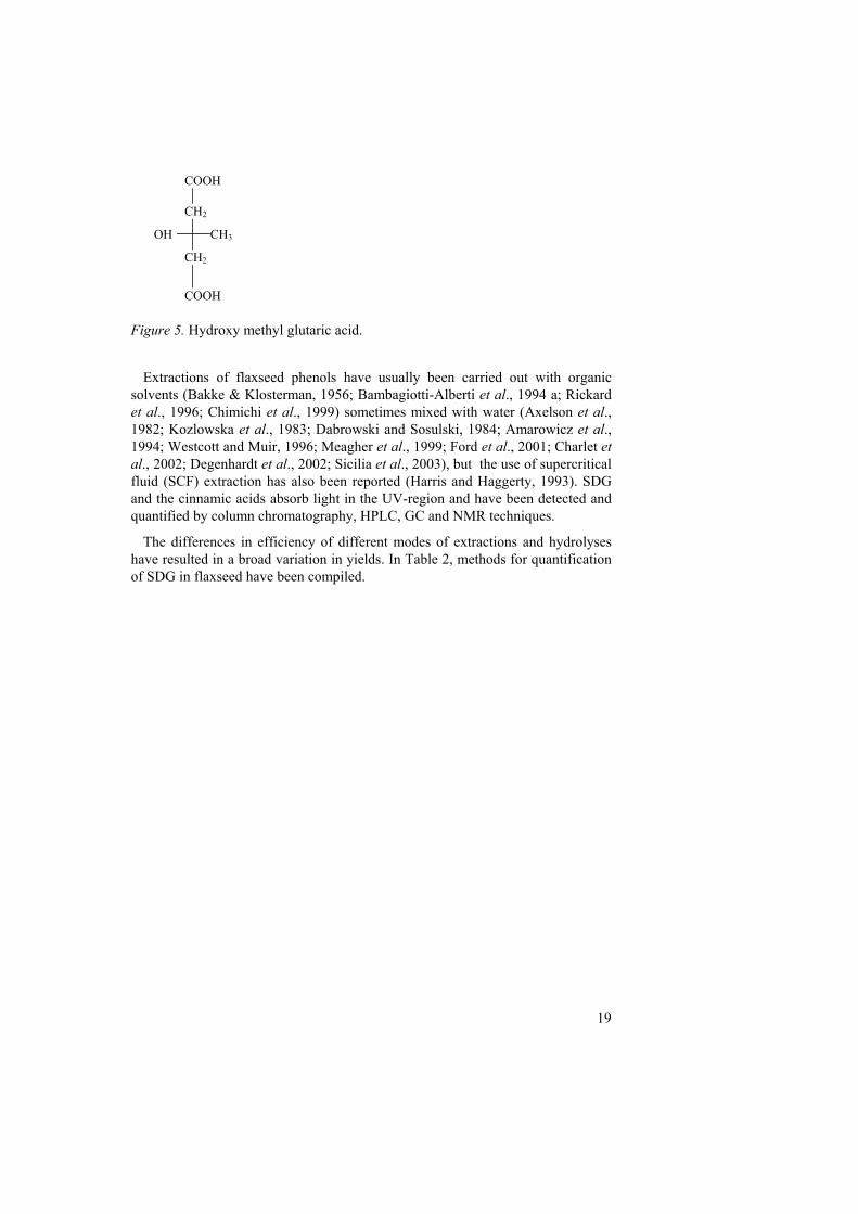
19
Figure 5. Hydroxy methyl glutaric acid.
Extractions of flaxseed phenols have usually been carried out with organic
solvents (Bakke & Klosterman, 1956; Bambagiotti-Alberti et al., 1994 a; Rickard et al., 1996; Chimichi et al., 1999) sometimes mixed with water (Axelson et al., 1982; Kozlowska et al., 1983; Dabrowski and Sosulski, 1984; Amarowicz et al., 1994; Westcott and Muir, 1996; Meagher et al., 1999; Ford et al., 2001; Charlet et al., 2002; Degenhardt et al., 2002; Sicilia et al., 2003), but the use of supercritical fluid (SCF) extraction has also been reported (Harris and Haggerty, 1993). SDG and the cinnamic acids absorb light in the UV-region and have been detected and quantified by column chromatography, HPLC, GC and NMR techniques.
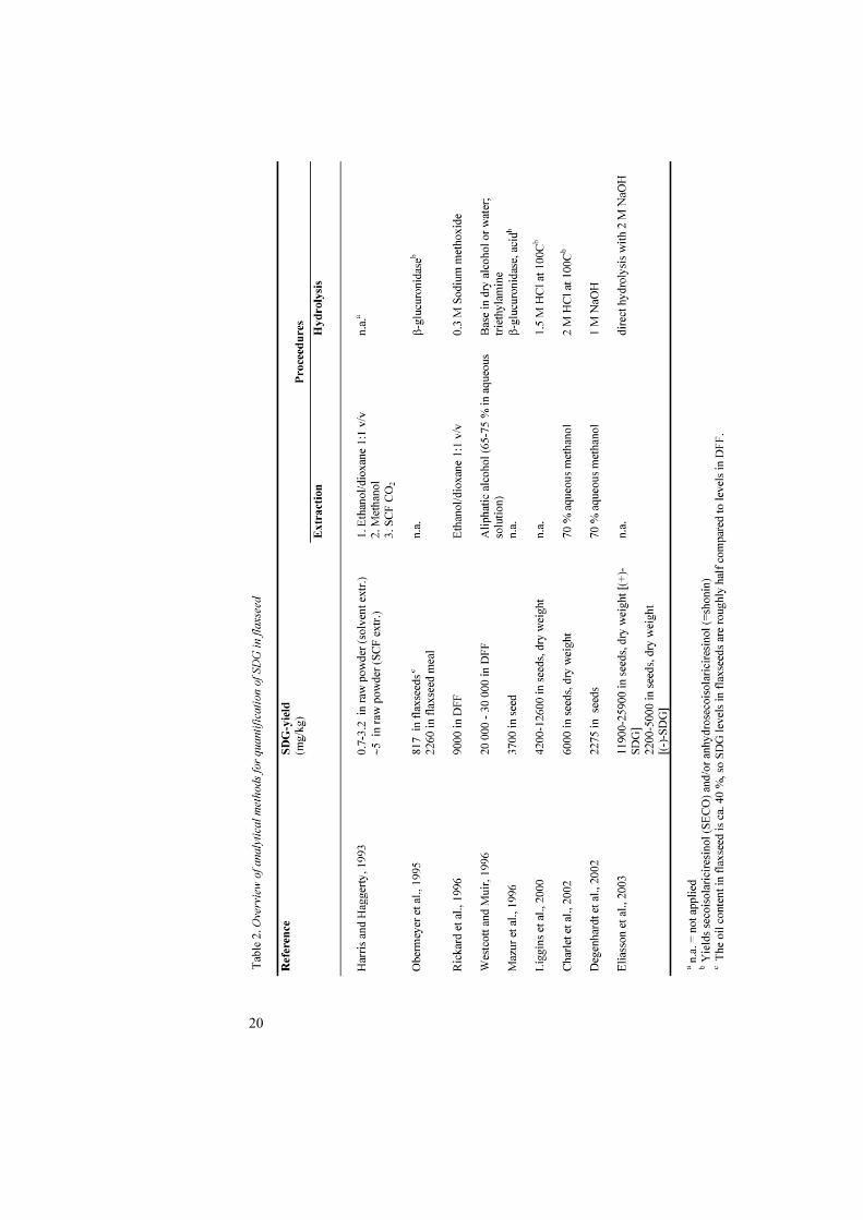
The differences in efficiency of different modes of extractions and hydrolyses have resulted in a broad variation in yields. In Table 2, methods for quantification of SDG in flaxseed have been compiled.
CH2
COOH
CH3
CH2
OH
COOH
20
21
Objectives
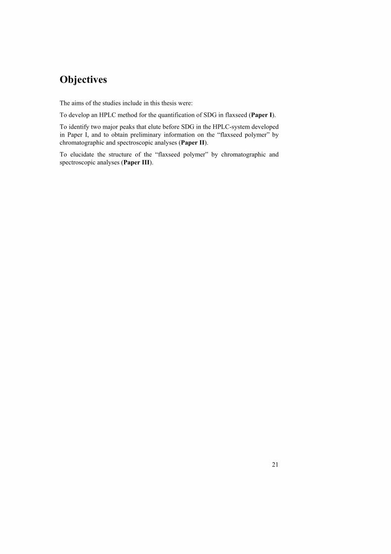
The aims of the studies include in this thesis were:
To develop an HPLC method for the quantification of SDG in flaxseed (Paper I).
To identify two major peaks that elute before SDG in the HPLC-system developed in Paper I, and to obtain preliminary information on the “flaxseed polymer” by chromatographic and spectroscopic analyses (Paper II).
To elucidate the structure of the “flaxseed polymer” by chromatographic and spectroscopic analyses (Paper III).

22
Analytical procedures
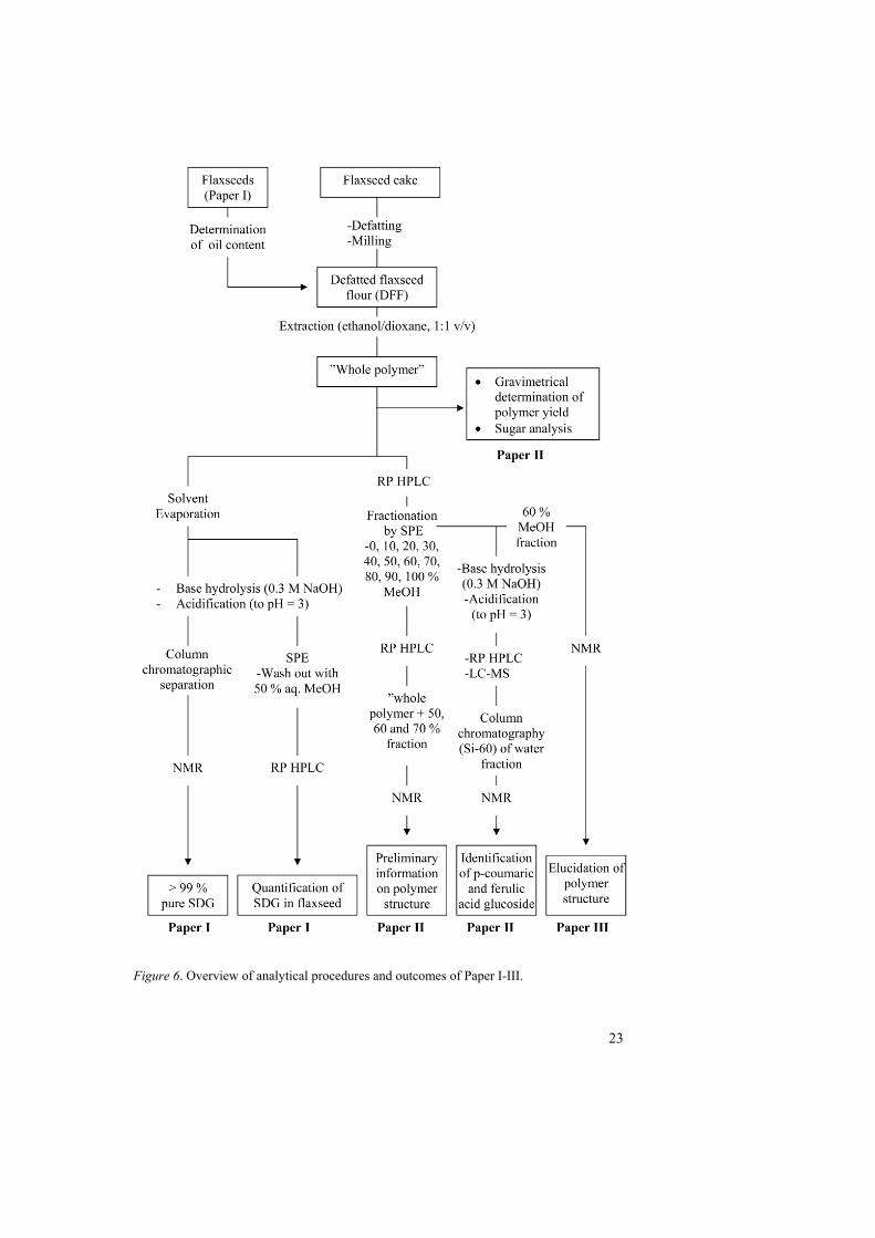
An outline of the work resulting in papers I-III is shown in Figure 6. (For more detailed descriptions, see Paper I-III).
In Paper I, SDG was purified from a flaxseed extract by reversed and normal phase column chromatography for use as an external standard in quantifications and for future reference. The identity and purity of the isolated SDG was determined by proton nuclear magnetic resonance (1H NMR). A high performance liquid chromatography (HPLC) method for the quantitative determination of SDG in flaxseed was developed and used for analysis of SDG in Swedish (n = 14) and Danish (n = 15) flaxseed cultivars. SDG was detected by UV- diode array detection (DAD) and its content in flaxseed was calculated using a six point linear standard curve (R2 = 0.999).
In Paper II, a flaxseed extract, “whole polymer”, was fractionated with 0-100 % methanol (in 10 %-unit intervals) on a C18 reversed phase solid phase extraction (SPE) column yielding 11 fractions. The fractions were analysed by HPLC. The “whole polymer” and its main UV-absorbing fractions, eluted with 50, 60 and 70 % aqueous methanol, were analysed by one- and two-dimensional NMR techniques (1H NMR, COSY, HSQC-DEPT and HMBC) in order to get structural information on the polymer. The same polymeric fractions were also subjected to alkaline hydrolysis and analysed by HPLC. SDG was identified by comparison with a standard (see above) and two other major peaks in the chromatograms were investigated by LC-MS and NMR. The compounds representing the two HPLC-peaks were isolated from an aqueous SPE fraction by normal phase column chromatography and identified by NMR. The sugar content and the yield of the “whole polymer” were determined by qualitative sugar analysis (Theander et al., 1995) and gravimetrical determination respectively.
To obtain the major fraction of the polymer for the experiments in Paper III, the “whole polymer” was washed on a SPE column with 50% aqueous methanol and then eluted with 60 % methanol. To elucidate the structure of the SDG-complex, the fraction was analysed by different NMR techniques (1H NMR, 13C NMR, TOCSY, HSQC-DEPT and HMBC). Standard SDG (see above) and HMGA (Sigma-Aldricht Chemie, Germany) were used as reference compounds.
23
Figure 6. Overview of analytical procedures and outcomes of Paper I-III.

25
Results and discussion
In 1998, at the beginning of this work, the nature of the extractable phenolic polymer first described by Klosterman and Smith (1954) was not known. Moreover, reports on the SDG/SECO content in flaxseed showed a great variation (Table 2), and values were often surprisingly low considering the high amounts of mammalian lignans that were excreted in urine of humans and animals (Axelson et al., 1982; Thompson et al., 1991) after flaxseed ingestion. Our aim was thus to develop a quantitative method for the analysis of SDG in flaxseed and to learn more about the complex in which SDG is included. This work is presented in three published papers (see Figure 6 and Appendix), and summarised hereafter.
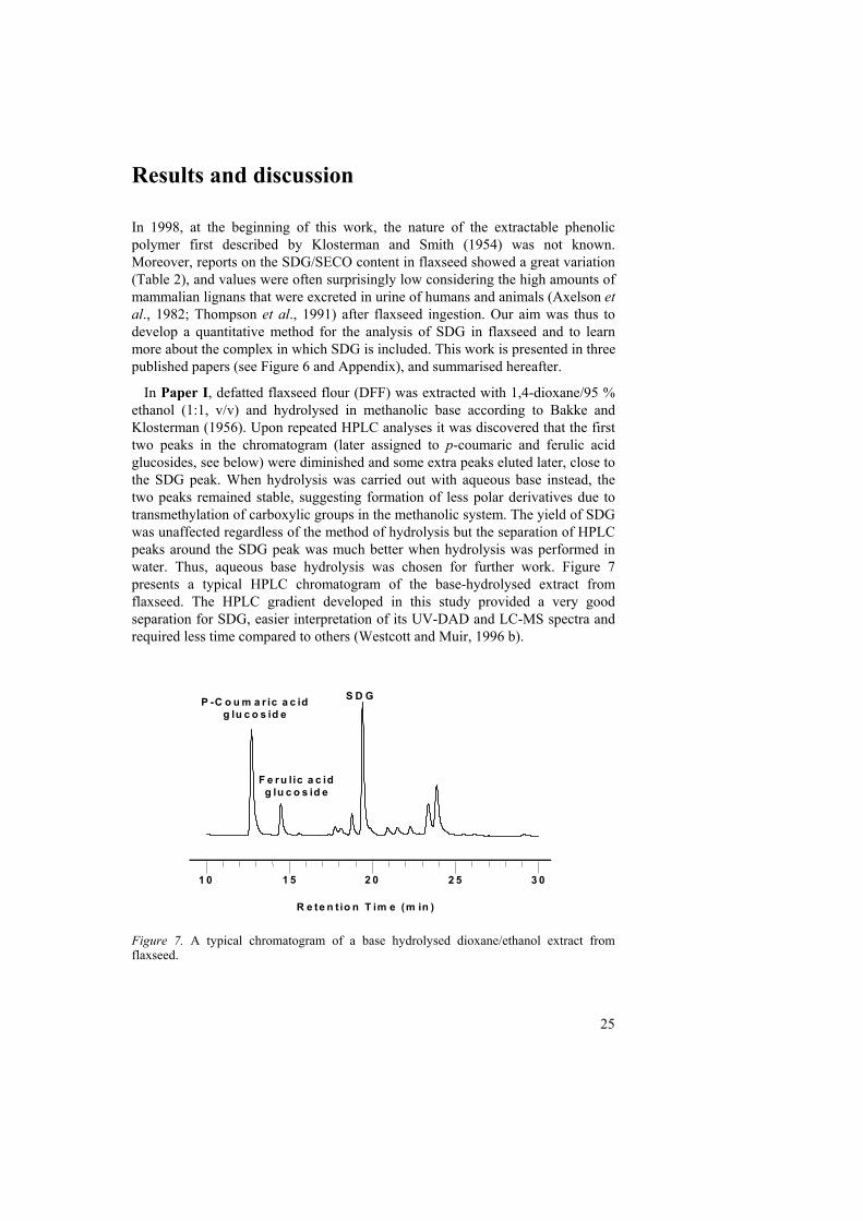
In Paper I, defatted flaxseed flour (DFF) was extracted with 1,4-dioxane/95 % ethanol (1:1, v/v) and hydrolysed in methanolic base according to Bakke and Klosterman (1956). Upon repeated HPLC analyses it was discovered that the first two peaks in the chromatogram (later assigned to p-coumaric and ferulic acid glucosides, see below) were diminished and some extra peaks eluted later, close to the SDG peak. When hydrolysis was carried out with aqueous base instead, the two peaks remained stable, suggesting formation of less polar derivatives due to transmethylation of carboxylic groups in the methanolic system. The yield of SDG was unaffected regardless of the method of hydrolysis but the separation of HPLC peaks around the SDG peak was much better when hydrolysis was performed in water. Thus, aqueous base hydrolysis was chosen for further work. Figure 7 presents a typical HPLC chromatogram of the base-hydrolysed extract from flaxseed. The HPLC gradient developed in this study provided a very good separation for SDG, easier interpretation of its UV-DAD and LC-MS spectra and required less time compared to others (Westcott and Muir, 1996 b).
Figure 7. A typical chromatogram of a base hydrolysed dioxane/ethanol extract from flaxseed.
1 0 1 5 2 0 2 5 3 0
R e te n t io n T im e (m in )
F e ru lic a c idg lu c o s id e
P -C o u m a r ic a c idg lu c o s id e
S D G
26
The method developed above was used to study the variation in SDG content in flaxseed cultivars grown in Sweden (n = 14) and Denmark (n = 15). Concentrations were calculated with the help of a standard curve produced using purified SDG at the concentrations 0, 20, 40, 80, 120 and 160 µg/ml (y = 3.69x – 9.21; R2 = 0.999). The level of SDG in these 29 samples varied between 11.7 and 24.1 mg/g in DFF and between 6.1 and 13.3 mg/g in whole seeds. The levels are in agreement with those obtained by Rickard et al. (1996), Westcott and Muir (1996 a), Liggins et al. (2000) and Charlet et al. (2002) (Table 2). Eliasson et al. (2003) reported a slightly higher yield for SDG with direct alkaline hydrolysis compared to the method in this study. When 27 different cultivars were analysed, the mean SDG yield was approximately two-fold compared to that found in this study (Table 2). The difference may be due to the efficiency of the analytical procedure, but may also be due to differences between years, cultivars or growing locations. Westcott and Muir (1996 b) found a double-fold variation in flaxseed lignan concentration and noted that the variation was mainly due to cultivation year, with secondary importance to variety, and less importance to cultivation location.
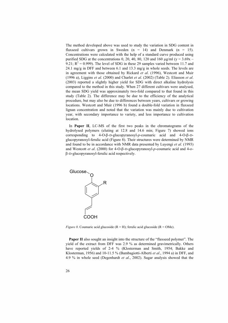
In Paper II, LC-MS of the first two peaks in the chromatograms of the hydrolysed polymers (eluting at 12.8 and 14.6 min; Figure 7) showed ions corresponding to 4-O-β-D-glucopyranosyl-p-coumaric acid and 4-O-β-D-glucopyranosyl-ferulic acid (Figure 8). Their structures were determined by NMR and found to be in accordance with NMR data presented by Luyengi et al. (1993) and Westcott et al. (2000) for 4-O-β-D-glucopyranosyl-p-coumaric acid and 4-ο-β-D-glucopyranosyl-ferulic acid respectively.
Figure 8. Coumaric acid glucoside (R = H); ferulic acid glucoside (R = OMe).
Paper II also sought an insight into the structure of the “flaxseed polymer”. The
yield of the extract from DFF was 2.9 % as determined gravimetrically. Others have reported yields of 2-4 % (Klosterman and Smith, 1954; Bakke and Klosterman, 1956) and 10-11.5 % (Bambagiotti-Alberti et al., 1994 a) in DFF, and 4.9 % in whole seed (Degenhardt et al., 2002). Sugar analysis showed that the
RO
COOH
Glucose
27
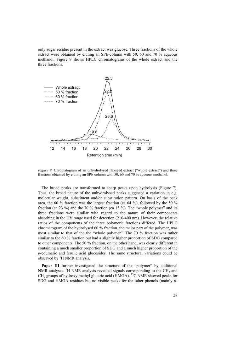
only sugar residue present in the extract was glucose. Three fractions of the whole extract were obtained by eluting an SPE-column with 50, 60 and 70 % aqueous methanol. Figure 9 shows HPLC chromatograms of the whole extract and the three fractions.
Figure 9. Chromatogram of an unhydrolysed flaxseed extract (“whole extract”) and three fractions obtained by eluting an SPE column with 50, 60 and 70 % aqueous methanol.
The broad peaks are transformed to sharp peaks upon hydrolysis (Figure 7).
Thus, the broad nature of the unhydrolysed peaks suggested a variation in e.g. molecular weight, substituent and/or substitution pattern. On basis of the peak area, the 60 % fraction was the largest fraction (ca 64 %), followed by the 50 % fraction (ca 23 %) and the 70 % fraction (ca 13 %). The “whole polymer” and its three fractions were similar with regard to the nature of their components absorbing in the UV range used for detection (210-400 nm). However, the relative ratios of the components of the three polymeric fractions differed. The HPLC chromatogram of the hydrolysed 60 % fraction, the major part of the polymer, was most similar to that of the the “whole polymer”. The 70 % fraction was rather similar to the 60 % fraction but had a slightly higher proportion of SDG compared to other components. The 50 % fraction, on the other hand, was clearly different in containing a much smaller proportion of SDG and a much higher proportion of the p-coumaric and ferulic acid glucosides. The same structural variations could be observed by 1H NMR analysis.
Paper III further investigated the structure of the “polymer” by additional NMR-analyses. 1H NMR analysis revealed signals corresponding to the CH3 and CH2 groups of hydroxy methyl glutaric acid (HMGA). 13C NMR showed peaks for SDG and HMGA residues but no visible peaks for the other phenols (mainly p-
12 14 16 18 20 22 24 26 28 30
Whole extract50 % fraction60 % fraction70 % fraction
22.3
Retention time (min)
22.2
23.6
19.6
28
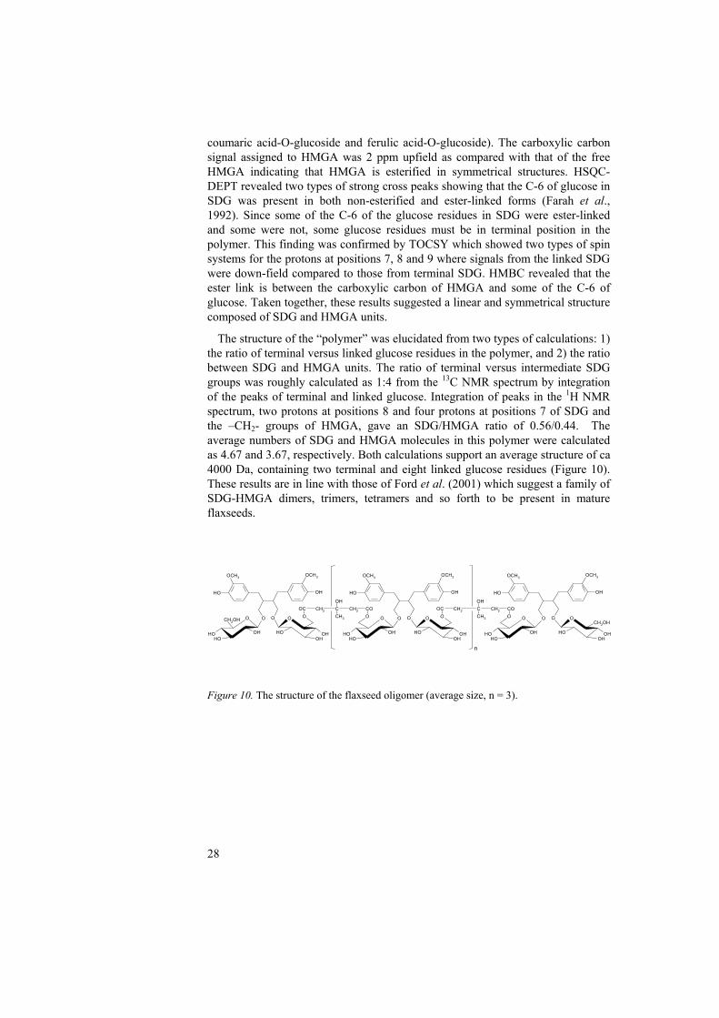
coumaric acid-O-glucoside and ferulic acid-O-glucoside). The carboxylic carbon signal assigned to HMGA was 2 ppm upfield as compared with that of the free HMGA indicating that HMGA is esterified in symmetrical structures. HSQC-DEPT revealed two types of strong cross peaks showing that the C-6 of glucose in SDG was present in both non-esterified and ester-linked forms (Farah et al., 1992). Since some of the C-6 of the glucose residues in SDG were ester-linked and some were not, some glucose residues must be in terminal position in the polymer. This finding was confirmed by TOCSY which showed two types of spin systems for the protons at positions 7, 8 and 9 where signals from the linked SDG were down-field compared to those from terminal SDG. HMBC revealed that the ester link is between the carboxylic carbon of HMGA and some of the C-6 of glucose. Taken together, these results suggested a linear and symmetrical structure composed of SDG and HMGA units.
The structure of the “polymer” was elucidated from two types of calculations: 1) the ratio of terminal versus linked glucose residues in the polymer, and 2) the ratio between SDG and HMGA units. The ratio of terminal versus intermediate SDG groups was roughly calculated as 1:4 from the 13C NMR spectrum by integration of the peaks of terminal and linked glucose. Integration of peaks in the 1H NMR spectrum, two protons at positions 8 and four protons at positions 7 of SDG and the –CH2- groups of HMGA, gave an SDG/HMGA ratio of 0.56/0.44. The average numbers of SDG and HMGA molecules in this polymer were calculated as 4.67 and 3.67, respectively. Both calculations support an average structure of ca 4000 Da, containing two terminal and eight linked glucose residues (Figure 10). These results are in line with those of Ford et al. (2001) which suggest a family of SDG-HMGA dimers, trimers, tetramers and so forth to be present in mature flaxseeds.
Figure 10. The structure of the flaxseed oligomer (average size, n = 3).
OH OH
OCH3OCH3
O O O
OHOHOH
OOC CH2 C
OH
CH3
CH2 CO
O
OHOH OH
O
OH OH
OCH3OCH3
O O O
OHOHOH
OOC CH2 C
OH
CH3
CH2 CO
O
OHOH OH
OO
OHOH
CH2OH
OH
OH OH
OCH3OCH3
O O O
OHOH
CH2OH
OH
n
29
Conclusions and future research
This work has led to the development of a method of analysis for SDG in flaxseed. Full spectroscopic data have been provided for 4-O-β-D-glucopyranosyl-p-coumaric acid and 4-O-β-D-glucopyranosyl-ferulic acid isolated from flaxseed extracts. Finally, the structures of SDG containing oligomers in flaxseed have been determined. Flaxseed is not a major food plant, and its use as food is limited by its laxative properties and content of cyanogenic glucosides. But, given its high levels of biologically active lignans, flaxseed may still be added to foods as a health promoting ingredient. It is also a good model plant for method development and for use (in whole, as a polymeric extract or as separate compounds) in in vitro and in vivo studies. The”polymer” may be of more value in relation to health compared to SDG because of its higher water solubility and ease of extraction. Moreover, other compounds in flaxseed, e.g. HMGA and hydroxycinnamic acids may also contribute to positive health effects. Thus, knowledge about the oligomeric structure and its components is of importance from a nutritional and biochemical point of view and is crucial for a complete understanding and possible exploitation.
The cinnamic acid glucosides are probably also incorporated into ester linked polymeric structures. This is supported by the fact that they too are released upon base hydrolysis. The structures of the polymers/oligomers containing these phenols have yet to be determined. It is not known if all SDG or cinammic acids are released by alkaline hydrolysis or if any other linkages, e.g. ether, are involved. Lam et al. (1992) showed that p-coumaric acid is present in ester and ether bound form in lignin-polysaccharide fractions from cell walls of wheat and Phalaris aquatica. Ferulic acid was mainly present in ester bound forms or included in ester-ether bridges. Further work is needed to unfold the bound structure(s) of these compounds.
30
References
Adlercreutz, H. 1984. Does fibre-rich food containing animal lignan precursors protect against both colon and breast cancer? An extension of the “fiber hypothesis, Gastroenterology, 86:761-766.
Adlercreutz, H. 1990. Western diet and Western diseases: Some hormonal and biochemical mechanisms and associations, The Scandinavian journal of clinical and laboratory investigation, 50, suppl. 201: 3-23.
Adlercreutz, H. 2002. Phyto-oestrogens and cancer, The Lancet oncology, 3:364-373. Adlercreutz, H. 2003. Phytoestrogens and breast cancer, Journal of steroid biochemistry
and molecular biology, 1803:1-6. Adlercreutz, H., Bannwart, C., Wähälä, K., Mäkelä, T., Brunow, G., Hase, T., Arosemena,
P.J., Kellis, J.T. and Vickery, L.E. 1993. Inhibition of human aromatase by mammalian lignans and isoflavonoids phytoestrogens, Journal of steroid biochemistry and molecular biology, 44:147-153.
Adlercreutz, H., Fotsis, T., Heikkinen, R., Dwyer, J.T., Woods, M., Goldin, B.R., Gorbach, S.L. 1982. Excretion of the lignans enterolactone and enterodiol and of equol in omnivorous and vegetarian postmenopausal women and in women with breast cancer, The Lancet, 320:1295-1298.
Amarowicz, R, Wanasundara, P.K.J.P.D, Shahidi, F. 1994. Chromatographic separation of flaxseed phenolics, Die Nahrung, 38:520-526.
Axelson, M. and Setchell, K.D.R. 1980. Conjugation of lignans in human urine, FEBS letters, 122:49-53.
Axelson, M. and Setchell, K.D.R. 1981. The excretion of lignans in rats – evidence for an intestinal bacterial source for this new group of compounds, FEBS letters, 123:337-342.
Axelson, M., Sjövall, J., Gustafsson, B.E., Setchell, K.D.R. 1982. Origin of lignans in mammals and identification of a precursor from plants, Nature, 298:659-660.
Ayres, D.C. and Loike, J.D. 1990. Lignans: Chemical, biological, and clinical properties, Cambridge University Press, Cambridge, pp 1-11; 85-112.
Bakke, J.E. and Klosterman, H.J. 1956. A new diglucoside from flaxseed, Proceedings of the North Dakota Academy of Science, July, Grand Forks, North Dakota, Volume X, pp 18-21.
Bambagiotti-Alberti, M., Coran, S.A., Ghiara, C., Giannelini, V., Raffaelli, A. 1994 a. Revealing the mammalian lignan precursor secoisolariciresinol diglucoside in flax seed by ionspray mass spectrometry, Rapid communications in mass spectrometry, 8:595-598.
Bambagiotti-Alberti, M., Coran, S.A., Ghiara, C., Moneti, G., Raffaelli, A. 1994 b. Investigation of mammalian lignan precursors in flax seed: First evidence of secoisolariciresinol diglucoside in two isomeric forms by liquid chromatography/mass spectrometry, Rapid communications in mass spectrometry, 8:929-932.
Bannwart, C., Adlercreutz, H., Wähälä, K., Brunow, G., Hase, T. 1989. Detection and identification of the plant lignans lariciresinol, isolariciresinol and secoisolariciresinol in human urine, Clinica chimica acta, 180:293-302.
Barclay, A.S. and Perdue, R.E. Jr. 1976. Distribution of anticancer activity in higher plants, Cancer treatment reports, 60:1081-1113.
Begum, A.N., Nicolle, C., Mila, I., Lapierre, C., Nagano, K., Fukushima, K., Heinonen, S-M., Adlercreutz, H., Rémésy, C., Scalbert, A. 2003. Dietary lignins are precursors of mammalian lignans in rats, Journal of nutrition, 134:120-127.
Bhatena, S.J. and Velasquez, M.T. 2002. Beneficial role of dietary phytoestrogens in obesity and diabetes, American journal of nutrition, 76:1191-1201.
Boccardo, F., Lunardi, G., Guglielmini, P., Parodi, M., Murialdo, R., Schettini, G., Rubagotti, A. 2004. Serum enterolactone levels and the risk of breast cancer in women with palpable cysts, European journal of cancer, 40:84-89.
Borrielo, S.P., Setchell, K.D.R., Axelson, M., Lawson, A.M. 1985. Production and metabolism of lignans by the human faecal flora, Journal of applied bacteriology, 58:37-43.
31
Charlet, S., Bensaddek, L., Raynaud, S., Gillet, F., Mesnard, F., Fliniaux, M-A. 2002. An HPLC method for the quantification of anhydrosecoisolariciresinol. Application to the evaluation of flax lignan content, Plant physiology and biochemistry, 40:225-229.
Chimichi, S., Bambagiotti-Alberti, M., Coran, S.A., Giannellini, V. and Biddau, B. 1999. Complete assignment of the 1H and 13C NMR spectra of secoisolariciresinol diglucoside, a mammalian lignan precursor isolated from Linum usitatissimum, Magnetic resonance in chemistry, 37:860-863.
Chisolm, G.M. and Steinberg, D. 2000. The oxidative modification hypothesis of atherogenesis: An overview, Free radical biology and medicine, 28:1815-1826.
Dabrowski, K.J. and Sosulski, F.W. 1984. Composition of free and hydolyzable acids in defatted flours of ten oilseeds, Journal of agricultural and food chemistry, 32:128-130.
Dai, Q., Franke, A.A., Jin, F., Shu, X-O., Hebert, J.R., Custer, L.J., Cheng, J., Gao, Y-T., Zheng, W. 2002. Urinary excretion of phytoestrogens and risk of breast cancer among Chinese women in Shanghai, Cancer epidemiology, biomarkers and prevention, 11:815-821.
Degenhardt, A., Habben, S., Winterhalter, P. 2002. Isolation of the lignan secoisolariciresinol diglucoside from flaxseed (Linum usitatissimum L.) by high-speed counter-current chromatography, Journal of chromatography A, 943:299-302.
den Tonkelaar, I., Keinan-Boker, L., Vant’t Veer, P., Arts, C.J.M., Adlercreutz, H., Thijssen, J.H.H., Peeters, P.H.M. 2001. Urinary phytoestrogens and postmenopausal breast cancer risk, Cancer epidemiology, biomarkers and prevention, 10:223-228.
Dunn, J.E. Jr. 1975. Cancer epidemiology in populations in the United States – with emphasis on Hawaii and California – and Japan, Cancer research, 35:3240-3245.
Eliasson, C., Kamal-Eldin, A., Andersson, R., Åman, P. 2003. High-performance liquid chromatographic analysis of secoisolariciresinol diglucoside and hydroxycinnamic acid glucosides in flaxseed by alkaline extraction, Journal of chromatography A, 1012:151-159.
Farah, H., Andersson, R., Samuelsson, G. 1992. Microdotin A and B: Two new aloin derivatives from Aloe microdonta, Planta Medica, 58:88-93.
Ford, J.D., Huang, K-S., Wang, H-B., Davin, L.B., Lewis, N.G. 2001. Biosynthetic pathway to the cancer chemopreventive secoisolariciresinol diglucoside-hydroxymethyl glutaryl ester-linked lignan oligomers in flax (Linum usitatissimum) seeds, Journal of natural products, 64:1388-1397.
Frank, J., Eliasson, C., Leroy-Nivad, D., Budek, A., Lundh, T., Vessby, B., Åman, P., Kamal-Eldin, A., 2004. Dietary secoisolariciresinol diglucoside and its oligomers with 3-hydroxy-3-methyl-glutaric acid decrease vitamin E levels in rats, British journal of nutrition, 92 (in press).
Harris, R.K. and Haggerty, W.J. 1993. Assays for potentially anticarcinogenic phytochemicals in Flaxseed, Cereal foods world, 38:147-151.
Hartwell, J.L. 1976. Types of anticancer agents isolated from plants, Cancer treatment reports, 60:1031-1067.
Heinonen, S., Nurmi, T., Liukkonen, K., Poutanen, K., Wähälä, K., Deyama, T., Nishibi, S., Adlercreutz, H. 2001. In vitro metabolism of plant lignans: New precursors of mammalian lignans enterolactone and enterodiol, Journal of agricultural and food chemistry, 49:3178-3186.
Hensley, K., Benaksas, E.J., Bolli, R., Comp, P., Grammas, P., Hamdheydari, L., Mou, S., Pye, Q.N., Stoddard, M.F., Wallis, G., Williamson, K.S., West, M., Wechter, W.J., Floyd, R.A. 2004. New perspectives on vitamin E: γ-tocopherol and carboxyethylhydroxychroman metabolites in biology and medicine, Free radical biology and medicine, 36:1-15.
Horner, N.K., Kristal, A.R., Prunty, J., Skor, H.E., Potter, J.D., Lampe, J.W. 2002. Dietary determinants of plasma enterolactone, Cancer epidemiology, biomarkers and prevention, 11:12-126.
Horwitz, C. and Walker, A.R.P. 1984. Lignans – Additional benefits from fiber? Nutrition and cancer, 6:73-76.
Ingram, D., Sanders, K., Kolybaba, M., Lopez, D. 1997. Case-control study of phyto-oestrogens and breast cancer, The Lancet, 350:990-994.
32
Jenab, M. and Thompson, L.U. 1996. The influence of flaxseed and lignans on colon carcinogenesis and β-glucuronidase activity, Carcinogenesis, 17:1343-1348.
Kitts, D.D., Yuan, Y.V., Wijewickreme, A.N., Thompson, L.U. 1999. Antioxidant activity of the lignan secoisolariciresinol diglycoside and its mammalian lignan metabolites enterodiol and enterolactone, Molecular and cellular biochemistry, 202:91-100.
Klosterman, H.J. and Smith, F. 1955. The isolation of β-hydroxy-β-methylglutaric acid from the seed of flax (Linum usitatissimum), Journal of the American Chemical Society, 76:1229-1230.
Klosterman, H.J., Smith, F., Clagett, C.O. 1954. The constitution of linocinnamarin, Journal of the American Chemical Society, 77:420-421.
Kozlowska, H., Zadernowski, R., Sosulski, F.W. 1983. Phenolic acids in oilseed flours, Nahrung, 27:449-453.
Kraushofer, T. and Sontag, G. 2002. Determination of matairesinol in flax seed by HPLC with coulometric electrode array detection, Journal of chromatography B, 777:61-66.
Kris-Etherton, P.M., Hecker, K.D., Bonanome, A., Coval, S.M., Binkoshi, A.E., Hilpert, K.F., Griel, A.E., Etherton, T.D. 2002. Bioactive compounds in food: Their role in the prevention of cardiovascular disease and cancer, The American journal of medicine, 113:71S-88S.
Kushi, L.H., Meyer, K.A., Jacobs, D.R. Jr. 1999. Cereals, legumes, and chronic disease risk reduction: Evidence from epidemiologic studies, American journal of clinical nutrition, 70 suppl.:451S-458S.
Lam, T.B.T., Iiyama, K., Stone, B.A. 1992. Cinnamic acid bridges between cell wall polymers in wheat and Phalaris internodes, Phytochemistry, 31:1179-1183.
Liggins, J., Grimwood, R., Bingham, S.A. 2000. Extraction and quantification of lignan phytoestrogens in food and human samples, Analytical biochemistry, 287:102-109.
Lin, X., Gingrich, J.R., Bao, W., Li, J., Haroon, Z.A., Demark-Wahnefried, W. 2002. Effect of flaxseed supplementation on prostatic carcinoma in transgenic mice, Urology, 60:919-924.
Luyengi, L., Pezzuto, J.M., Waller, D.P., Beecher, C.W.W., Fong, H.S. 1993. Linusitamarin, a new phenylpropanoid glucoside from Linum usitatissimum, Journal of natural products, 56:2012-2015.
MacRae, W.D. and Towers, G.H.N. 1984. Biological activities of lignans, Phytochemistry, 23:1207-1220.
Martin, M.E., Haourigui, M., Pelissero, C., Benassayag, C., Nunez, E.A. 1996. Interactions between phytoestrogens and human sex steroid binding protein, Life sciences, 58:429-436.
Mazur, W. 1998. Phytoestrogen content in foods, Baillière’s clinical endocrinology and metabolism, 12:729-742.
Mazur, W. and Adlercreutz, H. 1998. Natural and anthropogenic environmental oestrogens: the scientific basis for risk assessment, naturally occurring oestrogens in food, Pure and applied chemistry, 70:1759-1776.
Mazur, W., Duke, J.A., Wähälä, K., Rasku, S., Adlercreutz, H. 1998 a. Isoflavonoids and lignans in legumes: nutritional and health aspects in humans, Nutritional biochemistry, 9:193-200.
Mazur, W., Fotsis, T., Wähälä, K., Ojala, S., Salakka, A., Adlercreutz, H. 1996. Isotope dilution gas chromatographic-mass spectrometric method for the determination of isoflavonoids, coumestrol, and lignans in food samples, Analytical biochemistry, 233:169-180.
Mazur, W., Uehara, M., Wähälä, K., Adlercreutz, H. 2000. Phyto-oestrogen content of berries, and plasma concentrations and urinary excretion of enterolactone after a single strawberry-meal in human subjects, British journal of nutrition, 83:381-387.
Mazur, W., Wähälä, K., Salakka, A., Hase, T., Adlercreutz, H. 1998 b. Lignan and isoflavonoid concentrations in tea and coffee, British journal of nutrition, 79:37-45.
Meagher, L.P., Beecher, G.R., Flanagan, V.P., Li, B.W. 1999. Isolation and characterization of the lignans, isolariciresinol and pinoresinol, in flaxseed meal, Journal of agricultural and food chemistry, 47:3173-3180.
33
Morton, M.S., Chan, P.S.F., Cheng, C., Blacklock, N., Matos-Ferreira, A., Abranches-Monteiro, L., Correia, R., Lloyd, S., Griffiths, K. 1997. Lignans and isoflavonoids in plasma and prostatic fluid in men: samples from Portugal, Hong Kong, and the United Kingdom, The prostate, 32:122-128.
Mousavi, Y. and Adlercreutz, H. 1992. Enterolactone and estradiol inhibit each other’s proliferative effect on MCF-7 breast cancer cells in culture, Journal of steroid biochemistry and molecular biology, 41:615-619.
Muir, A.D. and Westcott, N.D. 2000. Quantitation of the lignan secoisolariciresinol diglucoside in baked goods containing flax seed or flax meal, Journal of agricultural and food chemistry, 48:4048-4052.
Namiki, M. 1995. The chemistry and physiological functions of sesame, Food reviews international, 11:281-329.
National Food Administration, Sweden. Mat och hälsa, råd och rekommendationer, http://www.slv.se (accessed 29/03/2004).
Nesbitt, P.D., Lam, Y., Thompson, L.U. 1999. Human metabolism of mammalian lignan precursors in raw and processed flaxseed, American journal of nutrition, 69:549-555.
Newmark, H.L. 1984. A hypothesis for dietary components as blocking agents of chemical carcinogenesis: plant phenolics and pyrrole pigments, Nutrition and cancer, 6:58-69.
Niemeyer, H.B. and Metzler, M. 2003. Differences in the antioxidant activity of plant and mammalian lignans, Journal of food engineering, 56:255-256.
Nurmi, T., Heinonen, S., Mazur, W., Deyama, T., Nishibe, S., Adlercreutz, H. 2003. Lignans in selected wines, Food chemistry, 83:303-309.
Obermeyer, W.R., Musser, S.M., Betz, J.M., Casey, R.E., Pohland, A.E., Page, S.W. Chemical studies of phytoestrogens and related compounds in dietary supplements: Flax and chaparral, Proceedings of the Society for Experimental Biology and Medicine, 208:6-12.
Oomah, B.D., Kenaschuck, E.O., Mazza, G. 1995. Phenolic acids in flaxseed, Journal of agricultural and food chemistry, 43:2016-2019.
Oomah, B.D., Mazza, G., Kenaschuck, E.O. 1996. Flavonoid content of flaxseed. Influence of cultivar and environment, Euphytica, 90:163-167.
Prasad, K. 1997. Hydroxyl radical-scavenging property of secoisolariciresinol diglucoside (SDG) isolated from flax-seed, Molecular and cellular biochemistry, 168:117-123.
Prasad, K. 1999. Reduction of serum cholesterol and hypercholesterolemic atherosclerosis in rabbits by secoisolariciresinol diglucoside isolated from flaxseed, Circulation, 99:1355-1362.
Prasad, K. 2000 a. Antioxidant activity of secoisolariciresinol diglucoside-derived metabolites, secoisolariciresinol, enterodiol and enterolactone, International journal of angiology, 9:220-225.
Prasad, K. 2000 b. Oxidative stress as a mechanism of diabetes in diabetic BB prone rats: Effect of secoisolariciresinol diglucoside (SDG), Molecular and cellular biochemistry, 209:89-96.
Prasad, K. 2001. Secoisolariciresinol diglucoside from flaxseed delays the development of type 2 diabetes in Zucker rat, Journal of laboratory and clinical medicine, 138:32-39.
Prasad, K., Mantha, S.V., Muir, A.D., Westcott, N.D. 1998. Reduction of hypercholesterolemic atherosclerosis by CDC-flaxseed with very low alpha-linolenic acid, Atherosclerosis, 136:367-375.
Qui, S-X., Lu, Z-Z., Luyengi, L., Lee, S.K., Pezzuto, J.M., Farnsworth, N.R., Thompson, L.U., Fong, H.S. 1999. Isolation and characterization of flaxseed (Linum usitatissimum) constituents, Pharmaceutical biology, 37:1-7.
Ratnayake, W.M.N., Behrens, W.A., Fischer, P.W.F., L’Abbé, M.R., Mongeau, R., Beare-Rogers, J.L 1992. Chemical and nutritional studies of flaxseed (variety Linott) in rats, The journal of nutritional biochemistry, 3:232-240.
Rickard, S.E., Orcheson, L.J., Seidl, M.M., Luyengi, L., Fong, H.S., Thompson, L.U. 1996. Dose-dependent production of mammalian lignans in rats and in vitro from the purified precursor secoisolariciresinol diglycoside in flaxseed, Journal of nutrition, 126:2012-2019.
34
Setchell, K.D.R, Lawson, A.M., Axelson, M., Adlercreutz, H. 1979. The excretion of two new phenolic compounds during the human menstrual cycle and in pregnancy, endocrinological cancer, ovarian function and disease, ed.: H. Adlercreutz, H., Bulbroock, R.D., van der Molen, H.J., Vermeulen, A. and Sciarra, S., Proceedings of the IX meeting of the international study group for steroid hormones, Rome, December 5-7, 515:207-215.
Setchell, K.D.R., Bull, R., Adlercreutz, H. 1980 a. Steroid excretion during the reproductive cycle and in pregnancy of the vervet monkey (Cercopithecus aethiopus pygerythrus), Journal of steroid biochemistry, 12:375-384.
Setchell, K.D.R., Lawson, A.M., Borrielo, S.P., Harkness, R., Gordon, H., Morgan, D.M.L., Kirk, D.N., Adlercreutz, H., Anderson, L.C. 1981. Lignan formation in man-microbial involvement and possible roles in relation to cancer, The Lancet, 318:4-7.
Setchell, K.D.R., Lawson, A.M., Mitchell, F.L., Adlercreutz, H., Kirk, D.N., Axelson, M. 1980 b. Lignans in man and in animal species, Nature, 287:740-742.
Sicilia, T., Niemeyer, H.B., Honig, D.M., Metzler, M. 2003. Identification and stereochemical characterization of lignans in flaxseed and pumpkin seeds, Journal of agricultural and food chemistry, 51:1181-1188.
Stich, S.R., Toumba, J.K., Groen, M.B., Funke, C.W., Leemhuis, J., Vink, J., Woods, G. 1980. Excretion, isolation and structure of a new phenolic constituent of female urine, Nature, 287:738-740.
Tham, D.M., Gardner, C.D., Haskell, W.D. 1998. Potential health benefits of dietary phytoestrogens: A review of the clinical, epidemiological, and mechanistic evidence, Journal of clinical endocrinology and metabolism, 83:2223-2235.
Theander, O., Åman, P., Westerlund, E., Andersson, R., Pettersson, D. 1995. Total dietary fiber determined as neutral sugar and uronic acid residues, and lignin (The Uppsala method): collaborative study, Journal of the Association of Official Analytical Chemists, 78:1030-1044.
Thompson, L.U., Rickard, S.E., Cheung, F., Kenaschuk, E.O., Obermeyer, W.R. 1997. Variability in anticancer lignan levels in flaxseed, Nutrition and cancer, 27:26-30.
Thompson, L.U., Rickard, S.E., Orcheson, L.J., Seidl, M.M. 1996 a. Flaxseed and its lignan and oil components reduce mammary tumor growth at a late stage of carcinogenesis, Carcinogenesis, 17:1373-1376.
Thompson, L.U., Robb, P., Serraino, M., Cheung, F. 1991. Mammalian lignan production from various foods, Nutrition and cancer, 16:43-52.
Thompson, L.U., Seidl, M.M., Rickard, S.E., Orcheson, L.J., Fong, H.S. 1996 b. Antitumorigenic effect of a mammalian lignan precursor from flaxseed, Nutrition and cancer, 26:159-165.
Tominaga, S. 1985. Cancer incidence in Japanese in Japan, Hawaii, and Western United States, The National Cancer Institute monographs, 69:83-92.
Tou, J.C.L. and Thompson, L.U. 1999. Exposure to flaxseed or its lignan component during different developmental stages influences rat mammary gland structures, Carcinogenesis, 20:1831-1835.
Tou, J.C.L., Chen, J., Thompson, L.U. 1998. Dose, timing, and duration of flaxseed exposure affect reproductive indices and sex hormone levels in rats, Journal of toxicology and environmental health, Part A, 56:555-570.
Wang, C., Mäkelä, T., Hase, T., Adlercreutz, H., Kurzer, M. 1994. Lignans and flavonoids inhibit aromatase enzyme in human preadipocytes, The journal of steroid biochemistry and molecular biology, 50:205-212.
Vanharanta, M., Voutilainen, S., Lakka, T.A., van der Lee, M., Adlercreutz, H., Salonen, J.T. 1999. Risk of acute coronary heart events according to serum concentrations of enterolactone: a prospective population-based case-control study, The Lancet, 354:2112-2115.
Vanharanta, M., Voutilainen, S., Nurmi, T., Kaikkonen, J., Roberts, L.J., Morrow, J.D., Adlercreutz, H., Salonen, J.T. 2002. Association between low serum enterolactone and increased plasma F2-isoprotanes, a measure of lipid peroxidation, Atherosclerosis, 160:465-469.
35
Waters, A.P. and Knowler, J.T. 1982. Effect of a lignan (HPMF) on RNA synthesis in the rat uterus, Journal of reproduction and fertility, 66:379-381.
Westcott, N.D. and Muir, A.D. 1996 a. Process for extracting and purifying lignans and cinnamic acid derivatives from flaxseed, PCT Patent WO9630468A2.
Westcott, N.D. and Muir, A.D. 1996 b. Variation in flax seed lignan concentration with variety, location and year, Proceedings of the 56th flax institute of the United States, March 20-22, Doublewood Inn, Fargo, North Dakota, pp 77-85.
Westcott, N.D., Hall, T.W.E., Muir, A.D. 2000. Evidence for the occurrence of ferulic acid derivatives in flaxseed meal, Proceedings of the 58th flax institute of the United States, March 23-25, Doublewood Inn, Fargo, North Dakota, pp 49-52.
World Cancer Research Fund, 1997. Food nutrition and the prevention of cancer: A global perspective, Washington, American Institute for Cancer Research.
36
Acknowledgements Thanks to all my colleagues, past and present, and my friends, for being nice, helpful and fun!
Speciellt vill jag tacka:
My main supervisor, Afaf. Thanks for your encouragement, your never-ending ideas and suggestions, which are, and have been, very stimulating! Moreover, your devotion to your work and other important “issues” in life is a great inspiration.
Min biträdande handledare, Per, för att du är så positiv, hjälpsam och peppande.
Roger, för hjälp med datorer, försöksupplägg och polymeranalyser.
Lennart Lundgren, för lärorika diskussioner, små knep och många aha-upplevelser.
Rolf Andersson, för utförande av NMR-analyser och tålmodigt förklarande av desamma.
Nienke Peerlkamp and Charlotta Jung. Thanks for performing many of the analyses resulting in this thesis!
Helena. Vad skulle jag ha gjort utan dig alla dagar, sena kvällar och helger, kämpandes med krånglande maskiner och omanalyser? Tack för sällskapet, ditt stöd och alla långa pratstunder.
Susanne, för stimulerande samtal om karriären och livet i övrigt!
Janne, för all hjälp med ovan nämnda maskiner och diskussioner om ditt och datt.
Christina, för att du låtit mig rota bland dina artiklar och rapporter.
Mangala. Thanks for all the good times we spent together, in Sweden, India, and Japan! I wish all the best for you and your family!
Alla mina forna kollegor på Livsmedelsverket. Monica, OTA PREV rapporterna har gett mig mycket kött på benen, det var kul att få jobba med dig! Ann, tack för gott samarbete med all möglig spannmål, din positiva attityd och din omtänksamhet!. Jag skulle kunna räkna upp er alla! Tack för tre roliga och trevliga år!
Kristina, Monica och Anna för allt roligt som vi upplevt tillsamans, från första traktorlektionen. Trots att vi alla är lite (?) dåliga på att höra av oss, så finns ni där.
Alla mina föräldrar, China, Filippa, Momma och Pappi, farmor och farfar och Mats för all kärlek, uppmuntran, stöd, minnen från min tids begynnelse, och trevliga stunder tillsammans.
Torbjörn, tack för allt kul vi haft hittills, hoppas att det blir mycket mer av samma slag! Jag är glad för att jag har dig!
KALSEC Inc. and VL-stiftelsen are gratefully acknowledged for financial support.