ph.d. synopsis submitted to gujarat technological
TRANSCRIPT

Medical Signal Analysis Using Artificial Intelligence
Ph.D. Synopsis Submitted to
Gujarat Technological University
Ahmedabad
For the Award of
Doctor of Philosophy In
Instrumentation and Control
by
Chintan P Shah (129990917001)
Under the guidance of
Dr. Vipul A. Shah
GUJARAT TECHNOLOGICAL UNIVERSITY
AHMEDABAD
MAY 2018

i
Index
Sr.
No.
Topic Page
no.
1 Abstract 1
2 State of the Art 2
3 Definition of Problem 5
4 Objective and scope of Work 5
5 Original Contribution by thesis 6
6 The Methodology of Research 6
7 Results/Comparison 8
8 Achievement with respect to Objectives 12
9 Conclusion 12
10 Paper Published 12

1
Title: Medical Signal Analysis Using Artificial Intelligence
Abstract:
Medical Signals are considered as vital signs for internal operations of the human body.
By closely observing and analyzing them we could infer the state of any important organs like
Heart, Lung, Brain etc. From all these medical signals Electroencephalogram (EEG) is most
complicated to understand. These signals are measured at the different location of head surface
and contain different frequency components in them with lower amplitude than any other
medical signal. Another important characteristic of EEG is that when we measure them at any
one location it contains signal with superimposition of signals far from measuring site. These
make the most challenging signal to analyze and interpret. But if we do handle them carefully
we can create an interface that can help persons with several amputees to their limbs. This
interface is called Brain Computer Interface (BCI). Essentially this system is rehabilitative in
nature so we can reinstate their partial limb movement. So in this research work, we have
focused on EEG signals and its end application is to determine movement of the upper limb.
Analysis of medical signal is done on signal attributes, which may be either frequency,
amplitude, time-frequency combine or any specific event. This analysis can be done by various
means but all signals are embedded with nonlinearity so, if we want to categorize the signal
than artificial intelligence is best suited for the purpose. We are focusing on EEG signals whose
main attributes are frequency and event related to some activity of the brain. So to analyze such
signal we can use their time-frequency combined characteristic as features for artificial
intelligence. The advantage of artificial intelligence over other classification method is that it
can adapt to any nonlinearity present in the signal. For BCI application we need to localize the
source of different activity in the brain so that we can improve the prediction of movement.
Since different individual’s signal attribute are different which makes a generalization of
algorithm very difficult. It is possible that algorithm which works on anyone database might
not produce the same result for another. This can be eradicated if we design an algorithm by
considering individual subject constraints. We have proposed a novel approach which localizes
source in spatial as well as temporal domain. We have considered the individual anatomical
structure to localize brain source in the spatial domain. While using Artificial intelligence we
have localized source in the temporal domain.

2
State of the art:
EEG signal is mainly attributed to their frequency components: Delta (0-4Hz), Theta
(4-8Hz), Alpha (8-12Hz), Beta (12-30Hz) and Gamma (25-100Hz). Mostly Alpha rhymes are
seen during ideal activity of the brain, and beta band seen during actual activity of the brain.
Several algorithms have been implemented which focuses on the frequency band to detect
movement of the limb. Sang-Hoon Park et. al. have divided frequency band into sub-band of
4Hz each starting from 4 to 40Hz. In their approach, they have applied Common Spatial
Patterns (CSP) on each subband in order to extract features. CSP is a filtering technique which
finds an optimal filter which maximizes variance for one class and minimizes variance for other
class. Wide frequency band should be selected for CSP because band effective band changes
on individual bases. They have mentioned that small sample setting performance of CSP
decreases so they have used Regularized-CSP. Followed by ensemble classifier for classifying
movement. In another paper of Sang-Hoon Park et. al. they have used sub-band regularized
CSP using small sample selection for motor imagery (MI) detection.
Jun Lu et. al. have implemented an approach where Adaptive Spatio-Temporal (AST)
method which does the temporal filtering with a Gaussian kernel. Gaussian kernel models
smooth change in movement-related potential in the signal. Spatial filtering done by Linear
Ridge Regression (LRR), this method performs classification and regression task
simultaneously. AST frame work’s optimal parameters of the spatiotemporal filter, including
the center and radius of the Gaussian kernel and the regularization coefficient of LRR, are
automatically estimated by minimizing the error of leave-one-out.
Proloy Das et. al. have improvised the concept of spectral analysis using an overlapping
sliding window with multi-taper analysis. They have proposed dynamic Bayesian multi-taper
spectrum estimation technique. In their approach, they have avoided the use of overlapping
windows by modeling and estimating the dependence of the spectra across windows using
state-space models while retaining the favorable leakage properties of the multitaper analysis.
State space models in the context of multitaper analysis result in the adaptive weighting of the
estimates of the Eigen-coefficients or eigenspectra across windows. These adaptive weights
depend on the common dynamic trends shared across windows and hence result in capturing
the degree of smoothness inherent in the signal while producing estimates robust against
uncertainties due to observation noise and limited data.

3
Pawel Herman et. al. have provided a comparative analysis of all approaches related to
the spectral feature used for classification of imagery movement. They have tested features
from Power Spectrum Density (PSD) (Fourier Transform), Short-time Fourier Transform
(STFT), Continuous morlet Wavelet Transform, Daubechies wavelet decomposition and
autoregressive model. Along with Linear Discriminant Analysis, Support Vector Machine
(SVM) and SVM with the Gaussian kernel as a classifier. They observed that PSD gathers
feature which is far more effective than any other method. And for classification SVM with the
Gaussian kernel is the best classifier among they have tried.
Several researchers have used statistical features set for classifying movement from
EEG data. Linsey Roijendijk et. al. have proposed an algorithm that uses Sensor covariance
matrix. This matrix method has reduced two staged supervised learning into one stage with
added whitened covariance matrix so that all feature gets equally amplified. Johanna Metsomaa
et. al. used independent component analysis method to classify evoked potentially related to
movement. They have implemented momentary-uncorrelated component analysis (MUCA) to
separate out multi-trial EEG/MEG data into momentary-uncorrelated components, i.e.,
components that are uncorrelated at each latency after the stimulus. MUCA is based on the
estimated joint diagonalization (AJD) of covariance matrices estimated at separate latencies of
the evoked data.
Jaime F. Delgado et.al. have utilized two statistical methods Conditioned Random Field
(CRF) and Latent Dynamic CRF to investigate motor imagery signals. CRFs can in principle
be used to model the dynamics of sequential data, although CRFs can model the extrinsic
dynamics of the data (or features), which in asynchronous BCI corresponds to dynamics across
different tasks, they lack the ability to model intrinsic dynamics. So to address this problem
they have used CRF plus hidden state to model intrinsic and extrinsic dynamics simultaneously.
Vikram Shenoy et. al. have reduced number of the channel by utilizing an iterative
multi-objective optimization for channel selection (IMOCS) approach. This approach selects
effective channel iteratively by utilizing anatomical and functional relevance in EEG data.
They have implemented several other data reduction technique and checked their performance
against the proposed approach. The proposed approach yielded good result as compared to
other methods because this approach uses a reference point-based iterative method to determine
the optimal solution. In this procedure, the channel weights and the objective function are
updated in every iteration.

4
Another domain of interest in EEG signal analysis is source localization which detects
an active region in the brain from EEG data. These methods use anatomical structure to create
a forward solution of the brain and its compartment. And then inverse projecting data back to
a source located in the brain. Barry D. Van Veen et. al. have implemented localization method
based on spatial filtering. Spatial filters are designed that pass brain electrical activity from a
specified location while attenuating activity originating at other locations. The power at the
output of a spatial filter is an estimate of the neural power originating within the spatial
passband of the filter. A map of neural power as a function of location is obtained by designing
multiple spatial filters, each with a different passband, and depicting output power as a function
of passband location. Here the criterion to select filter coefficient is based on linearly
constrained minimum variance (LCMV).
J. Gross et. al. implemented Dynamic imaging of coherent sources (DICS) method uses
frequency components to localize an active area in the brain. In this method, cross-spectral
density is used in the same manner as in the previous approach. In this method two measures
are computed at each grid point: first, the estimated power and, second, the estimated coherence
with respect to a given reference point. One of the two measures is then thresholded and
displayed together with the individual magnetic resonance images. Mosher John C et. al. have
implemented multiple signal classification approach. In this approach dipolar localization is
done in presence of non-dipolar sources and available dipolar sources’ with an unknown
location, amplitude. The presence of other dipolar and non-dipolar activity complicates the
generation of a suitable gain matrix for a least-squares search. Multiple signal characterization
(MUSIC) method is used to address this problem. But MUSIC also presents some shortcomings
like correlated noise, different head model, etc. So the writer has tried to resolve shortcoming
by providing improvised MUSIC.
Fa-Hsuan Lin et. al. have implemented a combination of a cortically constrained
minimum norm estimate (MNE) and wavelet-based spectral analysis employing a complex
Morlet wavelet. Wavelets preserve a high temporal resolution in the gamma band at
approximately 40 Hz necessary to image rapidly time-varying oscillations. This methods finds
effective frequency sources using wavelet over whole sensor space and they have used prior
neural activation using fMRI so that correct localization power can be identified. Keyvan
Mahjoory et.al. have implemented different method from different toolboxes and compared
their result. They have implemented spatio-spectral decomposition (SSD) method to localize
active brain source. SSD method removes brain activity without strong alpha waves.

5
Definition of Problem:
EEG analysis methods for BCI applications are having following problems:
Brain dynamics are non-stationary and superimposition of different signal sources
on all sensor locations makes activity classification difficult.
Anatomical structure changes for different individual and so the algorithm suffers
in generalization.
Generally, brain maps available are inaccurate so functional mapping of activity
becomes difficult.
EEG signals amplitude are very minute and they are prone to noise arising from
artifacts due to eyes and muscle activity around the head.
Larger channel count makes data of very large dimension, which is major concerns
for any algorithm.
Objective and Scope of work:
Objectives:
To develop an algorithm for movement detection from EEG data which can be
customized for individual
To test and implement different forward and inverse model to create realistic brain
compartments and back project data.
To determine the region of interest on scalp surface for the individual so channel
reduction can be achieved
To develop an artificial intelligence based algorithm to classify nonlinear EEG data.
Scope:
we have worked on EEG signals instead of
We have to focus on determining point spread function so that we can map the
electrical activity of the brain to scalp surface.
Finding an effective area on scalp surface for the individual so that algorithm can
become robust and customized for the individual.
We have focused only on motor activity (eye blinking, jaw movement, motion
artifact are other activity in EEG) so we will collect database by minimizing other
activity. Because of superimposition of all activity at all electrode.
6
We have covered actual movement because intended movement will take much
time for training but it will be possible to detect intended movement with our
developed algorithm once we do the validation.
Original Contribution by thesis:
We have proposed methodology which localizes source in the brain using
spatiotemporal localization technique. Spatial localization is done by inverse projecting data to
the brain and temporal localization is done by long-term short-term learning network. Our
approach has yielded region of interest from the total scalp surface
The methodology of Research:
We have proposed an approach which is spatiotemporal localization of movement-related
brain signals from noninvasive EEG. Spatial localization is done by a beam-former approach
which require to develop forward solution and inverse solution for the individual. Whereas
temporal localization is done by implementing long-term short-term neural network. Forward
model generally contains details about neural current propagation from a cortical source in the
brain to Scalp Surface. For knowing above detail we require knowledge of geometry and
electrical conductivity of different part of brain i.e. Skull, Scalp, and Brain etc. It can be done
by getting individual geometric data from MRI structure of persons own brain. Mostly MRI is
an expensive to process so the generally colin27 head is taken as an anatomical template for
getting geometric information. We can extract different parts anatomical data out of MRI
structure which are then used for further processing of forwarding model.
After extracting different fields of the brain it is required to create the head model.
Different methods are available to create head models: 1) Boundary Element Method (BEM),
2) Finite Element Method (FEM). There is significant difference between these two techniques,
BEM requires less computational effort as compared to FEM. Other major difference is that
anisotropy and other structure of neighboring tissue to electrical source is neglected in BEM
but can be addressed by using FEM method. In other words conductivity of tissue surrounding
electrical source is considered as constant for small regions and realistic representation is
ignored. BEM is tradeoff between oversimplifying structure and mathematical solution of
spherically symmetrical structure. This model will utilize mean conductivity of small tissue
regions.
In inverse projection estimation of the source of EEG with distributed source models
consists first of distributing several dipole sources with fixed locations and orientations on

7
the cortical surface, then estimating their amplitudes from the data. Following methods
normally employed with above method to inverse project the data.
1. Linearly constrained minimum variances (LCMV)
2. Dynamic Imaging of Coherence Sources (DICS)
3. Minimum Norm Estimates (MNE)
4. Partial Canonical Correlation/Coherence (PCC)
5. Standardized Low-Resolution Electromagnetic Tomography (LORETA)
From above methods, we are interested in LCMV, DICS and PCC methods since they
localize brain activity base on the particular frequency band. They detect the power of
particular frequency band and locate active dipole from the forward model.
LCMV based on localization method based on the principles of spatial filtering. Spatial
filters are designed that pass brain electrical activity from a specified location while attenuating
activity originating at other locations. The power at the output of a spatial filter is an estimate
of the neural power originating within the spatial passband of the filter. The current dipole is a
key component of our model relating the surface measurements to the underlying neural
activity. An individual active neuron is reasonably modelled as a current dipole. From above
method, DCIS and LCMV work on localizing dipole based on different frequency component
present at the source level. DCIS comparatively gets a better response than another method.
Once we have back projected data we can get the region of interest on individual bases.
Normally we consider 2-3 channel from topological data of each subject. These channels have
temporal data and it is significant for temporal localization. Here we employ fundamentals of
artificial intelligence. A supervised method like artificial neural network (ANN) and Support
vector machine (SVM). Here we are interested in temporal event present in the selected channel
so we can use the layered recurrent network (RNN). This network is advantageous because it
can store pattern related to the event. This RNN can only work on long-term memory so instead,
we have implemented long short-term memory.
(LSTM) (deep learning network) because it can provide you input sequence data into a
network, and make predictions based on the individual time steps of the sequence data. As we
are dealing with multiple channel data LSTM suitable to classify sequences and it is based on
the recurrent network so past data is also used to identify the event. Main advantage of LSTM
network is that it has four layers inside each chain as compared recurrent network’s one layer.
Here LSTM has three layers consisting of sigmoidal function which works as gate and allow

8
data to pass only in certain cases. So due to networks long-term memory capability, it is easier
to train network for incoming events and classification in temporal domain is possible.
We have first tested the algorithm on database available from Physisonet website.
During this testing, we found algorithm detects a region of interest properly. During testing
phase, this region is averaged over several individuals EEG data. So we get the average region
of interest on scalp surface. We have reduced a number of the channel from 64 to 12 channel.
In turn, this is classified by SVM properly. Here another notable finding we get from above
approach is that frequency-based features give maximum accuracy, so we have used frequency
domain analysis afterward. Then this algorithm is validated by EEG recordings from GMERS
(Medical College), Gandhinagar. The number of channels reduced in EEG recording to 18
channels since that module is available with GMERS. We have performed the same approach
as we have done with database from Physionet.
Results\Comparison:
In frequency domain signals are filtered with a frequency range of 12 -30 Hz so that we
only have a beta band because an alpha band with 8-12Hz represent no activity. The data has
event marker in EDF file which we have extracted separately from event marker field. Once
we get that we have stripped the data for particular activity for 4 seconds in it. EEG data
sampled at 160 Hz so in total we have to extract 641 data points for 64 channels each. After
applying filter the data is that fed to frequency domain analysis where cross spectral density is
calculated for the following detail:
1. The frequency band of interest:12-40Hz
2. Filtering window: Multitapering filter with multiple frequency bins
3. The output of filtering: Power Spectrum Density and Cross-spectral Density
4. Time of interest: 0-4 second
We get the following result on 10 subjects average EEG data related to movement. In the figure
spectrogram of each channel is performed using above parameter and plotted on the left-hand
side. On right-hand side shows a figure of back-projected data on the brain surface. So we have
confirmed that activity of movement on scalp surface is due to activity on the motor cortex of
the brain. From the figure, we can suggest that central region of scalp gets activated for
frequency band 12-40 Hz during activity.
C3 C1 CZ C2 C4 C6 CP3 CP1 CPZ CP2 CP4 CP6
1 2 3 4 5 6 7 8 9 10 11 12

9
Fig. 1 Result of the algorithm on a database
We have got validation data from GMERS, Gandhinagar. The system to get subject
data is from RMS medical equipment, Quest32 with 21 EEG channels with the following
specification.
18-effective channel with 2 reference channel
Amplification up to 3uV(micro Volt)
256 Hz sampling rate of each channel
Event marked as left hand and right hand movement
Data exported to EDF
We have taken data of 7 healthy subjects and details are given in following table. With this
data in EDF form we have to extract trial based on event marker. Details of all subjects who
have provided EEG data for validation. All subject have provided 5 trials each for Right hand
and Left hand movement in 3 sessions. With this data we have done same analysis as we have
done on database.
Sr.
No.
Age Gender Medical History Consent
Provided?
Remarks
1 23 Female No Yes Nil
2 23 Male No Yes Nil
3 23 Female No Yes Nil
4 22 Female No Yes Nil
5 30 Male No Yes Nil
6 30 Male No Yes Nil
7 32 male No Yes Nil Table1: Data base analysis
Fp1 Fp2 F7 F3 Fz F4 F8 A2 T7 C3 Cz C4 T8 A1 P7 P3 Pz P4 P8 O1 O2

10
Here we have implemented frequency analysis using following settings:
1. Frequency of interest: 12-30Hz
2. Filtering method: multi tapers multiple convolution, with steps of 2Hz each taper
3. Filtering window: Hanning
4. Output: power spectrum at each frequency step
5. No. of electrode: 18 active electrode
Sub1
Sub3
Sub4 Fig. 2 results on validation data
The above results are consistent with previously done work and inverse projection of
the above subjects are also done and results are given in figure on next page.

11
Sub 1 Sub 3
Sub 7
Fig. 3 results of back projection data
From above results we can prove that anatomical information can’t be ignored. As this
information highly affect accuracy of algorithm. By this way we can preserve information from
anatomical perspective. The accuracy of algorithm has improved significantly which is evident
from following table.
Subject
Classification using
SVM classifier
PCA based features
Discriminant
classifier
Deep
Learning
Network
Linear
(%)
RBF (%) (%) (%)
Sub1 100 100 100 100
Sub2 65 85 90 100
Sub3 85 100 88 95
Sub4 100 100 90 100
Sub5 100 100 88 100
Sub6 65 100 90 100
Sub7 52 100 100 100 Table2: Results of classifiers
12
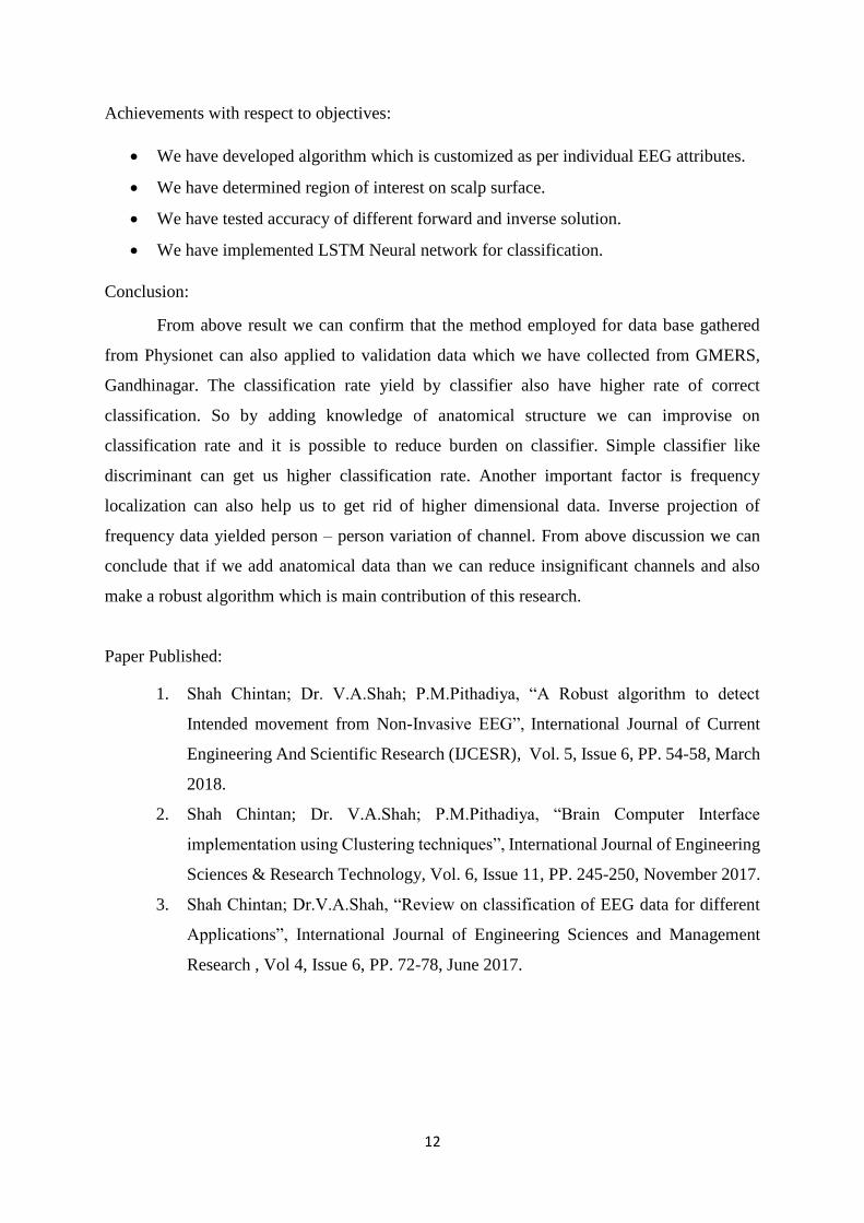
Achievements with respect to objectives:
We have developed algorithm which is customized as per individual EEG attributes.
We have determined region of interest on scalp surface.
We have tested accuracy of different forward and inverse solution.
We have implemented LSTM Neural network for classification.
Conclusion:
From above result we can confirm that the method employed for data base gathered
from Physionet can also applied to validation data which we have collected from GMERS,
Gandhinagar. The classification rate yield by classifier also have higher rate of correct
classification. So by adding knowledge of anatomical structure we can improvise on
classification rate and it is possible to reduce burden on classifier. Simple classifier like
discriminant can get us higher classification rate. Another important factor is frequency
localization can also help us to get rid of higher dimensional data. Inverse projection of
frequency data yielded person – person variation of channel. From above discussion we can
conclude that if we add anatomical data than we can reduce insignificant channels and also
make a robust algorithm which is main contribution of this research.
Paper Published:
1. Shah Chintan; Dr. V.A.Shah; P.M.Pithadiya, “A Robust algorithm to detect
Intended movement from Non-Invasive EEG”, International Journal of Current
Engineering And Scientific Research (IJCESR), Vol. 5, Issue 6, PP. 54-58, March
2018.
2. Shah Chintan; Dr. V.A.Shah; P.M.Pithadiya, “Brain Computer Interface
implementation using Clustering techniques”, International Journal of Engineering
Sciences & Research Technology, Vol. 6, Issue 11, PP. 245-250, November 2017.
3. Shah Chintan; Dr.V.A.Shah, “Review on classification of EEG data for different
Applications”, International Journal of Engineering Sciences and Management
Research , Vol 4, Issue 6, PP. 72-78, June 2017.

13
References:
[1] T. Oostendorp and A. Van Oosterom, “The Potential Distribution Generated by Surface
Electrodes in Inhomogeneous Volume Conductors of Arbitrary Shape,” IEEE Trans.
Biomed. Eng., vol. 38, no. 5, pp. 409–417, 1991.
[2] F. H. Lin, T. Witzel, M. S. Hämäläinen, A. M. Dale, J. W. Belliveau, and S. M.
Stufflebeam, “Spectral spatiotemporal imaging of cortical oscillations and interactions
in the human brain,” Neuroimage, vol. 23, no. 2, pp. 582–595, 2004.
[3] B. D. Van Veen, W. van Drongelen, M. Yuchtman, and A. Suzuki, “Localization of
brain electrical activity via linearly constrained minimum variance spatial filtering.,”
IEEE Trans. Biomed. Eng., vol. 44, no. 9, pp. 867–880, 1997.
[4] S. Baillet, J. J. Riera, G. Marin, J. F. Mangin, J. Aubert, and L. Garnero, “Evaluation of
inverse methods and head models for EEG source localization using a human skull
phantom,” Phys Med Biol, vol. 46, no. 1, pp. 77–96, 2001.
[5] J. C. Mosher, S. Baillet, and R. M. Leahy, “EEG source localization and imaging using
multiple signal classification approaches,” J. Clin. Neurophysiol., vol. 16, no. 3, pp.
225–238, 1999.
[6] J. C. Mosher, R. M. Leahy, and P. S. Lewis, “EEG and MEG: Forward solutions for
inverse methods,” IEEE Trans. Biomed. Eng., vol. 46, no. 3, pp. 245–259, 1999.
[7] J. Gross, J. Kujala, M. Hamalainen, L. Timmermann, A. Schnitzler, and R. Salmelin,
“Dynamic imaging of coherent sources: Studying neural interactions in the human
brain,” Proc. Natl. Acad. Sci., vol. 98, no. 2, pp. 694–699, 2001.
[8] S. Shoham, M. R. Fellows, N. G. Hatsopoulos, J. P. Donoghue, and R. A. Normann,
“Statistical encoding model for a primary motor cortical brain-machine interface.,”
IEEE Trans. Biomed. Eng., vol. 52, no. 7, pp. 1312–1322, 2005.
[9] V. S. Handiru and V. A. Prasad, “Optimized Bi-Objective EEG Channel Selection and
Cross-Subject Generalization with Brain-Computer Interfaces,” IEEE Trans. Human-
Machine Syst., vol. 46, no. 6, pp. 777–786, 2016.
[10] J. F. D. Saa and M. Çetin, “Discriminative Methods for Classi fi cation of
Asynchronous Imaginary Motor Tasks From EEG Data,” Ieee Trans. Neural Syst.
Rehabil. Eng., vol. 21, no. 5, pp. 716–724, 2013.
[11] L. Roijendijk, S. Gielen, and J. Farquhar, “Classifying Regularized Sensor Covariance
Matrices: An Alternative to CSP,” IEEE Trans. Neural Syst. Rehabil. Eng., vol. 24, no.
8, pp. 893–900, 2016.
[12] J. Metsomaa, J. Sarvas, and R. J. Ilmoniemi, “Blind Source Separation of Event-
Related EEG/MEG,” IEEE Trans. Biomed. Eng., vol. 64, no. 9, pp. 2054–2064, 2017.

14
[13] Y. Chen, A. Raymond, A. See, and S. Chen, “A review and experimental study on
application of classifiers and evolutionary algorithms in EEG based brain-machine
interface systems,” vol. 50, no. 10, pp. 8–9, 2014.
[14] S.-H. Park and S.-G. Lee, “Small Sample Setting and Frequency Band Selection
Problem Solving Using Subband Regularized Common Spatial Pattern,” IEEE Sens. J.,
vol. 17, no. 10, pp. 2977–2983, 2017.
[15] H. Higashi and T. Tanaka, “Simultaneous Design of FIR Filter Banks and Spatial
Patterns for EEG Signal Classification,” IEEE Trans. Biomed. Eng., vol. 60, no. 4, pp.
1–1, 2012.
[16] S.-H. Park, D. Lee, and S.-G. Lee, “Filter Bank Regularized Common Spatial Pattern
Ensemble for Small Sample Motor Imagery Classification,” IEEE Trans. Neural Syst.
Rehabil. Eng., vol. 26, no. 2, pp. 1–1, 2017.
[17] P. Das and B. Babadi, “Dynamic Bayesian Multitaper Spectral Analysis,” IEEE Trans.
Signal Process., vol. 66, no. 6, pp. 1394–1409, 2017.
[18] H. Yuan, A. J. Doud, A. Gururajan, and B. He, “Localization of Event-related ( de )
Synchronization of Cerebral Cortex during Online Control of Brain-Computer Interface
Using Minimum-norm Estimates in the Frequency Domain,” Int. J. Bioelectromagn.,
vol. 9, no. 2, pp. 109–110, 2007.
[19] P. Herman, G. Prasad, T. M. McGinnity, and D. Coyle, “Comparative analysis of
spectral approaches to feature extraction for EEG-based motor imagery classification,”
IEEE Trans. Neural Syst. Rehabil. Eng., vol. 16, no. 4, pp. 317–326, 2008.
[20] J. Lu, K. Xie, and D. J. McFarland, “Adaptive spatio-temporal filtering for movement
related potentials in EEG-based brain-computer interfaces,” IEEE Trans. Neural Syst.
Rehabil. Eng., vol. 22, no. 4, pp. 847–857, 2014.
[21] A. Paris, G. K. Atia, A. Vosoughi, and S. A. Berman, “A New Statistical Model of
Electroencephalogram Noise Spectra for Real-Time Brain-Computer Interfaces,” IEEE
Trans. Biomed. Eng., vol. 64, no. 8, pp. 1688–1700, 2017.
[22] S. Ge, R. Wang, Y. Leng, H. Wang, P. Lin, and K. Iramina, “A Double-Partial Least-
Squares Model for the Detection of Steady-State Visual Evoked Potentials,” IEEE J.
Biomed. Heal. Informatics, vol. 21, no. 4, pp. 897–903, 2017.
[23] K. D. I. K. Niazi, N. Jiang, O. Tiberghien, J. F. Nielsen, “Detection of movement
intention from single-trial movement-related cortical potentials,” J. Neural Eng., vol. 8,
no. 6, 2011.
[24] I. Journal, O. F. Engineering, B. Computer, I. Implementation, and U. Clustering,
“BRAIN COMPUTER INTERFACE IMPLEMENTATION USING CLUSTERING,”
vol. 6, no. 11, pp. 245–250, 2017.

15
[25] A. Bashashati, G. Birch, K. Navarro, M. Fatourechi, and S. Mason, “A Comprehensive
Survey of Brain Interface Technology Designs,” Ann. Biomed. Eng., vol. 35, no. 2, pp.
137–169, 2007.
[26] R. Palaniappan, “Brain computer interface design using band powers extracted during
mental tasks,” Proc. 2nd Int. IEEE EMBS Conf. Neural Eng. Arlingt., pp. 321–324,
2005.
[27] J. D. R. M. P. W. Ferrez, “EEG-based brain-computer inter- action: improved accuracy
by automatic single-trial error detection,” Proc. Adv. Neural Inf. Process. Syst., pp.
441–448, 2007.
[28] G. P. and C. Neuper, “Motor imagery and direct brain-com- puter communication,”
Proc. IEEE, vol. 89, no. 7, pp. 1123–1134, 2001.
[29] B. R. M. Bakker, F. P. De Lange, R. C. Helmich, R. Scheeringa and Bloem, “Cerebral
correlates of motor imagery of normal and precision gait,” Neuroimage, vol. 41, no. 3,
pp. 998–1010, 2008.
[30] Gerwin Schalk, “https://www.physionet.org/physiobank/database/eegmmidb/,” 2009. .
[31] A. G. arser S. M. Grigorescu, C. Fragkopoulos, M. Cyriacks, “A BCI-controlled robotic
assistant for quadriplegic people in domestic and professional life,” Robotica, vol. 30,
no. 3, pp. 419–431, 2012.
[32] B. J. Edelman, B. Baxter, and B. He, “EEG source imaging enhances the decoding of
complex right-hand motor imagery tasks,” IEEE Trans. Biomed. Eng., vol. 63, no. 1,
pp. 4–14, 2016.
[33] C. Lin, B.-H. Wang, N. Jiang, R. Xu, N. Mrachacz-Kersting, and D. Farina,
“Discriminative Manifold Learning Based Detection of Movement-Related Cortical
Potentials,” IEEE Trans. Neural Syst. Rehabil. Eng., vol. 24, no. 9, pp. 921–927, 2016.
[34] M. Severens, M. Perusquia-Hernandez, B. Nienhuis, J. Farquhar, and J. Duysens,
“Using actual and imagined walking related desynchronization features in a BCI,”
IEEE Trans. Neural Syst. Rehabil. Eng., vol. 23, no. 5, pp. 877–886, 2015.
[35] R. Kus, D. Valbuena, J. Zygierewicz, T. Malechka, and A. Graeser, “Asynchronous
BCI Based on Motor Imagery with Automated Calibration and Neur...: Search,” vol.
20, no. 6, pp. 823–835, 2012.
[36] S. Bhattacharyya, A. Konar, and D. N. Tibarewala, “Motor imagery and error related
potential induced position control of a robotic arm,” IEEE/CAA J. Autom. Sin., vol. 4,
no. 4, pp. 639–650, 2017.
[37] S. T. Foldes and D. M. Taylor, “Discreet discrete commands for assistive and
neuroprosthetic devices,” IEEE Trans. Neural Syst. Rehabil. Eng., vol. 18, no. 3, pp.
236–244, 2010.