phase serum proteins in vitamin c deficiency gulo-/- mice · this bacterium has been classified as...
TRANSCRIPT

American Journal of Research Communication www.usa-journals.com
Helicobacter pylori and diethylnitrosamine altered the expression of acute-phase serum proteins in vitamin C deficiency Gulo-/- mice
ARULKUMAR NAGAPPAN1, HYEON SOO PARK1, KWANG IL PARK2, JIN A KIM3, GYEONG EUN HONG1, SILVIA YUMNAM1, EUN HEE KIM4, WON SUP LEE5, WANG JAE LEE6, MYUNG JE CHO7, WOO KON LEE7, CHUNG KIL WON1 , GON SUP KIM1,*
1Research Institute of Life Science and College of Veterinary Medicine, Gyeongsang National University, 900 Gajwadong, Jinju, Gyeongnam 660-701, Republic of Korea
2Department of Biological Science, Center for Colon Cancer Research, University of South Carolina, Columbia, SC, 29208, USA
3Korea National Animal Research Resource Center, Korea National Animal Bio-resource Bank, Gyeongsang National University, Gazwa, Jinju, 660-701, Republic of Korea
4Department of Nursing Science, International University of Korea, Jinju, 660-759, Republic of Korea
5Department of Internal Medicine, Institute of Health Sciences, Gyeongsang National University School of Medicine, Jinju, Republic of Korea
6Department of Anatomy, Seoul National University College of Medicine, Seoul, Republic of Korea
7Department of Microbiology, Gyeongsang National University School of Medicine, Jinju, Republic of Korea
* Corresponding Author: Professor Gon Sup Kim, Research Institute of Life Science and College of Veterinary Medicine, Gyeongsang National University, 900 Gajwadong, Jinju,
Gyeongnam 660-701, Korea; Tel: +82-55-772-2346; Fax: +82-55-772-2349 E-mail: [email protected]
Abstract
Helicobacter pylori-infects approximately half of the world’s population that’s known to be an
important pathogen associated with various severe gastric diseases, including peptic ulcers,
chronic gastritis, and gastric cancer. Even though, diethylnitrosamine (DENA) is well known
hepatocarcinogen, it also provokes esophageal cancer in vivo. Hence, we established Gulo-/- mice
model like human (it can’t synthesis vitamin C) and, serum proteins were separated by using 2-
dimensional gel electrophoresis (2-DE) and differentially expressed proteins in response to
H.pylori-infection and followed by DENA treatment were identified by using matrix-assisted
Nagappan, et al., 2013: Vol 1(9) [email protected] 215

American Journal of Research Communication www.usa-journals.com
laser desorption/ionization time-of-flight mass spectrometry (MALDI-TOF-MS). By
comparative proteome analysis of Gulo-/- mice serum, a total of 47 statistically significant altered
expression protein spots were selected (spots which shows ≥2 fold in terms of expression and
p<0.05) and 38 proteins were successfully identified by using MALDI-TOF-MS and database
searching. The results showed that the altered expressions of Alpha-1B-glycoprotein,
Haptoglobin, Collagen alpha-2(IX) chain, Antithrombin-III, Hemopexin, Glutathione S-
transferase, Complement C3 and Apolipoprotein A-IV in H.pylori-infection and DENA group
when compared to control group. There proteins were involved in various physiological
pathways including acute phase response signaling, inflammatory regulation and immune
response. Moreover, this is the first report that combined effect of H. pylori-infection with
DENA in Gulo-/- mice serum. These findings provide new leads for researchers to better
understanding of the pathophysiologic mechanisms of H. pylori-induced gastric diseases and
DENA, early diagnosis, and therapy of H. pylori and DENA associated gastric disorders.
Key words: Gulo-/- mice; Vitamin C; Gastric cancer; Helicobacter pylori; Diethylnitrosamine;
Proteome analysis
{Citation: Arulkumar Nagappan, Hyeon Soo Park, Kwang Il Park, Jin A Kim, Gyeong Eun
Hong, Silvia Yumnam, Eun Hee Kim, Won Sup Lee, Wang Jae Lee, Myung Je Cho, Woo Kon
Lee, Chung Kil Won, Gon Sup Kim. Helicobacter pylori and diethylnitrosamine altered the
expression of acute-phase serum proteins in vitamin C deficiency Gulo-/- mice. American
Journal of Research Communication, 2013, 1(9): 215-239} www.usa-journals.com, ISSN: 2325-
4076.
Nagappan, et al., 2013: Vol 1(9) [email protected] 216

American Journal of Research Communication www.usa-journals.com
Introduction
Gastric cancer (stomach cancer) is second most common cause of cancer-related deaths
in the world, next to lung cancer (Gonzalez and Agudo, 2012). According to the Cancer Statistics
in Korea (2012) report, gastric cancer poses a major public health concern and a total of 28,078
(18,898 male and 9,180 female) new cases are estimated to occur in Korea in 2012 (Jung et al.,
2012). Most stomach cancers are an adenocarcinoma type, which accounts approximately 90%
(Kelley and Duggan, 2003). In advanced stage, gastric cancer is generally accompanied by
metastases to the peritoneum, lymph nodes, or other organs which increase the mortality rate of
patients. The average 5-year survival rate of patients with stage I was 91.2%, but patients with
stage IV was only 9.4%. Preventive measures for stomach cancer are insufficient and it’s difficult
to cure because most patients present with advanced disease. Therefore, it is important to
understand the molecular mechanism of carcinogenesis and metastasis of gastric cancer, and to
identify the biomarkers for the early diagnosis and effective treatments.
A global proteomic approach is being extensively applied in cancer research (Alaiya et al,
2000). This approach uses a combination of two-dimensional gel electrophoresis (2-DE), image
analysis, matrix-assisted laser desorption/ ionization-time of flight (MALDI-TOF) mass
spectrometry (MS), and bioinformatics analyses to comprehensively resolve, identify, and
characterize proteins in cells, tissues and animal models. The comparative proteome studies
between healthy and cancer cells with the purpose of developing biomarkers and therapeutic
targets. From cancer patients, serum is highly preferred sample for the early diagnosis because its
need less invasive methods to collect the samples. Presently, carcinoembryonic antigen (CEA),
carbohydrate antigen (CA) 19–9 and CA72–4 are the most widely serum biomarkers for gastric
cancer, however the specificity is low (Marrelli et al., 1999; Takahashi et al., 2003). Moreover,
Nagappan, et al., 2013: Vol 1(9) [email protected] 217

American Journal of Research Communication www.usa-journals.com
gastric cancer screening has been done using the serum ratio of pepsinogen I/II only in Asian
populations (Miki, 2006; Ang et al., 2005). However, there is no genuine serum biomarker
available for detection of gastric cancer at early-stage.
Vitamin C (ascorbic acid) is a water-soluble antioxidant and, an essential nutrient of most
living tissues (Acunzo et al., 2012). Humans must obtain vitamin C through daily diet due to lack
of L-Gulono-g-lactone oxidase (GULO), gene encoding the key enzyme in ascorbic acid
biosynthesis of most mammalian species. Even though humans overcome ascorbic acid
deficiency by diet, its requirements are vary greatly among individuals for optimal health. The
most common laboratory animals including the mice and rat possess a functional GULO gene
that can readily synthesize ascorbic acid when destitute of this vitamin in the diet. In such case,
ascorbic acid deficiency animal would be appropriate and prominent model for human disease
studies. Recently, Gulo-/- mice model have been used for in vivo experiment (Li et al., 2008) and
these Gulo-/- mice strain is known with deficiency, in which vitamin C intake can be controlled
by diet like human, would be valuable for investigating the molecular mechanism of various
diseases.
The primary factors for a high incidence of stomach cancer are Helicobacter pylori
infection or/and diet (Marchetti et al., 1995; Ray, 2005). H.pylori -infects approximately half of
the world’s population that colonizes the mucosal layer overlying the gastric epithelium of the
human stomach. This bacterium has been classified as a group 1 carcinogen by the World Health
Organization and it’s known to be an important pathogen associated with various severe gastric
diseases, including peptic ulcers, chronic gastritis, and gastric cancer (Houghton et al., 2002).
Epidemiological studies have reported that H. pylori infection is a risk factor for gastric
carcinoma and vitamin C deficiency humans linked to more severe H. pylori-associated gastritis
Nagappan, et al., 2013: Vol 1(9) [email protected] 218

American Journal of Research Communication www.usa-journals.com
and a gastric cancer risk also higher (Zhang et al., 1998; Correa et al., 1998). Moreover,
Diethylnitrosamine (DENA) is a well-known N-nitrous compound that provokes esophageal
cancer in laboratory animals (Sallet et al., 2002; Sallet et al., 2002). Presently, only limited
studies reported regarding DENA effects on gastric cancer. Hence, the study of both H. pylori
infection and DENA treatment in laboratory animals will provide further evidence supporting a
link between gastric cancer, H. pylori and DENA.
The aim of the present study was to separate proteins from serum of Gulo-/- mice for
quantitative comparison, and to identify proteins which showing altered expression by MALDI-
TOF-MS. To our knowledge, this study is the first report that profiling of protein changes in
Gulo-/- mice serum after H. pylori-infection alone and/or combined with DENA by proteome
analysis. The findings of this study will also provide new leads for researchers to better
understanding of the pathophysiologic mechanisms of H. pylori-induced gastric diseases and
combined effect of DENA, early diagnosis, and therapy of H. pylori and DENA associated
gastric disorders.
Materials and methods
Chemical and reagents
Materials and chemicals used for electrophoresis were obtained from BioRad (Hercules,
CA, USA). All other chemicals used in this study were purchased from AMRESCO (Solon, OH,
USA) and Sigma-Aldrich (St. Louis, MO, USA). All the chemicals used were of the highest
grade available commercially.
Nagappan, et al., 2013: Vol 1(9) [email protected] 219

American Journal of Research Communication www.usa-journals.com
Animals
Gulo-/- mice were kindly provided by Prof. Wang Jae Lee (Department of Anatomy, Seoul
National University College of Medicine). Gulo-/- breeding pairs were originally obtained from
the Mutant Mouse Regional Resource Centers, University of California at Davis. Genotypes of
the off springs were evaluated by PCR as recommended (Maeda et al., 2000). Female Gulo-/- and
C57BL/6 mice at 6-7 weeks of age were used, and they were maintained in specific pathogen
free condition in the animal facility at the Gyeongsang National University School of Medicine
with the animal experiments protocol reviewed and approved by Ethics Committee of the
Gyeongsang National University. Gulo-/- mice were supplemented with 1.0 g/L of vitamin C in
drinking water to prevent the death by vitamin C deficiency (Kim et al., 2012). We followed
animal Science guidelines for animal experimentation.
Experimental design and sample collection
Gulo-/- mouse was divided the animal groups into three groups such control, H. Pylori
infected group and H. Pylori infected followed by DENA treatment (Table 1). These all three
groups, vitamin C was supplemented (20mg/animal/day, PO) and only group 3 treated DENA
(10mg/L, PO). After 48 weeks, sacrificed all animals, serum were collected. Samples were stored
at -70°C until analysis.
Protein extraction and two-dimensional gel electrophoresis
100μg of serum protein was focused in the first dimension using the Ettan IPG Phor II
(GE Healthcare) at 50 V for 1 h, followed by 200 V for 1 h, 500 V for 30 min, 4000 V for 30 min,
4000 V for 1 h, 10000 V for 1 h, 10000 V for 13 h, and 50 V for 3 h. The focused strips were
Nagappan, et al., 2013: Vol 1(9) [email protected] 220

American Journal of Research Communication www.usa-journals.com
equilibrated twice for 15 min each time, first with 10mg/ml DTT and then with 40 mg/ml
iodoacetamide (IAA) prepared in equilibration buffer containing 50 mM Tris–HCl (pH 8.8), 6 M
urea, 30% (v/v) glycerol, 2% (w/v) SDS, and 0.002% (w/v) Bromophenol blue. The focused
proteins were then separated in the second dimension by 10% linear gradient SDS-PAGE with a
constant current of 20mA/gel at 20°C. Gels were run until the Bromophenol dye front reached
the end of the gel.
Table 1. Experimental procedure followed in Gulo-/- mice
Vitamin Ca H.pylori DENAb
Group I (Control) √ - -
Group II (H.pylori
infected group)
√
√
-
Group II (H.pylori
infected followed by
DENA treatment)
√
√
√
a Vitamin C - 20mg/animal/day, PO b DENA - 10mg/L, PO
Protein detection, analysis, and in-gel digestion
The gels were stained with silver nitrate, similar to the method described by (Swain and
Ross, 1995) with slight modifications. Three independent gels were performed in triplicate. Gels
were scanned and Image analysis was performed using Progenesis Samespots software
(Nonlinear Dynamics, Newcastle, UK). Using this software, the differentially expressed spots
were identified by automatic matching of the detected protein spots. Those spots differing
significantly (p<0.05, one-way ANOVA test) in their intensities with a fold-change ≥2 were used
Nagappan, et al., 2013: Vol 1(9) [email protected] 221

American Journal of Research Communication www.usa-journals.com
for further analysis. Selected protein spots were excised manually from the two-dimensional
electrophoresis (2-DE) gel and protein digestion was performed (Shevchenko et al., 1996) with
slight modifications. Briefly, the excised gel pieces were washed with 100 µl of 100mM
NH4HCO3 for 5 min and then dehydrated in 100µl of acetonitrile for 10 min. After dried in a
lyophilizer (SFDSM06, Samwon Freezing Engineering Co., Busan), the gel pieces were
rehydrated in 5–10 µl of 50mM NH4HCO3 containing 20ng/µl trypsin (Promega, Madison, WI,
USA) on ice. After 45 min, the trypsin solution was removed and replaced with 10–20 µl of
50mM NH4HCO3 without trypsin, and digestion was carried out for a minimum of 16 h at 37oC.
These peptide mixtures were collected and analyzed by mass spectrometry.
MALDI-TOF/MS analysis and database searching
Tryptic peptides obtained as described above were subsequently extracted by addition of
10µl of extraction buffer followed by addition of 10–15 µl of acetonitrile. Pooled extracts were
dried in a lyophilizer (SFDSM06, Samwon Freezing Engineering Co., Busan) and the extracts
were re-dissolved in 1µl of extraction buffer and 1µl of matrix solution (α-acyano- 4-
hydroxycinnamic acid, HCCA) and targeted onto a MALDI-TOF plate. After drying the samples
completely onto the targeting plate, MALDI-TOF/MS was conducted using a Voyager- DE STR
mass spectrometer (Applied Biosystems, Franklin Lakes, NJ, USA) equipped with delay ion
extraction. Mass spectra were obtained over a mass range of 800–3,000 Da. For identification of
proteins, the peptide mass fingerprinting data were used to search against the Swissprot database
using the Mascot program (http://www.matrixscience.com). The following parameters were used
for database searches: taxonomy, Mus musculus (Mouse); cleavage specificity, trypsin with one
missed cleavage allowed; peptide tolerance of 100ppm for the fragment ions; allowed
Nagappan, et al., 2013: Vol 1(9) [email protected] 222
American Journal of Research Communication www.usa-journals.com
modifications, Cys Carbamidomethyl (fixed), oxidation of Met (variable). Protein scores >56
were considered statistically significant (p<0.05).
Statistical analysis
The data represent mean±standard deviation (SD) of three independent experiments. The
statistical significance between control and test groups were calculated by the Student's t-test. A
p value <0.05 was considered as significant.
Results
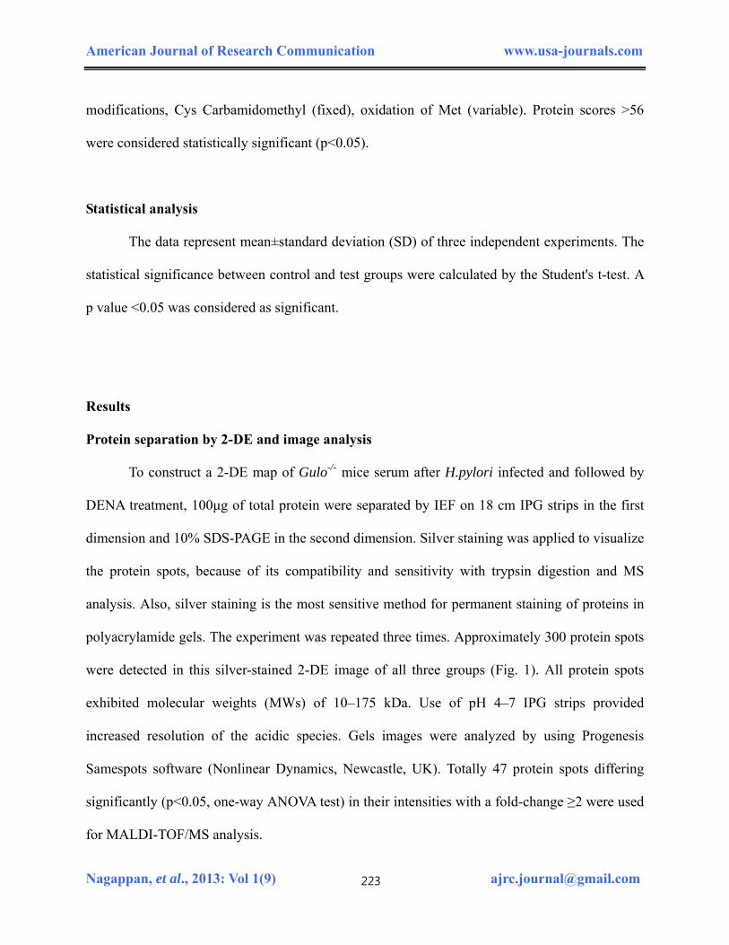
Protein separation by 2-DE and image analysis
To construct a 2-DE map of Gulo-/- mice serum after H.pylori infected and followed by
DENA treatment, 100μg of total protein were separated by IEF on 18 cm IPG strips in the first
dimension and 10% SDS-PAGE in the second dimension. Silver staining was applied to visualize
the protein spots, because of its compatibility and sensitivity with trypsin digestion and MS
analysis. Also, silver staining is the most sensitive method for permanent staining of proteins in
polyacrylamide gels. The experiment was repeated three times. Approximately 300 protein spots
were detected in this silver-stained 2-DE image of all three groups (Fig. 1). All protein spots
exhibited molecular weights (MWs) of 10–175 kDa. Use of pH 4–7 IPG strips provided
increased resolution of the acidic species. Gels images were analyzed by using Progenesis
Samespots software (Nonlinear Dynamics, Newcastle, UK). Totally 47 protein spots differing
significantly (p<0.05, one-way ANOVA test) in their intensities with a fold-change ≥2 were used
for MALDI-TOF/MS analysis.
Nagappan, et al., 2013: Vol 1(9) [email protected] 223

American Journal of Research Communication www.usa-journals.com
Figure 1. Two-dimension electrophoresis pattern of Gulo-/- mouse serum. (A) Control (only
vitamin C), (B) H.Pylori-infected, (C) H.Pylori-infected and DENA treated. 100µg of serum
total proteins were separated on IPG-strips with pH 4–7(18 cm) in the first dimension and then
on 12% polyacrylamide gels in the second dimension, which were stained silver nitrate. Protein
spots which showed significantly altered expression levels (Max fold change ≥2 and p<0.05)
were marked on the gels. Three independent experiments were performed.
Nagappan, et al., 2013: Vol 1(9) [email protected] 224

American Journal of Research Communication www.usa-journals.com
Nagappan, et al., 2013: Vol 1(9) [email protected] 225
Identification of differentially expressed proteins in H.pylori infected and combined with DENA treated Gulo-/- mice serum by MALDI-TOF- MS
The differentially expressed proteins in H.pylori infected and/or combined with DENA
treated Gulo-/- mice serum were investigated by 2-DE analysis in comparison with control. The
2-DE maps of three groups of Gulo-/- mice serum are shown in Fig. 1. A total of 47 statistically
significant altered protein spots were selected (≥2 fold, p<0.05) and successfully 38 proteins
were identified by a MALDI-TOF/MS using the MASCOT search engine and the SwissProt
database (Table 2). The name, accession number, number of matching peptides, theoretical
isoelectric point (pI), molecular weights, sequence coverage of the differentially expressed
protein identified are listed in Table 2. We noticed some of the important proteins such Alpha-
1B-glycoprotein, Haptoglobin, Collagen alpha-2 (IX) chain, Antithrombin-III, Hemopexin and
Glutathione S-transferase, Complement C3 and Apolipoprotein A-IV were differentially
expressed in H.pylori infection and followed by DENA groups. The expression patterns of
identified proteins were shown in Table 3. These differentially expressed proteins were involved
in various biological processes such as immune system process, response to stimulus, metabolic
process, cellular process, system process and cell communication due to this H.pylori-infection
and DENA treatment.
Discussion
Unlike most other mammals and animals, humans do not have the ability to make vitamin C
inside of their own bodies. Therefore, humans must obtain vitamin C through daily diet and its
requirements are vary greatly among individuals. In present study, we established Gulo-/- mice
like human (it does not have ability to synthesis vitamin C) must orally supplemented daily
because Gulo-/- mice can’t survive without vitamin C. Vitamin C deficiency mice will lost body

American Journal of Research Communication www.usa-journals.com
Table 2. List of statistically significant differentially expressed proteins identified in serum of Gulo-/- mice after infected with
H.pylori alone and/or combined treatement with DENA by MALDI-TOF- MS
S
pot
No.
Protein name1
Accessi
on
number1
Theoretical
Mr (kDa)2
Theoretical
pI Value2
Fold
change
p Value
(t-test)
Peptid
es
matched
Sequen
ce
Coverage
(%)
Mow
se
Score3
1 Haptoglobin Q61646 39.24 5.88 9.1 7.208e-005 6/20 22 61
2 Collagen alpha-2(IX) chain Q07643 65.51 9.40 15.6 1.254e-004 7/19 14 63
3 Collagen alpha-2(IX) chain Q07643 65.51 9.40 19.7 1.967e-004 8/20 13 62
4 Histidine protein
methyltransferase 1
homolog
Q4KM
84
40.55 5.54 4.7 2.454e-004 8/43 20 60
5 Haptoglobin Q61646 39.24 5.88 6.0 2.460e-004 8/48 30 58
6 Vasculin-like protein 1 Q3KR5
3
52.86 6.82 8.4 6.580e-004 5/10 15 60
7 Alpha-1B-glycoprotein Q19LI2 57.20 6.33 4.5 8.789e-004 15/45 22 126
8 Alpha-1B-glycoprotein Q19LI2 57.20 6.33 15.6 9.135e-004 56/16 25 123
9 Haptoglobin Q61646 39.24 5.88 5.9 0.001 8/26 24 69
10 Alpha-1B-glycoprotein Q19LI2 57.20 6.33 4.6 0.003 15/47 24 122
11 Alpha-1B-glycoprotein Q19LI2 57.20 6.33 3.7 0.003 17/47 29 149
12 Alpha-1B-glycoprotein Q19LI2 57.20 6.33 4.0 0.004 11/48 18 74
13 Growth arrest-specific
protein 6
Q63772 76.53 5.63 6.4 0.005 13/83 21 58
14 Alpha-1B-glycoprotein Q19LI2 57.20 6.33 4.8 0.005 10/37 17 76
15 Collagen alpha-2(IX) chain Q07643 65.51 9.40 6.1 0.005 7/22 14 58
Nagappan, et al., 2013: [email protected] 226

American Journal of Research Communication www.usa-journals.com
16 Antithrombin-III P32261 52.48 6.10 23.2 0.006 12/75 29 64
17 Apolipoprotein A-IV P06728 45.00 5.34 6.2 0.007 8/23 22 74
18 UPF0361 protein C3orf37
homolog
Q8R1M
0
40.77 8.86 3.2 0.007 15/109 33 72
19 Haptoglobin Q61646 39.24 5.88 3.0 0.008 7/28 24 64
20 Collagen alpha-2(IX) chain Q07643 65.51 9.40 2.7 0.008 7/21 10 58
21 Serum albumin P02770 70.68 6.09 4.0 0.010 10/37 22 72
22 Apolipoprotein A-IV P06728 45.00 5.34 4.3 0.013 13/67 30 81
23 Glutathione S-transferase P10649 26.07 7.71 2.1 0.013 7/38 32 58
24 Antithrombin-III P32261 52.48 6.10 3.9 0.013 9/36 27 73
25 Alpha-1B-glycoprotein Q19LI2 57.20 6.33 4.3 0.015 9/31 14 72
26 MACRO domain-
containing protein 1
Q922B
1
35.84 9.07 2.4 0.016 10/53 23 75
27 NF-kappa-B-repressing
factor
Q8BY0
2
78.38 9.08 4.6 0.020 7/16 9 60
28 Serum albumin P02770 70.68 6.09 3.8 0.024 10/51 21 57
29 Serum albumin P07724 70.70 5.75 3.1 0.027 14/97 26 62
30 Stabilin-2 Q8CF
M6
161.86 7.48 4.0 0.033 9/16 8 69
Nagappan, et al., 2013: [email protected] 227

American Journal of Research Communication www.usa-journals.com
Nagappan, et al., 2013: [email protected] 228
31 Hemopexin Q91X7
2
52.03 7.92 2.4 0.033 20/118 43 119
32 Creatine kinase M-type P07310 43.25 6.58 2.7 0.040 7/27 18 57
33 Microtubule-associated
tumor suppressor candidate
2 homolog
Q3UH
D3
147.95 8.42 2.8 0.044 12/39 12 58
34 G protein-regulated inducer
of neurite outgrowth 1
Q3UN
H4
96.07 8.14 6.3 0.046 16/93 21 59
35 1,25-dihydroxyvitamin D(3)
24-hydroxylase,
mitochondrial
Q64441 59.93 9.16 2.4 0.042 9/27 15 64
36 Complement C3 P01027 187.91 6.29 2.2 0.020 28/71 17 118
37 Hemopexin Q91X7
2
52.03 7.92 2.2 0.034 19/79 38 142
38 Haptoglobin Q61646 39.24 5.88 2.7 0.027 8/40 27 64
1Entry name, protein name and accession number from SWISS-PROT database identified by MALDI-TOF/MS
2Theoretical molecular weight (kDa) and pI from SWISS-PROT database
3Score is –10*log (p), where p is the probability that the observed match is a random event, Protein scores greater than 56 are significant (p<0.05).

American Journal of Research Communication www.usa-journals.com
Table 3. Expression patterns of differentially expressed proteins identified in serum of
Gulo-/- mice after infected with H.pylori alone and/or combined treatement with DENA by
MALDI-TOF- MS
Spot No.
Protein name1 Accession number1
Control Vs H. Pylori
infected2
Control Vs H.Pylori + DENA2
1 Haptoglobin Q61646 ↓ ↑
2 Collagen alpha-2(IX)
chain
Q07643 ↑ ↑
3 Collagen alpha-2(IX)
chain
Q07643 ↑ ↑
4 Histidine protein
methyltransferase 1
homolog
Q4KM84 ↑ ↑
5 Haptoglobin Q61646 ↓ ↑
6 Vasculin-like protein 1 Q3KR53 ↑ ↑ 7 Alpha-1B-glycoprotein Q19LI2 ↑ ↓ 8 Alpha-1B-glycoprotein Q19LI2 ↑ ↓ 9 Haptoglobin Q61646 ↓ ↑
10 Alpha-1B-glycoprotein Q19LI2 ↑ ↑ 11 Alpha-1B-glycoprotein Q19LI2 ↑ ↓ 12 Alpha-1B-glycoprotein Q19LI2 ↑ ↓ 13 Growth arrest-specific
protein 6
Q63772 ↓ ↑
14 Alpha-1B-glycoprotein Q19LI2 ↑ ↓ 15 Collagen alpha-2(IX)
chain
Q07643 ↑ ↑
16 Antithrombin-III P32261 ↑ ↑
17 Apolipoprotein A-IV P06728 ↓ ↓
18 UPF0361 protein
C3orf37 homolog
Q8R1M0 ↑ ↑
Nagappan, et al., 2013: Vol 1(9) [email protected] 229
American Journal of Research Communication www.usa-journals.com
19 Haptoglobin Q61646 ↓ ↑
20 Collagen alpha-2(IX)
chain
Q07643 ↑ ↑
21 Serum albumin P02770 ↑ ↑ 22 Apolipoprotein A-IV P06728 ↑ ↓
23 Glutathione S-
transferase
P10649 ↑ ↑
24 Antithrombin-III P32261 ↑ ↑ 25 Alpha-1B-glycoprotein Q19LI2 ↑ ↓ 26 MACRO domain-
containing protein 1
Q922B1 ↓ ↓
27 NF-kappa-B-repressing
factor
Q8BY02 ↓ ↓
28 Serum albumin P02770 ↑ ↑ 29 Serum albumin P07724 ↑ ↑ 30 Stabilin-2 Q8CFM6 ↓ ↓ 31 Hemopexin Q91X72 ↑ ↑ 32 Creatine kinase M-type P07310 ↓ ↓ 33 Microtubule-associated
tumor suppressor candidate
2 homolog
Q3UHD3 ↑ ↑
34 G protein-regulated
inducer of neurite
outgrowth 1
Q3UNH4 ↓ ↑
35 1,25-dihydroxyvitamin
D(3) 24-hydroxylase,
mitochondrial
Q64441 ↑ ↑
36 Complement C3 P01027 ↓ ↓ 37 Hemopexin Q91X72 ↑ ↑ 38 Haptoglobin Q61646 ↓ ↑
1Entry name, protein name and accession number from SWISS-PROT database identified by MALDI-TOF//MS 2Image analysis was performed using Progenesis Samespots software (Nonlinear Dynamics, Newcastle, UK). Those
spots differing significantly (p<0.05, one-way ANOVA test) in their intensities with a fold-change ≥2.
Nagappan, et al., 2013: Vol 1(9) [email protected] 230

American Journal of Research Communication www.usa-journals.com
weight and die within 6 weeks due to severe scurvy. With recent advances in proteomics protein
biomarker discovery is now a major area of proteome research. Using Gulo-/- mice model, we
investigated the protein expression pattern of serum in response to H.pylori infection and
followed by DENA treatment. Comparison of serum protein profiles of H.pylori infection group
and followed by DENA treated groups with control group revealed that the differentially
expressed proteins were involved in various physiological pathways including acute phase
response signaling, inflammatory regulation and immune response.
Complement components plays a pivotal role as mediators of inflammation and
regulation of the immune response (Jurianz et al., 1999). Complement C3 is protein of immune
system and plays a central role in the activation of the complement system (Sahu and Lambris,
2001; Markiewski and Lambris. 2007). Complement C3 is a multipotent protein that’s involved
in the immune response, as including complement activation, antigen presentation, cell-cell
interactions, and cell proliferation (Frade, 1999). The people with complement C3 deficiency are
susceptible to bacterial infection (Litzman et al, 2003). In this study, complement C3 (Spot no.
36) expression was significantly decreased in in H. Pylori infected group and H. Pylori infected
followed by DENA treated group when compare with control group (Fig.2A). The down-
regulation of complement C3 suggest that Gulo-/- mice infected with H.pylori and further DENA
enhanced the H.pylori effect and complement C3 might be involved in carcinogenesis of gastric
cancer.
An acute-phase serum proteins (APPs) such as Haptoglobin, hemopexin, Alpha-1B-glycoprotein
were significantly increased in in H. Pylori infected group and H. Pylori infected followed by
DENA treated group when compare with control group (Fig. 2B). APPs are circulating plasma
proteins which are mainly synthesized by liver parenchymal cells following simulation by
Nagappan, et al., 2013: Vol 1(9) [email protected] 231

American Journal of Research Communication www.usa-journals.com
Figure 2. Enlargements of differentially expressed proteins. (A) Complement C3, (B)
Haptoglobin, (C) Hemopexin, (D) Alpha-1B-glycoprotein and (E) Glutathione S-transferase
from 2-DE maps of Gulo-/- mouse serum corresponding sections of gels with protein spots
derived from control (only vitamin C), H.Pylori-infected, and H.Pylori-infected and DENA
treated. Three independent experiments were performed and the mean ± SD was plotted (*P <
0.05 compared with control).
Nagappan, et al., 2013: Vol 1(9) [email protected] 232

American Journal of Research Communication www.usa-journals.com
cytokines such as interleukin-1, interleukin-6 and tumor necrosis factor-α mainly from
macrophages at the inflammatory site (Moshage, 1997). The main biological functions of
haptoglobin are to play a role in modulating host defense responses to infection and
inflammation (Dobryszycka, 1997; Wassell, 2000). Haptoglobin has also been reported to be
involved in immune suppression in cancer (Oh et al., 1987' Oh et al., 1990). There are five
differentially expressed isoforms of Haptoglobin (Spot no.1, 5, 9, 19, 38) found in H. Pylori
infected group and H. Pylori infected followed by DENA treated group. Only little expressions
of Haptoglobin found in H. Pylori infected group but highly expressed in H. Pylori infected
followed by DENA treated group. These results revealed that that a possible association between
H.pylori and DENA with the disease pathogenesis and cancinogenesis.
Another APPs protein, Hemopexin two isoforms (spot no.31, 37) up-regulated protein H.
Pylori infected group and H. Pylori infected followed by DENA treated group when compare
with control group (Fig.2C). It provides the second line of defense against hemoglobin-mediated
oxidative damage during the intravascular hemolysis (Delanghe and Langlois, 2001). Moreover,
Serum hemopexin helps in scavenging free heme while the free iron is taken up and transported
by transferrin (Ferreira et al., 2008). However, the relationship between tumor development and
hemopexin is not very clear but matrix metallopeptidases containing a hemopexin domain have
been detected in the sera of gastric cancer patients with high expression of hemopexin. These
results suggest that, hemopexin might be involved H.pylori pathogenesis mechanism and gastric
cancer carcinogenesis.
Alpha-1B-glycoprotein, 7 isoforms (spot no.7, 8, 10, 11, 12, 14, 25) were up-regulated in
only H. Pylori infected group and completely not expressed in H. Pylori infected followed by
DENA treated group (Fig.2D). Alpha-1B-glycoprotein is a plasma secreted protein, member of
Nagappan, et al., 2013: Vol 1(9) [email protected] 233

American Journal of Research Communication www.usa-journals.com
the immunoglobulin superfamily, but its function is unknown (Ishioka et al., 1986). Recently
have reported that alpha-1B-glycoprotein was detected in urinary samples from bladder cancer
patients by using a glycoprotein profiling method (Kreunin et al., 2007). Also, overexpression of
alpha-1B-glycoprotein has reported in saliva from patients with head-and-neck squamous cell
carcinoma (Ohshiro et al., 2007). These findings suggest that alpha-1B-glycoprotein might be an
H.pylori associated cancer-related protein and its possible functions in carcinogenesis further to
be investigated.
The glutathione S-transferases (GSTs), a multigene family of dimeric enzymes that have a
significant role in the detoxification of electrophilic species by catalytic conjugation with
reduced glutathione (GSH) (Osada et al., 2002; Hayes and Pulford, 1995). Previous studies have
reported that pi-class GST is substantially increased in the early stages of rat liver carcinogenesis
(Farber, 1984; Sato et al., 1984; Satoh et al., 1985). GST pi is overexpressed in hepatic foci
arising spontaneously or in animals treated with carcinogens (Sato, 1989; Sawaki et al., 1990)
but almost undetectable in normal rat hepatocytes. In the present study, glutathione S-
transferases (GSTs) (Spot no. 23) was highly expressed in highly expressed in H. Pylori infected
followed by DENA treated group but no expression found in only H. Pylori infected group
(Fig.2E). This result clearly indicates that GSTs overexpressed by carcinogen DENA treatment
but H. Pylori not changed or involved in GSTs pathways.
In summary, we have performed a proteomic analysis to investigate the protein
expression pattern in Gulo-/- mice serum in response to H.pylori infection and followed by
DENA treatment. Comparison of protein profiles of Gulo-/- mice serum revealed that H.pylori
infection and followed by DENA treatment altered the expressions of Complement C3, Alpha-
1B-glycoprotein, Haptoglobin, Hemopexin and Glutathione S-transferase when compared to
Nagappan, et al., 2013: Vol 1(9) [email protected] 234

American Journal of Research Communication www.usa-journals.com
control group. The differentially expressed proteins were involved in various biological
processes such as acute phase response signaling, inflammatory regulation and immune response
due to this H.pylori infection and DENA. These protein expressions further needed to be
validated by immune-blotting. Moreover, identified proteins by MALDI-TOF/ MS in Gulo-/-
serum might be involved in mechanism of H. pylori disease pathogenesis and/or combined effect
of DENA, early diagnosis and therapy of H. pylori and DENA associated gastric disorders.
ACKNOWLEDGEMENTS
This work was supported by the National Research Foundation (NRF) of Korea Grant funded by
the Korean government (MEST) (No. 2012045015) and the National R&D Program for Cancer
Control, Ministry for Health, Welfare and Family affairs, Republic of Korea (No. 0820050).
REFERENCES
Acunzo J, Katsogiannou M, Rocchi P. Small heat shock proteins HSP27 (HspB1), alphaB-crystallin (HspB5) and HSP22 (HspB8) as regulators of cell death. The International Journal of Biochemistry & Cell Biology 2012; 44: 1622-1631.
Alaiya AA, Franzen B, Auer G, Linder S. Cancer proteomics: from identification of novel markers to creation of artifical learning models for tumor classification. Electrophoresis 2000; 21: 1210-1217.
Ang TL, Fock KM, Dhamodaran S, Teo EK, Tan J. Racial differences in Helicobacter pylori, serum pepsinogen and gastric cancer incidence in an urban Asian population. Journal of Gastroenterology and Hepatology 2005; 20: 1603-1609.
Correa P, Malcom G, Schmidt B, Fontham E, Ruiz B, Bravo JC, et al. Antioxidant micronutrients and gastric cancer. Alimentary Pharmacology & Therapeutics 1998; 12: 73-82.
Nagappan, et al., 2013: Vol 1(9) [email protected] 235

American Journal of Research Communication www.usa-journals.com
Delanghe JR, Langlois MR. Hemopexin: a review of biological aspects and the role in laboratory medicine. Clinica Chimica Acta 2001; 312: 13-23.
Dobryszycka W. Biological functions of haptoglobin--new pieces to an old puzzle. European Journal of Clinical Chemistry and Clinical Biochemistry 1997; 35: 647-654.
Farber E. The biochemistry of preneoplastic liver: a common metabolic pattern in hepatocyte nodules. Canadian Journal of Biochemistry and Cell Biology 1984; 62: 486-494.
Ferreira A, Balla J, Jeney V, Balla G, Soares MP. A central role for free heme in the pathogenesis of severe malaria: the missing link?. Journal of Molecular Medicine 2008; 86: 1097-1111.
Frade R. Structure and functions of proteases which cleave human C3 and are expressed on normal or tumor human cells: some are involved in tumorigenic and metastatic properties of human melanoma cells. Immunopharmacology 1999; 42: 39-45.
Gonzalez CA, Agudo A. Carcinogenesis, prevention and early detection of gastric cancer: where we are and where we should go. International Journal of Cancer 2012; 130: 745-753.
Hayes JD, Pulford DJ. The glutathione S-transferase supergene family: regulation of GST and the contribution of the isoenzymes to cancer chemoprotection and drug resistance. Critical Reviews in Biochemistry and Molecular Biology 1995; 30: 445-600.
Houghton J, Fox JG, Wang TC. Gastric cancer: laboratory bench to clinic. Journal of Gastroenterology and Hepatology 2002; 17: 495-502.
Ishioka N, Takahashi N, Putnam FW. Amino acid sequence of human plasma alpha 1B-glycoprotein: homology to the immunoglobulin supergene family. Proceedings of the National Academy of Sciences of the United States of America 1986; 83: 2363-2367.
Jung KW, Park S, Won YJ, Kong HJ, Lee JY, Seo HG, et al. Prediction of cancer incidence and mortality in Korea, 2012. Cancer Treatment and Research 2012; 44: 25-31.
Jurianz K, Ziegler S, Garcia-Schuler H, Kraus S, Bohana-Kashtan O, Fishelson Z, et al. Complement resistance of tumor cells: basal and induced mechanisms. Molecular Immunology 1999; 36: 929-939.
Kelley JR, Duggan JM. Gastric cancer epidemiology and risk factors. Journal of Clinical Epidemiology 2003; 56: 1-9.
Kim H, Bae S, Yu Y, Kim Y, Kim HR, Hwang YI, et al. The analysis of vitamin C concentration in organs of gulo(-/-) mice upon vitamin C withdrawal. Immune Network 2012;12: 18-26.
Nagappan, et al., 2013: Vol 1(9) [email protected] 236

American Journal of Research Communication www.usa-journals.com
Kreunin P, Zhao J, Rosser C, Urquidi V, Lubman DM, Goodison S. Bladder cancer associated glycoprotein signatures revealed by urinary proteomic profiling. Journal of Proteome Research 2007; 6: 2631-2639.
Li Y, Shi CX, Mossman KL, Rosenfeld J, Boo YC, Schellhorn HE. Restoration of vitamin C synthesis in transgenic Gulo-/- mice by helper-dependent adenovirus-based expression of gulonolactone oxidase. Human Gene Therapy 2008; 19: 1349-1358.
Litzman J, Freiberger T, Bartonkova D, Vlkova M, Thon V, Lokaj J. Early manifestation and recognition of C2 complement deficiency in the form of pyogenic infection in infancy. Journal of Paediatrics and Child Health2003; 39: 274-277.
Maeda N, Hagihara H, Nakata Y, Hiller S, Wilder J, Reddick R. Aortic wall damage in mice unable to synthesize ascorbic acid. Proceedings of the National Academy of Sciences of the United States of America 2000;.97: 841-846.
Marchetti M, Arico B, Burroni D, Figura N, Rappuoli R, Ghiara P. Development of a mouse model of Helicobacter pylori infection that mimics human disease. Science 1995; 267: 1655-1658.
Markiewski MM, Lambris JD. The role of complement in inflammatory diseases from behind the scenes into the spotlight. The American Journal of Pathology 2007; 171: 715-727.
Marrelli D, Roviello F, De Stefano A, Farnetani M, Garosi L, Messano A, e al. Prognostic significance of CEA, CA 19-9 and CA 72-4 preoperative serum levels in gastric carcinoma. Oncology 1999; 57: 55-62.
Miki K. Gastric cancer screening using the serum pepsinogen test method. Gastric Cancer 2006; 9: 245-253.
Moshage H. Cytokines and the hepatic acute phase response. The Journal of Pathology 1997; 181: 257-266.
Oh SK, Kim SH, Walker JE. Interference with immune response at the level of generating effector cells by tumor-associated haptoglobin. Journal of the National Cancer Institute 1990; 82: 934-940.
Oh SK, Very DL, Walker J, Raam S, Ju ST. An analogy between fetal haptoglobin and a potent immunosuppressant in cancer. Cancer Research 1987; 47: 5120-5126.
Ohshiro K, Rosenthal DI, Koomen JM, Streckfus CF, Chambers M, Kobayashi R, et al. Pre-analytic saliva processing affect proteomic results and biomarker screening of head and neck squamous carcinoma. International Journal of Oncology 2007; 30: 743-749.
Nagappan, et al., 2013: Vol 1(9) [email protected] 237

American Journal of Research Communication www.usa-journals.com
Osada H, Tatematsu Y, Yatabe Y, Nakagawa T, Konishi H, Harano T, et al. Frequent and histological type-specific inactivation of 14-3-3sigma in human lung cancers. Oncogene 2002; 21: 2418-2424.
Ray A. Cancer preventive role of selected dietary factors. Indian Journal of Cancer 2005; 42: 15-24.
Sahu A, Lambris JD. Structure and biology of complement protein C3, a connecting link between innate and acquired immunity. Immunological Reviews 2001; 180: 35-48.
Sallet JA, Zilberstein B, Andreollo NA, Eshkenazy R, Pajecki D. Experimental esophageal carcinogenesis: technical standardization and results. Diseases of the Esophagus 2002; 15: 278-281.
Sato K. Glutathione transferases as markers of preneoplasia and neoplasia. Advances in Cancer Research 1989; 52: 205-255.
Sato K, Kitahara A, Satoh K, Ishikawa T, Tatematsu M, Ito N. The placental form of glutathione S-transferase as a new marker protein for preneoplasia in rat chemical hepatocarcinogenesis. GANN Japanese Journal of Cancer Research 1984; 75: 199-202.
Satoh K, Kitahara A, Soma Y, Inaba Y, Hatayama I, Sato K.. Purification, induction, and distribution of placental glutathione transferase: a new marker enzyme for preneoplastic cells in the rat chemical hepatocarcinogenesis. Proceedings of the National Academy of Sciences of the United States of America 1985; 82: 3964-3968.
Sawaki M, Enomoto K, Takahashi H, Nakajima Y, Mori M. Phenotype of preneoplastic and neoplastic liver lesions during spontaneous liver carcinogenesis of LEC rats. Carcinogenesis 1990; 11: 1857-1861.
Shevchenko A, Wilm M, Vorm O, Mann M. Mass spectrometric sequencing of proteins silver-stained polyacrylamide gels. Analytical Chemistry 1996; 68: 850-858.
Swain M, Ross NW. A silver stain protocol for proteins yielding high resolution and transparent background in sodium dodecyl sulfate-polyacrylamide gels. Electrophoresis 1995; 16: 948-951.
Takahashi Y, Takeuchi T, Sakamoto J, Touge T, Mai M, Ohkura H, et al.The usefulness of CEA and/or CA19-9 in monitoring for recurrence in gastric cancer patients: a prospective clinical study. Gastric Cancer 2003; 6: 142-145.
Wassell J. Haptoglobin: function and polymorphism. Clinical Laboratory 2000; 46: 547-552.
Nagappan, et al., 2013: Vol 1(9) [email protected] 238

American Journal of Research Communication www.usa-journals.com
Nagappan, et al., 2013: Vol 1(9) [email protected] 239
Zhang ZW, Patchett SE, Perrett D, Katelaris PH, Domizio P, Farthing MJ. The relation between gastric vitamin C concentrations, mucosal histology, and CagA seropositivity in the human stomach. Gut 1998; 43: 322-326.