pharmacology subcutaneous administration in swine model
TRANSCRIPT

1/10https://vetsci.org
ABSTRACT
Background: Due to multiple similarities in the structure and physiology of human and pig skin, the pig model is extremely useful for biological drug testing after subcutaneous administration. Knowledge of the differences between subcutaneous injection sites could have a significant impact on the absorption phase and pharmacokinetic profiles of biological drugs.Objectives: This study aimed to analyze the impact of administration site on pharmacokinetics and selected biochemical and hematological parameters after a single subcutaneous administration of ustekinumab in pigs. Drug concentrations in blood plasma were analyzed by enzyme-linked immunosorbent assay. Pharmacokinetic analyses were performed based on raw data using Phoenix WinNonlin 8.1 software and ThothPro v 4.1.Methods: The study included 12 healthy, female, large white piglets. Each group received a single dose of ustekinumab given as a 1 mg/kg subcutaneous injection into the internal part of the inguinal fold or the external part of the inguinal fold.Results: The differences in absorption rate between the internal and external parts of the inguinal fold were not significant. However, the time of maximal concentration, clearance, area under the curve calculated between zero and mean residence time and mean residence time between groups were substantially different (p > 0.05). The relative bioavailability after administration of ustekinumab into the external part of the inguinal fold was 40.36% lower than after administration of ustekinumab into the internal part of the inguinal fold.Conclusions: Healthy breeding pigs are a relevant model to study the pharmacokinetic profile of subcutaneously administered ustekinumab.
Keywords: Swine; biologics; ustekinumab; skin; pharmacokinetics
INTRODUCTION
Pigs have been used previously to investigate the pharmacokinetics/pharmacodynamics and toxicology of small molecules and biologics [1-4]. The value of the pig model has been demonstrated in many studies, and due to multiple similarities in the structure
J Vet Sci. 2021 Sep;22(5):e47https://doi.org/10.4142/jvs.2021.22.e47pISSN 1229-845X·eISSN 1976-555X
Original Article
Received: Feb 2, 2021Revised: Apr 22, 2021Accepted: May 23, 2021Published online: May 27, 2021
*Corresponding author:Artur BurmańczukDepartment of Pharmacology, Toxicology and Environmental Protection, Faculty of Veterinary Medicine, University of Life Sciences, Akademicka 12, 20-033 Lublin, Poland.E-mail: [email protected]
© 2021 The Korean Society of Veterinary ScienceThis is an Open Access article distributed under the terms of the Creative Commons Attribution Non-Commercial License (https://creativecommons.org/licenses/by-nc/4.0) which permits unrestricted non-commercial use, distribution, and reproduction in any medium, provided the original work is properly cited.
ORCID iDsTomasz Grabowski https://orcid.org/0000-0002-3077-9698Artur Burmańczuk https://orcid.org/0000-0002-2354-7696Rafał Derlacz https://orcid.org/0000-0002-8254-3521Tadeusz Stefaniak https://orcid.org/0000-0002-8982-211XAnna Rząsa https://orcid.org/0000-0002-0903-3445Jacek Borkowski https://orcid.org/0000-0002-5535-5955
Tomasz Grabowski 1, Artur Burmańczuk 2,*, Rafał Derlacz 1, Tadeusz Stefaniak 3, Anna Rząsa 3, Jacek Borkowski 4
1Polpharma Biologics SA, 80-172 Gdańsk, Poland2 Sub-Department of Pharmacology, Toxicology and Environmental Protection, Faculty of Veterinary Medicine, University of Life Sciences, 20-033 Lublin, Poland
3 Department of Immunology, Pathophysiology and Veterinary Preventive Medicine, Wroclaw University of Environmental and Life Sciences, 50-375 Wrocław, Poland
4 Department of Physiology and Biochemistry, University School of Physical Education in Wroclaw, 51-612 Wrocław, Poland
Ustekinumab pharmacokinetics after subcutaneous administration in swine model
Pharmacology

FundingThis work was supported by The National Centre for Research and Development in Poland under grant number POIR.01.01.01-00-0407/17-00. The Ustekinumab PK pilot study was supported by The National Centre for Research and Development in Poland as a part of a development program for biosimilar ustekinumab candidates by Polpharma Biologics SA.
Conflict of InterestThe authors declare no conflicts of interest.
Author ContributionsConceptualization: Grabowski T; Formal analysis: Grabowski T, Stefaniak T, Rząsa A, Borkowski J; Investigation: Burmańczuk A; Methodology: Grabowski T; Resources: Burmańczuk A; Writing - original draft: Grabowski T, Stefaniak T, Rząsa A, Borkowski J; Writing - review & editing: Derlacz R.
and physiology of human and pig skin, is highly useful for testing subcutaneous (SC) formulations [5-10]. Moreover, the Fc receptor (Brambel receptor) in pigs, which is critical for the elimination half-life of therapeutic monoclonal antibodies (mAbs), is similar to the human Fc receptor [11]. Thus, the absorption process and general pharmacokinetic profile of the pig may provide a relevant and advantageous pharmacokinetic model for preclinical development of subcutaneously administered drugs [12,13]. The inguinal and scapular regions are most often used for SC mAb administration in pig model pharmacokinetic studies [9]. Distinct SC injection sites can significantly impact the shape of the absorption phase and the pharmacokinetics of biologics [14-17]. The key limiting factor related to absorption is lymphatic drainage and presence of nearby lymph nodes [18]. Thus, the administration site in SC studies can affect not only the pharmacokinetics of biological drugs but also their variability, making it difficult to optimize animal pharmacokinetics models of mAbs with SC administration [19].
Ustekinumab is a fully humanized immunoglobulin G1 (IgG1) mAb with high affinity to human cytokines interleukin (IL)-12 and IL-23. Neutralizing these cytokines' biological activity is important for plaque psoriasis and psoriatic arthritis treatment in humans [20]. A pharmacokinetic, as well as efficacy and safety, profile for the intravenous and SC administration of ustekinumab has previously been established [21]. The recommended injection sites in humans are the upper thigh or the area around the belly (abdomen), at least 5cm away from the navel (belly button). After abdominal SC injection in patients, biological drugs are absorbed into the inguinal, iliac and lumbar lymph nodes [22]. The inguinal fold of pigs is drained (inter alia) by afferent vessels of the inguinal lymph node [23].
This study aimed to analyze the impact of administration site on the pharmacokinetics and on select biochemical and hematological parameters after a single SC administration of ustekinumab in healthy pigs.
MATERIALS AND METHODS
AnimalsTwelve healthy, female, large white piglets weighing between 15.0 and 20.0 kg and aged 45 ± 10 days were used in this study. The animals were divided into 2 groups (n = 6) and housed with food and water ad libitum. Each group received a single dose of ustekinumab (Stelara; Janssen-Cilag International NV, Belgium) given in a single 1 mg/kg SC injection: the first group into the internal part of the inguinal fold (IF) and the second group on the external part of the inguinal fold (EF). Before (at time 0) and after drug administration, blood was sampled from the jugular vein (2 mL) at intervals of 6, 12, 24, 36, 48, 60, 72, 84, 96, 120, 144, 168, 288, 384 and 432 h (18 days) after injection into heparinized tubes using a vacutainer (BD Vacutainer Safety-Lok; BD Biosciences, USA) for pharmacokinetic and anti-drug-antibodies (ADA) analysis. The study was approved by the ethics committee of the University of Life Sciences, Lublin (51/2017). Animal experiments complied with the Animal Research: Reporting of In Vivo Experiments (ARRIVE) guidelines and were carried out in accordance with the UK Animals (Scientific Procedures) Act, 1986 and associated guidelines, as well as with EU Directive 2010/63/EU for animal experiments. The piglets were procured from the breeding farm of the University of Life Sciences in Lublin. After the study, the animals were used for breeding only.
2/10https://vetsci.org https://doi.org/10.4142/jvs.2021.22.e47
Ustekinumab pig model

Hematological and biochemical parametersThe blood samples were analyzed for basic parameters at the same sampling points used for pharmacokinetics analysis. The analyzed parameters included: cholesterol, bilirubin, urea, creatinine, phosphorus, calcium, WBC – number of white blood cells, LYM – number of lymphocytes, MID – number of types of WBC not classified as lymphocytes or granulocytes, GRAN – number of granulocytes, RBC – number of red blood cells, HTC – hematocrit, HGB – hemoglobin, MCV – mean corpuscular volume of red blood cells, MCH – mean corpuscular hemoglobin in red blood cells, MCHC – mean corpuscular hemoglobin concentration, RDWa – absolute red cell distribution width, RDW – red cell distribution width, PLT – platelet count, and MPV – mean platelet volume. All parameters were calculated using an automated hematology analyzer, Abacus Junior Vet (Diatron Group, Hungary).
Immunochemical analysisImmunization and production of immune serumThe production of pig-anti-ustekinumab serum was carried out with two additional pigs. The procedure was approved by the Local Ethics Committee for Animal Experiments in Wroclaw (07/18, 21.02.2018). Production of pig-anti-ustekinumab serum was a necessary step enabling the ADA analysis and analysis of concentration of ustekinumab in the experimental groups. Two pigs aged 2.5 months were immunized intramuscularly 4 times every 2 weeks with 100 mg of ustekinumab in 0.5 mL of saline emulsified immediately before injection with 0.5 mL of Freunds Adjuvant, Incomplete (F5506, Sigma, USA). Prior to each injection, a blood sample was taken from the vena cava cranialis. Two weeks after the last injection, around 200 mL of blood was taken using a 1.6 mm needle and 20 mL syringes. The hyperimmune serum was produced by centrifugation and stored until use at −20°C.
Then, monovalent porcine anti-ustekinumab IgG antibody was affinity-purified and conjugated with horseradish peroxidase (HRPO). IgG from pig's serum was purified by ion-exchange chromatography [24]. The IgG-rich fraction of the serum was precipitated using ammonium sulphate. The precipitate was dialyzed against 0.05 M phosphate buffer, pH 7.0. The supernatant (about 180 mL) (A280 = 55) was loaded onto a DE Cellulose (DE52) column (120 mL) and equilibrated with the same phosphate buffer. The non-bound fraction (210 mL, A280 = 28.5) was recognized as porcine IgG. Production of affinity-purified IgG anti-ustekinumab was performed using two steps. In the first step, 0.4 mL of ustekinumab solution (36 mg) was dialyzed against 0.05M carbonate buffer with 0.5 M NaCl to remove low-molecular substances possessing free amino-groups and bound with activated Sepharose 4B (CNBr Activated Sepharose 4B). In the second step, 50 mL of pig IgG containing ustekinumab antibody previously purified by ion-exchange chromatography was bound to ustekinumab, coupled to a Sepharose 4B column (3 mL). The column was washed with 0.05M phosphate buffer, pH 7.0, containing 0.1 M NaCl, then with the same buffer, containing 0.7 M NaCl, then with distilled water. The fraction containing affinity-purified porcine IgG anti-ustekinumab was eluted using 0.1 M glycine-HCl buffer, pH 2.2. Harvested fractions were neutralized using 1 M NaOH and 1M Tris base. Approximately 5.5 mL of affinity-purified IgG was obtained (A280 = 5.5). The preparation was dialyzed against a solution containing 0.5M NaCl and 0.05M NaHCO3. Next, 10% (v/v) glycerol was added and the antibody frozen until needed. Conjugation of affinity-purified porcine IgG anti-ustekinumab with HRPO was made according to [25]. Immune reactivity of the HRPO-conjugated porcine IgG antibody with ustekinumab was confirmed using direct enzyme-linked immunosorbent assay (ELISA). The control wells were coated with bovine serum albumin (A2153; Sigma) and the working concentration of harvested anti-ustekinumab antibody for coating the microplate wells was
3/10https://vetsci.org https://doi.org/10.4142/jvs.2021.22.e47
Ustekinumab pig model

established with preliminary testing on 5 mg/L, and the dilution of HRPO-conjugated anti-ustekinumab antibody was 1:100,000.
The assessment of ustekinumab in porcine serumAffinity-purified porcine IgG anti-ustekinumab was coated on a microplate (100 mL, 5 mg/L per well); the microplates were then washed 3 × 5 min with phosphate-buffered saline containing 0.05% Tween 20 (PBS-T). Following this, the swine blood serum samples (diluted 1:100, 100 mL/well) were added in duplicate and incubated for 2 h. After washing, HRPO-conjugated porcine IgG anti-ustekinumab was added (diluted 1:100,000, 100 mL/well). Following the next washing, a substrate solution (TMB, Sigma) was added (100 mL/well) and the microplates were incubated in the dark at room temperature for 10 min. The reaction was stopped with 50 mL of 1 M H2SO4 per well and read at l = 450 nm. A standard curve was prepared in duplicates with the following (ustekinumab) concentrations: 500, 250, 125, 62.5, 31.2, 15.625, 7.8125 and 0 ng/mL on every plate. The intra assay CV was 2.887%; all samples were tested within one assay. All measurements were made in duplicate in one series.
The determination of ADAThe HRPO-conjugated antibody against porcine IgM (rabbit anti-pig IgM secondary antibody [HRP] LS-C59959), (LifeSpan BioSciences Ltd., USA) was negatively absorbed using ustekinumab coupled to the Sepharose 4B column as described above. The antibody passing this column was evaluated as anti-pig IgM free of cross-reactivity with ustekinumab and was used for the determination of porcine IgM anti-ustekinumab by ELISA. Periods of microplate coating and respective steps and washings were performed as described above. Nunc Maxisorp Microplate wells were coated with ustekinumab (50 mL per well, 5 mg/L). After washing, swine blood serum samples (diluted 1:100, 50 mL/well) were added. After washing, anti-pig IgM HRPO, produced as described above (diluted 1:100, 50 mL/well), was added. After the next washing, substrate solution (TMB, Sigma) was added (50 mL/well) and the microplates were incubated in the dark at room temperature for 20 min. The reaction was stopped with 100 mL of 1M H2SO4 per well and read at l = 450 nm.
Pharmacokinetics and statistical analysesPharmacokinetic analyses were performed based on raw data using Phoenix WinNonlin 8.1 software (Certara L.P., USA) and ThothPro v 4.1 (ThothPro LLC, Poland). The calculations were based on the slope, height, area and moment analysis after SC administration for two different regions of the body. Key pharmacokinetic parameters were included in the comparative analysis, i.e., ka – absorption rate constant, kel – elimination rate constant, t1/2kel – elimination half-life, tmax – time to reach Cmax, Cmax – maximal concentration, AUC(0-t) – area under the curve calculated between zero and the last sampling point, AUC(0-Cmax) – area under the curve calculated between zero and tmax, AUC(0-MRT) – area under the curve calculated between zero and MRT(0-t), AUMC(0-t) – area under the first moment curve calculated between zero and the last sampling point, MRT(0-t) – mean residence time calculated for the last sampling point, CL/F – apparent clearance, and Vz/F – apparent volume of distribution.
Statistical analyses were carried out using Microsoft Excel 365 and GraphPad Prism 6.01 software (GraphPad Software Inc., USA). Predicted seroconversion time was calculated based on linear fit analysis, y = a × x + b, where y = ADA concentration, a = slope, x = abscissa of arithmetic means per time point and b = is the y-intercept. The difference between concentration variability for each time point of the concentration-time curve, external versus internal part of inguinal region, was shown as RSD%IF/RSD%EF where RSD%IF=percent of
4/10https://vetsci.org https://doi.org/10.4142/jvs.2021.22.e47
Ustekinumab pig model
relative standard deviation of concentrations after SC injection into internal part of inguinal region and RSD%EF=percent of relative standard deviation of concentrations after SC injection into external part of inguinal region.
The Student's t-test was used for IF versus EF comparisons and the two-tailed Mann-Whitney test for ADA value analysis. Differences in IF versus EF with p < 0.05 were considered statistically significant.
RESULTS
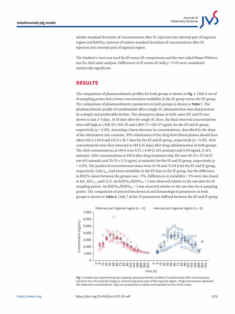
The comparison of pharmacokinetic profiles for both groups is shown in Fig. 1. Only 4 out of 14 sampling points had a lower concentration variability in the IF group versus the EF group. The comparison of pharmacokinetic parameters in both groups is shown in Table 1. The pharmacokinetic profile of ustekinumab after a single SC administration was characterized by a simple and predictable decline. The absorption phase in both cases (EF and IF) was shown to last 2–3 days. At 18 days after the single SC dose, the final observed concentrations were still high at 1,308.18 ± 201.20 and 1,850.73 ± 621.07 ng/mL for the EF and IF group, respectively (p > 0.05). Assuming a linear decrease in concentrations, described by the slope of the elimination rate constant, 99% elimination of the drug from blood plasma should have taken 102.6 ± 84.8 and 121.4 ± 36.7 days for the EF and IF group, respectively (p > 0.05). ADA concentrations were first observed at 144 h (6 days) after drug administration in both groups. The ADA concentrations at 144 h were 8.51 ± 4.49 (2 of 6 animals) and 0.04 ng/mL (1 of 6 animals). ADA concentrations at 432 h after drug treatment (day 18) were 40.19 ± 25.04 (5 out of 6 animals) and 20.55 ± 17.6 ng/mL (6 animals) for the EF and IF group, respectively (p > 0.05). The predicted seroconversion times were 10.96 and 73.04 h for the EF and IF group, respectively. Only tmax had lower variability in the EF than in the IF group, but the difference in RSD% values between the groups was < 5%. Differences in variability < 5% were also found in kel, AUC(0-t) and CL/F. An RSD%IF/RSD%EF > 1 was observed relative to the raw data for 10 sampling points. An RSD%IF/RSD%EF < 1 was observed relative to the raw data for 4 sampling points. The comparison of selected biochemical and hematological parameters in both groups is shown in Table 2. Only 7 of the 20 parameters differed between the EF and IF group
5/10https://vetsci.org https://doi.org/10.4142/jvs.2021.22.e47
Ustekinumab pig model
Conc
entr
atio
n (n
g/m
L)
Time (h)
External part inguinal region (n = 6) Internal part inguinal region (n = 6)
0
1,000
2,000
3,000
4,000
5,000
6,000
7,000
0 6 12 24 36 48 60 72 84 96 120
168
384
432
144
288 0 6 12 24 36 48 60 72 84 96 120
168
384
432
144
288
Fig. 1. Scatter plot representing two separate pharmacokinetic profiles of ustekinumab after subcutaneous injection into the external (rings) or internal (squares) part of the inguinal region. Rings and squares represent the observed concentrations. Data are presented as means and standard errors of the mean.

(p < 0.05). The physiological range of the measured parameters was exceeded only in the case of urea (EF and IF) and calcium levels (EF and IF) (Table 2). Differences in the variability of hematological and biochemical parameters between the groups were > 5% only in the case of MID (5.79%), LYM (6.09%) and cholesterol (6.20%). For these three parameters, the variability in the EF group was higher than in the IF group.
6/10https://vetsci.org https://doi.org/10.4142/jvs.2021.22.e47
Ustekinumab pig model
Table 1. Pharmacokinetic parameters of ustekinumab (arithmetic mean; standard deviation) after single subcutaneous administration 1 mg/kg BW into two different parts of the inguinal region of a pigParameter External part of inguinal region Internal part of inguinal region p value*ka (h−1) 0.141; 0.147 0.102; 0.103 > 0.05kel (h−1) 0.006; 0.004 0.003; 0.001 > 0.05t1/2kel (h) 246.254; 203.61 291.355; 88.047 > 0.05tmax (h) 40; 11.314 62.4; 20.646 0.03Cmax (ng/mL) 4,302.855; 982.453 4,329.768; 553.989 > 0.05AUC(0-t) (h×ng/mL) 524,279.414; 160,129.037 879,097.339; 235,094.091 0.00AUC(0-Cmax) (h×ng/mL) 962,451.95; 463,420.775 1,728,779.234; 697,387.859 > 0.05AUC(0-MRT) (h×ng/mL) 291,264.047; 89,329.841 433,319.142; 85,662.869 0.01AUMC(0-t) (h2×ng/mL) 61,272,689; 44,181,081.751 122,177,749.4; 38,626,379.669 0.02MRT(0-t) (h) 103.99; 43.513 149.176; 19.283 0.04Vz/F (mL/kg) 316.202; 118.027 256.702; 63.403 > 0.05CL/F (mL/h/kg) 1.267; 0.497 0.67; 0.233 0.01BW, body weight; ka, absorption rate constant; kel, elimination rate constant; t1/2kel, elimination half-life; tmax, time to reach Cmax; Cmax, maximal concentration; AUC(0-t), area under the curve calculated between zero and the last sampling point; AUC(0-Cmax), area under the curve calculated between zero and tmax; AUC(0-MRT), area under the curve calculated between zero and MRT(0-t); AUMC(0-t), area under the first moment curve calculated between zero and the last sampling point; MRT(0-t), mean residence time calculated for the last sampling point; Vz/F, apparent volume of distribution; CL/F, apparent clearance.*The p value representing the comparison of data between groups.
Table 2. Comparison of selected biochemical and hematological parameters (arithmetic mean; standard deviation) of pig blood after ustekinumab single subcutaneous administration 1 mg/kg BW into two different parts of the inguinal regionParameter External part of inguinal region Internal part of inguinal region p value*Cholesterol (mg/dL) 102.296; 16.594 103.855; 10.411 > 0.05Bilirubin (mg/dL) 0.017; 0.022 0.01; 0.012 > 0.05Urea (mg/dL) 30.021; 6.216↑ 27.532; 5.452↑ 0.00Creatinine (mg/dL) 1.025; 0.17 0.826; 0.14 0.00Calcium (mg/dL) 9.463; 1.423↓ 8.951; 1.269↓ > 0.05Phosphorus (mg/dL) 10.491; 1.184 11.255; 1.252 > 0.05WBC (×109/L) 22.836; 4.496 24.772; 4.097 0.01LYM (×109/L) 9.789; 1.819 10.09; 1.26 > 0.05MID (×109/L) 3.067; 0.775 3.343; 0.651 0.02GRAN (×109/L) 10.171; 2.922 11.419; 2.757 0.01RBC (×1012/L) 5.696; 0.489 5.599; 0.737 > 0.05HTC (%) 30.507; 2.871 29.284; 3.689 > 0.05HGB (g/dL) 10.098; 1.081 10.334; 1.453 > 0.05MCV (fL) 53.453; 1.941 53.266; 1.923 > 0.05MCH (pg) 17.759; 0.661 17.42; 0.832 > 0.05MCHC (g/dL) 33.236; 0.947 31.428; 1.125 0.00RDW (%) 20.441; 0.969 20.437; 1.404 > 0.05RDWa (fL) 36.892; 2.001 38.217; 2.684 0.03PLT (×109/L) 272.157; 93.548 274.355; 96.221 > 0.05MPV (fL) 7.372; 0.304 7.136; 0.241 > 0.05WBC, number of white blood cells; LYM, number of lymphocytes; MID, number of types of WBC not classified as lymphocytes or granulocytes; GRAN, number of granulocytes; RBC, number of red blood cells; HTC, hematocrit; HGB, hemoglobin; MCV, mean corpuscular volume of red blood cells; MCH, mean corpuscular hemoglobin in red blood cells; MCHC, mean corpuscular hemoglobin concentration; RDW, red cell distribution width; RDWa, absolute red cell distribution width; PLT, platelet count; MPV, mean platelet volume; ↑, ↓, mean value higher or lower than physiological range.*The p value representing the comparison of data between groups.

DISCUSSION
In the current literature, only a few studies have described the pharmacokinetic profiles of human mAbs in pigs after SC administration [6-9]. To date, published reports have described selected pharmacokinetic parameters for trastuzumab, anakinra, etanercept and adalimumab (in minipigs). The current study demonstrated the applicability of using a breeding pig model in ustekinumab preclinical studies. In general, the shape of the pharmacokinetic profile of ustekinumab did not deviate from observations made in other studies regarding trastuzumab, anakinra, etanercept and adalimumab, in minipigs. The pharmacokinetic profile of ustekinumab after a single SC dose showed a predictable and slow disposition.
As shown in other animal models, ADAs were also detected in the current study. In theory, the seroconversion time should have had a strong impact on the observed Cmax. However, in the current model, there was no impact on the Cmax value (p < 0.05) as a result of differences in the predicted seroconversion time (10.96 vs. 73.04 h). Here, it could be hypothesized that in the first two days after a single ustekinumab dose, the pig immune system had no influence on the drug pharmacokinetics. The differences in ka between the EF and IF group were not significant; however, the tmax between the groups was ultimately substantially different (p > 0.05). To conclude, the ka and Cmax up to tmax were the same in both groups and had no impact on drug bioavailability up to tmax; the differences in Cmax and AUC(0-Cmax) between the groups were not significant; and the relative bioavailability after EF administration of ustekinumab was 40.36% lower than after IF administration.
Large differences between the EF and IF group were provoked by a significant difference in CL/F. Only changes in CL/F led to differences between AUC(0-t) and AUC(0-MRT) (p < 0.05). In general, CL/F in the pig model was approximately 12 times faster than in humans [21]. The t1/2kel value in the current pig model was around 12 days, which is close to the lower limit in humans (duration from 15 to 32 days across various patient populations) [21]. However, it is noteworthy that Vz/F in the pig model was only approximately 2 times higher than in those with psoriasis [26].
The absorption process in the current model takes approximately 2 days, which is an advantage of the model. Such a time range allows accurate tracking of the absorption phase by means of an appropriate number of sampling points. The current study showed that differences in the administration site can strongly impact ustekinumab concentration variability and the variability of calculated pharmacokinetic parameters. The observations clearly indicate that the variability of ustekinumab concentration in the EF group is lower than in the IF group, especially in the absorption and early distribution phases within the first 3 days of administration. However, out of the entirety of pharmacokinetic parameters, only tmax variability was lower in the EF group. In the context of pig physiology, ustekinumab is a xenogenic molecule. Xenogenic Igs induce a humoral immune response, and immune complexes are involved in their rapid removal from the blood of the recipient [27]. The key mechanisms influencing the disposition of human mAbs in animal models involve numerous interactions with the immune system [18]. In the current study, it was shown that in the absorption phase (0-tmax) the interaction between ustekinumab and the pig immune system had no impact on pharmacokinetic parameters and did not generate any significant response in terms of ADA formation. There were no notable differences in the pharmacokinetic characteristics of ustekinumab between the EF and IF group in the absorption phase. This means that changing the administration site did not significantly affect the absorption of
7/10https://vetsci.org https://doi.org/10.4142/jvs.2021.22.e47
Ustekinumab pig model

the drug. However, differences in the administration site were shown to strongly impact the immune system response in the current study. This response is typically of a delayed nature [28]. Such a response was observed with regard to parameters extending beyond the absorption phase (MRT(0-t), AUC(0-t), AUC(0-MRT), AUMC(0-t), CL/F) and in differences between groups related to select hematological parameters.
Analysis of the hematological parameters showed that a single SC dose of ustekinumab at a 1 mg/kg body weight (BW) dose in pigs did not cause changes in excess of physiological limits. At the same time, it should be noted that the proposed model was sensitive enough to reveal an effect of administration site on select hematological parameters within their physiological ranges. Ustekinumab had no negative effects on renal function [29]. As such, it can be concluded that any changes in urea do not reflect an effect of ustekinumab on kidney physiology. In the case of LYM, MID, and GRAN, the single dose of ustekinumab used to treat the EF and IF groups induced different effects, with the IF group generating significantly higher values (p < 0.05). All three parameters remained within physiological values but proved that variations in injection site could generate different responses related to immunocompetent cells. It should be emphasized that in immunotoxicity studies, ustekinumab has been shown to have no impact on circulating lymphocyte subpopulations, which is in line with the findings of the current study [20]. The injection site impacted RDWa values in both groups. This finding could be valuable when considered in conjunction with recent results connecting cardiovascular disease risk in Crohn's disease with RDW and identification of RDW as a possible biomarker in psoriasis [30,31].
The proposed pig model is characterized by low inter-individual variability and the absence of a severe immune response. The pharmacokinetic profile is predictable without any fluctuations. The model allows for comparative studies regarding the analysis of the absorption process of ustekinumab after SC administration. In summary, the breeding pig can be proposed as a relevant and advantageous pharmacokinetic model to study SC-administered ustekinumab.
ACKNOWLEDGMENTS
The authors would like to express great appreciation to Neil Johnson, PhD for his professional guidance and valuable support in preparing the manuscript.
REFERENCES
1. Dalgaard L. Comparison of minipig, dog, monkey and human drug metabolism and disposition. J Pharmacol Toxicol Methods. 2015;74:80-92. PUBMED | CROSSREF
2. Ganderup NC. Chapter 3. Minipig models for toxicity testing and biomarkers. In: Gupta RC, editor. Biomarkers in Toxicology. 1st ed. Boston: Academic Press; 2014, 71-91.
3. Ganderup NC, Harvey W, Mortensen JT, Harrouk W. The minipig as nonrodent species in toxicology--where are we now? Int J Toxicol. 2012;31(6):507-528. PUBMED | CROSSREF
4. Burmańczuk A, Milczak A, Grabowski T, Osypiuk M, Kowalski C. The using of a piglets as a model for evaluating the dipyrone hematological effects. BMC Vet Res. 2016;12(1):263. PUBMED | CROSSREF
8/10https://vetsci.org https://doi.org/10.4142/jvs.2021.22.e47
Ustekinumab pig model

5. Helke KL, Nelson KN, Sargeant AM, Jacob B, McKeag S, Haruna J, et al. Pigs in toxicology: breed differences in metabolism and background findings. Toxicol Pathol. 2016;44(4):575-590. PUBMED | CROSSREF
6. van Mierlo GJ, Cnubben NH, Kuper CF, Wolthoorn J, van Meeteren-Kreikamp AP, Nagtegaal MM, et al. The Göttingen minipig® as an alternative non-rodent species for immunogenicity testing: a demonstrator study using the IL-1 receptor antagonist anakinra. J Immunotoxicol. 2013;10(1):96-105. PUBMED | CROSSREF
7. Bittner B, Richter WF, Hourcade-Potelleret F, McIntyre C, Herting F, Zepeda ML, et al. Development of a subcutaneous formulation for trastuzumab - nonclinical and clinical bridging approach to the approved intravenous dosing regimen. Arzneimittelforschung. 2012;62(9):401-409. PUBMED | CROSSREF
8. Harvey AJ, Kaestner SA, Sutter DE, Harvey NG, Mikszta JA, Pettis RJ. Microneedle-based intradermal delivery enables rapid lymphatic uptake and distribution of protein drugs. Pharm Res. 2011;28(1):107-116. PUBMED | CROSSREF
9. Zheng Y, Tesar DB, Benincosa L, Birnböck H, Boswell CA, Bumbaca D, et al. Minipig as a potential translatable model for monoclonal antibody pharmacokinetics after intravenous and subcutaneous administration. MAbs. 2012;4(2):243-255. PUBMED | CROSSREF
10. Gauthier BE, Penard L, Bordier NF, Briffaux JJ, Ruty BM. Specificities of the skin morphology in juvenile minipigs. Toxicol Pathol. 2018;46(7):821-834. PUBMED | CROSSREF
11. Stirling CM, Charleston B, Takamatsu H, Claypool S, Lencer W, Blumberg RS, et al. Characterization of the porcine neonatal Fc receptor--potential use for trans-epithelial protein delivery. Immunology. 2005;114(4):542-553. PUBMED | CROSSREF
12. U.S. Department of Health and Human Services; Food and Drug Administration; Center for Drug Evaluation and Research (CDER). Guidance for Industry: Nonclinical Safety Evaluation of Drug or Biologic Combinations. Rockville: Food and Drug Administration; 2006, 1-16.
13. U.S. Department of Health and Human Services; Food and Drug Administration; Center for Drug Evaluation and Research (CDER); Center for Biologics Evaluation and Research (CBER). Guidance for Industry: S6(R1) Preclinical Safety Evaluation of Biotechnology-Derived Pharmaceuticals. Geneva: International Council for Harmonisation of Technical Requirements for Pharmaceuticals for Human Use (ICH); 2011, 1-23.
14. Kagan L, Turner MR, Balu-Iyer SV, Mager DE. Subcutaneous absorption of monoclonal antibodies: role of dose, site of injection, and injection volume on rituximab pharmacokinetics in rats. Pharm Res. 2012;29(2):490-499. PUBMED | CROSSREF
15. McDonald TA, Zepeda ML, Tomlinson MJ, Bee WH, Ivens IA. Subcutaneous administration of biotherapeutics: current experience in animal models. Curr Opin Mol Ther. 2010;12(4):461-470.PUBMED
16. Kota J, Machavaram KK, McLennan DN, Edwards GA, Porter CJ, Charman SA. Lymphatic absorption of subcutaneously administered proteins: influence of different injection sites on the absorption of darbepoetin alfa using a sheep model. Drug Metab Dispos. 2007;35(12):2211-2217. PUBMED | CROSSREF
17. ter Braak EW, Woodworth JR, Bianchi R, Cerimele B, Erkelens DW, Thijssen JH, et al. Injection site effects on the pharmacokinetics and glucodynamics of insulin lispro and regular insulin. Diabetes Care. 1996;19(12):1437-1440. PUBMED | CROSSREF
18. Ryman JT, Meibohm B. Pharmacokinetics of monoclonal antibodies. CPT Pharmacometrics Syst Pharmacol. 2017;6(9):576-588. PUBMED | CROSSREF
19. Gradel AK, Porsgaard T, Lykkesfeldt J, Seested T, Gram-Nielsen S, Kristensen NR, et al. Factors affecting the absorption of subcutaneously administered insulin: effect on variability. J Diabetes Res. 2018;2018:1205121. PUBMED | CROSSREF
20. European Medicines Agency (EMA). Assessment Report for STELARA. London: European Medicines Agency (EMA); 2009, 1-58.
21. European Medicines Agency (EMA). Annex I. Summary of Product Characteristics. London: European Medicines Agency (EMA); 2018, 1-110.
9/10https://vetsci.org https://doi.org/10.4142/jvs.2021.22.e47
Ustekinumab pig model

22. Varkhede N, Forrest ML. Understanding the Monoclonal Antibody Disposition after Subcutaneous Administration using a Minimal Physiologically based Pharmacokinetic Model. J Pharm Pharm Sci. 2018;21(1s):130s-148s. PUBMED | CROSSREF
23. Ito R, Suami H. Lymphatic territories (lymphosomes) in swine: an animal model for future lymphatic research. Plast Reconstr Surg. 2015;136(2):297-304. PUBMED | CROSSREF
24. Stec J, Bicka L, Kuźmak J. Isolation and purification of polyclonal IgG antibodies from bovine serum by high performance liquid chromatography. Bull Vet Inst Pulawy. 2004;48(3):321-327.
25. Tijssen P, Kurstak E. Highly efficient and simple methods for the preparation of peroxidase and active peroxidase-antibody conjugates for enzyme immunoassays. Anal Biochem. 1984;136(2):451-457. PUBMED | CROSSREF
26. Food and Drug Administration. Ustekinumab Label. Rockville: Food and Drug Administration; 2014, 1-25.
27. Gąsowska A, Stefaniak T. Ocena efektów doustnego podania immunoglobuliny żółtka jaja (IgY) cielętom w okresie wchłaniania makromolekuł z jelita. Folia Univ Agric Stetin. 2003;233(45):87-92.
28. Shankar G, Arkin S, Cocea L, Devanarayan V, Kirshner S, Kromminga A, et al. Assessment and reporting of the clinical immunogenicity of therapeutic proteins and peptides-harmonized terminology and tactical recommendations. AAPS J. 2014;16(4):658-673. PUBMED | CROSSREF
29. Nimmannitya K, Tateishi C, Mizukami Y, Hamamoto K, Yamada S, Goto H, et al. Successful treatment with ustekinumab of psoriasis vulgaris in a patient undergoing hemodialysis. J Dermatol. 2016;43(1):92-94. PUBMED | CROSSREF
30. Doğan S, Atakan N. Red blood cell distribution width is a reliable marker of inflammation in plaque psoriasis. Acta Dermatovenerol Croat. 2017;25(1):26-31.PUBMED
31. Al Taii H, Yaqoob Z, Al-Kindi SG. Red cell distribution width (RDW) is associated with cardiovascular disease risk in Crohn's disease. Clin Res Hepatol Gastroenterol. 2017;41(4):490-492. PUBMED | CROSSREF
10/10https://vetsci.org https://doi.org/10.4142/jvs.2021.22.e47
Ustekinumab pig model