pharmacological amelioration of cone survival and vision ... · (samardzija et al., 2008) and rd12...
TRANSCRIPT

Neurobiology of Disease
Pharmacological Amelioration of Cone Survival and Visionin a Mouse Model for Leber Congenital Amaurosis
Songhua Li,1 Marijana Samardzija,2 Zhihui Yang,1,3 X Christian Grimm,2 and Minghao Jin1
1Department of Ophthalmology and Neuroscience Center, Louisiana State University School of Medicine, New Orleans, Louisiana 70112, and 2Departmentof Ophthalmology, University of Zurich, CH-8952 Schlieren, Switzerland
RPE65, an abundant membrane-associate protein in the retinal pigment epithelium (RPE), is a key retinoid isomerase of the visual cyclenecessary for generating 11-cis-retinal that functions not only as a molecular switch for activating cone and rod visual pigments inresponse to light stimulation, but also as a chaperone for normal trafficking of cone opsins to the outer segments. Many mutations inRPE65 are associated with Leber congenital amaurosis (LCA). A R91W substitution, the most frequent LCA-associated mutation, resultsin a severe decrease in protein level and enzymatic activity of RPE65, causing cone opsin mislocalization and early cone degeneration inthe mutation knock-in mouse model of LCA. Here we show that R91W RPE65 undergoes ubiquitination-dependent proteasomal degra-dation in the knock-in mouse RPE due to misfolding. The 26S proteasome non-ATPase regulatory subunit 13 mediated degradationspecifically of misfolded R91W RPE65. The mutation disrupted membrane-association and colocalization of RPE65 with lecithin:retinolacyltransferase (LRAT) that provides the hydrophobic substrate for RPE65. Systemic administration of sodium 4-phenylbutyrate (PBA),a chemical chaperone, increased protein stability, enzymatic activity, membrane-association, and colocalization of R91W RPE65 withLRAT. This rescue effect increased synthesis of 11-cis-retinal and 9-cis-retinal, a functional iso-chromophore of the visual pigments, ledto alleviation of S-opsin mislocalization and cone degeneration in the knock-in mice. Importantly, PBA-treatment also improved cone-mediated vision in the mutant mice. These results indicate that PBA, a U.S. Food and Drug Administration-approved safe oral medica-tion, may provide a noninvasive therapeutic intervention that delays daylight vision loss in patients with RPE65 mutations.
Key words: chemical chaperone; cone photoreceptor; Leber congenital amaurosis; retina; retinoid visual cycle; Rpe65
IntroductionVisual signal starts with photoisomerization of 11-cis-retinal toall-trans-retinal in the visual pigments of cone and rod photore-
ceptors. To sustain vision, 11-cis-retinal must be regenerated.RPE65, a retinal pigment epithelium (RPE)-specific microsomalprotein (Hamel et al., 1993), is a key retinoid isomerase in thevisual cycle necessary for regenerating 11-cis-retinal (Jin et al.,2005; Moiseyev et al., 2005; Redmond et al., 2005). RPE65 usesall-trans-retinyl esters synthesized by LRAT (Ruiz et al., 1999)and other enzymes (Kaschula et al., 2006; Kaylor et al., 2015) as itssubstrate (Moiseyev et al., 2003). Mice lacking RPE65 cannotsynthesize 11-cis-retinoids, thereby their photoreceptors almostcompletely lose light-sensitivity (Redmond et al., 1998; Pang et
Received Oct. 11, 2015; revised April 17, 2016; accepted April 20, 2016.Author contributions: S.L. and M.J. designed research; S.L., Z.Y., and M.J. performed research; M.S. and C.G.
contributed unpublished reagents/analytic tools; S.L. and M.J. analyzed data; S.L. and M.J. wrote the paper.This work was supported by grants from NIH (EY021208 to M. J. and GM103340 to LSU Neuroscience COBRE
facilities), RPB, and Lions Eye Foundation (to LSU Department of Ophthalmology). We thank Drs Marcin Golczak andKrzysztof Palczewski for the antibody against LRAT.
Correspondence should be addressed to Dr Minghao Jin, Department of Ophthalmology and Neuroscience Cen-ter, Louisiana State University School of Medicine, New Orleans, LA 70112. E-mail: [email protected].
The authors declare no competing financial interests.Z. Yang’s present address: Department of Psychiatry and Neuroscience, University of Florida, Gainesville, FL
32611.DOI:10.1523/JNEUROSCI.3857-15.2016
Copyright © 2016 the authors 0270-6474/16/365808-12$15.00/0
Significance Statement
LCA is a severe early onset retinal dystrophy. Recent clinical trials of gene therapy have implicated the need of an alternative orcombination therapy to improve cone survival and function in patients with LCA caused by RPE65 mutations. Using a mousemodel carrying the most frequent LCA-associated mutation (R91W), we found that the mutant RPE65 underwent ubiquitination-dependent proteasomal degradation due to misfolding. Treatment of the mice with a chemical chaperone partially correctedstability, enzymatic activity, and subcellular localization of R91W RPE65, which was also accompanied by improvement of conesurvival and vision. These findings identify an in vivo molecular pathogenic mechanism for R91W mutation and provide a feasiblepharmacological approach that can delay vision loss in patients with RPE65 mutations.
5808 • The Journal of Neuroscience, May 25, 2016 • 36(21):5808 –5819

al., 2005). Rpe65�/� mice also exhibit cone opsin mislocalizationand early cone degeneration (Rohrer et al., 2005; Znoiko et al.,2005; Fan et al., 2008).
The significance of RPE65 function in vision and retinalhealth is also reflected by the facts that many mutations in RPE65cause Leber congenital amaurosis (LCA) or retinitis pigmentosa(RP; Gu et al., 1997; Marlhens et al., 1997; Lotery et al., 2000;Thompson et al., 2000). RPE65 mutations are estimated to ac-count for �16% of LCA and 2% of recessive RP (RetNet). Al-though night blindness is the first significant symptom in mostpatients with RPE65 mutations (Felius et al., 2002), in vivo mi-croscopy of the fovea demonstrated that many patients exhibitedsevere cone photoreceptor loss at very early ages (Jacobson et al.,2005, 2009). The importance of RPE65 for the central vision isalso supported by its abundant expression and higher retinoidisomerase activity in the macaque central RPE layer localized tothe cone-rich area (Jacobson et al., 2007).
Recent clinical trials of gene therapy that express normalRPE65 in patients’ RPE have shown some improvement of visionin some patients (Bainbridge et al., 2008; Cideciyan et al., 2008;Hauswirth et al., 2008; Maguire et al., 2008). Subsequent studies,however, showed that gene therapy could not stop the progres-sive retinal degeneration and vision loss (Cideciyan et al., 2013;Bainbridge et al., 2015). The rate of loss of photoreceptors in thetreated retinas was the same as that in the untreated retina (Ja-cobson et al., 2015). In addition, topographic maps of visual sen-sitivity in the treated region indicated that the areas of improvedvision had progressive diminution (Jacobson et al., 2015). It istherefore urgently needed to develop an alternative therapy thatalleviates cone degeneration or enhances the efficacy of gene ther-apy in patients with RPE65 mutations.
A R91W substitution in RPE65 is one of the most frequentmutations causing LCA (Morimura et al., 1998; Thompson et al.,2000). Although patients with this mutation had useful vision inthe first decade of life, optical coherence tomography demon-strated that 3- and 7-year-old patients already had cone degener-ation (Jacobson et al., 2007, 2008). Similarly, a mouse modelcarrying R91W mutation displayed early cone degeneration (Sa-mardzija et al., 2009), due to shortage of 11-cis-retinal supply(Samardzija et al., 2008). RPE65 in the R91W mouse was de-creased to �5% of wild-type RPE65 level by an unknown mech-anism (Samardzija et al., 2008). In vitro and animal studiesshowed that not only R91W mutation but also many other mu-tations resulted in a drastic decrease of RPE65 (Chen et al., 2006;Takahashi et al., 2006; Bereta et al., 2008; Philp et al., 2009; Niko-laeva et al., 2010; Wright et al., 2013; S. Li et al., 2014, 2015). Invivo identification of the underlying molecular mechanism of thisphenotype is important not only for understanding the diseasemechanisms of RPE65 mutations but also for developing a ther-apeutic intervention that prevents or delays loss of RPE65 func-tion in patients.
In this study, we analyzed the molecular basis and pathwayleading to degradation of RPE65 in the R91W knock-in (KI)mouse RPE and tested whether a chemical chaperone-mediatedprotein repair approach can alleviate cone degeneration and vi-sion loss in the knock-in mouse model of LCA.
Materials and MethodsAnimals. Wild-type 129S2/Sv (Charles River Laboratories), R91W KI(Samardzija et al., 2008) and rd12 (The Jackson Laboratory; Pang et al.,2005) mice were maintained in 12 h cyclic light at �30 lux. RPE65 inthese mice has a leucine residue at position 450 (Leu450). All animalexperiments followed the ARVO statement for the use of animals in
ophthalmic and vision research and the protocols approved by the Insti-tutional Animal Care and Use Committee. Except where noted, animalsof either sex were daily injected intraperitoneally with 50 mg sodium4-phenylbutyrate (PBA) in saline per kilogram body weight for 3 or 8weeks, beginning on postnatal day (P)14. Although PBA is a FDA-approved oral medication, we performed intraperitoneal injection tostrictly control the amounts of PBA treated. Mice intraperitoneally in-jected with the same volume of saline were used as controls.
Eyecup ex vivo experiments. After removal of the anterior section andneural retina, the RPE in mouse eyecups were maintained in DMEM-F12medium (Thermo Fisher Scientific) supplemented with 10% fetal bovineserum, 100 U/ml penicillin G, and 100 �g/ml streptomycin at 37°C or30°C �5% CO2. Transfection of plasmid DNA (pcDNA, pEGFP, pRK5,and pPSMD13) and siRNA (PSMD13 siRNA and scrambled negativecontrol siRNA from OriGene Technologies) was performed using thePolyJet transfection reagent (SignaGen Laboratories) according to themanufacturer’s procedure. Forty hours post-transfection, the eyecupswere subjected to immunoblot analysis, immunoprecipitation or micro-scopic analysis. For proteasome and lysosome inhibitor experiments,mouse eyecups were incubated with increasing concentrations of MG132(15–25 �M), pepstatin A (10 –30 �M) or DMSO for 5 h.
Electroretinography. Overnight dark-adapted 10-week-old mice tre-ated with PBA or saline were anesthetized with intraperitoneal keta-mine and xylazine. The pupils were dilated with 1% tropicamide.Electroretinography (ERG) was recorded from the corneal surface usinga silver-silver chloride wire electrode referenced to a subcutaneous elec-trode in the mouth. A needle electrode in the tail served as the ground. Adrop of 2% methylcellulose was placed on the cornea to prevent cornealdesiccation. Animals were light adapted for 10 min by exposing to a white32 cd/m 2 light, and photopic ERG responses were obtained with whiteflashes (�1 to 2.4 log cd � s/m 2) on the rod-saturating background (32cd/m 2). Five responses to 10 s interval flashes were averaged for eachstep. Intensity-response amplitude data were displayed on log-linear co-ordinates using the SigmaPlot 11 software. For recording S-cone ERG,animals were light adapted for 10 min by exposing to a white 40 cd/m 2
light, and S-cone ERG responses were obtained on the 40 cd/m 2 back-ground with xenon flashes (0.1–1.4 log cd � s/m 2) equipped with a HoyaU-360 filter (360 nm peak, Edmund Optics; Oh et al., 2008).
Immunoblot analysis. Protein samples in the Laemmli sample buffercontaining 50 mM dithiothreitol were heated for 10 min at 70°C, sepa-rated by SDS-PAGE in a 10%, 12% or 4 –12% gradient polyacrylamidegel, and transferred to an Immobilon-P membrane (EMD Millipore).The membrane was incubated in blocking buffer, primary antibody, andhorseradish peroxidase-conjugated anti-rabbit or mouse IgG secondaryantibody. Antibodies against RPE65 (Abcam and EMD Millipore; Jin etal., 2007), PSMD13 (Proteintech), PSMD11 (Abnova), LRAT (Batten etal., 2004; Golczak et al., 2005), ubiquitin (Enzo Life Sciences), S-opsin(Santa Cruz Biotechnology), M-opsin (EMD Millipore), cone arrestin(EMD Millipore), cadherin-1 (Santa Cruz Biotechnology), or �-actin(Sigma-Aldrich) were used as the primary antibodies. Immunoblotswere visualized with the enhanced ECL-Prime and quantified as de-scribed previously (S. Li et al., 2013b).
Immunoprecipitation. RPE cells in mouse eyecups were lysed in a lysisbuffer (S. Li et al., 2013b) containing protease-inhibitor mixture (RocheApplied Science), 10 mM N-ethylmaleimide, and 100 �M PR619. Aftercentrifugation for 5 min at 1000 � g, the supernatants were incubatedwith an anti-RPE65 monoclonal antibody bound to the GammaBind GSepharose (GE Healthcare). Because protein levels of R91W RPE65 in theKI mice are �10% of RPE65 levels in WT mice (Samardzija et al., 2008),we used 200 �g of WT and 2 mg of KI mice RPE proteins for immuno-precipitation. The beads were washed three times with the lysis buffercontaining the inhibitors of proteases and deubiquitylating enzymes.Precipitated proteins were dissolved in Laemmli buffer and heated at70°C for 10 min for immunoblot analysis.
Preparation of mouse RPE membrane pellets. After removal of the an-terior section and neural retina, RPE in mouse eyecups were homoge-nized in ice-cold 10 mM HEPES buffer, pH 7.4, containing 280 mM
sucrose, 10 mM MgCl2, and protease-inhibitor mixture. This homoge-nate was used as total RPE cell lysate. A portion of each homogenate was
Li et al. • Delayed Cone Vision Loss in a Mouse Model of LCA J. Neurosci., May 25, 2016 • 36(21):5808 –5819 • 5809

centrifuged for 10 min at 1000 � g. The supernatants were collected andcentrifuged for 1 h at 100,000 � g. The resulting ultra-pellets were rinsedin the same buffer one time. The total cell lysates and ultra-pellets weresubjected to immunoblot analysis.
Proteasomal activity measurements. Chymotrypsin-like and trypsin-like proteolytic activities of proteasomes were measured with the fluoro-genic 7-Amino-4-methylcoumarin-conjugated peptide substrates asdescribed by Lobanova et al. (2013).
Retinoid isomerase assay. Mice eyecups without the neural retina wereirradiated for 10 min on ice with 365 nm light from a Spectroline ModelEN-140L ultraviolet light source to destroy endogenous retinoids. RPEhomogenates in 20 mM HEPES buffer were prepared from the eyecups.Each assay mixture contained 500 �g cell homogenate, 10 �M all-trans-retinol, and 6% bovine serum albumin. After incubating for 2 h in dark-ness at 37°C, retinoids were extracted with hexane and were analyzed byHPLC, as described below.
Analysis of retinoids. Retinoids in eyecups of overnight dark-adaptedmice were extracted with hexane as described previously and analyzed bynormal-phase HPLC (Jin et al., 2009). In brief, retinoids in hexane ex-tractions were evaporated, dissolved in 100 �l of hexane, and separatedon a silica column (Zorbax-Sil 5 �m, 250 � 4.6 mm, Agilent Technolo-gies) by gradient (0.2–10% dioxane in hexane at 2.0 ml/min flow rate) ornongradient (10% dioxane in hexane at 1.0 ml/min flow rate) elution ofmobile phase on an Agilent 1100 HPLC system equipped with a photo-diode array detector (Agilent Technologies). Spectral data were acquiredfor all eluted peaks. Quantitation was performed by comparison of peakareas to calibration curves established with authentic retinoid standards.
Immunohistochemistry. Mouse retinal cryosections were prepared asdescribed previously (Sato et al., 2013). Briefly, mouse eyeballs were fixedovernight with 4% paraformaldehyde in 0.1 M phosphate buffer (PB).After removing cornea and lens, eyecups were immersed in 15% sucrosein 0.1 M PB for 2 h, in 30% sucrose in 0.1 M PB for 2 h, and then in a 1:1mixture of 30% sucrose and Optimal Cutting Temperature (OCT) me-dium (Sakura Finetechnical) overnight at 4°C. After embedding eyecupsin OCT, 15-�m-thick sections were cut on a Shandon Cryotome SMEcryostat (Thermo Scientific). The sections were immunostained with theprimary antibodies listed in the method of immunoblot analysis andsecondary antibodies, as described previously (S. Li et al., 2013a). Nucleiwere labeled with DAPI (Sigma-Aldrich). Images were captured with aZeiss LSM710 Meta confocal microscope with a 40� oil-immersionobjective.
Quantitation of cone arrestin-positive cone and M-cone numbers. Reti-nal sections of 10-week-old mice were immunostained for cone arrestin(CAR) and M-opsin, as described above. Numbers of CAR-positive outersegments and middle-wavelength cones (M-cones) in whole retinal sec-tions were counted using an Olympus BX61VS microscope equippedwith VS-ASW FL software (Sato et al., 2013).
Quantification of S-opsin mislocalization. Immunostaining of S-opsinwas performed on retinal cryosections of 5-week-old WT and KI mice, asdescribed above. Fluorescence intensities in the outer segments (OS) andouter plexiform layer (OPL) were measured using the Olympus BX61VSmicroscope. We determined the fraction of mislocalized S-opsin accord-ing to the following formula. Mislocalization � [OPL fluorescence/(OSfluorescence � OPL fluorescence)] � 100%.
Statistics. Data were expressed as the mean � SD of three or moreindependent experiments. Differences between test and control groupswere determined with an unpaired two-tailed Student’s t test, using Sig-maPlot v11. P values �0.05 were considered statistically significant.
ResultsUbiquitination-dependent proteasomal degradation of R91WRPE65 in RPER91W mutation results in dramatic decrease of RPE65 in theR91W KI mouse RPE (Samardzija et al., 2008). To identify thepathway causing reduction of RPE65, we treated RPE in WT andKI eyecups with different concentrations of MG132 (a protea-some inhibitor) or pepstatin A (a lysosome inhibitor). Immuno-blot analysis of the eyecups showed that protein levels of R91WRPE65 were increased at least twofold in MG132-treated, but notpepstatin A-treated, eyecups (Fig. 1A). This result suggests thatthe proteasome plays a critical role in degrading the mutantRPE65. Because ubiquitination is critical for protein degradationin the proteasomes, we tested whether R91W RPE65 is ubiquiti-nated in the RPE. Under conditions that inhibit protein deubi-quitination, we performed immunoprecipitation of RPE65. Wethen analyzed the immunoprecipitates by immunoblot analysisusing antibodies against ubiquitin or RPE65. As shown in Figure1B, R91W RPE65 was strongly polyubiquitinated in the KI mouseRPE. MG132-treatment of the KI eyecup further increased thesignal intensities of the ubiquitinated R91W RPE65 (Fig. 1B). Weobserved very weak signals of ubiquitinated wild-type RPE65 un-der the same experimental conditions (Fig. 1B).
PSMD13 promoted degradation of misfolded RPE65 in RPEWe recently showed that the 26S proteasome non-ATPase regu-latory subunit 13 (PSMD13) promotes degradation of severalmutant RPE65s in culture cells (S. Li et al., 2014, 2015). To testwhether PSMD13 has the same function in vivo, we transfectedPSMD13-specific siRNA (100 � 200 nM) or control siRNA (200nM) into RPE cells in WT and KI eyecups. Immunoblot analysis
Figure 1. Ubiquitination-dependent proteasomal degradation of R91W RPE65 in mouse RPE. A, Immunoblot analysis of RPE65 and LRAT in WT and R91W, KI mice eyecups treated with DMSO orthe indicated concentrations of MG132 or pepstatin A. Beta-actin served as a loading control. Relative intensities of RPE65 immunoblots were quantified, normalized by the intensities of LRAT, andexpressed in the histograms as percentage of RPE65 in DMSO-treated eyecups. Asterisks indicate significant differences between DMSO- and MG132-treated groups (*p � 0.01, **p � 0.005). Errorbars show SD (n � 4). B, RPE65 in WT and KI eyecups treated with 20 �M MG132 or DMSO was immunoprecipitated, and the immunoprecipitates were probed with antibodies against ubiquitin (Ub)or RPE65. Polyubiquitinated RPE65 (Ub-RPE65) and monomeric RPE65 (arrowhead) are indicated.
5810 • J. Neurosci., May 25, 2016 • 36(21):5808 –5819 Li et al. • Delayed Cone Vision Loss in a Mouse Model of LCA

showed that R91W RPE65 was increased as PSMD13 decrease ineyecups transfected with PSMD13 siRNA (Fig. 2A–C). In agree-ment with this result, chymotrypsin-like proteolytic activity ofthe proteasomes was decreased in PSMD13 siRNA-transfectedWT and KI eyecups compared with control siRNA-transfectedeyecups (Fig. 2E). Transfection efficiency test showed that 40 �50% RPE cells were positive for EGFP in EGFP-transfected eye-cups whereas mock vector (pcDNA)-transfected eyecups had noEGFP-positive cells under the same experimental conditions(Fig. 2D). To confirm these results, we transfected pPSMD13 orpRK5 mock vector into RPE cells in WT and KI mice eyecups. Asshown in Figure 2F–H, R91W RPE65 was markedly decreasedin RPE overexpressing PSMD13. Both chymotrypsin-like andtrypsin-like proteolytic activities were increased 35 � 40% in
PSMD13-transfected KI eyecups compared with pRK5-trans-fected KI eyecups (Fig. 2I).
Mutation in the yeast Rpn9, a PSMD13 homolog, results in ac-cumulation of ubiquitinated proteins (Takeuchi et al., 1999). Wetherefore tested whether PSMD13 promotes degradation of R91WRPE65 via the ubiquitination-dependent pathway. We performedimmunoprecipitation of RPE65 in WT and KI eyecups transfectedwith PSMD13 siRNA or control siRNA. Immunoblot analysis of theimmunoprecipitates showed that ubiquitinated R91W RPE65 wasincreased in PSMD13 siRNA-transfected KI eyecups (Fig. 2J), sug-gesting that PSMD13 promoted degradation of ubiquitinated mu-tant RPE65 that might be misfolded.
To test whether PSMD13 promotes degradation of misfoldedRPE65 in vivo, we overexpressed PSMD13 in RPE of WT and KI
Figure 2. PSMD13-mediated ubiquitination-dependent degradation of misfolded R91W RPE65. A, Immunoblot analysis of RPE65 and PSMD13 in RPE of WT and KI eyecups transfected with theindicated amounts of PSMD13 siRNA (siPSMD13) or negative control siRNA (siCont, 200 nM). The 26S proteasome non-ATPase regulatory subunit 11 (PSMD11) and LRAT were detected as internalmaker for the 19S cap of the proteasome and RPE, respectively. B, C, Relative immunoblot intensities of RPE65 and PSMD13 normalized to LRAT were expressed as fold of RPE65 (B) or percentageof PSMD13 (C) in eyecups transfected with control siRNA. D, Confocal microscopic images of RPE cells in 129S2/Sv mice eyecups transfected with pcDNA or pcDNA encoding EGFP. Nuclei were stainedwith DAPI. E, Chymotrypsin-like and trypsin-like activities of the proteasomes in RPE of WT and KI mice eyecups transfected with 200 nM PSMD13 siRNA or control siRNA. F, Immunoblot analysis ofRPE65, PSMD13, and the indicated proteins in WT and KI eyecups transfected with pRK5 or pRK5 encoding PSMD13. G, H, Relative immunoblot intensities of RPE65 and PSMD13 normalized to LRATin F. I, Chymotrypsin-like and trypsin-like activities of the proteasomes in RPE of WT and KI mice eyecups transfected with pRK5 or PSMD13 construct (pPSMD13). J, RPE65 in WT and KI eyecupstransfected with siPSMD13 or siCont was immunoprecipitated and probed with an Ub antibody. K, Immunoblot analysis of RPE65 and PSMD13 in RPE of WT and KI eyecups maintained at 37°C or 30°Cafter transfecting pRK5 or pPSMD13. L, Relative immunoblot intensities of RPE65 and PSMD13 (bottom) in the KI mice eyecups in K. Asterisks indicate statistically significant differences between testand control groups (*p � 0.05, **p � 0.005). Error bars indicate SD (n � 4).
Li et al. • Delayed Cone Vision Loss in a Mouse Model of LCA J. Neurosci., May 25, 2016 • 36(21):5808 –5819 • 5811
eyecups at 37°C and low temperature, which has been shown tohelp proper folding of mutant proteins (Denning et al., 1992;Vollrath and Liu, 2006; S. Li et al., 2013b). Immunoblot analysisshowed that the full-length R91W RPE65 was further decreasedin the PSMD13-overexpressing KI eyecups at 37°C comparedwith control (pRK5-transfected) KI eyecups (Fig. 2K,L). In con-trast, cleaved fragments from the mutant RPE65, but not fromwild-type RPE65, were increased in the PSMD13-overexpressingeyecups at 37°C (Fig. 2K). At 30°C, however, the full-lengthR91W RPE65 was drastically increased in both KI eyecups over-expressing and not overexpressing PSMD13 (Fig. 2K,L). Theseresults are consistent with our previous studies showing that lowtemperature rescues membrane-association and the isomeraseactivity of several disease-causing RPE65 mutants, includingR91W RPE65, in culture cells (S. Li et al., 2014; 2015; Jin et al.,2016), and suggest that PSMD13 preferentially promoted degra-dation of misfolded mutant RPE65.
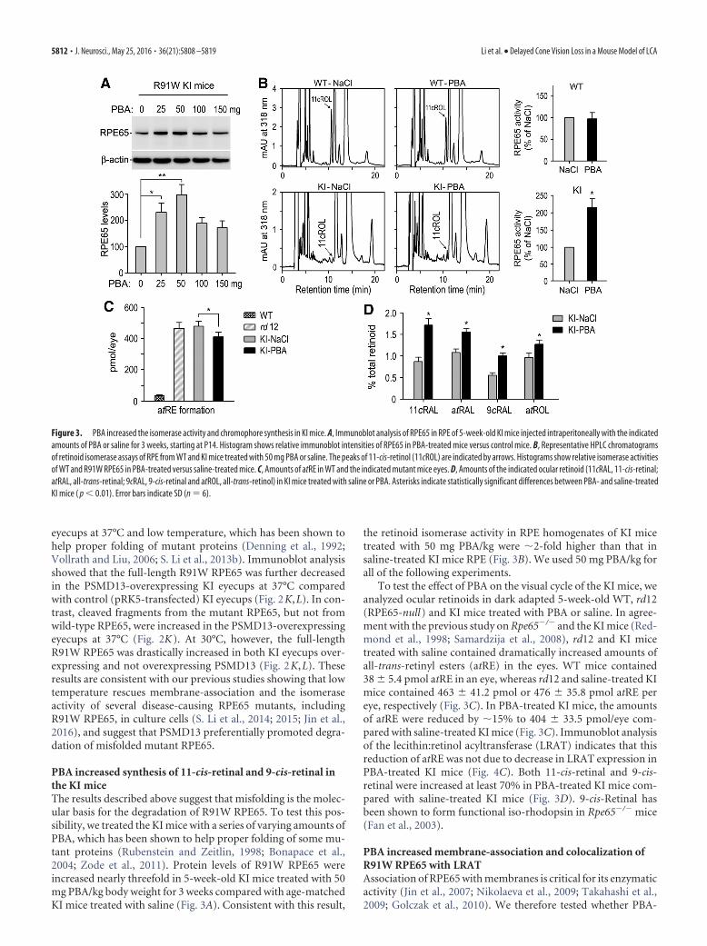
PBA increased synthesis of 11-cis-retinal and 9-cis-retinal inthe KI miceThe results described above suggest that misfolding is the molec-ular basis for the degradation of R91W RPE65. To test this pos-sibility, we treated the KI mice with a series of varying amounts ofPBA, which has been shown to help proper folding of some mu-tant proteins (Rubenstein and Zeitlin, 1998; Bonapace et al.,2004; Zode et al., 2011). Protein levels of R91W RPE65 wereincreased nearly threefold in 5-week-old KI mice treated with 50mg PBA/kg body weight for 3 weeks compared with age-matchedKI mice treated with saline (Fig. 3A). Consistent with this result,
the retinoid isomerase activity in RPE homogenates of KI micetreated with 50 mg PBA/kg were �2-fold higher than that insaline-treated KI mice RPE (Fig. 3B). We used 50 mg PBA/kg forall of the following experiments.
To test the effect of PBA on the visual cycle of the KI mice, weanalyzed ocular retinoids in dark adapted 5-week-old WT, rd12(RPE65-null) and KI mice treated with PBA or saline. In agree-ment with the previous study on Rpe65�/� and the KI mice (Red-mond et al., 1998; Samardzija et al., 2008), rd12 and KI micetreated with saline contained dramatically increased amounts ofall-trans-retinyl esters (atRE) in the eyes. WT mice contained38 � 5.4 pmol atRE in an eye, whereas rd12 and saline-treated KImice contained 463 � 41.2 pmol or 476 � 35.8 pmol atRE pereye, respectively (Fig. 3C). In PBA-treated KI mice, the amountsof atRE were reduced by �15% to 404 � 33.5 pmol/eye com-pared with saline-treated KI mice (Fig. 3C). Immunoblot analysisof the lecithin:retinol acyltransferase (LRAT) indicates that thisreduction of atRE was not due to decrease in LRAT expression inPBA-treated KI mice (Fig. 4C). Both 11-cis-retinal and 9-cis-retinal were increased at least 70% in PBA-treated KI mice com-pared with saline-treated KI mice (Fig. 3D). 9-cis-Retinal hasbeen shown to form functional iso-rhodopsin in Rpe65�/� mice(Fan et al., 2003).
PBA increased membrane-association and colocalization ofR91W RPE65 with LRATAssociation of RPE65 with membranes is critical for its enzymaticactivity (Jin et al., 2007; Nikolaeva et al., 2009; Takahashi et al.,2009; Golczak et al., 2010). We therefore tested whether PBA-
Figure 3. PBA increased the isomerase activity and chromophore synthesis in KI mice. A, Immunoblot analysis of RPE65 in RPE of 5-week-old KI mice injected intraperitoneally with the indicatedamounts of PBA or saline for 3 weeks, starting at P14. Histogram shows relative immunoblot intensities of RPE65 in PBA-treated mice versus control mice. B, Representative HPLC chromatogramsof retinoid isomerase assays of RPE from WT and KI mice treated with 50 mg PBA or saline. The peaks of 11-cis-retinol (11cROL) are indicated by arrows. Histograms show relative isomerase activitiesof WT and R91W RPE65 in PBA-treated versus saline-treated mice. C, Amounts of atRE in WT and the indicated mutant mice eyes. D, Amounts of the indicated ocular retinoid (11cRAL, 11-cis-retinal;atRAL, all-trans-retinal; 9cRAL, 9-cis-retinal and atROL, all-trans-retinol) in KI mice treated with saline or PBA. Asterisks indicate statistically significant differences between PBA- and saline-treatedKI mice ( p � 0.01). Error bars indicate SD (n � 6).
5812 • J. Neurosci., May 25, 2016 • 36(21):5808 –5819 Li et al. • Delayed Cone Vision Loss in a Mouse Model of LCA

treatment enhanced association of R91W RPE65s with mem-branes. We prepared membrane fractions from RPE of 10-week-old WT and KI mice treated with PBA or saline for 8 weeks.Immunoblot analysis of RPE65 in the membrane fractionsshowed that PBA-treatment increased membrane-association ofR91W RPE65 at least 3.5-fold, but had no influence on wild-typeRPE65 (Fig. 4A).
We then tested whether PBA-treatment promoted colocaliza-tion of R91W RPE65 with LRAT that synthesizes the hydropho-bic substrate of RPE65. As expected, immunohistochemistryshowed that the majority of RPE65 and LRAT proteins were co-localized and distributed throughout the RPE cells in WT retinas(Fig. 4B, top). In saline-treated KI mice, R91W RPE65 mainlylocalized to the basal side of the RPE and formed aggresome-likeinclusion bodies in close proximity to the nuclei of RPE cells (Fig.4B, middle). This distribution pattern was completely differentfrom that of LRAT in the same retinas (Fig. 4B, middle). In PBA-treated KI mice, however, many R91W mutant proteins distrib-uted to cytosol area and colocalized with LRAT (Fig. 4B, bottom).PBA-treatment for 8 weeks increased protein level of R91WRPE65, but not LRAT (Fig. 4C).
PBA reduced mislocalization of S-opsin in the KI miceBecause S-opsin is mislocalized in the KI mice (Samardzija et al.,2009), we tested whether PBA can prevent or reduce mislocaliza-tion of S-opsin in 5-week-old KI mice treated with PBA or salinefor 3 weeks. Immunohistochemistry showed that S-opsin wasmainly confined to the cone OS in WT retina (Fig. 5A). In KI micetreated with saline, a greater fraction of S-opsin immunoreactiv-ity was present in the OPL and some cell bodies of cones (Fig. 5A).In the PBA-treated KI mice, however, S-opsin immunoreactivity
was largely reduced in the OPL but increased in the OS (Fig. 5A).By measuring fluorescence intensity in the OS and OPL, we esti-mated the fraction of S-opsin mislocalization in WT mice, saline-treated, and PBA-treated KI mice. The fractional mislocalizationof S-opsin was reduced �50% in PBA-treated KI mice comparedwith saline-treated KI mice (Fig. 5B).
If S-opsin mislocalization is one of the pathogenic factorsleading to S-cone degeneration in the KI mice, the decrease inS-opsin mislocalization should be accompanied by increase ofS-opsin in the PBA-treated KI mice. To test this possibility, weperformed quantitative immunoblot analysis for S-opsin. Asshown in Figure 5C, S-opsin was increased �2-fold in PBA-treated KI mice compared with saline-treated KI mice.
PBA reduced cone degeneration in the KI miceTo test whether PBA can protect cones from early degeneration inthe KI mice, we performed immunohistochemistry for CAR andM-opsin on retinal sections of 10-week-old WT and KI micetreated with PBA or saline for 8 weeks. We first detected CAR,which expresses in both M- and S-cones. Low-magnification im-ages for whole retinal sections showed that the KI mice exhibiteda severe loss of cones in the inferior retina (Fig. 6A), which isconsistent with our previous result (Samardzija et al., 2009). Thisobservation was confirmed by high-magnification images forcentral areas of the superior and inferior retinas (Fig. 6B). Wethen compared with total numbers of CAR-positive cone OS inthe retinas of KI mice treated with PBA or saline. Numbers ofCAR-positive cone OS in the inferior and superior retinas ofPBA-treated KI mice were increased by 38% and 21%, respec-tively, compared with those in saline-treated KI mice (Fig.6C). CAR-positive cone OS in PBA-treated KI mice were no-
Figure 4. Increase in membrane-association and colocalization of R91W RPE65 with LRAT in KI mice treated with PBA. A, Immunoblot analyses of RPE65 and cadherin-1 (CDH1) in membranefractions of RPE from 10-week-old WT and KI mice treated with 50 mg PBA or saline for 8 weeks. Histograms show relative immunoblot intensities of RPE65 in the membrane fractions. B,Immunohistochemistry showing increase in colocalization of R91W RPE65 with LRAT in RPE of KI mice treated with PBA. Arrows indicate colocalization areas. Scale bar, 20 �m. C, Representativeimmunoblot analysis showing increase of R91W RPE65, but not LRAT, in RPE of KI mice treated with PBA for 8 weeks. Histogram shows relative expression levels of RPE65 and LRAT in WT mice andKI mice treated with saline or PBA. Asterisks indicate significant differences between PBA- and saline-treated KI mice ( p � 0.005). Error bars indicate SD (n � 4).
Li et al. • Delayed Cone Vision Loss in a Mouse Model of LCA J. Neurosci., May 25, 2016 • 36(21):5808 –5819 • 5813

tably longer than those in saline-treatedKI mice (Fig. 6B). Consistent with theseobservations, immunoblot analysisshowed that protein levels of CAR inretinal homogenates were increased by�65% in PBA-treated KI mice com-pared with saline-treated KI mice (Fig.6D).
To know whether PBA improvescone survival in the KI mice by partiallyrescuing of the mutant RPE65-mediatedvisual cycle, we treated rd12 (RPE65-null ) mice with PBA (50 mg/kg) or sa-line for 8 weeks, starting at P14.Immunoblot analysis showed that CARprotein levels were not significantly in-creased in PBA-treated rd12 mice com-pared with saline-treated rd12 mice(Fig. 6D).
We next counted numbers ofM-opsin-positive cone OS in WT and KImice retinal sections. M-cones were re-duced by 45% and 89% in the saline-treated KI mice superior and inferiorretinas, respectively, compared with thosein WT retinas (Fig. 7A,B). In PBA-treatment KI mice, the numbers ofM-cone OS were increased by 28% and41% in the superior and inferior retinas,respectively, compared with those insaline-treated KI mice (Fig. 7B). In addition, length of M-coneOS was notably longer in PBA-treated KI mice compared withthat in saline-treated KI mice (Fig. 7A). Consistent with theseobservations, quantitative immunoblot analysis showed thatM-opsin protein levels were increased by �55% in PBA-treatedKI retinas compared with saline-treated KI retinas (Fig. 7C).
PBA improved cone visual function in the KI mice but not inrd12 miceThe partial rescue of RPE65 function (Fig. 3) and cone survival (Figs.6, 7) prompted us to test whether PBA can improve cone visualfunction in the KI mice. We measured visual function by ERG on10-week-old WT and KI mice treated with PBA or saline for 8 weeks.ERG responses were elicited by stimulating mice with a series ofachromatic light flashes under a rod-saturating background light (32cd/m2). Amplitudes of photopic ERG responses to high flash inten-sities were increased by 30–45% in PBA-treated KI mice comparedwith saline-treated KI mice (Fig. 8A,B). To confirm whether S-conevision is improved in PBA-treated KI mice, we recorded ERG re-sponses using 360 nm UV light flashes under a 40 cd/m2 backgroundlight. As shown in Figure 8C,D, b-wave amplitudes of S-cone ERGresponses to 0.78 and 1.08 log cd � s/m2 UV light flashes were in-creased �90% and �52%, respectively, in PBA-treated KI micecompared with those in saline-treated KI mice. In contrast, S-coneERG responses, elicited with the same UV light flashes, in 10-week-old rd12 mice treatment with PBA for 8 weeks were similar to thosein rd12 mice treatment with saline for 8 weeks (Fig. 8C,D).
DiscussionThis study identified misfolding as the molecular basis for thePSMD13-promoted degradation of ubiquitinated mutant RPE65in the KI mouse RPE proteasomes. We further demonstrated thatPBA, an aromatic short-chain fatty acid functioning as a chemical
chaperone, partially rescued R91W RPE65-catalyzed synthesis ofthe visual chromophores, thereby improved cone survival andvision in the mutant mouse model of LCA.
RPE65 is a highly abundant enzyme (Hamel et al., 1993) withvery low catalytic activity (Winston and Rando, 1998; Jin et al.,2005). Therefore, decrease in protein stability will result in a per-nicious effect on its function. In this study, we found that thesevere decrease of R91W RPE65 in RPE was due to proteasomaldegradation (Fig. 1). The copious polyubiquitination and abnor-mal subcellular localization (Figs. 1B,4B) suggest that R91WRPE65 is misfolded. Our previous studies showed that manyother RPE65 mutants were also degraded via the ubiquitin-proteasome pathway (UPP) in culture cells (S. Li et al., 2014,2015). These results suggest that degradation of RPE65 via theUPP is a common pathogenic mechanism for many missensemutations in RPE65.
The UPP plays a pivotal role in the cellular protein qualitycontrol. In this study, we identified PSMD13 as a critical player ineliminating misfolded mutant RPE65 via the UPP in the KImouse RPE. Knockdown of PSMD13 markedly increased R91WRPE65, including ubiquitinated mutant RPE65, possibly due toreduction of the proteasome activity (Fig. 2A–C,E, J). Con-versely, overexpression of PSMD13 reduced full-length mutantRPE65 at 37°C (Fig. 2F–H,K,L), and significantly increasedRPE65 fragments cleaved only in the KI, but not in WT, RPE (Fig.2K). These results are consistent with the previous study in yeast.The yeast PSMD13 homolog (Rpn9) is required for the protea-some activity. It participates in the assembly and/or stability ofthe 26S proteasome (Takeuchi et al., 1999).
Low temperature has been shown to promote folding of mu-tant proteins (Denning et al., 1992; Vollrath and Liu, 2006; S. Li etal., 2013b). Consistent with these studies, full-length R91WRPE65 was dramatically increased in RPE at 30°C, even under
Figure 5. PBA attenuated S-opsin mislocalization in KI mice. A, Representative S-opsin immunoreactivity in retinal sections ofWT and KI mice treated with PBA (50 mg/kg) or saline. ONL, Outer nuclear layer; INL, inner nuclear layer. Scale bar, 20 �m. B,Percentage of S-opsin (S-op) mislocalization estimated by dividing S-opsin immunofluorescence in the OPL by the sum of immu-nofluorescence in OPL and OSs. Note the decrease in S-opsin mislocalization in KI mice after administering PBA. C, Immunoblotanalysis of S-opsin in retinas of WT and KI mice treated with PBA or saline. Histogram shows relative expression levels of S-opsin inPBA- or saline-treated KI mice versus saline-treated WT mice. Asterisks indicate significant differences between PBA- and saline-treated KI mice ( p � 0.007). Error bars indicate SD (n � 4).
5814 • J. Neurosci., May 25, 2016 • 36(21):5808 –5819 Li et al. • Delayed Cone Vision Loss in a Mouse Model of LCA

overexpressing PSMD13 (Fig. 2K,L). Membrane-associationand isomerase activity of R91W RPE65 were also increased at30°C (S. Li et al., 2015). This result and the similar amounts ofmutant RPE65 fragments in RPE at 30°C and 37°C suggest thatthe increase of full-length R91W RPE65 was due to improvedfolding of R91W RPE65 rather than decrease in the proteasomeactivity at 30°C. These results also suggest that PSMD13 pro-motes degradation of misfolded, but not properly folded, RPE65.High expression level of RPE65 and proteasomal overload ob-
served in multiple forms of inherited retinal degeneration (Lo-banova et al., 2013) might cause incomplete degradation ofmisfolded R91W RPE65 (Fig. 2K), which formed aggresome-likestructure in RPE (Fig. 4B).
We recently suggested that whether a mutation is mapped onthe active or near the active site of RPE65 is a critical parameter indetermining whether the isomerase function of the affectedRPE65 can be rescued (S. Li et al., 2014, 2015). We found that theisomerase activity of many pathogenic RPE65s with nonactive
Figure 6. Prolonged cone survival in KI mice treated with PBA. A, Low-magnification images of immunohistochemistry for CAR on retinal sections of 10-week-old WT and KI mice treated withPBA or saline for 8 weeks. ON, Optic nerve. Scale bar, 500 �m. B, High-magnification images showing the superior and inferior central regions highlighted in A. Scale bar, 20 �m. C, Counts ofCAR-positive cone OS in the superior and inferior retinas from the indicated mice. D, Quantitative immunoblot analysis of CAR in WT mice, as well as KI and rd12 mice treated with PBA or saline for8 weeks. Asterisks indicate significant differences between PBA- and saline-treated KI mice ( p � 0.01). Note that CAR expression levels in rd12 mice treated with PBA are similar to those in rd12 micetreated with saline. Error bars indicate SD (n � 4).
Li et al. • Delayed Cone Vision Loss in a Mouse Model of LCA J. Neurosci., May 25, 2016 • 36(21):5808 –5819 • 5815

site mutations, but not with active site mutations, could be res-cued at 30°C and/or with chemical chaperones (S. Li et al., 2014,2015). Importantly, the majority of disease-causing missensemutations are mapped onto the nonactive sites of RPE65. Similarto many disease-causing missense mutations, R91W was mappedonto the nonactive site of the RPE65 crystal structure (Kiser et al.,2012; S. Li et al., 2015), and the activity of R91W RPE65 could berescued partially by treating cells with low temperature (S. Li etal., 2015). Although most of missense mutations have not beenstudied in vivo, our in vitro results (S. Li et al., 2014, 2015; Jin etal., 2016) and the results presented in this study suggest that PBAmay also rescue the isomerase function in vivo for many disease-associated RPE65s with nonactive site mutations.
Both 11-cis-retinal and 9-cis-retinal function as the light-sensitive chromophore of the visual pigments (Crouch et al.,1975). Administration of QLT091001 (9-cis-retinyl acetate) inpatients with RPE65 or LRAT mutation improved visual function(Koenekoop et al., 2014). However, QLT091001 caused someside effects in patients (Koenekoop et al., 2014). In contrast, PBAis a FDA-approved safe medication (Perlmutter, 2002). PBA in-creased the retinoid isomerase activity and synthesis of 11-cis-retinal and 9-cis-retinal in the KI mice (Fig. 3). Although theisomerase activity in the PBA-treated KI mice RPE was still �5%of WT activity, a recent study on a mouse model with RPE65P25L knock-in mutation (Y. Li et al., 2015) suggests that thepartial rescue of the isomerase function in PBA-treated KI mice is
Figure 7. PBA reduced M-cone degeneration in KI mice. A, Representative M-opsin immunoreactivity in the superior retinas of 10-week-old WT and KI mice treated with PBA or saline. Scale bar,20 �m. B, Total numbers of M-cones in the superior and inferior retinal sections of WT and KI mice treated with PBA or saline. C, Immunoblot analysis showing increase of M-opsin in KI mice treatedwith PBA. Asterisks indicate significant differences between PBA- and saline-treated KI mice ( p � 0.01). Error bars indicate SD (n � 4).
Figure 8. PBA improved cone visual function in KI mice but not in rd12 mice. A, Representative photopic ERG in 10-week-old WT and KI mice treated with PBA or saline for 8 weeks. ERG responseswere elicited with the indicated flash intensities of white light under a rod-saturating background light. B, Amplitudes of photopic ERG b-waves, evoked with the indicated achromatic light flashes,in WT and KI mice treated with PBA or saline. C, Representative ERG responses of S-cones in 10-week-old WT, KI, and rd12 mice treated with PBA or saline for 8 weeks. Data were acquired using theindicated flash intensities of 360 nm UV light under a rod-saturating background light. D, Amplitudes of S-cone b-waves, evoked with the indicated UV light flashes, in KI and rd12 mice treated withPBA or saline. Asterisks indicate statistically significant differences between PBA- and saline-treated KI mice ( p � 0.01). Note the similar b-wave amplitudes in PBA- and saline-treated rd12 mice.Error bars indicate SD (n � 7).
5816 • J. Neurosci., May 25, 2016 • 36(21):5808 –5819 Li et al. • Delayed Cone Vision Loss in a Mouse Model of LCA

very important. The P25L RPE65 had �10% of WT activity(Lorenz et al., 2008). However, the P25L knock-in mice exhibitedalmost normal scotopic and photopic ERG responses.
It has been known that association of RPE65 with membranesis critical for its substrate-binding and enzymatic activity (Mata etal., 2004; Jin et al., 2007; Nikolaeva et al., 2009; Takahashi et al.,2009; Golczak et al., 2010). Several missense mutations have beenshown to cause aggregation and mislocalization of RPE65 in cul-ture cells (Chen et al., 2006; Takahashi et al., 2006; S. Li et al.,2014, 2015). Similarly, we observed that R91W RPE65 formedaggresome-like inclusion bodies in the RPE (Fig. 4B). This abnor-mal subcellular distribution might accelerate the decrease of 11-cis-retinol synthesis by dissociating RPE65 from the endoplasmicreticulum where LRAT synthesizes the substrates for RPE65. Inaddition, the aggregate formation of misfolded RPE65 may beinvolved in the pathogenic mechanisms for RPE atrophy ob-served in LCA patients with recessive RPE65 missense mutations(Cideciyan et al., 2015). Importantly, PBA-treatment signifi-cantly increased normal subcellular distribution of R91W RPE65evidenced by the increase in membrane-association and colocal-ization of the mutant RPE65 with LRAT (Fig. 4A,B). These re-sults and the partial rescue of isomerase activity (Fig. 3B) suggestthat PBA enhanced proper folding of R91W RPE65 and reduceddegradation of mutant RPE65 by the protein quality control sys-tem. Decrease in RPE65 misfolding could reduce the potentialcytotoxic effects of RPE65 mutants in patients’ RPE.
The most clinically significant result of this study is theimprovement of cone survival and vision in the PBA-treatedKI mice (Figs. 5– 8). Slowing the progression of cone losscould greatly extend the years of useful vision of patients.PBA-mediated alleviation of cone and vision loss may includeat least two different mechanisms: (1) PBA helped folding ofR91W RPE65, which in turn improved cone survival and func-tion by increasing supply of 11-cis-retinal and 9-cis-retinal;and (2) PBA promoted folding of cone opsins mislocalized dueto shortage of chromophore (Samardzija et al., 2009). InRpe65�/� mice, cone opsins are mislocalized (Rohrer et al.,2005) and M-opsin undergoes proteasomal degradation (Satoet al., 2012). In Lrat�/� retina lacking 11-cis-retinal (Batten etal., 2004), S-opsin is mislocalized (Zhang et al., 2008) andcolocalized with ubiquitin (Zhang et al., 2011). These obser-vations suggest that cone opsins are misfolded in the absenceor shortage of 11-cis-retinal. As a chemical chaperone, PBAmight act on misfolded R91W RPE65 and cone opsins in theKI mice. However, the lack of increase in CAR protein leveland S-cone ERG responses in PBA-treated rd12 mice indicatesthat PBA-mediated partial rescue of R91W RPE65 functioncontributed to the improvement of the visual cycle, cone sur-vival, and vision in KI mice.
It is remarkable that we detected S-cone function with UV-stimulation even in saline-treated KI mice. Our earlier geneticexamination of rod and cone function in the KI mice suggestedthat rods entrap most of the available 11-cis-retinal leading toundetectable cone function after stimulation with white light(Samardzija et al., 2009). In contrast to white light, however,UV-light of 360 nm activates S-cones with a highly increasedefficiency because S-cones have their absorption optimum at�355 nm (Jacobs et al., 1991; Lyubarsky et al., 1999). Thus, asmall amount of bleachable cone opsin may lead to a recordableresponse after UV stimulation.
Recently, several proteins, including MYO7A, fatty acid trans-port proteins (FATPs), and elongation of very long-chain fattyacids-like 1 (ELOVL1), have been identified as regulators of
RPE65 (Guignard et al., 2010; Lopes et al., 2011; S. Li et al., 2013a;Eroglu et al., 2016). In addition, lignoceroyl (C24:0)-CoA, a ma-jor product of FATP, has also been shown to inhibit synthesis of11-cis-retinol catalyzed by RPE65 (S. Li et al., 2013a). BecauseFATP4 inhibits synthesis of 11-cis-retinol by competing withRPE65 for the substrate of RPE65 (S. Li et al., 2013a), it would beinteresting to test whether knock-out of FATP4 can increase syn-thesis of 11-cis-retinol and 11-cis-retinal in the KI mice RPE.
Although PBA can be used as a noninvasive stand-alone treat-ment, it can also supplement RPE65 gene therapy for patientswith RPE65 mutations. A combined application of PBA, AAV-RPE65, and tauroursodeoxycholic acid (Zhang et al., 2012) maybe a powerful therapeutic intervention that facilitates long-termimprovement of the visual cycle, cone survival and daylight vi-sion in patients with RPE65 mutations.
ReferencesBainbridge JW, Smith AJ, Barker SS, Robbie S, Henderson R, Balaggan K,
Viswanathan A, Holder GE, Stockman A, Tyler N, Petersen-Jones S, Bhat-tacharya SS, Thrasher AJ, Fitzke FW, Carter BJ, Rubin GS, Moore AT, AliRR (2008) Effect of gene therapy on visual function in Leber’s congeni-tal amaurosis. N Engl J Med 358:2231–2239. CrossRef Medline
Bainbridge JW, Mehat MS, Sundaram V, Robbie SJ, Barker SE, Ripamonti C,Georgiadis A, Mowat FM, Beattie SG, Gardner PJ, Feathers KL, LuongVA, Yzer S, Balaggan K, Viswanathan A, de Ravel TJ, Casteels I, HolderGE, Tyler N, Fitzke FW, et al. (2015) Long-term effect of gene therapyon Leber’s congenital amaurosis. N Engl J Med 372:1887–1897. CrossRefMedline
Batten ML, Imanishi Y, Maeda T, Tu DC, Moise AR, Bronson D, Possin D,Van Gelder RN, Baehr W, Palczewski K (2004) Lecithin-retinol acyl-transferase is essential for accumulation of all-trans-retinyl esters in theeye and in the liver. J Biol Chem 279:10422–10432. CrossRef Medline
Bereta G, Kiser PD, Golczak M, Sun W, Heon E, Saperstein DA, Palczewski K(2008) Impact of retinal disease-associated RPE65 mutations on retinoidisomerization. Biochemistry 47:9856 –9865. CrossRef Medline
Bonapace G, Waheed A, Shah GN, Sly WS (2004) Chemical chaperonesprotect from effects of apoptosis-inducing mutation in carbonic anhy-drase IV identified in retinitis pigmentosa 17. Proc Natl Acad Sci U S A101:12300 –12305. CrossRef Medline
Chen Y, Moiseyev G, Takahashi Y, Ma JX (2006) Impacts of two point mu-tations of RPE65 from Leber’s congenital amaurosis on the stability, sub-cellular localization and isomerohydrolase activity of RPE65. FEBS Lett580:4200 – 4204. CrossRef Medline
Cideciyan AV, Aleman TS, Boye SL, Schwartz SB, Kaushal S, Roman AJ, PangJJ, Sumaroka A, Windsor EA, Wilson JM, Flotte TR, Fishman GA, HeonE, Stone EM, Byrne BJ, Jacobson SG, Hauswirth WW (2008) Humangene therapy for RPE65 isomerase deficiency activates the retinoid cycleof vision but with slow rod kinetics. Proc Natl Acad Sci U S A 105:15112–15117. CrossRef Medline
Cideciyan AV, Jacobson SG, Beltran WA, Sumaroka A, Swider M, Iwabe S,Roman AJ, Olivares MB, Schwartz SB, Komaromy AM, Hauswirth WW,Aguirre GD (2013) Human retinal gene therapy for Leber congenitalamaurosis shows advancing retinal degeneration despite enduring visualimprovement. Proc Natl Acad Sci U S A 110:E517–E525. CrossRefMedline
Cideciyan AV, Swider M, Jacobson SG (2015) Autofluorescence imagingwith near-infrared excitation: normalization by reflectance to reduce sig-nal from choroidal fluorophores. Invest Ophthalmol Vis Sci 56:3393–3406. CrossRef Medline
Crouch R, Purvin V, Nakanishi K, Ebrey T (1975) Isorhodopsin II: artificialphotosensitive pigment formed from 9,13-dicis retinal. Proc Natl AcadSci U S A 72:1538 –1542. CrossRef Medline
Denning GM, Anderson MP, Amara JF, Marshall J, Smith AE, Welsh MJ(1992) Processing of mutant cystic fibrosis transmembrane conduc-tance regulator is temperature-sensitive. Nature 358:761–764. CrossRefMedline
Eroglu A, Gentleman S, Poliakov E, Redmond TM (2016) Inhibition ofRPE65 retinol isomerase activity by inhibitors of lipid metabolism. J BiolChem 291:4966 – 4973. CrossRef Medline
Fan J, Rohrer B, Moiseyev G, Ma JX, Crouch RK (2003) Isorhodopsin rather
Li et al. • Delayed Cone Vision Loss in a Mouse Model of LCA J. Neurosci., May 25, 2016 • 36(21):5808 –5819 • 5817

than rhodopsin mediates rod function in RPE65 knock-out mice. ProcNatl Acad Sci U S A 100:13662–13667. CrossRef Medline
Fan J, Rohrer B, Frederick JM, Baehr W, Crouch RK (2008) Rpe65 �/� andLrat �/� mice: comparable models of Leber congenital amaurosis. InvestOphthalmol Vis Sci 49:2384 –2389. CrossRef Medline
Felius J, Thompson DA, Khan NW, Bingham EL, Jamison JA, Kemp JA,Sieving PA (2002) Clinical course and visual function in a family withmutations in the RPE65 gene. Arch Ophthalmol 120:55– 61. CrossRefMedline
Golczak M, Kuksa V, Maeda T, Moise AR, Palczewski K (2005) Positivelycharged retinoids are potent and selective inhibitors of the trans-cisisomerization in the retinoid (visual) cycle. Proc Natl Acad Sci U S A102:8162– 8167. CrossRef Medline
Golczak M, Kiser PD, Lodowski DT, Maeda A, Palczewski K (2010) Impor-tance of membrane structural integrity for RPE65 retinoid isomerizationactivity. J Biol Chem 285:9667–9682. CrossRef Medline
Gu SM, Thompson DA, Srikumari CR, Lorenz B, Finckh U, Nicoletti A,Murthy KR, Rathmann M, Kumaramanickavel G, Denton MJ, Gal A(1997) Mutations in RPE65 cause autosomal recessive childhood-onsetsevere retinal dystrophy. Nat Genet 17:194 –197. CrossRef Medline
Guignard TJ, Jin M, Pequignot MO, Li S, Chassigneux Y, Chekroud K, Guil-lou L, Richard E, Hamel CP, Brabet P (2010) FATP1 inhibits 11-cis ret-inol formation via interaction with the visual cycle retinoid isomeraseRPE65 and lecithin:retinol acyltransferase. J Biol Chem 285:18759 –18768. CrossRef Medline
Hamel CP, Tsilou E, Pfeffer BA, Hooks JJ, Detrick B, Redmond TM (1993)Molecular cloning and expression of RPE65, a novel retinal pigmentepithelium-specific microsomal protein that is post-transcriptionally reg-ulated in vitro. J Biol Chem 268:15751–15757. Medline
Hauswirth WW, Aleman TS, Kaushal S, Cideciyan AV, Schwartz SB, Wang L,Conlon TJ, Boye SL, Flotte TR, Byrne BJ, Jacobson SG (2008) Treatmentof leber congenital amaurosis due to RPE65 mutations by ocular subreti-nal injection of adeno-associated virus gene vector: short-term results of aphase I trial. Hum Gene Ther 19:979 –990. CrossRef Medline
Jacobs GH, Neitz J, Deegan JF 2nd (1991) Retinal receptors in rodents max-imally sensitive to ultraviolet light. Nature 353:655– 656. CrossRefMedline
Jacobson SG, Aleman TS, Cideciyan AV, Sumaroka A, Schwartz SB, WindsorEA, Traboulsi EI, Heon E, Pittler SJ, Milam AH, Maguire AM, PalczewskiK, Stone EM, Bennett J (2005) Identifying photoreceptors in blind eyescaused by RPE65 mutations: prerequisite for human gene therapy success.Proc Natl Acad Sci U S A 102:6177– 6182. CrossRef Medline
Jacobson SG, Aleman TS, Cideciyan AV, Heon E, Golczak M, Beltran WA,Sumaroka A, Schwartz SB, Roman AJ, Windsor EA, Wilson JM, AguirreGD, Stone EM, Palczewski K (2007) Human cone photoreceptor depen-dence on RPE65 isomerase. Proc Natl Acad Sci U S A 104:15123–15128.CrossRef Medline
Jacobson SG, Cideciyan AV, Aleman TS, Sumaroka A, Windsor EA, SchwartzSB, Heon E, Stone EM (2008) Photoreceptor layer topography in chil-dren with leber congenital amaurosis caused by RPE65 mutations. InvestOphthalmol Vis Sci 49:4573– 4577. CrossRef Medline
Jacobson SG, Aleman TS, Cideciyan AV, Roman AJ, Sumaroka A, WindsorEA, Schwartz SB, Heon E, Stone EM (2009) Defining the residual visionin leber congenital amaurosis caused by RPE65 mutations. Invest Oph-thalmol Vis Sci 50:2368 –2375. CrossRef Medline
Jacobson SG, Cideciyan AV, Roman AJ, Sumaroka A, Schwartz SB, Heon E,Hauswirth WW (2015) Improvement and decline in vision with genetherapy in childhood blindness. N Engl J Med 372:1920 –1926. CrossRefMedline
Jin M, Li S, Moghrabi WN, Sun H, Travis GH (2005) Rpe65 is the retinoidisomerase in bovine retinal pigment epithelium. Cell 122:449 – 459.CrossRef Medline
Jin M, Yuan Q, Li S, Travis GH (2007) Role of LRAT on the retinoid isomer-ase activity and membrane association of Rpe65. J Biol Chem 282:20915–20924. CrossRef Medline
Jin M, Li S, Nusinowitz S, Lloyd M, Hu J, Radu RA, Bok D, Travis GH (2009)The role of interphotoreceptor retinoid-binding protein on the translo-cation of visual retinoids and function of cone photoreceptors. J Neurosci29:1486 –1495. CrossRef Medline
Jin M, Li S, Hu J, Jin HH, Jacobson SG, Bok D (2016) Functional rescue ofretinal degeneration-associated mutant RPE65 proteins. Adv Exp MedBiol 854:525–532. CrossRef Medline
Kaschula CH, Jin MH, Desmond-Smith NS, Travis GH (2006) Acyl CoA:retinol acyltransferase (ARAT) activity is present in bovine retinal pig-ment epithelium. Exp Eye Res 82:111–121. CrossRef Medline
Kaylor JJ, Radu RA, Bischoff N, Makshanoff J, Hu J, Lloyd M, Eddington S,Bianconi T, Bok D, Travis GH (2015) Diacylglycerol o-acyltransferasetype-1 synthesizes retinyl esters in the retina and retinal pigment epithe-lium. PLoS One 10:e0125921. CrossRef Medline
Kiser PD, Farquhar ER, Shi W, Sui X, Chance MR, Palczewski K (2012)Structure of RPE65 isomerase in a lipidic matrix reveals roles for phos-pholipids and iron in catalysis. Proc Natl Acad Sci U S A 109:E2747–E2756. CrossRef Medline
Koenekoop RK, Sui R, Sallum J, van den Born LI, Ajlan R, Khan A, denHollander AI, Cremers FP, Mendola JD, Bittner AK, Dagnelie G,Schuchard RA, Saperstein DA (2014) Oral 9-cis retinoid for childhoodblindness due to Leber congenital amaurosis caused by RPE65 or LRATmutations: an open-label phase 1b trial. Lancet 384:1513–1520. CrossRefMedline
Li S, Lee J, Zhou Y, Gordon WC, Hill JM, Bazan NG, Miner JH, Jin M (2013a)Fatty acid transport protein 4 (FATP4) prevents light-induced degenera-tion of cone and rod photoreceptors by inhibiting RPE65 isomerase.J Neurosci 33:3178 –3189. CrossRef Medline
Li S, Yang Z, Hu J, Gordon WC, Bazan NG, Haas AL, Bok D, Jin M (2013b)Secretory defect and cytotoxicity: the potential disease mechanisms forthe retinitis pigmentosa (RP)-associated interphotoreceptor retinoid-binding protein (IRBP). J Biol Chem 288:11395–11406. CrossRefMedline
Li S, Izumi T, Hu J, Jin HH, Siddiqui AA, Jacobson SG, Bok D, Jin M (2014)Rescue of enzymatic function for disease-associated RPE65 proteins con-taining various missense mutations in non-active sites. J Biol Chem 289:18943–18956. CrossRef Medline
Li S, Hu J, Jin RJ, Aiyar A, Jacobson SG, Bok D, Jin M (2015) Temperature-sensitive retinoid isomerase activity of RPE65 mutants associated withLeber congenital amaurosis. J Biochem 158:115–125. CrossRef Medline
Li Y, Yu S, Duncan T, Li Y, Liu P, Gene E, Cortes-Pena Y, Qian H, Dong L,Redmond TM (2015) Mouse model of human RPE65 P25L hypomorphresembles wild type under normal light rearing but is fully resistant toacute light damage. Hum Mol Genet 24:4417– 4428. CrossRef Medline
Lobanova ES, Finkelstein S, Skiba NP, Arshavsky VY (2013) Proteasomeoverload is a common stress factor in multiple forms of inheritedretinal degeneration. Proc Natl Acad Sci U S A 110:9986 –9991.CrossRef Medline
Lopes VS, Gibbs D, Libby RT, Aleman TS, Welch DL, Lillo C, Jacobson SG,Radu RA, Steel KP, Williams DS (2011) The Usher 1B protein, MYO7A,is required for normal localization and function of the visual retinoidcycle enzyme, RPE65. Hum Mol Genet 20:2560 –2570. CrossRef Medline
Lorenz B, Poliakov E, Schambeck M, Friedburg C, Preising MN, RedmondTM (2008) A comprehensive clinical and biochemical functional studyof a novel RPE65 hypomorphic mutation. Invest Ophthalmol Vis Sci49:5235–5242. CrossRef Medline
Lotery AJ, Namperumalsamy P, Jacobson SG, Weleber RG, Fishman GA,Musarella MA, Hoyt CS, Heon E, Levin A, Jan J, Lam B, Carr RE, FranklinA, Radha S, Andorf JL, Sheffield VC, Stone EM (2000) Mutation analysisof 3 genes in patients with Leber congenital amaurosis. Arch Ophthalmol118:538 –543. CrossRef Medline
Lyubarsky AL, Falsini B, Pennesi ME, Valentini P, Pugh EN Jr (1999) UV-and midwave-sensitive cone-driven retinal responses of the mouse: a pos-sible phenotype for coexpression of cone photopigments. J Neurosci 19:442– 455. Medline
Maguire AM, Simonelli F, Pierce EA, Pugh EN Jr, Mingozzi F, Bennicelli J,Banfi S, Marshall KA, Testa F, Surace EM, Rossi S, Lyubarsky A, ArrudaVR, Konkle B, Stone E, Sun J, Jacobs J, Dell’Osso L, Hertle R, Ma JX, et al.(2008) Safety and efficacy of gene transfer for Leber’s congenital amau-rosis. N Engl J Med 358:2240 –2248. CrossRef Medline
Marlhens F, Bareil C, Griffoin JM, Zrenner E, Amalric P, Eliaou C, Liu SY,Harris E, Redmond TM, Arnaud B, Claustres M, Hamel CP (1997) Mu-tations in RPE65 cause Leber’s congenital amaurosis. Nat Genet 17:139 –141. CrossRef Medline
Mata NL, Moghrabi WN, Lee JS, Bui TV, Radu RA, Horwitz J, Travis GH(2004) Rpe65 is a retinyl ester binding protein that presents insolublesubstrate to the isomerase in retinal pigment epithelial cells. J Biol Chem279:635– 643. CrossRef Medline
Moiseyev G, Crouch RK, Goletz P, Oatis J Jr, Redmond TM, Ma JX (2003)
5818 • J. Neurosci., May 25, 2016 • 36(21):5808 –5819 Li et al. • Delayed Cone Vision Loss in a Mouse Model of LCA

Retinyl esters are the substrate for isomerohydrolase. Biochemistry 42:2229 –2238. CrossRef Medline
Moiseyev G, Chen Y, Takahashi Y, Wu BX, Ma JX (2005) RPE65 is theisomerohydrolase in the retinoid visual cycle. Proc Natl Acad Sci U S A102:12413–12418. CrossRef Medline
Morimura H, Fishman GA, Grover SA, Fulton AB, Berson EL, Dryja TP(1998) Mutations in the RPE65 gene in patients with autosomal recessiveretinitis pigmentosa or leber congenital amaurosis. Proc Natl Acad SciU S A 95:3088 –3093. CrossRef Medline
Nikolaeva O, Takahashi Y, Moiseyev G, Ma JX (2009) Purified RPE65 showsisomerohydrolase activity after reassociation with a phospholipid mem-brane. FEBS J 276:3020 –3030. CrossRef Medline
Nikolaeva O, Takahashi Y, Moiseyev G, Ma JX (2010) Negative charge ofthe glutamic acid 417 residue is crucial for isomerohydrolase activityof RPE65. Biochem Biophys Res Commun 391:1757–1761. CrossRefMedline
Oh EC, Cheng H, Hao H, Jia L, Khan NW, Swaroop A (2008) Rod differen-tiation factor NRL activates the expression of nuclear receptor NR2E3 tosuppress the development of cone photoreceptors. Brain Res 1236:16 –29.CrossRef Medline
Pang JJ, Chang B, Hawes NL, Hurd RE, Davisson MT, Li J, Noorwez SM,Malhotra R, McDowell JH, Kaushal S, Hauswirth WW, Nusinowitz S,Thompson DA, Heckenlively JR (2005) Retinal degeneration 12 (rd12):a new, spontaneously arising mouse model for human Leber congenitalamaurosis (LCA). Mol Vis 11:152–162. Medline
Perlmutter DH (2002) Chemical chaperones: a pharmacological strategyfor disorders of protein folding and trafficking. Pediatr Res 52:832– 836.CrossRef Medline
Philp AR, Jin M, Li S, Schindler EI, Iannaccone A, Lam BL, Weleber RG,Fishman GA, Jacobson SG, Mullins RF, Travis GH, Stone EM (2009)Predicting the pathogenicity of RPE65 mutations. Hum Mutat 30:1183–1188. CrossRef Medline
Redmond TM, Yu S, Lee E, Bok D, Hamasaki D, Chen N, Goletz P, Ma JX,Crouch RK, Pfeifer K (1998) Rpe65 is necessary for production of 11-cis-vitamin A in the retinal visual cycle. Nat Genet 20:344 –351. CrossRefMedline
Redmond TM, Poliakov E, Yu S, Tsai JY, Lu Z, Gentleman S (2005) Muta-tion of key residues of RPE65 abolishes its enzymatic role as isomerohy-drolase in the visual cycle. Proc Natl Acad Sci U S A 102:13658 –13663.CrossRef Medline
Rohrer B, Lohr HR, Humphries P, Redmond TM, Seeliger MW, Crouch RK(2005) Cone opsin mislocalization in Rpe65 �/� mice: a defect that canbe corrected by 11-cis retinal. Invest Ophthalmol Vis Sci 46:3876 –3882.CrossRef Medline
Rubenstein RC, Zeitlin PL (1998) Use of protein repair therapy in the treat-ment of cystic fibrosis. Curr Opin Pediatr 10:250 –255. CrossRef Medline
Ruiz A, Winston A, Lim YH, Gilbert BA, Rando RR, Bok D (1999) Molec-ular and biochemical characterization of lecithin retinol acyltransferase.J Biol Chem 274:3834 –3841. CrossRef Medline
Samardzija M, von Lintig J, Tanimoto N, Oberhauser V, Thiersch M, RemeCE, Seeliger M, Grimm C, Wenzel A (2008) R91W mutation in Rpe65leads to milder early-onset retinal dystrophy due to the generation of lowlevels of 11-cis-retinal. Hum Mol Genet 17:281–292. CrossRef Medline
Samardzija M, Tanimoto N, Kostic C, Beck S, Oberhauser V, Joly S, ThierschM, Fahl E, Arsenijevic Y, von Lintig J, Wenzel A, Seeliger MW, Grimm C
(2009) In conditions of limited chromophore supply rods entrap 11-cis-retinal leading to loss of cone function and cell death. Hum Mol Genet18:1266 –1275. CrossRef Medline
Sato K, Ozaki T, Ishiguro S, Nakazawa M (2012) M-opsin protein degrada-tion is inhibited by MG-132 in Rpe65�/� retinal explant culture. Mol Vis18:1516 –1525. Medline
Sato K, Li S, Gordon WC, He J, Liou GI, Hill JM, Travis GH, Bazan NG, Jin M(2013) Receptor interacting protein kinase-mediated necrosis contrib-utes to cone and rod photoreceptor degeneration in the retina lackinginterphotoreceptor retinoid-binding protein. J Neurosci 33:17458 –17468. CrossRef Medline
Takahashi Y, Chen Y, Moiseyev G, Ma JX (2006) Two point mutations ofRPE65 from patients with retinal dystrophies decrease the stability ofRPE65 protein and abolish its isomerohydrolase activity. J Biol Chem281:21820 –21826. CrossRef Medline
Takahashi Y, Moiseyev G, Ablonczy Z, Chen Y, Crouch RK, Ma JX (2009)Identification of a novel palmitylation site essential for membraneassociation and isomerohydrolase activity of RPE65. J Biol Chem 284:3211–3218. CrossRef Medline
Takeuchi J, Fujimuro M, Yokosawa H, Tanaka K, Toh-e A (1999) Rpn9 isrequired for efficient assembly of the yeast 26S proteasome. Mol Cell Biol19:6575– 6584. CrossRef Medline
Thompson DA, Gyurus P, Fleischer LL, Bingham EL, McHenry CL,Apfelstedt-Sylla E, Zrenner E, Lorenz B, Richards JE, Jacobson SG, SievingPA, Gal A (2000) Genetics and phenotypes of RPE65 mutations in in-herited retinal degeneration. Invest Ophthalmol Vis Sci 41:4293– 4299.Medline
Vollrath D, Liu Y (2006) Temperature sensitive secretion of mutant myoci-lins. Exp Eye Res 82:1030 –1036. CrossRef Medline
Winston A, Rando RR (1998) Regulation of isomerohydrolase activity inthe visual cycle. Biochemistry 37:2044 –2050. CrossRef Medline
Wright CB, Chrenek MA, Foster SL, Duncan T, Redmond TM, Pardue MT,Boatright JH, Nickerson JM (2013) Complementation test of Rpe65knock-out and tvrm148. Invest Ophthalmol Vis Sci 54:5111–5122.CrossRef Medline
Zhang H, Fan J, Li S, Karan S, Rohrer B, Palczewski K, Frederick JM, CrouchRK, Baehr W (2008) Trafficking of membrane-associated proteins tocone photoreceptor outer segments requires the chromophore 11-cis-retinal. J Neurosci 28:4008 – 4014. CrossRef Medline
Zhang T, Zhang N, Baehr W, Fu Y (2011) Cone opsin determines the timecourse of cone photoreceptor degeneration in Leber congenital amauro-sis. Proc Natl Acad Sci U S A 108:8879 – 8884. CrossRef Medline
Zhang T, Baehr W, Fu Y (2012) Chemical chaperone TUDCA preservescone photoreceptors in a mouse model of Leber congenital amaurosis.Invest Ophthalmol Vis Sci 53:3349 –3356. CrossRef Medline
Znoiko SL, Rohrer B, Lu K, Lohr HR, Crouch RK, Ma JX (2005) Downregu-lation of cone-specific gene expression and degeneration of cone photo-receptors in the Rpe65 �/� mouse at early ages. Invest Ophthalmol Vis Sci46:1473–1479. CrossRef Medline
Zode GS, Kuehn MH, Nishimura DY, Searby CC, Mohan K, Grozdanic SD,Bugge K, Anderson MG, Clark AF, Stone EM, Sheffield VC (2011) Re-duction of ER stress via a chemical chaperone prevents disease pheno-types in a mouse model of primary open angle glaucoma. J Clin Invest121:3542–3553. CrossRef Medline
Li et al. • Delayed Cone Vision Loss in a Mouse Model of LCA J. Neurosci., May 25, 2016 • 36(21):5808 –5819 • 5819