pharmacologic conseq uences cholesterol concentration ... · metabolism and reduction in plasma...
TRANSCRIPT

Pharmacologic conseq uences of cholesterol absorption inhibition: alteration in cholesterol metabolism and reduction in plasma cholesterol concentration induced by the synthetic saponin P-tigogen i n cel lobioside (CP-88818; tiq ueside)’
H. James Harwood, Jr.,*** Charles E. Chandler: Lorraine D. Pellarin,’ Faan Wen Bangerter,’ Robert W. Wilkins,* Catherine A. Long,* Patricia G. Cosgrove,* M. Rene Malinow,S Carol A. Marzetta: Judith L. Pettini: Yvette E. Savoy: and James T. Maynet
Departments of Metabolic Diseases* and Exploratory Toxicology, t Pfizer Central Research, Pfizer, Inc., Eastern Point Road, Groton, CT 06340, and Oregon Regional Primate Center,§ Beaverton, OR 97006
Abstract Natural and synthetic saponins inhibit cholesterol absorption and reduce plasma cholesterol levels in experimental animals and are therefore of potential pharmacologic utility in the treatment of hypercholesterolemia. To determine the effects of this class of compounds on cholesterol absorption and metabolism, we evaluated the effects of the synthetic saponin, 0- tigogenin cellobioside (tiqueside; CP-88818), on male golden Syrian hamsters. When administered as either a single oral bo- lus or as a dietary supplement for up to 2 weeks, tiqueside in- hibited cholesterol absorption in a dose-dependent manner in both the presence and absence of dietary cholesterol. Adminis- tration of tiqueside to chow-fed hamsters as a 0.2% dietary sup- plement (150 mg/kg per day) for 4 days resulted in a 68% decrease in intestinal cholesterol absorption with no change in either bile absorption or cholesterol 7a-hydroxylase activity, suggesting that tiqueside inhibits cholesterol absorption without interfering with enterohepatic bile acid recirculation. Under these conditions, hepatic cholesterol levels were also reduced in a dose-dependent manner. Hepatic cholesterol reduction was highly correlated with cholesterol absorption inhibition, and in- duced compensatory increases in both hepatic HMG-CoA reductase activity and hepatic low density lipoprotein (LDL) receptor levels. Compensatory increases in intestinal HMG- CoA reductase activity were also noted after tiqueside adminis- tration, and are consistent with a luminal mechanism for tique- side action. As a consequence of these changes to cholesterol metabolism, tiqueside administration induced plasma cholesterol reductions that were highly correlated with both hepatic cholesterol reduction and cholesterol absorption inhibi- tion. Tiqueside also produced comparable plasma cholesterol lowering in a variety of other species fed either cholesterol-free diets (hamster, rat, mouse, dog) or cholesterol-containing diets (hamster, rat, rabbit, mouse, cynomolgus monkey, rhesus mon- key, SEA quail) indicating the ubiquity of tiqueside action. For all species evaluated except the dog, the reduction in plasma cholesterol was due primarily to a reduction in circulating non- HDL cholesterol levels with little or no change in HDL cholesterol levels. Taken together, these results indicate that
inhibition of cholesterol absorption by tiqueside produces pro- found effects on cholesterol metabolism without affecting bile acid metabolism, and that these changes lead to reductions primarily in plasma non-HDL cholesterol concentrations. The synthetic saponin, tiqueside, may thus represent a prototypical form of therapy for the treatment of hypercholesterolemia. - Hanvood, H. J., Jr., C. E. Chandler, L. D. Pellarin, F. W. Bangerter, R. W. Wilkins, C. A. Long, P. G. Cosgrove, M. R. Malinow, C. A. Marzetta, J. L. Pettini, Y. E. Savoy, and J. T. Mayne. Pharmacologic consequences of cholesterol ab- sorption inhibition: alteration in cholesterol metabolism and reduction in plasma cholesterol concentration induced by the synthetic saponin 0-tigogenin cellobioside (CP-88818; tique- side). J Lipid Res. 1993. 34: 377-395.
Supplementary key words HMG-CoA reductase cholesterol 7a- hydroxylase LDL receptor bile acid absorption acyl-CoA:choles- terol acyltransferase hepatic cholesterol LDL cholesterol - HDL cholesterol
Absorption of cholesterol from the intestinal lumen has been shown to play a role in controlling the steady state concentration of cholesterol in the plasma (1). Since a causative link between elevated plasma cholesterol levels,
Abbreviations: LDL, low density lipoprotein; HDL, high density lipoprotein; ACAT, acyl-coenzyme A:cholesteroI acyltransferase; HMG, 3-hydroxy-3-methylglutaryl; SEA, Susceptible to Experimental Atheros- clerosis; TBS, Tris-buffered saline.
‘Portions of these studies were presented at the Tenth International Drugs Affecting Lipid Metabolism Meeting (60), at the 1990 Annual Meeting of the American Society for Biochemistry and Molecular Biol- ogy (61), and at the 1990 (62), 1991 (63), and 1993 (64) Annual Meetings of the Federation of American Societies for Experimental Biology.
2To whom correspondence should be addressed.
Journal of Lipid Research Volume 34, 1993 377
by guest, on June 13, 2018w
ww
.jlr.orgD
ownloaded from

atherosclerosis, and coronary heart disease has been firmly established (2, 3), the potential exists for reducing the incidence of atherosclerosis and coronary heart dis- ease by interfering with intestinal cholesterol absorption.
A variety of natural products (4-8) have been shown to inhibit cholesterol absorption from the intestinal lumen in experimental animals, and consequently reduce the con- centration of cholesterol in the plasma. Among these natural products, the plant saponins exhibit many of the characteristics desirable for long term hyperlipidemic therapy and thus may represent a novel form of therapy for the treatment of hypercholesterolemia. These natural products, which are widely distributed in nature, form a heterogeneous group of triterpenoid and steroid glyco- sides of diverse biological activity that are themselves poorly absorbed from the intestinal tract, and interact with sterols in the intestinal lumen to prevent sterol ab- sorption (5). Plant saponins thus have the potential to act nonsystemically to interfere with absorption of both dietary cholesterol and endogenous cholesterol entering the intestinal lumen via the bile or from desquamated in- testinal cells, and should be effective in reducing plasma cholesterol concentrations even in combination with low cholesterol diets.
Examples of plant saponins that inhibit intestinal cholesterol absorption and reduce plasma cholesterol con- centrations in a variety of experimental animal models in- clude the steroid glycosides digitonin (9), tomatine (lo), and sarsasaponin ( 5 ) , and triterpenoid glycosides of al- falfa (6, ll), soya (5, 12), quillaja (5, 13), saponaria (5, 14), gypsophila (15), and ginseng (16, 17). Several of these plant saponins have also been shown to prevent the de- velopment of, and induce regression of diet-induced atherosclerosis in experimental animals (6, 18-20). Whereas steroid glycosides such as digitonin and toma- tine bind to cholesterol to induce its precipitation in vitro (9, 10, 21, 22) and inhibit cholesterol absorption without affecting bile acid absorption in vivo (10, 23), many of the triterpenoid saponins interfere with micelle size and structure (24, 25) and alter bile acid absorption in addi- tion to inhibiting cholesterol absorption (5, 12-14, 24).
Although of potential pharmacologic interest, the util- ity of the naturally occurring plant saponins as hypolipi- demic agents is limited by difficulties in obtaining large quantities from natural sources. This is due in part to the presence of multiple saponins of differing structure and biological activity within a given species (5), making isola- tion of individual plant saponins in pure form on a large scale problematic. Indeed, in many reports assessing the hypocholesterolemic activity of plant saponins, a ‘saponin fraction’ containing multiple related structures was used rather than individually isolated saponins (5, 11-15,
In an effort to circumvent these difficulties, Malinow prepared a variety of synthetic saponins in which the complex carbohydrate moiety of various plant saponins was replaced with a simplified carbohydrate moiety such as glucose or cellobiose (28, 30). Similar to their naturally occurring counterparts, these synthetic saponins also as- sociate with cholesterol in such a way as to induce its precipitation in vitro (28), inhibit cholesterol absorption in vivo (23, 28-30), and reduce plasma cholesterol con- centrations in experimental animals (23, 28). Such syn- thetic saponins can be readily prepared in large scale (31) and thus are of pharmacologic interest.
The synthetic spirostane-based steroid glycoside, /3- tigogenin cellobioside (tiqueside; CP-88818; Fig. l), for example, precipitates cholesterol from micellar solution in vitro (28), and reduces plasma cholesterol absorption in rats (28-30) through a mechanism that is currently thought to be independent of either association of the saponin with the intestinal mucosal surface or absorption of the saponin molecule (12). As a consequence of this in- hibition, tiqueside has been shown to reduce plasma cholesterol concentrations in cynomolgus monkeys (28).
In an attempt to better understand the alterations to cholesterol metabolism that occur after inhibition of intes- tinal cholesterol absorption, and to determine the mechanism of action of tiqueside that leads to reductions in plasma cholesterol concentrations, we evaluated the effects of dietary administration of tiqueside to male golden Syrian hamsters fed cholesterol-poor (chow) and
24-27).
no n+:+i\N i
O H O H
Fig. 1. fl-tigogenin cellobioside.
Structure of tiqueside (CP-88818), (3fl,5or ,25R)-spirostan-3-yl 4-0-fl-D-g~ucopyranosy~-fl-D-g~ucopyranoside,
378 Journal of Lipid Research Volume 34, 1993
by guest, on June 13, 2018w
ww
.jlr.orgD
ownloaded from

cholesterol-containing diets. In these studies we found that tiqueside inhibits absorption of both dietary cholesterol and endogenous cholesterol excreted into the intestinal lumen via the bile, without interfering with bile acid absorption or synthesis. In addition, we found that by limiting the amount of luminal cholesterol available for systemic utilization, tiqueside induces a reduction in hepatic cholesterol concentration, compensatory in- creases in both nascent hepatic cholesterol formation and hepatic internalization of circulating LDL cholesterol, and a reduction in plasma cholesterol levels. Through similar evaluations in a variety of species, we have deter- mined that tiqueside alters cholesterol metabolism and reduces plasma cholesterol concentrations ubiquitously whether animals are administered cholesterol-poor or cholesterol-containing diets and that reduction in plasma cholesterol concentration is primarily the result of decreases in plasma non-HDL cholesterol concentrations.
MATERIALS AND METHODS
Chemicals Leupeptin, TPCK, TLCK, iodoacetamide, PMSF,
EDTA, EGTA, Tween-80, methylcellulose, carbox- ymethylcellulose, cholic acid, cholesterol, sodium dodecylsulfate, Triton X-100, P-mercaptoethylamine, key- hole limpet hemocyanin, hydrogen peroxide, and glucose-6-phosphate dehydrogenase were from Sigma Chemical Co. (St. Louis, MO). Gelatin, Tween-20, 4-chloro-1-naphthol, electrophoresis grade Tris, and gly- cine were from Bio-Rad Laboratories (Richmond, CA). Soybean trypsin inhibitor, glucose-6-phosphate, dithiothreitol, and NADP+ were from US. Biochemicals (Cleveland, OH). [3-14C]HMG-CoA (57 mCi/mmol), [ 5 -3H]mevalonolactone (24 C i/mmol), [ 1,2- 3H( N)]choles- terol (46 Ci/mmol), [4-14C]cholesterol (50 mCi/mmol), [1,2-3H]polyethylene glycol (1.3 'mCi/g; averagr MW 4000 kDa), [carboxyl-14C]cholic acid (47 mCi/mmol), and Aquasol-2 were from New England Nuclear (Boston, MA). EcoLite scintillation fluid was from ICN Biomedi- cals Inc. (Irvine, CA). A-Gent cholesterol and triglyceride reagent kits were from Abbott Laboratories (North Chicago, IL). Singlevial cholesterol reagent kit was from Boehringer Mannheim Diagnostics (Indianapolis, IN). Oxytest recovery standards were from Radiomatic Instru- ments and Chemical Co. (Tampa, FL). Goat anti-rabbit IgG-horseradish peroxidase conjugate (cat # AP132P) was from Chemicon (El Segundo, CA). Prestained molecular weight standards were from Bethesda Research Laboratories (Gaithersburg, MD). BA-85 nitrocellulose sheets were from Schleicher and Schuell (Keene, NH). Silica gel TLC plates were from Eastman Kodak (Rochester, NY). Cholestyramine (Questran) was from Mead Johnson & Co. (Evansville, IN). Colestipol (Coles-
tid) was from Upjohn (Kalamazoo, MI). A premixed, 37.5:l (w/w), liquid preparation of acrylamide and bi- sacrylamide was from Amersco (Solon, OH). A 15-amino acid peptide, corresponding to the sequence of the C- terminal tail of the bovine LDL receptor was prepared through custom synthesis by Cambridge Research Bi- ochemicals Ltd. (Button End, Harston Cambridge CB2 5NX, England). All other chemicals were from previously listed sources (32-35).
Diets AXN diet #76A, and Hegsted salt mixture were from
U.S. Biochemicals (Cleveland, OH). AIN mineral mix #76, and AIN vitamin mix #76, were from ICN Bio- chemicals Inc. (Irvine, CA). RMH 3200 laboratory meal and Respond 2000 laboratory dog chow were from Agway Inc. (Syracuse, NY). Liquid hamster diet (catalog # F-0739) and monkey OWP vitamin mixture were from Bio-Serv (Frenchtown, NJ). TD-89021, a rabbit diet con- taining 0.4% cholesterol and 5% peanut oil was from Tek- lad (Madison, WI). Semi-synthetic diet A contained (w/w) 40% soybean meal, 20% wheat flour, 20% non-fat dried milk, 10% lard, 5% calcium carbonate, 4% AIN mineral mix #76, 0.5% AIN vitamin mix #76, and 0.5% cholesterol. Semi-synthetic diet B contained (w/w) 15.2% casein, 33.7% sugar, 8.4% honey, 8.4% banana (wet weight), 15.2% butter, 3% olive oil, 2.5% soy oil, 1.7% vitamin mix, 3.4% mineral mix, 0.01% vitamin D (8000 U/ml), 8.4% Alphacel, and 0.09% cholesterol. Semi- synthetic diet C contained (w/w) 18% casein, 30% sugar, 10% honey, 10% banana (wet weight), 3% butter, 8.5% coconut oil, 2.5% safflower oil, 2 % Monkey OWP vita- min mix, 4% Hegsted salt mixture, 0.2% vitamin D (2000 U/ml), 12% Alphacel, and 0.1% cholesterol.
Buffers and solutions
TEDK buffer contained 50 mM tris (pH 7.5), 1.0 mM EDTA, 5.0 mM dithiothreitol, and 70 mM KCl. PEDSKF buffer contained 40 mM phosphate (pH 7.4), 5 mM EDTA, 5 mM DTT, 250 mM sucrose, 50 mM KCI, and 50 mM KF. PEDKF buffer contained 40 mM phosphate (pH 7.4), 5 mM EDTA, 5 mM DTT, 50 mM KCl, and 50 mM KF. LDL receptor homogenization buffer contained 20 mM Tris (pH 8.0), 150 mM NaCl, 1 mM CaC12, 5 mM EDTA, 5 mM EGTA, 0.1 mM iodoacetamide, 1 mM PMSF, 0.3 mM leupeptin, 0.3 mM TPCK (added from a 0.5 M solution in DMSO), and 0.3 mM TLCK (added from a 0.5 M solution in DMSO). Sucrose buffer con- tained 20 mM Tris (pH 8.0), 1 mM CaC12, 0.1 mM iodoa- cetamide, 1 mM PMSF, 0.3 mM leupeptin, 0.3 mM TPCK, 0.3 mM TLCK, and 30% (w/v) sucrose. LDL receptor solubilization buffer I contained 250 mM Tris maleate (pH 6.0), 2 mM CaC12, 10 mM EIXA, 10 mM EGTA, 0.2 mM iodoacetamide, 1 mM PMSF, 0.6 mM leupeptin, 0.6 mM TPCK, and 0.6 mM TLCK. LDL
Hamood et al. Effects of tiqueside on cholesterol metabolism 379
by guest, on June 13, 2018w
ww
.jlr.orgD
ownloaded from
receptor solubilization buffer I1 contained 2 mM CaC12, 0.32 mM NaCl, and 1.5% (v/v) Triton X-100. Electropho- resis sample buffer contained 320 mM Tris (pH 6.8), 8% (w/v) SDS, and 0.8 M sucrose. Tris-buffered saline (TBS) contained 20 mM Tris (pH 7.5) and 500 mM NaCl.
Synthesis of tiqueside
(3/3,5a! ,25R)-spirostan-3-y1 4-0-fl-D-glucopyranosyl-fl- D-glucopyranoside, 0-tigogenin cellobioside (tiqueside; CP-88818; Fig. l), was prepared by condensation of 0- tigogenin with heptaacetyl cellobiosyl bromide followed by deacetylation as previously described (31, 36).
Production of anti-LDL receptor C-terminal peptide antisera
Polyclonal, monospecific antiserum was prepared against a 15-amino acid peptide corresponding to the C- terminal sequence of the bovine LDL receptor through custom synthesis by Cambridge Research Biochemicals Ltd. (Button End, Harston Cambridge CB2 5NX, En- gland) as previously described (37). Briefly, the C- terminal LDL receptor peptide (Cys-Tyr-Pro-Ser-Arg- Gln-Met-Val-Ser-Leu-Glu-Asp-Asp-Val-Ala) was con- jugated to keyhole limpet hemocyanin using m- maleimidobenzoic acid N-hydroxylsuccinimide ester as a heterobifunctional crosslinking agent to produce an N- terminally bound peptide conjugate as described by Green et al. (38). Five female New Zealand White/Sandy Half Lop rabbits, weighing between 1.5 kg and 2.0 kg, were immunized by subcutaneous injections at multiple sites in their hindquarters with 100 mg of the peptide- hemocyanin conjugate in either Freund's complete adju- vant (injection 1) or Freund's incomplete adjuvant (injec- tions 2-7) every 2 weeks for 16 weeks. Blood samples were collected prior to the start of the immunization schedule and 7 days after the second through seventh injections. Serum was separated and samples were stored at - 20°C. All serum samples were assayed for the presence of anti- bodies against the LDL receptor peptide by ELISA using unimmunized rabbit sera and LDL receptor peptide ab- sorbed antisera as controls. Immunoglobulins contained in serum samples possessing antibodies to the LDL recep- tor peptide were partially purified by caprylic acid precipitation and molecular exclusion chromatography and were stored at -80°C.
Experimental protocol: mechanistic studies Single dose experimental design. For experiments in which
the effects of tiqueside on intestinal cholesterol absorption were measured after a single oral administration, male golden Syrian hamsters, weighing 100-200 g, were divided into groups of four animals each and were given free access to water and either a cholesterol-poor diet (Ag-
way R M H 3200; 28 ppm cholesterol; chow) or a semi- purified diet (AIN#76A) supplemented with 1% cholesterol and 0.5% cholic acid for 4 days. The following day, food was withdrawn and animals were fasted for 18 h. At the end of the 18-h fast, control animals received a 1.5-ml oral bolus of a saline solution containing 0.25% methylcellulose, 0.6% Tween 80, and 10% ethanol. Ex- perimental animals received an identical oral bolus that also contained tiqueside. Immediately following bolus ad- ministration, animals received a second 1.5-ml oral bolus of liquid hamster diet containing 1% [3H]cholesterol (2.0 pCi/animal; 115 dpm/nmol) and 0.5% cholic acid, and were then fasted for an additional 24 h. Animals were then sacrificed and blood and liver samples were collected and assessed for radioactivity as described below.
Multi-dose experimental design. Male golden Syrian ham- sters, weighing 100-120 g, were housed in a reversed light- ing cycle room (light between 3:OO PM and 3:OO AM) and received a cholesterol-poor diet (Agway RMH 3200; 28 ppm cholesterol; chow) and water ad libitum for 1 week prior to use. Unless otherwise indicated, animals were as- signed to groups of six animals each and were given free access to water and chow that contained various amounts of either tiqueside, colestipol, or cholestyramine resin (Questran@ contains 4 g resin per 9 g powder) for 4 days. At 9:00 AM on the morning of the fourth day of the study, animals received a 1.0-ml oral bolus of liquid hamster diet containing 15 mg of [3H]cholesterol (2.25 pCi; sp act 129 dpmhmol) and 7.5 mg cholic acid for determining cholesterol absorption. At 9:00 AM on the morning of the fifth day (peak of the diurnal cycles for HMG-CoA reduc- tase and cholesterol 7a-hydroxylase activities), animals were anesthetized with pentobartibal and blood samples were obtained by cardiac puncture for determining plasma cholesterol and triglyceride levels, and cholesterol absorption. Livers were removed, weighed, rinsed in 4OC saline, and apportioned for determining hepatic cholesterol levels, hepatic HMG-CoA reductase, and cholesterol 7a-hydroxylase activities, hepatic LDL recep- tor concentration, and cholesterol absorption. In selected experiments, intestinal segments from the pylorus to the cecum were also obtained and mucosal scrapings were used for measurement of intestinal HMG-CoA reductase activity. The interval between sacrifice of the first and last animal in a study generally ranged between 60 and 90 min.
In selected studies, on the morning of the second day of the study, animals were housed in pairs in plastic cages with wire cage bottoms and administered a 1.0-ml bolus of water containing 50 nmol ['*C]cholic acid (0.5 pCi; 22 dpm/pmol) and 2.5 mg [3H]polyethylene glycol (0.25 pCi; 220 dpm/pg), for determining rates of bile acid fecal ex- cretion and as a measure of intestinal motility. Fecal Sam- ples were collected over the next 3 days and assessed for radioactivity to determine the effects of administered drugs on bile acid excretion.
380 Journal of Lipid Research Volume 34, 1993
by guest, on June 13, 2018w
ww
.jlr.orgD
ownloaded from

Experimental protocol: plasma cholesterol lowering studies
Expniments wing cholarterol-poor diets. Male golden Syrian hamsters (approx. 150 g), male Sprague-Dawley rats (ap- prox. 250 g), male CD1 mice (approx. 30 g), and an equal distribution of male and female beagle dogs (approx. 10 kg) were assigned to groups of six animals each. Hamsters were given free access to water and Agway R M H 3200 chow containing various amounts of tiqueside for up to 2 weeks. Mice were given free access to water and Agway R M H 3200 chow supplemented with 5% coconut oil and 0.25% cholylsarcosine for 3 days, and then the same diet containing various amounts of tiqueside for 4 days. Rats were given free access to water and AIN diet #76A for 2 weeks, and received a 1.5-ml aqueous bolus containing 0.25 % carboxymethylcellulose, 0.6% Tween-80, and the indicated amounts of tiqueside by oral gavage daily at the mid-point of the dark (feeding) cycle. Dogs were fed Ag- way Respond 2000 laboratory dog chow (89 ppm cholesterol) twice daily for up to 4 weeks and just prior to each meal received a 10-ml aqueous bolus containing 0.5 % carboxymethylcellulose and 50% of the indicated daily amount of tiqueside by oral gavage. Control animals in all studies received corresponding diets or oral bolus administrations that lacked tiqueside addition. Intermit- tently (rats and dogs), and/or at the time of sacrifice (all species), plasma samples were obtained for determination of plasma total and HDL cholesterol concentrations as described below.
Experiments wing cholesterol-supplemented diets. Male golden Syrian hamsters (approx. 150 g), male Sprague- Dawley rats (approx. 250 g) fed AIN diet #76A for 2 weeks, male CD1 mice (approx. 30 g), male New Zealand white rabbits (approx. 3.5 kg) fed Teklad diet TD-89021 for 4 weeks, and male SEA (Susceptible to Experimental Atherosclerosis) quail (approx. 100 g) were assigned to groups of six animals each and were given free access to water and the following diets supplemented with tiqueside for up to 3 weeks; Agway R M H 3200 chow supplemented with 0.2% cholesterol and 0.1% cholic acid (hamsters and mice); AIN diet #76A supplemented with 0.1% cholesterol and 0.05% cholic acid (rats); Tekiad TD-89021 diet (rabbits); and semi-synthetic diet A (SEA quail). Control animals in all studies received the corresponding diet without tiqueside addition. Plasma samples were ob- tained intermittently through ear vein (rabbit) or tail vein (rat) bleeding, and/or at sacrifice (all species), for determi- nation of plasma total and HDL cholesterol concentra- tions as described below.
Male rhesus monkeys (approx. 10 kg), and female cynomolgus monkeys (approx. 3 kg) were assigned to groups of six animals each and were given free access to water and either semi-synthetic diet B (rhesus monkeys) or semi-synthetic diet C (cynomolgus monkeys), sup- plemented with various amounts of tiqueside for 3 weeks.
As previously described (23, 28) studies at various doses of tiqueside were performed sequentially in a single group of animals using each animal as its own base-line control. Diet-induced increases in plasma cholesterol concentra- tions were determined during control three-week intervals in which cholesterol-containing diets lacked added tique- side. After each 3-week interval of cholesterol/tiqueside- containing diet administration, and prior to initiating the next 3-week segment of the study, animals received a cor- responding diet for 4 weeks that lacked added cholesterol and tiqueside. Plasma samples were obtained prior to, and at the end of each 3-week treatment interval for deter- mination of total and HDL cholesterol concentrations.
Hepatic microsomal isolation
Hepatic microsomes for measurement of hepatic HMG-CoA reductase activity and cholesterol 7a-hydroxylase activity were prepared as described by Harwood, Schneider, and Stacpoole (33) and Junker and Story (39), respectively, with the following modifications. For meas- urement of HMG-CoA reductase activity, 0.5-g liver pieces were immediately homogenized at 4OC in TEDK buffer (2 ml TEDK buffer per gram of liver) using 15 strokes of a Dounce tissue homogenizer. For measure- ment of cholesterol 7a-hydroxylase activity, 0.5-g liver pieces were immediately homogenized at 4OC in PEDSKF buffer (2 ml PEDSKF buffer per gram of liver) using 20 strokes of a Dounce homogenizer. Homogenates were first centrifuged at 4OC for 20 min at 10,000 g and the resultant supernatant was then centrifuged at 4OC for 90 min at 178,000 g. The resulting microsomal pellets were resuspended in either 1.0 ml TEDK per gram liver (HMG-CoA reductase determination) or 1 ml PEDSKF per gram liver (cholesterol 7a-hydroxylase determination) by five strokes of a Potter-Elvehjem pestle and were stored frozen in liquid NP. For most experiments, 0.5-g liver pieces obtained for each of the six animals in a group were homogenized together in either 6.0 ml TEDK (HMG- CoA reductase determination) or 6.0 ml PEDSKF (cholesterol 7a-hydroxylase determination). In selected experiments in which liver samples from individual animals were processed separately, inter-animal varia- tions for both determinations averaged 20%.
Intestinal microsomal isolation
Microsomes isolated from intestinal mucosal scrapings for measurement of intestinal HMG-CoA reductase ac- tivity were prepared as described by Sugano, Okamatsu, and Takahara (40) with the following modifications. Intes- tinal segments from the pylorus to the cecum were re- moved, washed free of fecal material with saline, and split longitudinally. The mucosa was then scraped free from the musculature with a glass slide. Intestinal scrapings from individual animals were processed separately. Mucosal scrapings were weighed and immediately
Hamood et al. Effects of tiqueside on cholesterol metabolism 381
by guest, on June 13, 2018w
ww
.jlr.orgD
ownloaded from

homogenized at 4OC with TEDK buffer containing 0.33 mg/ml soybean trypsin inhibitor (6.0 ml per gram mucosa) using a Dounce homogenizer. The homogenate was centrifuged at 4OC for 15 min at 12,000 g and the resulting supernatant was centrifuged at 4OC for 60 min at 105,000 g. The resulting microsomal pellets were resuspended in TEDK buffer containing 0.33 mg/ml soy- bean trypsin inhibitor (0.7 ml per gram mucosa) and stored frozen in liquid N P .
Isolation and extraction of hepatic LDL receptor-containing membranes
For quantitation of LDL receptor protein concentra- tion, membrane fractions were isolated as described by Cosgrove, Gaynor, and Harwood (41). Liver pieces, 1.5 g, were homogenized with a Polytron homogenizer (setting 5, 30 sec) in LDL receptor homogenization buffer at a ra- tio of 1 ml buffer per gram of liver, and then centrifuged at 500 g for 5 min at 4OC to remove cellular debris. Ali- quots, 6 ml, of the resulting supernatant were layered onto an equal volume of sucrose buffer and were cen- trifuged at 4OC in a Beckman SW40 rotor for 17 h at 35000 g. The membrane fraction at the homogenate/su- crose interface contained the full complement of LDL receptor-containing membranes and was removed by gen- tle aspiration. This fraction (1.5 ml approximate volume), was diluted to 3.5 ml with LDL receptor homogenization buffer and centrifuged at 4OC for 1 h at 200,000 g. The resulting pellets were resuspended in 400 pl of LDL receptor solubilization buffer I with gentle mixing. The suspension was then drawn three times through a 20-gauge needle and three times through a 25-gauge nee- dle. LDL receptor solubilization buffer 11, 400 pl, was then added to the suspension. The mixture was mixed vigorously, incubated at 4OC for 10 min, and centrifuged at 4OC for 1 h at 100,000 g. The resulting supernatants (approximate concentration, 18 mg extracted protedml) were retained as the solubilized extract and stored frozen in liquid N2. No detectable levels of LDL receptor re- mained in the pellet (41).
Measurement of hepatic HMG-GOA reductase activity
HMG-CoA reductase (E.C. 1.1.1.34) activity was meas- ured as described by Harwood et al. (33) with the follow- ing modifications. Microsomal protein, 50 pg, was in- cubated for 30 min at 37°C in a final volume of 75 pl of TEDK buffer containing 3.4 mM NADP+, 30 mM glucose-6-phosphate, 0.2 U glucose-6-phosphate de- hydrogenase, 66.7 p~ ['~CJHMG-COA (10 cpm/pmol), 15,000-20,000 cpm [3H]mevalonate (0.6-1.2 Ci/mmoI) as an internal standard, and 68 mM EDTA to prevent con- version of mevalonate to phosphomevalonate during incu- bation. After incubation, 10 p1 of 6 M HCl was added to terminate the enzymatic reaction and to convert the newly formed mevalonate into mevalonolactone. The
mevalonolactone was then separated from unreacted sub- strate by silica gel thin-layer chromatography. After de- velopment in toluene-acetone 1:1, the region of the chro- matogram corresponding to Rf = 0.4-1.0 was removed, immersed in liquid scintillation fluid, and counted, using a dual-channel 3H/14C program. HMG-CoA reductase activity is expressed as pmoles of mevalonate formed from HMG-CoA per minute of incubation at 37°C per mg of microsomal protein. Variations in HMG-CoA reductase activity in replicate samples averaged 3% to 5%.
Measurement of intestinal HMG-CoA reductase activity
Intestinal HMG-CoA reductase activity was measured essentially as described above for hepatic HMG-CoA reductase activity with the following modification. Due to the high levels of proteases present in intestinal homogenates (40), the 0.33 mg/ml concentration of soy- bean trypsin inhibitor included in the buffers used to iso- late intestinal mucosal microsomes was also included in the assay for enzymatic activity.
Measurement of cholesterol 7a-hydroxylase activity
Cholesterol 7a-hydroxylase (E.C. 1.14.13.17) activity was measured in liver microsomes by the method of Junker and Story (39) with two major modifications: 1) the specific activity of the added radiolabeled cholesterol was increased to enhance the sensitivity of the method, and 2) substrate specific activities were calculated for each microsomal fraction analyzed based on the sum of the amount of radiolabeled cholesterol and endogenous microsomal cholesterol added to the assay.3 Briefly, 0.5 mg of microsomal protein, in a final volume of 200 pl PEDKF buffer, was mixed in a 16 x 125 mm test tube with 100 pl of PEDKF containing 0.5 pCi ["C]cholesterol (55 mCi/mmol), 0.5 mg Triton WR-1339, and 5 pl acetone.
3Through these modifications, the ability of the radiotracer methodol- ogy to accurately reflect the rate of conversion of cholesterol to 7a- hydroxycholesterol through the action of cholesterol 7a-hydroxylase ap- proaches that noted with HPLC methodologies (65) that directly meas- ure the quantity of 7a-hydroxycholesterol formed. When the concentra- tion of 7a-hydroxycholesteroI formed by cholesterol 'la-hydroxylase was assessed in identical aliquots of a reaction mixture (prepared as described above), by both analytical methodologies (e.g., TLC separa- tion of radiolabeled cholesterol and radiolabeled 7a-hydroxycholesterol vs. HPLC separation and detection of the oxidized products following cholesterol oxidase treatment), essentially identical rates of conversion of cholesterol to 7a-hydroxycholesterol per mg microsomal protein were calculated. This observation indicates that measurements of cholesterol 7a-hydroxylase activity made using this radiotracer methodology ac- curately reflect the rate at which the enzyme converts cholesterol to 7a- hydroxycholesterol and suggests that near equilibrium between added radiolabeled and endogenous unlabeled cholesterol is achieved during the 60-min, 37OC preincubation of microsomal membranes with radi- olabeled cholesterol.
382 Journal of Lipid Research Volume 34, 1993
by guest, on June 13, 2018w
ww
.jlr.orgD
ownloaded from

The mixture was incubated in a covered, circulating water bath at 37OC for 1 h to allow exogenously added [ ~4C]cholesterol to equilibrate with the unlabeled microsomal cholesterol The enzymatic reaction was then initiated by addition of 250 p1 of 70 mM phosphate buffer (pH 7.4) containing 1.25 mM NADP+, 10 mM glucose-6-phosphate, 2 U glucose-6-phosphate dehydro- genase, and 10 mM 0-mercaptoethylamine. After a 30-min incubation at 37OC, the reaction was terminated by addition of 7.5 ml chloroform-methanol 2:l and vigorously mixed for 20 sec. Distilled water, 1.0 ml, was then added and the mixtures were vigorously shaken for an additional 10 sec. After centrifugation at 2500 rpm for 5 min in a Sorvall RT-6000 benchtop centrifuge at 4OC, the chloroform layer was removed to a 13 x 100 mm test tube and evaporated to dryness under nitrogen. The resultant lipid residue was then dissolved in 100 p1 chloro- form and 20 pl aliquots were applied to silica gel thin- layer plates and developed in toluene ethyl acetate 1:9. Following development, the region of the chromatogram corresponding to 7a-hydroxycholesterol was removed from the plate, mixed with 10 ml of liquid scintillation fluid and assessed for radioactivity. Cholesterol 7a- hydroxylase activity is expressed as pmoles of 7a- hydroxycholesterol formed from cholesterol per minute of incubation at 37°C per mg of microsomal protein. The substrate specific activity for each microsomal sample was calculated by dividing the total substrate radioactivity by the total amount of cholesterol present in the assay. Varia- tions in cholesterol 7a-hydroxylase activity in replicate samples averaged 5% to 7%.
Measurement of hepatic LDL receptor levels
Hepatic LDL receptor levels were measured by enzyme immunoblotting analysis as described by Cosgrove, Gay- nor, and Harwood (41). Briefly, 100- to 150-p1 portions of the soluble extracts prepared as described above, that con- tained between 100 and 600 pg protein (maximum volume 150 pl), were adjusted to 2 % SDS and 0.2 M su- crose by addition of 0.33 volumes of Electrophoresis Sam- ple Buffer. Mixtures were applied without heating and without addition of @-mercaptoethanol to 6-mm wells of a 7.5% (w/w) 0.1% SDS-containing polyacrylamide slab gel of 1.5 mm thickness. Electrophoresis was conducted at room temperature with a constant current of 15 mA/gel, as described by Laemmli (42). Prestained molecular weight markers were included in a separate lane to moni- tor separation. After electrophoresis, proteins migrating into the gel were electrophoretically transferred to S&S BA85 nitrocellulose membranes at 18OC with a constant voltage of 120 V for 6-8 h in 25 mM Tris, 192 mM glycine buffer (pH 8.3), containing 20% (v/v) methanol. After transfer, the nitrocellulose paper was incubated with 100 ml of TBS containing 3% gelatin for a minimum of 30 min at room temperature with gentle shaking. After incu-
bation, the nitrocellulose sheet was removed from the blocking solutions, immersed, without rinsing, into 50 ml TBS containing 1% gelatin and 250 p1 of anti-LDL recep- tor peptide antiserum (final antiserum dilution 1:200) and incubated for 2 h at room temperature with gentle shak- ing. The nitrocellulose sheet was then washed twice for 10 min each with TBS containing 0.05% Tween-20 and once for 10 min with TBS. The washed nitrocellulose sheet was then incubated with 50 ml of TBS containing 1% gelatin and 100 pl of goat anti-rabbit I@-horseradish conjugate (final conjugate dilution 1:500) at room temperature for 1 h with gentle shaking. After incubation, the nitrocellulose was washed as described above. During this final TBS wash, 40 mg of 4-chloro-1-naphthol was dissolved in 10 ml room-temperature methanol, and 50 pl cold 30% hydro- gen peroxide was added to 50 ml of TBS containing 1% gelatin. After draining the final TBS wash from the nitrocellulose, the two solutions were mixed and immedi- ately added to the nitrocellulose. The color development reaction was incubated at room temperature with gentle shaking until the desired color development was observed. The nitrocellulose sheet was then washed with running tap water for 15 min. The nitrocellulose was dried be- tween pieces of filter paper and the intensity of color for- mation was quantitated by reflectance densitometry using a Hoefer Scientific Instruments GS300 Transmit- tance/Reflectance Scanning Densitometer. Color inten- sity, which is a linear function of the amount of LDL receptor in the analyzed fraction (41), is expressed in terms of mm peak height (arbitrary reflectance units). Variations between replicate samples averaged 7%. Inter- animal variation averaged 20%.
Measurement of cholesterol absorption
Intestinal cholesterol absorption was estimated from the sum of radioactivity present in the liver and plasma 24 h after an oral bolus of [ 3H ]cholesterol as described by Kelley and Tsai (43) with the following modifications. For assessment of hepatic radioactivity, 1.0-g liver pieces were placed in 50-ml polypropylene tubes and incubated with 2.5 ml of 2.5 M KOH for 2 h at 75OC. After allowing the saponification reaction mixture to cool to room tempera- ture, 5.0 ml of 80% (v/v) ethanol, 0.1 ml of [ 14C]cholesterol in ethanol (40,000 dpm; 15 dpmhmol) as a carrier and extraction standard, and 10 ml of hexane were added. Tubes were capped and shaken vigorously for 1 min. Mixtures were permitted to stand to allow phase separation, and were then shaken vigorously for an addi- tional minute. Mixtures were then centrifuged at 1000 g for 5 min. Duplicate 3.0-ml aliquots of each hexane layer were removed, transferred to 20-ml liquid scintillation vials, mixed with 10 ml of liquid scintillation fluid, and counted using a dual channel 3H/14C program. After cor- rection for recovery losses, hepatic radioactivity was cal- culated based on total liver weights. For assessment of to-
Hurwood et ul. Effects of tiqueside on cholesterol metabolism 383
by guest, on June 13, 2018w
ww
.jlr.orgD
ownloaded from

tal plasma radioactivity, 200-pl aliquots of plasma were added to 20 ml liquid scintillation vials and decolorized by addition of 25 pl of hydrogen peroxide. Aqueous liquid scintillation fluid, 10 ml, was then added and the mixture was assessed for radioactivity. Total radioactivity in the plasma was calculated based on the assumption that ham- sters possess approximately 4.0 ml plasma per 100 g body weight. The degree of cholesterol absorption is expressed as a percentage of the total radioactivity administered that is present in the liver plus plasma 24 h after bolus ad- ministration.
Measurement of fecal bile acid excretion Fecal bile acid excretion and intestinal transit time were
estimated after oral administration of [IQC ]cholic acid and [ 3H]polyethylene glycol. Daily fecal samples were weighed, transferred to 20-ml plastic scintillation vials, and lyophylized overnight. The dry fecal weight was then recorded and either one 12-mm chrome steel ball bearing or two 9-mm steel ball bearings were added to the scintil- lation vials. Scintillation vials were then capped, immobi- lized in the bottom of an empty paint can, and vigorously shaken for 3 min using a Red Devil 5410 paint shaker to obtain a homogeneous mixture of the dried feces. Samples of 200-400 mg of the powdered fecal samples were then combusted in a Packard model 306 tissue oxidizer and the resultant %02 and 3H20 were collected in separate scin- tillation vials in liquid scintillation fluid. Oxytest recovery standards were burned prior to and after sample oxida- tion and were used to quantitate recovery of radioactivity from the oxidized fecal samples. Data are expressed as a percentage of administered [ '4C]cholic acid and [ 3H]poly- ethylene glycol excreted in a 24-h period and the cumula- tive total excreted radioactivity over the 1, 2 , and 3 days was calculated.
Hepatic cholesterol determination For determination of hepatic cholesterol concentra-
tions, a 200-p1 aliquot of the liver homogenate obtained after Dounce homogenization in TEDK buffer (see above) was mixed with 350 pl of TEDK buffer in a 16 x 125 mm test tube. Chloroform-methanol 2:l (7.5 ml) was then added to the tube and the suspension was vigorously mixed for 1 min. The sample was allowed to sit for 20 min, after which time 1 ml deionized water was added and the mixture was vigorously mixed for 10 sec. The mixture was next centrifuged at 6000 rpm for 15 min at room temperature in a Sorvall RT-6000 benchtop cen- trifuge. The chloroform-methanol layer was removed, placed in a 13 x 100 mm test tube, and evaporated to dry- ness under nitrogen at 37OC. The lipid residue was resuspended in 200 pl isopropanol and the cholesterol concentration was determined using an Abbott ABA-200 bichromatic analyzer as described below. Totd hepatic
cholesterol concentrations are expressed as pg cholesterol per mg hepatic protein.
Plasma lipid and lipoprotein determination
Blood samples isolated as described above were treated with either EDTA or heparin to prevent clotting and were then centrifuged at 2500 rpm for 30 min at 4OC. The resultant plasma samples were removed and analyzed for cholesterol and triglyceride content using either A-Gent cholesterol and triglyceride reagent kits or Singlevial cholesterol reagent kits. HDL cholesterol concentration was measured directly after phosphotungstic acid/MgC12 precipitation of apoB-containing lipoproteins as previ- ously described (44). Non-HDL cholesterol concentration (LDL + IDL + VLDL) was estimated by subtraction. In studies in which tiqueside was added to plasma sam- ples of known cholesterol concentration, tiqueside at con- centrations of up to 33 mg/dl did not interfere with plasma cholesterol determination using these cholesterol oxidase-based cholesterol reagent kits. In addition, tique- side was not recognized as a substrate by the cholesterol oxidase enzyme. When analyzed using the A-Gent cholesterol reagent kit, solutions of tiqueside up to 33 mg/dl did not yield detectable absorbances greater than control solutions.
Measurement of protein concentration
Protein concentrations were determined by the method of Bradford (45) using bovine serum albumin as standard.
RESULTS
Inhibition of intestinal cholesterol absorption by tiqueside
The synthetic saponin, P-tigogenin cellobioside (CP-88818, tiqueside; Fig. 1) interacts with cholesterol in vitro to induce its precipitation from micellar solution (28). When administered to chow-fed hamsters as a single oral bolus, tiqueside inhibited cholesterol absorption in a dose-dependent manner with an ED50 of 95 mg/kg (Fig. 2). Tiqueside also inhibited cholesterol absorption after a single oral dose in hamsters fed a 1% cholesterol- containing diet with an EDs0 of 40 mg/kg per day (Fig. 2). When tiqueside was administered to chow-fed ham- sters as a dietary supplement for 4 days, cholesterol ab- sorption was inhibited in a dose-dependent manner (ED50 of 65 mg/kg per day), with greater than 90% inhibition of cholesterol absorption observed when tiqueside was ad- ministered as a 0.6% (approx. 450 mg/kg per day) dietary supplement (Fig. 3). Comparable levels of cholesterol ab- sorption inhibition were noted whether tiqueside was ad- ministered as a single oral dose or as a dietary supplement for up to 11 days.
384 Journal of Lipid Research Volume 34, 1993
by guest, on June 13, 2018w
ww
.jlr.orgD
ownloaded from

120 Cholesterol-containing diet * Cholesterol-freediet
I
200 04 0 50 100 1 SO
Single Dose CP-88618 (mglkg)
Fig. 2. Dose-dependent inhibition of cholesterol absorption in chow- fed and cholesterol-fed hamsters after a single oral dose of tiqueside. Forty male golden Syrian hamsters were assigned to 10 groups of four animals each and administered either a cholesterol-poor diet or a diet supplemented with 1% cholesterol and 0.5% cholic acid for 4 days as described in Materials and Methods. The following day, animals were fasted for 18 h, then administered a 1.5-ml oral bolus of water containing 0.25% methylcellulose, 0.6% Tween 80, 10% ethanol, and the indicated concentrations of tiqueside. Immediately after bolus administration, animals received a second 1.5-ml oral bolus of liquid hamster diet con- taining 1% [3H]cholesterol (2.0 pcilanimd, 210 dpmhmol) and 0.5% cholic acid, and were fasted for an additional 24 h. At the end of this sec- ond fasting period animals were sacrificed and blood samples were collected and assessed for radioactivity as described in Materials and Methods. Hepatic samples were also collected, homogenized, decolorized by addi- tion of 100 c1 of hydrogen peroxide to 100 c1 homogenate, and assessed for radioactivity as described above. Total plasma radioactivity was cal- culated based on body weight and on the assumption that hamsters pos- sess approximately 4.0 ml plasma per 100 g body weight. Total hepatic radioactivity was calculated based on measured liver weights. The degree of cholesterol absorption is expressed as a percentage of the total radioactivity administered as an oral bolus that are present in the liver plus plasma 24 h after bolus administration. Shown is the percent of con- trol cholesterol absorption (* SD) as a function of the dose of tiqueside administered to chow-fed (0) and cholesterol-fed (a) hamsters. In con- trol hamsters, approximately 10-25% (chow-fed hamsters) and 25-40% (cholesterol-fed hamsters) of the orally administered [3H]cholesterol was present in the liver plus plasma 24 h after bolus administration.
Inhibition of cholesterol absorption by tiqueside was not the result of inhibition of intestinal acyl- CoA:cholesterol acyltransferase (ACAT; E.C. 2.3.1.26) ac- tivity as tiqueside did not inhibit the ACAT enzyme in isolated rat liver microsomes at concentrations of up to 50 pM. By comparison, in parallel analyses using the same methodology (46), the ACAT inhibitors CL-277082 and PD-124706 inhibited enzyme activity with IC50 values of 4.5 pM and 0.5 pM, respectively.
Tiqueside does not interfere with bile acid metabolism Although a number of natural triterpenoid saponins
such as those from saponaria, quillaja, and soya inhibit intestinal absorption of both cholesterol and bile acids (5, 12-14, 24), tiqueside specifically inhibits cholesterol ab- sorption without affecting bile acid absorption. As shown in Fig. 4, fecal recovery of orally administered radiola- beled bile acids was not increased after tiqueside adminis- tration to chow-fed hamsters as a 0.2% dietary supple- ment (approximately 150 mg/kg per day). At this dosage, however, tiqueside inhibited cholesterol absorption by ap- proximately 80% (Fig. 3). In contrast, fecal bile acid ex- cretion was dramatically increased by equi-efficacious doses (relative to plasma cholesterol reduction) of the bile acid sequestrants cholestyramine and colestipol (Fig. 4). No significant differences in intestinal transit time were noted between control animals and animals administered tiqueside, cholestyramine, or colestipol, indicating that differences in bile acid fecal excretion were not the conse- quence of altered intestinal motility
" 0.0% 0.1% 0.2% 0.4% 0.6%
CP-88818 in the Dlet Fig. 3. Dose-dependent inhibition of cholesterol absorption in chow- fed hamsters following multi-dose administration of tiqueside. Thirty male golden Syrian hamsters were assigned to five groups of six animals each and administered chow containing the indicated amounts of tiqueside (w/w) for 4 days as described in Materials and Methods. Twenty-four houn prior to sacrifice, each animal received a 1.0-ml oral bolus of liquid hamster diet containing 15 mg of [3H]cholesterol(Z.25 pCi; sp act 100 dpmhmol) and 7.5 mg cholic acid. Animals were then anesthetized with pentobarbital and blood (cardiac puncture) and liver samples were obtained and assessed for radioactivity as described in Materials and Methods. Total hepatic radioactivity was calculated based on the meas- ured total liver weight. Total plasma radioactivity was calculated based on the assumption that hamsters possess approximately 4.0 ml plasma per 100 g body weight. The degree of cholesterol absorption is expressed as a percentage of the total radioactivity administered as an oral bolus that is present in the liver and plasma 24 h after bolus administration. Shown is the percentage of the oral bolus of radiolabeled cholesterol ab- sorbed (* SD) as a function of the amount of tiqueside in the diet. Based on daily food consumption and body weight measurements, 0.2% dietary tiqueside is roughly equivalent to a dosage of 150 mg/kg/day.
Harwood et al. Effects of tiqueside on cholesterol metabolism 385
by guest, on June 13, 2018w
ww
.jlr.orgD
ownloaded from

100
E 0 .- c a 80 6 % x o w u
0
Chow % Cholestyramine
' p < 0.05 vs Chow
W 2% Colestipol T 0.2%CP-88818
7 - r
t
D 1 D 1+2 D 1+2+3 Days (cumulative)
Fig. 4. Bile acid absorption is not inhibited by tiqueside. Twenty-four male golden Syrian hamsters were assigned to four groups of six animals each and administered either chow containing no additions, chow con- taining 0.2% (w/w) tiqueside, chow containing 2.0% (w/w) colestipol, or chow containing 2.0% (w/w) cholestyramine resin (Questran@ contains 4 g resin per 9 g powder) for 4 days (0.2% tiqueside, 2% cholestyramine resin, and 2% colestipol are approximately equi-efficacious doses with regard to plasma cholesterol reduction). On the morning of the second day, animals were transferred to plastic cages with wire racks, housed in pairs, and administered a 1.0-ml bolus of water containing 50 nmol ['+C]cholic acid (0.5 pCi; 22 dpm/pmol) and 2.5 mg [3H]polyethylene glycol (0.25 pCi; 220 dpmlpg), for determining bile acid excretion rates and as a measure of rates of intestinal motility, respectively. Daily fecal samples, collected from under the wire rack of the plastic cages, were weighed, transferred to 20-ml plastic scintillation vials, lyophylized, homogenized, and combusted in a Packard model 306 tissue oxidizer for quantitation of W02 and 3H20 as described in Materials and Methods. Data are expressed as a percentage of administered radiolabeled cholic acid and polyethylene glycol excreted in a 24(D1), 48 (D1 + 2), and 72 (D1 + 2 + 3) hour period (f SD). No significant differences were noted in the rates of intestinal transit between control animals and animals ad- ministered either tiqueside, cholestyramine, or colestipol.
Consistent with the lack of effect of tiqueside on bile acid excretion, tiqueside also had no effect on the activity of cholesterol 7a-hydroxylase (Fig. 5), the rate-limiting enzyme in hepatic bile acid synthesis (47), at doses that inhibited cholesterol absorption by 80% (Fig. 3). Tique- side also failed to markedly stimulate cholesterol 7a- hydroxylase activity in chow-fed rats and dogs ad- ministered tiqueside by oral gavage at doses of up to 2000 mg/kg per day for 2 weeks, indicating that tiqueside does not affect bile acid metabolism even at these supra-effica- cious doses (data not shown). In contrast, equi-efficacious doses of cholestyramine resulted in an 8-fold elevation in cholesterol 7a-hydroxylase activity within 4 days of ad- ministration (Fig. 5). Stimulation of cholesterol 7a- hydroxylase activity by cholestyramine occurred concomi- tantly with the increase in fecal bile acid excretion shown in Fig. 4 and is indicative of increased hepatic bile acid
production, to replace the bile acids not returned to the liver as a consequence of interruption of enterohepatic bile acid recirculation.
Cholesterol absorption inhibition by tiqueside prevents luminal cholesterol from entering the intestinal mucosa and induces compensatory increases in intestinal cholesterol synthesis
Consistent with the proposed luminal mechanism for tiqueside action, administration of tiqueside to chow-fed hamsters as a 0.2% dietary supplement (approx. 150 mg/kg per day) for 4 days reduced the concentration of lu- minally derived cholesterol within the intestinal mucosa by 20%. As a consequence, intestinal HMG-CoA reduc- tase activity was markedly increased after tiqueside ad- ministration. Treatment of chow-fed hamsters with tique- side as a 0.1% dietary supplement (approximately 75 mg/kg per day) for 4 days resulted in an approximately %fold increase in intestinal HMG-CoA reductase activity relative to untreated animals [94.3 5 4.4 (SD) vs. 32.7 f 6.3 pmol/min per mg, respectively].
+ %Cholestyramine
U ' - 0 2 4 6 8 10 2
Duration of Administration (days)
Fig. 5. Cholesterol 7a-hydroxylase activity is not increased by tique- side. Sixty male golden Syrian hamsters were assigned to three groups of 18 animals each and administered either chow containing no additions (O), chow containing 0.1% (wlw) tiqueside (a), or chow containing 2.0% (w/w) cholestyramine resin (0) (Questran" contains 4 g resin per 9 g powder) for 2 weeks (0.1% tiqueside and 2% cholestyramine resin are approximately equi-efficacious doses with regard to plasma cholesterol reduction). At the indicated times after initiating diet ad- ministration, six animals from each of the three groups were sacrificed at 9:OO AM (corresponding to the diurnal peak of cholesterol 7a- hydroxylase activity) and their livers were removed, weighed, and rinsed in 4OC saline. An additional group of six animals was sacrificed at the beginning of the experiment as an assessment of basal cholesterol 7a- hydroxylase activity. Hepatic microsomes were isolated and cholesterol 7a-hydroxylase activity was measured as described in Materials and Methods. Shown is cholesterol 7a-hydroxylase activity, as a function of duration of drug administration.
386 Journal of Lipid Research Volume 34, 1993
by guest, on June 13, 2018w
ww
.jlr.orgD
ownloaded from
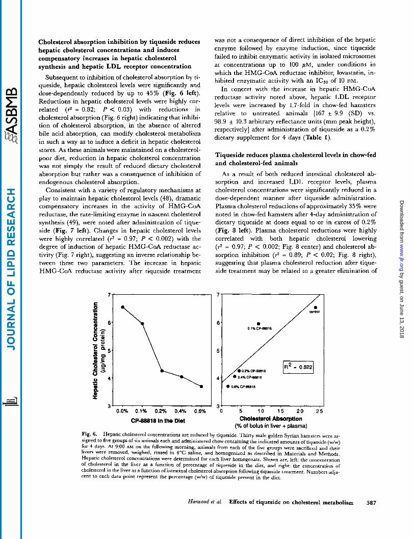
Cholesterol absorption inhibition by tiqueside reduces hepatic cholesterol concentrations and induces compensatory increases in hepatic cholesterol synthesis and hepatic LDL receptor concentration
Subsequent to inhibition of cholesterol absorption by ti- queside, hepatic cholesterol levels were significantly and dose-dependently reduced by up to 45% (Fig. 6 left). Reductions in hepatic cholesterol levels were highly cor- related (r2 = 0.82; P < 0.03) with reductions in cholesterol absorption (Fig. 6 right) indicating that inhibi- tion of cholesterol absorption, in the absence of altered bile acid absorption, can modify cholesterol metabolism in such a way as to induce a deficit in hepatic cholesterol stores. As these animals were maintained on a cholesterol- poor diet, reduction in hepatic cholesterol concentration was not simply the result of reduced dietary cholesterol absorption but rather was a consequence of inhibition of endogenous cholesterol absorption.
Consistent with a variety of regulatory mechanisms at play to maintain hepatic cholesterol levels (48), dramatic compensatory increases in the activity of HMG-CoA reductase, the rate-limiting enzyme in nascent cholesterol synthesis (49), were noted after administration of tique- side (Fig. 7 left). Changes in hepatic cholesterol levels were highly correlated (9 = 0.97; P < 0.002) with the degree of induction of hepatic HMG-CoA reductase ac- tivity (Fig. 7 right), suggesting an inverse relationship be- tween these two parameters. The increase in hepatic HMG-CoA reductase activity after tiqueside treatment
was not a consequence of direct inhibition of the hepatic enzyme followed by enzyme induction, since tiqueside failed to inhibit enzymatic activity in isolated microsomes at concentrations up to 100 p M , under conditions in which the HMG-CoA reductase inhibitor, lovastatin, in- hibited enzymatic activity with an ICso of 10 nM.
In concert with the increase in hepatic HMG-CoA reductase activity noted above, hepatic LDL receptor levels were increased by 1.7-fold in chow-fed hamsters relative to untreated animals [167 f 9.9 (SD) vs. 98.9 f 10.3 arbitrary reflectance units (mm peak height), respectively] after administration of tiqueside as a 0.2% dietary supplement for 4 days (Table 1).
Tiqueside reduces plasma cholesterol levels in chow-fed and cholesterol-fed animals
As a result of both reduced intestinal cholesterol ab- sorption and increased LDL receptor levels, plasma cholesterol concentrations were significantly reduced in a dose-dependent manner after tiqueside administration. Plasma cholesterol reductions of approximately 35 % were noted in chow-fed hamsters after 4-day administration of dietary tiqueside at doses equal to or in excess of 0.2% (Fig. 8 left). Plasma cholesterol reductions were highly correlated with both hepatic cholesterol lowering (r2 = 0.97; P < 0.002; Fig. 8 center) and cholesterol ab- sorption inhibition (r2 = 0.89; P < 0.02; Fig. 8 right), suggesting that plasma cholesterol reduction after tique- side treatment may be related to a greater elimination of
0 0.6% CPB80W
R w - - 0.0% 0.1% 0.2Yo 0.4% 0.6% O 5 10 1 5 2 0 2 5
Cholesterol Absorption (“A of bolus in liver + plasma)
CP-88818 in the Diet
Fig. 6. Hepatic cholesterol concentrations are reduced by tiqueside. Thirty male golden Syrian hamsters were as- signed to five groups of six animals each and administered chow containing the indicated amounts of tiqueside (w/w) for 4 days. At 9:OO AM on the following morning, animals from each of the five groups were sacrificed and their livers were removed, weighed, rinsed in 4OC saline, and homogenized as described in Materials and Methods. Hepatic cholesterol concentrations were determined for each liver homogenate. Shown are, left: the concentration of cholesterol in the liver as a function of percentage of tiqueside in the diet, and right: the concentration of cholesterol in the liver as a function of intestinal cholesterol absorption following tiqueside treatment. Numbers adja- cent to each data point represent the percentage (w/w) of tiqueside present in the diet.
Hurwood et ul. Effects of tiqueside on cholesterol metabolism 387
by guest, on June 13, 2018w
ww
.jlr.orgD
ownloaded from

1: 300
fc" 250
- > - c.
0
a - CI 50 n
2 0 0.0% 0.1% 0.2% 0.4% 0.6%
CP-88818 in the Diet
0.1% CP-BBBlB 150-
501 " . 3 4 5 6
Hepatic Chdeaterd Concentration (ug/mg protein)
Fig. 7. Dose-dependent increase in hepatic HMG-CoA reductase activity by tiqueside. Thirty male golden Syrian hamsters were assigned to five groups of six animals each and administered chow containing the indicated amounts of tiqueside (w/w) for 4 days. At 9:00 AM on the fifth day of the study (corresponding to the mid-dark cycle diurnal peak of hepatic HMG-CoA reductase activity) animals were sacrificed and their livers were removed, weighed, and rinsed in 4OC saline. Hepatic microsomes were isolated and hepatic HMG-CoA reductase activity was measured. Shown are, left: the activity of hepatic HMG-CoA reductase, as a function of percentage of tiqueside in the diet, and right: the activity of hepatic HMG-CoA reductase, as a function of hepatic cholesterol concentration following tiqueside treatment. Numbers adjacent to each data point represent the percentage (w/w) of tiqueside present in the diet.
cholesterol in the feces and also to a greater removal of cir- culating LDL cholesterol from the plasma through in- creased hepatic LDL receptor activity. The degree of reduction of plasma cholesterol after tiqueside treatment
was not changed when dose administration was extended for up to 14 days, suggesting that plasma cholesterol reduction through cholesterol sequestration is not only rapid but it is sustained.
TABLE 1. Summary of the effects of tiqueside on cholesterol metabolism in the chow-fed hamster
Modulation of Cholesterol Metabolism in Chow-Fed Hamsters by Tiqueside
Administered as a 0.2% Dietary Supplement
68 ? 4.7% reduction
Variable
Cholesterol absorption Bile acid absorption (excretion) unchanged Cholesterol 7u-hydroxylase unchanged Hepatic cholesterol HMG-CoA reductase
16 * 7% reduction
Hepatic 6.8 ~t 0.7-fold increase Intestinal 3.2 -t 0.1-fold increase
1.7 ~t 0.1-fold increase LDL receptor Plasma cholesterol 16 * 4% reduction Plasma triglycerides unchanged
Twelve male golden Syrian hamsters were assigned to two groups of six animals each and maintained on either chow containing no additions or chow containing 0.2% (wlw) tiqueside (approximately 150 mg/kg per day) for 4 days as described in Materials and Methods. On the morning of day 2, animals were transferred to plastic cages with wire racks, housed in pairs, and administered a 1.0-ml bolus of water containing 50 nmol ['+C]cholic acid (0.5 pCi; 22 dpm/pmol) and 2.5 mg [SH]polyethylene glycol (0.25 pCi; 220 dpmlpg). Fecal samples were collected over the next 3 days and rates of bile acid excretion were determined as described in Materials and Methods. At 9:00 AM on the morning of the fourth day, each animal received a 1 .O-ml oral bolus of liquid hamster diet containing 15 mg of [SH]cholesterol (2.25 pCi; sp act 100 dpmhmol) and 7.5 mg cholic acid for determining cholesterol ab- sorption. The next morning, animals were anesthetized with pentobartibal at 9:OO AM. Terminal blood samples were obtained by cardiac puncture for determination of plasma cholesterol levels, plasma triglyceride levels, and cholesterol absorption, as described in Materials and Methods. Livers were removed, weighed, rinsed in 4OC sa- line, and apportioned for determining hepatic cholesterol concentration, HMG-CoA reductase, and cholesterol 7cu- hydroxylase activities, hepatic LDL receptor concentration, and cholesterol absorption, as described in Materials and Methods. Intestinal segments from the stomach to the cecum were obtained and intestinal mucosal scrapings were used for measurement of intestinal microsomal HMG-CoA reductase activity as described in Materials and Methods. Data are the average of the measured modulations in the indicated variables induced by tiqueside ad- ministration relative to control animals for five independent experiments * SD.
388 Journal of Lipid Research Volume 34, 1993
by guest, on June 13, 2018w
ww
.jlr.orgD
ownloaded from

” 0.0% 0.1% 0.2% 0.4% 0.6%
CP-88818 in the DW
1701 IR2, 1
150.
140.
90. 3 4 5 6
Haptic Chokrtrrol Concentration (wmo protein)
170-
180. J
150-
140-
130.
/” / U R2= 0.893
0.4nc~ae.18 120L 110 O d X C P ~ l 8
0 5 10 15 20 25 Choimtwd Absarptlon
(% of bolus in liver + plasma)
Fig. 8. Dose dependent reduction in plasma cholesterol concentration in chow-fed hamsters after tiqueside treatment. Thirty male golden Syrian hamsters were assigned to five groups of six animals each and administered chow containing the indicated amounts of tiqueside (w/w) for 4 days. Animals from each of the five groups were anesthetized with pentobarbital and terminal blood samples were obtained by cardiac puncture. Plasma was isolated and analyzed for total cholesterol content as described in Materials and Methods. Shown are, left: the average concentration of cholesterol in the plasma (i SD) as a function of percentage of tiqueside in the diet; center: plasma cholesterol concentration as a function of hepatic cholesterol concentration following tiqueside treatment; and right: plasma cholesterol concentration as a function of intestinal cholesterol absorption following tiqueside treatment. Numbers adjacent to each data point represent the percentage (w/w) of tiqueside present in the diet.
Similar reductions in plasma cholesterol concentrations after tiqueside treatment were also noted in a variety of other species fed cholesterol-free as well as cholesterol- containing diets, indicating the ubiquity with which ti- queside reduces plasma cholesterol concentrations in ex- perimental animals. Administration of tiqueside to ham- sters, rats, mice, and dogs receiving diets essentially free of cholesterol resulted in plasma cholesterol reductions of up to 40% within 2 weeks (Table 2). Since these animals were maintained on diets that essentially lacked ex-
ogenous cholesterol, plasma cholesterol reductions were not the result of reduced dietary cholesterol absorption but rather were a consequence of inhibition of en- dogenous cholesterol absorption. Administration of tique- side to cholesterol-fed hamsters, rats, mice, rabbits, SEA quail, rhesus monkeys, and cynomolgus monkeys also resulted in marked plasma cholesterol reductions within 2-3 weeks (Table 3). In many instances, at the upper doses evaluated, tiqueside not only prevented the dietary cholesterol-induced rise in plasma cholesterol but also
TABLE 2, Plasma cholesterol reductions in various cholesterol-poor diet-fed species after treatment with tiqueside
Plasma Percent Cholesterol
SDecies Cholesterol Dose Animals Administration Concentration Reduction Cholesterol Dietary Number of Duration of
Hamster Hamster Hamster Hamster Rat Rat Mouse Mouse Dog Dog Dog Dog Dog
%
0.003 0.003 0.003 0.003 0.000 0.000 0.003 0.003 0.009 0.009 0.009 0.009 0.009
m d k s 0
75 150 300
0 50 0
280 0
250 500
1000 2000
12 6 6 6 6 6 6 6 8 2 8 8 6
days
4 4 4 4
14 14 4 4
14 14 14 14 14
mg/dl
175 i 15 155 i 11 118 f 8 117 * 20 116 + 25 103 * 15 124 i 12 92 + 19
176 + 44 141 i 3 125 i 28 127 i 25 107 * 35
%
12 33 33
12
26
20 29 28 39
Hamsters and mice received cholesterol-poor diets supplemented with tiqueside as described in Materials and Methods. Rats and dogs received cholesterol-poor diets and were administered tiqueside by oral gavage at the time of food consumption as described. At the indicated time plasma samples were obtained and plasma cholesterol con- centrations were determined as described. Data are the average plasma cholesterol concentrations for the indicated number of animals SD. For hamsters and mice, doses of tiqueside were calculated based on food consumption, body weights, and the percentage of tiqueside present in the diet.
Harwood et al. Effects of tiqueside on cholesterol metabolism 389
by guest, on June 13, 2018w
ww
.jlr.orgD
ownloaded from

TABLE 3. Plasma cholesterol reductions in various cholesterol-fed species after treatment with tiqueside
Plasma Prrcent Dietary Number of Duratlon of Cholesterol Cholesterol
Species Cholesterol Dose Animals Administration Concentration Reduction
% mP-/kg days mg/dl %
Hamster 0.2 0 24 14 185 f 18 Hamster 0.2 150 12 14 145 f 17
150 f 25 Rat 0.1 0 6 14 158 f 31 Rat 0.1 5 0 6 14 123 & 10 Rabbit 0.4 0 12 21 981 k 563 Rabbit 0.4 63 6 21 785 k 589 Rabbit 0.4 125 6 21 540 f 326 Rabbit 0.4 375 4 21 138 f 31 Mouse 0.2 0 6 4 183 +_ 21 Mouse 0.2 15 6 4 160 f 21 Mouse 0.2 75 6 4 149 I 29 Mouse 0.2 300 6 4 144 k 26 SEA quail 0.5 0 6 7 836 +- 154
SEA quail 0.5 50 6 7 300 f 87
179 k 25 Rhesus monkey 0.1 24 6 21 162 I 11 Rhesus monkey 0. I 49 6 21 145 & 34 Rhesus monkey 0.1 98 6 21 114 k 19 Cynomolgus monkey" 0.1 0 6 21 316 i 21 Cynomolgus monkey" 0.1 120 6 21 202 I 8
Hamster 0.2 200 12 14
SEA quail 0.5 20 6 7 393 112
SEA quail 0.5 100 6 7 210 * 55 Rhesus monkey 0.1 0 6 21
22 20
23
20 45 86
13 19 21
53 64 75
9 19 36
36
Animals were fed cholesterol-containing diets supplemented with tiqueside as described in Materials and Methods. At the indicated times, plasma samples were obtained and plasma cholesterol levels were determined. Data are the average plasma cholesterol concentrations for the indicated number of animals f SD. Baseline (pre-cholesterol diet) plasma cholesterol concentrations were as follows: hamster, 125 mg/dl; rat, 74 mg/dl; rabbit, 42 mg/dl; mouse, 107 mg/dl; SEA quail, 177 mg/dl; rhesus monkey, 151 mg/dl; cynomolgus monkey, 228 mg/dl. Doses oftiqueside were calculated based on food consumption, body weights, and the percentage of tiqueside present in the diet.
"Data from reference 28.
reduced plasma cholesterol concentrations to below base- line levels (Table 3), an observation consistent with the reductions from baseline noted in animals fed cholesterol- poor diets (Table 2). Although there was some inter- species variability in responsiveness, in general, tiqueside administration at doses of approximately 100 mg/kg per day resulted in 20-25% reductions in plasma cholesterol concentrations (Table 3). In studies of longer duration (dogs, rabbits) somewhat greater reductions in plasma cholesterol levels were observed. However, most of the effect occurred within the first 1-2 weeks of administration.
Plasma cholesterol reduction by tiqueside is primarily due to a reduction in non-HDL cholesterol levels
With the exception of the dog, which carries its plasma cholesterol mainly in HDL, the plasma cholesterol reduc- tions noted in Table 2 and Table 3 were primarily, and in many cases exclusively, the result of a reduction in non- HDL (e.g., LDL, IDL, VLDL) cholesterol concentrations with little or no change in HDL cholesterol levels (Table 4). In contrast, plasma cholesterol reductions in the chow-fed dog, which was the least responsive to tiqueside-induced plasma cholesterol lowering of all species evaluated (Table
2 and Table 3), were primarily the result of reductions in HDL cholesterol levels.
DISCUSSION
In the studies outlined in this report, we have demon- strated that the synthetic steroid saponin, 0-tigogenin cel- lobioside (CP-88818, tiqueside), inhibits cholesterol ab- sorption without affecting bile acid absorption. As a consequence of this inhibition, the supply of intestinally absorbed cholesterol reaching the liver is decreased and hepatic cholesterol levels are reduced. This deficit in hepatic cholesterol induces compensatory increases in both hepatic cholesterolgenesis and in hepatic uptake of circulating LDL cholesterol, which, in combination with reduced cholesterol absorption, leads to a reduction in plasma cholesterol levels. The series of modulations of cholesterol metabolism induced by tiqueside that lead to plasma cholesterol reduction are summarized in Table 1. That hepatic cholesterol reduction, compensatory induc- tion of hepatic cholesterolgenesis, and plasma cholesterol lowering are all highly correlated with inhibition of intes- tinal cholesterol absorption strongly suggests that these al-
390 Journal of Lipid Research Volume 34, 1993
by guest, on June 13, 2018w
ww
.jlr.orgD
ownloaded from

TABLE 4. Change in lipoprotein cholesterol concentrations in various species after treatment with tiqueside
Species Dose
Hamster'
Hamster
Rata
Rat
Mouse"
Mouse
Rabbit
SEA quail
Rhesus monkey
Cynomolgus monkeyb
Dog"
m d k control
300
control 150
control 50
control 50
control 280
control 15 75
300
control 375
control 20 50 100
control 22 44 88
control 120
control 250 500
1000 2000
Total Non-HDL HDL Cholesterol Cholesterol Cholesterol
106 mg/dl 187 mg/dl 81 mg/dl - 23% - 63% + 8%
187 mg/dl 112 mg/dl 75 mg/dl - 23% - 25% - 19%
116 mg/dl 90 mg/dl 26 mg/dl - 12% - 15% 0%
158 mg/dl 137 mg/dl 21 mg/dl - 23% - 23% - 19%
124 mg/dl 48 mg/dl 75 mg/dl - 26% - 56% - 5 %
183 mg/dl 78 mg/dl 109 mg/dl - 13% - 30% - 8% - 19% - 25% - 18% - 2 1 % - 59% + 3%
1046 mg/dl 979 mg/dl 67 mg/dl - 87% - 93% - 3 %
836 mg/dl 700 mg/dl 136 mg/dl - 53% - 63% - 1 % - 64% - 75% - 8% - 75% - 90% + 2%
179 mg/dl 93 mg/dl 78 mg/dl - 9% - 2 1 % + 9 % - 19% - 29% + 7% - 36% - 50% - 24%
316 mg/dl 177 mg/dl 139 mg/dl - 36% - 78% + 17%
176 mgidl 35 mg/dl 143 mg/dl - 20% - 26% - 19% - 29% - 3 1 % - 27% - 27% - 29% - 27% - 39% - 29% - 39%
Data for total cholesterol concentrations are those of Tables 2 and 3. HDL and non-HDL cholesterol concentra- tions were determined as described in Materials and Methods. Data are presented as the average cholesterol values in mg/dl for control animals and as a percentage of control values for tiqueside-treated animals.
"Indicates animals fed cholesterol-poor diets. 'Data from reference 28.
terations are all secondary to inhibition of cholesterol ab- sorption. Furthermore, the observation that tiqueside reduces plasma cholesterol concentrations in the absence of added dietary cholesterol also suggests that inhibition of reabsorption of endogenous cholesterol is at least as im- portant to the efficacy of tiqueside as is its ability to inhibit absorption of dietary cholesterol.
Qualitatively similar results have also been obtained in a variety of species, indicating that tiqueside acts to lower plasma cholesterol levels through a similar mechanism in all species evaluated. The ubiquity with which tiqueside reduces plasma cholesterol concentrations in a variety of diverse species whether fed cholesterol-free or cholesterol- containing diets further indicates the ability of tiqueside to inhibit both absorption of dietary cholesterol and reab- sorption of endogenous cholesterol. Furthermore, in all
species except the chow-fed dog, which uses HDL as the primary lipoprotein involved in cholesterol transport, reductions in plasma cholesterol were mainly and in many instances exclusively the result of reductions in non-HDL (LDL, VLDL, IDL) cholesterol, with little or no change in HDL cholesterol.
Taken together, these observations suggest that tique- side has the potential to reduce LDL cholesterol without significantly affecting HDL cholesterol concentrations in humans even when administered in combination with a strict low cholesterol diet. In addition, 'as lowering plasma cholesterol levels has been shown to halt and reverse the progression of atherosclerosis and to reduce the incidence of myocardial infarctions (2, 3), the potential also exists for tiqueside to exhibit these beneficial effects with respect to coronary heart disease. In this regard, the naturally oc-
Harwood et al. Effects of tiqueside on cholesterol metabolism 391
by guest, on June 13, 2018w
ww
.jlr.orgD
ownloaded from

curring saponins of alfalfa have been shown to prevent progression of, as well as induce regression of, experimen- tal atherosclerosis in a variety of species including mon- keys (6, 18, 19), and ginseng saponins have been shown to retard the progression of experimental atherosclerosis in cholesterol-fed rabbits (20). Whether tiqueside will lower plasma cholesterol levels in humans with the efficacy that it has exhibited in experimental animals, and whether ti- queside will also inhibit progression of, and induce regres- sion of atherosclerosis in experimental animals and in hu- mans, remain to be determined.
The luminal mechanism of action whereby tiqueside reduces plasma cholesterol concentrations is distinctly different from the mechanisms of action of currently available lipid-lowering therapies. These differences re- side primarily in the mechanism by which hepatic cholesterol levels are reduced. For example, cholesterol bi- osynthesis inhibitors, such as the HMG-CoA reductase inhibitors lovastatin, simvastatin, and pravastatin (50-52), act systemically to inhibit cholesterol synthesis in the liver and consequently reduce hepatic cholesterol levels. In contrast, bile acid-binding resins such as cholestyramine and colestipol (7, 53) act nonsystemically to sequester bile acids in the intestinal lumen and prevent their reabsorption. These sequestered bile acids are ex- creted in the feces, resulting in a deficit in the bile acid pool that in turn induces an increase in hepatic cholesterol utilization for bile acid synthesis thereby causing a reduc- tion in hepatic cholesterol levels. By comparison, tique- side acts nonsystemically to inhibit cholesterol absorption without affecting bile acid absorption. Bile acids are thus recycled normally and bile acid synthesis is not induced. However, the cholesterol that is prevented from being ab- sorbed does not reach the liver, and this decreased supply of cholesterol to the liver results in a reduction in hepatic cholesterol concentrations. Regardless of the mechanism by which hepatic cholesterol concentration is reduced, its reduction induces compensatory increases in both hepatic HMG-CoA reductase and LDL receptor levels, and the resulting increase in receptor-mediated hepatic uptake of circulating LDL cholesterol contributes to the observed reduction in plasma cholesterol levels.
Because of its ability to inhibit cholesterol absorption without affecting bile acid metabolism, tiqueside should be useful in further characterizing the distinct roles of en- terohepatic bile acid recirculation and intestinal cholesterol absorption in regulating cholesterol homeosta- sis. Also, as net hepatic cholesterol synthesis is increased secondarily to inhibition of cholesterol absorption, the potential exists for tiqueside to act synergistically with cholesterol synthesis inhibitors, in much the same manner as seen with the bile acid binding resins and the HMG- CoA reductase inhibitors (50-52). Whether tiqueside and cholesterol synthesis inhibitors will also act synergistically
to lower plasma cholesterol levels in experimental animals and in humans, however, remains to be determined.
The mechanism by which tiqueside inhibits cholesterol absorption is also distinctly different from that of inhibi- tors of intestinal ACAT activity. ACAT inhibitors act in- tracellularly to inhibit the esterification of cholesterol in the enterocyte following its uptake from the intestinal lu- men and as a consequence prevent its packaging into chylomicrons and secretion into the circulation (54-56). The free cholesterol that is retained within the enterocyte is presumably excreted via the feces as the enterocytes turn over. In contrast, a luminal mode of action for tique- side has been inferred both from the ability of tiqueside to associate with cholesterol in vitro to induce its precipi- tation (28), and from experiments conducted in isolated, perfused rat intestinal segments (12). In these latter studies, inhibition of cholesterol absorption by tiqueside and by soya saponins was independent of any association with the intestinal wall or need for saponin absorption for activity (12). Our observations that the concentration of intestinally derived cholesterol within the intestinal mucosa is reduced after tiqueside treatment, and that this occurs concomitant with a marked increase in intestinal HMG-CoA reductase activity, also support a mechanism whereby luminal cholesterol is prevented from entering the intestinal mucosa. That tiqueside fails to induce an in- crease in intestinally derived mucosal cholesterol concen- trations under conditions in which 2-fold to 5-fold in- creases are typically noted following treatment with ACAT inhibitors (57), also argues against an intracellular mechanism for tiqueside action. Thus, based on the above observations it is unlikely that tiqueside acts intracellu- larly to interfere with either cholesterol esterification or chylomicron assembly, but rather, appears to interact with luminal cholesterol prior to its uptake by mucosal cells.
Interestingly, the increase in intestinal HMG-CoA reductase that occurs after inhibition of luminal cholesterol entry into the intestinal mucosal cells by tique- side suggests that this cholesterol pool which is thought to be trafficked toward chylomicron formation (54), and in- tracellular pools of cholesterol that are thought to regulate intestinal cholesterol synthesis (54), may not be totally distinct. Thus, in response to a marked reduction in lumi- nal cholesterol uptake by enterocytes, newly synthesized cholesterol may be commandeered for use in chylomicron formation. Whether this increase in intestinal HMG-CoA reductase activity is driven by a reduction in the cholesterol concentration in specific pools within the en- terocyte or by an increase in enterocyte triglyceride con- tent resulting from reduced chylomicron formation without concomitant reductions in triglyceride absorption (54), however, remains to be evaluated.
A variety of mechanisms exist whereby tiqueside could inhibit cholesterol absorption through actions solely in the
392 Journal of Lipid Research Volume 34, 1993
by guest, on June 13, 2018w
ww
.jlr.orgD
ownloaded from

intestinal lumen. For example, 1) tiqueside could associ- ate with cholesterol in the intestinal lumen to induce its precipitation, possibly through complex formation; 2) ti- queside could interact with and alter the size and/or sta- bility of cholesterol-containing micelles in the intestinal lumen and thereby alter the efficiency with which cholesterol is delivered to the mucosal surface; or 3) tique- side could interact with mucosal cell membranes to ex- tract cholesterol or disrupt transport functions. An inter- action of tiqueside with mucosal cell membranes as a primary mechanism for inhibiting cholesterol absorption is unlikely in view of the observations of Sidhu, Upson, and Malinow (E'), in which pre-exposure of isolated rat intestinal segments to tiqueside (or soya saponins), in situ, followed by removal of compound and subsequent ex- posure to radiolabeled cholesterol failed to result in a reduction in cholesterol absorption. In contrast, simul- taneous addition of tiqueside (or soya saponins) and radi- olabeled cholesterol to the intestinal segments resulted in a marked inhibition of cholesterol absorption.
A more plausible possibility is that tiqueside alters the size and/or stability of cholesterol-containing micelles in the intestinal lumen and thereby alters the efficiency with which cholesterol is delivered to the mucosal surface (54). Such a mechanism has been evoked to explain the action of various plant sterols (58, 59) and neomycin (7). However, the observation that tiqueside and other steroi- dal saponins (10, 23) inhibit cholesterol absorption without interfering with bile acid absorption, whereas a number of plant saponins that alter micellar size (24, 25) affect both bile acid and cholesterol absorption (5, 12-14, 24), suggests that micellar modification may not play a primary role in the mechanism of cholesterol absorption inhibition by tiqueside.
The most probable mechanism for tiqueside action is that it induces cholesterol precipitation in the intestinal lumen via either cholesterol/tiqueside complex formation, competition for micellar space (micellar exclusion), or both. Indeed, evidence has accumulated suggesting that inhibitory saponins associate with cholesterol in vitro to induce its precipitation (9, 10, 21, 22, 26-28). In this regard, studies examining a variety of saponidcholesterol interactions have demonstrated that, in many instances, specific structural requirements for sterol binding exist (21, 22, 27), and that the avidity and stoichiometry of the saponin/sterol interactions vary depending on the partic- ular saponin and sterol that are associating (21, 22, 27). Based on these observations, the ability of plant saponins to precipitate cholesterol from micellar solution and render it unavailable for absorption has generally been regarded as the mechanism whereby these agents inhibit cholesterol absorption and hence reduce plasma cholesterol levels in experimental animals. Tiqueside and related synthetic saponins have also been shown to cause cholesterol to precipitate from micellar solution in vitro
(28). Thus, as with a variety of plant saponins, the possi- bility exists that tiqueside inhibits cholesterol absorption in vivo by causing cholesterol in the intestinal lumen to precipitate. Whether tiqueside causes cholesterol precipi- tation in vivo through complex formation or by micellar exclusion, however, remains to be determined. I
We thank David A. Beebe for establishing an HPLC methodol- ogy for measuring cholesterol 7a-hydroxylase activity and com- paring that methodology with the radiometric methodology used in this study, B. J. Gaynor for her assistance in isolating hamster intestinal microsomes and for determining the concen- tration of intestinally derived cholesterol contained in tiqueside- treated hamster intestinal mucosa, Berkeley W. Cue and associ- ates for preparing the tiqueside used in these studies, and James B. Moberly and Thomas A. Beyer for their critical review of this manuscript.
Manuscript received 16 March 1992 and in revised form 15 September 1992.
REFERENCES
1.
2.
3.
4.
5.
6.
7.
8
9
10.
11.
Harwood et al.
Kesaniemi, Y. A,, and T. A. Miettinen. 1987. Cholesterol absorption efficiency regulates plasma cholesterol level in the Finnish population. Eur. J. Clin. Invest. 17: 391-395. Lipid Research Clinics Program. 1984. The Lipid Research Clinics coronary primary prevention trials results; I. Reduction in incidence of coronary heart disease; 11. The relationship of reduction in incidence of coronary heart dis- ease to cholesterol lowering. J. Am. Med. h o c . 251:
Blankenhorn, D. H., S. A. Nessim, R. L. Johnson, M. E. SanMarco, S. P. Azen, and L. Cashin-Hemphill. 1987. Beneficial effects of combined colestipol-niacin therapy on coronary atherosclerosis and coronary venous bypass grafts. J. Am. Med. Assoc. 257: 3233-3240. Pollak, 0. J. 1985. Effect of plant sterols on serum lipids and atherosclerosis. Phannacol. Ther. 31: 177-208. Price, K. R., I. T. Johnson, andG. R. Fenwick. 1987. The chemistry and biological significance of saponins in foods and feedstuffs. CRC Crit. Rev. Food Sci. Nut% 26: 27-135. Malinow, M. R. 1984. Triterpenoid saponins in mammals: effects on cholesterol metabolism and atherosclerosis. In Isopentenoids in Plants, Biochemistry and Function. W. D. Nes, G. Fuller, and L. S. Tsai, editors. Marcel Dekker Inc., New York. 229-246. Miettinen, T. A. 1976. Methods for evaluation of hypolipi- demic drugs in man: mechanisms of their actions. I n Lipid Pharmacology. 11. R. Paoletti and C. J. Glueck, editors. Academic Press, New York. Chapter 3. 83-125. Cayen, M. N., and D. Dvomik. 1979. Effect of diosgenin on lipid metabolism in rats. J. Lipid Res. 20: 162-174. Malinow, M. R., P. McLaughlin, and C. Stafford. 1978. Prevention of hypercholesterolemia in monkeys (Macaca fa- cicularis) by digitonin. Am. J. Clin. Nutr. 31: 814-818. Cayen, M. N. 1971. Effect of dietary tomatine on cholesterol metabolism in the rat.J. Lipid Res. 12: 482-490. Malinow, M. R., P. McLaughlin, L. Papworth, C. Stafford, G. 0. Kohler, A. L. Livingston, and P. R. Cheeke. 1987. Effect of alfalfa saponins on intestinal cholesterol absorp- tion in rats. Am. J Clin. Nutt: 30: 2061-2067.
351-374.
Effects of tiquaide on cholesterol metabolism 393
by guest, on June 13, 2018w
ww
.jlr.orgD
ownloaded from

12.
13.
14.
15.
16.
17.
18.
19.
20.
21.
22.
23.
24.
25.
26.
27.
28.
29.
Sidhu, G. S., B. Upson, and M . R. Malinow. 1987. Effects of soy saponins and tigogenin cellobioside on intestinal up- take of cholesterol, cholate and glucose. Nutr Rep. Znt. 35:
Oakenfull, D. G., D. L. Topping, R. J. Illman, and D. E. Fenweick. 1984. Prevention of dietary hypercholesterolemia in the rat by soya bean and quillaja saponins. Nut,: Rep. Znt. 29: 1039-1046. Topping, D. L., G. B. Storer, G. D. Calvert, R. J. Illman, D. G. Oakenfull, and R. A. Weller. 1980. Effects of dietary saponins on fecal bile acids and neutral sterols, plasma lipids, and lipoprotein turnover in the pig. Am. J. Clin. Nutr.
Southon, S., I. T. Johnson, J. M. Gee, and K. R. Price. 1988. The effect of Gypsophila saponins in the diet on mineral status and plasma cholesterol concentration in the rat. B,: J . Nut,: 59: 49-55. Ha, C. J., and S. H. Kim. 1984. Effects of ginseng saponins on cholesterol solubility. Saengyak Hakhoechi. 15: 134-138. Sakakibara, K., Y. Shibata, T. Higashi, S. Sanada, and J. Shoji. 1975. Effect of ginseng saponins on cholesterol metabolism. I. The level and the synthesis of serum and liver cholesterol in rats treated with ginsenosides. Chem. Pharm. Bull. 23: 1009-1016. Malinow, M. R., P. McLaughlin, H. K. Naito, L. A. Lewis, and W. P. Nulty. 1978. Effect of alfalfa meal on shrinkage (regression) of atherosclerotic plaques during cholesterol feeding in monkeys. Atherosclerosis. 30: 27-43. Malinow, M. R., P. McLaughlin, C. Stafford, A. L. Living- ston, and J. W. Senner. 1983. Effects of alfalfa saponins on regression of atherosclerosis in monkeys. Zn Proceedings of the Second Munster International Atherosclerosis Sympo- sium W. H. Hauss, editor. Westdeutscher Verlag, Opladen, Germany. Chapter 15. 241-254. Koo, J. H. 1983. The effect of ginseng saponin on the de- velopment of experimental atherosclerosis. Hanyang Uidae Haksulchi. 3: 273-286. Haslam, R. M., and W. Klyne. 1953. The precipitation of 3P-hydroxysteroids by digitonin. Biochem. J . 55: 340-346. Takagi, S., H. Otsuka, T. Akiyama, and U. Sankawa. 1982. Digitonin-cholesterol complex formation: effects of varying the length of the side-chain. Chem. Pharm. Bull. 30:
Malinow, M. R., W. H. Elliott, P. McLaughlin, and B. Up- son. 1987. Effects of synthetic glycosides on sterol balance in Macaca fascicularis. J . Lipid Res. 28: 1-9. Sidhu, G. S., and D. G. Oakenfull. 1986. A mechanism for the hypocholesterolaemic activity of saponins. Br. J. Nutr.
Oakenfull, D. 1986. Aggregation of saponins and bile acids in aqueous solution. Aut. J. Chem. 3 9 1671-1683. Story, J. A,, S. L. LePage, M. S. Petro, L. G. West, M. M. Cassidy, F. G. Lightfoot, and G. V. Vahouny. 1984. Interac- tions of alfalfa plant and sprout saponins with cholesterol in vitro and in cholesterol-fed rats. Am. J Clin. Nut,: 39:
Gestetner, B., Y. Assa, Y. Henis, Y. Tencer, M. Rotman, Y. Birk, and A. Bondi. 1972. Interaction of lucerne saponins with sterols. Biochim. Biophys. Acta. 270: 181-187. Malinow, M. R. 1986. U.S. patent applications 4,602,003 and 4,602,005. Malinow,.M. R., J. 0. Gardner, J. T. Nelson, P. McLaugh- lin, B. Upson, and R. Aigner-Held. 1986. Effects of a and 0-tigogenin cellobiosides on cholesterol absorption. Steroids.
615-623.
33: 783-786.
3485-3492.
55: 643-649.
917-929.
48: 197-211.
30.
31.
32.
33.
34.
35.
36.
37.
38.
39.
40.
41.
42.
43.
44.
45.
46.
47.
48.
Malinow, M. R. 1985. Effects of synthetic glycosides on cholesterol absorption. Ann. N Y Acad. Sci. 454: 23-27. Urban, E J., B. S. Moore, and R. Breitenbach. 1990. Syn- thesis of tigogenyl @-0-cellobioside heptaacetate and glyco- side tetraacetate via Schmidt’s trichloroacetimidate method: some new observations. Tetrahedron Lett. 31:
Harwood, H. J., Jr., I. M. Alvarez, Y. J. Greene, G. C. Ness, and P. W. Stacpoole. 1987. Development of a non- competitive, solid phase, bridged biotin-avidin enzyme im- munoassay for measurement of human leukocyte microsomal HMG-CoA reductase protein concentration. J. Lipid Res. 28: 292-304. Harwood, H. J., Jr., M. Schneider, and P. W. Stacpoole. 1984. Measurement of human leukocyte microsomal HMG-CoA reductase activity. J. Lipid Res. 25: 967-978. Harwood, H. J., Jr., Y. J. Greene, and P. W. Stacpoole. 1986. Inhibition of human leukocyte HMG-CoA reductase activity by ascorbic acid. J. Biol. Chem. 261: 7127-7135. Harwood, H. J., Jr., I. M. Alvarez, W. D. Noyes, and P. W. Stacpoole. 1991. In vivo regulation of human leukocyte HMG-CoA reductase: increased enzyme protein concen- tration and catalytic efficiency in human leukemia and lym- phoma. J Lipid Res. 32: 1237-1252. McCarthy, P. A. 1990. Synthesis of [5,6-3H2]CP-88818 (p- [ 5,6-3H2]tigogenin cellobioside). J. Labelled Comp. & Radi- opharm. 28: 1149-1159. Russell, D. W., W. J. Schneider, T. Yamamoto, K. L. Luskey, M . S. Brown, and J. L. Goldstein. 1984. Domain map of the LDL receptor: sequence homology with the epidermal growth factor precursor. Cell. 37: 577-585. Green, N., H. Alexander, A. Olson, S. Alexander, T. M. Shinnick, J. G. Sutcliffe, and R. A. Lerner. 1982. Immuno- genic structure of the influenza virus hemagglutinin. Cell.
Junker, L. H., and J. A. Story. 1985. An improved assay for cholesterol 7a-hydroxylase activity using phospholipid liposome-solubilized substrate. Lipids. 20: 712-718. Sugano, M., T. Ide, H. Okamatsu, and H. Takahara. 1977. Requirement of trypsin inhibitor for measurement of HMG-CoA reductase in intestinal mucosa of rat. Biochem. Biophys. Res. Commun. 79: 1092-1097. Cosgrove, P. G., B. J. Gaynor, and H. J. Harwood, Jr. 1992. Quantitation of hepatic low density lipoprotein receptor levels in the hamster. FASEB J 4: A533. Laemmli, U. K. 1970. Cleavage of structural proteins dur- ing the assembly of the head of bacteriophage T4. Nature. 227: 680-685. Kelley, J. J., and A. C. Tsai. 1978. Effect of pectin, gum arabic and agar on cholesterol absorption, synthesis and turnover in rats. J. Nutr. 108: 630-639. Assmann, G., H. Shriewer, G. Schmitz, and E-0. Hasele. 1983. Quantitation of HDL cholesterol by precipitation with phosphotungstic acid/MgC12. Clin. Chem. 29:
Bradford, M. M. 1976. A rapid and sensitive method for the quantitation of microgram quantities of protein using the principle of protein dye-binding. Anal. Biochem. 72:
Billheimer, J. T. 1985. Cholesterol acyltransferase. Methods Enzymol. 111: 286-293. Myant, N. B., and. K. A. Mitropoulos. 1977. Cholesterol 7a-hydroxylase. J. Lipid Res. 18: 135-153. Goldstein, J. L., and M. S. Brown. 1990. Regulation of the mevalonate pathway. Nature. 343: 425-430.
4421-4424.
28: 477-487.
2026-2030.
248-254.
394 Journal of Lipid Research Volume 34, 1993
by guest, on June 13, 2018w
ww
.jlr.orgD
ownloaded from

49.
50.
51.
52.
53.
54.
55.
56.
57.
58.
Rodwell, V. W., J. L. Nordstrom, and J. J. Mitschelen. 1976. Regulation of HMG-CoA reductase. Ado. Lipid Res.
Todd, P. A,, and K. L. Goh. 1990. Simvastatin: a review of its pharmacological properties and therapeutic potential in hypercholesterolemia. Drugs. 4 0 583-607. Anon. 1989. Lovastatin. A review of the scientific literature on lovastatin, an inhibitor of HMG-CoA reductase and the first of a new class of drugs for the treatment of hyper- cholesterolemia. In SCRIP'S New Product Review ffl31. PJB Publications. Ltd. 18-20 Hill Rise, Richmond. Surrey TWlO 6UA, UK. Arai, M., N. Serizawa, A. Terahara, Y. Tsujita, M. Tanaka, H. Masuda, and S. Ishikawa. 1988. Pravastatin sodium (CS-514), a novel cholesterol-lowering agent which inhibits HMG-CoA reductase. Sankyo Kenkyusho Nempo. 40: 1-38. Casdorph, H. R. 1976. Cholestyramine and ion exchange resins. In Lipid Pharmacology. Vol. 11. R. Paoletti and C. J. Glueck, editors. Academic Press, New York. Chapter 7. 221-256. Stange, E. E, and J. M. Dietschy. 1985. Cholesterol absorp- tion and metabolism by the intestinal epithelium. In Sterols and Bile Acids. H. Danielsson and J. Sjovall, editors. El- sevier Amsterdam. Chapter 5. 121-149. Spector, A. A., S. N. Mathur, and T. L. Kaduce. 1979. Role of ACAT in cholesterol metabolism. Prog. Lipid Res. 18:
Bennett Clark, S., and A. M. Tercyak. 1984. Reduced cholesterol transmucosal transport in rats with inhibited mucosal acyl CoA:cholesterol acyltransferase and normal pancreatic function. J. Lipid Res. 25: 148-159. Gaynor, B. J., J. B. Moberly, and D. I . Goldberg. 1992. In- testinal accumulation of unesterified cholesterol in hamsters following inhibition of acyl coenzyme A-cholesterol acyl transferase (ACAT). XIth International Symposium on Drugs Affecting Lipid Metabolism. Book of Abstracts. 87. Seki, K., T. Watanabe, and T. Suga. 1988. Studies on hypolipidemic agents. V. Influence of 5-tridecylpyrazole-3-
14: 1-74.
31-53.
carboxylic acid, a new hypolipidemic agent, on intestinal cholesterol absorption in rats. Chem. Pharm. Bull. 36:
59. Ikeda, I . , K. Tanaka, M. Sugano, G. V. Vahouny, and L. L. Gallo. 1988. Inhibition of cholesterol absorption in rats by plant sterols. J. Lipid Res. 29: 1573-1582. Harwood, H. J., Jr., C. E. Chandler, L. D. Pellarin, R. W. Wilkins, F. W. Bangerter, L. Mikolay, and L. Zaccaro. 1989. Effects of the cholesterol sequestrant, CP-88818, on cholesterol metabolism in the chow-fed hamster. Xth Inter- national Symposium on Drugs Affecting Lipid Metabolism. Book of Abstracts. 93. Harwood, H. J., Jr., C. E. Chandler, L. D. Pellarin, P. G. Cosgrove, R. W. Wilkins, and F. W. Bangerter. 1990. Effects of the cholesterol sequestrant, 6-tigogenin cellobio- side (CP-888181, on cholesterol metabolism in the chow-fed hamster. FASEBJ. 4: A1746.
62. Pellarin, L. D., R. W. Wilkins, F. W. Bangerter, C. E. Chandler, and H. J. Harwood, Jr. 1990. Effects of the cholesterol sequestrant, P-tigogenin cellobioside (CP-88818), on cholesterol metabolism in the chow-fed hamster. FASEB J 4: A532.
63. Harwood, H. J., Jr., L. D. Pellarin, C. A. Long, Y. E. Sa- voy, and J. T. Mayne. 1991. The cholesterol sequestrant, p- tigogenin cellobioside (CP-88818), inhibits cholesterol ab- sorption and lowers plasma cholesterol levels in chow-fed and cholesterol-fed animals. FASEB J. 5: A947.
64. Harwood, H. J., Jr., L. D. Pellarin, P. G. Cosgrove, C. E. Chandler, F. W. Bangerter, R. W. Wilkins, C. A. Marzetta, J. L. Pettini, M. R. Malinow, and J. T. Mayne. 1992. Phar- macologic consequences of cholesterol absorption inhibi- tion: alteration in cholesterol metabolism and reduction in plasma cholesterol levels induced by the cholesterol seques- trant P-tigogenin cellobioside (CP-88818). FASEB J. 6: A1206.
65. Ogishima, T., and K. Okuda. 1986. An improved method for assay of cholesterol 7a-hydroxylase activity. Anal. Bio- chem. 158: 228-232.
1117-1121.
60.
61.
Harwood et al. Effects of tiqueside on cholesterol metabolism 395
by guest, on June 13, 2018w
ww
.jlr.orgD
ownloaded from