pharmacol rev 61:62–97, 2009 printed in u.s.a. cerebral ... filecerebral blood flow regulation by...
TRANSCRIPT

Cerebral Blood Flow Regulation by Nitric Oxide:Recent Advances
NOBORU TODA, KAZUHIDE AYAJIKI, AND TOMIO OKAMURA
Toyama Institute for Cardiovascular Pharmacology Research, Osaka, Japan (N.T.); and Department of Pharmacology,Shiga University of Medical Science, Shiga, Japan (N.T., K.A., T.O.)
Abstract . . . . . . . . . . . . . . . . . . . . . . . . . . . . . . . . . . . . . . . . . . . . . . . . . . . . . . . . . . . . . . . . . . . . . . . . . . . . . . . . . 63I. Introduction . . . . . . . . . . . . . . . . . . . . . . . . . . . . . . . . . . . . . . . . . . . . . . . . . . . . . . . . . . . . . . . . . . . . . . . . . . . . . 64
II. Synthesis and actions of nitric oxide. . . . . . . . . . . . . . . . . . . . . . . . . . . . . . . . . . . . . . . . . . . . . . . . . . . . . . . . 64A. Nitric-oxide synthase isoforms and nitric oxide production . . . . . . . . . . . . . . . . . . . . . . . . . . . . . . . . 64B. Nitric oxide actions and signal transduction . . . . . . . . . . . . . . . . . . . . . . . . . . . . . . . . . . . . . . . . . . . . . 65C. Nitric oxide degradation . . . . . . . . . . . . . . . . . . . . . . . . . . . . . . . . . . . . . . . . . . . . . . . . . . . . . . . . . . . . . . . 65
III. Physiological control of cerebral blood flow . . . . . . . . . . . . . . . . . . . . . . . . . . . . . . . . . . . . . . . . . . . . . . . . . . 65A. Cerebral blood flow regulation by endothelium-derived nitric oxide. . . . . . . . . . . . . . . . . . . . . . . . . 65
1. Basal release of nitric oxide . . . . . . . . . . . . . . . . . . . . . . . . . . . . . . . . . . . . . . . . . . . . . . . . . . . . . . . . . 662. Stimulated release of endothelium-derived relaxing factor . . . . . . . . . . . . . . . . . . . . . . . . . . . . . . 673. Role of superoxide anions . . . . . . . . . . . . . . . . . . . . . . . . . . . . . . . . . . . . . . . . . . . . . . . . . . . . . . . . . . . 684. Role of estrogen and other hormones . . . . . . . . . . . . . . . . . . . . . . . . . . . . . . . . . . . . . . . . . . . . . . . . . 685. Studies on humans . . . . . . . . . . . . . . . . . . . . . . . . . . . . . . . . . . . . . . . . . . . . . . . . . . . . . . . . . . . . . . . . . 69
B. Autoregulation . . . . . . . . . . . . . . . . . . . . . . . . . . . . . . . . . . . . . . . . . . . . . . . . . . . . . . . . . . . . . . . . . . . . . . . . 691. Studies on experimental animals. . . . . . . . . . . . . . . . . . . . . . . . . . . . . . . . . . . . . . . . . . . . . . . . . . . . . 692. Studies on humans . . . . . . . . . . . . . . . . . . . . . . . . . . . . . . . . . . . . . . . . . . . . . . . . . . . . . . . . . . . . . . . . . 70
C. Influences of carbon dioxide, oxygen, and carbon monoxide . . . . . . . . . . . . . . . . . . . . . . . . . . . . . . . . 701. Hypercapnia . . . . . . . . . . . . . . . . . . . . . . . . . . . . . . . . . . . . . . . . . . . . . . . . . . . . . . . . . . . . . . . . . . . . . . . 70
a. Studies on experimental animals. . . . . . . . . . . . . . . . . . . . . . . . . . . . . . . . . . . . . . . . . . . . . . . . . . 70b. Studies on humans . . . . . . . . . . . . . . . . . . . . . . . . . . . . . . . . . . . . . . . . . . . . . . . . . . . . . . . . . . . . . . 71
2. Hyperbaric oxygen. . . . . . . . . . . . . . . . . . . . . . . . . . . . . . . . . . . . . . . . . . . . . . . . . . . . . . . . . . . . . . . . . . 71a. Studies on experimental animals. . . . . . . . . . . . . . . . . . . . . . . . . . . . . . . . . . . . . . . . . . . . . . . . . . 71b. Studies on humans . . . . . . . . . . . . . . . . . . . . . . . . . . . . . . . . . . . . . . . . . . . . . . . . . . . . . . . . . . . . . . 72
3. Hypoxia . . . . . . . . . . . . . . . . . . . . . . . . . . . . . . . . . . . . . . . . . . . . . . . . . . . . . . . . . . . . . . . . . . . . . . . . . . . 72a. Studies on experimental animals. . . . . . . . . . . . . . . . . . . . . . . . . . . . . . . . . . . . . . . . . . . . . . . . . . 72b. Studies on humans . . . . . . . . . . . . . . . . . . . . . . . . . . . . . . . . . . . . . . . . . . . . . . . . . . . . . . . . . . . . . . 72
4. Carbon monoxide. . . . . . . . . . . . . . . . . . . . . . . . . . . . . . . . . . . . . . . . . . . . . . . . . . . . . . . . . . . . . . . . . . . 73D. In vitro cerebral arterial tone as affected by endothelial nitric oxide. . . . . . . . . . . . . . . . . . . . . . . . 73
1. Basal release of nitric oxide . . . . . . . . . . . . . . . . . . . . . . . . . . . . . . . . . . . . . . . . . . . . . . . . . . . . . . . . . 732. Stimulated release of nitric oxide . . . . . . . . . . . . . . . . . . . . . . . . . . . . . . . . . . . . . . . . . . . . . . . . . . . . 74
E. Involvement of nerve-derived nitric oxide in the regulation of cerebral blood flow andvascular tone . . . . . . . . . . . . . . . . . . . . . . . . . . . . . . . . . . . . . . . . . . . . . . . . . . . . . . . . . . . . . . . . . . . . . . . . . 751. In vivo studies . . . . . . . . . . . . . . . . . . . . . . . . . . . . . . . . . . . . . . . . . . . . . . . . . . . . . . . . . . . . . . . . . . . . . 75
a. Basal release of nitric oxide . . . . . . . . . . . . . . . . . . . . . . . . . . . . . . . . . . . . . . . . . . . . . . . . . . . . . . 75b. Stimulated release of nitric oxide . . . . . . . . . . . . . . . . . . . . . . . . . . . . . . . . . . . . . . . . . . . . . . . . . 75c. Involvement of neurogenic nitric oxide in diseases . . . . . . . . . . . . . . . . . . . . . . . . . . . . . . . . . . 76d. Interactions between nitric oxide from nitrergic nerves and other neurotransmitters . . . 77e. Histological demonstration of nitrergic innervation in cerebral vasculature . . . . . . . . . . . . 77f. Summary of the possible role of neuronal nitric-oxide synthase-derived nitric oxide . . . . 77
2. In vitro studies. . . . . . . . . . . . . . . . . . . . . . . . . . . . . . . . . . . . . . . . . . . . . . . . . . . . . . . . . . . . . . . . . . . . . 78a. Basal release of nitric oxide . . . . . . . . . . . . . . . . . . . . . . . . . . . . . . . . . . . . . . . . . . . . . . . . . . . . . . 78b. Stimulated release of nitric oxide . . . . . . . . . . . . . . . . . . . . . . . . . . . . . . . . . . . . . . . . . . . . . . . . . 78c. Interactions between nitrergic nerve and other autonomic nerves . . . . . . . . . . . . . . . . . . . . 78
Address correspondence to: Dr. Noboru Toda, Professor Emeritus, Shiga University of Medical Science, Toyama Institute for Cardiovas-cular Pharmacology Research, 7-13, 1-Chome, Azuchi-machi, Chuo-ku, Osaka 541-0052, Japan. E-mail: [email protected]
This article is available online at http://pharmrev.aspetjournals.org.doi:10.1124/pr.108.000547.
0031-6997/09/6101-62–97$20.00PHARMACOLOGICAL REVIEWS Vol. 61, No. 1Copyright © 2009 by The American Society for Pharmacology and Experimental Therapeutics 547/3454208Pharmacol Rev 61:62–97, 2009 Printed in U.S.A.
62
by guest on May 2, 2016
pharmrev.aspetjournals.org
Dow
nloaded from

F. Astrocyte-derived nitric oxide and other vasodilators. . . . . . . . . . . . . . . . . . . . . . . . . . . . . . . . . . . . . . 79IV. Cerebral blood flow regulation by nitric oxide under pathological conditions . . . . . . . . . . . . . . . . . . . . 79
A. Cerebral ischemia and stroke. . . . . . . . . . . . . . . . . . . . . . . . . . . . . . . . . . . . . . . . . . . . . . . . . . . . . . . . . . . 791. Ischemia and endothelial nitric-oxide synthase-derived nitric oxide. . . . . . . . . . . . . . . . . . . . . . 792. Ischemia and neuronal nitric-oxide synthase . . . . . . . . . . . . . . . . . . . . . . . . . . . . . . . . . . . . . . . . . . 803. Ischemia and inducible nitric-oxide synthase . . . . . . . . . . . . . . . . . . . . . . . . . . . . . . . . . . . . . . . . . . 814. Ischemic preconditioning . . . . . . . . . . . . . . . . . . . . . . . . . . . . . . . . . . . . . . . . . . . . . . . . . . . . . . . . . . . . 815. Studies on patients with stroke . . . . . . . . . . . . . . . . . . . . . . . . . . . . . . . . . . . . . . . . . . . . . . . . . . . . . 816. Therapeutic measures for cerebral ischemic injury . . . . . . . . . . . . . . . . . . . . . . . . . . . . . . . . . . . . . 82
a. L-Arginine and nitric oxide donors. . . . . . . . . . . . . . . . . . . . . . . . . . . . . . . . . . . . . . . . . . . . . . . . . 82b. Nitric oxide modulators . . . . . . . . . . . . . . . . . . . . . . . . . . . . . . . . . . . . . . . . . . . . . . . . . . . . . . . . . . 82c. Nitric-oxide synthase inhibitors . . . . . . . . . . . . . . . . . . . . . . . . . . . . . . . . . . . . . . . . . . . . . . . . . . . 83d. Drugs possessing anti inflammatory effects . . . . . . . . . . . . . . . . . . . . . . . . . . . . . . . . . . . . . . . . 83e. Miscellaneous . . . . . . . . . . . . . . . . . . . . . . . . . . . . . . . . . . . . . . . . . . . . . . . . . . . . . . . . . . . . . . . . . . . 83
B. Cerebral vasospasm after subarachnoid hemorrhage . . . . . . . . . . . . . . . . . . . . . . . . . . . . . . . . . . . . . 831. Studies on experimental animals. . . . . . . . . . . . . . . . . . . . . . . . . . . . . . . . . . . . . . . . . . . . . . . . . . . . . 832. Therapeutic measures in animals . . . . . . . . . . . . . . . . . . . . . . . . . . . . . . . . . . . . . . . . . . . . . . . . . . . . 843. Studies on human materials and patients. . . . . . . . . . . . . . . . . . . . . . . . . . . . . . . . . . . . . . . . . . . . . 85
C. Traumatic head injury . . . . . . . . . . . . . . . . . . . . . . . . . . . . . . . . . . . . . . . . . . . . . . . . . . . . . . . . . . . . . . . . . 851. Role of constitutive nitric-oxide synthase-derived nitric oxide . . . . . . . . . . . . . . . . . . . . . . . . . . . 852. Role of inducible nitric-oxide synthase-derived nitric oxide. . . . . . . . . . . . . . . . . . . . . . . . . . . . . . 863. Therapeutic measures . . . . . . . . . . . . . . . . . . . . . . . . . . . . . . . . . . . . . . . . . . . . . . . . . . . . . . . . . . . . . . 86
V. Pharmacological implications of nitric oxide and nitric oxide related agents for miscellaneousbrain diseases . . . . . . . . . . . . . . . . . . . . . . . . . . . . . . . . . . . . . . . . . . . . . . . . . . . . . . . . . . . . . . . . . . . . . . . . . . . . 87A. L-Arginine . . . . . . . . . . . . . . . . . . . . . . . . . . . . . . . . . . . . . . . . . . . . . . . . . . . . . . . . . . . . . . . . . . . . . . . . . . . . 87B. Nitric oxide inhalation . . . . . . . . . . . . . . . . . . . . . . . . . . . . . . . . . . . . . . . . . . . . . . . . . . . . . . . . . . . . . . . . . 87C. Nitric oxide donors . . . . . . . . . . . . . . . . . . . . . . . . . . . . . . . . . . . . . . . . . . . . . . . . . . . . . . . . . . . . . . . . . . . . 87D. Phosphodiesterase-5 inhibitors . . . . . . . . . . . . . . . . . . . . . . . . . . . . . . . . . . . . . . . . . . . . . . . . . . . . . . . . . 87
1. Studies on experimental animals. . . . . . . . . . . . . . . . . . . . . . . . . . . . . . . . . . . . . . . . . . . . . . . . . . . . . 872. Studies on patients or healthy subjects . . . . . . . . . . . . . . . . . . . . . . . . . . . . . . . . . . . . . . . . . . . . . . . 87
E. 3-Hydroxy-3-methylglutaryl coenzyme A reductase inhibitors (statins). . . . . . . . . . . . . . . . . . . . . . 881. Studies on experimental animals. . . . . . . . . . . . . . . . . . . . . . . . . . . . . . . . . . . . . . . . . . . . . . . . . . . . . 882. Studies on patients . . . . . . . . . . . . . . . . . . . . . . . . . . . . . . . . . . . . . . . . . . . . . . . . . . . . . . . . . . . . . . . . . 88
F. Erythropoietin . . . . . . . . . . . . . . . . . . . . . . . . . . . . . . . . . . . . . . . . . . . . . . . . . . . . . . . . . . . . . . . . . . . . . . . 88G. Other therapeutic agents . . . . . . . . . . . . . . . . . . . . . . . . . . . . . . . . . . . . . . . . . . . . . . . . . . . . . . . . . . . . . . 89
1. Dietary factors and Chinese herbs . . . . . . . . . . . . . . . . . . . . . . . . . . . . . . . . . . . . . . . . . . . . . . . . . . . 89VI. Summary and conclusion . . . . . . . . . . . . . . . . . . . . . . . . . . . . . . . . . . . . . . . . . . . . . . . . . . . . . . . . . . . . . . . . . 90
Acknowledgments . . . . . . . . . . . . . . . . . . . . . . . . . . . . . . . . . . . . . . . . . . . . . . . . . . . . . . . . . . . . . . . . . . . . . . . . 90References . . . . . . . . . . . . . . . . . . . . . . . . . . . . . . . . . . . . . . . . . . . . . . . . . . . . . . . . . . . . . . . . . . . . . . . . . . . . . . . 90
Abstract——Nitric oxide (NO) is undoubtedly quite animportant intercellular messenger in cerebral and pe-ripheral hemodynamics. This molecule, formed by con-stitutive isomers of NO synthase, endothelial nitric-ox-ide synthase, and neuronal nitric-oxide synthase, playspivotal roles in the regulation of cerebral blood flow andcell viability and in the protection of nerve cells or fibersagainst pathogenic factors associated with cerebralischemia, trauma, and hemorrhage. Cerebral blood flowis increased and cerebral vascular resistance is de-creased by NO derived from endothelial cells, autonomicnitrergic nerves, or brain neurons under resting andstimulated conditions. Somatosensory stimulation alsoevokes cerebral vasodilatation mediated by neurogenicNO. Oxygen and carbon dioxide alter cerebral bloodflow and vascular tone mainly via constitutively formed
NO. Endothelial dysfunction impairs cerebral hemody-namics by reducing the bioavailability of NO and in-creasing the production of reactive oxygen species(ROS). The NO-ROS interaction is an important issue indiscussing blood flow and cell viability in the brain. Re-cent studies on brain circulation provide quite usefulinformation concerning the physiological roles of NOproduced by constitutive isoforms of nitric-oxide syn-thase and how NO may promote cerebral pathogenesisunder certain conditions, including cerebral ischemia/stroke, cerebral vasospasm after subarachnoid hemor-rhage, and brain injury. This information would contrib-ute to better understanding of cerebral hemodynamicregulation and its dysfunction and to development ofnovel therapeutic measures to treat diseases of the cen-tral nervous system.
NO AND CEREBRAL BLOOD FLOW 63

I. Introduction
The discovery of nitric oxide (NO1), a lipophilic gaseousmolecule constitutively generated from endothelial cellsand nerve cells or fibers, opened a new era of understand-ing mechanisms underlying the regulation of cardiovascu-lar functions and their disturbances (Ignarro et al., 1987;Palmer et al., 1987; Furchgott, 1988). Developmental in-vestigations on NO synthesis inhibitors, chemically syn-thesized (Palmer et al., 1988; Mulsch and Busse, 1990;Toda et al., 1990) or endogenously generated (Vallance etal., 1992), enabled biological researchers to clarify how NOcontributes to cardiovascular and central/peripheral ner-vous system functions. NO formed via endothelial (eNOS)and neuronal NO synthase (nNOS) causes vasodilatation,hypotension, blood flow increase, inhibitions of plateletaggregation and adhesion, and a decrease in smooth mus-cle proliferation and exerts other beneficial actions as anantioxidant. It also acts as a neurotransmitter or neuro-modulator. On the other hand, unregulated production ofNO through inducible or immunological NOS (iNOS) dur-ing inflammation or NO formed by nNOS in the brainevokes nitrative stress, leading to neurodegeneration andapoptosis.
Cerebral blood flow is one of the important factors inthe regulation of brain functions. Cerebral blood flowand vascular smooth muscle tone are regulated by NOderived from endothelial cells (Moncada et al., 1991),autonomic nitrergic nerves (Toda and Okamura, 2003),or brain neurons (Bhardwaj et al., 2000). NO plays piv-otal roles in regulating cerebrovascular effects of in-creased or decreased oxygen, elevated carbon dioxide,and carbon monoxide and cerebrovascular autoregula-tion as well. Information from recent studies on cerebralblood flow regulation by NO is expected to provide newinsight about the physiological control of cerebrovascu-lar functions and the pathological mechanisms involvedin genesis of central nervous system diseases and tosupply clues for developing novel therapeutic strategiesfor cerebral dysfunctions.
The literature since the discovery of endothelium-de-rived relaxing factor by Furchgott and Zawadzki (1980)contains numerous reports about the interactions be-tween NO and cardiovascular function and dysfunctionin the brain. This review article covers recent advancesin these investigations, mainly including those pub-lished in this century, on the regulation by NO of cere-bral blood flow under physiological and pathological con-ditions, including cerebral ischemia, stroke and trauma,and vasospasm after subarachnoid hemorrhage (SAH).Possible therapeutic measures using NO, NO release-modulating substances, and NOS inhibitors for thesediseases are also summarized.
II. Synthesis and Actions of Nitric Oxide
A. Nitric-Oxide Synthase Isoforms and Nitric OxideProduction
NO is synthesized together with L-citrulline by NOSfrom the precursor L-arginine in the presence of oxygenand cofactors, including NADPH, tetrahydrobiopterin(BH4), heme, FAD, FMN, and calmodulin (Alderton et al.,2001). The enzyme has domains for each of the cofactorsexcept for BH4, which is required for dimerization of theenzyme (Andrew and Mayer, 1999). All of the cofactors arerequired to produce NO, and BH4 insufficiency results inuncoupled NOS, which produces superoxide anions in-stead of NO. It is known that eNOS uncoupling causesendothelial dysfunction, leading to vascular and metabolicdisorders such as hypertension, hyperlipidemia, athero-sclerosis, insulin resistance, and diabetes mellitus (Chan-non, 2004; Shinozaki et al., 2004). Recently, eNOS uncou-pling in the cerebral artery has been reported toparticipate in hypoxic-ischemic brain injury (Fabian et al.,2008). The increase in the intracellular Ca2� concentrationvia transmembrane influx and/or release from intracellu-lar storage sites activates constitutive NOS (eNOS andnNOS), but iNOS does not require Ca2� for activation.eNOS can also be activated through phosphorylation ofSer1177/1179 via the phosphatidylinositol-3 (PI3) kinase-serine/threonine protein kinase (Akt) pathway (Dimmeler
1 Abbreviations: NO, nitric oxide; 7-NI, 7-nitroindazol; ACh, acetyl-choline, BK, bradykinin; ADMA, asymmetric dimethylarginine; AF-DX116, 11-([2-[(diethylamino)methyl]-1-piperdinyl]acetyl)-5,11-dihydro-6H-pyrido[2,3-b][1,4]benzodiazepine-6-one; AF-SX 384, N-[2-[2-[(dipro-pylamino)methyl]-1-piperidinyl]ethyl]-5, 6-dihydro-6-oxo-11H-pyrid-[2,3-b][1,4]benzodiazepine-11-carboxamide; Akt, serine/threonineprotein kinase; ANG, angiotensin; ARL 17477, N-[4-(2-{[3-chloro-phenyl)methyl]amino}ethyl)phenyl]-2-thiophenecarboximidamidedihydrochloride; AT1, angiotensin type 1 receptor; BH4, tetrahydro-biopterin; CO, carbon monoxide; COX, cyclooxygenase; DDAH-1,dimethylarginine dimethylaminohydrolase-1; DY-9760e, 3-(2-(4-(3-chloro-2-methylphenyl)1-piperazinyl)ethyl)5,6-dimethoxy-1-(4-imidazolylmethyl)-1H-indazol dihydrochloride 3.5 hydrate; EDHF,endothelium-derived hyperpolarizing factor; EET, eicosatrienoicacid; EGCG, (�)-epigallocatechin gallate; eNOS, endothelial NOS;EPO, erythropoietin; ERK, extracellular signal-regulated kinase;ET, endothelin; GR, glucocorticoid receptor; HDT, head-down tailsuspension; HO, heme oxygenase; ICI 118,551, (�)-1-[2,3-(dihydro-7-methyl-1H-inden-4-yl)oxy]-3-[(1-methylethyl)amino]-2-butanol; ICI 182,780, fulvestrant; ICI 192,605, 4(Z)-6-(2-o-chlorophenyl-4-o-hydroxyphenyl-1,3-dioxan-cis-5-yl)hexenoic acid; IL,interleukin; iNOS, immunological or inducible NOS; IPC, ischemic pre-conditioning; L-NA, NG-nitro-L-arginine; L-NAME, L-NA methylester;L-NMMA, NG-monomethyl-L-arginine; LPS, lipopolysaccharide; MEK,mitogen-activated protein kinase/ERK kinase; MK-801, 5H-dibenzo-[a,d]cyclohepten-5,10-imine (dizocilpine maleate); NMDA, N-methyl-D-aspartate; nNOS, neuronal NOS; NOS, nitric-oxide synthase; NOx,NO2 � NO3; ODQ, 1H-[1,2,4]oxalodiazolol[4,3a]quinoxalin-1-one; OVX,ovariectomized; oxyHb, oxyhemoglobin; PARP-1, PARP, poly(ADP-ribose polymerase) 1; PDE-5, phosphodiesterase type 5; PG, prostaglan-din; PGI2, prostacyclin; PI3, phosphatidylinositol-3; rHuEPO, recombi-nant human EPO; ROS, reactive oxygen species; SAH, subarachnoidhemorrhage; SHR, spontaneously hypertensive rat(s); SHRSP, stroke-prone SHR; SIN-1, 3-morpholinosydnonimine; SNP, sodium nitroprus-side; SOD, superoxide dismutase; TNF-�, tumor necrosis factor-�; tPA,tissue plasminogen activator; U-II, urotensin-II; VEGF, vascular endo-thelial growth factor; WKY, Wistar-Kyoto rat(s); Y-27632,trans-4-[(1R)-1-aminoethyl]-N-4-pyridinylcyclohexanecarboxamide dihydrochloride.
64 TODA ET AL.

et al., 1999). eNOS binds to caveolin-1 in the caveolae andmicrodomains of the endothelial plasma membrane andintracellularly migrates in response to increased cytosolicCa2� in the presence of calmodulin; thereby, the enzymebecomes activated for NO synthesis. On the other hand,nNOS is mostly a soluble enzyme present in the cytoplasm.NO production can be affected by L-arginine availability.L-Arginine is produced from L-citrulline through arginino-succinate synthetase and argininosuccinate lyase insidethe cell (Wiesinger, 2001). L-Arginine is also supplied byuptake from the extracellular space through a cationamino acid transporter system (system y�).
Several NOS inhibitors, which have been used to dem-onstrate the functional roles of endogenous NO, are asfollows: NG-monomethyl-L-arginine (L-NMMA) (Palmer etal., 1988), NG-nitro-L-arginine (L-NA) (Rees et al., 1990;Toda et al., 1990), L-NA methyl ester (L-NAME) (Rees etal., 1990), and asymmetric dimethylarginine (ADMA)(Vallance et al., 1992) as nonselective inhibitors; 7-ni-troindazol (7-NI) (Moore et al., 1993) as a relativelyselective inhibitor against nNOS; and aminoguanidine(Griffiths et al., 1993), N6-iminoethyl-L-lysine (Moore etal., 1994), and W1400 (Mancinelli et al., 2001) as iNOS-selective inhibitors.
B. Nitric Oxide Actions and Signal Transduction
NO derived from endothelial cells causes vasodilata-tion, a decrease in vascular resistance, lower blood pres-sure, inhibition of platelet aggregation and adhesion,inhibition of leukocyte adhesion and migration, and re-duction of smooth muscle proliferation, thus leading toprevention of atherosclerosis. These NO functions aremediated by cyclic GMP synthesized through solubleguanylyl cyclase, a heme-containing enzyme, which isdirectly activated by NO. The cyclic GMP-dependent mech-anism is involved in vasodilatation caused by various NOdonors such as nitroglycerin, sodium nitroprusside (SNP),and sodium nitrite (Moncada et al., 1991).
NO derived from peripheral efferent (nitrergic) nervesinnervating blood vessels (Toda and Okamura, 1990b,2003), gastrointestinal tract, (Bult et al., 1990; Toda andHerman, 2005), penis (Ignarro et al., 1990; Toda et al.,2005), and trachea (Tucker et al., 1990) also causes vaso-dilatation, blood flow increase, smooth muscle relaxation,and penile erection. Cyclic GMP also mediates these re-sponses. Phosphodiesterase type 5 (PDE-5) degrades cyclicGMP to yield 5�-GMP. PDE-5 inhibitors such as sildenafilpotentiate and prolong the actions of NO and cyclic GMP.Recently, a cyclic GMP-independent mechanism such asS-glutathiolation or activation of sarco/endoplasmic retic-ulum Ca2� ATPase (Cohen and Adachi, 2006) and NOdonors that activate Ca2�-dependent K� channels (Bolo-tina et al., 1994; Plane et al., 1998) have been reported toparticipate in NO-induced muscle relaxation.
In the central nervous system, NO derived from nNOSacts as a neuromodulator or neurotransmitter for theregulation of synaptic plasticity, the sleep-wake cycle,
and hormone secretion. Glutamate and related aminoacids, such as N-methyl-D-aspartate (NMDA), stimulateNO formation via increasing intracellular Ca2� concen-tration. Overproduction of NO synthesized by nNOS hasbeen reported in many clinical disorders, including acuteand chronic neurodegenerative diseases (Calabrese etal., 2007).
NO synthesized by iNOS modulates inflammationthrough multiple pathways and plays an important role inthe regulation of immune reactions. Overproduction of NOby iNOS has been implicated in various pathological pro-cesses, including tissue injury and cell apoptosis caused byischemia and inflammation.
Excessive production of NO is clearly neurotoxic, butthe mechanism underlying the toxic action is not fullyunderstood. NO is metabolized to peroxynitrite in thepresence of superoxide anion (O2
.), which binds directlyto DNA and causes its structure to change, resulting incell injury. On the other hand, NO is known to inhibitcytochrome c oxidase, the terminal complex of the mito-chondrial respiratory chain (Erusalimsky and Moncada,2007). In most cells, including neurons and astrocytes,NO reversibly and irreversibly modulates oxygen con-sumption, a phenomenon through which NO induces asignal to specific pathways relevant to neuronal sur-vival. NO may modulate the balance between glucoseconsumption through the glycolytic pathway and thepentose phosphate pathway in neurons (Bolanos et al.,2007). This may relate to the mechanisms of neurode-generation and enhancement of apoptosis due to oxida-tive and nitrosative stress.
C. Nitric Oxide Degradation
NO synthesized by NOS or generated from NO donorsvia nonenzymatic processes is rapidly inactivated by oxi-dation to nitrite or nitrate. NO is scavenged by superoxide-generating agents such as pyrogallol and hydroquinone,oxyhemoglobin (oxyHb), and carboxy-2-phenyl-4,4,5,5-tetrametyl-imidazoline-okyl 3-oxide (Akaike et al., 1993).Methylene blue, oxyHb, and 1H-[1,2,4]oxadiazolo[4,3a]-quinoxalin-1-one (ODQ) (Garthwaite et al., 1995) inhibitthe activity of soluble guanylyl cyclase. Under certain con-ditions in which superoxide and NO are sufficiently gen-erated, these molecules form peroxynitrite, which is an-other free radical species that elicits biological actions.
III. Physiological Control of Cerebral Blood Flow
A. Cerebral Blood Flow Regulation by Endothelium-Derived Nitric Oxide
There are number of reports implicating the involve-ment of basal NO release in cerebral blood flow (Faraci,1990), vascular tone, vascular resistance, and vasculargrowth (Baumbach et al., 2004) under resting conditionsin various mammals including mice, rats, dogs, pigs, andgoats (reviewed by Toda and Okamura, 2003). This re-lease has been elucidated by determining the decrease in
NO AND CEREBRAL BLOOD FLOW 65

blood flow and vasoconstriction induced by NOS inhibi-tors, NO scavengers, guanylyl cyclase inhibitors (Sobeyand Faraci, 1997), and endothelial disruption. On theother hand, stimulated release of NO from the endothe-lium has been demonstrated in cerebral vasculatures inresponse to acetylcholine (ACh), bradykinin (BK), or theCa2� ionophore A23187.
1. Basal Release of Nitric Oxide. Rats injected intra-cisternally with replication-defective adenovirus contain-ing the bovine eNOS gene developed transient increases incerebral blood flow (Luders et al., 2000). There is evidencesuggesting that adenosine-induced vasodilatation via acti-vation of A2B-adenosine receptors in the rat pial artery iscoupled to the production of NO (Shin et al., 2000). Afteradministration of L-NAME to near-term fetal sheep, corti-cal blood flow decreased, arterial blood pressure increased,and cerebrovascular resistance increased (Hunter et al.,2003). In lambs, L-NA infusion produced increases in cere-bral vascular resistance and decreases in cerebral bloodflow during sleep, with the greatest changes occurring inactive sleep, which was characterized by widespread neu-ral activation and elevated cerebral blood flow; character-istic differences in cerebral vascular resistance and bloodflow among active sleep, quiet sleep, and quiet wakeful-ness disappeared after treatment with L-NA (Zoccoli et al.,2001). Acute NOS blockade by intravenous L-NAME injec-tion induced a dramatic decrease in hypothalamic bloodflow in rats, whereas chronic oral administration of L-NAME did not alter the blood flow; reversal of chronic NOSblockade by L-arginine infusion evoked hypothalamic hy-peremia, suggesting the appearance of a compensatoryvasodilator mechanism (Hortobagyi et al., 2007). Theseauthors obtained data indicating that the compensatorymechanism is independent of vasodilator prostanoids butreduced release of vasoconstrictor prostanoids may con-tribute to the normalization of hypothalamic blood flowafter chronic loss of NO. Chronic treatment for 3 monthswith L-NAME in the drinking water increased mean cere-bral arteriolar pressure and pulse pressure in Wistar-Kyoto rats (WKY) to levels lower than those in stroke-prone spontaneously hypertensive rats (SHRSP). Thecross-sectional area of the vessel wall was greater inL-NAME-treated WKY and SHRSP than in untreatedWKY, and the external diameter was less in L-NAME-treated WKY than in untreated WKY but greater than inSHRSP (Chillon and Baumbach, 2004). Cerebral arteriolesseem to undergo hypertrophy and remodeling in WKYwith L-NAME-induced hypertension. Jesmin et al. (2004a)noted that there was a marked reduction in the profile ofcerebral blood flow and angiogenic factors of SHRSP, com-pared with that in age-matched WKY and SHR, and re-duction in levels of endothelial growth factor (VEGF) andfibroblast growth factor occurred earlier in developmentbefore the juvenile period and probably preceded reductionin regional cerebral blood flow in SHRSP. They also ob-tained evidence suggesting that the decrease in NO, pos-sibly responsible for reduced blood flow, may largely be due
to a decrease in eNOS expression in juvenile male SHRSP.Vascular expression of dimethylarginine dimethylamin-ohydrolase-1 (DDAH-1), an ADMA-hydrolyzing enzyme,was increased and plasma levels of ADMA were reduced inDDAH-1 transgenic mice. Contraction of the aorta to L-NAME was increased, and relaxation of the carotid arteryto ACh was enhanced in the transgenic mice; in vivo,ADMA reduced responses of cerebral arterioles to ACh innontransgenic mice, and this inhibitory effect was absentin the transgenic mice (Dayoub et al., 2008). Overexpres-sion of DDAH-1 seems to increase basal levels of vascularNO and protects against ADMA-induced endothelial dys-function in the cerebral circulation.
In addition to subprimate mammals, Japanese mon-keys also responded to NOS inhibitors with constrictionof the anterior and middle cerebral arteries, and theeffect was reversed by L-arginine (Toda et al., 2000a).Joshi et al. (2003) noted that in healthy baboons duringisoflurane anesthesia, intracarotid SNP did not increasecerebral blood flow, although it decreased proximal ar-terial tone, as demonstrated by the increase in cyclicGMP content measured in these vessels in vitro.
Chronic head-down tail (HDT) suspension in rats toinduce headward fluid shifts and elevations in arterialpressure was suggested to result in an attenuated basalrelease of NO or a diminished sensitivity of cerebralartery smooth muscle cells to NO (Geary et al., 1998).Basal tone and vasoconstrictor responses to increases intransmural pressure, shear stress, and K� were greaterin middle cerebral arteries isolated from HDT rats com-pared with controls. L-NAME and endothelium denuda-tion abolished these differences between HDT and con-trol rats, and HDT was associated with lower levels ofmiddle cerebral artery eNOS protein; cerebral blood flowin select regions was lower and cerebral vascular resis-tance higher during standing and head-up tilt in HDTrats, indicating that chronic cephalic fluid shifts en-hanced basal tone and vasoconstriction through a down-regulation of eNOS signaling mechanism (Wilkerson etal., 2005). Alterations in cerebral autoregulation associ-ated with microgravity or prolonged bed rest may be theresult of diminished levels of cerebral artery endothelialNO.
There is evidence suggesting that cyclic AMP has op-posing effects on NO-stimulated cyclic GMP increases(Xu et al., 2004). Results from studies on pial arteriolardiameter changes in anesthetized rats that receivedknockdown of the cyclic GMP efflux protein multidrugresistance protein 5 or were treated with inhibitors ofphosphodiesterase-5 (PDE-5) led the authors to concludethat the effect of cyclic AMP to reduce cyclic GMP effluxseems to predominate over cyclic AMP stimulation ofcyclic GMP hydrolysis.
In summary, on the basis of studies using NOS inhib-itors or those on measurement of plasma ADMA andDDAH-1 levels, there is evidence suggesting that basalrelease of NO produced by eNOS contributes to in-
66 TODA ET AL.

creases in cerebral blood flow in various mammals in-cluding monkeys and also to prevention of cerebrovas-cular hypertrophy and remodeling.
2. Stimulated Release of Endothelium-Derived NitricOxide. Cerebral vasodilatation can occur by stimulatedrelease of endothelium-derived relaxing factor (NO,EDHF, and vasodilator prostaglandins). In anesthetizedrats, L-NA almost abolished cerebral vasodilator responsesto ACh and BK. 5-N,N-Hexamethyleneamiloride, an inhib-itor of the Na�/H� exchanger, did not affect the baselinediameter of the basilar artery but inhibited the vasodilatorresponse to ACh and BK, without affecting vasodilatationproduced by SNP; monomethylamine hydrochloride, whichcauses intracellular alkalinization, enhanced ACh-inducedvasodilatation in the presence of a Na�/H� exchangerinhibitor (Kitazono et al., 2001). Intracellular alkaliniza-tion produced by activation of the Na�/H� exchangermay enhance NO production in the basilar arterial en-dothelium. ACh lost the ability to dilate cerebral arteriesand arterioles in mice lacking the muscarinic ACh re-ceptor M5 subtype (Yamada et al., 2001). Light/dye en-dothelial injury inhibited cerebral vasodilator responsesto hypercapnia and BK in anesthetized newborn andjuvenile pigs. Juvenile, but not newborn, hypercapnicand BK vascular responses were sensitive to solubleguanylyl cyclase inhibition, and indomethacin inhibitedBK responses in newborns, whereas juvenile responseswere markedly inhibited by L-NAME and mildly de-creased by indomethacin (Willis and Leffler, 2001). Itseems that newborn cerebrovascular responses are largelyNO-independent, but NO becomes more important withmaturation. Topical application of the inhibitor of copper/zinc superoxide dismutase (SOD) attenuated cerebral va-sodilator responses to ACh, BK, and arachidonate but notto SNP in anesthetized rabbits, and these inhibitory effectswere reversed by a superoxide scavenger (Didion et al.,2001). Dilatation of cerebral arterioles to ACh was reducedin copper/zinc SOD knockout mice compared with wild-type mice (Didion et al., 2002). These results suggest thatendogenous SOD limits superoxide levels in the cerebralmicrocirculation and that NOS- and cyclooxygenase(COX)-mediated responses are dependent on normal activ-ity of SOD. On the basis of data indicating that the re-sponses of cerebrocortical microflow to L-NAME, indo-methacin, and ACh were abolished in rats subjected tochronic vasopressin-induced hyponatremia, Kozniewskaand Radomska (2001) suggested that attenuation of cere-bral blood flow during chronic hyponatremia is, at least inpart, due to the withdrawal of basal vasodilator tone pro-duced by NO and PGI2 in cerebral circulation under phys-iological conditions. Pial arteriolar dilatation in responseto hypercapnia or BK was inhibited by L-NAME only inindomethacin-treated piglets, and NOS activity, but noteNOS expression, increased after chronic indomethacintreatment, suggesting that chronic inhibition of COX canincrease the contribution of NO to cerebrovascular circu-latory control (Zhang and Leffler, 2002).
In awake goats, injection of vasopressin into thecerebral circulation increased the resting cerebrovas-cular resistance, and this effect was reduced by anantagonist of vasopressin V1 receptors and was aug-mented by L-NAME but not by indomethacin (Fernan-dez et al., 2001). The vasopressin-induced vasocon-striction may be mediated by V1 receptors and alsomodulated by NO. From studies on isolated canineciliary arteries, the contractions and NO release fromthe endothelium induced by vasopressin are both sug-gested to be mediated via vasopressin V1 receptors(Okamura et al., 1997). There are data indicating thatNO is involved in acetazolamide-induced cerebral bloodflow stimulation possibly by acting as a modulator ratherthan as a mediator in rats (Tuettenberg et al., 2001). Pialarteriolar dilatation in response to ACh and ADP wasreduced by L-NAME but not by an nNOS-selective inhibi-tor, and suffusion of the heat shock protein 90 blocker17-(allylamino)-17-demethoxygeldanamycin, the PI3 ki-nase inhibitor wortmannin, or the tyrosine kinaseblocker tryphostin 47 was accompanied by reductions inACh-induced dilatation but no changes in the responsesto ADP (Xu et al., 2002). Muscarinic and purinergicreceptor-mediated eNOS activation in cerebral arte-rioles was suggested to involve distinctly different signaltransduction pathways.
Intravenous angiotensin (ANG) II attenuated the ce-rebral blood flow increase induced by mechanical stim-ulation of vibrissae, and this effect was blocked by theAT1 receptor antagonist losartan or by SOD and was notobserved in mice lacking the gp91 phox subunit ofNAD(P)H oxidase; AT1 and gp91 phox immunoreactivi-ties were present in the endothelium and adventitia ofneocortical arterioles (Kazama et al., 2004). ANG IIseems to impair functional hyperemia, possibly medi-ated by neurogenic NO, by activating AT1 receptors andinducing reactive oxygen species (ROS) production via agp91 phox-containing NAD(P)H oxidase. ANG II atten-uated the cerebral blood flow increase in mice producedby topical application of ACh and by whisker stimula-tion, and it also increased the nitration marker 3-nitro-tyrosine in cerebral blood vessels, an effect dependent onNO and NOX-2-derived ROS (Girouard et al., 2007).Peroxynitrite, formed from NO and superoxide, seems tocontribute to the deleterious cerebrovascular effects ofANG II.
Local application of ADP in anesthetized wild-typemice produced vasodilatation that was not altered byindomethacin but was partially reduced by L-NA orODQ. In eNOS-deficient mice, responses to ADP werepreserved, and in the absence of L-NA, responses to ADPwere markedly reduced by charybdotoxin plus apaminin both wild-type and eNOS-deficient mice (Faraci et al.,2004). A significant portion of the response to ADP incerebral microvessels may be mediated by an EDHF butnot by an eNOS-dependent mechanism. In intact andovariectomized (OVX), 17�-estradiol-treated nascent fe-
NO AND CEREBRAL BLOOD FLOW 67
male rats, both chronic and acute NOS inhibition byL-NA, combined with indomethacin, depressed ADP-in-duced pial arteriolar dilatation, and subsequent appli-cation of the gap junction inhibitory peptide Gap 27 hadno further effect; however, ADP reactivity was retainedin OVX rats after combined treatment with L-NA andindomethacin, but it was attenuated by Gap 27 (Xu etal., 2003). The increased EDHF-like function in chronicestrogen-depleted rats is not due to eNOS deficiency butpossibly is attributable to a direct effect of estrogen tomodulate EDHF-mediated cerebral vasodilatation. Theimportance of EDHF relative to NO has also been re-ported in association with nitroglycerin (Watanabe etal., 2008) and anesthetics (Bryan et al., 2005).
Dilatation of the rat basilar artery in vivo inducedby raising cerebrospinal fluid K� concentrations (3–30mM) was inhibited by the inwardly rectifying K�
channel inhibitor BaCl2, but neither ouabain nor L-NAhad any effect on K�-induced vasodilatation; the ad-ministration of K� hyperpolarized smooth muscle inisolated segments of basilar artery, with the effectbeing abolished by BaCl2 (Chrissobolis et al., 2000).The induced basilar artery dilatation in vivo seemsto partly involve hyperpolarization mediated by in-wardly rectifying K� channel activity and possiblyanother mechanism that does not involve hyperpolar-ization, activation of Na�,K�-ATPase or NO. On theother hand, K� (0.5–10 mM)-induced relaxations inisolated canine basilar, middle cerebral, and posteriorcerebral arteries were abolished or reversed to con-tractions by ouabain or substitution of LiCl for NaClin the bathing media, enhanced by a reduction ofextracellular K� concentrations, and decreased whenthe temperature of the bathing media was lowered(Toda, 1974). Similar findings were also obtained inhuman, feline, and rabbit cerebral arteries (Toda,1976). Stimulation of the electrogenic Na� pumpseems to be involved in the K�-induced cerebroarte-rial dilatation in the animal species used. Whether thedifferent mechanisms involved are due to differencesin animal species (rat versus dog, cat, rabbit, andhuman) or experimental methods (in vivo versus invitro) remains to be determined.
3. Role of Superoxide Anions. In anesthetized miceequipped with a cranial window, superfusion withNAD(P)H increased cerebral blood flow, with the effectbeing attenuated by the free radical scavenger manga-nese(III) tetrakis(4-benzoic acid)porphyrin chloride, a pep-tide inhibitor of NAD(P)H oxidase (gp91ds-tat), or by L-NA,the stimulating effect of NAD(P)H was inhibited in gp91-null mice, and NAD(P)H increased the production of ROS,an effect not observed in gp91-null mice (Park et al., 2004).The authors suggested that the mechanisms of cerebralblood flow increases produced by NAD(P)H includeNAD(P)H oxidase-dependent and -independent factors. Onthe other hand, NADH produced Tiron (a scavenger ofsuperoxide)-sensitive dilatation of rabbit cerebral arte-
rioles in vivo. NADH in low concentrations produced relax-ations, whereas higher concentrations produced contrac-tions of the isolated basilar artery, NAD(P)H producedonly contractions, and NADH- and NAD(P)H-induced in-creases in superoxide levels were reduced in the presenceof SOD or a NAD(P)H oxidase inhibitor (Didion and Faraci,2002). NADH- or NADPH-induced changes in cerebralvascular tone seem to be mediated by superoxide that isprobably produced via NAD(P)H oxidase but not xanthineoxidase or NOS. Faraci (2006) obtained data suggestingthat low concentrations of ROS may function as signalingmolecules involved in normal regulation of cerebral vascu-lar tone. In 12-month-old mice, the cerebral blood flowincrease evoked by whisker stimulation or by ACh and BKwas attenuated in association with increased ROS produc-tion in neurons and cerebral blood vessels; the cerebrovas-cular impairment was reversed by a ROS scavenger or bygp91 ds/tat, and it was not observed in mice lacking theNOX-2 subunit of NAD(P)H oxidase (Park et al., 2007).NOX-2 may be a critical source of the neurovascular oxi-dative stress mediating the deleterious cerebrovasculareffects associated with increasing age. In the mice withdominant-negative mutation of peroxisome proliferator-ac-tivated receptor �, ACh-induced vasodilation was impairedand superoxide levels were increased in cerebral arterioles,suggesting that peroxisome proliferator-activated receptor� plays a role in protecting blood vessels by suppression ofoxidative stress (Beyer et al., 2008). In short, superoxideanions mainly produced via NAD(P)H oxidase interactwith NO, involving decreases in cerebral blood flow, endo-thelial dysfunction, and impairment of cell viability.
4. Role of Estrogen and Other Hormones. In estro-gen-depleted rats, decreased eNOS function in pial ar-terioles in vivo was not reversed by the up-regulation ofeNOS or the down-regulation of caveolin-1 but normal-ized only when eNOS up-regulation and caveolin-1down-regulation were combined (Xu et al., 2001). Thelong-term deprivation of estrogen by ovariectomy in ratsdecreased serum levels of estradiol and plasma concen-trations of NO metabolites, and the thrombotic tendencywas increased and vessel diameter and blood flow in pialarterioles were reduced after ovariectomy, suggestingthat estrogen may mediate beneficial effects on the ce-rebral microcirculation and modulate cerebral throm-botic mechanisms via increased NO bioavailability infemale rats (Ono et al., 2002). Stirone et al. (2003) notedthat in vivo estrogen treatment resulted in a 100% in-crease in eNOS mRNA copy number and increasedeNOS protein levels by 47% in mouse cerebral bloodvessels. Estrogen seems to modulate eNOS at the tran-scriptional level in blood vessels in vivo. The Rho-kinaseinhibitor Y-27632 was more potent as a cerebral vasodi-lator in male rats than in female rats. In OVX rats,vasodilator responses to Y-27632 resembled responsesin males, and treatment of OVX rats with 17�-estradiolnormalized the effect of Y-27632 so that they were equiv-alent to the responses in intact female controls; L-NAME
68 TODA ET AL.

caused greater constriction of the basilar artery in fe-males than males (Chrissobolis et al., 2004). It seemsthat vascular Rho-kinase function is suppressed by es-trogen in female rats, and the higher NO activity infemales is estrogen-independent. In cerebral blood ves-sels from control mice, chronic treatment with estro-gen increased protein levels of eNOS but had no effecton COX-1 protein and PGI2 production, in L-NAME-treated mice, cerebrovascular COX-1 levels and PGI2production, as well as eNOS protein, were greater inestrogen-treated animals, and in vessels from eNOS-knockout mice, estrogen also increased levels ofCOX-1 protein, but no effect on PGI2 production wasdetected (Li et al., 2004). It seems that estrogen in-creases eNOS protein in cerebral microvessels anddecreases cerebrovascular Rho-kinase function, thusimproving cerebral microcirculation.
Levels of vascular VEGF, VEGF receptors, eNOS, phos-phorylated Akt, estrogen receptor-�, aromatase, and cere-bral capillary density in juvenile male SHRSP, a modelfor attention-deficit/hyperactivity disorder, were down-regulated, whereas androgen receptor levels were up-regulated, compared with age-matched controls; castra-tion, estrogen, and an androgen receptor antagonist (flu-tamide) counteracted these effects (Jesmin et al., 2004b).The authors postulated that changes in androgen me-tabolism in the frontal cortex of male SHRSP may lowerlevels and/or activity of VEGF and its signaling cascadeand, subsequently, reduce regional cerebral blood flow.These findings could help explain the pathogenesis ofreduced regional cerebral blood flow and male prepon-derance in attention deficit/hyperactivity disorder.
Tripeptide thyrotropin-releasing hormone induced anincrease in cerebral blood flow in rats, and L-NAME, butnot 7-NI and D-NAME, reduced this effect, indicatingthat the cerebral vasodilatation induced by this hor-mone may be mediated by endothelial NO (Koskinenand Koch, 2003).
5. Studies on Humans. Basal NO release seems to beimportant in controlling human cerebral blood flow (Whiteet al., 1998; Joshi et al., 2000). On intravenous infusion ofL-arginine, cerebral blood flow velocity increased and meanblood pressure decreased in healthy volunteers; however,the hypotensive effect did not differ between older (averageage 70.2 years) and younger (28.8 years) groups. Thecerebral circulation in the older group showed a blunted,smaller, and more easily saturated vasomotor responsecompared with that in the younger group (Okamoto etal., 2001). NO may be involved in the increased cerebralblood flow velocity. Basal cerebral blood flow was lowerin old (78 years) compared with young subjects (25 years).Intravenous infusion of L-NMMA increased mean arterialpressure in both groups, decreased cerebral blood flow inthe old subjects, and did not influence cerebral circulationin the young subjects (Kamper et al., 2004). In elderlyindividuals, cerebral blood flow seems to be impaired anddependent on the intactness of the NO pathway. In healthy
volunteers, intravenous L-NMMA decreased regionalcerebral blood flow, but did not affect middle cerebralartery blood velocity; L-NMMA did not affect acetazol-amide-induced increases in middle cerebral artery bloodvelocity nor regional cerebral blood flow, suggesting thatthe basal tone of human cerebral arterioles but not thatof the conduit arteries is NO-dependent (Lassen et al.,2005). Katona et al. (2006) provided evidence indicatingthat cerebral blood flow velocities but not cerebral vas-cular resistance values may be associated with serumNO and endothelin (ET)-1 concentrations in healthy andhypertensive adolescents.
The use of NO synthase inhibitors in humans islimited because only relatively low concentrations ofL-NMMA are permitted in human experimental use.Therefore, the role of NO may be underestimated.
Intracarotid administration of SNP in doses sufficientto decrease mean arterial pressure in sedated humansdid not augment cerebral blood flow (Joshi et al., 2002).Intravenous nitroglycerin at therapeutic doses in awakehumans did not alter global or regional cerebral bloodflow (White et al., 2000a). Intravenous infusion of SNPdecreased mean blood pressure and regional cerebralvascular resistance, resulting in unaffected cerebralblood flow in healthy subjects. SNP infusion blunted thevasoconstrictor responses to hypocapnia and augmentedthe vasodilator response to hypercapnia, indicating thatexogenously administered NO selectively affects theCO2-dependent, chemoregulatory mechanism (Lavi etal., 2003). Therefore, the CO2-NO axis may be a cardinalpathway for cerebral blood flow regulation in humans.
In short, basal and stimulated release of NO from theendothelium plays important roles in the cerebral bloodflow increase in humans. Elderly individuals seem tohave impaired NO availability and also increased pro-duction of counteracting molecules, ROS.
B. Autoregulation
In vital organs, particularly the brain, blood flow isconstantly maintained when blood pressure changesrapidly within a limited range. The mechanism of thisphenomenon, termed autoregulation, has been inten-sively investigated; however, the involvement of NO inthis mechanism is still controversial.
1. Studies on Experimental Animals. There is evi-dence supporting the idea that NO is involved in thephysiological autoregulation of cerebral blood flow in rats(Tanaka et al., 1993; Preckel et al., 1996; Jones et al., 1999;Sugimoto et al., 2000), cats (Kobari et al., 1994), and new-born pigs (Hardy et al., 1999). NO synthesis exerted animportant influence on the pressure-flow relationships ofthe internal and external carotid artery circulations, asL-NMMA increased input perfusion pressure at any givenflow rate; however, in the presence of NO synthesis, hy-draulic conductance increased rapidly with flow in theinternal carotid artery, thereby stabilizing perfusion pres-sures over a wide range of flow rates, whereas this phe-
NO AND CEREBRAL BLOOD FLOW 69

nomenon was not evident in the external carotid arteryterritory (Ujiie et al., 2002). A possible explanation for theapparently greater functional effects of NO in the internalcarotid artery than in the external carotid artery may bethat there are differences in the NO availability betweenthe two circulations. Jones et al. (2003) found that NOSinhibition depressed the autoregulatory pattern in rats,decreasing the seemingly paradoxical increase in cerebralblood flow as blood pressure decreased. NO probably in-creases cerebral blood flow near the lower limit and aug-ments the hypotensive portion of the autoregulatory curve.Newborn lambs showed cerebral autoregulation before se-vere hypoxia-ischemia but lacked the autoregulatory abil-ity of the cerebral vascular bed after hypoxia-ischemia;autoregulation was restored in L-NA-treated lambs, sug-gesting a role for NO-induced vasodilatation in the impair-ment of cerebral blood flow autoregulation after birth as-phyxia (Dorrepaal et al., 2001).
In contrast, no effects of NOS inhibitors on autoregu-lation have been reported in anesthetized (Wang et al.,1992; Buchanan and Phillis, 1993) and conscious rats(Kelly et al., 1994; Takahashi et al., 1995), dogs (Saitoet al., 1994), and cynomolgus monkeys (Thompson et al.,1996). Low concentrations of superoxide anions gener-ated by subdural perfusion of xanthine/xanthine oxidaseand catalase decreased basal cerebral blood flow in rats,whereas higher concentrations of superoxide increasedbasal blood flow and impaired autoregulation; neitherinhibition of NO synthesis nor the addition of deferox-amine had any effect at higher concentrations of super-oxide; superoxide increased the activity of Ca2� acti-vated K� channels in cerebral vascular smooth musclecells (Zagorac et al., 2005). Superoxide anions seem toincrease basal cerebral blood flow and impair autoregu-lation, probably through the activation of Ca2�-acti-vated K� channels but not NO production.
Sevoflurane increased brain tissue NO2 and impairedcerebral blood flow autoregulation in rats, and adminis-tration of L-NAME restored the autoregulation, suggest-ing that this volatile anesthetic impairs cerebrovascularautoregulation by mechanisms secondary to increases inperivascular NO availability (Werner et al., 2005).
2. Studies on Humans. To calculate the autoregula-tion index in healthy subjects, the rate of rise of middlecerebral artery blood flow velocity, compared with thatof arterial blood pressure, was determined after a step-wise fall in arterial blood pressure (White et al., 2000b).The authors noted that the mean change in autoregula-tory index after norepinephrine at a similar pressor dosewas greater than the change after the bolus injection ofL-NMMA, and they concluded that NO seems to at leastpartly mediate the dynamic phase of cerebral autoregu-lation, and reduced NO release may play a role in theimpaired cerebral autoregulation seen in patients withcerebral ischemia.
On the other hand, in healthy volunteers, intravenousL-NMMA increased mean blood pressure but did not
change cerebral blood flow velocity, resulting in anincreased cerebrovascular resistance index. Similarchanges were observed after phenylephrine infusion,and during baseline tilt, steady-state blood pressureincreased and cerebral blood flow velocity decreased toa similar extent in L-NMMA-treated subjects and un-treated subjects (Zhang et al., 2004). Inhibition oftonic production of NO does not seem to alter dynamiccerebral autoregulation in humans. Pressure-depen-dent autoregulation was preserved in patients withendothelial dysfunction (Lavi et al., 2006).
Whether or not endogenous NO participates in theautoregulation of cerebral blood flow in a variety ofexperimental animals and healthy subjects is still con-troversial. The basis of these discrepancies in reportedfindings has yet to be elucidated.
C. Influences of Carbon Dioxide, Oxygen, and CarbonMonoxide
1. Hypercapniaa. Studies on Experimental Animals. The increase in
cerebral blood flow by hypercapnia has been reported tobe either dependent on or independent of endogenousNO (reviewed by Toda and Okamura, 2003). The hyper-capnia-induced cerebral blood flow increase was reducedby either 7-NI or indomethacin in rats; the attenuationby indomethacin was diminished by 7-NI, and 7-NI hadless effect in the presence of indomethacin (Heinert etal., 1999). It seems that the pathways involved in thehypercapnic responses mediated by NO formed by nNOSare distinct from those mediated by COX products; how-ever both may interact synergically. Light/dye endothe-lial injury inhibited hypercapnic cerebrovascular dilata-tion in anesthetized juvenile pigs, which were sensitiveto L-NAME and soluble guanylyl cyclase inhibition, in-dicating that endothelial NO may participate signifi-cantly in the hypercapnic vasodilatation (Willis and Lef-fler, 2001). L-NAME inhibited pial arteriolar dilatationin response to hypercapnia in piglets chronically treatedwith indomethacin but not in controls, topical SNP oriloprost, a stable analog of PGI2, restored hypercapnia-induced dilatation, and pial arterioles of control pigletsand those chronically treated with L-NAME constrictedin response to ACh, whereas those of indomethacin-treated piglets dilated in response to ACh; this responsewas inhibited by L-NAME (Zhang and Leffler, 2002).Chronic inhibition of COX may increase the contributionof NO to cerebrovascular circulatory control. L-NAMEcompletely and indomethacin markedly inhibited thehypercapnia-induced increase in corticocerebral bloodflow in conscious rabbits (Csete et al., 2001). Ances et al.(2001) obtained data suggesting that estrogen may mod-ulate the up-regulation of the cerebral blood flow re-sponse observed after transient hypercapnia in femalerats. There are findings supporting the idea that estro-gen increases NO availability in cerebral vasculatures(see Section III.A.4). On the other hand, Xu et al. (2003)
70 TODA ET AL.

reported that in the presence of L-NAME, normal CO2reactivity was observed in female rats, whereas a 50%reduction in CO2 reactivity was seen in males.
Ventilation of pigs with high CO2 caused an increasein cerebral blood flow at 30 min, which declined andgradually rose at 6 to 8 h; the latter increase was asso-ciated with PGE2 elevation, nitrite formation, eNOSmRNA expression, and in situ NOS reactivity. Treat-ment of pigs with the COX inhibitor diclofenac or L-NAprevented the secondary cerebral blood flow increaseduring hypercapnic acidosis. In addition, acidosis pro-duced changes similar to those seen with hypercapnia,which were prevented by diclofenac, by a Ca2� channelinhibitor, and by the ATP-sensitive K�-channel blockerglybenclamide (Najarian et al., 2000). In prolonged hy-percapnic acidosis, the secondary increase in cerebralblood flow is closely associated with induction of eNOSexpression; this seems to be mediated by PGE2 gener-ated by an ATP-sensitive K� and Ca2� channel-depen-dent process. In superfused brain slices from rat cere-bral cortex, ATP-sensitive K� channels were suggestedto play a major role in cerebral vasodilator responses todecreased pH accompanied by hypercapnia, but not hy-percapnia itself (Nakahata et al., 2008). The CO2-evokedincrease in cerebral arteriolar diameter in rats was de-pressed by selective adenosine A2A receptor antagonistsand also by nonselective NOS inhibitors (Phillis et al.,2004). Pial arterial vasodilatation to hypercapnia wasreduced by intravenous infusion of L-glutamine in rats,and coinfusion of L-arginine with glutamine maintainedthe hypercapnic vasodilatation; infusion of ammoniumacetate at a rate to produce increases in cortical tissueglutamine concentrations resulted in no hypercapnic va-sodilatation, and coinfusion of L-arginine reversed theeffect (Okada et al., 2000). Glutamine probably acts bylimiting L-arginine availability. Puscas et al. (2000) pro-vided evidence suggesting that inhibitions by NO andPG of carbonic anhydrase I activity is involved in cere-bral vasodilatation produced by hypercapnia.
b. Studies on Humans. In healthy young subjects,hypercapnia increased mean flow velocity in the middlecerebral artery, and the response was blunted by L-NMMA infusion, supporting the concept that NO has arole in hypercapnia-induced vasodilatation in humans(Schmetterer et al., 1997). In patients with impairedvasomotor reactivity to CO2, L-arginine infusion in-creased CO2 vasoreactivity (Zimmermann and Haberl,2003). In diabetic or hypertensive patients with endo-thelial dysfunction, cerebral CO2 vasoreactivity was im-paired, and SNP offset this disparity, indicating that NOmay be involved in CO2-dependent cerebral blood flowregulation (Lavi et al., 2006). On the other hand, Mead-ows et al. (2005) noted the lack of correlation betweentotal nitrite � nitrate levels and hypercapnic cerebralvascular reactivity in healthy male individuals. Accord-ing to Ide et al. (2007), hypercapnia-induced increasesin mean arterial pressure, heart rate, and middle
cerebral artery blood velocity were similar with andwithout L-NMMA in young adult volunteers, suggest-ing that NO is not required for the cerebrovascularresponses to hypercapnia
In summary, the hypercapnia-induced cerebral bloodflow increase in experimental animals seems to be asso-ciated with NO formed by nNOS, eNOS, or both andwith eNOS mRNA expression. Studies in volunteers pro-vide evidence for and against the role of NO. Again,these studies are limited by the low doses of L-NMMAused in human studies.
2. Hyperbaric Oxygen. Hyperbaric oxygen has beenadvocated for the treatment of various vascular dysfunc-tions, including ischemic stroke, air embolism, and car-bon monoxide (CO) poisoning.
a. Studies on Experimental Animals. In rats exposedto hyperbaric oxygen at 5 atm, regional cerebral bloodflow decreased, brain NOx levels decreased, and esti-mated hydroxyl radical production increased; the de-crease in cerebral blood flow in response to hyperbaricoxygen was abolished by manganese SOD administra-tion into the circulation, suggesting that impaired cere-bral blood flow with hyperbaric oxygen is associatedwith a decrease in effective NO concentrations and anincrease in ROS production in the brain (Demchenko etal., 2000). Decreases in regional cerebral blood flow inresponse to hyperoxia were observed in wild-type andSOD3-transgenic mice, whereas SOD3-deficient micedid not show cerebral vasoconstriction. Cerebral bloodflow was reduced in these three groups, but SOD3-trans-genic mice showed larger decreases in cerebral bloodflow, implying that extracellular SOD promotes NO va-sodilatation by scavenging superoxide anions, whereashyperoxia opposes NO and promotes cerebral vasocon-striction by enhancing endogenous superoxide genera-tion and decreasing the basal vasodilator effects of NO(Demchenko et al., 2002). The decrease in the diameterof perfused middle cerebral artery branches induced byexposure to L-NAME was less in the arteries from new-born lambs exposed to extracorporeal membrane oxy-genation compared with those in control animals, sug-gesting that oxygenation leads to a decrease in basalproduction of NO, possibly associated with an impair-ment of cerebral arterial endothelial function (Ingyinnet al., 2000). Mice lacking eNOS and nNOS genesshowed decreased cerebrovascular reactivity to L-NAMEand ACh. In response to hyperbaric oxygen, wild-typeand nNOS-knockout mice showed decreases in regionalcerebral blood flow over 30 min, but eNOS-knockoutmice did not, and after 60 min of exposure to hyperoxia,cerebral blood flow increased more in wild type micethan in eNOS-knockout or nNOS-knockout mice(Atochin et al., 2003b). Modulation of eNOS-derived NOby hyperbaric oxygen may be responsible for the earlyvasoconstrictor responses, whereas late vasodilatationinduced by hyperoxia probably depends on both eNOSand nNOS. In rat brains exposed to hyperbaric oxygen,
NO AND CEREBRAL BLOOD FLOW 71
ADMA and L-arginine levels were higher and the NOxlevel was lower than control levels; significant correla-tions between ADMA and L-arginine and betweenADMA and NOx were detected (Akgul et al., 2007).ADMA-induced NOS inhibition may be responsible forthe early-phase hyperoxic vasoconstriction.
Before convulsive activity evoked by hyperbaric oxy-gen appeared on the electroencephalogram in rats, cere-bral blood flow increased; pretreatment with L-NAME or7-NI prevented the development of hyperoxic hyperemiaand paroxysmal spikes on the electroencephalogram(Moskvin et al., 2003). Hyperbaric oxygen is suggestedto induce changes in cerebral blood flow, which modu-lates its neurotoxic action via NO synthesized possiblyby nNOS.
b. Studies on Humans. In young healthy subjects,L-NMMA did not affect the hyperoxia-induced decreaseof mean flow velocity in the middle cerebral artery(Schmetterer et al., 1997).
3. Hypoxiaa. Studies on Experimental Animals. In 0.75 gesta-
tion sheep, L-NAME blunted the increase in cerebralblood flow after hypoxia, compared with that seen dur-ing normoxia, suggesting that NO plays a role in fetalcerebrovascular control during normoxia and hypoxia(Coumans et al., 2003). In near-term fetal sheep, in-creases in cortical blood flow and cortical release of cyclicGMP evoked by hypoxia were attenuated after L-NAMEadministration (Hunter et al., 2003). The increase in theexpression of nNOS in the cortex of newborn rats seenduring the early postnatal days was higher in the hy-poxic group than that in the control group; immunore-activity for iNOS was also higher in the cortex of hypoxicrats (Fernandez et al., 2003). The authors suggestedthat overproduction of NO in the brain of hypoxic ani-mals may constitute an effort to reestablish normalblood flow. In the llama fetus during normoxemia,L-NAME produced an increase in mean blood pres-sure, an increase in cerebral vascular resistance, andno change in cerebral blood flow. During hypoxemia,vascular resistance was decreased compared with thatduring normoxemia, and it was increased after treat-ment with L-NAME, suggesting that NO has an im-portant role in maintaining normal vasodilator toneduring normoxemia and hypoxemia in cerebral vascu-lar beds and that NOS inhibition unmasks other va-sodilator substances that seem to play a role in thecontrol of cerebral hemodynamic responses to acutehypoxemia (Sanhueza et al., 2005).
In contrast, there was evidence suggesting no con-tribution of NO to the vascular response to hypoxia.Administration of L-NAME increased blood pressureand decreased microsphere-determined forebrain ce-rebral blood flow during normoxia in fetal sheep, andincreases in cerebral blood flow by hypoxia in controland L-NAME-treated groups were similar at 0.6 and0.9 gestations, suggesting that NO does not play an
important role in hypoxic vasodilatation in brain ateither 0.6 or 0.9 gestation (Harris et al., 2001). Therewas no change in the activity or subcellular distribu-tion of NOS activity in brain tissues from llama fe-tuses after a 24-h exposure to hypoxia (Galleguillos etal., 2001).
In isolated guinea pig basilar arteries, hypoxia enhancedthe vasodilator response to the NO donor S-nitroso-N-acetylpenicillamine, and in the presence of L-NA, hypoxiano longer enhanced this response, suggesting that thisresponse enhancement may be explained by hypoxia-induced inhibition of basal NO synthesis (Movahed etal., 2003). The hypoxia-induced increase in transport of[14C]sucrose across primary bovine brain microvesselendothelial cell monolayers compared with normoxiawas attenuated by either posthypoxic reoxygenation orinhibition of NOS; total NO and expression of iNOS wereincreased in the endothelial cells after hypoxic exposure(Mark et al., 2004). Hypoxia-induced blood-brain barrierbreakdown may be diminished by NOS inhibition anddecreased the concentration of NO metabolites and/orreoxygenation. Exposure to hypoxia of brain capillaryendothelial cells resulted in increased tight junction per-meability, with a decrease in transendothelial electricalresistance, and induced the expression of both occludinand glucose transporter 1 mRNA in endothelial cells.The decrease in the electrical resistance due to hypoxiawas inhibited by anti-interleukin-1 antibody and NOSinhibitor (Yamagata et al., 2004). Based on these re-sults, the authors concluded that the expression of oc-cludin and glucose transporter 1 mRNA may be sensi-tive to exposure to hypoxia and that the changes inpermeability in endothelial cells are associated with in-terleukin-1� and NO.
b. Studies on Humans. In young volunteers, admin-istration of L-NMMA during normoxia did not affectcerebral blood flow, blood pressure, or heart rate; hyp-oxia increased cerebral blood flow, and after L-NMMA,the augmented cerebral blood flow returned to baseline,indicating that hypoxia-induced cerebral vasodilatationin humans seems to be mediated by NO (Van Mil et al.,2002). On the other hand, Ide et al. (2007) found thathypoxia-induced increases in mean arterial pressure,heart rate, and middle cerebral artery blood velocity inhumans were similar with and without administrationof L-NMMA. In this study, administration of L-NMMAelevated blood pressure and decreased heart rate undernormoxia. Therefore, systemic effects of L-NMMA on thebaseline hemodynamics under normoxia may affect theresponsiveness of the cerebral artery to L-NMMA. In thehuman endothelial cell line, RhoA protein levels andRho-kinase expression were increased, and eNOS ex-pression was decreased after a 5-h exposure to hypoxia;the hypoxia-induced decrease in eNOS expression wasinhibited when the endogenous Rho-kinase activity wasinhibited and enhanced by expression of the constitu-tively active form of Rho-kinase (Jin et al., 2006b). At-
72 TODA ET AL.

tempts to down-regulate RhoA and Rho-kinase by mul-tiple drugs, such as statins and Rho-kinase inhibitors,may provide endothelial and cardiovascular benefitsthrough up-regulation of eNOS. In summary, endoge-nous NO seems to play important roles in increasingcerebral blood flow in the fetus and in newborn animals.This may also be the case in young subjects.
4. Carbon Monoxide. CO, an endogenously derivedgas formed from the breakdown of heme by the enzymeheme oxygenase (HO), exerts a beneficial effect in main-taining cardiovascular homeostasis in relation to NOavailability. The vasodilatory (nonhypoxic) effects of COon cerebral blood flow in adult rats were mediated byNO; aging rats exposed to CO and to air under the sameconditions did not show significant changes in cerebralblood flow, indicating that the brain vasodilatory re-sponse to CO does not seem to be active in the aging rat(Mendelman et al., 2000). Intraperitoneal administra-tion of the HO inhibitor zinc deuteroporphyrin 2,4-bisglycol had no effect on the resting cerebral blood flow inrats but increased hypothalamic NOS activity withoutchanging cerebrospinal fluid cyclic GMP concentration.After treatment with NOS inhibitors, the diminishedcerebral blood flow was further reduced by the HO in-hibitor (Horvath et al., 2003). Endogenous CO may con-tribute to the cerebral vasodilatation in pathophysiolog-ical states associated with diminished NO synthesis. Inanesthetized pigs, pial arterioles dilated in response toSNP, CO, or the CO-releasing molecule Mn2CO10, andthis molecule did not cause an increase in the cerebro-spinal fluid cyclic GMP concentration. A cyclic GMPclamp with a threshold dilator level of 8-bromo-cyclicGMP and ODQ restored the dilatation to Mn2CO10 thathad been blocked by ODQ alone. Inhibition of the pialarteriolar dilatation to glutamate by L-NA was similar tothat by HO inhibitors (Koneru and Leffler, 2004). Theseauthors suggested that the permissive role of NO in CO-and glutamate-induced vasodilatation involves main-taining the cyclic GMP levels necessary to allow CO tocause dilatation independently of increasing cyclic GMP.Leffler et al. (2005a) obtained data indicating that acti-vation of cyclic GMP-dependent protein kinase seems tobe the predominant mechanism of the permissive ac-tions of NO and PGI2 for CO-induced pial arteriolardilatation in piglets. In isolated cerebral microvesselsfrom piglets, L-NA or ODQ blocked glutamate stimula-tion of HO-2 activity and CO production (Leffler et al.,2005b). Glutamate may activate NOS, producing NOthat leads to CO synthesis via a cyclic GMP-dependentelevation of HO-2 catalytic activity. In newborn and7-week-old pigs, L-NA inhibited the cerebrovascular di-latation to CO; in contrast, the CO-induced dilatation injuvenile piglets or rats was not reduced by NOS inhibi-tion (Holt et al., 2007).
In rats, blockade of the HO activity by zinc protoporphy-rin IX dilated pial arterioles, and the dilator effect wasdependent on the local NOS activities and was abolished
by CO supplementation. Upon CO synthesis inhibition,regional NO formation was augmented in subdural me-sothelial cells, and this effect was attenuated by theapplication of CO (Ishikawa et al., 2005). CO derivedfrom HO-2 seems to serve as a tonic vasoregulator an-tagonizing NO-mediated vasodilatation in the rat cere-bral microcirculation. There is evidence suggesting thatCO is not a dilator of rat, mouse (Andresen et al., 2006),and rabbit cerebral arteries (Brian et al., 1994). Moredata are required to resolve this discrepancy.
NO concentrations and cerebral blood flow were in-creased by exposure to CO poisoning associated withactivation of nNOS that was inhibited by MK-801, andelevations of NO were inhibited by infusion of SOD.When injected with MK-801 or 7-NI, rats did not exhibitCO-mediated nitrotyrosine formation; elevation of nitro-tyrosine was seen in CO-poisoned wild-type mice but notin nNOS-knockout mice (Thom et al., 2004). It seemsthat CO exposure initiates perivascular processes in-cluding oxidative stress that triggers activation ofNMDA nNOS.
In short, cerebral vasodilator effects of CO are medi-ated by NO or by NO-independent mechanisms; differ-ent mechanisms may be partly dependent on the animalspecies used. Cyclic GMP may be involved in the vascu-lar response. CO poisoning probably triggers activationof the NMDA neuronal NOS pathway.
D. In Vitro Cerebral Arterial Tone as Affected byEndothelial Nitric Oxide
1. Basal Release of Nitric Oxide. There are literaturereports indicating that NOS inhibitors produced cere-bral vasoconstriction in isolated cerebral arteries andarterioles from various animal species (Toda and Oka-mura, 1992; Rosenblum, 1997; Yamakawa et al., 1997),including Japanese monkeys (Toda et al., 1993a). L-NAelicited a gradual constriction in isolated, perfused bo-vine and human intracortical penetrating arterioles, in-dicating the presence of constitutive NO release (Elhus-seiny and Hamel, 2000). In isolated rat middle cerebralarteries, blocking the basal release of NO by L-NA orguanylyl cyclase inhibition by ODQ induced vasomotion,which was enhanced and became irregular after UTPadministration; the thromboxane receptor antagonistICI 192,605 attenuated the vasomotion induced by L-NAand UTP, suggesting that the lack of NO in cerebralvessels provokes vulnerability to chaotic vasomotionthat is mediated by activation of thromboxane receptors(Lacza et al., 2001). In cannulated and pressurized en-dothelium-intact cerebral arteries, indomethacin pro-duced no effect on vascular tone in either neonatal oradult mice, but subsequent addition of L-NAME con-stricted both neonatal and adult arteries at various pres-sures. In the presence of indomethacin and L-NAME,intrinsic tone was greater in neonatal arteries than inadult arteries, but when the endothelium was removed,tone was similar in neonatal and adult arteries at all
NO AND CEREBRAL BLOOD FLOW 73

pressures (Geary et al., 2003). The contribution of endo-thelial vasoactive factors (NO and PGI2) to intrinsicvascular tone seems to be highly age-dependent. Gearyand Buchholz (2003) noted that aging alters rat middlecerebral artery tone and [Ca2�]i responses throughendothelium-derived NOS-sensitive and -insensitivemechanisms. Treatment with L-NAME and indometh-acin revealed that both endothelium-derived NO andvasodilator prostanoids were responsible for the re-duced vascular tone in arteries from near-term fetalsheep (Geary et al., 2004). The authors suggested thatdevelopment is associated with alterations in cerebralvascular smooth muscle, endothelium, NOS, and COXresponses to intravascular pressure. In pressurizedcerebral arteries, L-NA-induced contraction was signifi-cantly greater in the postpartum rats compared withnonpregnant or late-pregnant rats, suggesting thatbasal release of NO increases after delivery (Cipolla etal., 2004).
In cerebral blood vessels from control mice, estrogenincreased protein levels of eNOS but had no effect onCOX-1 protein and PGI2 production; after treatmentwith L-NAME, cerebrovascular COX-1 levels and PGI2production, as well as eNOS protein, were greater inestrogen-treated mice. In vessels from eNOS-knockoutmice, estrogen increased levels of COX-1 protein, but noeffect on PGI2 production was detected (Li et al., 2004).Cerebral blood vessels of control mice do not seem toexhibit effects of estrogen on the PGI2 pathway; how-ever, when NO production is dysfunctional, the impactof estrogen on a COX-sensitive vasodilator is probablyrevealed. Stirone et al. (2005) found that treatment with17�-estradiol increased NO production in cerebral arter-ies isolated from OVX rats, and this effect was decreasedby membrane cholesterol depletion, the estrogen recep-tor antagonist ICI 182,780, and inhibitors of PI3 kinase.17�-Estradiol increased phosphorylation of both eNOSand Akt. In addition, long-term estrogen exposure in-creased levels of cerebrovascular phosphorylated Aktand phosphorylated eNOS as well as basal NO produc-tion. The authors suggested that estrogen signaling vianontranscriptional kinase mechanisms has long-termconsequences for vascular function. Treatment of bovinemicrovascular and human umbilical endothelial cellswith 17�-estradiol increased phosphorylation of Akt fol-lowed by phosphorylation of eNOS, and ICI 182,780inhibited eNOS phosphorylation. 17�-Estradiol dilatedcerebral microvascular vessels, and this effect wasblocked by wortmannin (Florian et al., 2004).
Martens and Kojda (2001) provided evidence suggest-ing that porcine cerebral conductance arteries seem tohave a reduced expression and/or activity of a cellularenzymatic electron transport systems, such as the cyto-chrome P450 enzymes, which are necessary to biocon-vert organic nitrates to NO.
2. Stimulated Release of Nitric Oxide. Chemical andphysical stimuli release vasodilator factors from the ce-
rebral vascular endothelium. In an isolated, perfusedguinea pig brain preparation, ACh exerted two simulta-neous and opposite effects, characterized by a slow di-rect constriction concealed in physiological conditions bya fast vasodilatation mediated through the release of NOfrom endothelial cells (Librizzi et al., 2000). ACh dilatedisolated and pressurized bovine and human intracorticalpenetrating arterioles, and the effect was blocked byL-NA; the rank order of potency of muscarinic receptorantagonists was 1,1-dimethyl-4-diphenylacetoxypiperi-dinium iodide �� pirenzepine � AF-DX 384, suggestingthe involvement of the M5 muscarinic receptor subtypein NO-mediated dilatation in bovine and human cere-bral arterioles (Elhusseiny and Hamel, 2000). NO-medi-ated, ACh-induced vasodilatation in isolated perfusedbasilar arteries was not altered in heterozygous manga-nese SOD-deficient mice compared with wild-type con-trols, but vasoconstriction induced by arginine vasopres-sin was increased in manganese SOD-deficient mice(Faraci et al., 2006). On the other hand, vasodilatorresponses to ACh were reduced in pial arterioles inmanganese SOD-deficient mice in vivo. In the cerebralmicrocirculation, there may be superoxide-mediated im-pairment of the response to ACh.
Acidosis induced dilatation of isolated rat cerebralarterioles, and this effect was inhibited by KCl, glyben-clamide, or BaCl2 and also by L-NMMA or L-NA, sug-gesting that endothelial NO and smooth muscle ATP-sensitive K� channels contribute to acidosis-inducedcerebroarterial dilatation (Horiuchi et al., 2002). In hu-man brain capillary endothelial cells, ET-1-stimulatedCa2� mobilization was inhibited by NOR-1, an NO do-nor, and this inhibition was prevented by ODQ or Rp-8-CPT-cyclic guanine monophosphate synthetase, aninhibitor of protein kinase G; NOR-1 reduced Ca2� mo-bilization induced by the G protein activator mastopa-ran, inositol trisphosphate, or the Ca2�-ATPase inhibi-tor thapsigargin (Chen et al., 2003). The functionalinterrelationship between ET-1 and NO seems to play arole in regulating human cerebral capillary tone andmicrocirculation.
In isolated rat middle cerebral arteries, the addition ofK� (20–60 mM) caused dilatation. Although the vasodi-lator effect of 20 mM K� was unaffected by L-NA, NOSinhibition resulted in vasoconstriction at 40 mM K� orhigher, suggesting a modulator role of NO in the actionsof K� at 40 mM or higher (Schuh-Hofer et al., 2001).Similar results were also obtained in rat cerebral arter-ies exposed to high K�/osmolarity (Golding et al., 2001).Toda (1974, 1976) suggested that relaxations of canineand human cerebral arteries in response to K� (0.5–10mM) are mediated by activation of the electrogenic Na�
pump. Yamazaki and Kitamura (2001) provided evi-dence from a patch-clamp study on pieces of arteriolesdissected from the rat cerebral pial membrane that BKseems to cause NO to move from the endothelium tosmooth muscle, where it inhibits an ET-1-evoked oscil-
74 TODA ET AL.
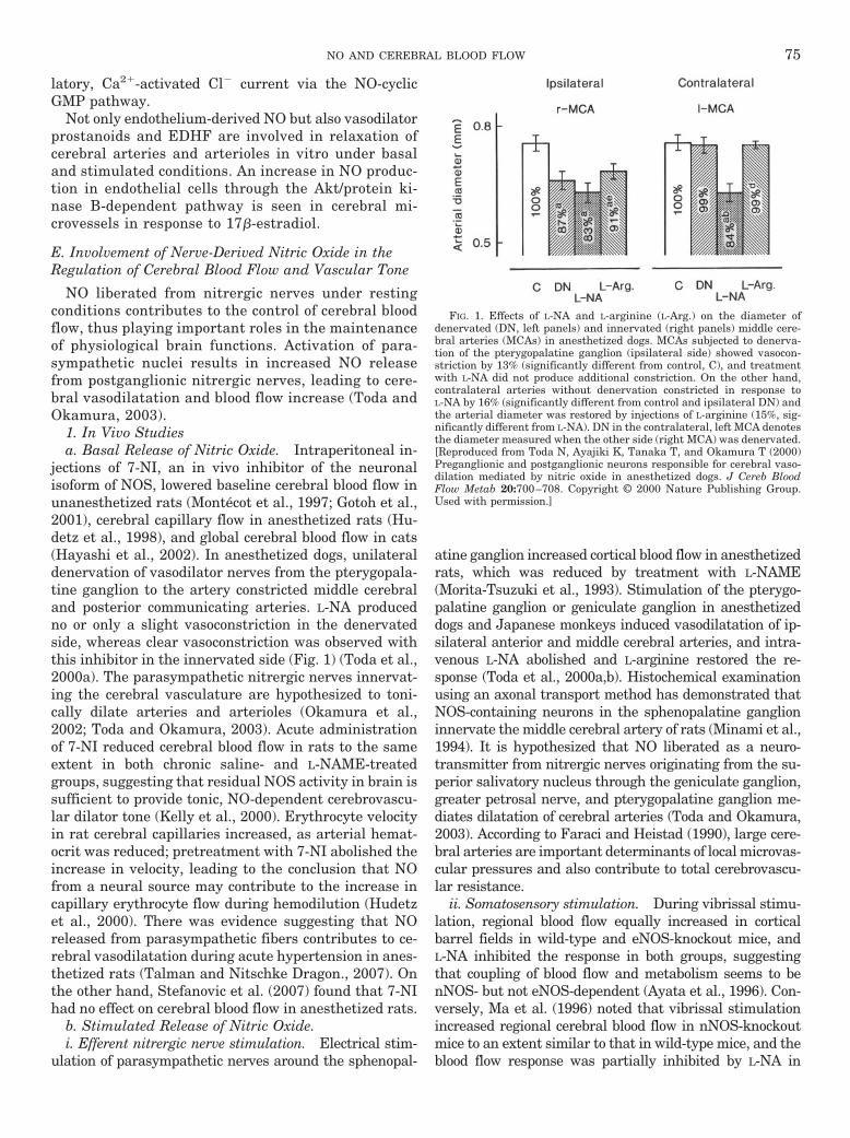
latory, Ca2�-activated Cl� current via the NO-cyclicGMP pathway.
Not only endothelium-derived NO but also vasodilatorprostanoids and EDHF are involved in relaxation ofcerebral arteries and arterioles in vitro under basaland stimulated conditions. An increase in NO produc-tion in endothelial cells through the Akt/protein ki-nase B-dependent pathway is seen in cerebral mi-crovessels in response to 17�-estradiol.
E. Involvement of Nerve-Derived Nitric Oxide in theRegulation of Cerebral Blood Flow and Vascular Tone
NO liberated from nitrergic nerves under restingconditions contributes to the control of cerebral bloodflow, thus playing important roles in the maintenanceof physiological brain functions. Activation of para-sympathetic nuclei results in increased NO releasefrom postganglionic nitrergic nerves, leading to cere-bral vasodilatation and blood flow increase (Toda andOkamura, 2003).
1. In Vivo Studiesa. Basal Release of Nitric Oxide. Intraperitoneal in-
jections of 7-NI, an in vivo inhibitor of the neuronalisoform of NOS, lowered baseline cerebral blood flow inunanesthetized rats (Montecot et al., 1997; Gotoh et al.,2001), cerebral capillary flow in anesthetized rats (Hu-detz et al., 1998), and global cerebral blood flow in cats(Hayashi et al., 2002). In anesthetized dogs, unilateraldenervation of vasodilator nerves from the pterygopala-tine ganglion to the artery constricted middle cerebraland posterior communicating arteries. L-NA producedno or only a slight vasoconstriction in the denervatedside, whereas clear vasoconstriction was observed withthis inhibitor in the innervated side (Fig. 1) (Toda et al.,2000a). The parasympathetic nitrergic nerves innervat-ing the cerebral vasculature are hypothesized to toni-cally dilate arteries and arterioles (Okamura et al.,2002; Toda and Okamura, 2003). Acute administrationof 7-NI reduced cerebral blood flow in rats to the sameextent in both chronic saline- and L-NAME-treatedgroups, suggesting that residual NOS activity in brain issufficient to provide tonic, NO-dependent cerebrovascu-lar dilator tone (Kelly et al., 2000). Erythrocyte velocityin rat cerebral capillaries increased, as arterial hemat-ocrit was reduced; pretreatment with 7-NI abolished theincrease in velocity, leading to the conclusion that NOfrom a neural source may contribute to the increase incapillary erythrocyte flow during hemodilution (Hudetzet al., 2000). There was evidence suggesting that NOreleased from parasympathetic fibers contributes to ce-rebral vasodilatation during acute hypertension in anes-thetized rats (Talman and Nitschke Dragon., 2007). Onthe other hand, Stefanovic et al. (2007) found that 7-NIhad no effect on cerebral blood flow in anesthetized rats.
b. Stimulated Release of Nitric Oxide.i. Efferent nitrergic nerve stimulation. Electrical stim-
ulation of parasympathetic nerves around the sphenopal-
atine ganglion increased cortical blood flow in anesthetizedrats, which was reduced by treatment with L-NAME(Morita-Tsuzuki et al., 1993). Stimulation of the pterygo-palatine ganglion or geniculate ganglion in anesthetizeddogs and Japanese monkeys induced vasodilatation of ip-silateral anterior and middle cerebral arteries, and intra-venous L-NA abolished and L-arginine restored the re-sponse (Toda et al., 2000a,b). Histochemical examinationusing an axonal transport method has demonstrated thatNOS-containing neurons in the sphenopalatine ganglioninnervate the middle cerebral artery of rats (Minami et al.,1994). It is hypothesized that NO liberated as a neuro-transmitter from nitrergic nerves originating from the su-perior salivatory nucleus through the geniculate ganglion,greater petrosal nerve, and pterygopalatine ganglion me-diates dilatation of cerebral arteries (Toda and Okamura,2003). According to Faraci and Heistad (1990), large cere-bral arteries are important determinants of local microvas-cular pressures and also contribute to total cerebrovascu-lar resistance.
ii. Somatosensory stimulation. During vibrissal stimu-lation, regional blood flow equally increased in corticalbarrel fields in wild-type and eNOS-knockout mice, andL-NA inhibited the response in both groups, suggestingthat coupling of blood flow and metabolism seems to benNOS- but not eNOS-dependent (Ayata et al., 1996). Con-versely, Ma et al. (1996) noted that vibrissal stimulationincreased regional cerebral blood flow in nNOS-knockoutmice to an extent similar to that in wild-type mice, and theblood flow response was partially inhibited by L-NA in
FIG. 1. Effects of L-NA and L-arginine (L-Arg.) on the diameter ofdenervated (DN, left panels) and innervated (right panels) middle cere-bral arteries (MCAs) in anesthetized dogs. MCAs subjected to denerva-tion of the pterygopalatine ganglion (ipsilateral side) showed vasocon-striction by 13% (significantly different from control, C), and treatmentwith L-NA did not produce additional constriction. On the other hand,contralateral arteries without denervation constricted in response toL-NA by 16% (significantly different from control and ipsilateral DN) andthe arterial diameter was restored by injections of L-arginine (15%, sig-nificantly different from L-NA). DN in the contralateral, left MCA denotesthe diameter measured when the other side (right MCA) was denervated.[Reproduced from Toda N, Ayajiki K, Tanaka T, and Okamura T (2000)Preganglionic and postganglionic neurons responsible for cerebral vaso-dilation mediated by nitric oxide in anesthetized dogs. J Cereb BloodFlow Metab 20:700–708. Copyright © 2000 Nature Publishing Group.Used with permission.]
NO AND CEREBRAL BLOOD FLOW 75
wild-type mice but was not affected in nNOS-knockoutmice. The authors suggested that in mice deficient innNOS expression, endothelial NO production does not me-diate the blood flow coupling to neuronal activity, andNO-independent mechanisms couple regional cerebralblood flow and metabolism during whisker stimulation.However, the following data support the participation ofneurogenic NO in the stimulation-induced increase in ce-rebral blood flow. Whisker stimulation for 10 min led to acontinuous and persistent cortical blood flow increase inanesthetized rats; after nNOS inhibition, the blood flowresponse was depressed (Bonvento et al., 2000). Regulationof the cerebral blood flow in response to increased neuronalactivity seems to be a dynamic and tonic process, in whichnNOS plays an essential role. Increases in cerebral bloodflow during vibrissal stimulation in unanesthetized ratswere reduced by L-NAME in the ventroposteromedial tha-lamic nucleus and reduced by 7-NI in both the ventropos-teromedial thalamic nucleus and barrel cortex (Gotoh etal., 2001). In wild-type mice, upper lip stimulation in-creased blood flow in Crus II, a region of the cerebellarcortex that receives trigeminal sensory afferents, and theblood flow response was reduced in cyclin D2-null micethat had a profound reduction in the number of stellateneurons in the cerebellar monolayer; 7-NI attenuated thevascular response to Crus II activation in wild-type micebut not in mice lacking cyclin D2 (Yang et al., 2000).Stellate neurons are probably the major source of NO me-diating the vascular response. Increased blood flow in theCrus II in response to upper lip stimulation seen in wild-type mice was attenuated in nNOS-null mice; the increasein cerebellar blood flow by superfusion of Crus II withglutamate or by systemic administration of harmaline wasalso attenuated in nNOS-knockout mice, implying thatnNOS-derived NO is a critical link between glutamatergicsynaptic activity and blood flow in the activated cerebel-lum (Yang et al., 2003).
Ayajiki et al. (2005) provided evidence indicating thatNO released from parasympathetic nerves and neuropep-tide(s) released antidromically from sensory nerves seemto be responsible for the increase in cerebral blood flow inthe rat. Electrical somatosensory stimulation in the uni-lateral forepaw elicited an increase in cerebral blood flowin the cat contralateral somatosensory cortex and the ipsi-lateral cerebellum, and 7-NI decreased cerebral blood flowboth during rest and activation, suggesting that neuronalNO has an important role as a mediator of regional neu-rovascular coupling (Hayashi et al., 2002). 7-NI alsoattenuated cerebral blood flow responses to electricalstimulation of the forepaw and decreased the amplitudeof somatosensory-evoked potentials (Stefanovic et al.,2007). Electrical forepaw stimulation resulted in an im-mediate increase in NO in the rat somatosensory cortexfollowed by cortical blood flow increase (Buerk et al.,2003). Some evidence suggested that an epoxygenase,presumably localized in astrocytes, and NOS are bothrequired for generating the cortical hyperemic response
in rats evoked by electrical forepaw stimulation (Peng etal., 2004). It was suggested that the increase in cerebralblood flow in the hippocampus during walking in rats isattributable to activation of nicotinic receptors by AChreleased from vasodilator nerves projecting to the hip-pocampus, resulting in the production of NO (Nakajimaet al., 2003).
iii. Hypercapnia and hyperoxia. Administration of7-NI reduced the vasodilator and cerebral blood flowresponses to hypercapnia in anesthetized rats, suggest-ing that NO synthesized by nNOS participates in hyper-capnic hyperemia (Wang et al., 1995; Okamoto et al.,1997). From studies using mice lacking nNOS or eNOSand wild-type mice, Atochin et al. (2003b) obtained evi-dence suggesting that cerebral vasodilatation inducedby 60-min exposure to hyperbaric oxygen depends onboth nNOS and eNOS. The cerebral blood flow responseas well as blood oxygenation level-dependent signal in-tensity changes after electrical stimulation were abol-ished after application of 7-NI in rats (Burke andBuhrle, 2006).
iv. Response to N-methyl-D-aspartate. NMDA (Faraciand Breese, 1993) and kainic acid (Faraci et al., 1994)increase cerebral blood flow via neuronally derived NO.Microdialysis perfusion of NMDA doubled blood flow inrat striatum and increased NO production; perfusion ofL-NA inhibited the blood flow response to NMDA, and anepoxygenase substrate inhibitor blocked the increase inblood flow without decreasing NO recovery (Bhardwaj etal., 2000). These authors concluded that both the P450epoxygenase and NOS pathways seem to be involved inthe local cerebral blood flow response to NMDA receptoractivation. A direct cortical application of NMDA in-creased regional blood flow and oxygen consumption inrats, and 7-NI attenuated the effects of NMDA on cere-bral blood flow and decreased the oxygen consumptionduring NMDA receptor stimulation (Chi et al., 2003).NMDA induced both NO flux and cerebellum vasodi-latation that were abolished by treatment of rats withan NOS inhibitor and tetrodotoxin, suggesting thatNO-producing interneurons mediate this vasomotorresponse (Rancillac et al., 2006). These authors alsodemonstrated that electrical stimulation of single ni-trergic stellate cells was sufficient to release NO anddilate intraparenchymal and upstream pial microves-sels. Harris et al. (2008) obtained findings suggestingthat NO-dependent cerebral vasodilatation in response toNMDA receptor activation was present as early as 0.65gestation in fetal sheep and increased further during thelast trimester.
c. Involvement of Neurogenic Nitric Oxide in Diseases.There are recorded studies indicating the participationof neurogenic NO in some diseases. The selective nNOSinhibitor ARL 17477 and the nonselective inhibitor L-NAreduced intraischemic cortical cerebral blood flow to asimilar extent in rats subjected to forebrain ischemia,suggesting that the nNOS contribution to intraischemic
76 TODA ET AL.

vasodilatation in vulnerable brain regions is greaterthan that of eNOS (Santizo et al., 2000). Combinedtreatment with 7-NI and propranolol but not treatmentwith 7-NI or propranolol alone was accompanied by areduction in the hypoglycemia-induced pial arterial di-latation; in the presence of tetrodotoxin, the arterialresponse was lost in rats (Santizo et al., 2001). �-Adre-noceptors and nNOS-derived NO seem to interact incontributing to hypoglycemia-induced cerebral vasodila-tation, and the vasodilating signal may be trans-mitted via a neuronal pathway. In anesthetized rats,without the nNOS inhibitor propyl-L-arginine, the pialarterial diameter abruptly increased when mean arte-rial pressure rose, whereas with nNOS inhibition thediameter increased to a much lesser extent even whenarterial pressure was similarly raised; the nNOS inhib-itor attenuated pial arterial dilatation induced byNMDA but not that induced by ACh delivered into acranial window (Talman and Nitschke Dragon, 2007).NO released from parasympathetic fibers seems to con-tribute to cerebral vasodilatation during acute hyperten-sion. Topical application of NMDA or kinate producedpial arteriolar dilatation in alcohol-fed (8–12 weeks)rats to a lesser extent than nonalcohol-fed rats; treat-ment with L-NAME inhibited dilatation of pial arteriolesin response to NMDA, implying that chronic alcoholconsumption impairs nNOS-dependent pial arteriolardilatation (Sun et al., 2002). Impaired reactivity of cere-bral blood vessels to neuronal activation may contributeto the pathogenesis of cerebrovascular disorders ob-served during chronic alcohol consumption. At clinicallyrelevant concentrations, halothane dilated rat intrace-rebral arterioles (Staunton et al., 2000). This dilatationin hippocampal microvessels was mediated by neuro-genic NO but not NO derived from the endothelium.
d. Interactions between Nitric Oxide from NitrergicNerves and Other Neurotransmitters. The nicotine-induced increase in the rat cortical blood flow, mediatedby nerve-derived NO, was attenuated by propranololand the �2-adrenoceptor antagonist ICI 118,551, but notby the �1-adrenoceptor antagonist metoprolol (Uchida etal., 2002). According to Toda and Okamura (2003), the�-adrenoceptor-cyclic AMP system in nitrergic nerveterminals does not participate in the regulation of re-lease and synthesis of NO in peripheral arteries andveins from various mammals.
Increases in rat meningeal blood flow caused by NOhas been suggested to depend on the release and vaso-dilatory action of calcitonin gene-related peptide fromdural afferents (Strecker et al., 2002a,b). The interactionof NO and calcitonin gene-related peptide may be rele-vant for the development of vascular headaches.
e. Histological Demonstration of Nitrergic Innervationin Cerebral Vasculature. Numerous histochemical andimmunohistochemical studies have demonstrated theinnervation of nitrergic nerves in cerebral arteries andarterioles from a variety of mammals (Toda and Oka-
mura, 2003), including humans (Nozaki et al., 1993;Gorelova et al., 1996) and monkeys (Yoshida et al., 1994;Toda et al., 1997). It was demonstrated on the electronmicroscopic level that most perivascular NOS neuronalelements corresponded to nerve terminals and a major-ity of these were located in the immediate vicinity of theblood vessels; NOS terminals abutting on cortical NOSneurons were primarily nonjunctional (Tong and Hamel,2000). The authors raised the possibility that nitrergicbasal forebrain neurons are involved in the blood flowresponse observed after stimulation of the basal fore-brain. Using the fluorescent imaging method, Brown etal. (2000) provided evidence suggesting that tonic activ-ity in perivascular nitrergic nerve fibers lying in closeproximity to intraparenchymal microvessels may be asource of dilator tone within the parenchyma. Double-labeling studies revealed that cytochrome P450 2C11epoxygenase and soluble epoxide hydrolase immunore-activity predominantly colocalized with nNOS immuno-reactivity within perivascular nerves (Iliff et al., 2007).The presence of enzymes involved in production andinactivation of eicosatrienoic acids within extrinsic para-sympathetic and sensory vasodilator fibers suggests anovel role for eicosatrienoic acids in the neurogenic con-trol of cerebral arteries.
From studies on Wistar rats of various postnatal days,Morys et al. (2002) found that NOS and neuropeptide Yimmunoreactivities in rat hippocampus were weak inthe newborn rat. Then the levels increased until the 7thday of postnatal life, and thereafter until day 14 theywere maintained at the similar levels; until the 10th dayof life, the immunoreactive cells were immature.
f. Summary of the Possible Role of Neuronal Nitric-Oxide Synthase-Derived Nitric Oxide. The hypothesisthat NO formed through nNOS contributes to the in-crease in cerebral circulation is supported by the follow-ing observations. 1) Cerebral vasodilatation or increasedcerebral blood flow is evoked by efferent nerve stimula-tion by electrical pulses or ganglionic stimulants such asnicotine. The responses are susceptible to NOS inhibi-tors and soluble guanylyl cyclase inhibitors, and lightand/or electron microscopy reveals that nNOS-immuno-reactive nerve fibers are located in close proximity tovascular smooth muscle cells. 2) Cerebral arteriolar di-latation or a blood flow increase induced by somatosen-sory stimulation is blocked by selective nNOS inhibitors,such as 7-NI, concomitant with a histochemical deter-mination of nNOS-positive nerve fibers in the immedi-ate vicinity of intracerebral arterioles. 3) Cerebral arte-riolar dilatation or the blood flow increase induced byNMDA receptor agonists is inhibited by 7-NI, and in-creased NO levels are also observed in brain tissues. 4)Cerebral vasodilator responses to stimulation of efferentor afferent nerves are reduced in nNOS-, but not ineNOS- and iNOS-deficient mice. Figure 2 summarizesthe origins of NO formed by nNOS and eNOS that acts
NO AND CEREBRAL BLOOD FLOW 77
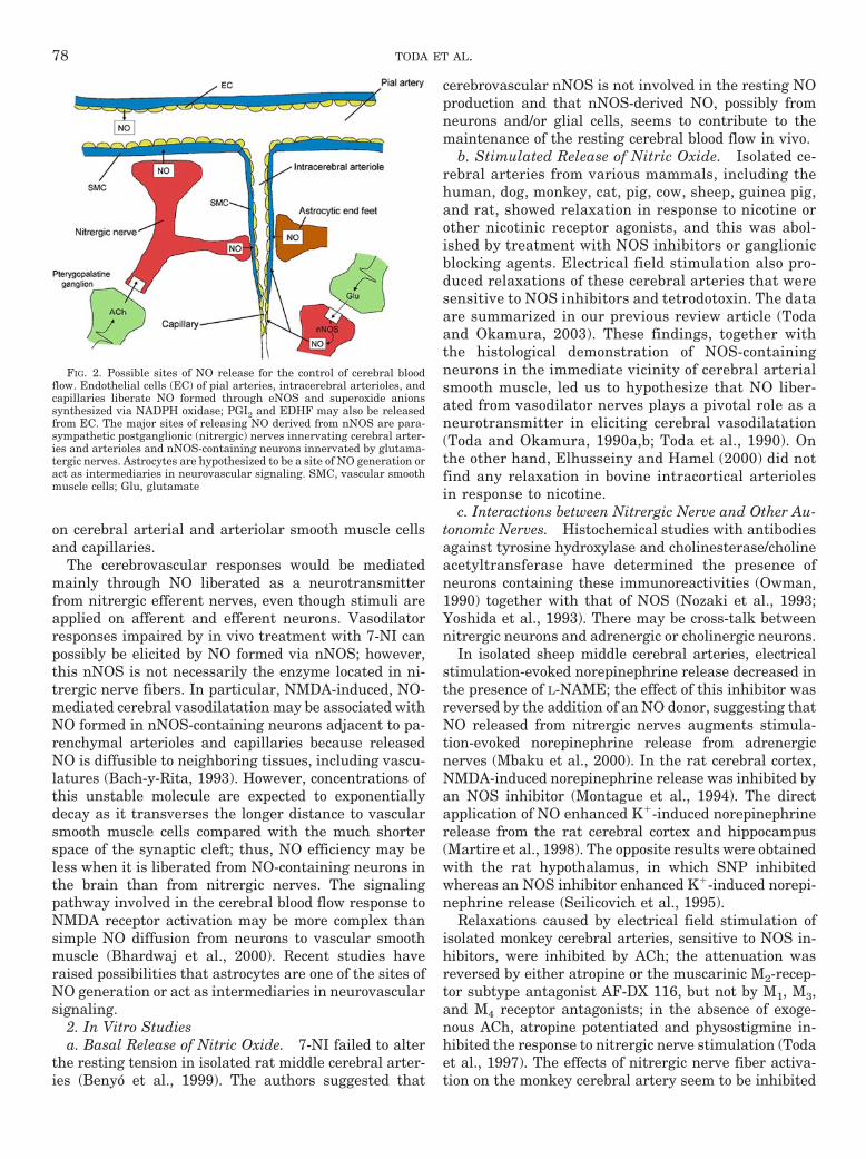
on cerebral arterial and arteriolar smooth muscle cellsand capillaries.
The cerebrovascular responses would be mediatedmainly through NO liberated as a neurotransmitterfrom nitrergic efferent nerves, even though stimuli areapplied on afferent and efferent neurons. Vasodilatorresponses impaired by in vivo treatment with 7-NI canpossibly be elicited by NO formed via nNOS; however,this nNOS is not necessarily the enzyme located in ni-trergic nerve fibers. In particular, NMDA-induced, NO-mediated cerebral vasodilatation may be associated withNO formed in nNOS-containing neurons adjacent to pa-renchymal arterioles and capillaries because releasedNO is diffusible to neighboring tissues, including vascu-latures (Bach-y-Rita, 1993). However, concentrations ofthis unstable molecule are expected to exponentiallydecay as it transverses the longer distance to vascularsmooth muscle cells compared with the much shorterspace of the synaptic cleft; thus, NO efficiency may beless when it is liberated from NO-containing neurons inthe brain than from nitrergic nerves. The signalingpathway involved in the cerebral blood flow response toNMDA receptor activation may be more complex thansimple NO diffusion from neurons to vascular smoothmuscle (Bhardwaj et al., 2000). Recent studies haveraised possibilities that astrocytes are one of the sites ofNO generation or act as intermediaries in neurovascularsignaling.
2. In Vitro Studiesa. Basal Release of Nitric Oxide. 7-NI failed to alter
the resting tension in isolated rat middle cerebral arter-ies (Benyo et al., 1999). The authors suggested that
cerebrovascular nNOS is not involved in the resting NOproduction and that nNOS-derived NO, possibly fromneurons and/or glial cells, seems to contribute to themaintenance of the resting cerebral blood flow in vivo.
b. Stimulated Release of Nitric Oxide. Isolated ce-rebral arteries from various mammals, including thehuman, dog, monkey, cat, pig, cow, sheep, guinea pig,and rat, showed relaxation in response to nicotine orother nicotinic receptor agonists, and this was abol-ished by treatment with NOS inhibitors or ganglionicblocking agents. Electrical field stimulation also pro-duced relaxations of these cerebral arteries that weresensitive to NOS inhibitors and tetrodotoxin. The dataare summarized in our previous review article (Todaand Okamura, 2003). These findings, together withthe histological demonstration of NOS-containingneurons in the immediate vicinity of cerebral arterialsmooth muscle, led us to hypothesize that NO liber-ated from vasodilator nerves plays a pivotal role as aneurotransmitter in eliciting cerebral vasodilatation(Toda and Okamura, 1990a,b; Toda et al., 1990). Onthe other hand, Elhusseiny and Hamel (2000) did notfind any relaxation in bovine intracortical arteriolesin response to nicotine.
c. Interactions between Nitrergic Nerve and Other Au-tonomic Nerves. Histochemical studies with antibodiesagainst tyrosine hydroxylase and cholinesterase/cholineacetyltransferase have determined the presence ofneurons containing these immunoreactivities (Owman,1990) together with that of NOS (Nozaki et al., 1993;Yoshida et al., 1993). There may be cross-talk betweennitrergic neurons and adrenergic or cholinergic neurons.
In isolated sheep middle cerebral arteries, electricalstimulation-evoked norepinephrine release decreased inthe presence of L-NAME; the effect of this inhibitor wasreversed by the addition of an NO donor, suggesting thatNO released from nitrergic nerves augments stimula-tion-evoked norepinephrine release from adrenergicnerves (Mbaku et al., 2000). In the rat cerebral cortex,NMDA-induced norepinephrine release was inhibited byan NOS inhibitor (Montague et al., 1994). The directapplication of NO enhanced K�-induced norepinephrinerelease from the rat cerebral cortex and hippocampus(Martire et al., 1998). The opposite results were obtainedwith the rat hypothalamus, in which SNP inhibitedwhereas an NOS inhibitor enhanced K�-induced norepi-nephrine release (Seilicovich et al., 1995).
Relaxations caused by electrical field stimulation ofisolated monkey cerebral arteries, sensitive to NOS in-hibitors, were inhibited by ACh; the attenuation wasreversed by either atropine or the muscarinic M2-recep-tor subtype antagonist AF-DX 116, but not by M1, M3,and M4 receptor antagonists; in the absence of exoge-nous ACh, atropine potentiated and physostigmine in-hibited the response to nitrergic nerve stimulation (Todaet al., 1997). The effects of nitrergic nerve fiber activa-tion on the monkey cerebral artery seem to be inhibited
FIG. 2. Possible sites of NO release for the control of cerebral bloodflow. Endothelial cells (EC) of pial arteries, intracerebral arterioles, andcapillaries liberate NO formed through eNOS and superoxide anionssynthesized via NADPH oxidase; PGI2 and EDHF may also be releasedfrom EC. The major sites of releasing NO derived from nNOS are para-sympathetic postganglionic (nitrergic) nerves innervating cerebral arter-ies and arterioles and nNOS-containing neurons innervated by glutama-tergic nerves. Astrocytes are hypothesized to be a site of NO generation oract as intermediaries in neurovascular signaling. SMC, vascular smoothmuscle cells; Glu, glutamate
78 TODA ET AL.

by nerve-released ACh acting on prejunctional musca-rinic receptors, possibly of the M2 subtype. Voltage-dependent, �-conotoxin-sensitive Ca2� currents in thecultured rat sphenopalatine ganglion were inhibited byACh and a muscarinic M2 receptor agonist; the inhibi-tory effects were reversed by atropine and a M2 receptorantagonist but were not affected by M1, M3, and M4
receptor antagonists (Liu et al., 2002b). The sphenopal-atine/pterygopalatine ganglia are major sources ofnNOS and vasoactive intestinal peptide and send post-ganglionic nitrergic and peptidergic neurons to middlecerebral arteries (Edvinsson et al., 2001).
F. Astrocyte-Derived Nitric Oxide and OtherVasodilators
Histochemical study demonstrated the subcellulardistribution of NADPH-diaphorase (NOS) in the ratstriatal astroglia; electron microscopic examinationrevealed disposition of the NADPH-diaphorase reac-tion product predominantly in mitochondria of astro-cytic “end feet” (Calka and Wolf, 2003). The authorshave suggested that astroglial NADPH diaphorase/NOS may be involved in adaptation of the local bloodflow in the striatal microenvironment. The gap junc-tion protein connexin-43 and purinergic P2Y2 and P2Y4
receptors were expressed in end feet abutting capil-laries and larger cerebral vessels; ATP mobilized cy-tosolic Ca2� in astrocytic end feet, whereas electricalstimulation triggered Ca2� waves propagating alongthe vessel wall (Simard et al., 2003). It was speculatedthat Ca2� signaling may play a role in astrocytic func-tion related to blood flow regulation and that astro-cytes are in a position for sensing neuronal activityand communicating with blood vessels in the brainparenchyma. The dilatation of arterioles triggered byneural activity in rat cortical slices was dependent onglutamate-mediated [Ca2�]i oscillations in astrocytes,and inhibition of these Ca2� responses resulted in theimpairment of stimulated vasodilatation; a COX prod-uct was involved in this astrocyte-mediated control ofarterioles, suggesting that neuron-to-astrocyte signal-ing is a key mechanism in functional hyperemia(Zonta et al., 2003). Filosa et al. (2004) obtained datasuggesting that astrocytic Ca2� changes signal thecerebral microvasculature. Koehler et al. (2006) sug-gested that astrocytes act as intermediaries in neuro-vascular signaling and that neurally derived NO,COX-2 metabolites, epoxyeicosatrienoic acids, andadenosine couple increased cerebral blood flow to in-creased neuronal activity. The production of PGE2 orepoxyeicosatrienoic acid, the level of NO, and the ac-tivation of Ca2�-activated K� channels in astrocytesseem to participate in cerebral vasodilatation (Gordonet al., 2007). More work is needed to clarify the con-tributions of astrocytes to vascular dynamics.
IV. Cerebral Blood Flow Regulation by NitricOxide under Pathological Conditions
A. Cerebral Ischemia and Stroke
1. Ischemia and Endothelial Nitric-Oxide Synthase-Derived Nitric Oxide. In rats that underwent revers-ible occlusion of the right middle cerebral artery, eNOSprotein occurred in all blood vessels and microvessels inthe ischemic striatum after 24 to 168 h of reperfusion,and eNOS expression was also demonstrated; levels ofbrain NOS did not increase after ischemia (Veltkamp etal., 2002). Ischemia associated with occlusion of the bi-lateral common carotid arteries induced increases in NOend-products (NOx) during ischemia and prominentlyafter reperfusion, and such increases were abolished byL-NAME but only slightly reduced by 7-NI; the increasein the quantity of NO end-products became prominentand was persistent after reperfusion in rats in which thecerebral blood flow during ischemia was in the rangebetween 22.7 and 60 ml/100 g/min, whereas in animalsin which the cerebral blood flow during ischemia fellbelow 22.7 ml/100 g/min, the end-products decreasedduring ischemia increased transiently after reperfusion.(Uetsuka et al., 2002). The increase in NO seems to bederived from eNOS, and NO synthesis during and afterischemia may be related to cerebral blood flow. In anes-thetized goats subjected to a 60-min occlusion of the leftmiddle cerebral artery followed by a 60-min reperfusion,ipsilateral middle cerebral artery resistance was de-creased, and the cerebral vasodilatation to ADP but notto SNP was reduced; the relaxation to ADP but not toSNP was less in isolated left (ischemic) middle cerebralarteries than in right (control) ones (Sanchez et al.,2006). These authors obtained further evidence suggest-ing that ischemia-reperfusion produces cerebrovascularendothelial dysfunction, which may be associated withdecreased NO availability, decreased release of EDHF(Chen et al., 1988), and increased production of vaso-constrictor prostaglandins and that these alterationsmay be related with increased production of superoxideanions. In wild-type mouse brains subjected to middlecerebral artery occlusion, endothelial caveolin-1 andcaveolin-2 and eNOS protein levels were increased; con-versely, the protein levels of eNOS remained unchangedand the cerebral volume of infarction was markedly in-creased in caveolin-1-knockout, ischemic mousebrains, suggesting that caveolin-1-deficient, ischemicbrains show impaired angiogenesis and increased ap-optotic cell death (Jasmin et al., 2007). Adiponectin-deficient mice exhibited enlarged brain infarction andincreased neurological deficits after ischemia-reperfu-sion and also decreased cerebral blood flow duringischemia compared with wild-type mice; phosphoryla-tion of eNOS in ischemic brain tissues and the pro-duction of NO metabolites in plasma were attenuatedin adiponectin-deficient mice. Conversely, adenovirus-mediated supplementation of adiponectin reduced cere-
NO AND CEREBRAL BLOOD FLOW 79

bral infarct size in wild-type and adiponectin-knockoutmice but did not affect brain infarction in mice deficientin eNOS (Nishimura et al., 2008). Adiponectin seems toexert a cerebroprotective action through an eNOS-de-pendent mechanism.
In three groups of rats subjected to chronic brain hypo-perfusion and treated with 7-NI, aminoguanidine, or imi-noethyl-L-ornithine (relatively selective inhibitors ofnNOS, iNOS, and eNOS, respectively), only rats adminis-tered the eNOS inhibitor showed a worsened water mazeperformance, suggesting that vascular NO derived fromeNOS may play a critical role in spatial memory functionduring brain hypoperfusion by keeping cerebral blood flowoptimal (de la Torre and Aliev, 2005). NOx levels inplasma, ischemic brain tissue, and cerebrospinal fluid in-creased after transient middle cerebral artery occlusion inrats. ADMA levels were unchanged in plasma, but de-creased in cerebrospinal fluid after the occlusion; theADMA/NOx ratio in cerebrospinal fluid decreased afterischemia (Xu et al., 2007). The combined increase in NOxand decrease in ADMA suggests occurrence of markedactivation of NOS. There is evidence indicating that RhoA/Rho-kinase activity is increased in cerebral vascular dis-eases, such as ischemic brain injury, cerebral vasospasm,hypertension, and diabetes, not only in vascular musclebut also in the endothelium that results in decreases in NOproduction and increases in cerebral vascular tone (Didionet al., 2005; Chrissobolis and Sobey, 2006). Sercombe et al.(2001) provided evidence indicating that in the SHR withfocal cerebral ischemia, cerebral infarct expansion may belimited by chronic treatment with L-NAME through a pro-gressive reduction in cerebral artery response to vasocon-strictor neurotransmitters, concomitant with augmentedresponses to the guanylyl cyclase-independent vasodilatorpapaverine and some recovery of NO-mediated dilatationto ACh.
Intrauterine ischemia-hypoxia insult applied by uter-ine artery occlusion on day 21 of pregnancy caused in-creases in both activities and mRNA levels of eNOS,whereas there were no changes in nNOS mRNA levels,suggesting that eNOS may contribute to the mainte-nance of cerebral blood flow against ischemia or hypoxiain fetal rats (Nakata et al., 2002). Brachiocephalic occlu-sion increased nNOS (hippocampus and brainstem),iNOS (hypothalamus), and eNOS (hippocampus and cor-tex) at the protein level in fetal sheep brain, estradiolincreased nNOS (brainstem and cortex), iNOS (hip-pocampus and hypothalamus), and eNOS (brainstemand cerebellum), and the combination of occlusion andestradiol produced smaller changes in NOS isomersthan those produced by either stimulus (Wood and Gir-oux, 2006).
Voluntary training on running wheels or exercise ontreadmill apparatus reduced cerebral infarct size andfunctional deficits, improved endothelium-dependentvasodilatation, and augmented cerebral blood flow inwild-type mice, whereas the neuroprotective effects were
absent in eNOS-deficient mice, indicating that the en-hanced eNOS activity by physical training seems to bethe mechanism by which this modality protects againstcerebral injury (Endres et al., 2003). Continuous volun-tary running on wheels conferred long-term up-regula-tion of eNOS in the vasculature and of endothelial pro-genitor cells in the spleen and bone marrow: 4 weeksafter the ischemic insult, trained animals showed highernumbers of newly generated vascular cells, increaseddensity of perfused microvessels, and sustained aug-mentation of cerebral blood flow within the ischemicstriatum; the protective effects of physical training wereabolished in animals treated with NOS inhibitors or inthose lacking eNOS expression (Gertz et al., 2006).
On the other hand, there is evidence for neuroprotec-tion by NOS inhibition against ischemic injury. Thenovel calmodulin-dependent NOS inhibitor DY-9760e,which inhibits eNOS activity and, in turn, protein ty-rosine nitration, inhibited blood-brain barrier disruptioninduced by microsphere embolism and also inhibitedcleavage of poly(ADP-ribose) polymerase 1 (PARP-1) asa marker of the apoptotic pathway in vascular endothe-lial cells in rats (Han et al., 2006). Microsphere embo-lism-induced eNOS expression in vascular endothelialcells probably mediates blood-brain barrier disruptionand, in turn, brain edema.
Urotensin-II (U-II) is a vasoactive factor with pleio-tropic effects that is identified in mammalian speciesincluding humans (Bousette and Giaid, 2006). U-II andits receptor are expressed as mRNA and protein in ratneuromicrovascular endothelial cells, and U-II exerts apro-angiogenic effect (Spinazzi et al., 2006). Administra-tion of U-II into the lateral cerebral ventricle increasedcerebral blood flow in anesthetized rats, and this effectwas reduced by NOS inhibition. U-II administrationafter the induction of cerebral ischemia failed to alterresidual cerebral blood flow in the affected hemisphere,but after reperfusion, U-II-treated rats displayed a hy-perperfusion; treatment with U-II increased the volumeof infarction, suggesting an exacerbation by U-II of braindamage associated with an ischemic insult (Chuquet etal., 2008).
In summary, ischemia with reversible occlusion of thecerebral artery causes increments in endothelial caveo-lin and eNOS protein levels, plasma NOx concentra-tions, and cerebral blood flow that seem to play a role inminimizing impaired memory function. Physical train-ing protects against cerebral injury after ischemic insultdue possibly to enhanced eNOS activity and NO forma-tion. The increased RhoA/Rho-kinase activity in isch-emic brain injury decreases NO production.
2. Ischemia and Neuronal Nitric-Oxide Synthase. Fe-male mice lacking the nNOS gene exhibited exacerbatedhistological injury after middle cerebral artery occlusionrelative to that in wild-type females, unlike the protectionobserved in male nNOS-knockout mice, and treatmentwith 7-NI increased infarction in female wild-type mice,
80 TODA ET AL.

but protected male mice. Unlike male PARP-1 knockoutmice, female PARP-1 knockout littermates sustainedgrossly increased ischemic damage relative to that insex-matched wild-type mice, and loss of PARP-1 resultedin reversal of the neuroprotective activity by the femalesex steroid (McCullough et al., 2005). Cell death path-ways involving NO and PARP ischemic neurotoxicitymay be operant solely in male brain, and the integrity ofnNOS/PARP-1 signaling is paradoxically protective inthe female. Although deletion of HO-2 resulted ingreater stroke damage (Dore et al., 2000), the pharma-cological inhibition of nNOS or its gene deletion con-ferred neuroprotection in mice with transient cerebralischemia (Eliasson et al., 1999; Wei et al., 1999). Indouble knockout (HO-2 and nNOS) mice lacking bothgenes, no difference was observed in the volume of in-farction, neurological signs, and decrease in relative ce-rebral blood flow during ischemia associated with mid-dle cerebral artery occlusion compared with wild-typemice, suggesting that the deleterious action of nNOSwould counteract the role of HO-2 in neuroprotection(Namiranian et al., 2005).
3. Ischemia and Inducible Nitric-Oxide Synthase.Nitrotyrosine, a marker of peroxynitrite, was detectedafter a 15-h reperfusion only in wild-type mice, in whichthe left middle cerebral artery was occluded for 2 h, butnot in iNOS-knockout or sham-operated mice; nitroty-rosine was seen predominantly in the vascular wall inthe peri-infarct region of the cerebral cortex in wild-typemice (Hirabayashi et al., 2000). It seems that iNOS isresponsible for nitrotyrosine formation in the laterphase of reperfusion and that vascular endothelium isthe major site of the reaction. The expression of iNOSwas detected at 22 to 70 h after reperfusion in thevascular wall and cortex of rats subjected to transientmiddle cerebral artery occlusion, and nitrotyrosine inthe vascular wall and cortex and NOx levels in thecirculation increased 4 to 46 h after reperfusion, sug-gesting that marked increases in the NOx level mightreflect the expression of iNOS (Suzuki et al., 2002).Hyperglycemia induced before ischemia in rats resultedin earlier infarction, which correlated with increasedimmunoreactivity for iNOS, manganese SOD, and nitro-tyrosine (Ste-Marie et al., 2001). Based on the observedtime courses of the occurrence of ischemic infarction,oxidative stress, iNOS expression, and neutrophil infil-tration, Lerouet et al. (2002) concluded that the delayediNOS activity and neutrophil infiltration may not con-tribute to ischemic brain damage in rats. Pruss et al.(2008) noted that with the use of three different strainsof iNOS-deficient mice and wild-type controls, infarctsize was independent of iNOS deletion and thus sug-gested that iNOS is not a universal mediator of braindamage after cerebral ischemia.
4. Ischemic Preconditioning. Huang (2004) summa-rized the effects of NO after cerebral ischemia, the sig-
naling pathways through which NO acts, and its poten-tial roles in cerebral ischemic preconditioning (IPC).
Wild-type mice subjected to IPC showed decreases inthe neurological deficit and infarct size induced by isch-emic insult, whereas neither eNOS nor nNOS knockoutmice showed protection from IPC; blood flow decreasesin core ischemic areas were the same in all groups,suggesting that NO formed by eNOS or nNOS may playa role in the molecular mechanisms of protection(Atochin et al., 2003a). The early and late phases of localIPC and the late phase of remote IPC were found to haveendothelium-protecting actions evidenced as improve-ments in the recovery of cerebral blood flow in the post-ischemic period in preconditioned rats, with lower levelsof endothelial desquamation and cerebral edema (Vlasovet al., 2005). Preconditioning with lipopolysaccharide(LPS), as a preconditioning stimulus, prevented isch-emic cell death by reducing lesion size, and reduction oflocal cerebral blood flow in the peri-infarct area wasinhibited in the LPS group, which was correlated witheNOS expression (Furuya et al., 2005). Preservation oflocal cerebral blood flow, which is relevant to sustainedup-regulation of eNOS, may be one factor exerting aninhibitory effect against infarct evolution in the LPS-induced tolerant state.
On the other hand, infarct volume in the brain wasreduced 24 h after IPC, treatment with aminoguanidineabolished the IPC-induced protection, and IPC failed toinduce ischemic tolerance in iNOS-null mice. IPC in-creased the resistance to Ca2�-mediated depolarizationin isolated brain mitochondria in wild-type mice,whereas in iNOS-null mice, IPC failed to induce suchresistance (Cho et al., 2005). NO derived from iNOS maybe beneficial by promoting ischemic tolerance throughsignaling, resulting in mitochondrial protection. Admin-istration of LPS as a preconditioning stimulus, 24 hbefore ischemic insult reduced the infarct volume andimproved ischemic cerebral blood flow, and these effectswere not observed in mice lacking iNOS or the NOX-2subunit of the superoxide-producing enzyme NAD(P)Hoxidase, suggesting a mediation by peroxynitrite formedfrom iNOS-derived NO and NOX-2-derived superoxidein neuroprotection and vasoprotection against ischemicinsult (Kunz et al., 2007).
In short, neurological deficit and infarct size inducedby ischemic insult are decreased by ischemic precondi-tioning through increased NO production by eithercNOS or iNOS.
5. Studies on Patients with Stroke. In patients withischemic stroke, differences in the mean middle cerebralarterial velocity between rest (prestimulation) and L-arginine stimulation were lower in the ischemic hemi-spheres compared with the healthy ones, but did notdiffer between healthy hemispheres in healthy and isch-emic groups, suggesting that cerebrovascular reactivityto L-arginine is impaired in patients with recent stroke(Zvan et al., 2002). The mean middle cerebral arterial
NO AND CEREBRAL BLOOD FLOW 81
velocity increase during L-arginine infusion was im-paired in asymptomatic patients with cardiovascularrisk factors, compared with the healthy control subjects;however, patients with lacunar infarctions did not showany additional impairment of cerebral endothelial func-tion (Pretnar-Oblak et al., 2006b). A negative correlationwas established between the initial levels of blood NOand the erythrocyte aggregability index, as well as be-tween the blood NO2 and NO initial levels and theischemic lesion size (Beridze et al., 2004). Endothelium-derived NO seems to have a positive impact on restora-tion of cerebral blood flow in the initial stage of acutebrain ischemia.
Patients with a history of stroke or transient ischemicattack had higher cerebral blood flow velocity responsesto L-arginine than patients with cardiovascular risk fac-tors but no previous cerebrovascular event; there was asignificant association of enhanced L-arginine reactivitywith previous stroke/transient ischemic attack and ele-vated fibrinogen levels but not with age, intima-mediathickness, hypertension, cholesterol, or other risk fac-tors (Zimmermann et al., 2004). The authors concludedthat enhanced L-arginine reactivity is a potentialmarker for cerebral endothelial dysfunction. Taffi et al.(2008) noted that plasma levels of peroxynitrite werehigher in patients with ischemic stroke, whereas plasmaNO levels were higher in control subjects; peroxynitritelevels were higher among patients with nonlacunarstroke, whereas NO levels were higher among lacunarstroke patients. Changes in NO metabolism were sug-gested to be markers of brain injury in patients withischemic stroke.
6. Therapeutic Measures for Cerebral IschemicInjury
a. L-Arginine and Nitric Oxide Donors. L-Argininewas effective in reducing the areas of necrosis in ratswith ischemic stroke (Hong and Hwang, 2000). In therabbit cerebral ischemia-reperfusion model, intracarotidadministration of the NO donor proline NO decreasedfree radical levels, and in the rat ischemia-reperfusionmodel, proline NO reduced the brain infarction volume.In both experimental groups, proline NO did not affectregional cerebral blood flow (Pluta et al., 2001). Thesebeneficial effects of the NO donor may be associated withROS scavenging by NO. Administration of the NO donor3-morpholinosydnonimine (SIN-1) reduced infarctionvolume in normoglycemic rats and, to a lesser extent, inhyperglycemic rats (Coert et al., 2002). The same au-thors (Coert et al., 2003) noted that 7-NI also reducedinfarction volume that was dependent on serum glucoselevels, as did SIN-1. The authors hypothesized that theNO donor and nNOS inhibitor have varying efficiencies,depending on the intracellular pH under cerebral isch-emia and different serum glucose levels. However, thedynamic changes of NO in decreasing cellular pH maybe too speculative, and a more valid rationale for thediscrepant findings would be required. It may be plau-
sible that the beneficial effect of SIN-1 is due to improve-ment of cerebral blood flow, and the neuroprotectiveeffect of 7-NI is associated with decreased production ofneurogenic NO that participates in ischemic brain in-jury. The ischemia-induced increase in NO productionmay enhance intracellular synthesis of more neurotoxicradicals such as peroxynitrite.
Sodium nitrite reduced infarction volume and en-hanced local cerebral blood flow and functional recoveryin ischemic rats, and the effects were abolished by car-boxy-2-phenyl-4,4,5,5-tetrametyl-imidazoline-okyl 3-ox-ide (Jung et al., 2006). An NO donor reduced neuro-pathologic injury resulting from hypoxia-ischemia in thepostnatal day 7 rat. NO levels decreased in both isch-emic and contralateral hemispheres during hypoxia-ischemia, and this response was prevented by treatmentwith an NO donor. After prolonged hypoxia-ischemia,cerebral blood flow remained decreased in the ischemichemisphere, and this effect was prevented by an NOdonor (Wainwright et al., 2007). The neuroprotectiveeffects of the NO donor may be due to an increase in therate of cerebral blood flow recovery. On the basis of theliterature reported so far, Willmot et al. (2005b) pro-posed that NO donors and L-arginine reduce stroke le-sion in permanent and transient models of ischemia andthat this may be mediated, in part, by increased cerebralperfusion in permanent models of ischemia. Administra-tion of S-nitrosoglutathione after the onset of focal cere-bral ischemia in rats reduced infarct volume, improvedcerebral blood flow, and reduced the expression of tumornecrosis factor-� (TNF-�), interleukin (IL)-1�, and iNOS;the number of apoptotic cells and the activity of caspase-3were also decreased, suggesting that this compound pro-tects the brain against ischemia-reperfusion injury bymodulating the NO system, resulting in a reduction ininflammation and neuronal cell death (Khan et al., 2005)
b. Nitric Oxide Modulators. Administration of L-kin-urenine (precursor of kinurenic acid, known as an endog-enous antagonist of the excitatory amino acid receptors)increased corticocerebral blood flow in conscious rabbits,and this effect was pronounced in rabbits with carotidocclusion; atropine or L-NAME prevented L-kinurenine-induced increase in the normal and the ischemic cerebralblood flow, suggesting that the effect of L-kinurenine wasmediated through activation of the cholinergic and ni-trergic pathways (Sas et al., 2003). Several modalities,including HMG-CoA reductase inhibitors (statins), steroidhormones, nutrients, and physical activity, that up-regu-late eNOS expression and/or activity have been identifiedto lead to enhanced cerebral blood flow and protection fromischemic stroke (Endres et al., 2004). Bolus infusion ofL-3,5,3�-triiodothyronine to mice undergoing transient fo-cal cerebral ischemia increased Akt activity in the brain,reduced cerebral infarct volume, and improved neurologi-cal deficit scores; these effects were attenuated or absent ineNOS-knockout and thyroid hormone receptor (TR�1, �)-knockout mice and were abolished in wild-type mice
82 TODA ET AL.
treated with a triiodothyronine antagonist, indicating thatthe activation of Akt and eNOS seems to contribute to thevasodilatory and neuroprotective effects of thyroid hor-mone (Hiroi et al., 2006). Rho-kinase inhibitors reducedthe area of ischemic cortex in wild-type mice but not ineNOS-knockout mice (Shin et al., 2007). The effect of Rho-kinase inhibitors on cerebral blood flow in the ischemiccortex is probably endothelium-dependent, and Rho-ki-nase may negatively regulate eNOS activity in ischemicbrain.
c. Nitric-Oxide Synthase Inhibitors. In contrast tothe neuroprotective effects of NO donors, there are re-ports in the literature suggesting that NO synthesisinhibition is beneficial in reducing functional and histo-logical impairments induced by ischemic insult. Pre-treatment of newborn pigs with L-NAME induced severehypotension and reduced cerebral microcirculation dur-ing hypoxia, and NO synthesis inhibition during reoxy-genation blunted the increase in NO concentration with-out the undesirable side effect of reducing cerebral bloodflow, suggesting a beneficial effect of L-NAME in thereoxygenation phase (Kutzsche et al., 2002). SelectivenNOS and iNOS inhibitors may be candidate treatmentsfor acute ischemic stroke (Willmot et al., 2005a). Ami-noguanidine attenuated heatstroke-induced hyperther-mia, arterial hypotension, intracranial hypertension, ce-rebral ischemia, and neuronal damage and inhibited theincrease in iNOS-dependent NO formation in the brain.iNOS inhibition also attenuated the increase in hypo-thalamic ischemia and damage markers associated withheatstroke (Chang et al., 2004).
d. Drugs Possessing Anti-Inflammatory Effects. N-Acetylcysteine, a precursor of glutathione and a potentantioxidant, administered after ischemic events in ex-perimental stroke in rats decreased the infarct area andinfarct volume, improved neurological scores and gluta-thione levels, and reduced the expression of proinflam-matory cytokines and apoptotic cell death in ischemicbrain, suggesting that administration of N-acetylcyste-ine even after ischemia onset may protect the brain fromfree radical injury, apoptosis, and inflammation (Khanet al., 2004). Administration of the peroxisome prolifera-tor-activated receptor-� activators troglitazone or piogli-tazone before and at the time of cerebral infarction aftermiddle cerebral occlusion in rats reduced infarctionvolume, improved neurological function, and reducedinflammation, as evidenced by decreased protein andmRNA for interleukin-1�, COX-2, and iNOS (Sundarara-jan et al., 2005). The beneficial effects of these drugs areprobably due to reduced expression of inflammatory medi-ators, which exacerbate ischemic injury after stroke. Inanesthetized rats, the heatstroke-induced arterial hypo-tension, cerebral ischemia, and hypoxia and increased lev-els of iNOS-dependent NO in the striatum were reduced bypretreatment with human umbilical cord blood cells butnot with human peripheral mononuclear cells (Chen et al.,2006). Khan et al. (2007) provided evidence suggesting
that caffeic acid phenethyl ester is a promising drug can-didate for ischemic stroke treatment owing to its inhibitionof oxidative stress and inflammation.
e. Miscellaneous. Reperfusion after forebrain isch-emia increased the phosphorylation level of extracellu-lar signal-regulated kinase (ERK) 2 in the gerbil hip-pocampus, and administration of a specific inhibitor ofMEK (mitogen-activated protein kinase/ERK kinase) re-duced infarct volume after focal ischemia in mice, indi-cating that MEK inhibition may protect the brainagainst reperfusion injury (Namura et al., 2001). Inaddition, the MEK inhibitor protected mouse primarycultured cortical neurons against oxygen deprivation aswell as NO toxicity. There has been evidence for aninvolvement of the MEK/ERK pathway in oxidative in-jury and NO-induced apoptosis (Bhat and Zhang, 1999;Ishikawa et al., 2000; Satoh et al., 2000). Intravenoushexasulfobutylated C60, a free radical scavenger, re-duced the total volume of infarction and increased theNO content in the plasma of rats subjected to focalcerebral ischemia by clipping the common carotid ar-tery, suggesting that the beneficial effect may be relatedto its antioxidant property and to the up-regulation ofNO production (Huang et al., 2001). The Rho-kinaseinhibitor hydroxyfasudil increased eNOS mRNA andprotein expression in human vascular endothelial cellsthat correlated with an increase in eNOS activity andNO production. Fasudil increased cerebral blood flow toboth ischemic and nonischemic brain areas and reducedcerebral infarct size in mice; however, the neuroprotec-tive effects were absent in eNOS knockout mice (Riki-take et al., 2005). Collagen-stimulated expression of theplatelet activation marker P-selectin was increased inOVX rats after reperfusion in rats subjected to forebrainischemia, and this effect was reversed by estrogen treat-ment. No differences were detected in platelet eNOSexpression between intact, OVX, and OVX plus estro-gen-treated rats or in cerebral blood flow between OVXand OVX plus estrogen-treated rats (Littleton-Kearneyet al., 2005).
B. Cerebral Vasospasm after SubarachnoidHemorrhage
1. Studies on Experimental Animals. In anesthetizeddogs treated with phentolamine, intracisternal applica-tions of oxyHb and L-NA reduced the basilar arterydiameter by about 50% (Toda et al., 1991) and 35% (Todaet al., 1993b), respectively. In Japanese monkeys, thebasilar artery diameter was decreased by 28 and 49%after cisternal injections of L-NA (Okamura et al., 1995)and oxyHb (Toda et al., 1991), respectively. OxyHb, pos-sibly produced from blood clots via erythrocyte lysis inanimals subjected to SAH, is a strong scavenger of NO(Martin et al., 1985). Cerebral vasospasm after SAHwould therefore be associated with impaired bioavail-ability of NO derived from the nitrergic nerve and endo-thelium and also with non-NO factors such as vasocon-
NO AND CEREBRAL BLOOD FLOW 83

strictor COX products, ROS, ET-1, and RhoA/Rho-kinase (Toda and Okamura, 2003).
From studies on rats receiving intravenous L-NAMEbefore or 15, 30, or 60 min after SAH, Schwartz et al.(2000) obtained data suggesting that NO-mediated cere-bral vasodilatation is limited during the first 30 min ofSAH and is restored by 60 min after SAH. Corticalarterioles isolated from SAH rats demonstrated at-tenuated dilatation to the endothelium-dependent vaso-dilator adenosine; eNOS protein concentration was de-creased, but eNOS mRNA was increased after SAH,indicating that acute microvascular endothelial dysfunc-tion may occur after SAH and contribute to microvascu-lar vasospasm (Park et al., 2001). During the acute stageafter SAH, the lower limit of cerebral blood flow auto-regulation shifted to the higher arterial pressure in as-sociation with suppressed vasodilatation in response toacute hypotension, which was accompanied by increasedexpression of eNOS mRNA and increased production ofsuperoxide anions in rat cerebral vessels. In addition,the increased superoxide production was further enhancedunder pretreatment with L-NAME, and L-arginine admin-istration restored hemodynamic alterations and reducedsuperoxide levels; rats that received polyethylene glycolsuperoxide dismutase and catalase showed recovery of im-paired autoregulatory vasodilatation (Cho et al., 2003).Endogenous NO seems to play an important role in thepreservation of cerebral blood flow autoregulation duringthe acute stage after SAH via its capability to scavengesuperoxide anions. Cerebrospinal fluid levels of ADMAcorrelated directly with the degree of delayed vasospasmafter SAH in cynomolgus monkeys, and levels of NO2 andNO3 as well as those of L-citrulline in cerebrospinal fluidswere decreased in animals with vasospasm, suggestingthat endogenous inhibition of NOS by ADMA may be in-volved in the development of delayed cerebral vasospasm(Jung et al., 2004).
Despite the narrowing of arterial diameter and re-duced expression of eNOS, expressions of phosphory-lated Akt and phosphorylated eNOS were increased inspastic basilar arteries of rabbits subjected to SAH; genetransfer of human erythropoietin reversed the cerebralvasospasm, augmented endothelium-dependent relax-ations to ACh, and further increased the expression ofphosphorylated Akt and eNOS (Santhanam et al., 2005).Phosphorylation of Akt/eNOS may contribute to the pro-tective effect of erythropoietin against cerebral vaso-spasm induced by SAH (Santhanam and Katusic, 2006).
Takeuchi et al. (2006) provided evidence that the fallin cerebral blood flow after SAH was largely due to therelease of vasoactive factors by clotting blood ratherthan the scavenging of NO by hemoglobin in rats andthat 20-hydroxyeicosatetraenoic acid contributed to thevasoconstrictor response of cerebral vessels to both he-moglobin and blood. Immunohistochemical expression ofeNOS was comparable in control rats and those sacri-ficed on day 3 and day 5 after SAH; the soluble guanylyl
cyclase �- and �-subunits were diminished on day 3, butrecovered by day 5; the relaxation of basilar arteries toACh and 8-bromo-cyclic GMP was virtually identical incontrols and during cerebral vasospasm (Vatter et al.,2007b). Relaxations dependent on the endothelium, NO,and cyclic GMP seem to persist during cerebral vaso-spasm in rats. There was evidence for a role of oxyHb inimpaired autoregulation (i.e., enhanced myogenic tone)in small cerebral arteries during SAH in rabbits (Ish-iguro et al., 2002).
There are findings suggesting that endothelial andnonendothelial NO counteract the contractile responseof cerebral arteries to ET-1. Furthermore, although SAHdoes not modify the effect of nonendothelial NO, theabsence of endothelial NO after SAH may contributeto the hyperreactivity of cerebral arteries to ET-1 and,thereby, to the development of cerebral vasospasm (Ala-badí et al., 1997). Regulatory features of NO and ET-1and the possibility of their relationship to SAH are re-viewed in a brief report (Thomas, 1997). In basilar ar-tery rings obtained from rats with SAH, relaxation tothe selective ETB receptor agonist sarafotoxin 6c wasreduced compared with that in artery rings from sham-operated rats, whereas endothelium-dependent ACh-in-duced relaxation was not altered. Immunoreactivity forthe ETB receptor was observed exclusively in the endo-thelium, suggesting that the loss of the ETB receptor-mediated relaxation after SAH is independent of endo-thelial NO (Konczalla et al., 2006; Vatter et al., 2007a).
2. Therapeutic Measures in Animals. In anesthetizedrats, the NO donor S-nitrosoglutathione reversed acutevasoconstriction and prevented ischemic brain injuryafter SAH (Sehba et al., 1999). The authors implied thatacute vasoconstriction contributes to SAH-induced isch-emic brain injury and this is mediated by decreasedavailability of NO. In a rabbit SAH model, Goksel et al.(2001) provided results supporting the contribution ofthe “NO shortage” concept in the pathogenesis of cere-bral vasospasm. Overconsumption of L-arginine duringthe post-SAH period may cause this shortage; therefore,L-arginine supplementation may be useful for the pro-phylaxis and treatment of cerebral vasospasms. SAHinduced an immediate and persistent decrease in re-gional cerebral blood flow in rats, decreased serum NO2/NO3 levels, increased plasma ET-1 levels, and damage ofneurons in the hippocampus CA1 region. Treatmentwith intraperitoneal L-arginine alleviated these patho-logical alterations (Sun et al., 2003b). L-Arginine seemsto increase cerebral blood flow by enhancing NO levelsand decreasing ET-1 levels in blood, resulting in a pro-tective effect on cerebral ischemic injury after SAH.Cerebral blood flow measurements in rabbits revealedresolution of vasospasm after SAH with short-term in-tracisternal and intracarotid L-arginine infusion (Ozumet al., 2007). On the other hand, in cynomolgus monkeys,brief intracarotid and continuous intravenous infusionof L-arginine did not influence the incidence or degree of
84 TODA ET AL.

cerebral vasospasm after SAH (Pluta et al., 2000).Whether the discrepancy in effectiveness of L-arginine isdue to differences in animal species, route of drug ad-ministration, or dosage has not been determined. If anincrease in cerebrospinal fluid ADMA levels is one ofpathogenic factors provoking vasospasm after SAH,then increased supply of the NOS substrate, L-arginine,would be expected to minimize the action of this endog-enous NOS inhibitor.
The endogenous NOS inhibitor ADMA in cerebrospi-nal fluids was increased by the presence of bilirubin-oxidized fragments, and disappearance of ADMA-hydro-lyzing enzyme immunoreactivity in cerebral arteries inspasm decreased ADMA elimination; cerebrospinal fluidADMA levels were associated with the degree and timesof vasospasm (Pluta, 2005, 2006). Therefore, stimulationof ADMA-hydrolyzing enzymes and/or exogenous deliv-ery of NO were suggested as therapeutic modalities toprevent and treat SAH.
In copper/zinc SOD-1 transgenic rats, copper/zinc SOD-1activity increased in association with the increase in cop-per/zinc SOD-1 mRNA and protein expression in cerebralvasculature of both sham-operated and SAH animals. TheSAH-induced increase in superoxide anions was sup-pressed with increased NO production, and blunted ce-rebral vasodilatation and impaired autoregulation afterSAH were restored, providing a rational basis for appli-cation of copper/zinc SOD gene therapy (Shin et al.,2003).
These findings, together with those seen in SAH ani-mals treated with PDE-5 inhibitors (refer to sectionV.D), lead to the hypothesis that NO has a potential toprevent cerebral vasospasm and ischemic brain injuryafter SAH through mechanisms including cerebral bloodflow increase, platelet aggregation inhibition, superox-ide scavenging, antagonism against the production ofET-1 (Ahlborg and Lundberg, 1997), and inhibition ofthe Rho-kinase activity (Chitaley and Webb, 2002) orinhibition of the Rho-kinase mediated vasoconstriction(Mills et al., 2001).
3. Studies on Human Materials and Patients. In tis-sues from SAH patients, decreased extracellular concen-trations of NOx in cerebral white matter of the vascularterritory have been demonstrated. Furthermore, overallNOx correlated intraindividually with energy- or dam-age-related compounds, glucose, lactate, and glutamate(Sakowitz et al., 2001, 2002). These alterations may berelated to reduced NO availability because of the hy-pothetical NO scavenging by products of hemolysis.Woszczyk et al. (2003) noted that patients with cere-bral vasospasm after SAH showed higher levels ofNOx in cerebrospinal fluids than patients with a un-complicated follow-up between day 2 and 8; the in-crease in NOx correlated with the flow velocities intranscranial Doppler measurement. Raabe et al. (2002)found that in 6 of 13 patients with severe, medicallyrefractory vasospasm, intracerebroventricular SNP im-
proved cerebral oxygenation and blood flow and sug-gested that NO may be more effective when initiatedearly and administered continuously. Even thoughtransdermal nitroglycerin reduced blood pressure, low-ered intracarotid transcranial Doppler velocities, andincreased the Lindegaad ratio, higher cerebral bloodflow was measured in the nitroglycerin group (Reinert etal., 2004). The authors suggested that SAH therapy withnitroglycerin might be an effective treatment with min-imal risk of increasing the incidence of delayed ischemicneurological deficits; however, the exact timing of onset,duration, and reduction of nitroglycerin administrationwith respect to the appearance of vasospasm may have astrong impact on the success of such a therapy. Accord-ing to Keyrouz and Diringer (2007), NO donors, HMG-CoA reductase, ET-1 antagonists, and magnesium sul-fate are the most promising for SAH therapy. Starke etal. (2008) provided evidence that patients with the Tallele of the eNOS gene are more likely to have severecerebral vasospasm after aneurysmal SAH.
Impaired bioavailability of NO and vasoconstrictorfactors such as prostanoids, ROS, ET-1, ADMA, andRhoA/Rho-kinase are involved in the genesis of cerebralvasospasm after SAH. Reduced ability of NO to interferewith the synthesis of ET-1 and RhoA/Rho-kinase and theactions of ROS and ADMA may contribute to promotionof the vasospastic response.
C. Traumatic Head Injury
1. Role of Constitutive Nitric-Oxide Synthase-DerivedNitric Oxide. In a closed-head injury model, femalerats showed a less dramatic reduction in and betterrecovery of cerebral blood flow than males; postinjurycerebral blood flow was higher in female and male ratsgiven 17�-estradiol injections that did not alter meanblood pressure (Roof and Hall, 2000). The authors hy-pothesized, based on known effects of estrogen, that thebeneficial microvascular effects of estrogen are mostlikely a combination of eNOS induction and an antioxi-dant effect. Sustained severe head injury resulted in auniform decline in the tissue concentrations of NO me-tabolites in rat cortex, hippocampus, cerebellum, andbrainstem that occurred in the first 5 min after impacttrauma, and there seemed to be a relationship betweendegree of decline in NO levels and amount of trauma(Tuzgen et al., 2003). This decline may be linked to andmay even cause the global decrease in cerebral bloodflow that occurs in the initial stages of traumatic braininjury. In the contused brain tissue at the impact siteafter injury, the eNOS-deficient mice had a greater re-duction in cerebral blood flow than in the wild-type mice.L-Arginine administration increased blood flow postin-jury and reduced contusion volume only in the wild-typemice, suggesting an important role for NO produced byeNOS in the preservation of cerebral blood flow in con-tused brain (Hlatky et al., 2003b). In anesthetized new-born pigs, topical application of urokinase or tissue plas-
NO AND CEREBRAL BLOOD FLOW 85
minogen activator (tPA) elicited pial artery dilatationassociated with increased cyclic GMP in cerebrospinalfluid that was blunted by L-NA; the artery dilatationwas blocked after fluid percussion brain injury (Arm-stead et al., 2004). Altered NO function seems to con-tribute to the impairment of urokinase- and tPA-inducedcerebrovasodilatation after brain injury. In patientswith severe traumatic injury, microdialysate NOx val-ues were the highest within the first 24 h after traumaand gradually declined over the 5 postinjury days. Therewas a significant relationship between regional cerebralblood flow and dialysate NOx levels, and the NOx levelsin patients with critical reduction of regional cerebralblood flow were lower than those in patients with nor-mal cerebral blood flow (Hlatky et al., 2003a). NO mayplay some role in the abnormalities of cerebral bloodflow that occur after traumatic injury.
Western blot analysis and immunohistochemistry re-vealed that phosphorylated eNOS was expressed in theendothelial cells of microvessels within the gray matterduring the subacute stage after spinal cord injury inmice, suggesting that eNOS activation and an increasein spinal cord blood flow may be involved in protectingand repairing the responses (Osuka et al., 2008).
In summary, there is a relationship between cerebralblood flow/NO levels and the amount of trauma in ex-perimental animals and patients with head injury. NOproduced by eNOS seems to play an important role inthe preservation of cerebral blood flow in traumaticbrain injuries.
2. Role of Inducible Nitric-Oxide Synthase-DerivedNitric Oxide. After head trauma in rats, cortical per-fusion was reduced. A reduction in the lumen of mi-crovessels and distortion of their shape, together withET-1 expression and up-regulation of iNOS mRNAsynthesis in the endothelial cells, were observed afterthe 4th h post-trauma; only ET-1 expression was fur-ther increased at 24 h after injury (Petrov and Rafols,2001). A reciprocal interaction in the synthesis ofthese two molecules may underlie the control of mi-crovascular autoregulation after traumatic brain in-jury. Steiner et al. (2004) provided evidence suggest-ing that NO, generated primarily by iNOS, suppressesET-1 production and that a decrease in NO results inup-regulation of ET-1 via transcriptional and transla-tional mechanisms. Laser Doppler flow velocity de-creased in rats subjected to moderate or severe trau-matic brain injury; brain tissue NO levels decreasedafter moderate injury, and IL-6 and TNF-� mRNAexpression increased after both moderate and severeinjury (Ahn et al., 2004). There were reductions incerebral blood flow in the hemisphere and cortex at24 h after controlled cortical impact. Recovery of ce-rebral blood flow at 72 h after injury was reduced iniNOS knockout mice versus wild-type mice, suggest-ing a contribution of iNOS to delayed post-traumatichyperemia (Foley et al., 2008). In short, there is a recip-
rocal interaction between iNOS expression and ET-1up-regulation that may underlie the control of cerebralblood flow after traumatic brain injury.
3. Therapeutic Measures. The cerebral vasodilatationto CO2 was attenuated in rats with cortical spreadingdepression elicited by a controlled cortical impact, andtopical superfusion with the NO donor S-nitroso-N-acetylpenicillamine restored the hypercapnic vasodila-tation, supporting the hypothesis that NO production isreduced after traumatic brain injury and that the NOdonor has a potential beneficial role in the clinical man-agement of head injury (Zhang et al., 2002). In anesthe-tized mice and rats that were subjected to cortical im-pact injury, cerebral blood flow decreased at the impactsite, and L-arginine administration restored the bloodflow (Liu et al., 2002a). L-Arginine administration re-sulted in an increase in tissue NO concentrations and animprovement in cerebral blood flow at the site of trau-matic brain injury in rats, and similar beneficial effectswere elicited by administration of SOD in combinationwith catalase (Cherian and Robertson, 2003). Free rad-ical production after trauma may contribute to the re-duction in cerebral blood flow by inactivating NO. Ad-ministration of L-arginine increased regional cerebralblood flow in the injured brain tissue in rats, but braintissue partial pressure of oxygen was not increased(Mendez et al., 2004). L-Arginine and BH4 administra-tion both resulted in preservation of tissue NO concen-trations and an improvement in cerebral blood flow(Cherian et al., 2004a). In mice, cortical blood flow in thetraumatized brain was increased by L-arginine at 3 h,but not at 24 h, after trauma, and brain water contentwas lower in the L-arginine-treated group than in thevehicle group only at 3 h after trauma (Lundblad andBentzer, 2007). L-Arginine seems to improve corticalblood flow and reduce brain edema formation in theearly phase after a brain trauma, whereas no circulatoryeffects are seen after prolonged treatment. On the otherhand, Prough et al. (2006) noted that the improvementin cerebral blood flow, intracranial pressure, and meanarterial pressure produced by hypertonic saline aloneafter traumatic brain injury was not enhanced by theaddition of L-arginine.
Treatment with L-NAME alone or combined with thenitrone radical scavenger 2-sulfo-phenyl-N-tert-butylnitrone reduced eNOS and nNOS activity but did notaffect iNOS activity or iNOS immunoreactivity; thesetreatments reduced neuronal degeneration and nitroty-rosine immunoreactivity at 24 h and increased neuronalsurvival at 6 days after secondary traumatic brain in-jury (Gahm et al., 2005). Post-traumatic treatment withthese compounds does not seem to inhibit early bene-ficial NO-related effects but may limit peroxynitriteformation, promoting neuronal survival. In short, in-creased production of NO by eNOS or iNOS seems toplay a role in the preservation of cerebral blood flowunder traumatic brain injury.
86 TODA ET AL.

V. Pharmacological Implications of Nitric Oxideand Nitric Oxide-Related Agents for
Miscellaneous Brain Diseases
A. L-Arginine
L-Arginine or nitroglycerin reversed the attenuationof cerebral vasodilatation to hypercapnia seen underketamine anesthesia in rabbits (Nagase et al., 2002). Inpatients with cardiovascular risk factors and impairedvasomotor reactivity, L-arginine infusion improved im-paired reactivity to CO2 of cerebral blood vessels (Zim-mermann and Haberl, 2003). L-Arginine administrationduring the acute phase improved symptoms in patientswith mitochondrial myopathy, encephalopathy, andstroke-like episodes (Kubota et al., 2004; Koga et al.,2005, 2006).
B. Nitric Oxide Inhalation
In fetal sheep, acute pulmonary vasodilatation causedby NO inhalation did not change left ventricular output,cerebral blood flow, or cerebral oxygen consumption,despite an increased systemic-to-pulmonary shuntacross the ductus arteriosus (Rosenberg et al., 1995). Inanesthetized pigs, inhaled NO increased cerebral bloodvolume and cerebral transit time, whereas cerebralblood flow remained unchanged, suggesting a vasodila-tor action of inhaled NO in the cerebral vasculature,which may occur preferentially in the venous component(Kuebler et al., 2003). From studies evaluating the im-pact of inhaled NO on brain growth and cerebral injuryin a premature baboon model, Rees et al. (2007) providedevidence indicating that there was neither a protectivenor a major injurious effect of NO therapy on the devel-oping brain.
There was evidence that inhaled NO therapy, com-pared with 100% oxygen therapy, was associated witha decreased risk of cerebral palsy in preterm infantswith persistent pulmonary hypertension of the newborn(Tanaka et al., 2007b). After the onset of inhaled NO, thevelocity time integral of flow in the middle cerebralartery was acutely decreased in newborns with pulmo-nary hypertension, who experienced an acute increase inoxygenation (Day, 2001). In a child with traumatic braininjury, peripheral vascular resistance was decreasedduring the inhaled NO trial, but middle cerebral arteryblood flow, jugular bulb oxygen saturations, and intra-cranial pressure were not changed (Vavilala et al.,2001).
C. Nitric Oxide Donors
In patients with recent ischemic or hemorrhagicstroke, transdermal nitroglycerin lowered blood pres-sure but did not affect platelet aggregation or expressionof adhesion molecules (Bath et al., 2001). In addition,there was no change in cerebral blood flow, cerebralperfusion pressure, or cerebral steal in these patientstreated with the NO donor (Willmot et al., 2006). In
hypertensive pregnant patients, estimated cerebral per-fusion pressure was unaltered by sublingual isosorbidedinitrate, despite decreases in maternal blood pressureand blood flow velocities in the middle cerebral artery(Nevo et al., 2003).
D. Phosphodiesterase-5 Inhibitors
PDE-5 inhibitors are therapeutic measures for penileerectile dysfunction and pulmonary hypertension (Gold-stein et al., 1998; Sastry et al., 2004), the effects beingassociated with a reduced degradation of intracellularcyclic GMP formed via NO liberated from cavernosalnitrergic nerves and endothelial cells (Toda et al., 2005).
1. Studies on Experimental Animals. In SAH rats,vasodilator responses to ACh, SNP, and the PDE-5 in-hibitor zaprinast were impaired; in contrast, vasodilata-tion induced by adenosine and 8-Br-cyclic GMP weresimilar in control and SAH rats, suggesting that anincreased rate of cyclic GMP hydrolysis by PDE-5 maybe a major factor contributing to the impairment ofNO-mediated cerebral vasodilatation after SAH (Sobeyand Quan, 1999). In the vasospastic artery in a canineSAH model, oral sildenafil produced vasodilatation andincreased the activity and expression of PDE-5 (Inoha etal., 2002). The authors suggested that PDE-5 inhibitorsmay be useful therapeutic agents for the prevention ofvasospasm after SAH. Sildenafil dilated the basilar ar-teries in both the sham surgery and SAH groups ofrabbits; numbers of the apoptotic endothelial cells percross-section after SAH in the control and sildenafil-treated groups were similar (Atalay et al., 2006). Zapri-nast increased regional cerebral blood flow in the isch-emic brain of rats, despite the decreased mean bloodpressure, whereas it did not affect cerebral blood flow inthe contralateral side. The volume of cerebral infarctionwas decreased by zaprinast administration (Gao et al.,2005).
In isolated rabbit basilar arteries, sildenafil inducedvasodilatation, prevented vasoconstriction, and potenti-ated the effect of other NO-dependent vasodilators, pos-sibly by enhancing the NO/cyclic GMP signaling path-way (Salom et al., 2006). On the other hand, Kruuse etal. (2005) found that sildenafil in vitro was a poor dilatorof guinea pig cerebral arteries unless an NO donor wascoadministered.
2. Studies on Patients or Healthy Subjects. In pa-tients suffering from severe pulmonary hypertension,oral sildenafil provoked a reduction of pulmonary arte-rial pressure and vascular resistance, accompanied byminor changes in systemic vascular resistance; in con-trast, cerebral vascular reactivity was improved by thePDE inhibitor, indicative of an improvement in neuro-vascular coupling in these patients (Rosengarten et al.,2006). In a double-blind, placebo-controlled study in pa-tients with erectile dysfunction, a significant improve-ment in cerebrovascular reactivity, measured by means
NO AND CEREBRAL BLOOD FLOW 87

of the breath holding index, was observed by oral admin-istration of 50 mg of sildenafil (Diomedi et al., 2005).
E. 3-Hydroxy-3-methylglutaryl Coenzyme A ReductaseInhibitors (Statins)
1. Studies on Experimental Animals. The HMG-CoAreductase inhibitor simvastatin increased cerebral bloodflow to ischemic regions of the brain, and mice treatedwith simvastatin showed smaller cerebral infarctionsafter middle cerebral artery occlusion. No neuroprotec-tion was observed in eNOS-knockout mice (Laufs et al.,2000a). After eNOS up-regulation by chronic simvasta-tin treatment, L-arginine amplified and sustained theL-arginine-induced hyperemia; pretreatment with sim-vastatin enhanced the blood flow within ischemic braintissue after middle cerebral artery occlusion (Yamada etal., 2000). The HMG-CoA inhibitor atorvastatin up-reg-ulated eNOS in thrombocytes, decreased platelet activa-tion in vivo, and protected from cerebral ischemia innormocholesterolemic mice (Laufs et al., 2000b). Similarresults were also obtained with mevastatin (Amin-Han-jani et al., 2001) and rosuvastatin (Laufs et al., 2002).The stroke-protective effects of statins seem to be medi-ated by eNOS up-regulation. Statins increased eNOSand tPA mRNA levels; in eNOS knockout mice, atorva-statin reduced the volume of ischemic tissue after arte-rial occlusion by blood clot emboli, whereas, in contrast,statins did not have protective effects in tPA knockoutmice after embolic focal ischemia (Asahi et al., 2005).Statins may protect against stroke by multiple mecha-nisms involving both eNOS and tPA. Zhang et al. (2005)also obtained evidence that combined treatment withatorvastatin and recombinant human tPA exerted aneuroprotective effect in mice when administered afterstroke. The survival ratio at 19 weeks of age in SHRSPwas higher in the atorvastatin-treated group than in thevehicle group, and statin administration reduced theADMA levels without affecting the levels of plasma lip-ids, suggesting that the efficacy of the statin for strokeprotection may be involved in the improvement of endo-thelial function via NO production and reduction ofADMA (Tanaka et al., 2007a).
Although neither simvastatin nor dipyridamole aloneat subtherapeutic doses changed cerebral blood flow andaortic cyclic GMP levels in mice, the combination ofsubtherapeutic doses of these drugs increased cerebralblood flow, conferred stroke protection, and improvedneurologic motor deficits, all of which were absent ineNOS-deficient mice, suggesting that the combinedtherapy has greater benefit in stroke protection thanstatin alone (Kim et al., 2008).
2. Studies on Patients. Statin administration re-sults in greater NO availability, improved endothelialfunction, enhanced cerebral blood flow, decreasedplatelet aggregation, and antioxidant activity (Reissand Wirkowski, 2007). Recent evidence suggests thatsudden withdrawal of statin treatment leads to a re-
bound effect with down-regulation of NO production,resulting in impaired vascular function.
In patients with subcortical small vessel disease, thecerebral blood flow velocity increase by bolus injectionof acetazolamide was greater after pravastatin treat-ment; there was a negative correlation between thestatin-induced enhancement of vasomotor reactivityand the pretreatment blood flow velocity increase(Sterzer et al., 2001). This pilot study provided evidencefor an improvement of cerebral vasomotor reactivity bystatins in these patients. In patients with lacunar in-farctions, L-arginine reactivity was decreased comparedwith that of healthy control subjects, and systemic en-dothelial function, evaluated by flow-mediated vasodila-tation, was also impaired; atorvastatin treatment re-versed the impairments seen in these patients (Pretnar-Oblak et al., 2006a).
F. Erythropoietin
EPO, a hypoxia-inducible hormone, is essential fornormal erythropoiesis in bone marrow. EPO receptorsare widely distributed in the cardiovascular system(Smith et al., 2003). Tsukahara et al. (1997) found thatchronic administration of recombinant human erythro-poietin (rHuEPO) increased the steady-state release ofNO formed by eNOS in rats. Thoracic aorta segmentsfrom rHuEPO-treated rats revealed an increase in NO-dependent relaxation elicited by ACh, and NOS expres-sion in the rHuEPO aorta and plasma NO levels wereincreased (Kanagy et al., 2003). Injections of EPO intothe hippocampal tissue in rats increased NOx levels thatwere eliminated in the presence of anti-EPO antibody;the increases in NOx levels were blunted by nicardipinebut not by MK-801, suggesting that EPO increases NOproduction by activating voltage-gated Ca2� channelsbut not through NMDA receptors (Yamamoto et al.,2004). In cross-country skiers, EPO levels significantlyincreased after intensive training at a moderate altitudeof 3100 m. Nitrite/nitrate baseline values were higher intrained subjects compared with untrained subjects, andadaptation to moderate altitude increased nitrite/ni-trate levels in untrained subjects (Schena et al., 2002).
Administration of rHuEPO reversed the vasoconstric-tion of the basilar artery and reduced total damagedneurons in rabbits with experimental SAH, suggestingthat EPO is effective in attenuating ischemic brain in-jury after SAH (Grasso, 2001), thereby reducing thepost-SAH morbidity (Grasso et al., 2002). In cerebralarteries made spastic by SAH in rabbits, expressionof eNOS was reduced, and expressions of phosphory-lated Akt and phosphorylated eNOS were increased.Gene transfer of rHuEPO reversed the vasospasm, aug-mented NO-mediated relaxation to ACh, and furtherincreased the expressions of phosphorylated Akt andeNOS (Santhanam et al., 2005). The vascular protectiveeffect of rHuEPO against cerebral vasospasm may bemediated by phosphorylation of Akt/eNOS. d’Uscio et al.
88 TODA ET AL.

(2007) provided evidence that the absence of eNOStransformed EPO from a vasoprotective agent to onethat promoted hypertension and adverse and aberrantremodeling of the injured vasculature. In rats that un-derwent controlled cortical impact injury and weretreated with EPO postinjury, the contusion volume wasreduced and the neuron density in the CA1 and CA3regions of the hippocampus was increased (Cherian etal., 2007). The neuroprotective action of EPO againsttraumatic brain injury may be due to increased cerebralblood flow in response to increased NO production. Onthe other hand, Calapai et al. (2000) found that treat-ment with rHuEPO after brain ischemic injury in thegerbil reduced hippocampal CA1 neuronal loss and de-creased nitrite/nitrate levels in the hippocampus thathad been increased by ischemia, suggesting that theneuroprotective effect of EPO could be due to inhibitionof NO overproduction instead.
G. Other Therapeutic Agents
Enalapril prevented diabetes-induced impairment ofNOS-dependent pial arteriolar dilatation; eNOS proteinwas higher in diabetic rats than in nondiabetic rats, andenalapril did not produce a further increase in eNOS,suggesting that the protective role of angiotensin-con-verting enzyme inhibition may be independent of analteration in eNOS protein in cerebral microvessels(Trauernicht et al., 2003). The middle cerebral artery ofSHR exhibited decreased lumen diameter and increasedmedia thickness, in association with decreased eNOS andincreased iNOS protein and mRNA, and these morpholog-ical and enzymatic alterations were reversed by long-termtreatment with the AT1 receptor antagonist candesartan(Yamakawa et al., 2003). Their findings are supported bythe fact that chronic administration of angiotensin II pro-duced cerebral artery contraction mediated by AT1 recep-tors, Rho-kinase, and superoxide (Faraci et al., 2006). Inhypertensive patients, candesartan decreased carotidintima-media thickness, possibly by enhancing NO pro-duction and decreasing oxidative stress (Ono et al., 2008).High-dose corticosteroids increased eNOS activity, aug-mented regional cerebral blood flow, and reduced cere-bral infarct size, and these effects were abolished byglucocorticoid receptor (GR) blockade and by PI3 kinaseinhibition and were absent in eNOS knockout mice. GRand a mutant GR, which cannot dimerize and bind toDNA, activated PI3 kinase and Akt in response to corti-costeroids (Limbourg et al., 2002). Non-nuclear GRseems to activate eNOS through the PI3 kinase/Aktpathway that mediates the neuroprotective effect of cor-ticosteroids through augmentation of cerebral bloodflow.
In double-blind, placebo-controlled studies on patientswith Fabry disease, which is a lysosomal deficiency of�-galactosidase A, Moore et al. (2001) noted that theresting regional cerebral blood flow in the patientstreated with �-galactosidase A was reduced, with a de-
crease in nitrotyrosine staining in dermal blood vessels.A chronic alteration of the NO pathway in Fabry diseasewith critical protein nitration may be reversible withenzyme replacement therapy.
1. Dietary Factors and Chinese Herbs. Intraperito-neal resveratrol, a polyphenolic antioxidant, either dur-ing or after common carotid artery occlusion, decreaseddelayed neuronal cell death as well as glial cell activa-tion in gerbils, suggesting that resveratrol seems tocross the blood-brain barrier and exerts protective ef-fects against ischemic injury (Wang et al., 2002). Duringcerebral ischemia in rats, the hydroxy radical level inthe hippocampus was elevated. Intravenous administra-tion of resveratrol increased the NO level, decreased thehydroxy radical level, and attenuated the reduction ofcerebral blood flow and neuronal cell death (Lu et al.,2006). The neuroprotective effect of this phytoestrogenagainst cerebral ischemia may be attributed to free rad-ical scavenging and cerebral blood flow elevation due toNO release. Evidence for the important role of NOformed by eNOS in the neuroprotective effect of resvera-trol was also obtained in rats subjected to focal cerebralischemia (Tsai et al., 2007).
In intact rat hippocampus, ischemia increased NOxconcentrations, whereas (�)-epigallocatechin gallate(EGCG) inhibited the NO increase without affecting hip-pocampal blood flow; treatment with SNP reduced theviability of cultured rat hippocampal neurons, whereasEGCG reversed the effect of SNP, suggesting that EGCGmay protect against ischemic neuronal damage by act-ing as an antioxidant (Nagai et al., 2002). Administra-tion of the flavonoid baicalin before the start of heatexposure reduced the hyperthermia, intracranial hy-pertension, increased levels of NO2 and glutamate andthe lactate/pyruvate ratio in the hypothalamus that oc-curred during heatstroke; baicalin also suppressed theheatstroke-induced increase in IL-1� and TNF-� levels(Chang et al., 2007). Baicalin may protect against cere-brovascular dysfunction and brain inflammation inheatstroke. Flavonol-rich cocoa induces consistent vaso-dilatation in healthy subjects by improving endothelialfunction in an NO-dependent manner (Toda, 2007). Onthe basis of studies, although still preliminary, onhealthy older subjects, Fisher et al. (2006) raised theprospect that increasing cerebral perfusion with cocoaflavanols is promising as a preventive measure againstdecreased cerebral perfusion with dementia.
In rats fed a diet containing homocysteine that ledto hyperhomocysteinemia, cerebral eNOS and glucosetransporter-1 levels were reduced and the level of vas-cular cell adhesion molecule-1 expression was increased.Dietary folic acid supplementation decreased the plasmahomocysteine levels and reversed the effects of homocys-teine on the endothelial function, glucose transporterprotein, and cell adhesion molecule (Lee et al., 2004). InSHRSP, L-arginine, antioxidants, and voluntary exer-cise reduced blood pressure and thrombotic tendency in
NO AND CEREBRAL BLOOD FLOW 89

cerebral microvessels; A number of dietary factors (vita-min E, sesamin, and Ginkgo biloba extract), possessingantioxidant activity, showed preventive effects on hyper-tension, cerebral blood flow dysfunction, thrombus for-mation, and neuronal cell death (Noguchi et al., 2004).
G. biloba extract effectively antagonized SAH-inducedchanges in rats, including a reduction in the regionalcerebral blood flow, a decrease in serum NO levels, andan increase in brain tissue NO, leading to relief fromcerebral ischemic damage due to SAH (Sun et al., 2000).These authors (Sun et al., 2003a) also obtained evidencethat G. biloba extract improved microcirculation in SAHrats by antagonizing the overproduction of ET-1. Jin etal. (2006a) noted that an extract of G. biloba promotedthe development of NOS- and acetylcholinesterase-pos-itive neurons in the rat embryonic basal forebrain.Shengmai San, a Chinese herbal medicine, and amino-guanidine attenuated heat stress-induced arterial hy-pertension, cerebral ischemia, and increased levels ofbrain iNOS-dependent NO formation and serum cyto-kine formation, suggesting that the neuroprotective ef-fects may be associated with an inhibition of NO over-production and excessive accumulation of cytokines(Wang et al., 2005).
VI. Summary and Conclusion
This review article summarizes information concern-ing recent advances in research on cerebral blood flowregulation in reference to NO generated through activa-tion of eNOS, nNOS, and iNOS under physiological andpathophysiological conditions. NO derived from eNOSactivation under basal conditions and in response tochemical (neurotransmitters, hormones, and autacoidsas well as oxygen and carbon dioxide) and physical stim-uli (blood flow and shear stress) contributes to cerebralarterial and arteriolar dilatation, increases in cerebralblood flow, and decreases in cerebral vascular resis-tance. These NO effects are mediated by soluble guany-lyl cyclase activation that leads to increased productionof cyclic GMP in vascular smooth muscle cells. NO isalso generated for vasodilatation and the blood flowincrease from parasympathetic postganglionic (ni-trergic) nerve fibers innervating pial arteries and intra-cerebral arterioles and from neurons in the vicinity ofarterioles and capillaries, in which nNOS is activated byelevated [Ca2�]i through stimulation of NMDA receptorsby the neurotransmitter glutamate in the brain. Astro-cytes adjacent to parenchymal arterioles and capillariesmay be one of the sites of NO generation or act asintermediaries in neurovascular signaling.
Impaired synthesis and actions of NO derived fromconstitutive NOS are widely regarded as some of theserious pathogenic factors in various brain diseases,such as ischemic stroke and traumatic head injury thatelicit cerebral dysfunction and neuronal cell death andcerebral vasospasm after SAH. Substrates of NO syn-
thesis, NO donors, phosphodiesterase-5 inhibitors, andHMG-CoA reductase inhibitors are beneficial, because ofincreasing NO bioavailability, in the treatment of pa-tients suffering from the above-mentioned diseases hav-ing impaired cerebral blood flow. On the other hand,long-lasting overproduction of NO by iNOS and nNOS,in conjunction with the generation of superoxide anions,participates in neurodegeneration. Inhibitors of NOS(particularly iNOS), together with antioxidants, are ef-fective in reversing their deteriorating actions.
Information in this article was obtained mainly fromexperimental animals. Findings in healthy subjects andpatients with cerebral circulatory dysfunction are stilllimited and conflicting. Determination of the neural net-works via which NO regulates cerebral blood flow andthe elucidation of the functional roles of astrocytes innitrergic mechanisms remain important but yet to beachieved goals. Further efforts devoted to advance ourunderstanding of the physiological and pathophysiolog-ical actions of NO and its counteractive molecules, ROS,on cerebral blood flow regulation will contribute to de-velopment of novel ways of preventing and treating ce-rebrovascular dysfunction.
Acknowledgments. We thank Dr. K. Noda, Nippon-Shinyaku Co.,for assistance in arranging the references.
REFERENCES
Ahlborg G and Lundberg JM (1997) Nitric oxide-endothelin-1 interaction in humans.J Appl Physiol 82:1593–1600.
Ahn MJ, Sherwood ER, Prough DS, Lin CY, and DeWitt DS (2004) The effects oftraumatic brain injury on cerebral blood flow and brain tissue nitric oxide levelsand cytokine expression. J Neurotrauma 21:1431–1442.
Akaike T, Yoshida M, Miyamoto Y, Sato K, Kohno M, Sasamoto K, Miyazaki K, UedaS, and Maeda H (1993) Antagonistic action of imidazolineoxyl N-oxides againstendothelium-derived relaxing factor/NO through a radical reaction. Biochemistry32:827–832.
Akgul EO, Cakir E, Ozcan O, Yaman H, Kurt YG, Oter S, Korkmaz A, Bilgi C, andErbil MK (2007) Pressure-related increase of asymmetric dimethylarginine causedby hyperbaric oxygen in the rat brain: a possible neuroprotective mechanism.Neurochem Res 32:1586–1591.
Alabadí JA, Torregrosa G, Miranda FJ, Salom JB, Centeno JM, and Alborch E (1997)Impairment of the modulatory role of nitric oxide on the endothelin-1-elicitedcontraction of cerebral arteries: a pathogenetic factor in cerebral vasospasm aftersubarachnoid hemorrhage? Neurosurgery 41:245–252; discussion 252–253.
Alderton WK, Cooper CE, and Knowles RG (2001) Nitric oxide synthases: structure,function and inhibition. Biochem J 357:593–615.
Amin-Hanjani S, Stagliano NE, Yamada M, Huang PL, Liao JK, and Moskowitz MA(2001) Mevastatin, an HMG-CoA reductase inhibitor, reduces stroke damage andupregulates endothelial nitric oxide synthase in mice. Stroke 32:980–986.
Ances BM, Greenberg JH, and Detre JA (2001) Sex differences in the cerebral bloodflow response after brief hypercapnia in the rat. Neurosci Lett 304:57–60.
Andresen JJ, Shafi NI, Durante W, and Bryan RM Jr (2006) Effects of carbonmonoxide and heme oxygenase inhibitors in cerebral vessels of rats and mice. Am JPhysiol Heart Circ Physiol 291:H223–H230.
Andrew PJ and Mayer B (1999) Enzymatic function of nitric oxide synthase. Car-diovasc Res 43:521–531.
Armstead WM, Cines DB, and Al-Roof Higazi A (2004) Altered NO function contrib-utes to impairment of uPA and tPA cerebrovasodilation after brain injury. J Neu-rotrauma 21:1204–1211.
Asahi M, Huang Z, Thomas S, Yoshimura S, Sumii T, Mori T, Qiu J, Amin-HanjaniS, Huang PL, Liao JK, et al. (2005) Protective effects of statins involving botheNOS and tPA in focal cerebral ischemia. J Cereb Blood Flow Metab 25:722–729.
Atalay B, Caner H, Cekinmez M, Ozen O, Celasun B, and Altinors N (2006) Systemicadministration of phosphodiesterase V inhibitor, sildenafil citrate, for attenuationof cerebral vasospasm after experimental subarachnoid hemorrhage. Neurosur-gery 59:1102–1108.
Atochin DN, Clark J, Demchenko IT, Moskowitz MA, and Huang PL (2003a) Rapidcerebral ischemic preconditioning in mice deficient in endothelial and neuronalnitric oxide synthases. Stroke 34:1299–1303.
Atochin DN, Demchenko IT, Astern J, Boso AE, Piantadosi CA, and Huang PL(2003b) Contributions of endothelial and neuronal nitric oxide synthases to cere-brovascular responses to hyperoxia. J Cereb Blood Flow Metab 23:1219–1226.
Ayajiki K, Fujioka H, Shinozaki K, and Okamura T (2005) Effects of capsaicin andnitric oxide synthase inhibitor on increase in cerebral blood flow induced by
90 TODA ET AL.
sensory and parasympathetic nerve stimulation in the rat. J Appl Physiol 98:1792–1798.
Ayata C, Ma J, Meng W, Huang P, and Moskowitz MA (1996) L-NA-sensitive rCBFaugmentation during vibrissal stimulation in type III nitric oxide synthase mutantmice. J Cereb Blood Flow Metab 16:539–541.
Bach-y-Rita P (1993) Nonsynaptic diffusion neurotransmission (NDN) in the brain.Neurochem Int 23:297–318.
Bath PM, Pathansali R, Iddenden R, and Bath FJ (2001) The effect of transdermalglyceryl trinitrate, a nitric oxide donor, on blood pressure and platelet function inacute stroke. Cerebrovasc Dis 11:265–272.
Baumbach GL, Sigmund CD, and Faraci FM (2004) Structure of cerebral arteriolesin mice deficient in expression of the gene for endothelial nitric oxide synthase.Circ Res 95:822–829.
Benyo Z, Lacza Z, Gorlach C, and Wahl M (1999) Selective inhibition of neuronalnitric oxide synthase fails to alter the resting tension and the relaxant effect ofbradykinin in isolated rat middle cerebral arteries. Acta Physiol Hung 86:161–165.
Beridze M, Momtselidze N, Shakarishvili R, and McHedlishvili G (2004) Effect ofnitric oxide initial blood levels on erythrocyte aggregability during 12 hours fromischemic stroke onset. Clin Hemorheol Microcirc 30:403–406.
Beyer AM, Baumbach GL, Halabi CM, Modrick ML, Lynch CM, Gerhold TD, Gho-neim SM, de Lange WJ, Keen HL, Tsai YS, et al. (2008) Interference with PPAR�signaling causes cerebral vascular dysfunction, hypertrophy, and remodeling.Hypertension 51:867–871.
Bhardwaj A, Northington FJ, Carhuapoma JR, Falck JR, Harder DR, Traystman RJ,and Koehler RC (2000) P-450 epoxygenase and NO synthase inhibitors reducecerebral blood flow response to N-methyl-D-aspartate. Am J Physiol Heart CircPhysiol 279:H1616–H1624.
Bhat NR and Zhang P (1999) Hydrogen peroxide activation of multiple mitogen-activated protein kinases in an oligodendrocyte cell line: role of extracellularsignal-regulated kinase in hydrogen peroxide-induced cell death. J Neurochem72:112–119.
Bolanos JP, Herrero-Mendez A, Fernandez-Fernandez S, and Almeida A (2007)Linking glycolysis with oxidative stress in neural cells: a regulatory role for nitricoxide. Biochem Soc Trans 35:1224–1227.
Bolotina VM, Najibi S, Palacino JJ, Pagano PJ, and Cohen RA (1994) Nitric oxidedirectly activates calcium-dependent potassium channels in vascular smooth mus-cle. Nature 368:850–853.
Bonvento G, Cholet N, and Seylaz J (2000) Sustained attenuation of the cerebrovas-cular response to a 10 min whisker stimulation following neuronal nitric oxidesynthase inhibition. Neurosci Res 37:163–166.
Bousette N and Giaid A (2006) Urotensin-II and cardiovascular diseases. CurrHypertens Rep 8:479–483.
Brian JE Jr, Heistad DD, and Faraci FM (1994) Effect of carbon monoxide on rabbitcerebral arteries. Stroke 25:639–643; discussion 643–644.
Brown LA, Key BJ, and Lovick TA (2000) Fluorescent imaging of nitric oxideproduction in neuronal varicosities associated with intraparenchymal arterioles inrat hippocampal slices. Neurosci Lett 294:9–12.
Bryan RM Jr, You J, Golding EM, and Marrelli SP (2005) Endothelium-derivedhyperpolarizing factor: a cousin to nitric oxide and prostacyclin. Anesthesiology102:1261–1277.
Buchanan JE and Phillis JW (1993) The role nitric oxide in the regulation of cerebralblood flow. Brain Res 610:248–255.
Buerk DG, Ances BM, Greenberg JH, and Detre JA (2003) Temporal dynamics ofbrain tissue nitric oxide during functional forepaw stimulation in rats. Neuroim-age 18:1–9.
Bult H, Boeckxstaens GE, Pelckmans PA, Jordaens FH, Van Maercke YM, andHerman AG (1990) Nitric oxide as an inhibitory non-adrenergic non-cholinergicneurotransmitter. Nature 345:346–347.
Burke M and Buhrle Ch (2006) BOLD response during uncoupling of neuronalactivity and CBF. Neuroimage 32:1–8.
Calabrese V, Mancuso C, Calvani M, Rizzarelli E, Butterfield DA, and Stella AM(2007) Nitric oxide in the central nervous system: neuroprotection versus neuro-toxicity. Nat Rev Neurosci 8:766–775.
Calapai G, Marciano MC, Corica F, Allegra A, Parisi A, Frisina N, Caputi AP, andBuemi M (2000) Erythropoietin protects against brain ischemic injury by inhibi-tion of nitric oxide formation. Eur J Pharmacol 401:349–356.
Calka J and Wolf G (2003) An ultrastructural demonstration of NADPH-diaphorase/nitric oxide synthase activity in the rat striatal astroglia. Folia Morphol (Warsz)62:251–253.
Chang CP, Huang WT, Cheng BC, Hsu CC, and Lin MT (2007) The flavonoid baicalinprotects against cerebrovascular dysfunction and brain inflammation in experi-mental heatstroke. Neuropharmacology 52:1024–1033.
Chang CP, Lee CC, Chen SH, and Lin MT (2004) Aminoguanidine protects againstintracranial hypertension and cerebral ischemic injury in experimental heat-stroke. J Pharmacol Sci 95:56–64.
Channon KM (2004) Tetrahydrobiopterin: regulator of endothelial nitric oxide syn-thase in vascular disease. Trends Cardiovasc Med 14:323–327.
Chen G, Suzuki H, and Weston AH (1988) Acetylcholine releases endothelium-derived hyperpolarizing factor and EDRF from rat blood vessels. Br J Pharmacol95:1165–1174.
Chen SH, Chang FM, Tsai YC, Huang KF, Lin CL, and Lin MT (2006) Infusion ofhuman umbilical cord blood cells protect against cerebral ischemia and damageduring heatstroke in the rat. Exp Neurol 199:67–76.
Chen Y, McCarron RM, Golech S, Bembry J, Ford B, Lenz FA, Azzam N, and SpatzM (2003) ET-1 and NO-mediated signal transduction pathway in human braincapillary endothelial cells. Am J Physiol Cell Physiol 284:C243–C249.
Cherian L, Goodman JC, and Robertson C (2007) Neuroprotection with erythropoi-etin administration following controlled cortical impact injury in rats. J Pharma-col Exp Ther 322:789–794.
Cherian L, Hlatky R, and Robertson CS (2004a) Comparison of tetrahydrobiopterin
and L-arginine on cerebral blood flow after controlled cortical impact injury in rats.J Neurotrauma 21:1196–1203.
Cherian L and Robertson CS (2003) L-Arginine and free radical scavengers increasecerebral blood flow and brain tissue nitric oxide concentrations after controlledcortical impact injury in rats. J Neurotrauma 20:77–85.
Chi OZ, Liu X, and Weiss HR (2003) Effects of inhibition of neuronal nitric oxidesynthase on NMDA-induced changes in cerebral blood flow and oxygen consump-tion. Exp Brain Res 148:256–260.
Chillon JM and Baumbach GL (2004) Effects of chronic nitric oxide synthase inhi-bition on cerebral arterioles in Wistar-Kyoto rats. J Hypertens 22:529–534.
Chitaley K and Webb RC (2002) Nitric oxide induces dilation of rat aorta viainhibition of rho-kinase signaling. Hypertension 39:438–442.
Cho HG, Shin HK, Shin YW, Lee JH, and Hong KW (2003) Role of nitric oxide in theCBF autoregulation during acute stage after subarachnoid haemorrhage in ratpial artery. Fundam Clin Pharmacol 17:563–573.
Cho S, Park EM, Zhou P, Frys K, Ross ME, and Iadecola C (2005) Obligatory role ofinducible nitric oxide synthase in ischemic preconditioning. J Cereb Blood FlowMetab 25:493–501.
Chrissobolis S, Budzyn K, Marley PD, and Sobey CG (2004) Evidence that estrogensuppresses rho-kinase function in the cerebral circulation in vivo. Stroke 35:2200–2205.
Chrissobolis S and Sobey CG (2006) Recent evidence for an involvement of rho-kinase in cerebral vascular disease. Stroke 37:2174–2180.
Chrissobolis S, Ziogas J, Chu Y, Faraci FM, and Sobey CG (2000) Role of inwardlyrectifying K� channels in K�-induced cerebral vasodilatation in vivo. Am J PhysiolHeart Circ Physiol 279:H2704–H2712.
Chuquet J, Lecrux C, Chatenet D, Leprince J, Chazalviel L, Roussel S, MacKenzieET, Vaudry H, and Touzani O (2008) Effects of urotensin-II on cerebral blood flowand ischemia in anesthetized rats. Exp Neurol 210:577–584.
Cipolla MJ, Vitullo L, and McKinnon J (2004) Cerebral artery reactivity changesduring pregnancy and the postpartum period: a role in eclampsia? Am J PhysiolHeart Circ Physiol 286:H2127–H2132.
Coert BA, Anderson RE, and Meyer FB (2002) Effects of the nitric oxide donor3-morpholinosydnonimine (SIN-1) in focal cerebral ischemia dependent on intra-cellular brain pH. J Neurosurg 97:914–921.
Coert BA, Anderson RE, and Meyer FB (2003) Is neuroprotective efficacy of nNOSinhibitor 7-NI dependent on ischemic intracellular pH? Am J Physiol Heart CircPhysiol 284:H151–H159.
Cohen RA and Adachi T (2006) Nitric-oxide-induced vasodilatation: regulation byphysiologic S-glutathiolation and pathologic oxidation of the sarcoplasmic endo-plasmic reticulum calcium ATPase. Trends Cardiovasc Med 16:109–114.
Coumans AB, Garnier Y, Supcun S, Jensen A, Hasaart TH, and Berger R (2003) Therole of nitric oxide on fetal cardiovascular control during normoxia and acutehypoxia in 0.75 gestation sheep. J Soc Gynecol Investig 10:275–282.
Csete K, Barzo P, Bodosi M, and Papp JG (2001) Influence of nitrovasodilators andcyclooxygenase inhibitors on cerebral vasoreactivity in conscious rabbits. EurJ Pharmacol 412:301–309.
Day RW (2001) Cerebral blood flow velocity acutely decreases in newborns whorespond to inhale nitric oxide. Am J Perinatol 18:185–194.
Dayoub H, Rodionov RN, Lynch C, Cooke JP, Arning E, Bottiglieri T, Lentz SR, andFaraci FM (2008) Overexpression of dimethylarginine dimethylaminohydrolaseinhibits asymmetric dimethylarginine-induced endothelial dysfunction in the ce-rebral circulation. Stroke 39:180–184.
de la Torre JC and Aliev G (2005) Inhibition of vascular nitric oxide after rat chronicbrain hypoperfusion: spatial memory and immunocytochemical changes. J CerebBlood Flow Metab 25:663–672.
Demchenko IT, Boso AE, O’Neill TJ, Bennett PB, and Piantadosi CA (2000) Nitricoxide and cerebral blood flow responses to hyperbaric oxygen. J Appl Physiol88:1381–1389.
Demchenko IT, Oury TD, Crapo JD, and Piantadosi CA (2002) Regulation of thebrain’s vascular responses to oxygen. Circ Res 91:1031–1037.
Didion SP and Faraci FM (2002) Effects of NADH and NADPH on superoxide levelsand cerebral vascular tone. Am J Physiol Heart Circ Physiol 282:H688–H695.
Didion SP, Hathaway CA, and Faraci FM (2001) Superoxide levels and function ofcerebral blood vessels after inhibition of Cu/Zn-SOD. Am J Physiol Heart CircPhysiol 281:H1697–H1703.
Didion SP, Lynch CM, Baumbach GL, and Faraci FM (2005) Impaired endothelium-dependent responses and enhanced influence of Rho-kinase in cerebral arteriolesin type II diabetes. Stroke 36:342–347.
Didion SP, Ryan MJ, Didion LA, Fegan PE, Sigmund CD, and Faraci FM (2002)Increased superoxide and vascular dysfunction in CuZnSOD-deficient mice. CircRes 91:938–944.
Dimmeler S, Fleming I, Fisslthaler B, Hermann C, Busse R, and Zeiher AM (1999)Activation of nitric oxide synthase in endothelial cells by Akt-dependent phosphor-ylation. Nature 399:601–605.
Diomedi M, Sallustio F, Rizzato B, Ferrante F, Leone G, Spera E, Scarfini M, andBernardi G (2005) Sildenafil increases cerebrovascular reactivity: a transcranialDoppler study. Neurology 65:919–921.
Dore S, Goto S, Sampei K, Blackshaw S, Hester LD, Ingi T, Sawa A, Traystman RJ,Koehler RC, and Snyder SH (2000) Heme oxygenas-2 acts to prevent neuronaldeath in brain cultures and following transient cerebral ischemia. Neuroscience99:587–592.
Dorrepaal CA, Steendijk P, Baan J, and van Bel F (2001) Inhibition of nitric oxidesynthesis following severe hypoxia-ischemia restores autoregulation of cerebralblood flow in newborn lambs. Early Hum Dev 60:159–170.
d’Uscio LV, Smith LA, Santhanam AV, Richardson D, Nath KA, and Katusic ZS(2007) Essential role of endothelial nitric oxide synthase in vascular effects oferythropoietin. Hypertension 49:1142–1148.
Edvinsson L, Elsås T, Suzuki N, Shimizu T, and Lee TJ (2001) Origin and co-localization of nitric oxide synthase, CGRP, PACAP, and VIP in the cerebralcirculation of the rat. Microsc Res Tech 53:221–228.
NO AND CEREBRAL BLOOD FLOW 91

Elhusseiny A and Hamel E (2000) Muscarinic—but not nicotinic—acetylcholinereceptors mediate a nitric oxide-dependent dilation in brain cortical arterioles: apossible role for the M5 receptor subtype. J Cereb Blood Flow Metab 20:298–305.
Eliasson MJ, Huang Z, Ferrante RJ, Sasamata M, Molliver ME, Snyder SH, andMoskowitz MA (1999) Neuronal nitric oxide synthase activation and peroxynitriteformation in ischemic stroke linked to neural damage. J Neurosci 19:5910–5918.
Endres M, Gertz K, Lindauer U, Katchanov J, Schultze J, Schrock H, Nickenig G,Kuschinsky W, Dirnagl U, and Laufs U (2003) Mechanisms of stroke protection byphysical activity. Ann Neurol 54:582–590.
Endres M, Laufs U, Liao JK, and Moskowitz MA (2004) Targeting eNOS for strokeprotection. Trends Neurosci 27:283–289.
Erusalimsky JD and Moncada S (2007) Nitric oxide and mitochondrial signaling:from physiology to pathophysiology. Arterioscler Thromb Vasc Biol 27:2524–2531.
Fabian RH, Perez-Polo JR, and Kent TA (2008) Perivascular nitric oxide and super-oxide in neonatal cerebral hypoxia-ischemia. Am J Physiol Heart Circ Physiol295:H1809–H1814.
Faraci FM (1990) Role of nitric oxide in regulation of basilar artery tone in vivo. Am JPhysiol 259:H1216–H1221.
Faraci FM (2006) Reactive oxygen species: influence on cerebral vascular tone.J Appl Physiol 100:739–743.
Faraci FM and Breese KR (1993) Nitric oxide mediates vasodilatation in response toactivation of N-methyl-D-aspartate receptors in brain. Circ Res 72:476–480.
Faraci FM, Breese KR, and Heistad DD (1994) Responses of cerebral arterioles tokainate. Stroke 25:2080–2083; discussion 2084.
Faraci FM and Heistad DD (1990) Regulation of large cerebral arteries and cerebralmicrovascular pressure. Circ Res 66:8–17.
Faraci FM, Lamping KG, Modrick ML, Ryan MJ, Sigmund CD, and Didion SP (2006)Cerebral vascular effects of angiotensin II: new insights from genetic models.J Cereb Blood Flow Metab 26:449–455.
Faraci FM, Lynch C, and Lamping KG (2004) Responses of cerebral arterioles toADP: eNOS-dependent and eNOS-independent mechanisms. Am J Physiol HeartCirc Physiol 287:H2871–H2876.
Faraci FM, Modrick ML, Lynch CM, Didion LA, Fegan PE, and Didion SP (2006)Selective cerebral vascular dysfunction in Mn-SOD-deficient mice. J Appl Physiol100:2089–2093.
Fernandez AP, Alonso D, Lisazoaín I, Serrano J, Leza JC, Bentura ML, Lopez JC,Manuel Encinas J, Fernandez-Vizarra P, Castro-Blanco S, et al. (2003) Postnatalchanges in the nitric oxide system of the rat cerebral cortex after hypoxia duringdelivery. Brain Res Dev Brain Res 142:177–192.
Fernandez N, Martínez MA, García-Villalon AL, Monge L, and Dieguez G (2001)Cerebral vasoconstriction produced by vasopressin in conscious goats: role ofvasopressin V1 and V2 receptors and nitric oxide. Br J Pharmacol 132:1837–1844.
Filosa JA, Bonev AD, and Nelson MT (2004) Calcium dynamics in cortical astrocytesand arterioles during neurovascular coupling. Circ Res 95:e73–e81.
Fisher ND, Sorond FA, and Hollenberg NK (2006) Cocoa flavanols and brain perfu-sion. J Cardiovasc Pharmacol 47 (Suppl 2):S210–S214.
Florian M, Lu Y, Angle M, and Magder S (2004) Estrogen induced changes inAkt-dependent activation of endothelial nitric oxide synthase and vasodilation.Steroids 69:637–645.
Foley LM, Hitchens TK, Melick JA, Bayir H, Ho C, and Kochanek PM (2008) Effectof inducible nitric oxide synthase on cerebral blood flow after experimental trau-matic brain injury in mice. J Neurotrauma 25:299–310.
Furchgott RF (1988) Studies on relaxation of rabbit aorta by sodium nitrite: the basisfor the proposal that the acid-activatable inhibitory factor from retractor penis isinorganic nitrite and the endothelium-derived relaxing factor is nitric oxide, inVasodilatation: Vascular Smooth Muscle, Peptides, Autonomic Nerve and Endo-thelium (Vanhoutte PM ed) pp 401–414, Raven Press Ltd, New York.
Furchgott RF and Zawadzki JV (1980) Obligatory role of endothelial cells in therelaxation of arterial smooth muscle by acetylcholine. Nature 288:373–376.
Furuya K, Zhu L, Kawahara N, Abe O, and Kirino T (2005) Differences in infarctevolution between lipopolysaccharide-induced tolerant and nontolerant conditionsto focal cerebral ischemia. J Neurosurg 103:715–723.
Gahm C, Danilov A, Holmin S, Wiklund PN, Brundin L, and Mathiesen T (2005)Reduced neuronal injury after treatment with NG-nitro-L-arginine methyl ester(L-NAME) or 2-sulfo-phenyl-N-tert-butyl nitrone (S-PBN) following experimentalbrain contusion. Neurosurgery 57:1272–1281.
Galleguillos M, Valenzuela MA, Riquelme R, Sanhueza E, Sanchez G, Figueroa JP,and Llanos AJ (2001) Nitric oxide synthase activity in brain tissues from llamafetuses submitted to hypoxemia. Comp Biochem Physiol A Mol Integr Physiol129:605–614.
Gao F, Sugita M, and Nukui H (2005) Phosphodiesterase 5 inhibitor, zaprinast,selectively increases cerebral blood flow in the ischemic penumbra in the rat brain.Neurol Res 27:638–643.
Garthwaite J, Southam E, Boulton CL, Nielsen EB, Schmidt K, and Mayer B (1995)Potential and selective inhibition of nitric oxide-sensitive guanylate cyclase by1H[1,2,4]oxadiazolo[4,3-a]quinpxalin-1-one. Mol Pharmacol 48:184–188.
Geary GG and Buchholz JN (2003) Selected contribution: effects of aging on cere-brovascular tone and [Ca2�]i. J Appl Physiol 95:1746–1754.
Geary GG, Buchholz JN, and Pearce WJ (2003) Maturation depresses mouse cere-brovascular tone through endothelium-dependent mechanisms. Am J PhysiolRegul Integr Comp Physiol 284:R734–R741.
Geary GG, Krause DN, Purdy RE, and Duckles SP (1998) Simulated microgravityincreases myogenic tone in rat cerebral arteries. J Appl Physiol 85:1615–1621.
Geary GG, Osol GJ, and Longo LD (2004) Development affects in vitro vascular toneand calcium sensitivity in ovine cerebral arteries. J Physiol 558:883–896.
Gertz K, Priller J, Kronenberg G, Fink KB, Winter B, Schrock H, Ji S, Milosevic M,Harms C, Bohm M, et al. (2006) Physical activity improves long-term strokeoutcome via endothelial nitric oxide synthase-dependent augmentation of neovas-cularization and cerebral blood flow. Circ Res 99:1132–1140.
Girouard H, Park L, Anrather J, Zhou P, and Iadecola C (2007) Cerebrovascular
nitrosative stress mediates neurovascular and endothelial dysfunction induced byangiotensin II. Arterioscler Thromb Vasc Biol 27:303–309.
Goksel HM, Ozum, and Oztoprak I (2001) The therapeutic effect of continuousintracisternal L-arginine infusion on experimental cerebral vasospasm. Acta Neu-rochir (Wien) 143:277–285.
Golding EM, Steenberg ML, Johnson TD, and Bryan RM (2001) Nitric oxide in thepotassium-induced response of the rat middle cerebral artery: a possible permis-sive role. Brain Res 889:98–104.
Goldstein I, Lue TF, Padma-Nathan H, Rosen RC, Steers WD, and Wicker PA (1998)Oral sildenafil in the treatment of erectile dysfunction. Sildenafil Study Group.N Engl J Med 338:1397–1404.
Gordon GR, Mulligan SJ, and MacVicar BA (2007) Astrocyte control of the cerebro-vasculature. Glia 55:1214–1221.
Gorelova E, Loesch A, Bodin P, Chadwick L, Hamlyn PJ, and Burnstock G (1996)Localization of immunoreactive factor VIII, nitric oxide synthase, substance P,endothelin-1, and 5-hydroxytryptamine in human postmortem middle cerebralartery. J Anat 188:97–107.
Gotoh J, Kuang TY, Nakao Y, Cohen DM, Melzer P, Itoh Y, Pak H, Pettigrew K, andSokoloff L (2001) Regional differences in mechanisms of cerebral circulatory re-sponse to neuronal activation. Am J Physiol Heart Circ Physiol 280:H821–H829.
Grasso G (2001) Neuroprotective effect of recombinant human erythropoietin inexperimental subarachnoid hemorrhage. J Neurosurg Sci 45:7–14.
Grasso G, Buemi M, Alafaci C, Sfacteria A, Passalacqua M, Sturiale A, Calapai G, DeVico G, Piedimonte G, Salpietro FM, et al. (2002) Beneficial effects of systemicadministration of recombinant human erythropoietin in rabbits subjected to sub-arachnoid hemorrhage. Proc Natl Acad Sci U S A 99:5627–5631.
Griffiths MJ, Messent M, MacAllister RJ, and Evans TW (1993) Aminoguanidineselectively inhibits inducible nitric oxide synthase. Br J Pharmacol 110:963–968.
Han F, Shirasaki Y, and Fukunaga K (2006) Microsphere embolism-induced endo-thelial nitric oxide synthase expression mediates disruption of the blood-brainbarrier in rat brain. J Neurochem 99:97–106.
Hardy P, Nuyt AM, Dumont I, Peri KG, Hou X, Varma DR, and Chemtob S (1999)Developmentally increased cerebrovascular NO in newborn pigs curtails cerebralblood flow autoregulation. Pediatr Res 46:375–382.
Harris AP, Helou S, Gleason CA, Traystman RJ, and Koehler RC (2001) Fetalcerebral and peripheral circulatory responses to hypoxia after nitric oxide syn-thase inhibition. Am J Physiol Regul Integr Comp Physiol 281:R381–R390.
Harris AP, Ohata H, and Koehler RC (2008) Role of nitric oxide in cerebrovascularreactivity to NMDA and hypercapnia during prenatal development in sheep. Int JDev Neurosci 26:47–55.
Hayashi T, Katsumi Y, Mukai T, Inoue M, Nagahama Y, Oyanagi C, Yamauchi H,Shibasaki H, and Fukuyama H (2002) Neuronal nitric oxide has a role as aperfusion regulator and a synaptic modulator in cerebellum but not in neocortexduring somatosensory stimulation—an animal PET study. Neurosci Res 44:155–165.
Heinert G, Nye PC, and Paterson DJ (1999) Nitric oxide and prostaglandin pathwaysinteract in the regulation of hypercapnic cerebral vasodilatation. Acta PhysiolScand 166:183–193.
Hirabayashi H, Takizawa S, Fukuyama N, Nakazawa H, and Shinohara Y (2000)Nitrotyrosine generation via inducible nitric oxide synthase in vascular wall infocal ischemia-reperfusion. Brain Res 852:319–325.
Hiroi Y, Kim HH, Ying H, Furuya F, Huang Z, Simoncini T, Noma K, Ueki K,Nguyen NH, Scanlan TS, et al. (2006) Rapid nongenomic actions of thyroid hor-mone. Proc Natl Acad Sci U S A 103:14104–14109.
Hlatky R, Goodman JC, Valadka AB, and Robertson CS (2003a) Role of nitric oxidein cerebral blood flow abnormalities after traumatic brain injury. J Cereb BloodFlow Metab 23:582–588.
Hlatky R, Lui H, Cherian L, Goodman JC, O’Brien WE, Contant CF, and RobertsonCS (2003b) The role of endothelial nitric oxide synthase in the cerebral hemody-namics after controlled cortical impact injury in mice. J Neurotrauma 20:995–1006.
Holt DC, Fedinec AL, Vaughn AN, and Leffler CW (2007) A brief communication: ageand species dependence of pial arteriolar responses to topical carbon monoxide invivo. Exp Biol Med (Maywood) 232:1465–1469.
Hong SJ and Hwang JH (2000) Reduction of neuronal damage in ischemic strokeusing a combination therapy of TMB-8 with L-arginine. Kaohsiung J Med Sci16:170–180.
Horiuchi T, Dietrich HH, Hongo K, Goto T, and Dacey RG Jr (2002) Role of endo-thelial nitric oxide and smooth muscle potassium channels in cerebral arteriolardilation in response to acidosis. Stroke 33:844–849.
Hortobagyi L, Kis B, Hrabak A, Horvath B, Huszty G, Schweer H, Benyo B, SandorP, Busija DW, and Benyo Z (2007) Adaptation of the hypothalamic blood flow tochronic nitric oxide deficiency is independent of vasodilator prostanoids. Brain Res1131:129–137.
Horvath B, Hrabak A, Kaldi K, Sandor P, and Benyo Z (2003) Contribution of theheme oxygenase pathway to the maintenance of the hypothalamic blood flowduring diminished nitric oxide synthesis. J Cereb Blood Flow Metab 23:653–657.
Huang PL (2004) Nitric oxide and cerebral ischemic preconditioning. Cell Calcium36:323–329.
Huang SS, Tsai SK, Chih CL, Chiang LY, Hsieh HM, Teng CM, and Tsai MC (2001)Neuroprotective effect of hexasulfobutylated C60 on rats subjected to focal cerebralischemia. Free Radic Biol Med 30:643–649.
Hudetz AG, Shen H, and Kampine JP (1998) Nitric oxide from nNOS plays crucialrole in cerebral capillary flow response to hypoxia. Am J Physiol 274:H982–H989.
Hudetz AG, Wood JD, and Kampine JP (2000) 7-Nitroindazole impedes erythrocyteflow response to isovolemic hemodilution in the cerebral capillary circulation.J Cereb Blood Flow Metab 20:220–224.
Hunter CJ, Blood AB, White CR, Pearce WJ, and Power GG (2003) Role of nitricoxide in hypoxic cerebral vasodilatation in the ovine fetus. J Physiol 549:625–633.
Ide K, Worthley M, Anderson T, and Poulin MJ (2007) Effects of the nitric oxide
92 TODA ET AL.
synthase inhibitor L-NMMA on cerebrovascular and cardiovascular responses tohypoxia and hypercapnia in humans. J Physiol 584:321–332.
Ignarro LJ, Buga GM, Wood KS, Byrns RE, and Chaudhuri G (1987) Endothelium-derived relaxing factor produced and released from artery and vein is nitric oxide.Proc Natl Acad Sci U S A 84:9265–9269.
Ignarro LJ, Bush PA, Buga GM, Wood KS, Fukuto JM, and Rajfer J (1990) Nitricoxide and cyclic GMP formation upon electrical field stimulation cause relaxationof corpus cavernosum smooth muscle. Biochem Biophys Res Commun 170:843–850.
Iliff JJ, Close LN, Selden NR, and Alkayed NJ (2007) A novel role for P450 eico-sanoids in the neurogenic control of cerebral blood flow in the rat. Exp Physiol92:653–658.
Ingyinn M, Lee J, Short BL, and Viswanathan M (2000) Venoarterial extracorporealmembrane oxygenation impairs basal nitric oxide production in cerebral arteries ofnewborn lambs. Pediatr Crit Care Med 1:161–165.
Inoha S, Inamura T, Ikezaki K, Nakamizo A, Amano T, and Fukui M (2002) Type Vphosphodiesterase expression in cerebral arteries with vasospasm after subarach-noid hemorrhage in a canine model. Neurol Res 24:607–612.
Ishiguro M, Puryear CB, Bisson E, Saundry CM, Nathan DJ, Russell SR, TranmerBI, and Wellman GC (2002) Enhanced myogenic tone in cerebral arteries from arabbit model of subarachnoid hemorrhage. Am J Physiol Heart Circ Physiol 283:H2217–H2225.
Ishikawa M, Kajimura M, Adachi T, Maruyama K, Makino N, Goda N, YamaguchiT, Sekizuka E, and Suematsu M (2005) Carbon monoxide from heme oxygenase-2is a tonic regulator against NO-dependent vasodilatation in the adult rat cerebralmicrocirculation. Circ Res 97:e104–e114.
Ishikawa Y, Ikeuchi T, and Hatanaka H (2000) Brain-derived neurotrophic factoraccelerates nitric oxide donor-induced apoptosis of cultured cortical neurons.J Neurochem 75:494–502.
Jasmin JF, Malhotra S, Singh Dhallu M, Mercier I, Rosenbaum DM, and Lisanti MP(2007) Caveolin-1 deficiency increases cerebral ischemic injury. Circ Res 100:721–729.
Jesmin S, Togashi H, Mowa CN, Ueno K, Yamaguchi T, Shibayama A, Miyauchi T,Sakuma I, and Yoshioka M (2004a) Characterization of regional cerebral bloodflow and expression of angiogenic growth factors in the frontal cortex of juvenilemale SHRSP and SHR. Brain Res 1030:172–182.
Jesmin S, Togashi H, Sakuma I, Mowa CN, Ueno K, Yamaguchi T, Yoshioka M, andKitabatake A (2004b) Gonadal hormones and frontocortical expression of vascularendothelial growth factor in male stroke-prone, spontaneously hypertensive rats,a model for attention-deficit/hyperactivity disorder. Endocrinology 145:4330–4343.
Jin GH, Huang Z, Tan XF, Tian ML, Zhang XH, Qin JB, Xu HJ, Yew DT, and MakYT (2006a) Effects of Ginkgolide on the development of NOS and AChE positiveneurons in the embryonic basal forebrain. Cell Biol Int 30:500–504.
Jin HG, Yamashita H, Nagano Y, Fukuba H, Hiji M, Ohtsuki T, Takahashi T,Kohriyama T, Kaibuchi K, and Matsumoto M (2006b) Hypoxia-induced upregula-tion of endothelial small G protein RhoA and Rho-kinase/ROCK2 inhibits eNOSexpression. Neurosci Lett 408:62–67.
Jones SC, Easley KA, Radinsky CR, Chyatte D, Furlan AJ, and Perez-Trepichio AD(2003) Nitric oxide synthase inhibition depresses the height of the cerebral bloodflow-pressure autoregulation curve during moderate hypotension. J Cereb BloodFlow Metab 23:1085–1095.
Jones SC, Radinsky CR, Furlan AJ, Chyatte D, and Perez-Trepichio AD (1999)Cortical NOS inhibition raises the lower limit of cerebral blood flow-arterialpressure autoregulation. Am J Physiol 276:H1253–H1262.
Joshi S, Hartl R, Sun LS, Libow AD, Wang M, Pile-Spellman J, Young WL, ConnollyES, and Hirshman CA (2003) Despite in vitro increase in cyclic guanosine mono-phosphate concentrations, intracarotid nitroprusside fails to augment cerebralblood flow of healthy baboons. Anesthesiology 98:412–419.
Joshi S, Young WL, Duong H, Aagaard BA, Ostapkovich ND, Connolly ES, andPile-Spellman J (2002) Intracarotid nitroprusside does not augment cerebral bloodflow in human subjects. Anesthesiology 96:60–66.
Joshi S, Young WL, Duong DH, Ostapkovich ND, Aagaard BD, Hashimoto T, andPile-Spellman J (2000) Intracarotid infusion of the nitric oxide synthase inhibitor,L-NMMA, moderately decreases cerebral blood flow in human subjects. Anesthe-siology 93:699–707.
Jung CS, Iuliano BA, Harvey-White J, Espey MG, Oldfield EH, and Pluta RM (2004)Association between cerebrospinal fluid levels of asymmetric dimethyl-L-arginine,an endogenous inhibitor of endothelial nitric oxide synthase, and cerebral vaso-spasm in a primate model of subarachnoid hemorrhage. J Neurosurg 101:836–842.
Jung KH, Chu K, Ko SY, Lee ST, Sinn DI, Park DK, Kim JM, Song EC, Kim M, andRoh JK (2006) Early intravenous infusion of sodium nitrite protects brain againstin vivo ischemia-reperfusion injury. Stroke 37:2744–2750.
Kamper AM, Spilt A, de Craen AJ, van Buchem MA, Westendorp RG, and Blauw GJ(2004) Basal cerebral blood flow is dependent on the nitric oxide pathway in elderlybut not in young healthy men. Exp Gerontol 39:1245–1248.
Kanagy NL, Perrine MF, Cheung DK, and Walker BR (2003) Erythropoietin admin-istration in vivo increases vascular nitric oxide synthase expression. J CardiovascPharmacol 42:527–533.
Katona E, Settakis G, Varga Z, Juhasz M, Paragh G, Bereczki D, Fulesdi B, and PallD (2006) Both nitric oxide and endothelin-1 influence cerebral blood flow velocityat rest and after hyper- and hypocapnic stimuli in hypertensive and healthyadolescents. Kidney Blood Press Res 29:152–158.
Kazama K, Anrather J, Zhou P, Girouard H, Frys K, Milner TA, and Iadecola C(2004) Angiotensin II impairs neurovascular coupling in neocortex throughNADPH oxidase-derived radicals. Circ Res 95:1019–1026.
Kelly PA, Ritchie IM, and McBean DE (2000) 7-Nitroindazole reduces cerebral bloodflow following chronic nitric oxide synthase inhibition. Brain Res 885:295–297.
Kelly PA, Thomas CL, Ritchie IM, and Arbuthnott GW (1994) Cerebrovascular
autoregulation in response to hypertension induced by NG-nitro-L-arginine methylester. Neuroscience 59:13–20.
Keyrouz SG and Diringer MN (2007) Clinical review: prevention and therapy ofvasospasm in subarachnoid hemorrhage. Crit Care 11:220.
Khan M, Elango C, Ansari MA, Singh I, and Singh AK (2007) Caffeic acid phenethylester reduces neurovascular inflammation and protects rat brain following tran-sient focal cerebral ischemia. J Neurochem 102:365–377.
Khan M, Sekhon B, Giri S, Jatana M, Gilg AG, Ayasolla K, Elango C, Singh AK, andSingh I (2005) S-Nitrosoglutathione reduces inflammation and protects brainagainst focal cerebral ischemia in a rat model of experimental stroke. J CerebBlood Flow Metab 25:177–192.
Khan M, Sekhon B, Jatana M, Giri S, Gilg AG, Sekhon C, Singh I, and Singh AK(2004) Administration of N-acetylcysteine after focal cerebral ischemia protectsbrain and reduces inflammation in a rat model of experimental stroke. J NeurosciRes 76:519–527.
Kim HH, Sawada N, Soydan G, Lee HS, Zhou Z, Hwang SK, Waeber C, MoskowitzMA, and Liao JK (2008) Additive effects of statin and dipyridamole on cerebralblood flow and stroke protection. J Cereb Blood Flow Metab 28:1285–1293.
Kitazono T, Kamouchi M, Ago T, Ooboshi H, Nakamura H, Fujishima M, andIbayashi S (2001) Role of Na�/H� exchanger in dilator responses of rat basilarartery in vivo. Brain Res 906:101–106.
Kobari M, Fukuuchi Y, Tomita M, Tanahashi N, and Takeda H (1994) Role of nitricoxide in regulation of cerebral microvascular tone and autoregulation of cerebralblood flow in cats. Brain Res 667:255–262.
Koehler RC, Gebremedhin D, and Harder DR (2006) Role of astrocytes in cerebro-vascular regulation. J Appl Physiol 100:307–317.
Koga Y, Akita Y, Junko N, Yatsuga S, Povalko N, Fukiyama R, Ishii M, andMatsuishi T (2006) Endothelial dysfunction in MELAS improved by l-argininesupplementation. Neurology 66:1766–1769.
Koga Y, Akita Y, Nishioka J, Yatsuga S, Povalko N, Tanabe Y, Fujimoto S, andMatsuishi T (2005) L-Arginine improves the symptoms of strokelike episodes inMELAS. Neurology 64:710–712.
Konczalla J, Vatter H, Weidauer S, Raabe A, and Seifert V (2006) Alteration of thecerebrovascular function of endothelin B receptor after subarachnoid hemorrhagein the rat. Exp Biol Med (Maywood) 231:1064–1068.
Koneru P and Leffler CW (2004) Role of cGMP in carbon monoxide-induced cerebralvasodilation in piglets. Am J Physiol Heart Circ Physiol 286:H304–H309.
Koskinen LO and Koch ML (2003) Nitric oxide inhibition by L-NAME but not 7-NIinduces a transient increase in cortical cerebral blood flow and affects the cere-brovasodilation induced by TRH. Peptides 24:579–583.
Kozniewska E and Radomska L (2001) Suppression of basal NO- and prostacyclin-dependent tone in cerebral microcirculation during chronic hyponatremia in fe-male rats. Med Sci Monit 7:641–645.
Kruuse C, Khurana TS, Rybalkin SD, Birk S, Engel U, Edvinsson L, and Olesen J(2005) Phosphodiesterase 5 and effects of sildenafil on cerebral arteries of man andguinea pig. Eur J Pharmacol 521:105–114.
Kubota M, Sakakihara Y, Mori M, Yamagata T, and Momoi-Yoshida M (2004)Beneficial effect of L-arginine for stroke-like episode in MELAS. Brain Dev 26:481–483; discussion 480.
Kuebler WM, Kisch-Wedel H, Kemming GI, Meisner F, Bruhn S, Koehler C, FlondorM, Messmer K, and Zwissler B (2003) Inhaled nitric oxide induces cerebrovasculareffects in anesthetized pigs. Neurosci Lett 348:85–88.
Kunz A, Park L, Abe T, Gallo EF, Anrather J, Zhou P, and Iadecola C (2007)Neurovascular protection by ischemic tolerance: role of nitric oxide and reactiveoxygen species. J Neurosci 27:7083–7093.
Kutzsche S, Solas AB, Lyberg T, and Saugstad OD (2002) Nitric oxide synthesisinhibition during cerebral hypoxemia and reoxygenation with 100% oxygen innewborn pigs. Biol Neonate 82:197–206.
Lacza Z, Herman P, Gorlach C, Hortobagyi T, Sandor P, Wahl M, and Benyo Z (2001)NO synthase blockade induces chaotic cerebral vasomotion via activation of throm-boxane receptors. Stroke 32:2609–2614.
Lassen L, Sperling B, Andersen A, and Olesen J (2005) The effect of i.v. L-NG
methylarginine hydrochloride (L-NMMA: 546C88) on basal and acetazolamide(Diamox) induced changes of blood velocity in cerebral arteries and regionalcerebral blood flow in man. Cephalalgia 25:344–352.
Laufs U, Endres M, Stagliano N, Amin-Hanjani S, Chui DS, Yang SX, Simoncini T,Yamada M, Rabkin E, Allen PG, et al. (2000a) Neuroprotection mediated bychanges in the endothelial actin cytoskeleton. J Clin Invest 106:15–24.
Laufs U, Gertz K, Dirnagl U, Bohm M, Nickenig G, and Endres M (2002) Rosuvas-tatin, a new HMG-CoA reductase inhibitor, upregulates endothelial nitric oxidesynthase and protects from ischemic stroke in mice. Brain Res 942:23–30.
Laufs U, Gertz K, Huang P, Nickenig G, Bohm M, Dirnagl U, and Endres M (2000b)Atorvastatin upregulates type III nitric oxide synthase in thrombocytes, decreasesplatelet activation, and protects from cerebral ischemia in normocholesterolemicmice. Stroke 31:2442–2449.
Lavi S, Egbarya R, Lavi R, and Jacob G (2003) Role of nitric oxide in the regulationof cerebral blood flow in humans: chemoregulation versus mechanoregulation.Circulation 107:1901–1905.
Lavi S, Gaitini D, Milloul V, and Jacob G (2006) Impaired cerebral CO2 vasoreac-tivity: association with endothelial dysfunction. Am J Physiol Heart Circ Physiol291:H1856–H1861.
Lee H, Kim HJ, Kim JM, and Chang N (2004) Effects of dietary folic acid supple-mentation on cerebrovascular endothelial dysfunction in rats with induced hyper-homocysteinemia. Brain Res 996:139–147.
Leffler CW, Balabanova L, Fedinec AL, and Parfenova H (2005a) Nitric oxideincreases carbon monoxide production by piglet cerebral microvessels. Am JPhysiol Heart Circ Physiol 289:H1442–H1447.
Leffler CW, Fedinec AL, Parfenova H, and Jaggar JH (2005b) Permissive contribu-tions of NO and prostacyclin in CO-induced cerebrovascular dilation in piglets.Am J Physiol Heart Circ Physiol 289:H432–H438.
Lerouet D, Beray-Berthat V, Palmier B, Plotkine M, and Margaill I (2002) Changes
NO AND CEREBRAL BLOOD FLOW 93
in oxidative stress, iNOS activity and neutrophil infiltration in severe transientfocal cerebral ischemia in rats. Brain Res 958:166–175, 202.
Li X, Geary GG, Gonzales RJ, Krause DN, and Duckles SP (2004) Effect of estrogenon cerebrovascular prostaglandins is amplified in mice with dysfunctional NOS.Am J Physiol Heart Circ Physiol 287:H588–H594.
Librizzi L, Folco G, and de Curtis M (2000) Nitric oxide synthase inhibitors unmaskacetylcholine-mediated constriction of cerebral vessels in the in vitro isolatedguinea-pig brain. Neuroscience 101:283–287.
Limbourg FP, Huang Z, Plumier JC, Simoncini T, Fujioka M, Tuckermann J, SchutzG, Moskowitz MA, and Liao JK (2002) Rapid nontranscriptional activation ofendothelial nitric oxide synthase mediates increased cerebral blood flow andstroke protection by corticosteroids. J Clin Invest 110:1729–1738.
Littleton-Kearney MT, Gaines JM, Callahan KP, Murphy SJ, and Hurn PD (2005)Effects of estrogen on platelet reactivity after transient forebrain ischemia in rats.Biol Res Nurs 7:135–145.
Liu H, Goodman JC, and Robertson CS (2002a) The effects of L-arginine on cerebralhemodynamics after controlled cortical impact injury in the mouse. J Neuro-trauma 19:327–334.
Liu J, Evans MS, and Lee TJ (2002b) Presynaptic muscarinic M2-receptor-mediatedinhibition of N-type Ca2� channels in cultured sphenopalatine ganglion: directevidence for acetylcholine inhibition of cerebral nitrergic neurogenic vasodilation.J Pharmacol Exp Ther 302:397–405.
Lu KT, Chiou RY, Chen LG, Chen MH, Tseng WT, Hsieh HT, and Yang YL (2006)Neuroprotective effects of resveratrol on cerebral ischemia-induced neuron lossmediated by free radical scavenging and cerebral blood flow elevation. J AgricFood Chem 54:3126–3131.
Luders JC, Weihl CC, Lin G, Ghadge G, Stoodley M, Roos RP, and Macdonald RL(2000) Adenoviral gene transfer of nitric oxide synthase increases cerebral bloodflow in rats. Neurosurgery 47:1206–1214; discussion 1214–1215.
Lundblad C and Bentzer P (2007) Effects of L-arginine on cerebral blood flow,microvascular permeability, number of perfused capillaries, and brain water con-tent in the traumatized mouse brain. Microvasc Res 74:1–8.
Ma J, Ayata C, Huang PL, Fishman MC, and Moskowitz MA (1996) Regionalcerebral blood flow response to vibrissal stimulation in mice lacking type I geneexpression. Am J Physiol 270:H1085–H1090.
Mancinelli R, Fabrizi A, Vargiu R, Morrone L, Bagetta G, and Azzena GB (2001)Functional role of inducible nitric oxide synthase on mouse colonic motility. Neu-rosci Lett 311:101–104.
Mark KS, Burroughs AR, Brown RC, Huber JD, and Davis TP (2004) Nitric oxidemediates hypoxia-induced changes in paracellular permeability of cerebral micro-vasculature. Am J Physiol Heart Circ Physiol 286:H174–H180.
Martens D and Kojda G (2001) Impaired vasodilator response to organic nitrates inisolated basilar arteries. Br J Pharmacol 132:30–36.
Martin W, Villani GM, Jothianandan D, and Furchgott RF (1985) Selective blockadeof endothelium-dependent and glyceryl trinitrate induced relaxation by hemoglo-bin and by methylene blue in the rabbit aorta. J Pharmacol Exp Ther 232:708–716.
Martire M, Altobelli D, Cannizzaro C, and Preziosi P (1998) Effects of nitric oxidedonors on basal and K�-evoked release of [3H]noradrenaline from rat cerebralcortex synaptosomes. Eur J Pharmacol 350:345–351.
Mbaku EN, Zhang L, Duckles SP, and Buchholz J (2000) Nitric oxide synthase-containing nerves facilitate adrenergic transmitter release in sheep middle cere-bral arteries. J Pharmacol Exp Ther 293:397–402.
McCullough LD, Zeng Z, Blizzard KK, Debchoudhury I, and Hurn PD (2005) Isch-emic nitric oxide and poly-ADP-ribose polymerase-1 in cerebral ischemia: maletoxicity, female protection. J Cereb Blood Flow Metab 25:502–612.
Meadows GE, Kotajima F, Vazir A, Kostikas K, Simonds AK, Morrell MJ, andCorfield DR (2005) Overnight changes in the cerebral vascular response to isocap-nic hypoxia and hypercapnia in healthy humans: protection against stroke. Stroke36:2367–2372.
Mendelman A, Zarchin N, Rifkind J, and Mayevsky A (2000) Brain multiparametricresponses to carbon monoxide exposure in the aging rat. Brain Res 867:217–222.
Mendez DR, Cherian L, and Robertson CS (2004) Laser Doppler flow and braintissue PO2 after cortical impact injury complicated by secondary ischemia in ratstreated with arginine. J Trauma 57:244–250.
Mills TM, Chitaley K, and Lewis RW (2001) Vasoconstrictors in erectile physiology.Int J Impot Res 13 (Suppl 5):S29–S34.
Minami Y, Kimura H, Aimi Y, and Vincent SR (1994) Projections of nitric oxidesynthase-containing fibers from the sphenopalatine ganglion to cerebral arteriesin the rat. Neuroscience 60:745–759.
Moncada S, Palmer RM, and Higgs EA (1991) Nitric oxide: physiology, pathophysi-ology, and pharmacology. Pharmacol Rev 43:109–142.
Montague PR, Gancayco CD, Winn MJ, Marchase RB, and Friedlander MJ (1994)Role of production of NMDA-receptor mediated neurotransmitter release in cere-bral cortex. Science 263:973–977.
Montecot C, Borredon J, Seylaz J, and Pinard E (1997) Nitric oxide of neural originis involved in cerebral blood flow increase during seizures induced by kainate.J Cereb Blood Flow Metab 17:94–99.
Moore DF, Scott LT, Gladwin MT, Altarescu G, Kaneski C, Suzuki K, Pease-Fye M,Ferri R, Brady RO, Herscovitch P, et al. (2001) Regional cerebral hyperperfusionand nitric oxide pathway dysregulation in Fabry disease: reversal by enzymereplacement therapy. Circulation 104:1506–1512.
Moore PK, Babbedge RC, Wallace P, Gaffen ZA, and Hart SL (1993) 7-Nitroindazole,an inhibitor of nitric oxide synthase, exhibits anti-nociceptive activity in the mousewithout increasing blood pressure. Br J Pharmacol 108:296–297.
Moore WM, Webber RK, Jerome GM, Tjoeng FS, Misko TP, and Currie MG (1994)L-N6-(1-iminoethyl) lysine: a selective inhibitor of inducible nitric oxide synthase.J Med Chem 37:3886–3888.
Morita-Tsuzuki Y, Hardebo JE, and Bouskela E (1993) Inhibition of nitric oxidesynthase attenuates the cerebral blood flow response to stimulation of postgangli-onic parasympathetic nerves in the rat. J Cereb Blood Flow Metab 13:993–997.
Morys JM, Kowianski P, and Morys J (2002) Distribution of nitric oxide synthase andneuropeptide Y neurons during the development of the hippocampal formation inthe rat. Folia Morphol (Warsz) 61:221–232.
Moskvin AN, Zhilyaev SY, Sharapov OI, Platonova TF, Gutsaeva DR, Kostkin VB,and Demchenko IT (2003) Brain blood flow modulates the neurotoxic action ofhyperbaric oxygen via neuronal and endothelial nitric oxide. Neurosci BehavPhysiol 33:883–888.
Movahed P, Hogestatt ED, and Petersson J (2003) Effect of hypoxia on vasodilatorresponses to S-nitroso-N-acetylpenicillamine and levcromakalim in guinea pigbasilar artery. Naunyn Schmiedebergs Arch Pharmacol 367:532–537.
Mulsch A and Busse R (1990) NG-Nitro-L-arginine (N5-[imino(nitro-amino)methyl]-L-ornithine) impairs endothelium-dependent dilators by inhibiting cytosolic nitricoxide synthesis from L-arginine. Naunyn Schmiedebergs Arch Pharmacol 341:143–147.
Nagai K, Jiang MH, Hada J, Nagata T, Yajima Y, Yamamoto S, and Nishizaki T(2002) (�)-Epigallocatechin gallate protects against NO stress-induced neuronaldamage after ischemia by acting as an anti-oxidant. Brain Res 956:319–322.
Nagase K, Iida H, and Dohi S (2002) L-Arginine and nitroglycerin restore hypercap-nia-induced cerebral vasodilation in rabbits after its attenuation by ketamine.Anesth Analg 94:954–958.
Najarian T, Marrache AM, Dumont I, Hardy P, Beauchamp MH, Hou X, Peri K,Gobeil F Jr, Varma DR, and Chemtob S (2000) Prolonged hypercapnia-evokedcerebral hyperemia via K� channel- and prostaglandin E2-dependent endothelialnitric oxide synthase induction. Circ Res 87:1149–1156.
Nakahata K, Kinoshita H, Hama-Tomioka K, Ishida Y, Matsuda N, Hatakeyama N,Haba M, Kondo T, and Hatano Y (2008) Cholinesterase inhibitor donepezil dilatescerebral parenchymal arterioles via the activation of neuronal nitric oxide syn-thase. Anesthesiology 109:124–129.
Nakajima K, Uchida S, Suzuki A, Hotta H, and Aikawa Y (2003) The effect ofwalking on regional blood flow and acetylcholine in the hippocampus in consciousrats. Auton Neurosci 103:83–92.
Nakata M, Anno K, Sugino N, Matsumori LT, Nakamura Y, and Kato H (2002) Effectof intrauterine ischemia-hypoxia on endothelial nitric oxide synthase in fetal brainin rats. Biol Neonate 82:117–121.
Namiranian K, Koehler RC, Sapirstein A, and Dore S (2005) Stroke outcomes in micelacking the genes for neuronal heme oxygenase-2 and nitric oxide synthase. CurrNeurovasc Res 2:23–27.
Namura S, Iihara K, Takami S, Nagata I, Kikuchi H, Matsushita K, Moskowitz MA,Bonventre JV, and Alessandrini A (2001) Intravenous administration of MEKinhibitor U0126 affords brain protection against forebrain ischemia and focalcerebral ischemia. Proc Natl Acad Sci U S A 98:11569–11574.
Nevo O, Thaler I, Shik V, Vortman T, and Soustiel JF (2003) The effect of isosorbidedinitrate, a donor of nitric oxide, on maternal cerebral blood flow in gestationalhypertension and preeclampsia. Am J Obstet Gynecol 188:1360–1365.
Nishimura M, Izumiya Y, Higuchi A, Shibata R, Qiu J, Kudo C, Shin HK, MoskowitzMA, and Ouchi N (2008) Adiponectin prevents cerebral ischemic injury throughendothelial nitric oxide synthase dependent mechanisms. Circulation 117:216–223.
Noguchi T, Ikeda K, Sasaki Y, and Yamori Y (2004) Nutritional prevention onhypertension, cerebral hemodynamics and thrombosis in stroke-prone spontane-ously hypertensive rats. Cell Mol Neurobiol 24:599–638.
Nozaki K, Moskowitz MA, Maynard KI, Koketsu N, Dawson TM, Bredt DS, andSnyder SH (1993) Possible origins and distribution of immunoreactive nitric oxidesynthase-containing nerve fibers in cerebral arteries. J Cereb Blood Flow Metab13:70–79.
Okada T, Watanabe Y, Brusilow SW, Traystman RJ, and Koehler RC (2000) Inter-action of glutamine and arginine on cerebrovascular reactivity to hypercapnia.Am J Physiol Heart Circ Physiol 278:H1577–H1584.
Okamoto H, Hudetz AG, Roman RJ, Bosnjak ZJ, and Kampine JP (1997) NeuronalNOS-derived NO plays permissive role in cerebral blood flow response to hyper-capnia. Am J Physiol 272:H559–H566.
Okamoto M, Etani H, Yagita Y, Kinoshita N, and Nukada T (2001) Diminishedreserve for cerebral vasomotor response to L-arginine in the elderly: evaluation bytranscranial Doppler sonography. Gerontology 47:131–135.
Okamura T, Ayajiki K, Fujioka H, Shinozaki K, and Toda N (2002) Neurogeniccerebral vasodilation mediated by nitric oxide. Jpn J Pharmacol 88:32–38.
Okamura T, Ayajiki K, and Toda N (1995) Basilar arterial constriction caused byintracisternal NG-nitro-L-arginine in anesthetized monkeys. Cardiovasc Res 30:663–667.
Okamura T, Toda M, Ayajiki K, and Toda N (1997) Receptor subtypes involved inrelaxation and contraction by arginine vasopressin in canine isolated short poste-rior ciliary arteries. J Vasc Res 34:464–472.
Ono H, Minatoguchi S, Watanabe K, Yamada Y, Mizukusa T, Kawasaki H, Taka-hashi H, Uno T, Tsukamoto T, Hiei K, et al. (2008) Candesartan decreases carotidintima-media thickness by enhancing nitric oxide and decreasing oxidative stressin patients with hypertension. Hypertens Res 31:271–279.
Ono H, Sasaki Y, Bamba E, Seki J, Giddings JC, and Yamamoto J (2002) Cerebralthrombosis and microcirculation of the rat during the oestrous cycle and afterovariectomy. Clin Exp Pharmacol Physiol 29:73–78.
Osuka K, Watanabe Y, Takagi T, Usuda N, Atsuzawa K, Yoshida J, and TakayasuM (2008) Activation of endothelial nitric oxide synthase following spinal cordinjury in mice. Neurosci Lett 436:265–268.
Owman C (1990) Peptidergic vasodilator nerves in the peripheral circulation and inthe vascular beds of the heart and brain. Blood Vessels 27:73–93.
Ozum U, Aslan A, Karadag O, Gurelik M, Tas A, and Zafer Kars H (2007) Intracis-ternal versus intracarotid infusion of L-arginine in experimental cerebral vaso-spasm. J Clin Neurosci 14:556–562.
Palmer RM, Ferrige AG, and Moncada S (1987) Nitric oxide release accounts for thebiological activity of endothelium-derived relaxing factor. Nature 327:524–526.
Palmer RM, Rees DD, Ashton DS, and Moncada S (1988) L-Arginine is the physio-
94 TODA ET AL.
logical precursor for the formation of nitric oxide in endothelium-dependent relax-ation. Biochem Biophys Res Commun 153:1251–1256.
Park KW, Metais C, Dai HB, Comunale ME, and Sellke FW (2001) Microvascularendothelial dysfunction and its mechanism in a rat model of subarachnoid hem-orrhage. Anesth Analg 92:990–996.
Park L, Anrather J, Girouard H, Zhou P, and Iadecola C (2007) Nox2-derivedreactive oxygen species mediate neurovascular dysfunction in the aging mousebrain. J Cereb Blood Flow Metab 27:1908–1918.
Park L, Anrather J, Zhou P, Frys K, Wang G, and Iadecola C (2004) ExogenousNADPH increases cerebral blood flow through NADPH oxidase-dependent and-independent mechanisms. Arterioscler Thromb Vasc Biol 24:1860–1865.
Peng X, Zhang C, Alkayed NJ, Harder DR, and Koehler RC (2004) Dependency ofcortical functional hyperemia to forepaw stimulation on epoxygenase and nitricoxide synthase activities in rats. J Cereb Blood Flow Metab 24:509–517.
Petrov T and Rafols JA (2001) Acute alterations of endothelin-1 and iNOS expressionand control of the brain microcirculation after head trauma. Neurol Res 23:139–143.
Phillis JW, Lungu CL, Barbu DE, and O’Regan MH (2004) Adenosine’s role inhypercapnia-evoked cerebral vasodilatation in the rat. Neurosci Lett 365:6–9.
Plane F, Wiley KE, Jeremy JY, Cohen RA, and Garland CJ (1998) Evidence thatdifferent mechanisms underlie smooth muscle relaxation to nitric oxide and nitricoxide donors in the rabbit isolated carotid artery. Br J Pharmacol 123:1351–1358.
Pluta RM (2005) Delayed cerebral vasospasm and nitric oxide: review, new hypoth-esis, and proposed treatment. Pharmacol Ther 105:23–56.
Pluta RM (2006) Dysfunction of nitric oxide synthase as a cause and therapeutictarget in delayed cerebral vasospasm after SAH. Neurol Res 28:730–737.
Pluta RM, Afshar JK, Thompson BG, Boock RJ, Harvey-White J, and Oldfield EH(2000) Increased cerebral blood flow but no reversal or prevention of vasospasm inresponse to L-arginine infusion after subarachnoid hemorrhage. J Neurosurg92:121–126.
Pluta RM, Rak R, Wink DA, Woodward JJ, Khaldi A, Oldfield EH, and Watson JC(2001) Effects of nitric oxide on reactive oxygen species production and infarctionsize after brain reperfusion injury. Neurosurgery 48:884–892; discussion 892–893.
Preckel MP, Leftheriotis G, Ferber C, Degoute CS, Banssillon V, and Saumet JL(1996) Effect of nitric oxide blockade on the lower limit of the cortical cerebralautoregulation in pentobarbital-anesthetized rats. Int J Microcirc Clin Exp 16:277–283.
Pretnar-Oblak J, Sabovic M, Sebestjen M, Pogacnik T, and Zaletel M (2006a) Influ-ence of atorvastatin treatment on L-arginine cerebrovascular reactivity and flow-mediated dilatation in patients with lacunar infarctions. Stroke 37:2540–2545.
Pretnar-Oblak J, Zaletel M, Zvan B, Sabovic M, and Pogacnik T (2006b) Cerebro-vascular reactivity to L-arginine in patients with lacunar infarctions. CerebrovascDis 21:180–186.
Prough DS, Kramer GC, Uchida T, Stephenson RT, Hellmich HL, and Dewitt DS(2006) Effects of hypertonic arginine on cerebral blood flow and intracranialpressure after traumatic brain injury combined with hemorrhagic hypotension.Shock 26:290–295.
Pruss H, Prass K, Ghaeni L, Milosevic M, Muselmann C, Freyer D, Royl G, ReuterU, Baeva N, Dirnagl U, et al. (2008) Inducible nitric oxide synthase does notmediate brain damage after transient focal cerebral ischemia in mice. J CerebBlood Flow Metab 28:526–539.
Puscas I, Coltau M, Domuta G, Baican M, Puscas C, and Pasca R (2000) Carbonicanhydrase I inhibition by nitric oxide: implications for mediation of the hypercap-nia-induced vasodilator response. Clin Exp Pharmacol Physiol 27:95–99.
Raabe A, Zimmermann M, Setzer M, Vatter H, Berkefeld J, and Seifert V (2002)Effect of intraventricular sodium nitroprusside on cerebral hemodynamics andoxygenation in poor-grade aneurysm patients with severe, medically refractoryvasospasm. Neurosurgery 50:1006–1013; discussion 1013–1014.
Rancillac A, Rossier J, Guille M, Tong XK, Geoffroy H, Amatore C, Arbault S, HamelE, and Cauli B (2006) Glutamatergic control of microvascular tone by distinctGABA neurons in the cerebellum. J Neurosci 26:6997–7006.
Rees DD, Palmer RM, Schulz R, Hodson HF, and Moncada S (1990) Characterizationof three inhibitors of endothelial nitric oxide synthase in vivo and in vitro. Br JPharmacol 101:746–752.
Rees SM, Camm EJ, Loeliger M, Cain S, Dieni S, McCurnin D, Shaul PW, Yoder B,McLean C, and Inder TE (2007) Inhaled nitric oxide: effects on cerebral growth andinjury in a baboon model of premature delivery. Pediatr Res 61:552–558.
Reinert M, Wiest R, Barth L, Andres R, Ozdoba C, and Seiler R (2004) Transdermalnitroglycerin in patients with subarachnoid hemorrhage. Neurol Res 26:435–439.
Reiss AB and Wirkowski E (2007) Role of HMG-CoA reductase inhibitors in neuro-logical disorders: progress to date. Drugs 67:2111–2120.
Rikitake Y, Kim HH, Huang Z, Seto M, Yano K, Asano T, Moskowitz MA, and LiaoJK (2005) Inhibition of Rho kinase (ROCK) lead to increased cerebral blood flowand stroke protection. Stroke 36:2251–2257.
Roof RL and Hall ED (2000) Estrogen-related gender difference in survival rate andcortical blood flow after impact-acceleration head injury in rats. J Neurotrauma17:1155–1169.
Rosenberg AA, Kinsella JP, and Abman SH (1995) Cerebral hemodynamics anddistribution of left ventricular output during inhalation of nitric oxide. Crit CareMed 23:1391–1397.
Rosenblum WI (1997) Tetrahydrobiopterin, a cofactor for nitric oxide synthase,produces endothelium-dependent dilation of mouse pial arterioles. Stroke 28:186–189.
Rosengarten B, Schermuly RT, Voswinckel R, Kohstall MG, Olschewski H, Weiss-mann N, Seeger W, Kaps M, Grimminger F, and Ghofrani HA (2006) Sildenafilimproves dynamic vascular function in the brain: studies in patients with pulmo-nary hypertension. Cerebrovasc Dis 21:194–200.
Saito S, Wilson DA, Hanley DF, and Traystman RJ (1994) Nitric oxide synthase doesnot contribute to cerebral autoregulatory phenomenon in anesthetized dogs. JAuton Nerv Syst 49(Suppl):S73–S76.
Sakowitz OW, Wolfrum S, Sarrafzadeh AS, Stover JF, Dreier JP, Dendorfer A,
Benndorf G, Lanksch WR, and Unterberg AW (2001) Relation of cerebral energymetabolism and extracellular nitrite and nitrate concentrations in patients afteraneurysmal subarachnoid hemorrhage. J Cereb Blood Flow Metab 21:1067–1076.
Sakowitz OW, Wolfrum S, Sarrafzadeh AS, Stover JF, Lanksch WR, and UnterbergAW (2002) Temporal profiles of extracellular nitric oxide metabolites followinganeurysmal subarachnoid hemorrhage. Acta Neurochir Suppl 81:351–354.
Salom JB, Burguete MC, Perez-Asensio FJ, Castello-Ruiz M, Torregrosa G, andAlborch E (2006) Relaxant effect of sildenafil in the rabbit basilar artery. VasculPharmacol 44:10–16.
Sanchez A, Fernandez N, Monge L, Salcedo A, Climent B, Luis García-Villalon A,and Dieguez G (2006) Goat cerebrovascular reactivity to ADP after ischemia-reperfusion: role of nitric oxide, prostanoids and reactive oxygen species. Brain Res1120:114–123.
Sanhueza EM, Riquelme RA, Herrera EA, Giussani DA, Blanco CE, Hanson MA, andLlanos AJ (2005) Vasodilator tone in the llama fetus: the role of nitric oxide duringnormoxemia and hypoxemia. Am J Physiol Regul Integr Comp Physiol 289:R776–R783.
Santhanam AV and Katusic ZS (2006) Erythropoietin and cerebral vascular protec-tion: role of nitric oxide. Acta Pharmacol Sin 27:1389–1394.
Santhanam AV, Smith LA, Akiyama M, Rosales AG, Bailey KR, and Katusic ZS(2005) Role of endothelial NO synthase phosphorylation in cerebrovascular pro-tective effect of recombinant erythropoietin during subarachnoid hemorrhage-induced cerebral vasospasm. Stroke 36:2731–2737.
Santizo R, Baughman VL, and Pelligrino DA (2000) Relative contributions fromneuronal and endothelial nitric oxide synthases to regional cerebral blood flowchanges during forebrain ischemia in rats. Neuroreport 11:1549–1553.
Santizo RA, Koenig HM, and Pelligrino DA (2001) �-Adrenoceptor and nNOS-derived NO interactions modulate hypoglycemic pial arteriolar dilation in rats.Am J Physiol Heart Circ Physiol 280:H562–H568.
Sas K, Csete K, Vecsei L, and Papp JG (2003) Effect of systemic administration ofL-kynurenine on corticocerebral blood flow under normal and ischemic conditionsof the brain in conscious rabbits. J Cardiovasc Pharmacol 42:403–409.
Sastry BK, Narasimhan C, Reddy NK, and Raju BS (2004) Clinical efficacy ofsildenafil in primary pulmonary hypertension: a randomized, placebo-controlled,double-blind, crossover study. J Am Coll Cardiol 43:1149–1153.
Satoh T, Nakatsuka D, Watanabe Y, Nagata I, Kikuchi H, and Namura S (2000)Neuroprotection by MAPK/ERK kinase inhibition with U0126 against oxidativestress in a mouse neuronal cell line and rat primary cultured cortical neurons.Neurosci Lett 288:163–166.
Schena F, Cuzzolin L, Rossi L, Pasetto M, and Benoni G (2002) Plasma nitrite/nitrateand erythropoietin levels in cross-country skiers during altitude training. J SportsMed Phys Fitness 42:129–134.
Schmetterer L, Findl O, Strenn K, Graselli U, Kastner J, Eichler HG, and Wolzt M(1997) Role of NO in the O2 and CO2 responsiveness of cerebral and ocularcirculation in humans. Am J Physiol 273:R2005–R2012.
Schuh-Hofer S, Lobsien E, Brodowsky R, Vogt J, Dreier JP, Klee R, Dirnagl U, andLindauer U (2001) The cerebrovascular response to elevated potassium—role ofnitric oxide in the in vitro model of isolated rat middle cerebral arteries. NeurosciLett 306:61–64.
Schwartz AY, Sehba FA, and Bederson JB (2000) Decreased nitric oxide availabilitycontributes to acute cerebral ischemia after subarachnoid hemorrhage. Neurosur-gery 47:208–214; discussion 214–215.
Sehba FA, Ding WH, Chereshnev I, and Bederson JB (1999) Effects of S-nitrosoglutathione on acute vasoconstriction and glutamate release after sub-arachnoid hemorrhage. Stroke 30:1955–1961.
Seilicovich A, Lasaga M, Befumo M, Duvilanski BH, del Carmen Diaz M, Rettori V,and McCann SM (1995) Nitric oxide inhibits the release of norepinephrine anddopamine from the medial basal hypothalamus of the rat. Proc Natl Acad SciU S A 92:11299–11302.
Sercombe R, Issertial O, Seylaz J, and Pinard E (2001) Effects of chronic L-NAMEtreatment on rat focal cerebral ischemia and cerebral vasoreactivity. Life Sci69:2203–2216.
Shin HK, Lee JH, Kim CD, Kim YK, Hong JY, and Hong KW (2003) Prevention ofimpairment of cerebral blood flow autoregulation during acute stage of subarach-noid hemorrhage by gene transfer of Cu/Zn SOD-1 to cerebral vessels. J CerebBlood Flow Metab 23:111–120.
Shin HK, Salomone S, Potts EM, Lee SW, Millican E, Noma K, Huang PL, Boas DA,Liao JK, Moskowitz MA, et al. (2007) Rho-kinase inhibition acutely augmentsblood flow in focal cerebral ischemia via endothelium mechanisms. J Cereb BloodFlow Metab 27:998–1009.
Shin HK, Shin YW, and Hong KW (2000) Role of adenosine A2B receptors invasodilation of rat pial artery and cerebral blood flow autoregulation. Am J PhysiolHeart Circ Physiol 278:H339–H344.
Shinozaki K, Kashiwagi A, Masada M, and Okamura T (2004) Molecular mecha-nisms of impaired endothelial function associated with insulin resistance. CurrDrug Targets Cardiovasc Haematol Disord 4:1–11.
Simard M, Arcuino G, Takano T, Liu QS, and Nedergaard M (2003) Signaling at thegliovascular interface. J Neurosci 23:9254–9262.
Smith KJ, Bleyer AJ, Little WC, and Sane DC (2003) The cardiovascular effects oferythropoietin. Cardiovasc Res 59:538–548.
Sobey CG and Faraci FM (1997) Effects of a novel inhibitor of guanylyl cyclase ondilator responses of mouse cerebral arterioles. Stroke 28:837–842; discussion842–843.
Sobey CG and Quan L (1999) Impaired cerebral vasodilator responses to NO andPDE V inhibition after subarachnoid hemorrhage. Am J Physiol 277:H1718–H1724.
Spinazzi R, Albertin G, Nico B, Guidolin D, Di Liddo R, Rossi GP, Ribatti D, andNussdorfer GG (2006) Urotensin-II and its receptor (UT-R) are expressed in ratbrain endothelial cells, and urotensin-II via UT-R stimulates angiogenesis in vivoand in vitro. Int J Mol Med 18:1107–1112.
Starke RM, Kim GH, Komotar RJ, Hickman ZL, Black EM, Rosales MB, Kellner CP,
NO AND CEREBRAL BLOOD FLOW 95
Hahn DK, Otten ML, Edwards J, et al. (2008) Endothelial nitric oxide synthasegene single-nucleotide polymorphism predicts cerebral vasospasm after aneurys-mal subarachnoid hemorrhage. J Cereb Blood Flow Metab 28:1204–1211, 2008.
Staunton M, Drexler C, Schmid PG 3rd, Havlik HS, Hudetz AG, and Farber NE(2000) Neuronal nitric oxide synthase mediates halothane-induced cerebral mi-crovascular dilation. Anesthesiology 92:125–132.
Stefanovic B, Schwindt W, Hoehn M, and Silva AC (2007) Functional uncoupling ofhemodynamic from neuronal response by inhibition of neuronal nitric oxide syn-thase. J Cereb Blood Flow Metab 27:741–754.
Steiner J, Rafols D, Park HK, Katar MS, Rafols JA, and Petrov T (2004) Attenuationof iNOS mRNA exacerbates hypoperfusion and upregulates endothelin-1 expres-sion in hippocampus and cortex after brain trauma. Nitric Oxide 10:162–169.
Ste-Marie L, Hazell AS, Bemeur C, Butterworth R, and Montgomery J (2001)Immunohistochemical detection of inducible nitric oxide synthase, nitrotyrosineand manganese superoxide dismutase following hyperglycemic focal cerebral isch-emia. Brain Res 918:10–19.
Sterzer P, Meintzschel F, Rosler A, Lanfermann H, Steinmetz H, and Sitzer M (2001)Pravastatin improves cerebral vasomotor reactivity in patients with subcorticalsmall-vessel disease. Stroke 32:2817–2820.
Stirone C, Boroujerdi A, Duckles SP, and Krause DN (2005) Estrogen receptoractivation of phosphoinositide-3 kinase, Akt, and nitric oxide signaling in cerebralblood vessels: rapid and long-term effects. Mol Pharmacol 67:105–113.
Stirone C, Chu Y, Sunday L, Duckles SP, and Krause DN (2003) 17�-Estradiolincreases endothelial nitric oxide synthase mRNA copy number in cerebral bloodvessels: quantification by real-time polymerase chain reaction. Eur J Pharmacol478:35–38.
Strecker T, Dux M, and Messlinger K (2002a) Increase in meningeal blood flow bynitric oxide—interaction with calcitonin gene-related peptide receptor and pros-taglandin synthesis inhibition. Cephalalgia 22:233–241.
Strecker T, Dux M, and Messlinger K (2002b) Nitric oxide releases calcitonin-gene-related peptide from rat dura mater encephali promoting increases in meningealblood flow. J Vasc Res 39:489–496.
Sugimoto A, Goto K, Ishige A, Komatsu Y, and Miyamoto KI (2000) Effect ofChoto-san, a Kampo medicine, on the cerebral blood flow autoregulation in spon-taneously hypertensive rats. Jpn J Pharmacol 83:135–142.
Sun BL, Xia ZL, Yang MF, and Qiu PM (2000) Effects of Ginkgo biloba extract onsomatosensory evoked potential, nitric oxide levels in serum and brain tissue inrats with cerebral vasospasm after subarachnoid hemorrhage. Clin HemorheolMicrocirc 23:139–144.
Sun BL, Zhang J, Wang XC, Xia ZL, Yang MF, Zhang SM, Ye WJ, and Yuan H(2003a) Effects of extract Ginkgo biloba on spasm of the basilar artery and cerebralmicrocirculatory perfusion in rats with subarachnoid hemorrhage. Clin HemorheolMicrocirc 29:231–238.
Sun BL, Zhang SM, Xia ZL, Yang MF, Yuan H, Zhang J, and Xiu RJ (2003b)L-Arginine improves cerebral blood perfusion and vasomotion of microvesselsfollowing subarachnoid hemorrhage in rats. Clin Hemorheol Microcirc 29:391–400.
Sun H, Patel KP, and Mayhan WG (2002) Impairment of neuronal nitric oxidesynthase-dependent dilation of cerebral arterioles during chronic alcohol consump-tion. Alcohol Clin Exp Res 26:663–670.
Sundararajan S, Gamboa JL, Victor NA, Wanderi EW, Lust WD, and Landreth GE(2005) Peroxisome proliferator-activated receptor-� ligands reduce inflammationand infarction size in transient focal ischemia. Neuroscience 130:685–696.
Suzuki M, Tabuchi M, Ikeda M, and Tomita T (2002) Concurrent formation ofperoxynitrite with the expression of inducible nitric oxide synthase in the brainduring middle cerebral artery occlusion and reperfusion in rats. Brain Res 951:113–120.
Taffi R, Nanetti L, Mazzanti L, Bartolini M, Vignini A, Raffaelli F, Pasqualetti P,Vernieri F, Provinciali L, and Silvestrini M (2008) Plasma levels of nitric oxide andstroke outcome. J Neurol 255:94–98.
Takahashi S, Cook M, Jehle J, Kennedy C, and Sokoloff L (1995) Preservation ofautoregulatory cerebral vasodilator responses to hypotension after inhibition ofnitric oxide synthesis. Brain Res 678:21–28.
Takeuchi K, Miyata N, Renic M, Harder DR, and Roman RJ (2006) Hemoglobin, NO,and 20-HETE interactions in mediating cerebral vasoconstriction following SAH.Am J Physiol Regul Integr Comp Physiol 290:R84–R89.
Talman WT and Nitschke Dragon D (2007) Neuronal nitric oxide mediates cerebralvasodilatation during acute hypertension. Brain Res 1139:126–132.
Tanaka K, Fukuuchi Y, Gomi S, Mihara B, Shirai T, Nogawa S, Nozaki H, andNagata E (1993) Inhibition of nitric oxide synthesis impairs autoregulation of localcerebral blood flow in the rat. Neuroreport 4:267–270.
Tanaka N, Katayama Y, Katsumata T, Otori T, and Nishiyama Y (2007a) Effects oflong-term administration of HMG-CoA reductase inhibitor, atorvastatin, on strokeevents and local cerebral blood flow in stroke-prone spontaneously hypertensiverats. Brain Res 1169:125–132.
Tanaka Y, Hayashi T, Kitajima H, Sumi K, and Fujimura M (2007b) Inhaled nitricoxide therapy decreases the risk of cerebral palsy in preterm infants with persis-tent pulmonary hypertension of the newborn. Pediatrics 119:1159–1164.
Thom SR, Fisher D, Zhang J, Bhopale VM, Cameron B, and Buerk DG (2004)Neuronal nitric oxide synthase and N-methyl-D-aspartate neurons in experimen-tal carbon monoxide poisoning. Toxicol Appl Pharmacol 194:280–295.
Thomas JE (1997) Molecular biological considerations in cerebral vasospasm follow-ing aneurysmal subarachnoid hemorrhage. Neurosurg Focus 15:e3.
Thompson BG, Pluta RM, Girton ME, and Oldfield EH (1996) Nitric oxide mediationof chemoregulation but not autoregulation of cerebral blood flow in primates.J Neurosurg 84:71–78.
Toda N (1974) Responsiveness to potassium and calcium ions of isolated cerebralarteries. Am J Physiol 227:1206–1211.
Toda N (1976) Potassium-induced relaxation in isolated cerebral arteries contractedwith prostaglandin F2�. Pflugers Arch 364:235–242.
Toda N (2007) Nitric oxide and dietary factors: Part III—Minerals, vitamins andother dietary and lifestyle factors. Vasc Dis Prevent 4:59–75.
Toda N, Ayajiki K, and Okamura T (1993a) Endothelial modulation of contractionscaused by oxyhemoglobin and NG-nitro-L-arginine in isolated dog and monkeycerebral arteries. Stroke 24:1584–1589.
Toda N, Ayajiki K, and Okamura T (1993b) Neural mechanism underlying basilararterial constriction by intracisternal L-NNA in anesthetized dogs. Am J Physiol265:H103–H107.
Toda N, Ayajiki K, and Okamura T (1997) Inhibition of nitroxidergic nerve functionby neurogenic acetylcholine in monkey cerebral arteries. J Physiol 498:453–461.
Toda N, Ayajiki K, and Okamura T (2005) Nitric oxide and penile erectile function.Pharmacol Ther 106:233–266.
Toda N, Ayajiki K, Tanaka T, and Okamura T (2000a) Preganglionic and postgan-glionic neurons responsible for cerebral vasodilation mediated by nitric oxide inanesthetized dogs. J Cereb Blood Flow Metab 20:700–708.
Toda N and Herman AG (2005) Gastrointestinal function regulation by nitrergicefferent nerves. Pharmacol Rev 57:315–338.
Toda N, Kawakami M, and Yoshida K (1991) Constrictor action of oxyhemoglobin inmonkey and dog basilar arteries in vivo and in vitro. Am J Physiol 260:H420–H425.
Toda N, Minami Y, and Okamura T (1990) Inhibitory effects of L-NG-nitro-arginineon the synthesis of EDRF and the cerebroarterial response to vasodilator nervestimulation. Life Sci 47:345–351.
Toda N and Okamura T (1990a) Modification by L-NG-monomethyl arginine (L-NMMA) of the response to nerve stimulation in isolated dog mesenteric andcerebral arteries. Jpn J Pharmacol 52:170–173.
Toda N and Okamura T (1990b) Possible role of nitric oxide in transmitting infor-mation from vasodilator nerve to cerebroarterial muscle. Biochem Biophys ResCommun 170:308–313.
Toda N and Okamura T (1992) Different susceptibility of vasodilator nerve, endo-thelium and smooth muscle functions to Ca�� antagonists in cerebral arteries.J Pharmacol Exp Ther 261:234–239.
Toda N and Okamura T (2003) The pharmacology of nitric oxide in the peripheralnervous system of blood vessels. Pharmacol Rev 55:271–324.
Toda N, Tanaka K, Ayajiki K, and Okamura T (2000b) Cerebral vasodilatationinduced by stimulation of the pterygopalatine ganglion and greater petrosal nervein anesthetized monkeys. Neuroscience 96:393–398.
Tong XK and Hamel E (2000) Basal forebrain nitric oxide synthase (NOS)-containingneurons project to microvessels and NOS neurons in the rat neocortex: cellularbasis for cortical blood flow regulation. Eur J Neurosci 12:2769–2780.
Trauernicht AK, Sun H, Patel KP, and Mayhan WG (2003) Enalapril preventsimpaired nitric oxide synthase-dependent dilatation of cerebral arterioles in dia-betic rats. Stroke 34:2698–2703.
Tsai SK, Hung LM, Fu YT, Cheng H, Nien MW, Liu HY, Zhang FB, and Huang SS(2007) Resveratrol neuroprotective effects during focal cerebral ischemia injury vianitric oxide mechanism in rats. J Vasc Surg 46:346–353.
Tsukahara H, Hiraoka M, Hori C, Hata I, Okada T, Gejyo F, and Sudo M (1997)Chronic erythropoietin treatment enhances endogenous nitric oxide production inrats. Scand J Clin Lab Invest 57:487–493.
Tucker JF, Brave SR, Charalambous L, Hobbs AJ, and Gibson A (1990) L-NG-Nitroarginine inhibits non-adrenergic, non-cholinergic relaxation of guinea-pig isolatedtracheal smooth muscle. Br J Pharmacol 100:663–664.
Tuettenberg J, Heimann A, and Kempski O (2001) Nitric oxide modulates cerebralblood flow stimulation by acetazolamide in the rat cortex: a laser Doppler scanningstudy. Neurosci Lett 315:65–68.
Tuzgen S, Tanriover N, Uzan M, Tureci E, Tanriverdi T, Gumustas K, and Kuday C(2003) Nitric oxide levels in rat cortex, hippocampus, cerebellum, and brainstemafter impact acceleration head injury. Neurol Res 25:31–34.
Uchida S, Kawashima K, and Lee TJ (2002) Nicotine-induced NO-mediated increasein cortical cerebral blood flow is blocked by �2-adrenoceptor antagonists in theanesthetized rats. Auton Neurosci 96:126–130.
Uetsuka S, Fujisawa H, Yasuda H, Shima H, and Suzuki M (2002) Severe cerebralblood flow reduction inhibits nitric oxide synthesis. J Neurotrauma 19:1105–1116.
Ujiie H, Edwards DH, and Griffith TM (2002) Endogenous nitric oxide synthesisdifferentially modulates pressure-flow and pressure-conductance relationships inthe internal and external carotid artery circulations of the rat. Neurol Med Chir(Tokyo) 42:527–534.
Vallance P, Leone A, Calver A, Collier J, and Moncada S (1992) Accumulation of anendogenous inhibitor of nitric oxide synthesis in chronic renal failure. Lancet339:572–575.
Van Mil AH, Spilt A, Van Buchem MA, Bollen EL, Teppema L, Westendorp RG, andBlauw GJ (2002) Nitric oxide mediates hypoxia-induced cerebral vasodilation inhumans. J Appl Physiol 92:962–966.
Vatter H, Konczalla J, Weidauer S, Preibisch C, Raabe A, Zimmermann M, andSeifert V (2007a) Characterization of the endothelin-B receptor expression andvasomotor function during experimental cerebral vasospasm. Neurosurgery 60:1100–1109.
Vatter H, Weidauer S, Dias S, Preibisch C, Ngone S, Raabe A, Zimmermann M, andSeifert V (2007b) Persistence of the nitric oxide-dependent vasodilator pathway ofcerebral vessels after experimental subarachnoid hemorrhage. Neurosurgery 60:179–187; discussion 187–188.
Vavilala MS, Roberts JS, Moore AE, Newell DW, and Lam AM (2001) The influenceof inhaled nitric oxide on cerebral blood flow and metabolism in a child withtraumatic brain injury. Anesth Analg 93:351–353.
Veltkamp R, Rajapakse N, Robins G, Puskar M, Shimizu K, and Busija D (2002)Transient focal ischemia increases endothelial nitric oxide synthase in cerebralblood vessels. Stroke 33:2704–2710.
Vlasov TD, Korzhevskii DE, and Polyakova EA (2005) Ischemic preconditioning ofthe rat brain as a method of endothelial protection from ischemic/reperfusioninjury. Neurosci Behav Physiol 35:567–572.
Wainwright MS, Grundhoefer D, Sharma S, and Black SM (2007) A nitric oxide
96 TODA ET AL.

donor reduces brain injury and enhances recovery of cerebral blood flow afterhypoxia-ischemia in the newborn rat. Neurosci Lett 415:124–129.
Wang NL, Chang CK, Liou YL, Lin CL, and Lin MT (2005) Shengmai San, a Chineseherbal medicine, protects against rat heatstroke by reducing inflammatory cyto-kines and nitric oxide formation. J Pharmacol Sci 98:1–7.
Wang Q, Paulson OB, and Lassen NA (1992) Is autoregulation of cerebral blood flowin rats influenced by nitro-L-arginine, a blocker of the synthesis of nitric oxide?Acta Physiol Scand 145:297–298.
Wang Q, Pelligrino DA, Baughman VL, Koenig HM, and Albrecht RF (1995) The roleof neuronal nitric oxide synthase in regulation of cerebral blood flow in normocap-nia and hypercapnia in rats. J Cereb Blood Flow Metab 15:774–778.
Wang Q, Xu J, Rottinghaus GE, Simonyi A, Lubahn D, Sun GY, and Sun AY (2002)Resveratrol protects against global cerebral ischemic injury in gerbils. Brain Res958:439–447.
Watanabe Y, Kusama N, and Itoh T (2008) Effects of chronic in vivo administrationof nitroglycerine on ACh-induced endothelium-dependent relaxation in rabbitcerebral arteries. Br J Pharmacol 153:132–139.
Wei G, Dawson VL, and Zweier JL (1999) Role of neuronal and endothelial nitricoxide synthase in nitric oxide generation in the brain following cerebral ischemia.Biochem Biophys Acta 1455:23–34.
Werner C, Lu H, Engelhard K, Unbehaun N, and Kochs E (2005) Sevofluraneimpairs cerebral blood flow autoregulation in rats: reversal by nonselective nitricoxide synthase inhibition. Anesth Analg 101:509–516.
White RP, Deane C, Hindley C, Bloomfield PM, Cunningham VJ, Vallance P, BrooksDJ, and Markus HS (2000a) The effect of the nitric oxide donor glyceryl trinitrateon global and regional cerebral blood flow in man. J Neurol Sci 178:23–28.
White RP, Deane C, Vallance P, and Markus HS (1998) Nitric oxide synthaseinhibition in humans reduces cerebral blood flow but not the hyperemic responseto hypercapnia. Stroke 29:467–472.
White RP, Vallance P, and Markus HS (2000b) Effect of inhibition of nitric oxidesynthase on dynamic cerebral autoregulation in humans. Clin Sci (Lond) 99:555–560.
Wiesinger H (2001) Arginine metabolism and the synthesis of nitric oxide in thenervous system. Prog Neurobiol (Oxford) 64:365–391.
Wilkerson MK, Lesniewski LA, Golding EM, Bryan RM Jr, Amin A, Wilson E, andDelp MD (2005) Simulated microgravity enhances cerebral artery vasoconstrictionand vascular resistance through endothelial nitric oxide mechanism. Am J PhysiolHeart Circ Physiol 288:H1652–H1661.
Willis AP and Leffler CW (2001) Endothelial NO and prostanoid involvement innewborn and juvenile pig pial arteriolar responses. Am J Physiol Heart CircPhysiol 281:H2366–H2377.
Willmot M, Ghadami A, Whysall B, Clarke W, Wardlaw J, and Bath PM (2006)Transdermal glyceryl trinitrate lowers blood pressure and maintains cerebralblood flow in recent stroke. Hypertension 47:1209–1215.
Willmot M, Gibson C, Gray L, Murphy S, and Bath P (2005a) Nitric oxide synthaseinhibitors in experimental ischemic stroke and their effects on infarct size andcerebral blood flow: a systemic review. Free Radic Biol Med 39:412–425.
Willmot M, Gray L, Gibson C, Muphy S, and Bath PM (2005b) A systematic reviewof nitric oxide donors and L-arginine in experimental stroke: effects n infarct sizeand cerebral blood flow. Nitric Oxide 12:141–149.
Wood CE and Giroux D (2006) Expression of nitric oxide synthase isoforms in theovine fetal brain: alteration by hormonal and hemodynamic stimuli. J Soc GynecolInvestig 13:329–337.
Woszczyk A, Deinsberger W, and Boker DK (2003) Nitric oxide metabolites incisternal CSF correlate with cerebral vasospasm in patients with a subarachnoidhaemorrhage. Acta Neurochir (Wien) 145:257–263.
Xu HL, Feinstein DL, Santizo RA, Koenig HM, and Pelligrino DA (2002) Agonist-specific differences in mechanisms mediating eNOS-dependent pial arteriolardilation in rats. Am J Physiol Heart Circ Physiol 282:H237–H243.
Xu HL, Galea E, Santizo RA, Baughman VL, and Pelligrino DA (2001) The key roleof caveolin-1 in estrogen-mediated regulation of endothelial nitric oxide synthasefunction in cerebral arterioles in vivo. J Cereb Blood Flow Metab 21:907–913.
Xu HL, Santizo RA, Baughman VL, and Pelligrino DA (2003) Nascent EDHF-mediated cerebral vasodilation in ovariectomized rats is not induced by eNOSdysfunction. Am J Physiol Heart Circ Physiol 285:H2045–H2053.
Xu HL, Wolde HM, Gavrilyuk V, Baughman VL, and Pelligrino DA (2004) cAMPmodulates cGMP-mediated cerebral arteriolar relaxation in vivo. Am J PhysiolHeart Circ Physiol 287:H2501–H2509.
Xu L, Wang B, Kaur K, Kho MF, Cooke JP, and Giffard RG (2007) NOx and ADMAchanges with focal ischemia, amelioration with the chaperonin GroEL. NeurosciLett 418:201–204.
Yamada M, Huang Z, Dalkara T, Endres M, Laufs U, Waeber C, Huang PL, Liao JK,and Moskowitz MA (2000) Endothelial nitric oxide synthase-dependent cerebralblood flow augmentation by L-arginine after chronic statin treatment. J CerebBlood Flow Metab 20:709–717.
Yamada M, Lamping KG, Duttaroy A, Zhang W, Cui Y, Bymaster FP, McKinzie DL,Felder CC, Deng CX, Faraci FM, et al. (2001) Cholinergic dilation of cerebral bloodvessels is abolished in M5 muscarinic acetylcholine receptor knockout mice. ProcNatl Acad Sci U S A 98:14096–14101.
Yamagata K, Tagami M, Takenaga F, Yamori Y, and Itoh S (2004) Hypoxia-inducedchanges in tight junction permeability of brain capillary endothelial cells areassociated with IL-b and nitric oxide. Neurobiol Dis 17:491–499.
Yamakawa H, Jezova M, Ando H, and Saavedra JM (2003) Normalization of endo-thelial and inducible nitric oxide synthase expression in brain microvessels ofspontaneously hypertensive rats by angiotensin II AT1 receptor inhibition. J CerebBlood Flow Metab 23:371–380.
Yamakawa N, Ohhashi M, Waga S, and Itoh T (1997) Role of endothelium inregulation of smooth muscle membrane potential and tone in the rabbit middlecerebral artery. Br J Pharmacol 121:1315–1322.
Yamamoto M, Koshimura K, Sohmiya M, Murakami Y, and Kato Y (2004) Effect oferythropoietin on nitric oxide production in the rat hippocampus using in vivobrain microdialysis. Neuroscience 128:163–168.
Yamazaki J and Kitamura K (2001) Cell-to-cell communication via nitric oxidemodulation of oscillatory Cl- currents in rat intact cerebral arterioles. J Physiol536:67–78.
Yang G, Huard JM, Beitz AJ, Ross ME, and Iadecola C (2000) Stellate neuronsmediate functional hyperemia in the cerebellar molecular layer. J Neurosci 20:6968–6973.
Yang G, Zhang Y, Ross ME, and Iadecola C (2003) Attenuation of activity-inducedincreases in cerebellar blood flow in mice lacking neuronal nitric oxide synthase.Am J Physiol Heart Circ Physiol 285:H298–H304.
Yoshida K, Okamura T, Kimura H, Bredt DS, Snyder SH, and Toda N (1993) Nitricoxide synthase-immunoreactive nerve fibers in dog cerebral and peripheral arter-ies. Brain Res 629:67–72.
Yoshida K, Okamura T, and Toda N (1994) Histological and functional studies on thenitroxidergic nerve innervating monkey cerebral, mesenteric and temporal arter-ies. Jpn J Pharmacol 65:351–359.
Zagorac D, Yamaura K, Zhang C, Roman RJ, and Harder DR (2005) The effect ofsuperoxide anion on autoregulation of cerebral blood flow. Stroke 36:2589–2594.
Zhang F, Sprague SM, Farrokhi F, Henry MN, Son MG, and Vollmer DG (2002)Reversal of attenuation of cerebrovascular reactivity to hypercapnia by a nitricoxide donor after controlled cortical impact in a rat model of traumatic braininjury. J Neurosurg 97:963–969.
Zhang L, Zhang ZG, Ding GL, Jiang Q, Liu X, Meng H, Hozeska A, Zhang C, Li L,Morris D, et al. (2005) Multitargeted effects of statin-enhanced thrombolytictherapy for stroke with recombinant human tissue-type plasminogen activator inthe rat. Circulation 112:3486–3494.
Zhang R, Wilson TE, Witkowski S, Cui J, Crandall GG, and Levine BD (2004)Inhibition of nitric oxide synthase does not alter dynamic cerebral autoregulationin humans. Am J Physiol Heart Circ Physiol 286:H863–H869.
Zhang Y and Leffler CW (2002) Compensatory role of NO in cerebral circulation ofpiglets chronically treated with indomethacin. Am J Physiol Regul Integr CompPhysiol 282:R400–R410.
Zimmermann C and Haberl RL (2003) L-Arginine improves diminished cerebra CO2reactivity in patients. Stroke 34:643–647.
Zimmermann C, Wimmer M, and Haberl RL (2004) L-Arginine-mediated vasoreac-tivity in patients with a risk of stroke. Cerebrovasc Dis 17:128–133.
Zoccoli G, Grant DA, Wild J, and Walker AM (2001) Nitric oxide inhibition abolishessleep-wake differences in cerebral circulation. Am J Physiol Heart Circ Physiol280:H2598–H2606.
Zonta M, Angulo MC, Gobbo S, Rosengarten B, Hossmann KA, Pozzan T, andCarmignoto G (2003) Neuron-to astrocyte signaling is central to the dynamiccontrol of brain microcirculation. Nat Neurosci 6:43–50.
Zvan B, Zaletel M, Pogacnik T, and Kiauta T (2002) Testing of cerebral endotheliumfunction with L-arginine after stroke. Int Angiol 21:256–259.
NO AND CEREBRAL BLOOD FLOW 97